Page 1
09/28/2015
1
Ulnar sided wrist pain “The low back pain of the wrist”
R. Colin Brabender, M.D.
Hand and Upper Extremity Surgeon
Allegheny Health Network
Disclosures
• I have no disclosures/conflicts of interest relevant to this talk
• Often referred to as “black box” of the wrist
– Complex anatomy
– Challenging differential diagnosis
– Variable treatment outcomes
Page 2
09/28/2015
2
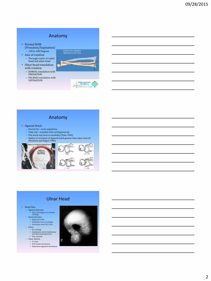
Anatomy
• Normal ROM (Pronation/Supination) – 150 to 180 Degrees
• Axis of rotation – Through center of radial
head and ulnar head
• Ulnar head translation with rotation – DORSAL translation with
PRONATION
– PALMAR translation with SUPINATION
Anatomy
• Sigmoid Notch – Dorsal rim – acute angulation
– Volar rim – rounded with cartilaginous lip
– Flat notch may lead to instability (Tolat 1996)
– Radius of curvature of sigmoid notch greater than ulnar seat (Af Ekenstam and Hagert 1985)
Ulnar Head
• Distal Ulna – Sigmoid Articular
• Up to 220 degrees of articular cartilage
– Distal Articular • Spherical to flat
• Semilunar area of cartilage
• Articulates with TFCC Disc
– Fovea • No cartilage
• Attachment site for Radioulnar and Ulnocarpal ligaments
• Very vascular
– Ulnar Styloid • 2-6 mm
• ECU sheath attachment
• Radioulnar ligament attachment
Page 3
09/28/2015
3
Joint Reactive Forces
• Neutral Position (af Ekenstam 1984) – Capitate through SLL to articular ridge of distal radius
– 84% of load transmitted through radius
• Ulnar Deviation – Through central articular disk
– Force can increase 150%
• Ulnar variance (Palmer 1988) – Neutral Variance = 16-18% load through ulna
– Shortening by 2.5mm = 4% ulnar load
– Lengthening by 2.5mm = 42% ulnar load
DRUJ Stability
• Intrinsic – Dorsal and
palmar radioulnar ligaments
• Extrinsic
– ECU – ECU Sheath – Pronator
quadratus – Interosseous
ligament of forearm
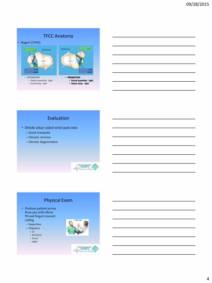
TFCC Anatomy
Superficial Radioulnar Ligaments ◦ Insert onto STYLOID ◦ Acute angle of insertion
Deep Radioulnar Ligaments ◦ Insert onto FOVEA ◦ Obtuse angle of insertion is
mechanically advantageous ◦ Ligamentum subcruentum
(Kauer 1975)
Page 4
09/28/2015
4
TFCC Anatomy
– SUPINATION
• Palmar superficial - tight
• Dorsal deep - tight
• Hagert (1994)
– PRONATION
Dorsal superficial - tight
Palmar deep - tight
Evaluation
• Divide ulnar sided wrist pain into
– Acute traumatic
– Chronic overuse
– Chronic degenerative
Physical Exam
• Position patient across from you with elbow 90 and fingers toward ceiling
– Inspection
– Palpation • LT
• ECU/FCU
• Fovea
• DRUJ
Page 5
09/28/2015
5
Special maneuvers
• LT snuff box test
• LT shuck
• Foveal Sign
• Ulnocarpal stress test
• Piano key test
• ECU synergy test
• ECU subluxation test
Imaging
• X-rays – Standard PA and Lateral
• Don’t depend on lateral to dx DRUJ dislocation/subluxation
– Check for ulnar variance • May use clenched fist to eval for dynamic variance
• May change >1mm from pronation to supination
– Lateral Stress Views
– Signs of DRUJ injury • Ulnar styloid base frx
• Widening of DRUJ
• >20° dorsal radial angulation
• >5mm shortening of distal radius
Imaging
• CT Scan – Helpful in identifying pathology of DRUJ
• Malunions, degenerative changes
– Eval both wrists: neutral, supinated, pronated
• MRI – Variable sensitivity, specificity for TFCC tears – Arthrogram improves
• Better at detecting central TFCC tears and SL tears than peripheral TFCC and LT tears
• Ultrasound – Low cost and non-invasive – Can be used with hardware without artifact issues
Page 6
09/28/2015
6
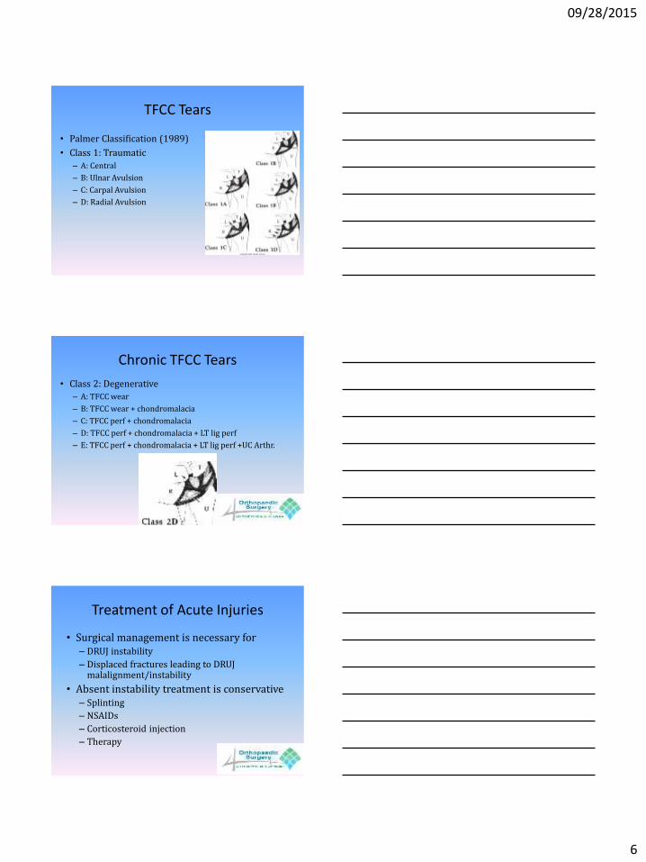
TFCC Tears
• Palmer Classification (1989)
• Class 1: Traumatic
– A: Central
– B: Ulnar Avulsion
– C: Carpal Avulsion
– D: Radial Avulsion
Chronic TFCC Tears
• Class 2: Degenerative
– A: TFCC wear
– B: TFCC wear + chondromalacia
– C: TFCC perf + chondromalacia
– D: TFCC perf + chondromalacia + LT lig perf
– E: TFCC perf + chondromalacia + LT lig perf +UC Arthr.
Treatment of Acute Injuries
• Surgical management is necessary for – DRUJ instability
– Displaced fractures leading to DRUJ malalignment/instability
• Absent instability treatment is conservative – Splinting
– NSAIDs
– Corticosteroid injection
– Therapy
Page 7
09/28/2015
7
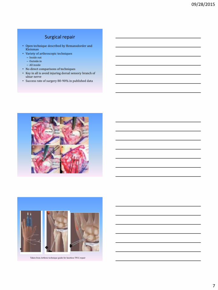
Surgical repair
• Open technique described by Hemansdorder and Kleinman
• Variety of arthroscopic techniques – Inside out
– Outside in
– All inside
• No direct comparisons of techniques
• Key in all is avoid injuring dorsal sensory branch of ulnar nerve
• Success rate of surgery 80-90% in published data
Taken from Arthrex technique guide for knotless TFCC repair
Page 8
09/28/2015
8
Chronic TFCC Tears
• Most of Palmer Class 2 tears result from excessive loading between the distal ulna and triquetrum
• Degenerative tears are not amenable to repair
• Treatment: Debridement +/- ulnar shortening
Ulnar Impaction Syndrome
• Due to acquired or developmental ulnar positive variance
• Exam – ulnar sided wrist pain and
swelling – ↑ pain with pronated ulnar
deviation and grip
• Imaging – X-ray
– Consider stress radiographs
– Dynamic ulnar variance
– MRI
Ulnar Impaction Syndrome
• Treatment
– Conservative tx first (splinting,
activity mod, NSAIDS, injection)
– Surgery if conservative tx fails
– Wafer Procedure (Feldon 1992)
• Partial distal ulnar resection (2-4mm
max)
• Preserves styloid and fovea
• Arthroscopic or open
– Ulnar shortening osteotomy
• Contraindicated with DRUJ arthritis
Page 9
09/28/2015
9
Ulnar shortening osteotomy
• Converts ulnar positive wrist to ulnar negative • Generally performed in distal 1/3rd • Standard technique is compression plating
with transverse or oblique osteotomy – Variety of cutting guides now available
• Results of USO overall very good – Complications
• 0-5% nonunion • Hardware irritation
• Avoid in patients with DRUJ arthritis and dorsal DRUJ dislocations
Wafer Procedure
• First described as open procedure by Feldon in 1992. – Reported good to excellent results in 12 of 13 patients
• Arthroscopic technique gaining popularity – Create 2-3mm ulnar neg variance using power burr
through defect in TFCC – Can resect about 5mm of ulna using this technique
• Bernstein et al compared USO to wafer – Found similar results overall – Recommend wafer
• No hardware issues • No risk of nonunion
LT ligament injuries
• Seen in isolation or in combination with other radiocarpal and intercarpal injuries
• Isolated injuries can be seen from fall on outstretched wrist or direct blow
• Present with ulnar sided pain and swelling
• On exam may show positive provocative maneuvers
• Imaging – VISI pattern on lateral
radiograph – MR arthogram best test
Page 10
09/28/2015
10
Treatment
• Acute stable injuries
– Cast immobilize in neutral for 4-6 weeks
• Late stable injuries
– Corticosteroid injection into midcarpal joint
Failure of conservative treatment
• Arthroscopic evaluation
– Evaluate Radiocarpal and Midcarpal joint
– Geissler I and II • Debride tear
– Geissler III and IV • Repair
• Pinning
• Fusion
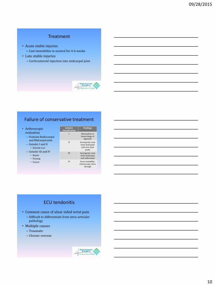
Geissler Classification
Findings
I Attenuation or hemorrhage of
ligament
II Incongruity seen from midcarpal joint-less than
probe
III Incongruity seen from midcarpal and radiocarpal
IV Gross instability-arthroscopic drive
through
ECU tendonitis
• Common cause of ulnar sided wrist pain
– Difficult to differentiate from intra-articular pathology
• Multiple causes
– Traumatic
– Chronic overuse
Page 11
09/28/2015
11
Treatment
• Start with conservative therapy
– Bracing
– NSAIDs
• If diagnosis unclear
– MRI
– Diagnostic Injection
• Fill sheath with local +/- corticosteroid
Operative Intervention
• Complete release of fibro-osseous tunnel of 6th dorsal compartment
– Possible to cause tendon subluxation
• In advanced cases may need to debride tendon
– Consider interposition graft with severe tendon damage in chronic cases
ECU subluxation
• Can result from trauma
– Direct blow with wrist supinated and ulnar deviation
• Exam
– Tenderness over ECU
– Illicit subluxation
Page 12
09/28/2015
12
Treatment
• Initial treatment with immobilization
– Long arm cast with wrist extended, pronated and radial deviated (4-6weeks)
– Transition to short arm cast (4 weeks)
– Physical therapy
Operative Intervention
• Repair
– Can be difficult in chronic cases
– Consider deepening of the groove
• Reconstruction
– Variety of techniques
• Use local retinaculum flap
• Use of palmaris graft
MacLennon et al. Recon for ECU subluxation. JHS 2008.