Ultra Sensitive, Super Speedy and Much more... Lumitein TM Protein Gel Stain HIGHLIGHTS Highly Sensitive At least as sensitive as silver stain by detecting as little as 1 ng or less protein. Extremely Simple & Fast Staining Fixation and staining is a single combined step. Use the 30-min Rapid Protocol for excellent result, or the 90-min Basic Protocol for the ultimate sensitivity; no overstaining with longer staining time. (protocol downloadable) Excellent Compatibility with Existing Instruments Can be used with either a simple UV-box (designed for DNA gel viewing), a Dark Reader, or a high-end laser scanner (See Figure 2 for spectra). Wide Linear Detection Range At least three orders of magnitude. Perfectly Compatible with Downstream Analysis Compatible with MS and sequencing. Economical Supplied as a 100X concentrated solution to reduce manufacturing cost and shipping cost, resulting in significant saving for customers. Stable Both the 100X concentrated solution and the 1X working solution are stable at room temperature for at least 1 year. Glowing Products for Science TM Figure 1. Top panel: Two-fold serial dilutions of Precision Plus protein standard (Bio-Rad) were separated via SDS-PAGE and then stained with Lumitein TM for 90 minutes without a separate fixation step. Images were taken with GE Typhoon Trio using 532 nm excitation and 610BP30 emission filter. Bottom panel: 2-D gel of human liver protein lysate stained with Lumiteinand again imaged with GE Typhoon Trio. The three circled spots were picked for MS analysis by Applied Biomics, Inc. (Hayward, CA). The result confirmed that Lumiteingel staining is fully compatible with MS analysis (data not shown). Biotium, Inc. Tel: (510)265-1027; Fax: (510)265-1352 Email: [email protected]; web: www.biotium.com 1 2 3 1 2 1 2 3 ~0.2 ng ~0.2 ng
Transcript
Ultra Sensitive, Super Speedy and Much more...
LumiteinTM Protein Gel Stain
HIGHLIGHTS
Highly SensitiveAt least as sensitive as silver stain by detecting as little
as 1 ng or less protein.
Extremely Simple & Fast StainingFixation and staining is a single combined step. Use the
30-min Rapid Protocol for excellent result, or the 90-minBasic Protocol for the ultimate sensitivity; no overstainingwith longer staining time. (protocol downloadable)
Excellent Compatibility with Existing InstrumentsCan be used with either a simple UV-box (designed for
DNA gel viewing), a Dark Reader, or a high-end laserscanner (See Figure 2 for spectra).
Wide Linear Detection RangeAt least three orders of magnitude.
Perfectly Compatible with Downstream AnalysisCompatible with MS and sequencing.
EconomicalSupplied as a 100X concentrated solution to reduce
manufacturing cost and shipping cost, resulting in significantsaving for customers.
StableBoth the 100X concentrated solution and the 1X working
solution are stable at room temperature for at least 1 year.
Glowing Products for ScienceTM
Figure 1. Top panel: Two-fold serial dilutions of Precision Plus protein standard (Bio-Rad)were separated via SDS-PAGE and then stained with LumiteinTM for 90 minutes without aseparate fixation step. Images were taken with GE Typhoon Trio using 532 nm excitationand 610BP30 emission filter. Bottom panel: 2-D gel of human liver protein lysate stainedwith Lumitein and again imaged with GE Typhoon Trio. The three circled spots werepicked for MS analysis by Applied Biomics, Inc. (Hayward, CA). The result confirmed thatLumitein gel staining is fully compatible with MS analysis (data not shown).
Figure 3. Linear detection range of Lumitein for 4 different proteins. Various amountsof each protein were separated via SDS-PAGE. Gel images were taken by GE TyphoonTrio gel scanner using 532 nm excitation and 610BP30 emission filter. The bands werequantitated using ImageQuant volume analysis. Log luminescence intensity was plot-ted against log protein amount per band for each protein.
Carbonic anhyrase 103106 (R2=0.9986)
Log (Protein/ng)0 1 2 3
Log
(Flu
ores
ence
)
5.4
5.6
5.8
6
6.2
6.4
6.6
6.8
7
7.2
7.4Carbonic anhyrase 103106 (R2=0.9986)
Log (Protein/ng)0 1 2 3
Log
(Flu
ores
ence
)
5.4
5.6
5.8
6
6.2
6.4
6.6
6.8
7
7.2
7.4
Glucose Oxidase 103106 (R2=0.9971)
Log (Protein/ng)0 1 2 3
Log
(Flu
ores
ence
)
5.6
5.8
6
6.2
6.4
6.6
6.8
7
7.2
7.4
7.6
Glucose Oxidase 103106 (R2=0.9971)
Log (Protein/ng)0 1 2 3
Log
(Flu
ores
ence
)
5.6
5.8
6
6.2
6.4
6.6
6.8
7
7.2
7.4
7.6
BSA 103106 R2=0.9972)
Log (Protein/ng)0 1 2 3
Log
(Flu
ores
ence
)
4.6
5
5.4
5.8
6.2
6.6
7
BSA 103106 R2=0.9972)
Log (Protein/ng)0 1 2 3
Log
(Flu
ores
ence
)
4.6
5
5.4
5.8
6.2
6.6
7
Ovalbumin 103106 (R2=0.9967)
Log (Protein/ng)0.6 1.4 2.2 3
Log
(Flu
ores
ence
)
4.9
5.1
5.3
5.5
5.7
5.9
6.1
6.3
6.5
6.7Ovalbumin 103106 (R2=0.9967)
Log (Protein/ng)0.6 1.4 2.2 3
Log
(Flu
ores
ence
)
4.9
5.1
5.3
5.5
5.7
5.9
6.1
6.3
6.5
6.7
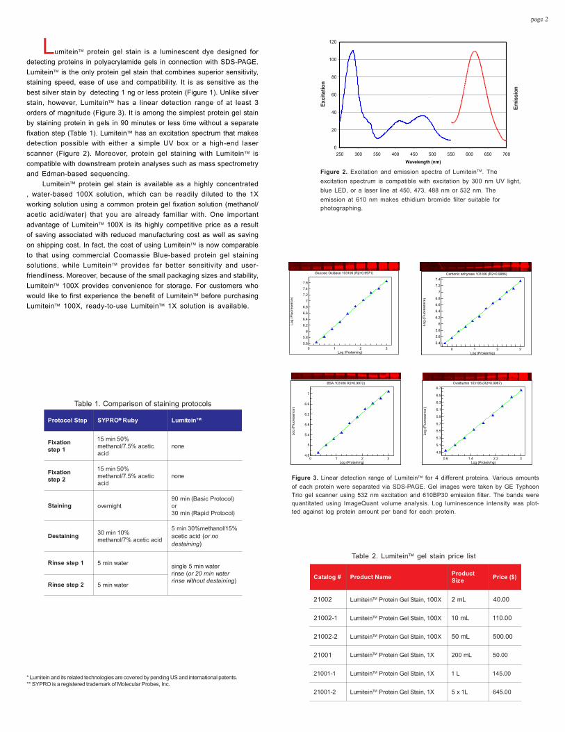
Figure 2. Excitation and emission spectra of LumiteinTM. Theexcitation spectrum is compatible with excitation by 300 nm UV light,blue LED, or a laser line at 450, 473, 488 nm or 532 nm. Theemission at 610 nm makes ethidium bromide filter suitable forphotographing.
Lumitein protein gel stain is a luminescent dye designed fordetecting proteins in polyacrylamide gels in connection with SDS-PAGE.Lumitein is the only protein gel stain that combines superior sensitivity,staining speed, ease of use and compatibility. It is as sensitive as thebest silver stain by detecting 1 ng or less protein (Figure 1). Unlike silverstain, however, Lumitein has a linear detection range of at least 3orders of magnitude (Figure 3). It is among the simplest protein gel stainby staining protein in gels in 90 minutes or less time without a separatefixation step (Table 1). Lumitein has an excitation spectrum that makesdetection possible with either a simple UV box or a high-end laserscanner (Figure 2). Moreover, protein gel staining with Lumitein iscompatible with downstream protein analyses such as mass spectrometryand Edman-based sequencing.
Lumitein protein gel stain is available as a highly concentrated, water-based 100X solution, which can be readily diluted to the 1Xworking solution using a common protein gel fixation solution (methanol/acetic acid/water) that you are already familiar with. One importantadvantage of Lumitein 100X is its highly competitive price as a resultof saving associated with reduced manufacturing cost as well as savingon shipping cost. In fact, the cost of using Lumitein is now comparableto that using commercial Coomassie Blue-based protein gel stainingsolutions, while Lumitein provides far better sensitivity and user-friendliness. Moreover, because of the small packaging sizes and stability,Lumitein 100X provides convenience for storage. For customers whowould like to first experience the benefit of Lumitein before purchasingLumitein 100X, ready-to-use Lumitein 1X solution is available.
* Lumitein and its related technologies are covered by pending US and international patents.** SYPRO is a registered trademark of Molecular Probes, Inc.
#golataC emaNtcudorP tcudorPeziS )$(ecirP
20012 nietimuL MT 1,niatSleGnietorP 00 X Lm2 00.04
1-20012 nietimuL MT 1,niatSleGnietorP 00 X Lm01 00.011
2-20012 nietimuL MT 1,niatSleGnietorP 00 X Lm05 00.005
Storage and HandlingLumitein 1X staining solution can be stored at room temperature or in a
refrigerator for a year.
Product DescriptionLumitein Protein Gel Stain is a luminescent dye designed for
detecting proteins in polyacrylamide gels. The dye has many of the samedesirable features that SYPRO Ruby possesses, such as detection of aslittle as 1 ng or less proteins, compatibility with both UV and visible lightexcitation, excellent photostability and a detection linearity range of at least3 orders of magnitude. However, Lumitein has some importantadvantages over SYPRO Ruby. Protein staining using Lumitein is farsimpler and faster than that using SYPRO Ruby. With Lumitein, proteinfixation and staining is a single combined 90-minute incubation process.Afterwards, the stained gel is briefly destained and washed, or simplywashed in deionized water for 20 minutes before it is ready for viewing/imaging. The linearity of protein quantitation plot with Lumitein extends forat least three logs. Lumitein produces a smaller slope than SYPRO Rubyfor the linearity plot. As a result, highly abundant proteins may appearbrighter in SYPRO Ruby-stained gels than in Lumitein-stained gels.However, less abundant proteins in Lumitein-stained gels generallyappear as bright as, or even brighter than those in SYPRO Ruby-stainedgels. This slope difference may prove advantageous for 2-D gels stainedwith Lumitein because fluorescent spots of less abundant proteins are lesslikely to be overwhelmed by those of highly abundant proteins nearby.Finally, Lumitein protein gel staining is fully compatible with downstreamprotein analyses such as mass spectrometry and Edman-basedsequencing.
Preparations of Destaining Solution Destaining may be accomplished by soaking the stained gels in de-ionized water for 20 minutes on a shaker (See staining procedure below).For even lower background and faster destaining, gels may be destained ina destaining solution containing 30% methanol, 15% acetic acid and 55%water. To prepare approximately 100 mL destaining solution, mix 30 mLmethanol, 15 mL acetic acid and 55 mL deionized water in a clean container.
Staining ProtocolThe following protocol is optimized for standard 1 mm thick, 8 cm X 8cm SDS-PAGE minigels.
Important: Before protein staining with Lumitein, do NOT fix the gels.Staining of pre-fixed gels with Lumitein may not produce optimal results.
1. After electrophoresis, the gel is directly placed in a clean gel-staining container (such as a polypropylene container) withoutfixation. Add at least 60 mL of Lumitein 1X staining solution. Agitateon an orbital shaker or a Nutator Mixer. For ultimate sensitivity,incubate the gel for 90 minutes. For more rapid results, incubate for30 minutes.Note: The staining solution contains sufficient organic solvents to fix the
proteins during staining.The gel may be left in the staining solution indefinitely without overstaining.For larger gels, more staining solution and/or longer staining time may be
required. In general, the amount of the staining solution should be sufficient sothat it freely flows over the surface of the gel during incubation.
2. Place the stained gel in a dust-free container. Add 100 mL ofthe destaining solution and agitate on an orbital shaker or a NutatorMixer for 5 min. Decant the destaining solution, add at least 100 mLdeionized water and agitate for at least another 5 min beforeviewing/imaging. Alternatively, destaining and rinsing can beaccomplished in a single step by placing the stained gel in at least100 mL deionized water on a shaker or Nutator for 20 min. Leavingthe gel in water overnight does not significantly reduce thesensitivity.Note: The single-step destaining/rinsing in water may produce slightly higher
background than the two-step destaining/rinsing procedure. The single-stepprocedure is adequate for 1-D gels or applications where signal/noise ratio isrelatively less demanding.
Table 1. List of suitable excitation sources/filters andemission filters
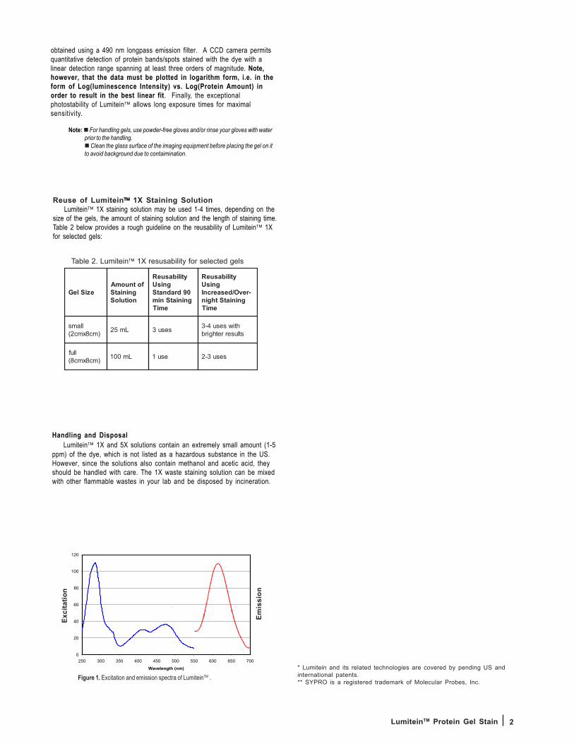
Viewing and Photographing the GelLumitein has a UV excitation maximum at around ~280 nm and a broad
visible excitation maximum centered around ~450 nm (Figure 1). It emitsbright red fluorescence at around ~610 nm. As a result, gels stained with thedye can be viewed using a standard 300 nm UV transilluminator, a 470 nmblue LED transilluminator, or a laser scanner with a laser line at 450, 473, 488or 532 nm. For maximum sensitivity, a 490 nm longpass filter should beused. More comprehensive lists of various suitable excitation sources andemission filters are summarized in Table 1 below:
The stained gel may be imaged using either a photographic camerasuch as a Polaroid camera or a CCD camera. When using a Polaroidcamera with Polaroid 667 black-and-white print film, the best result may be
(C) 2003 COSMO BIO CO.,LTD.
Added by COSMO BIO CO.,LTD.
2LumiteinTM Protein Gel Stain
Handling and DisposalLumitein 1X and 5X solutions contain an extremely small amount (1-5
ppm) of the dye, which is not listed as a hazardous substance in the US.However, since the solutions also contain methanol and acetic acid, theyshould be handled with care. The 1X waste staining solution can be mixedwith other flammable wastes in your lab and be disposed by incineration.
* Lumitein and its related technologies are covered by pending US andinternational patents.** SYPRO is a registered trademark of Molecular Probes, Inc.
Figure 1. Excitation and emission spectra of LumiteinTM .
0
20
40
60
80
100
120
250 300 350 400 450 500 550 600 650 700
Wavelength (nm)
`
Exci
tatio
n
Emis
sion
Table 2. Lumitein 1X resusability for selected gels
Reuse of Lumitein 1X Staining SolutionLumitein 1X staining solution may be used 1-4 times, depending on the
size of the gels, the amount of staining solution and the length of staining time.Table 2 below provides a rough guideline on the reusability of Lumitein 1Xfor selected gels:
obtained using a 490 nm longpass emission filter. A CCD camera permitsquantitative detection of protein bands/spots stained with the dye with alinear detection range spanning at least three orders of magnitude. Note,however, that the data must be plotted in logarithm form, i.e. in theform of Log(luminescence Intensity) vs. Log(Protein Amount) inorder to result in the best linear fit. Finally, the exceptionalphotostability of Lumitein allows long exposure times for maximalsensitivity.
Note: For handling gels, use powder-free gloves and/or rinse your gloves with waterprior to the handling.
Clean the glass surface of the imaging equipment before placing the gel on itto avoid background due to contaimination.


![Ultra Sensitive Analysis Of Polycyclic Aromatic ... › 2014annualmeeting › ... · Ultra Sensitive Analysis Of Polycyclic Aromatic Hydrocarbon Dibenzo[def,p]chrysene Pharmacokinetics](https://static.documents.pub/doc/80x56/5f0f44867e708231d443508b/ultra-sensitive-analysis-of-polycyclic-aromatic-a-2014annualmeeting-a-.jpg)


