Abstract— Gigahertz hard X-ray imaging for the proposedmatter-radiation interaction in extreme project presents anunprecedented challenge to front imager in both speed andradiation hardness. Novel fast scintillators are to be developedto face these challenges. This paper presents an investigationon the optical and scintillation properties for a set of fastinorganic scintillators. Transmittance, emission, light output, anddecay time were measured. Based on this investigation, we planto take two approaches to develop inorganic scintillators withsubnanoseconds of decay time for the gigahertz hard X-rayimaging. One is yttrium-doped barium fluoride single crystals,and another is based on gallium-doped ZnO nanoparticles.
Index Terms— Hard X-ray imaging, light output (LO),self-absorption, transmittance, ultrafast decay time, ultrafastscintillator.
I. INTRODUCTION
A IMING at studying the dynamics of material evolutionrelated to the nuclear Big Bang, the matter–radiation
interaction in extreme (MaRIE) experimental facility wasproposed at Los Alamos [1], [2], where gigahertz hard X-ray(>20 keV) imaging is required. The proposed X-ray energyand the interframe time are 30 keV and 2 ns in phase I andup to 126 keV and 300 ps in phase II, respectively [3]. Theseultrafast interframe times require ultrafast sensors to captureand store the dense spatial and temporal signals. To mitigatepileup effect for such high frame rates, it is important to havea temporal response of less than 2 ns and 300 ps, respectively,for the Type I and II imagers. The development of sensorswith ultrafast time response is thus important as well [3].
Fig. 1 is a schematic showing an inorganic scintillator-based total absorption imager concept featured with a pixelatedultrafast scintillator screen, a pixelated ultrafast photodetector,and ultrafast readout electronics [3].
Table I summarizes the basic properties of various fastscintillators investigated in the recent years for gigahertzhard X-ray imaging with a reference of cerium-doped
Manuscript received January 14, 2018; accepted February 15, 2018. Dateof publication February 19, 2018; date of current version August 15, 2018.This work was supported by the U.S. Department of Energy, Office of HighEnergy Physics Program, under Award DE-SC0011925.
C. Hu, L. Zhang, and R.-Y. Zhu are with the Physics Department,California Institute of Technology, Pasadena, CA 91125 USA (e-mail:[email protected]).
A. Chen and Z. Wang are with the Los Alamos National Laboratory,Los Alamos, NM 87545 USA.
L. Ying and Z. Yu are with the Materials Science and EngineeringDepartment, University of Wisconsin, Madison, WI 53706 USA.
Color versions of one or more of the figures in this paper are availableonline at http://ieeexplore.ieee.org.
Digital Object Identifier 10.1109/TNS.2018.2808103
Fig. 1. Schematic showing total absorption concept of a pixelated crystal-based front imager for the proposed gigahertz X-ray imaging.
lutetium yttrium oxyorthosilicate LYSO:Ce crystal(Lu2(1−x)Y2xSiO5:Ce or LYSO) and yttrium oxyorthosilicate(Y2SiO5:Ce or YSO:Ce), which are chosen because of theirexcellent performance and wide applications.
The crystal candidates listed in Table I can be classifiedinto four groups according to their scintillation mechanism.The first group is the direct-gap semiconductor crystals, suchas gallium-doped ZnO (ZnO:Ga). The second group is thecore-valence luminescence crystals, such as BaF2. The thirdgroup is Yb3+ activated crystals featured with fast decaytime and thermal quenching, such as YAlO3:Yb (YAP:Yb)and Y3Al5O12:Yb (YAG:Yb). The fourth group is Ce3+-activated bright and fast crystals, such as YAlO3 (YAP:Ce),Lu3Al5O12:Ce (LuAG:Ce), and LaBr3:Ce.
The bright and fast halides, such as LaBr3:Ce and CeBr3,are highly hygroscopic, so that their application needs extraengineering work. Among the nonhygroscopic inorganic crys-tal scintillators listed in Table I, BaF2 crystals with sub-nanoseconds of fast decay time provide high scintillationphoton yield in the first nanoseconds. This fast scintillationcomponent promises a solution for an ultrafast front imager.BaF2 is also featured with the shortest attenuation length for40-keV X-rays, promising a compact sensor for gigahertz hardX-ray imaging. Recent investigation on ZnO:Ga nanoparticlesimbedded in polystyrene shows its ultrafast scintillation lightwith subnanoseconds decay time (0.5 ns) [5], which promisesanother solution for an ultrafast front imager for gigahertz hardX-ray imaging.
2098 IEEE TRANSACTIONS ON NUCLEAR SCIENCE, VOL. 65, NO. 8, AUGUST 2018
TABLE I
CANDIDATE SCINTILLATORS FOR GIGAHERTZ HARD X-RAY IMAGING
Fig. 2. Photograph showing samples investigated in this paper.
In this paper, we report the results on fast inorganicscintillators investigated at the Caltech HEP CrystalLaboratory. Their optical and scintillation properties, such astransmittance, emission, light output (LO), and decay time,are measured. The pros and cons of each of the four groupsare discussed. For gigahertz hard X-ray imaging, yttrium-doped BaF2 crystals (BaF2:Y) and ZnO:Ga nanoparticle-basedscintillators are most promising.
II. SAMPLES AND EXPERIMENTAL DETAILS
Fig. 2 shows photograph of inorganic crystal scintil-lator samples investigated. Their corresponding dimension
TABLE II
BASIC PROPERTIES FOR THE SAMPLES INVESTIGATED IN THIS PAPER
information is listed in Table II. Two ZnO:Ga samples wereproduced in the Fujian Institute of Research on the Structureof Matter. The large-sized BaF2 crystal and the LuAG:Ceceramic samples were produced in the Shanghai Institute ofCeramics, Chinese Academy of Sciences. The BaF2:Y crystalswere produced in the Beijing Glass Research Institute. TheYAP:Ce, YAP:Yb, and YAG:Yb crystals were produced inChengdu Dongjun Laser Co., Ltd. The LaBr3 crystal wasproduced in Saint-Gobain Crystals.
Photoluminescence (PL) was measured by an EdinburghInstrument FLS920 fluorescence spectrometer. The signalswere detected by a Hamamatsu R928P PMT through a mono-chromator. Measured PL spectrum was corrected using acalibrated light source. Longitudinal transmittance (LT) was
HU et al.: ULTRAFAST INORGANIC SCINTILLATORS FOR GHZ HARD X-RAY IMAGING 2099
measured using a Perkin Elmer Lambda-950 spectrometerequipped with double-beam, double-monochromator, and ageneral purpose optical bench. The systematic uncertainty ofthe LT measurement is about 0.2%.
Scintillation pulse shape and decay time were measured bya Hamamatsu R2059 PMT and an Agilent 9254 digital scopewith a temporal response time of 1.3 and 0.14 ns, respectively,which limited our ability to measure subnanoseconds decaytime. In order to measure subnanoseconds of decay time,a Photek MCP-PMT240 and a Tektronix MSO 72304DX werealso used for BaF2 and BaF2:Y samples, where the corre-sponding temporal response time is reduced to 0.2 and 0.02 ns,respectively.
LO was measured using either a 22Na or a 137Cs γ -raysource, where the 22Na source provided a coincidence trig-ger, which helps to mitigate residual phosphorescence in thesamples [6], [7]. A Hamamatsu R2059 or a HamamatsuR1306 PMT was used in the LO measurements, where thePMT R1306 provides a lower gain than the R2059 so wasused for samples with a high LO.
The LO of the LaBr3 crystal and LuAG:Ce ceramic sampleswere measured by the R1306 PMT using 0.662-MeV γ -raysfrom the 137Cs source. The LO of BaF2 and BaF2:Y crystalswas measured by the R2059 PMT using 0.511-MeV γ -raysfrom a 22Na source with a coincidence trigger. The LO ofYAP:Ce crystals was measured by the R1306 PMT using0.511-MeV γ -rays from the 22Na source with a coincidencetrigger.
LO was also measured by the R2059 PMT using 5.486-MeVα-rays from an 241Am source for two kinds of samples witha low LO. They are Yb-doped samples suffering with seriousthermal quenching, and ZnO:Ga crystals suffering with seriousself-absorption. It is well known that the nominal LO valuesmeasured by γ -rays and α-rays are different in terms ofp.e./MeV, so are not directly comparable. No attempt wasmade to compare the LO values measured using γ -rays andα-rays. The readers are advised to pay an attention to thisdifference when reading the LO values listed in Table I.
In all these LO measurements, the samples were wrappedwith two layers of Tyvek paper, and the PMTs were coupledto the samples via a thin layer of Down-Corning 200 opticalfluid. The systematic uncertainty of the LO measurement isabout 1%.
III. EXPERIMENTAL RESULTS
A. ZnO:Ga Plates
Gallium-doped ZnO crystal was developed as a Wannierexciton-based scintillator with subnanoseconds of decaytime [8], [9], and is commercially available [10]. In suchdirect-gap semiconductors, the electrons in the conductionband recombine directly with holes in the valence bandresulting in near band-edge luminescence [9]. Because of thesmall Stokes shift, most of the generated emission light insuch scintillators is self-absorbed at room temperature.
Fig. 3 shows the transmittance spectra for two ZnO:Gacrystal samples of 2- and 0.3-mm thickness. Also shown in thefigure is the photoluminescence spectrum (blue dashed line).
Fig. 3. Transmittance spectra for the ZnO:Ga crystal samples of33 × 30 × 2 mm3 and 22 × 22 × 0.3 mm3.
Fig. 4. Pulse height spectra for the ZnO:Ga crystal samples of 33 × 30 ×2 mm3 and 22 × 22 × 0.3 mm3 excited by 5.03-MeV α-rays.
Since the scintillation emission of ZnO:Ga peaks at 380 nmwhich is shorter than its intrinsic cutoff edge of 400 nm, a largefraction of the emission spectrum is self-absorbed in the crystalbulk.
Fig. 4 shows very different peak values of 5-MeV α-rays forthese two samples with different thickness. The 5-MeV α-raysexcited only a thin-layer crystal at the crystal’s surface becauseof their very short absorption length in the crystal. Sincemost scintillation photons generated by the α-rays propagatedacross the entire thickness before reaching the photodetector,the pulse height of the 5-MeV α-rays is thus sample thicknessdependent. Consequently, the 0.3-mm thick sample providesa much higher LO for 5-MeV α-rays than the 2-mm-thicksample.
2100 IEEE TRANSACTIONS ON NUCLEAR SCIENCE, VOL. 65, NO. 8, AUGUST 2018
Fig. 5. Pulse shape for the ZnO:Ga samples of 33 × 30 × 2 mm3 and22 × 22 × 0.3 mm3 excited by 22Na γ -rays.
Fig. 6. Schematic showing the multilayer detector architecture for efficientand fast imaging of diffracted X-rays [11].
Fig. 5 shows the pulse shape of two ZnO:Ga samples excitedby 0.511-MeV γ -ray from a 22Na source. The observed decaytime is a few nanoseconds, which is longer than the 0.5-nsdecay time observed from the ZnO:Ga nanoparticles embeddedin polystyrene [5]. This difference is partly due to the temporalresponse time of the R2059 PMT used in this measurement.
The self-absorption restricts applications of ZnO:Gacrystal in bulk for a total absorption front imager. An alterna-tive approach is to develop ZnO:Ga nanoparticle-based frontimager for gigahertz hard X-ray imaging [11]. Fig. 6 showsthis imager concept for gigahertz X-ray imaging featuredwith a multilayer high QE photocathode coated with ZnO:Gananoparticles. Recently discovered enhanced UV emissionin Ag/Au ZnO core–shell nanoparticles hints an interestingapproach to develop such thin film-based concept [12].
B. BaF2 and BaF2:Y Crystals
In the core valence transition scintillators, such as BaF2and CsF, the energy gap between the valence band and the
Fig. 7. Transmittance spectrum for the BaF2 sample of 50 × 50 × 5 mm3.
uppermost core band is less than the fundamental bandgap[13], [14]. A photon is emitted when an electron in thevalence band fills an ionization hole in the top core band.This is an allowed process with a decay time of an orderof 1 ns or less [15]. The overall light yield of core valencescintillation is low due to the inefficiency of creating holes inan upper core band. Because of its fast decay time, however,the light yield in the first nanoseconds is comparable tobright scintillators, such as LYSO:Ce. Such a scintillator hasattracted the HEP community pursuing ultrafast calorimeterand/or ultrafast timing.
Fig. 7 shows transmittance spectrum for the 5-mm-thickBaF2 crystal sample. Also shown in this plot is the numer-ical values of the emission weighted longitudinal transmit-tance (EWLT) for the fast (220 nm with subnanoseconds ofdecay time) and slow (300 nm with 600-ns decay time) com-ponents. We note that BaF2 has a good transmittance withoutself-absorption, and the slow component has an intensity of afactor of five of the fast component measured by a PMT with abialkali photocathode. The slow scintillation component wouldcause pileup, so needs to be suppressed for these applications.
Fig. 8 shows the pulse height spectrum with a full-width athalf-maximum (FWHM) resolution of 54.9% for 0.511-MeVγ -rays from a 22Na source for the BaF2 sample. The LO is209 p.e./MeV measured with an integration time of 50 ns.
Fig. 9 compares the pulse shape for BaF2 samplesmeasured by a Hamamatsu PMT R2059 (top) and a PhotekMCP-PMT240 (bottom), respectively. While R2059 datashow both rising and decay times of 1.4 ns, which is limitedby PMT’s temporal response time, they are 0.26 and 0.52 ns,respectively, measured by an MCP-PMT240, indicating thatthe intrinsic rising time is very small, and its fast scintillationdecay time is less than 0.6 ns. We also note that BaF2 isthe only crystal scintillator that can provide subnanosecondspulsewidth, so is the best choice when ultrafast timing isrequired.
HU et al.: ULTRAFAST INORGANIC SCINTILLATORS FOR GHZ HARD X-RAY IMAGING 2101
Fig. 8. Pulse height spectrum for the BaF2 sample of 50 × 50 × 5 mm3.
Fig. 9. Normalized pulse shape for a BaF2 sample excited by 22Na γ -rays,measured by a Hamamatsu R2059 PMT (top) and a Photek MCP-PMT240(bottom), respectively.
Fig. 10 compares the LO as a function of integration timemeasured by a Hamamatsu R2059 PMT for an undoped BaF2and a yttrium-doped BaF2:Y samples of the same dimension.Simple exponential fits were used to extract amplitudes fora fast scintillation component and a slow component and itsdecay time. It is interesting to note that yttrium doping greatlysuppressed the slow component. Yttrium-doped BaF2:Y hasthus a great potential to be developed to an ultrafast scintillatorin bulk. While the initial result of the BaF2:Y crystal wasreported elsewhere [16], it is our plan to further investigatethis material for its use in total absorption front imager shownin Fig. 1 for gigahertz hard X-ray imaging.
Fig. 10. LO and decay kinetics for BaF2 and BaF2:Y samples.
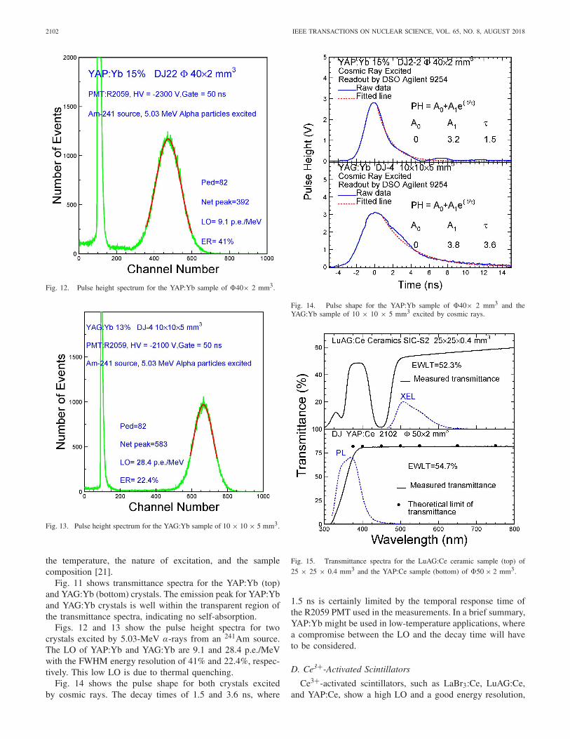
Fig. 11. Transmittance spectra for the YAP:Yb sample (top) of�40 × 2 mm3 and the YAG:Yb sample (bottom) of 10 × 10 × 5 mm3.
C. Yb3+ Activated Scintillators
Yb-doped yttrium perovskites (YAlO3, YAP) [17] andaluminum garnets (Y3Al5O12, YAG) [18] were commer-cially developed as IR laser media. Yb3+ ions embeddedinto various host lattices show charge transfer (CT) lumi-nescence [19], [20]. CT luminescence has two bands UV(CT state →2 F7/2) and visible (CT state →2 F5/2), andexhibits with a strong thermal quenching. Scintillation per-formances of YAP:Yb and YAG:Yb are very similar andvery fast, from a few to tens of nanoseconds depending on
2102 IEEE TRANSACTIONS ON NUCLEAR SCIENCE, VOL. 65, NO. 8, AUGUST 2018
Fig. 12. Pulse height spectrum for the YAP:Yb sample of �40× 2 mm3.
Fig. 13. Pulse height spectrum for the YAG:Yb sample of 10 × 10 × 5 mm3.
the temperature, the nature of excitation, and the samplecomposition [21].
Fig. 11 shows transmittance spectra for the YAP:Yb (top)and YAG:Yb (bottom) crystals. The emission peak for YAP:Yband YAG:Yb crystals is well within the transparent region ofthe transmittance spectra, indicating no self-absorption.
Figs. 12 and 13 show the pulse height spectra for twocrystals excited by 5.03-MeV α-rays from an 241Am source.The LO of YAP:Yb and YAG:Yb are 9.1 and 28.4 p.e./MeVwith the FWHM energy resolution of 41% and 22.4%, respec-tively. This low LO is due to thermal quenching.
Fig. 14 shows the pulse shape for both crystals excitedby cosmic rays. The decay times of 1.5 and 3.6 ns, where
Fig. 14. Pulse shape for the YAP:Yb sample of �40× 2 mm3 and theYAG:Yb sample of 10 × 10 × 5 mm3 excited by cosmic rays.
Fig. 15. Transmittance spectra for the LuAG:Ce ceramic sample (top) of
25 × 25 × 0.4 mm3 and the YAP:Ce sample (bottom) of �50 × 2 mm3.
1.5 ns is certainly limited by the temporal response time ofthe R2059 PMT used in the measurements. In a brief summary,YAP:Yb might be used in low-temperature applications, wherea compromise between the LO and the decay time will haveto be considered.
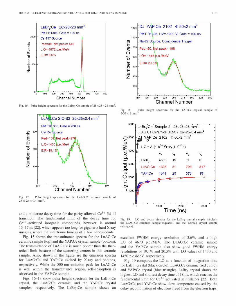
D. Ce3+-Activated Scintillators
Ce3+-activated scintillators, such as LaBr3:Ce, LuAG:Ce,and YAP:Ce, show a high LO and a good energy resolution,
HU et al.: ULTRAFAST INORGANIC SCINTILLATORS FOR GHZ HARD X-RAY IMAGING 2103
Fig. 16. Pulse height spectrum for the LaBr3:Ce sample of 28×28×28 mm3.
Fig. 17. Pulse height spectrum for the LuAG:Ce ceramic sample of25 × 25 × 0.4 mm3.
and a moderate decay time for the parity-allowed Ce3+ 5d–4ftransition. The fundamental limit of the decay time forCe3+-activated inorganic compounds, however, is around15–17 ns [22], which appears too long for gigahertz hard X-rayimaging where the interframe time is of a few nanoseconds.
Fig. 15 shows the transmittance spectra for the LuAG:Ceceramic sample (top) and the YAP:Ce crystal sample (bottom).The transmittance of LuAG:Ce is much poorer than the theo-retical limit because of the scattering centers in this ceramicsample. Also, shown in the figure are the emission spectrafor LuAG:Ce and YAP:Ce excited by X-ray and photons,respectively. While the 500-nm emission peak for LuAG:Ceis well within the transmittance region, self-absorption isobserved in the YAP:Ce sample.
Figs. 16–18 show pulse height spectrum for the LaBr3:Cecrystal, the LuAG:Ce ceramic, and the YAP:Ce crystalsamples, respectively. The LaBr3:Ce sample shows an
Fig. 18. Pulse height spectrum for the YAP:Ce crystal sample of�50 × 2 mm3.
Fig. 19. LO and decay kinetics for the LaBr3 crystal sample (circles),the LuAG:Ce ceramics sample (squares), and the YAP:Ce crystal sample(triangles).
excellent FWHM energy resolution of 3.6%, and a highLO of 4670 p.e./MeV. The LuAG:Ce ceramic sampleand the YAP:Ce sample also show good FWHM energyresolutions of 19.1% and 20.5% with LO values of 1430 and1450 p.e./MeV, respectively.
Fig. 19 compares the LO as a function of integration timefor LaBr3 crystal (black circle), LuAG:Ce ceramic (red cubic),and YAP:Ce crystal (blue triangle). LaBr3 crystal shows thehighest LO and shortest decay time of 18 ns, which reaches thefundamental limit for Ce3+ activated scintillators [22]. BothLuAG:Ce and YAP:Ce show slow component caused by thedelay recombination of electrons freed from the electron traps.
2104 IEEE TRANSACTIONS ON NUCLEAR SCIENCE, VOL. 65, NO. 8, AUGUST 2018
IV. CONCLUSION
A comparative study on four groups of scintillators wascarried out for gigahertz hard X-ray imaging. The direct-gapsemiconductor ZnO:Ga crystals show a very short decay time,but a low LO because of self-absorption. An imager consistingof ZnO:Ga nanoparticles-based multilayers is one detectorconcept to be pursued for gigahertz hard X-ray imaging forthe proposed MaRIE project.
BaF2 provides a fast scintillation component with anultrafast decay time of less than 0.6 ns and an ultrashortFWHM pulsewidth of less than nanoseconds, providing a solidfoundation for an ultrafast scintillator. Its slow scintillationcomponent with 600-ns decay time may be suppressed effec-tively by Y3+ doping. An imager consisting of bulk BaF2:Ycrystals may serve as a total absorption detector concept forgigahertz hard X-ray imaging for the proposed MaRIE project.
The Yb3+-doped crystals show very short decay time, butthey have low LO due to thermal quenching. The Ce3+-dopedscintillators show very high light yield and good FWHMenergy resolution. Their decay time, however, is too long forgigahertz hard X-ray imaging.
REFERENCES
[1] R. W. Garnett and M. S. Gulley, “Matter-radiation interactionsin extremes,” in Proc. Linear Accelerator Conf. (LINA), 2010,pp. 485–487.
[2] Z. Wang et al., “Gigahertz (GHz) hard X-ray imaging using fastscintillators,” Proc. SPIE, Hard X-Ray, Gamma-Ray, Neutron DetectorPhys. XV, vol. 8852, p. 88521A, Sep. 2013.
[3] P. Denes, S. Gruner, M. Stevens, and Z. Wang, “Ultrafast and high-energy X-ray imaging technologies and applications,” Los Alamos Nat.Lab., Santa Fe, NM, USA, Tech. Rep. LA-UR-17-22085, Aug. 2016.[Online]. Available: http://www.lanl.gov/science-innovation/science-facilities/marie/_assets/docs/workshops/ultrafast-high-energy-x-ray.pdf
[4] S. Liu et al., “Towards bright and fast Lu3Al5O12:Ce,Mg opticalceramics scintillators,” Adv. Opt. Mater., vol. 4, no. 5, pp. 731–739,2016.
[5] P. Lecoq, “The 10 ps timing-of-flight PET challenge,” in Proc. SCINTConf., Chamonix, France, 2017.
[6] R. Y. Zhu et al., “A study on the properties of lead tungstate crystals,”Nucl. Instrum. Methods Phys. Res. A, Accel. Spectrom. Detect. Assoc.Equip., vol. 376, no. 3, pp. 319–334, 1996.
[7] J. Chen, R. Mao, L. Zhang, and R. Y. Zhu, “Large size LSO and LYSOcrystals for future high energy physics experiments,” IEEE Trans. Nucl.Sci., vol. 54, no. 3, pp. 718–724, Jun. 2007.
[8] W. Lehmann, “Edge emission of n-type conducting Zno and CdS,”Solid-State Electron., vol. 9, nos. 11–12, pp. 1107–1110, 1966.
[9] S. E. Derenzo, M. J. Weber, and M. K. Klintenberg, “Tempera-ture dependence of the fast, near-band-edge scintillation from CuI,HgI2, PbI2, ZnO:Ga and CdS:In,” Nucl. Instrum. Methods Phys. Res.A, Accel. Spectrom. Detect. Assoc. Equip., vol. 486, pp. 214–219,Jun. 2002.
[10] E. Ohshima et al., “Growth of the 2-in-size bulk ZnO single crystalsby the hydrothermal method,” J. Crystal Growth, vol. 260, nos. 1–2,pp. 166–170, 2004.
[11] Z. Wang et al., “Thin scintillators for ultrafast hard X-ray imag-ing,” Proc. SPIE, Photon Counting Appl., vol. 9504, p. 95040N,May 2015.
[12] E. J. Guidelli, O. Baffa, and D. R. Clarke, “Enhanced UV emission fromsilver/ZnO and gold/ZnO core-shell nanoparticles: Photoluminescence,radioluminescence, and optically stimulated luminescence,” Sci. Rep.,vol. 5, Sep. 2015, Art. no. 14004.
[13] A. P. Shpak, O. A. Glike, A. G. Dmitriev, P. A. Rodnyi,A. S. Voloshinovskii, and S. M. Pidzyrailo, “Radiative core-valencetransitions in wide-gap crystals,” J. Electron. Spectrosc. Related Phe-nomena, vol. 68, pp. 335–338, May 1994.
[14] P. A. Rodnyi, “Core-valence transitions in wide-gap ionic-crystals,” Soviet Phys. Solid-State, vol. 34, no. 7, pp. 1053–1066,1992.
[15] M. J. Weber, “Scintillation: mechanisms and new crystals,” Nucl.Instrum. Methods Phys. Res. A, Accel. Spectrom. Detect. Assoc. Equip.,vol. 527, nos. 1–2, pp. 9–14, 2004.
[16] R.-Y. Zhu, “Applications of very fast inorganic crystal scintillatorsin future HEP experiments,” in Proc. Technol. Instrum. ParticlePhys., Beijing, China, May 2017. [Online]. Available: http://www.hep.caltech.edu/~zhu/talks/ryz_170522_Fast_Crystals.pdf
[17] V. E. Kisel, S. V. Kurilchik, A. S. Yasukevich, S. V. Grigoriev,S. A. Smirnova, and N. V. Kuleshov, “Spectroscopy and femtosecondlaser performance of Yb3+:YAlO3 crystal,” Opt Lett., vol. 33, no. 19,pp. 2194–2196, 2008.
[18] A. R. Reinberg, L. A. Riseberg, R. M. Brown, R. W. Wacker, andW. C. Holton, “GaAs: Si LED pumped Yb-doped YAG laser,” Appl.Phys. Lett., vol. 19, no. 1, p. 11, 1971.
[19] L. van Pieterson, M. Heeroma, E. de Heer, and A. Meijerink,“Charge transfer luminescence of Yb3+,” J. Lumin., vol. 91, nos. 3–4,pp. 177–193, 2000.
[20] R. Chipaux et al., “Ytterbium-based scintillators, a new class ofinorganic scintillators for solar neutrino spectroscopy,” Nucl. Instrum.Methods Phys. Res. A, Accel. Spectrom. Detect. Assoc. Equip., vol. 486,pp. 228–233, Jun. 2002.
[21] S. Belogurov, G. Bressi, G. Carugno, and Y. Grishkin, “Properties ofYb-doped scintillators: YAG, YAP, LuAG,” Nucl. Instrum. MethodsPhys. Res. A, Accel. Spectrom. Detect. Assoc. Equip., vol. 516, no. 1,pp. 58–67, 2004.
[22] P. Dorenbos, “Fundamental limitations in the performance of Ce3+}−,Pr3+−, Eu2+−activated scintillators,” IEEE Trans. Nucl. Sci., vol. 57,no. 3, pp. 1162–1167, Jun. 2010.