16
UltraMicroscope Blaze™ The automation of light sheet microscopy
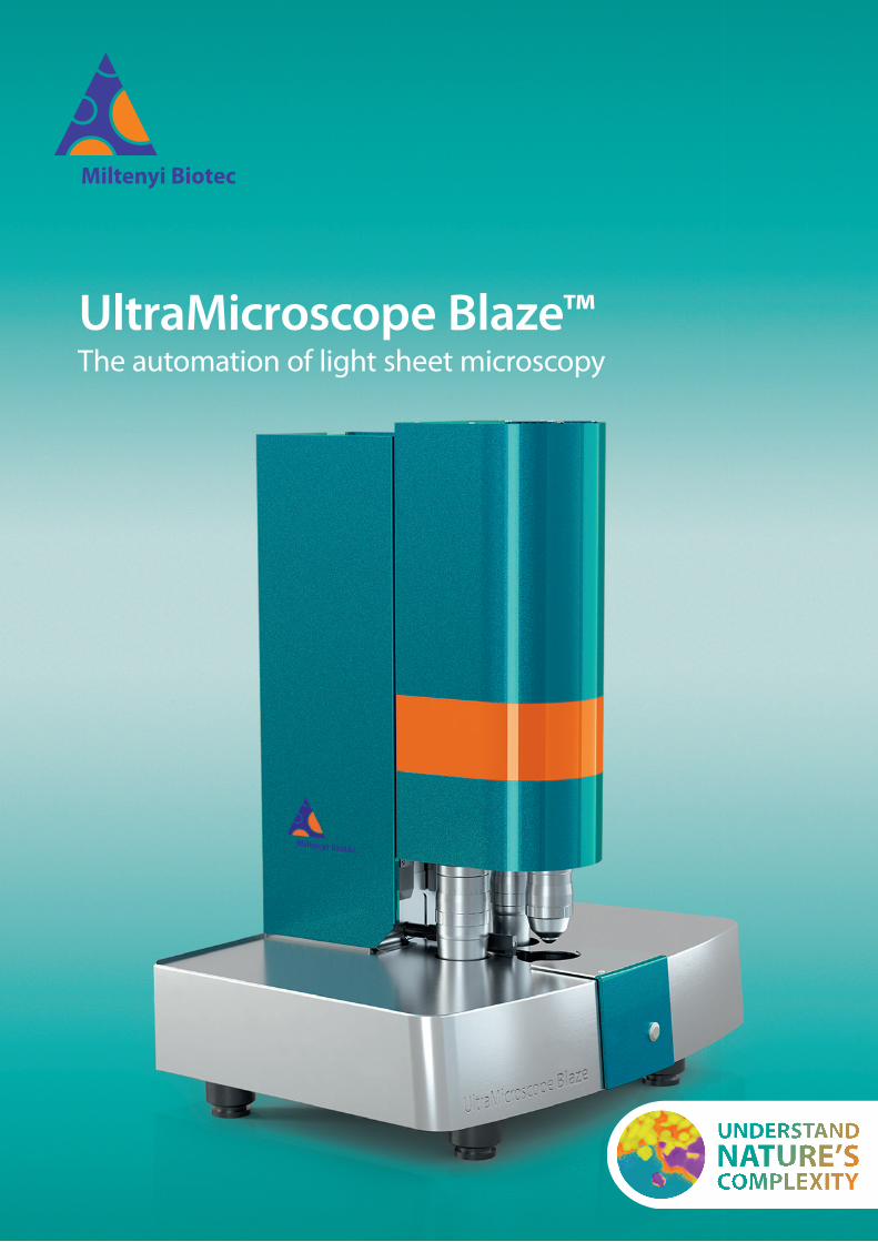
UltraMicroscope Blaze™The automation of light sheet microscopy
22
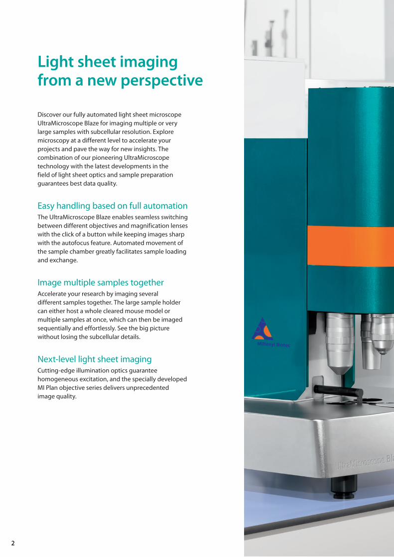
Discover our fully automated light sheet microscope UltraMicroscope Blaze for imaging multiple or very large samples with subcellular resolution. Explore microscopy at a different level to accelerate your projects and pave the way for new insights. The combination of our pioneering UltraMicroscope technology with the latest developments in the field of light sheet optics and sample preparation guarantees best data quality.
Easy handling based on full automationThe UltraMicroscope Blaze enables seamless switching between different objectives and magnification lenses with the click of a button while keeping images sharp with the autofocus feature. Automated movement of the sample chamber greatly facilitates sample loading and exchange.
Image multiple samples togetherAccelerate your research by imaging several different samples together. The large sample holder can either host a whole cleared mouse model or multiple samples at once, which can then be imaged sequentially and effortlessly. See the big picture without losing the subcellular details.
Next-level light sheet imagingCutting-edge illumination optics guarantee homogeneous excitation, and the specially developed MI Plan objective series delivers unprecedented image quality.
Light sheet imaging from a new perspective
The UltraMicroscope Blaze originates from a decade of experience and is designed to expedite your research projects. Our users’ feedback has been the driving force to create this new member of the UltraMicroscope family.
Loading a sample into the microscope and switching between different magnifications has never been easier. Enter the fast lane with the new UltraMicroscope Blaze and pave the way for new insights.
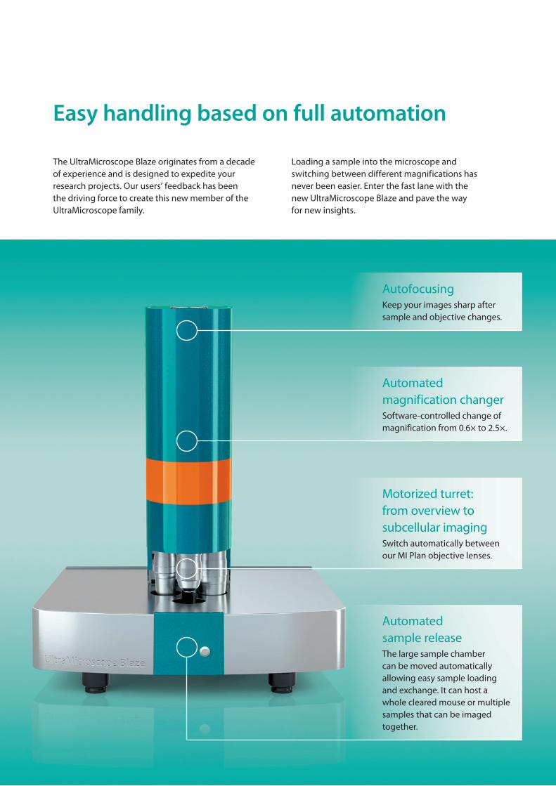
Easy handling based on full automation
AutofocusingKeep your images sharp after sample and objective changes.
Automated magnification changerSoftware-controlled change of magnification from 0.6× to 2.5×.
Motorized turret:from overview to subcellular imagingSwitch automatically between our MI Plan objective lenses.
Automated sample releaseThe large sample chamber can be moved automatically allowing easy sample loading and exchange. It can host a whole cleared mouse or multiple samples that can be imaged together.
55
Image multiple samples together
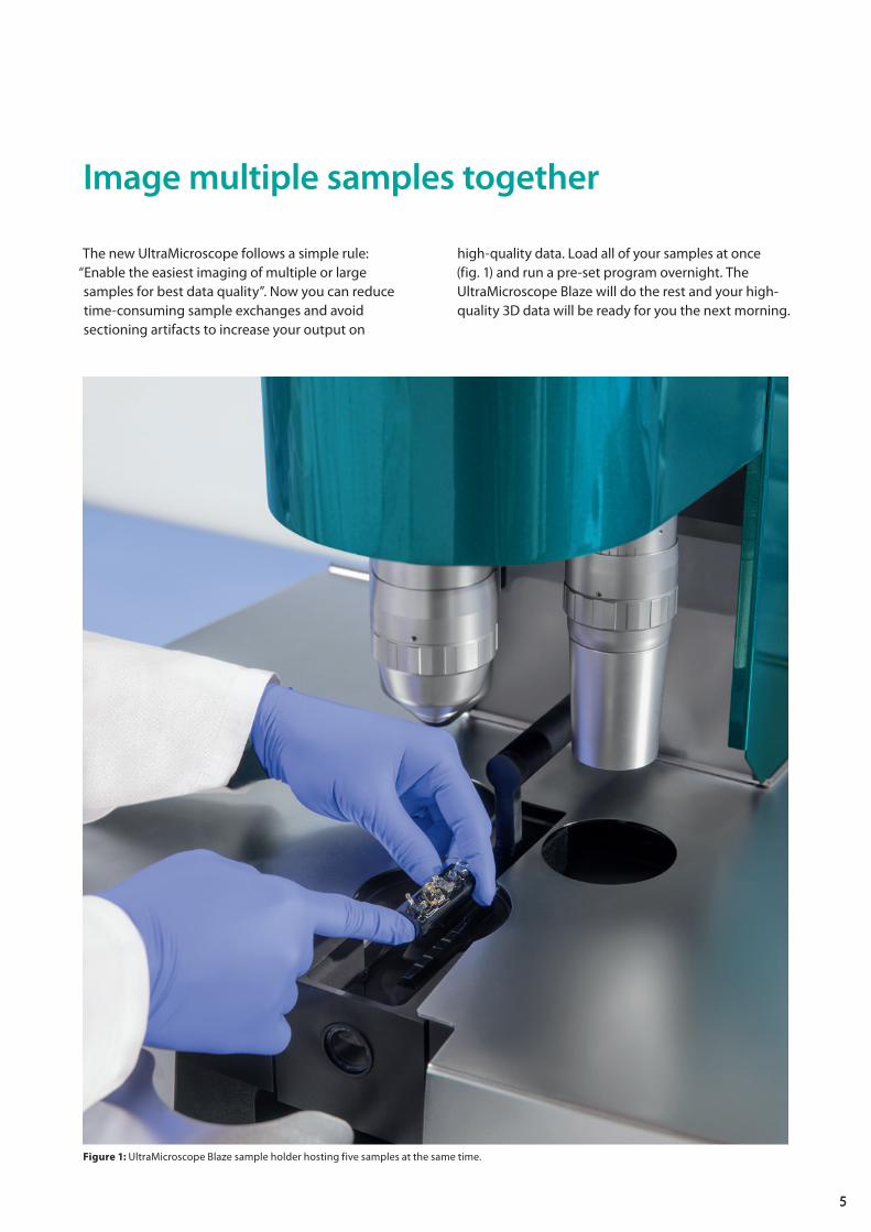
The new UltraMicroscope follows a simple rule: “Enable the easiest imaging of multiple or large samples for best data quality”. Now you can reduce time-consuming sample exchanges and avoid sectioning artifacts to increase your output on
high-quality data. Load all of your samples at once (fig. 1) and run a pre-set program overnight. The UltraMicroscope Blaze will do the rest and your high-quality 3D data will be ready for you the next morning.
Figure 1: UltraMicroscope Blaze sample holder hosting five samples at the same time.

6
The combination of our successful UltraMicroscope technology with the latest developments in the field of light sheet optics guarantees the best data quality. Flat-field correction in addition to long working distances makes the MI Plan objective lens series well suited for high-resolution imaging of large samples (fig. 2). In addition, they are compatible with all imaging solutions from water to solvents with high refractive indices. Explore our broad range of magnification options, from panoptic imaging at 0.66× to subcellular imaging at 30×.
Next-level light sheet imaging
Figure 2: The UltraMicroscope Blaze can host up to three MI Plan objectives at the same time. Six light sheets provide homogeneous fluorescence excitation.
working distances makes the MI Plan objective lens series well suited for high-resolution imaging of large samples (fig. 2). In addition, they are compatible with all imaging solutions from water to solvents with high refractive indices. Explore our broad range of magnification options, from panoptic imaging at 0.66× to subcellular imaging at 30×.
Six light sheets evenlyilluminate the sampleThe UltraMicroscope Blaze uses cutting-edge illumination optics to slightly tilt 2×3 bidirectional light sheets, with their Rayleigh lengths overlapping in the entire field of view. All six light sheets converge on the focal plane to illuminate all areas of the sample and minimize shadow artifacts. Get the most out of your sample with improved optical sectioning.
The combination of our successful UltraMicroscope technology with the latest developments in the field of light sheet optics guarantees the best data quality. Flat-field correction in addition to long working distances makes the MI Plan objective lens
Next-level light sheet imaging
working distances makes the MI Plan objective lens series well suited for high-resolution imaging of large samples (fig. 2). In addition, they are compatible with all imaging solutions from water to solvents with high refractive indices. Explore our broad range of magnification options, from panoptic imaging at
The UltraMicroscope Blaze uses cutting-edge illumination optics to slightly tilt 2×3 bidirectional light sheets, with their Rayleigh lengths overlapping in the entire field of view. All six light sheets converge on the focal plane to illuminate all areas of the sample and minimize shadow artifacts. Get the most out of your sample with improved optical sectioning.
Light sheet technology tailored to your sampleThe light sheet approximates a plane only over a given horizontal range. This is where the light sheet is thinnest and where fluorescence detection takes place. To achieve an appropriate illumination for a particular sample, the planar range of the light sheet has to be matched with the sample size and the desired field of view (FOV). The light sheet of the UltraMicroscope Blaze is tailored to the sample size by using adjustable parameters. A low numerical aperture (NA) of illumination results in a broad FOV at the expense of a low z-resolution when imaging large samples (A). In contrast, a high NA results in a high z-resolution and a narrow FOV suitable for imaging small samples (B), with a full range of gradations in between. Where both high z-resolution and a large FOV are needed, a sequential series of high-resolution images are taken across the desired FOV and automatically merged into a single high-quality image.
Figure 3: By adjusting the shape of the light sheets, illumination is tailored to sample size and imaging goals. A lower NA results in a broad field of view (A), and a higher NA results in a narrow field of view (B). While there is a tradeoff between the field of view, the thickness of the light sheet, and the z-resolution, the UltraMicroscope Blaze allows balancing these parameters to meet your specific requirements.
Lower NA
Sample
Lower z- resolution because light sheet is thicker
Broad field of view
A
Sample
Higher NA
Narrow field of view
Better z- resolution becauselight sheet is thinner
B
7
Smooth and hassle-free 3D imaging with a complete workflow solution
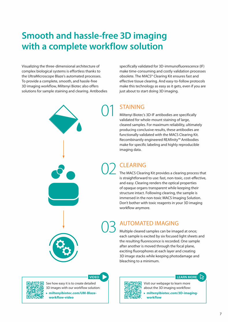
Visualizing the three-dimensional architecture of complex biological systems is effortless thanks to the UltraMicroscope Blaze’s automated processes. To provide a complete, smooth, and hassle-free 3D imaging workflow, Miltenyi Biotec also offers solutions for sample staining and clearing. Antibodies
specifically validated for 3D-immunofluorescence (IF) make time-consuming and costly validation processes obsolete. The MACS® Clearing Kit ensures fast and effective tissue clearing. And easy-to-follow protocols make this technology as easy as it gets, even if you are just about to start doing 3D imaging.
STAININGMiltenyi Biotec’s 3D-IF antibodies are specifically validated for whole-mount staining of large, cleared samples. For maximum reliability, ultimately producing conclusive results, these antibodies are functionally validated with the MACS Clearing Kit. Recombinantly engineered REAfinity™ Antibodies make for specific labeling and highly reproducible imaging data.
01
AUTOMATED IMAGINGMultiple cleared samples can be imaged at once; each sample is excited by six focused light sheets and the resulting fluorescence is recorded. One sample after another is moved through the focal plane, exciting fluorophores at each layer and creating 3D image stacks while keeping photodamage and bleaching to a minimum.
03
CLEARINGThe MACS Clearing Kit provides a clearing process that is straightforward to use: fast, non-toxic, cost-effective, and easy. Clearing renders the optical properties of opaque organs transparent while keeping their structure intact. Following clearing, the sample is immersed in the non-toxic MACS Imaging Solution. Don’t bother with toxic reagents in your 3D imaging workflow anymore.
02
Visit our webpage to learn moreabout the 3D imaging workflow:
miltenyibiotec.com/3D-imaging-workflow
LEARN MORE
See how easy it is to create detailed3D images with our workflow solution:
miltenyibiotec.com/UM-Blaze-workflow-video
VIDEO
8
Antibodies validated for 3D imaging of cleared tissues
Identifying appropriate antibodies to label structures of interest in large cleared samples is one of the most time-consuming steps in setting up the assays. Comprehensive screening and validation processes are needed to make sure that the antibodies give meaningful results. Miltenyi Biotec has already done this work for you: Recombinantly engineered REAfinity Antibodies are specifically validated and optimized for 3D-IF on tissues cleared with the MACS Clearing Kit.
• Validated and optimized for thorough whole-mount staining of large samples cleared with the MACS Clearing Kit
• Staining time decreased by 50% due to fluorochrome-conjugated primary antibodies
• Optimal signal-to-background ratios with primary antibodies conjugated to bright and photostable Vio® Dyes
• Recombinantly engineered for reproducible results and minimal background signals
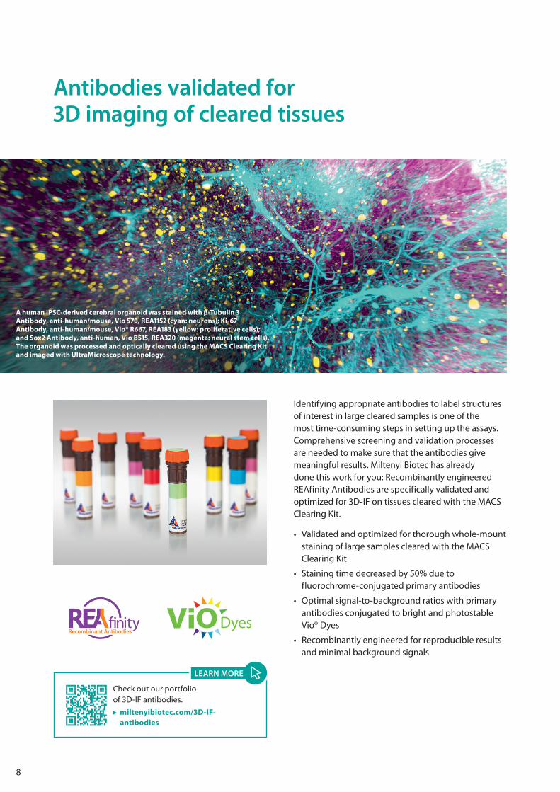
A human iPSC-derived cerebral organoid was stained with β-Tubulin 3 Antibody, anti-human/mouse, Vio 570, REA1152 (cyan: neurons); Ki-67 Antibody, anti-human/mouse, Vio® R667, REA183 (yellow: proliferative cells); and Sox2 Antibody, anti-human, Vio B515, REA320 (magenta: neural stem cells). The organoid was processed and optically cleared using the MACS Clearing Kit and imaged with UltraMicroscope technology.
Check out our portfolio of 3D-IF antibodies.
miltenyibiotec.com/3D-IF-antibodies
LEARN MORE
9
Mo
use
bra
in h
emis
ph
ere
Before clearing After clearing
Hu
man
ova
rian
tum
or
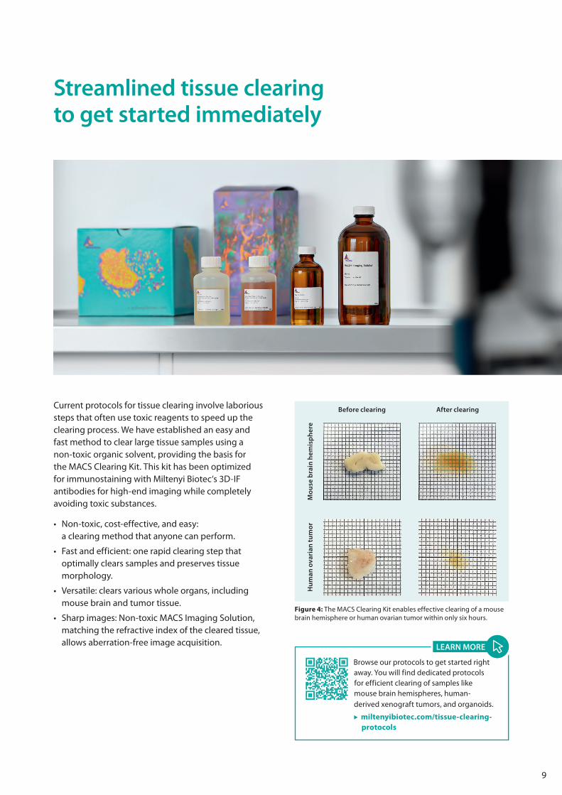
Figure 4: The MACS Clearing Kit enables effective clearing of a mouse brain hemisphere or human ovarian tumor within only six hours.
Current protocols for tissue clearing involve laborious steps that often use toxic reagents to speed up the clearing process. We have established an easy and fast method to clear large tissue samples using a non-toxic organic solvent, providing the basis for the MACS Clearing Kit. This kit has been optimized for immunostaining with Miltenyi Biotec‘s 3D-IF antibodies for high-end imaging while completely avoiding toxic substances.
• Non-toxic, cost-effective, and easy: a clearing method that anyone can perform.
• Fast and efficient: one rapid clearing step that optimally clears samples and preserves tissue morphology.
• Versatile: clears various whole organs, including mouse brain and tumor tissue.
• Sharp images: Non-toxic MACS Imaging Solution, matching the refractive index of the cleared tissue, allows aberration-free image acquisition.
Browse our protocols to get started right away. You will find dedicated protocols for efficient clearing of samples like mouse brain hemispheres, human-derived xenograft tumors, and organoids.
miltenyibiotec.com/tissue-clearing-protocols
LEARN MORE
Streamlined tissue clearing to get started immediately
10
3D microscopy and deep learning reveal the heterogeneity of crown-like structure microenvironments in intact adipose tissue.Geng, J. et al. (2021) Sci. Adv. 7: eabe2480.
Identification and characterization of a non-conventional CD45 negative perivascular macrophage population within the mouse brain.Siret, C. et al. (2021) Research Square: preprint. DOI: 10.21203/rs.3.rs-479980/v1
Cellular and molecular probing of intact human organs.Zhao, S. et al. (2020) Cell 180, 1–17.
Deep learning reveals cancer metastasis and therapeutic antibody targeting in the entire body.Pan, C. et al. (2019) Cell 179: 1661–1676.e19.
Locally renewing resident synovial macrophages provide a protective barrier for the joint.Culemann, S. et al. (2019) Nature 572: 670–675.
Glioblastoma multiforme restructures the topological connectivity of cerebrovascular networks.Hahn, A. et al. (2019) Scientific Reports 9, 11757.
Correlated MRI and Ultramicroscopy (MR-UM) of brain tumors reveals vast heterogeneity of tumor infiltration and neoangiogenesis in preclinical models and human disease.Breckwoldt, M.O. et al. (2019) Front. Neurosci. 12, 1004.
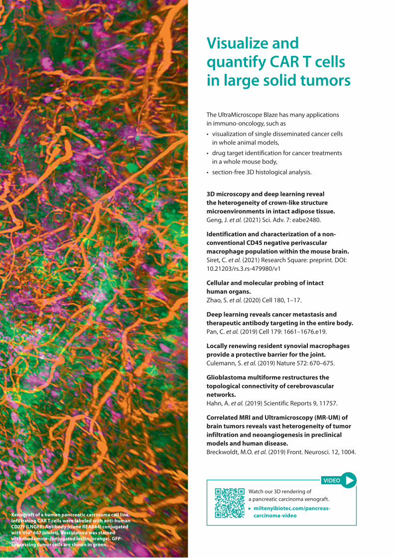
The UltraMicroscope Blaze has many applications in immuno-oncology, such as
• visualization of single disseminated cancer cells in whole animal models,
• drug target identification for cancer treatments in a whole mouse body,
• section-free 3D histological analysis.
Xenograft of a human pancreatic carcinoma cell line. Infiltrating CAR T cells were labeled with anti-human CD271 (LNGFR) Antibody (clone REA844) conjugated with Vio® 667 (violet). Vasculature was stained with rhodamine-conjugated lectin (orange). GFP-expressing tumor cells are shown in green.
Visualize and quantify CAR T cells in large solid tumors
Watch our 3D rendering of a pancreatic carcinoma xenograft.
miltenyibiotec.com/pancreas-carcinoma-video
VIDEO
11
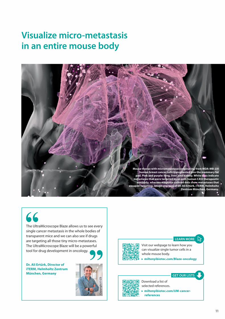
The UltraMicroscope Blaze allows us to see every single cancer metastasis in the whole bodies of transparent mice and we can also see if drugs are targeting all those tiny micro-metastases. The UltraMicroscope Blaze will be a powerful tool for drug development in oncology.
Dr. Ali Ertürk, Director of iTERM, Helmholtz Zentrum München, Germany
Cop
yrig
ht ©
IZB
Mouse thorax with micrometastases originating from MDA-MB-231 human breast cancer cells transplanted into the mammary fat
pad. Pink and purple: lung, liver, and kidney. White dots indicate metastases that were targeted by an anti-human CA12 therapeutic
antibody, whereas magenta-colored dots show metastases that escaped targeting. Image courtesy of Dr. Ali Ertürk, iTERM, Helmholtz
Zentrum München, Germany.
Visualize micro-metastasis in an entire mouse body
Download a list of selected references.
miltenyibiotec.com/UM-cancer-references
GET OUR LISTS
Visit our webpage to learn how you can visualize single tumor cells in a whole mouse body.
miltenyibiotec.com/Blaze-oncology
LEARN MORE
12
Microglia facilitate repair of demyelinated lesions via post-squalene sterol synthesis.Berghoff, S.A. et al. (2021) Nat. Neurosci. 24: 47–60.
Ventral arkypallidal neurons inhibit accumbal firing to promote reward consumption.Vachez, Y.M. et al. (2021) Nat. Neurosci. 24: 379–390.
Mapping the fine-scale organization and plasticity of the brain vasculature.Kirst, C. et al. (2020) Cell 180, 780–795.e25.
Circuit asymmetries underlie functional lateralization in the mouse auditory cortex.Levy, R.B. et al. (2019) Nat. Commun. 10: 2783.
GABAergic inhibition in dual-transmission cholinergic and GABAergic striatal interneurons is abolished in Parkinson disease.Lozovaya, N. et al. (2018) Nat. Commun. 9: 1422.
Three-dimensional study of Alzheimer’s disease hallmarks using the iDISCO clearing method.Liebmann, T. et al. (2016) Cell Rep. 6: 1138–1152.
Mapping of brain activity by automated volume analysis of immediate early genes.Renier, N. et al. (2016) Cell 165: 1789–1802.
3D neuroimaging across scales – from a whole mouse to single neurons
Understand the complex orchestration of neural circuits with whole-brain imaging at subcellular resolution. The UltraMicroscope Blaze offers many applications in neuroscience, such as
• system-level identification of neuronal circuits in whole brains at subcellular resolution,
• 3D study of the pathology of Alzheimer’s and Parkinson’s diseases in whole brains in unprecedented detail,
• holistic visualization of affected areas in the central and peripheral nervous system after stroke and traumatic brain injury.
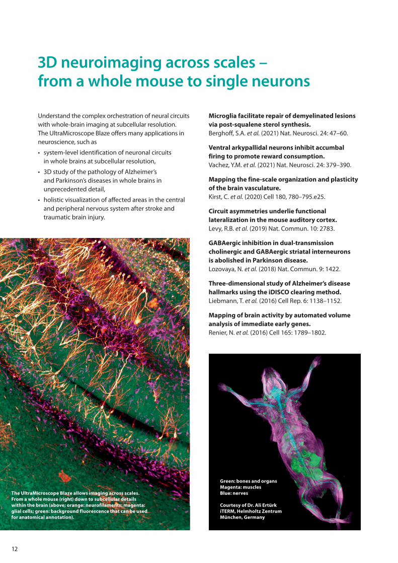
The UltraMicroscope Blaze allows imaging across scales. From a whole mouse (right) down to subcellular details within the brain (above; orange: neurofilaments; magenta: glial cells; green: background fluorescence that can be used for anatomical annotation).
Green: bones and organsMagenta: musclesBlue: nerves
Courtesy of Dr. Ali ErtürkiTERM, Helmholtz ZentrumMünchen, Germany
13
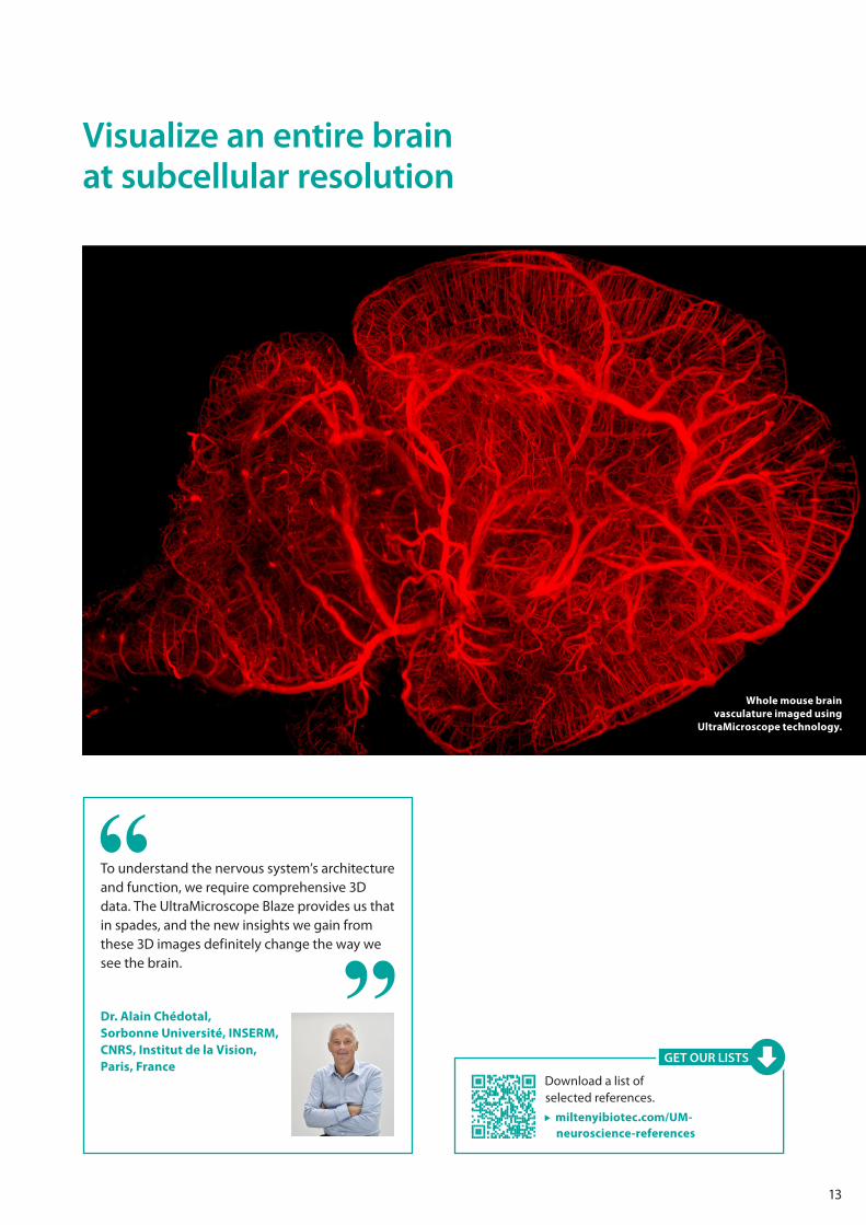
Visualize an entire brain at subcellular resolution
To understand the nervous system’s architecture and function, we require comprehensive 3D data. The UltraMicroscope Blaze provides us that in spades, and the new insights we gain from these 3D images definitely change the way we see the brain.
Dr. Alain Chédotal, Sorbonne Université, INSERM, CNRS, Institut de la Vision, Paris, France
Whole mouse brain vasculature imaged using
UltraMicroscope technology.
Download a list of selected references.
miltenyibiotec.com/UM-neuroscience-references
GET OUR LISTS
14
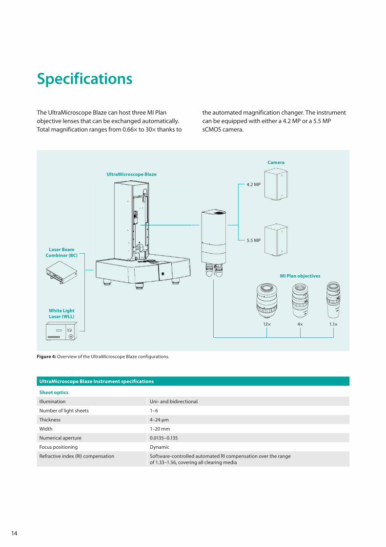
Specifications
The UltraMicroscope Blaze can host three MI Plan objective lenses that can be exchanged automatically. Total magnification ranges from 0.66× to 30× thanks to
the automated magnification changer. The instrument can be equipped with either a 4.2 MP or a 5.5 MP sCMOS camera.
UltraMicroscope Blaze
4.2 MP
Camera
5.5 MP
Laser Beam Combiner (BC)
White Light Laser (WLL)
MI Plan objectives
1.1×4×12×
Figure 4: Overview of the UltraMicroscope Blaze configurations.
UltraMicroscope Blaze Instrument specifications
Sheet optics
Illumination Uni- and bidirectional
Number of light sheets 1–6
Thickness 4–24 μm
Width 1–20 mm
Numerical aperture 0.0135–0.135
Focus positioning Dynamic
Refractive index (RI) compensation Software-controlled automated RI compensation over the range of 1.33–1.56, covering all clearing media
15
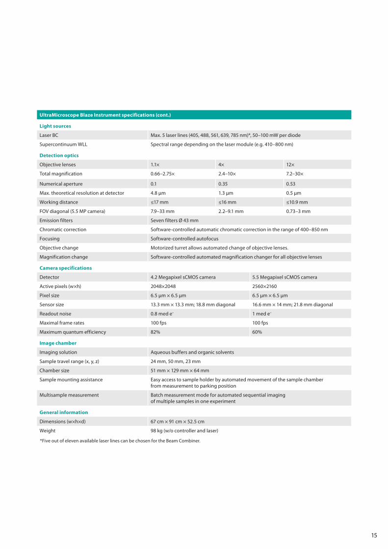
UltraMicroscope Blaze Instrument specifications (cont.)
Light sources
Laser BC Max. 5 laser lines (405, 488, 561, 639, 785 nm)*, 50–100 mW per diode
Supercontinuum WLL Spectral range depending on the laser module (e.g. 410–800 nm)
Detection optics
Objective lenses 1.1× 4× 12×
Total magnification 0.66–2.75× 2.4–10× 7.2–30×
Numerical aperture 0.1 0.35 0.53
Max. theoretical resolution at detector 4.8 μm 1.3 μm 0.5 μm
Working distance ≤17 mm ≤16 mm ≤10.9 mm
FOV diagonal (5.5 MP camera) 7.9–33 mm 2.2–9.1 mm 0.73–3 mm
Emission filters Seven filters Ø 43 mm
Chromatic correction Software-controlled automatic chromatic correction in the range of 400–850 nm
Focusing Software-controlled autofocus
Objective change Motorized turret allows automated change of objective lenses.
Magnification change Software-controlled automated magnification changer for all objective lenses
Camera specifications
Detector 4.2 Megapixel sCMOS camera 5.5 Megapixel sCMOS camera
Active pixels (w×h) 2048×2048 2560×2160
Pixel size 6.5 μm × 6.5 μm 6.5 μm × 6.5 μm
Sensor size 13.3 mm × 13.3 mm; 18.8 mm diagonal 16.6 mm × 14 mm; 21.8 mm diagonal
Readout noise 0.8 med e– 1 med e–
Maximal frame rates 100 fps 100 fps
Maximum quantum efficiency 82% 60%
Image chamber
Imaging solution Aqueous buffers and organic solvents
Sample travel range (x, y, z) 24 mm, 50 mm, 23 mm
Chamber size 51 mm × 129 mm × 64 mm
Sample mounting assistance Easy access to sample holder by automated movement of the sample chamber from measurement to parking position
Multisample measurement Batch measurement mode for automated sequential imaging of multiple samples in one experiment
General information
Dimensions (w×h×d) 67 cm × 91 cm × 52.5 cm
Weight 98 kg (w/o controller and laser)
*Five out of eleven available laser lines can be chosen for the Beam Combiner.
Germany/AustriaMiltenyi Biotec B.V. & Co. KGFriedrich-Ebert-Straße 6851429 Bergisch GladbachGermanyPhone +49 2204 8306-0Fax +49 2204 [email protected]
USA/CanadaMiltenyi Biotec, Inc.2303 Lindbergh StreetAuburn, CA 95602, USAPhone 800 FOR MACSPhone +1 866 811 4466Fax +1 877 591 [email protected]
AustraliaMiltenyi Biotec Australia Pty. Ltd.Unit 11, 2 Eden Park DriveMacquarie Park, NSW 2113AustraliaPhone +61 2 8877 7400Fax +61 2 9889 [email protected]
BeneluxMiltenyi Biotec B.V.Sandifortdreef 172333 ZZ LeidenThe [email protected] service The NetherlandsPhone 0800 4020120Fax 0800 4020100Customer service BelgiumPhone 0800 94016Fax 0800 99626Customer service LuxembourgPhone 800 24971Fax 800 24984
ChinaMiltenyi Biotec Technology & Trading (Shanghai) Co., Ltd.Room 401No. 1077, Zhangheng RoadPudong New Area201203 Shanghai, P.R. ChinaPhone +86 21 6235 1005-0Fax +86 21 6235 [email protected]
FranceMiltenyi Biotec SAS10 rue Mercoeur75011 Paris, FrancePhone +33 1 56 98 16 [email protected]
Hong KongMiltenyi Biotec Hong Kong Ltd.Unit 301, Lakeside 1 No. 8 Science Park West AvenueHong Kong Science Park Pak Shek Kok, New Territories Hong KongPhone +852 3751 6698Fax +852 3619 [email protected]
ItalyMiltenyi Biotec S.r.l.Via Paolo Nanni Costa, 3040133 Bologna ItalyPhone +39 051 6 460 411Fax +39 051 6 460 [email protected]
JapanMiltenyi Biotec K.K.NEX-Eitai Building 5F16-10 Fuyuki, Koto-kuTokyo 135-0041, JapanPhone +81 3 5646 8910Fax +81 3 5646 [email protected]
Nordics and BalticsMiltenyi Biotec Norden ABMedicon VillageScheeletorget 1223 81 [email protected] service SwedenPhone 0200 111 800Fax +46 280 72 99 Customer service DenmarkPhone 80 20 30 10Fax +46 46 280 72 99 Customer service Norway, Finland, Iceland, and Baltic countriesPhone +46 46 280 72 80Fax +46 46 280 72 99
SingaporeMiltenyi Biotec Asia Pacific Pte Ltd.438B Alexandra Road, Block BAlexandra Technopark#06-01Singapore 119968 Phone +65 6238 8183Fax +65 6238 [email protected]
South KoreaMiltenyi Biotec Korea Co., Ltd.Arigi Bldg. 8F562 Nonhyeon-roGangnam-guSeoul 06136, South KoreaPhone +82 2 555 1988Fax +82 2 555 [email protected]
SpainMiltenyi Biotec S.L.C/Luis Buñuel 2Ciudad de la Imagen28223 Pozuelo de Alarcón (Madrid)SpainPhone +34 91 512 12 90Fax +34 91 512 12 [email protected]
SwitzerlandMiltenyi Biotec Swiss AGGibelinstrasse 274500 SolothurnSwitzerlandPhone +41 32 623 08 47Fax +49 2204 [email protected]
United KingdomMiltenyi Biotec Ltd.Almac House, Church LaneBisley, Surrey GU24 9DR, UKPhone +44 1483 799 800Fax +44 1483 799 [email protected]
www.miltenyibiotec.com
130-
126-
220.
03
Miltenyi Biotec provides products and services worldwide. Visit www.miltenyibiotec.com/local to find your nearest Miltenyi Biotec contact.
Unless otherwise specifically indicated, Miltenyi Biotec products and services are for research use only and not for therapeutic or diagnostic use. Blaze, MACS, the Miltenyi Biotec logo, REAfinity, and Vio, are registered trademarks or trademarks of Miltenyi Biotec and/or its affiliates in various countries worldwide. Copyright © 2021 Miltenyi Biotec and/or its affiliates. All rights reserved.
miltenyibiotec.com


