Ultrasonographic and clinical evaluation of additional contribution of kinesiotaping to tendon and nerve gliding exercises in the treatment of carpal tunnel syndrome
Pınar YILDIRIM, Banu DİLEK*, Ebru ŞAHİN, Selmin GÜLBAHAR, Ramazan KIZILDepartment of Physical Medicine and Rehabilitation, Faculty of Medicine, Dokuz Eylül University, İzmir, Turkey
1. IntroductionCarpal tunnel syndrome (CTS) develops as a result of compression of the median nerve under the transverse carpal ligament during its course through the carpal tunnel (1). It is the most frequently seen and best known entrapment neuropathy. Its incidence has been reported as 0.1% to 3.8%. It is most commonly seen in middle age and it is seen threefold more in females than males. It causes several complaints such as numbness that becomes prominent particularly at night, pain, stiffness, and loss of function and skills (2–4).
Early diagnosis and treatment is important, as CTS is common, worsens quality of life, and leads to irreversible nerve damage if untreated. The diagnosis is based on the clinical findings in the presence of typical signs and symptoms. Electrophysiological tests are supportive for the diagnosis; however, they can be insufficient in the early stage. Furthermore, ultrasonography (USG) can be helpful for the diagnosis of CTS (5).
Conservative and surgical treatments are recommended in CTS. In conservative treatments, various methods such as splints, local steroid injections, physical therapy agents, and exercise are used. However, there is an ongoing debate about the effectiveness and superiority of these methods (6).
Kinesiotaping is a method that has been used in musculoskeletal system diseases in recent years and its effectiveness in certain diseases has been demonstrated (7–9). On the other hand, in the literature, there is a limited number of studies on the effectiveness of kinesiotaping in CTS. In these studies, it has been shown that kinesiotaping provides an additional contribution to symptoms and functions compared to placebos, and its use together with a splint increases these contributions (10,11). USG has not been used in the follow-up of kinesiotaping treatment in CTS before.
In the present study, we aimed to ultrasonographically and clinically evaluate the additional contribution of
Background and aim: This study aims to ultrasonographically and clinically evaluate the additive contribution of kinesiotaping to tendon and nerve gliding exercises in the treatment of mild or moderate carpal tunnel syndrome (CTS).
Materials and methods: Thirty-eight wrists of patients (n = 21) with CTS were randomized into two groups as the intervention group (n = 19) and the control group (n = 19). Tendon and nerve gliding exercises were given to both groups. In the intervention group, additional kinesiotaping was performed three times with 5-day intervals. Functional assessments were performed with the Boston Carpal Tunnel Syndrome Questionnaire and the Moberg pick-up test. Hand grip and pinch strength were evaluated. Cross-section area (CSA) of the median nerve was measured by ultrasonography. All assessments were performed at baseline and at 3 and 6 weeks after treatment.
Results: In the intervention group, there was a significant improvement in all clinical assessments and in the CSA of the median nerve at the level of proximal carpal bones. In the control group, a significant improvement was detected in all clinical parameters except grip strength and ultrasonographic measurements. There was no significant difference in the clinical and ultrasonographic findings between the groups at 6 weeks.
Conclusion: Kinesiotaping may provide a positive contribution to ultrasonographic and clinical outcomes in the treatment of mild or moderate CTS in the short term.
Received: 19.09.2017 Accepted/Published Online: 23.07.2018 Final Version: 31.10.2018
Research Article
This work is licensed under a Creative Commons Attribution 4.0 International License.
926
YILDIRIM et al. / Turk J Med Sci
kinesiotaping to tendon and nerve gliding exercises in the treatment of mild or moderate CTS.
2. Materials and methodsThe study protocol was approved by the Ethics Committee of Dokuz Eylül University Medical Faculty. The study was conducted in accordance with the principles of the Declaration of Helsinki.
This prospective, single-blind, randomized study included a total of 38 wrists of 21 patients aged between 18 and 60 years who were admitted to the physical medicine and rehabilitation outpatient clinic between January 2016 and October 2016 who were clinically and electrophysiologically diagnosed with mild or moderate CTS, who had the symptoms for at least 3 months, and for whom conservative treatment was planned. The clinical assessment of each patient was performed by a blind investigator (PY) and USG measurements were performed by another blind investigator (BD).
The exclusion criteria were as follows: electrophysiological diagnosis of severe CTS, thenar atrophy, local corticosteroid injection or physical therapy for CTS within the past 3 months, cervical disc hernia (which can affect symptoms), peripheral nerve damage, entrapment neuropathy at more proximal levels in an upper extremity, and pregnancy.
The wrists of the patients were randomized into two groups as the intervention group (n = 19) and the control group (n = 19). For patients who were diagnosed with bilateral CTS, both wrists were included in the group of the first (right) wrist.
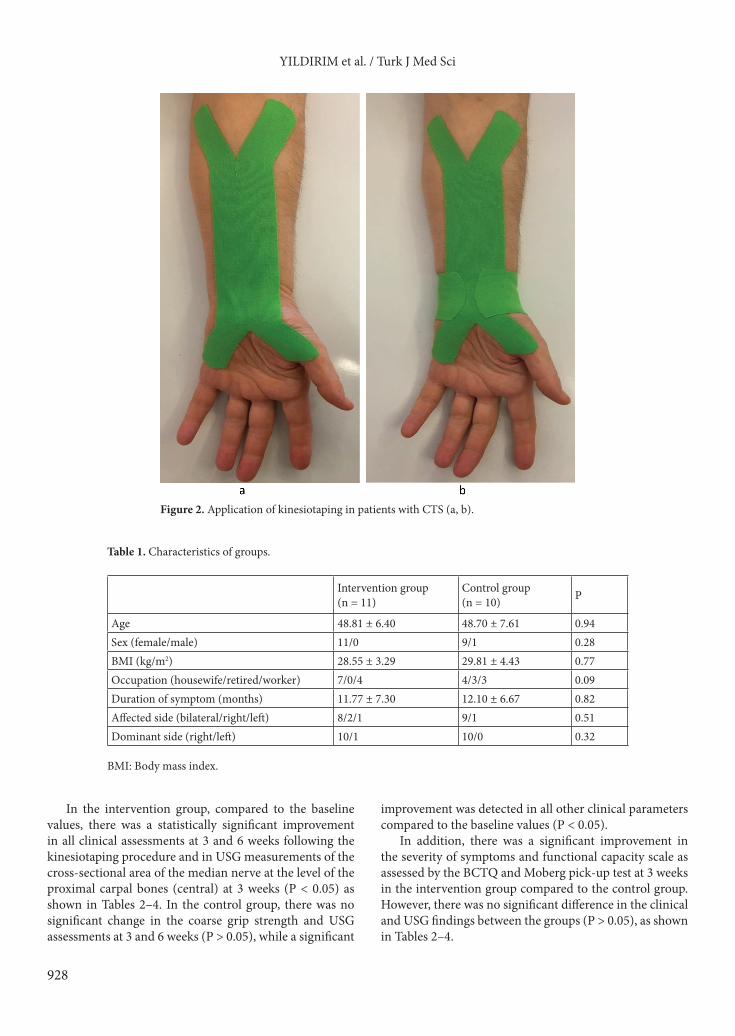
At baseline, written informed consent was obtained from each patient. Then demographical and clinical data of the patients such as age, sex, body mass index (BMI), dominant hand, affected side, occupation, and duration of the symptoms were recorded. Tendon and nerve gliding exercises were performed in both groups and kinesiotaping was performed additionally in the treatment group. Tendon and nerve gliding exercises were continued for 6 weeks. In the treatment group, kinesiological banding was performed three times with 5-day intervals.
The Boston Carpal Tunnel Syndrome Questionnaire (BCTQ) was used for the clinical evaluation, a hand dynamometer was used for grip strength, a pinchmeter was used for finger pinch (pulp to pulp), the Moberg pick-up test was used for hand skills, and USG was used for the evaluation of the carpal tunnel and median nerve. All patients were evaluated at baseline and at 3 and 6 weeks of treatment.
The BCTQ is a disease-specific questionnaire comprising two scales to evaluate the severity of symptoms and functional capacity (12). The Turkish version of the questionnaire was validated by Heybeli (13) and it can be
used in the evaluation of the effectiveness of treatment (13,14). With this scale, patients are evaluated using 11 questions, where each question consists of five answers and the symptoms are scored in a range from absent to very severe. High scores in the scoring of 11–55 indicate an increased severity of symptoms (12,13). In the functional capacity scale, eight questions consisting of daily activities in which hands are used are asked and patients are asked to answer these questions as “able to perform without difficulty” or “impossible to do”. The results are scored between 8 and 40 and high scores indicate functional impairment of the hand (12,13).
In the evaluation of grip strength, a hand dynamometer (JAMAR) was used for grip strength and a pinchmeter was used for the evaluation of the pulp to pulp grip (15). The measurements were performed with tightening at a maximum strength, while the elbow was at 90° flexion and the forearm and the wrist were in the neutral position, and three measurements were performed. The best of these measurements were recorded in kilograms.
In the Moberg pick-up test, for the evaluation of hand skills, the patient was positioned in the sitting position in a chair and a table was put in front of the patient, and 12 small objects (2 paperclips, 1 pencil, 1 needle, 2 nuts, 1 A4 paper, 2 coins, 1 screw, 1 ring, and 1 key) were put on the table (16). The patient was requested to pick up these objects using the affected hand without sliding on the surface and to put them in a box present on the table, and the duration was recorded in seconds.
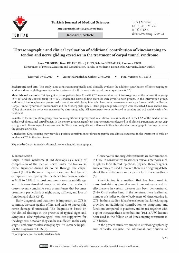
In the USG evaluation of CTS, a linear probe of 7–11 mHz was used (LOGIQ P5, GE, Solingen, Germany). All measurements were performed by drawing the median nerve at the largest point without including the nerve sheath, as shown in Figures 1a and 1b.
The median nerve cross-sectional area was measured at 4 cm proximal to the carpal tunnel, at the level of the radiocarpal joint at the entrance to the tunnel, at the level of the proximal carpal bones at the middle of the tunnel, and at the level of the distal carpal bones at the exit of the tunnel (17,18).
In both groups, tendon and nerve gliding exercises were performed three times a day with 15 repetitions each time for 6 weeks. For tendon gliding exercises, the fingers were put into five different positions as regular grip, hook grip, punch, tabletop, and regular punch. During median nerve gliding exercises, the fingers, hand, and wrist were put into six different positions. In the first position, the wrist was put in neutral position, while fingers and thumb were in flexion; in the second position, the wrist was in neutral position and the thumb and fingers were in extension; in the third position, the wrist and fingers were in extension while the thumb was in neutral position; in the fourth position, the wrist, thumbs, and fingers were in extension; in the fifth position, the forearm was in supination; and
927
YILDIRIM et al. / Turk J Med Sci
in the sixth position, gentle stretching was performed of the thumb with the other hand (19). An exercise diary was given to the patients.
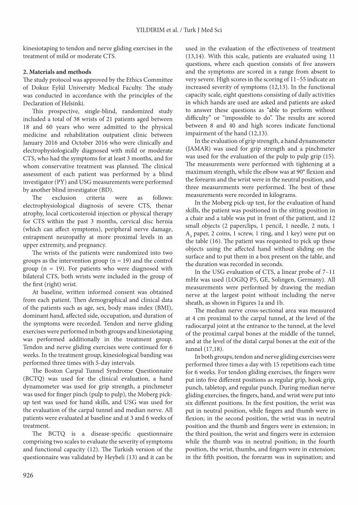
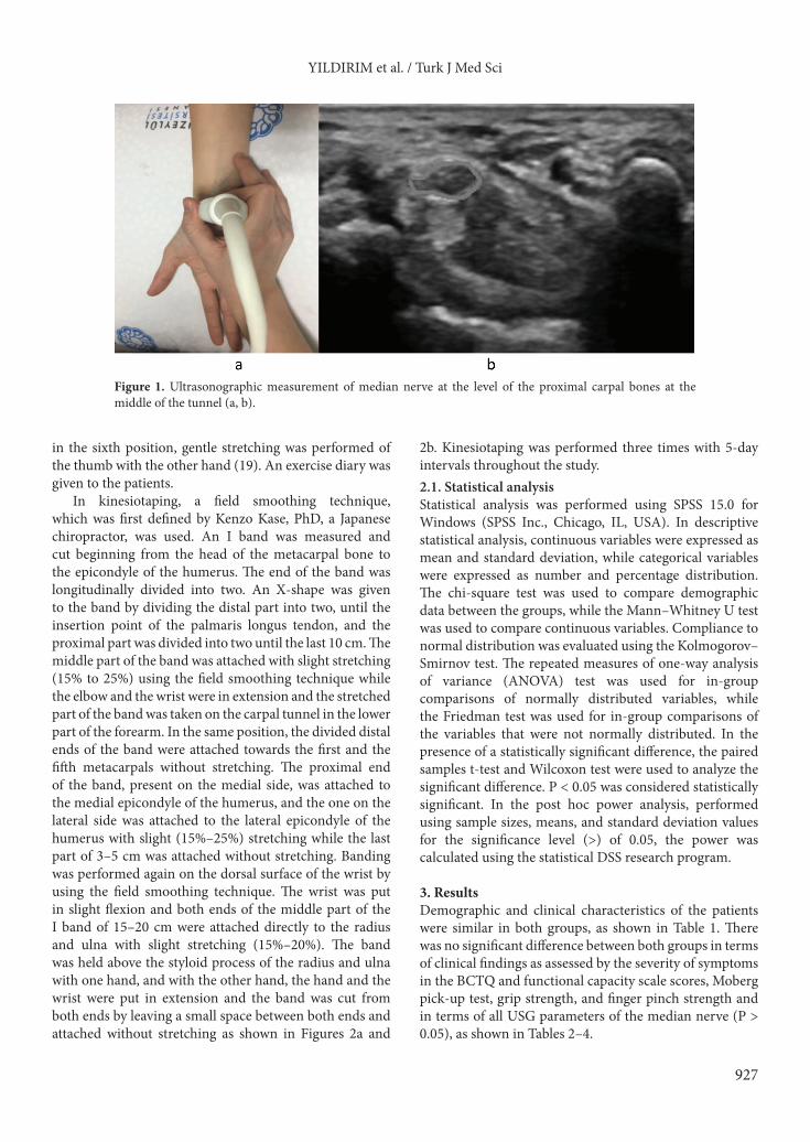
In kinesiotaping, a field smoothing technique, which was first defined by Kenzo Kase, PhD, a Japanese chiropractor, was used. An I band was measured and cut beginning from the head of the metacarpal bone to the epicondyle of the humerus. The end of the band was longitudinally divided into two. An X-shape was given to the band by dividing the distal part into two, until the insertion point of the palmaris longus tendon, and the proximal part was divided into two until the last 10 cm. The middle part of the band was attached with slight stretching (15% to 25%) using the field smoothing technique while the elbow and the wrist were in extension and the stretched part of the band was taken on the carpal tunnel in the lower part of the forearm. In the same position, the divided distal ends of the band were attached towards the first and the fifth metacarpals without stretching. The proximal end of the band, present on the medial side, was attached to the medial epicondyle of the humerus, and the one on the lateral side was attached to the lateral epicondyle of the humerus with slight (15%–25%) stretching while the last part of 3–5 cm was attached without stretching. Banding was performed again on the dorsal surface of the wrist by using the field smoothing technique. The wrist was put in slight flexion and both ends of the middle part of the I band of 15–20 cm were attached directly to the radius and ulna with slight stretching (15%–20%). The band was held above the styloid process of the radius and ulna with one hand, and with the other hand, the hand and the wrist were put in extension and the band was cut from both ends by leaving a small space between both ends and attached without stretching as shown in Figures 2a and
2b. Kinesiotaping was performed three times with 5-day intervals throughout the study. 2.1. Statistical analysisStatistical analysis was performed using SPSS 15.0 for Windows (SPSS Inc., Chicago, IL, USA). In descriptive statistical analysis, continuous variables were expressed as mean and standard deviation, while categorical variables were expressed as number and percentage distribution. The chi-square test was used to compare demographic data between the groups, while the Mann–Whitney U test was used to compare continuous variables. Compliance to normal distribution was evaluated using the Kolmogorov–Smirnov test. The repeated measures of one-way analysis of variance (ANOVA) test was used for in-group comparisons of normally distributed variables, while the Friedman test was used for in-group comparisons of the variables that were not normally distributed. In the presence of a statistically significant difference, the paired samples t-test and Wilcoxon test were used to analyze the significant difference. P < 0.05 was considered statistically significant. In the post hoc power analysis, performed using sample sizes, means, and standard deviation values for the significance level (>) of 0.05, the power was calculated using the statistical DSS research program.
3. ResultsDemographic and clinical characteristics of the patients were similar in both groups, as shown in Table 1. There was no significant difference between both groups in terms of clinical findings as assessed by the severity of symptoms in the BCTQ and functional capacity scale scores, Moberg pick-up test, grip strength, and finger pinch strength and in terms of all USG parameters of the median nerve (P > 0.05), as shown in Tables 2–4.
Figure 1. Ultrasonographic measurement of median nerve at the level of the proximal carpal bones at the middle of the tunnel (a, b).
928
YILDIRIM et al. / Turk J Med Sci
In the intervention group, compared to the baseline values, there was a statistically significant improvement in all clinical assessments at 3 and 6 weeks following the kinesiotaping procedure and in USG measurements of the cross-sectional area of the median nerve at the level of the proximal carpal bones (central) at 3 weeks (P < 0.05) as shown in Tables 2–4. In the control group, there was no significant change in the coarse grip strength and USG assessments at 3 and 6 weeks (P > 0.05), while a significant
improvement was detected in all other clinical parameters compared to the baseline values (P < 0.05).
In addition, there was a significant improvement in the severity of symptoms and functional capacity scale as assessed by the BCTQ and Moberg pick-up test at 3 weeks in the intervention group compared to the control group. However, there was no significant difference in the clinical and USG findings between the groups (P > 0.05), as shown in Tables 2–4.
Figure 2. Application of kinesiotaping in patients with CTS (a, b).
We could not calculate the sample size because there is no similar study. In the post hoc power analysis, performed using sample sizes, means, and standard deviation values for the significance level (alpha) of 0.05, the power was found to be 76% for the Moberg test and 72% for the BCTQ at 3 weeks. However, the power was below 50% for the other clinical and ultrasonographic measurements. 4. DiscussionIn the current study, we showed that kinesiotaping in addition to an exercise program provided a positive
contribution in the short term to the clinical outcomes in the treatment of mild or moderate CTS.
Kinesiotaping has physiological effects on the blood, lymphatic circulation, muscles, joints, and fascia. The basic goal of the technique is to support movement and to facilitate mobilization. Banding increases the space between the skin and the muscles by elevating the skin and decreases the pressure developing in this area. In this way, it helps blood circulation and lymphatic drainage. This effect in the blood circulation helps eliminate the tension
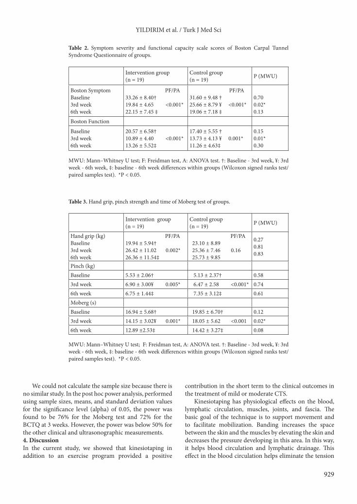
Table 2. Symptom severity and functional capacity scale scores of Boston Carpal Tunnel Syndrome Questionnaire of groups.
MWU: Mann–Whitney U test; F: Freidman test, A: ANOVA test. †: Baseline - 3rd week, ¥: 3rd week - 6th week, ‡: baseline - 6th week differences within groups (Wilcoxon signed ranks test/paired samples test). *P < 0.05.
930
YILDIRIM et al. / Turk J Med Sci
and tenderness in the damaged area, and it decreases stimulation of subcutaneous pain receptors. Owing to these features, kinesiological banding provides positive effects in the treatment of CTS (9).
For early stage and mild to moderate CTS, different conservative treatment methods including activity modification, physical treatment modalities, splints, local steroid administration, and exercise are recommended. Tendon and nerve gliding exercises in addition to different conservative treatment methods have also been shown to be effective (17). In a study by Akalin et al. (18) in which they evaluated effectiveness of tendon and nerve shifting exercises, the authors divided 28 patients who were clinically and electrophysiologically diagnosed with CTS (36 wrists) into two groups and recommended splint therapy for 4 weeks in both groups with tendon and nerve gliding exercises for the treatment group additionally. The rate of global improvement in both groups was not found to be significantly different. Similarly, in the present study, tendon and nerve gliding exercises were given for 6 weeks in both groups. In the treatment group, kinesiotaping was performed additionally during the first 2 weeks. A significant improvement in the clinical assessment parameters was detected in both groups. However, different from the study of Akalin et al. (18), there was no
statistically significant difference in the recovery rates at 6 weeks between the groups.
The use of kinesiotaping techniques in musculoskeletal diseases has gradually gained generality and popularity in recent years. Kinesiological banding is recommended as one of the conservative treatment methods in several peripheral nervous system diseases including CTS. In the literature, two studies on the use of kinesiological banding in the treatment of CTS have been reported (19,20).
In the study of Geler Külcü et al. (19), 65 wrists of 45 patients with the diagnosis of mild to moderate CTS were included. The patients were divided into three groups as kinesiotaping, placebo kinesiotaping, and splint groups. Tendon gliding exercises were given to all groups as a home-based program for 4 weeks. The patients were evaluated using a visual analog scale (VAS), the Douleur Neuropathique 4 questions (DN4), the BCTQ, and a hand dynamometer. The VAS and DN4 scores at 4 weeks following treatment decreased in all groups, although no significant difference was found among the groups. A significant recovery was also detected in the BCTQ scores in all groups; however, the recovery in the kinesiotaping group was statistically higher than in the placebo kinesiotaping group. Furthermore, the coarse grip strength significantly improved in the splint group only.
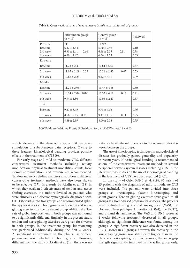
Table 4. Cross-sectional area of median nerve (mm2) in carpal tunnel of groups.
In the study of Oncu et al. (20) including 40 patients (60 wrists) with the diagnosis of mild or moderate idiopathic CTS, they investigated the effectiveness of kinesiotaping. The patients were randomized into four groups as the kinesiotaping group, splint group, kinesiotaping and splint group, and control group. Tendon and nerve gliding exercises were given to all groups as a home-based program. The patients were then evaluated using the BCTQ, hand dynamometer, pinchmeter, and Moberg tests at baseline, on day 25 following treatment, and at 2 and 3 months following treatment. Electrophysiological examination was also performed before treatment and at 3 months following treatment. There was a significantly higher recovery in the symptom severity, functions, and grip strength on day 25 and at the second and third month following treatment in the group where kinesiotaping and splint were used in combination, whereas a significant recovery was demonstrated in the Moberg pick-up test times only at the end of 3 months. Based on these findings, the authors concluded that addition of kinesiological banding and a splint together was more effective in clinical improvement and this effectiveness would last longer compared to kinesiotaping alone or splint alone. In the current study, the recovery in the BCTQ scores and the Moberg pick-up test results in the treatment group at 3 weeks were superior to the control group, while there was no significant difference in the recovery rates at 6 weeks. In addition, there was a significant recovery in both hand grip strength and finger pinch strength at 3 and 6 weeks following treatment with kinesiotaping compared to the baseline values, while there was a recovery in only finger pinch strength in patients doing the exercise program. While the study of Oncu et al. (20) demonstrated that kinesiotaping together with splints would make a contribution to hand skills in the long term, the findings of the current study suggest that kinesiological banding would make a mild additional contribution to hand skills.
Furthermore, Oncu et al. (20) used electrophysiological tests for the objective evaluation of CTS findings, while we used USG in the current study. The use of USG in the diagnosis of CTS has gradually increased in recent years. In 1993, the efficacy of USG in the diagnosis of CTS was accepted by the Report of the Quality Measurement and Reporting Subcommittee of the American Neurology Academy Quality Standards. USG is also helpful in the evaluation of carpal tunnel and soft tissues of the median nerve. The main advantages of USG are that it provides an opportunity for dynamic examination and it is easy, rapid, painless, cheap, and repeatable. To date, the use of USG in CTS has been limited to diagnosis and no study has been found related to its use in kinesiotaping treatment follow-up. In the current study, a statistically significant
recovery was observed in the cross-sectional area of the median nerve, which was measured at the level of the proximal carpal bones at 3 weeks in the treatment group, and the current study has superiority to similar studies due to the use of USG measurements during follow-up. However, this statistically significant difference is too small to be deemed a clinically important difference. Ultrasonography is a user-dependent method. This small difference may have resulted from measurement error. In the literature, the ultrasonography technique is shown with comparability of intrarater measurements about CTS (21). We did not determine intrarater and interrater reliability of measurements for this study.
Different techniques in kinesiotaping recommended by Kenzo Kase can be used in the treatment of CTS (9). The field smoothing technique when performed on only the dorsal or volar surface of the wrist can be insufficient. Therefore, we performed the field smoothing method in two fields. In this technique, the band does not extend towards the fingers of the patient, thereby providing a positive contribution in terms of patient comfort and adaptation. On the other hand, it can cause detachment of the band extending towards the hand from the skin. Detachment of the band from the skin may delay the technique, where the third and fourth fingers are passed through the hole opened at the end of the band. Different methods can yield different results.
The limitations of the current study are the absence of a control group in which no treatment was given, small sample size, application of kinesiotaping for a short period of time, and the lack of electrophysiological evaluation during follow-up. The positive results that were found in the intervention group might have resulted from a placebo effect of kinesiotaping.
In conclusion, our study results suggest that tendon and nerve gliding exercises may be effective in the short-term ultrasonographic and clinical outcomes of patients with the diagnosis of mild or moderate CTS. The addition of kinesiotaping by field smoothing technique to the tendon and nerve gliding exercises three times with 5-day intervals for a period of 15 days provides an additional, but limited, contribution. Tendon and nerve gliding exercises alone or their combination with kinesiological banding can yield a significant recovery in the USG measurement of the median nerve area at the middle of the tunnel at 3 weeks without a prominent effect at 6 weeks. Nonetheless, further large-scale, long-term, randomized controlled studies in which kinesiological banding is performed for different durations and/or by different techniques and in which electrophysiological examinations are included are required to evaluate the efficacy of this treatment method.
932
YILDIRIM et al. / Turk J Med Sci
References
1. Bengston KA, Brault JS. Hand disorders. In: Delisa JA, editor. Physical Medicine and Rehabilitation Principles and Practice. 3rd Ed. Philadelphia, PA, USA: Lippincott Williams & Wilkins, 2005. pp. 843-854.
2. Ertekin C. Santral ve Periferik EMG. 1st ed. İzmir, Turkey: Meta Basım Matbaacılık, 2006 (in Turkish).
3. Tanaka S, Wild DK, Seligman PJ, Behrens V, Cameron L, Putz-Anderson V. The US prevalence of self-reported carpal tunnel syndrome: 1988 National Health Interview Survey data. Am J Public Health 1994; 84: 1846-1848.
4. Stevens JC. AAEM Minimonograph #26: The electrodiagnosis of carpal tunnel syndrome. American Association of Electrodiagnostic Medicine. Muscle Nerve 1997; 20: 1477-1486.
5. Kara M, Özçakar L, De Muynck M, Tok F, Vanderstraeten G. Musculoskeletal ultrasound for peripheral nerve lesions. Eur J Phys Rehabil Med. 2012; 48: 665-674.
6. Page MJ, O’Connor D, Pitt V, Massy-Westropp N. Exercise and mobilisation interventions for carpal tunnel syndrome. Cochrane Database Syst Rev 2012; 6: CD009899
7. Thelen MD, Dauber JA, Stoneman PD. The clinical efficacy of kinesio tape for shoulder pain: a randomized, double-blinded, clinical trial. J Orthop Sports Phys Ther 2008; 38: 389-392.
8. Williams S, Whatman C, Hume PA, Sheerin K. Kinesio taping in treatment and prevention of sports injuries: a meta-analysis of the evidence for its effectiveness. Sports Med 2012; 42: 153-64.
9. Kase K, Wallis J, Kase T. Clinical Therapeutic Applications of the Kinesio Taping Method. Tokyo, Japan: Ken Ikai Co. Ltd.; 2003.
10. Levine DW, Simmons BP, Koris MJ, Daltroy LH, Hohl GG, Fossel AH. A self-administered questionnaire for the assessment of severity of symptoms and functional status in carpal tunnel syndrome. J Bone Joint Surg Am 1993; 75: 1585-1592.
11. Heybeli N, Özerdemoğlu RA, Aksoy OG, Mumcu EF. Karpal tünel sendromu: cerrahi tedavi izleminde fonksiyonel ve semptomatik skorlama. Acta Orthopaedica et Traumatologica Turcica 2001; 35: 147-151 (in Turkish).
12. Heybeli N, Kutluhan S, Demirci S, Kerman M, Mumcu EF. Assessment of outcome of carpal tunnel syndrome: a comparison of electrophysiological findings and a self-administered Boston questionnaire. J Hand Surg Br 2002; 27: 259-264.
13. Mathiowetz V, Weber K, Volland G, Kashman N. Reliability and validity of grip and pinch strength evaluations. J Hand Surg Am 1984; 9: 222-226.
14. Amirjani N, Ashworth NL, Olson JL, Morhart M, Chan KM. Discriminative validity and test-retest reliability of the Dellon-modified Moberg pick-up test in carpal tunnel syndrome patients. J Peripher Nerv Syst 2011; 16: 51-58.
15. Cartwright MS, Hobson-Webb LD, Boon AJ, Alter KE, Hunt CH, Flores VH, Werner RA, Shook SJ, Thomas TD, Primack SJ et al. Evidence-based guideline: neuromuscular ultrasound for the diagnosis of carpal tunnel syndrome. Muscle Nerve 2012; 46: 287-293.
16. Wong MS, Griffith JF, Hui AC, Tang A, Wong KS. Discriminatory sonographic criteria for the diagnosis of carpal tunnel syndrome. Arth Rheum 2002; 46: 1914-1921.
17. Rozmaryn LM, Dovelle S, Rothman ER, Gorman K, Olvey K, Bartko JJ. Nerve and tendon gliding exercises and the conservative management of carpal tunnel syndrome. J Hand Ther 1998; 11: 171-179.
18. Akalin E, El O, Peker O, Senocak O, Tamci S, Gülbahar S, Cakmur R, Oncel S. Treatment of carpal tunnel syndrome with nerve and tendon gliding exer cises. Am J Phys Med Rehabil 2002; 81: 108-113.
19. Geler Külcü D, Bursalı C, Aktaş İ, Bozkurt Alp S. Kinesiotaping as an alternative treatment method for carpal tunnel syndrome. Turk J Med Sci 2016; 46: 1042-1049.
20. Öncü J, İlişer R, Köymen Yılmaz F, Kuran B. Karpal tünel sendromu tedavisinde kinezyo bantlama tekniğinin hastalık semptomları, el fonksiyonu ve kavrama gücüne etkisi: tek kör randomize kontrollü çalışma. Türkiye Fiziksel Tıp ve Rehabilitasyon Dergisi 2014; 60: 43-51.
21. Fu T, Cao M, Liu f, Zhu J,Ye D, Feng x, Xu Y, Wang G, Bai G. Carpal tunnel syndrome assessment with ultrasonography: value of inlet-to-outlet median nerve area ratio in patients versus healthy volunteers. PLoS One 2015; 10: 1-11.