This manuscript has been published in the IVIS website with the permission of the congress organizers. To return to the Table of Content click here or go to http://www.ivis.org ULTRASONOGRAPHY OF THE REPRODUCTIVE SYSTEM OF THE COW: BASIC PRINCIPLES, PRACTICAL USES AND ECONOMIC ASPECTS OF THIS DIAGNOSTIC TOOL IN DAIRY PRODUCTION Luc DesCôteaux, Paul D. Carrière, Jean Durocher Faculté de Médecine Vétérinaire, Université de Montréal, St-Hyacinthe, Québec, Canada [email protected]1. INTRODUCTION In bovine practice, ultrasonography has become an important diagnostic tool for evaluating the female reproductive system. With the help of ultrasound technology, it is possible to view the entire reproductive system in a non-invasive manner (Carrière et al. 2002). The user needs to master the physical principles of ultrasonography in order to perform a worthwhile examination with the instrument. The quality of the images depends above all on the user's understanding of the interactions between the ultrasound wave and the organ tissue, as well as proper use of the instrument's controls (DesCôteaux et al. 2005). The goals of this presentation are to help understand some basic principles of ultrasonography, to present its principal uses in bovine reproduction, and the economic benefits of early pregnancy diagnosis. 2. BASIC PRINCIPLES, PROBE INDICATIONS AND TERMINOLOGY 2.1 General description of the images It is important to realize that the image on the ultrasound screen represents a fine section of an organ, grossly resembling a weakly-magnified histological cut. The probe therefore simulates the passage of a knife, slicing through an organ tissue from top to bottom. Ultrasonography thus presents a flattened two-dimensional image of a finely-cut section of tissue; whereas radiography is a two-dimensional superimposed view of the entire thickness of an animal or of a limb under observation. Ultrasound images are rapidly renewed and images are superimposed one over the other as the probe moves across a tissue surface. The rapid succession of tissue section views gives the impression that the structures are moving like an animated cartoon. When interpreting sectional WORLD BUIATRICS CONGRESS 2006 - NICE, FRANCE
Transcript
This manuscript has been published in the IVIS website with the permission of the congress organizers. To return to the Table of Content click here or go to http://www.ivis.org
ULTRASONOGRAPHY OF THE REPRODUCTIVE SYSTEM OF THE COW: BASIC PRINCIPLES, PRACTICAL USES AND ECONOMIC ASPECTS OF THIS DIAGNOSTIC
TOOL IN DAIRY PRODUCTION
Luc DesCôteaux, Paul D. Carrière, Jean Durocher
Faculté de Médecine Vétérinaire, Université de Montréal, St-Hyacinthe, Québec, Canada [email protected]
1. INTRODUCTION In bovine practice, ultrasonography has become an important diagnostic tool for evaluating the female reproductive system. With the help of ultrasound technology, it is possible to view the entire reproductive system in a non-invasive manner (Carrière et al. 2002). The user needs to master the physical principles of ultrasonography in order to perform a worthwhile examination with the instrument. The quality of the images depends above all on the user's understanding of the interactions between the ultrasound wave and the organ tissue, as well as proper use of the instrument's controls (DesCôteaux et al. 2005). The goals of this presentation are to help understand some basic principles of ultrasonography, to present its principal uses in bovine reproduction, and the economic benefits of early pregnancy diagnosis. 2. BASIC PRINCIPLES, PROBE INDICATIONS AND TERMINOLOGY 2.1 General description of the images It is important to realize that the image on the ultrasound screen represents a fine section of an organ, grossly resembling a weakly-magnified histological cut. The probe therefore simulates the passage of a knife, slicing through an organ tissue from top to bottom. Ultrasonography thus presents a flattened two-dimensional image of a finely-cut section of tissue; whereas radiography is a two-dimensional superimposed view of the entire thickness of an animal or of a limb under observation. Ultrasound images are rapidly renewed and images are superimposed one over the other as the probe moves across a tissue surface. The rapid succession of tissue section views gives the impression that the structures are moving like an animated cartoon. When interpreting sectional
views of an organ on the screen, it is essential to have a good appreciation of the three-dimensional shape of the organ in space. 2.2 Probe characteristics and resolutions The probe is the most fragile component of the ultrasound apparatus. In the veterinary market, usual probes have a frequency of 3.5, 5.0 or 7.5 MHz. The ability of the instrument to distinguish between two structures located very close together along the axis of the ultrasound beam is called its axial resolution (Figure 1). The axial resolution is best when the groups of waves emitted have a short wavelength. Since the number of cycles in each group of waves is set according to instrument design, the only way to shorten their length is to use a probe with a higher frequency. For example, the group of waves produced by a 7.5 MHz probe will be shorter than one produced by a 3.5 MHz probe and will provide better axial resolution.
Figure 1. Axial and lateral resolutions of the linear probe There are two types of probes: linear and sectorial (Figure 2). In theriogenology, linear probes are preferred for transrectal ultrasound examinations of the ovaries and uterus. This probe has a set row of crystals that are selected electronically to form a rectangular image. The linear probe provides good resolution for tissues located close to the probe. Sectorial probes have one or several crystals whose position produces a beam in the shape of a pie slice. The advantage of the sectorial probe is that it doesn't require a large surface of contact, and it scans a greater overall surface. The disadvantage is that the visual field and the lateral resolution (i.e. the ability of a system to differentiate between two adjacent structures) are more restricted close to the probe. The sectorial probe is ideal for viewing the small ruminant fetus by transabdominal ultrasound imaging and for ultrasound-guided transvaginal aspiration of bovine follicles.
WORLD BUIATRICS CONGRESS 2006 - NICE, FRANCE
Figure 2. Types of probes (A = sectorial and B = linear) The details contained in an ultrasound image (resolution) as well as the depth of the tissue observed depend on the frequency and the focalization of the scanning beam. With a lower frequency, tissue penetration will be deep, but the resolution will be lower. A higher frequency enables better resolution, but beam attenuation will be greater and it will not penetrate the tissue as deeply. Table I summarizes the indications of the different types of probes used in bovine theriogenology. Table I. Summary of indications and characteristics of different types of probes used in bovine reproduction
Follicles and corpus luteum Pregnancy diagnosis Fetal sexing
Follicles and corpus luteum Pregnancy diagnosis
2.3 Terminology and interpretation of images The description of ultrasound images is based on an evaluation of the shape, contour, size, and position of the structure being studied, as well as its echogenicity, which depends on the amplitude of the echoes received. An echogenic structure reflects the majority of soundwaves back to the probe and thus appears from white to different shades of grey on the screen. An anechogenic structure does not produce echoes; instead, it transmits the waves on to more deeply situated tissues. An example of an anechogenic structure is follicular fluid, which appears black on the screen. The terms hypoechogenic and hyperechogenic indicate respectively a decrease and an increase in relative echogenicity in comparison with the surrounding tissue, whereas the term isoechogenic is used to describe similar echogenicity with the surrounding tissue.
WORLD BUIATRICS CONGRESS 2006 - NICE, FRANCE
The following sections will present the principal uses of ultrasonography in bovine reproduction. 3. PRACTICAL APPLICATIONS OF ULTRASONOGRAPHY IN BOVINE REPRODUCTION 3.1 Ovarian and uterus evaluations Since the advent of bovine ovarian ultrasound in 1984, enormous progress has been made in our understanding of folliculogenesis and the development of the bovine corpus luteum (Durocher et al. 2005; Ginther, 1998; Pierson & Ginther, 1988; Sirois & Fortune, 1988). Ultrasound enables us to describe the dynamics of follicular growth in follicles greater than 1 mm in diameter (Durocher et al. 2005). In cows, we can find follicles larger than 1 mm throughout the estrous cycle. The same pattern of growth repeats itself approximately every seven days. It starts with the growth of a cohort of many follicles, known as a follicular wave, which includes a follicle that will pursue its growth and will be called dominant while the other follicles of the cohort will regress by atresia. The most frequently encountered pathological conditions of the ovaries in bovine practice are follicular and luteal cysts. Using ultrasound, the clinician can differentiate between these two types of cysts and apply the appropriate treatment. Exaggerated growth of a non-ovulating follicle may lead to the creation of a follicular or luteal cyst. An ovary is usually considered cystic when it contains a hollow structure greater than 25 mm that persists for more than 10 days. A follicular cyst can be differentiated from a luteal cyst by its thin wall and uniformly anechogenic follicular fluid (Ginther, 1998). The two most frequently observed uterine pathologies in ultrasound diagnosis are, by decreasing order of prevalence, the metritis complex (pyometra-pyometritis-endometritis) and mucometra. In the case of endometritis-pyometritis, the contents of the uterus may vary in appearance as anechogenic fluid in black, to the presence of echogenic material floating in a black background, to
WORLD BUIATRICS CONGRESS 2006 - NICE, FRANCE
a purulent exudate that is echogenic in appearance and similar to the surrounding tissue, or isoechogenic (Stroud, 1994). The aspect of mucometra resembles a pregnancy, except for the fact that it is impossible to view the embryo or its adjacent membranes. In this situation, it is important to seek the essential signs that would confirm a diagnosis of pregnancy. The chosen criteria for a positive diagnosis may also influence the reliability of the ultrasound diagnosis. For example, if the presence of anechogenic liquid (without visualization of the embryo) is accepted as the sole criterion of pregnancy, then an episode of mucometra may lead to a false positive diagnosis. This may even be the case during periestrus or in the presence of a follicular cyst, when a small volume of fluid is sometimes observed in the uterine lumen. 3.2 Early pregnancy diagnosis, diagnostic tool characteristics and embryonic death 3.2.1 When should you start performing early pregnancy diagnosis? Diagnosis of pregnancy in cows is an integral part of preventive health programs for dairy herds. The pregnancy examination is generally considered essential and profitable for dairy producers (DesCôteaux et al. 2002). Transrectal palpation has been used for the past 50 years to diagnose pregnancy. This diagnosis can be made after 30 to 35 days in adult cows and slightly earlier in heifers. However, it is important to note that the precision of early diagnosis by palpation varies according to the skill and experience of the veterinarian. The disadvantage of diagnosing by palpation in an early phase of pregnancy is the risk of causing embryonic death. Embryonic death is one of the most important factors associated with dairy cow infertility. It is defined as the loss of an embryo between the moment of fertilization and the first stages of differentiation, at around day 45 of pregnancy (Ayalon, 1978). The majority of embryonic deaths occur before day 25. However, the period between days 25 and 45 is critical for the fixation of the embryonic membranes to the uterine epithelium and corresponds to the period when early pregnancy examinations are performed on dairy cows. Ultrasonography is less traumatic and more precise than transrectal palpation for diagnosing pregnancy in dairy cows before day 35; it also allows us to decrease the risk of embryonic death. For the bovine practitioner whose activities are not strictly limited to reproduction and who wants to offer early pregnancy detection, ultrasound is a reliable and easy-to-use tool for preventive herd health visits. 3.2.2 How good is this diagnostic method for early pregnancy diagnosis? In general, ultrasound diagnosis of pregnancy can be made as early as day 25 after insemination (Hanzen & Delsaux, 1987). An evaluation of this diagnostic method has demonstrated excellent sensitivity (detection of pregnancy when the animal is truly gravid): greater than 95% at day 26 post-insemination (Filteau & DesCôteaux, 1998). From a diagnostic point of view, ultrasonography's greatest advantage for the practitioner is to obtain an excellent predictive value of non-pregnancy. This means that if the ultrasound exam reveals that the animal is not gravid, then she truly isn't - and won't calve. Using ultrasonography from day 28 of pregnancy allows us to obtain a predictive value of 95% for a negative test. This value rises to 98 and 100% if the pregnancy diagnosis is performed respectively after day 30 and day 31 post insemination (Filteau & DesCôteaux, 1998). This excellent characteristic enables veterinarians to suggest a plan for synchronizing estrus or ovulation on non-gravid cows as quickly as possible in order to proceed with a new insemination in the hope of obtaining a pregnancy in the briefest possible interval. Many factors affect the precision of early pregnancy diagnosis using ultrasonography. One of the most important is of course the period of pregnancy when the reproductive exam is performed.
WORLD BUIATRICS CONGRESS 2006 - NICE, FRANCE
Theoretically, the further advanced the pregnancy, the more space the amniotic vesicle and the embryo will occupy within the uterus, and the easier they will be to visualize. There are other factors that influence the accuracy of early pregnancy diagnosis, however. For example, the position of the uterus in the pelvic canal can influence the incidence of false negative diagnoses. In fact, there are more false negatives between days 24 and 33 after insemination when the uterus is located at the cranial part of the pelvic bone. It appears that a complete exam of the reproductive tract is more difficult when the uterus is located in this position. The cow's parity also has an impact on diagnostic accuracy, which has proven to be inversely proportional to the age of the cow being examined. Diagnostic accuracy is also influenced by the type of instrument and probe used. In bovine reproduction, where it is important to evaluate ovarian and uterine structures in detail, we prefer to use linear probes that produce frequencies of 5 to 7.5 MHz; these give a better resolution compared to probes with lower frequencies. 3.2.3 What is the impact of the ultrasound examination on embryonic death? It is important to note that the rate of embryonic or fetal death reported between days 27 and 90 of pregnancy varies between 6 and 20% (Filteau & DesCôteaux, 1998). This incidence is high and justifies in itself a confirmation exam of those cows diagnosed gravid by the early ultrasound method. In practice, we always suggest a re-check, by transrectal palpation,.of pregnant cows diagnosed before day 45. It is possible for the veterinarian to make the diagnosis with confidence using ultrasonography as early as day 27 post-insemination. However, he or she must remain conscious of the fact that the diagnosis was made during a high-risk period for embryonic death and that this constitutes a considerable source of diagnostic error. We therefore strongly recommend that veterinary practitioners who offer early pregnancy diagnosis by ultrasound should inform their clients of this fact and perform a confirmation exam after day 45 of pregnancy. Since there is about a 10% chance that a gravid cow before day 28 post-insemination will be declared non-pregnant (false negative), it is not recommended to administer prostaglandins before this stage unless the practitioner has taken into account all of the factors that may influence the accuracy of such an early diagnosis (Filteau & DesCôteaux, 1998). Finally, a threshold of 30 days post-insemination appears to be the safest one for the veterinarian who is less experienced with this diagnostic tool. 3.2.4 What are the signs of the embryo’s viability? During the exam to confirm pregnancy, it is also important to evaluate the embryo's viability by paying particular attention to the fetal heartbeat. It is generally visible at the centre of the embryo starting at day 25 of pregnancy, appearing as a twinkling light with a frequency that varies between 140 and 160 beats per minute (Kastelic et al. 1988). Another important sign of normal embryonic development is the appearance of the umbilical cord between the uterus and the embryo between days 40 and 45 of pregnancy. Starting on day 45, we may also observe the first movements of the fetus (Kastelic et al. 1988). Finally, the presence of a homogeneous, anechogenic liquid in sufficient quantities within the amnios is another way of confirming that viable fetal development is progressing normally. When the presence of many echogenic particles are noted within the uterine liquid, the veterinarian should question the viability of the embryo and wonder if embryonic death has occurred. At this time, it is important to review the various signs of embryonic vitality, such as the heartbeat, fetal movement as well as its length in relation to the stage of pregnancy. The presence of a large amount of debris within the amnionic fluid is generally the result of embryonic degeneration (Kastelic et al. 1988). On occasion, it is also possible to observe a dead but nearly intact embryo. However, upon further examination, the embryo's contours may appear irregular and small particles may be found in the immediate surroundings. When the process of degeneration is more advanced, the observer will have the impression that a debris storm is taking place within the uterus, and that normal signs of pregnancy are unrecognizable.
WORLD BUIATRICS CONGRESS 2006 - NICE, FRANCE
4. WHAT ARE THE ECONOMIC MERITS OF EARLY PREGNANCY DIAGNOSIS? It has already been demonstrated that using ultrasound for early pregnancy diagnosis in cows between days 27 and 35 post-insemination is economically advantageous for the dairy producer and the practitioner (Table II) (DesCôteaux et Fetrow, 1998). Table II. Evaluation of the economic merits of the ultrasound equipment in 100 cow dairies as a diagnostic tool between 27 and 34 days post insemination compared to the evaluation of those pregnancies at a following herd health visit in 7, 14 or 30 days (DesCôteaux et Fetrow, 1998)
Herd health visit interval 7 days 14 days 30 days Number of ultrasound examinations per year 140 80 37 Number of ultrasound examinations influencing days open (30%) 42 24 11 Number of days open saved 294 days 336 days 330 days Money saved ($ or €) 1176 1344 1320 Break-even cost of ultrasound examinations $ or € per open cow $ or € per examination
28.00 8.40
56.00 16.80
120.00 35.68
Pay-back evaluations Cost of ultrasound examinations ($ or €) Number of herdsa to pay back the ultrasound equipment in: 2 years 3 years 4 years
5.00 13 10 8
8.00 15 11 9
15.00
17 12 10
a Number of herds that are necessary to pay back a 15 000.00 ($ or €) ultrasound machine plus annual additional cost of 1 600.00 ($ or €) in interest rate, insurance and maintenance. In most common situations, when herd health visits are scheduled monthly and bi-monthly, experienced dairy practitioners can easily pay-back their investment in three years when charging half of the break-even cost of ultrasound examination while servicing 12 well managed 100 cow dairies. In problem herds, when the proportion of pregnant cows at pregnancy examination decreases under 70%, the use of an ultrasound equipment pays even more for both producer and veterinarian. Furthermore, dairy practitioners can make profit from any extra ultrasonographic examination like pregnancy exams in replacement heifers, cystic ovaries, fetal sexing, teat cisterna and sphincter evaluation, and abdominal problems’ evaluation (fatty liver, traumatic reticuloperitonitis, etc.). Finally, the ultrasound machine is an added value to the veterinary clinic by improving the diversity and quality of services given to dairy clients. 5. EARLY PREGNANCY DIAGNOSIS: IMAGES, TRICKS AND TRAPS Early pregnancy diagnosis using ultrasonography shows the uterine lumen containing a variable amount of anechogenic liquid produced by the embryo. The amount of liquid depends largely on the stage of pregnancy and the relative size of the uterus. It is possible that between days 25 and 27 of pregnancy there is little accumulation of liquid within the uterus, which could give the impression of non-pregnancy and a false negative diagnosis at that early stage of a true pregnancy. The gravid uterus at such an early stage needs to be examined with great attention before the practitioner is able to confirm its status. To avoid diagnostic errors, we strongly recommend that practitioners do not initiate pregnancy exams with ultrasound before day 27 post-insemination. A 25 day, bovine embryo measures approximately one centimetre (0.4 inches) in length, with a relatively straight shape that modifies gradually into a C-shape by approximately day 30 post-insemination (Kastelic et al. 1988). Sometimes it is difficult to locate the embryo within the small amount of liquid present before day 30 of pregnancy. As well, the young embryo is located very
WORLD BUIATRICS CONGRESS 2006 - NICE, FRANCE
close to the uterine wall and can be hidden behind the endometrial folds. A careful examination of the anechogenic zone (where there is liquid), should enable the practitioner to discover the embryo near the folds for an early diagnosis of pregnancy. Starting at day 30, the amount of liquid is generally sufficient to distend the folds and move the embryo towards the centre of the anechogenic zone of accumulated liquid within the uterus. As well, it is now possible to detect an echogenic band (specular reflection) around the embryo. The specular reflection of this membrane represents the amnion; it is especially evident between days 30 and 60 of pregnancy (Kastelic et al. 1988) (Figure 3). Placentomes of 0.5 cm (0.2 inches) in length may be visible beginning on day 35 and are located close to the embryo. At around day 60, the placentomes measure 2 cm (0.8 inches) and can be found on a large surface of the gravid uterine horn. Starting on day 40, we can easily visualize the forelimb and hind buds (Kastelic et al. 1988).
Figure 3. Specular reflexion of the amnion around a 42-day embryo The embryo's age may be determined by plotting its length on a quadratic growth curve. The fetus growth is more rapid after day 50 of pregnancy than at any preceding period (Kastelic et al. 1988). Many ultrasound equipments come with a feature that enables the veterinarian to measure the length of the embryo and to estimate its age. The distance between the top of the head and the rump of the fetus (crown-rump length, or CRL) is the most frequently used measurement in determining the age of the embryo. For example, a 30 to 35-day-old embryo measures between 1 and 1.5 cm; an embryo of 40 days measures between 2 and 2.5 cm (Figure 3); a 50-day-old fetus is approximately 4 cm in length and a 60-day-old fetus is 8 cm. 6. FOETAL SEXING The ultrasound diagnosis of the foetal sexing involves three steps:
• locating and identifying the fetus(es), • verifying fetal viability, • determining fetal sex.
The practitioner must first locate the fetus in the gravid uterus. With a complete examination of the two uterine horns the user will be able to determine the number of fetuses present. During manipulation, the aspect of the amniotic liquid and the presence of a fetal heartbeat should be carefully observed to certify viability. At this stage, many interrupted pregnancies are the consequence of fetal mortality. Since the interval between the death of the fetus and its expulsion may be several days, this step should not be neglected. Finally, the user will determine the sex of the fetus (Durocher et al. 2002).
WORLD BUIATRICS CONGRESS 2006 - NICE, FRANCE
6.1 Review of the bovine fetal development of the external genital organs A brief review of the development of the external genital organs of the bovine fetus during the initial phase of pregnancy will help the practitioner in describing the anatomical structures involved in diagnosing fetal sex using ultrasound. At around day 40 of pregnancy, we can detect the presence of a small elevation that corresponds to the genital tubercle (GT) on the median line of the abdominal wall between the hind limbs (Figure 4). Midway between the umbilicus (OM) and the genital tubercle, in the lateral position, we can distinguish the genital swellings (GS). At this stage, there is no observable macroscopic difference between the two sexes. Starting on day 40, a vertical lengthening of the genital tubercle accompanies the appearance of the urogenital folds (UF). These develop at the base of the genital tubercle (Inomata et al. 1982).
Figure 4. Schematic representation of the bovine fetus at 40 days of pregnancy Both the male and female genital tubercles appear on the screen as a two-lobed white structure whose echogenicity is similar to that of bone tissue. In the male, the genital swellings (GS) and the urogenital folds (GF) are highly echogenic structures. 6.2 Male characteristics At around day 50, the genital tubercle (GT) migrates cranially along the median line towards the umbilicus (OM). This migration gradually increases the distance separating the genital tubercle from the tail of the fetus. At around day 58, the genital tubercle reaches its final destination, slightly caudal to the umbilicus (Inomata et al. 1982). Following this migration, the genital swellings (GS) are now in a caudal position in relation to the genital tubercle and have fused together near the median line (Figure 5). Between days 65 and 70 in the male fetus, we note a change in the appearance of the genital tubercle. The two-lobed structure observed earlier in the pregnancy gives way to a four-lobed structure following a view of the genital tubercle and the urogenital folds (UF). In the male, the genital tubercle (GT) is the origin of the penis, the urogenital folds form the prepuce and the genital swellings form the scrotum. The migration of the testicles toward the scrotum occurs later and is generally complete by day 140 of pregnancy.
WORLD BUIATRICS CONGRESS 2006 - NICE, FRANCE
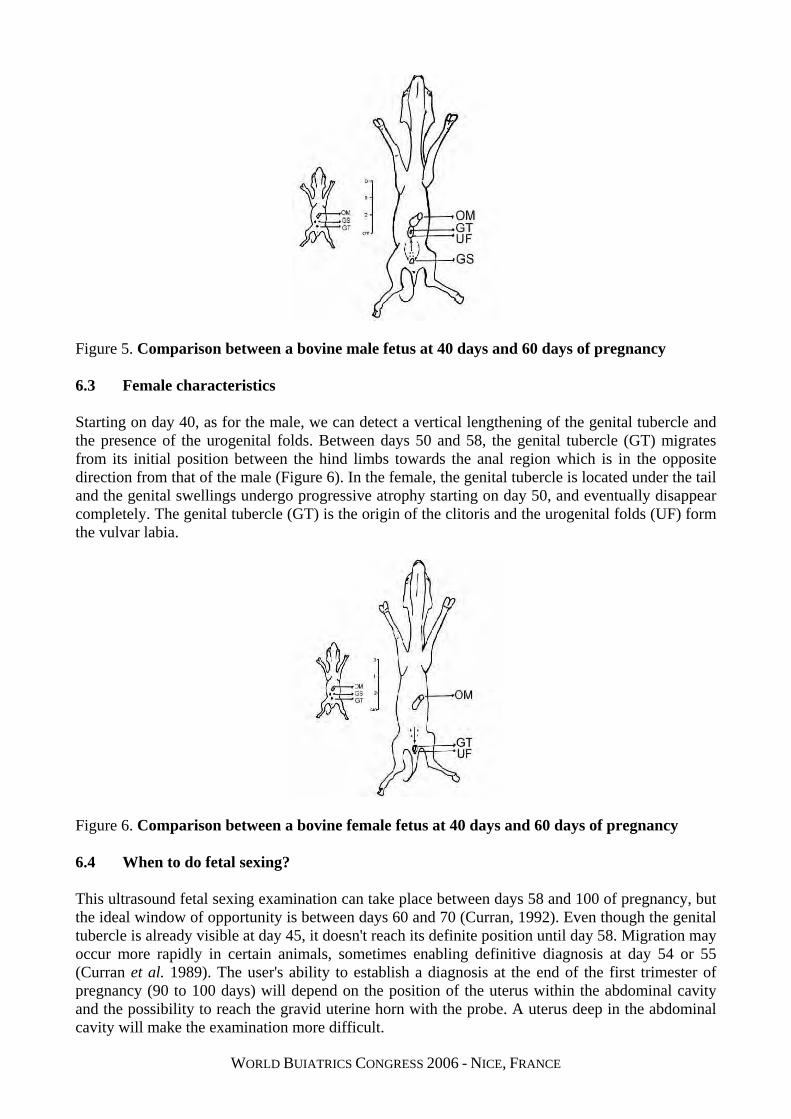
Figure 5. Comparison between a bovine male fetus at 40 days and 60 days of pregnancy 6.3 Female characteristics Starting on day 40, as for the male, we can detect a vertical lengthening of the genital tubercle and the presence of the urogenital folds. Between days 50 and 58, the genital tubercle (GT) migrates from its initial position between the hind limbs towards the anal region which is in the opposite direction from that of the male (Figure 6). In the female, the genital tubercle is located under the tail and the genital swellings undergo progressive atrophy starting on day 50, and eventually disappear completely. The genital tubercle (GT) is the origin of the clitoris and the urogenital folds (UF) form the vulvar labia.
Figure 6. Comparison between a bovine female fetus at 40 days and 60 days of pregnancy 6.4 When to do fetal sexing? This ultrasound fetal sexing examination can take place between days 58 and 100 of pregnancy, but the ideal window of opportunity is between days 60 and 70 (Curran, 1992). Even though the genital tubercle is already visible at day 45, it doesn't reach its definite position until day 58. Migration may occur more rapidly in certain animals, sometimes enabling definitive diagnosis at day 54 or 55 (Curran et al. 1989). The user's ability to establish a diagnosis at the end of the first trimester of pregnancy (90 to 100 days) will depend on the position of the uterus within the abdominal cavity and the possibility to reach the gravid uterine horn with the probe. A uterus deep in the abdominal cavity will make the examination more difficult.
WORLD BUIATRICS CONGRESS 2006 - NICE, FRANCE
6.5 What is the accuracy of fetal sexing? Diagnostic accuracy depends on the experience of the practitioner, the quality of the equipment used and the working conditions. The use of a squeeze chute is not required, but minimal restraint of the animal is necessary. A dim environment will facilitate the identification of structures on the screen. As well, it is recommended to place the ultrasound monitor so that the screen is at eyes level. A methodical and meticulous examination is necessary if the practitioner wishes to obtain satisfactory results. In ideal conditions, an experienced practitioner can obtain a definitive and accurate diagnosis in more than 99% of subjects (Curran, 1992; Durocher et al. 2002; Stroud, 1994), and the examination requires an average of five minutes per animal. 7. PRESENCE OF MULTIPLE FETUSES Detection of multiple fetuses is generally performed by the veterinarian at the same time as sexing the fetus, after day 57 of pregnancy (Stroud, 1994). With the advent of the embryo transfer technique, it has become possible to implant two embryos in a recipient. Given this situation, it is important to be able to determine the probability of twinning (Stroud, 1994). If the presence of twins is confirmed before day 55 of pregnancy, it is then important to do another ultrasound exam after day 60 to determine the sex of the fetuses and adopt a course of action according to the results. If the fetuses are male and female, the client will then have the opportunity to choose whether to maintain the pregnancy and run the risk of obtaining a freemartin heifer or to abort the cow. 8. FETAL ANOMALIES During the ultrasound examination for pregnancy, and especially when determining the sex of the fetus, it is very important to ensure that the fetus is viable and its appearance is normal. Although the following defects are rare, it is important to be able to recognize them: siamese twins; schistosomus reflexus; amorphous globosus. Finally, certain acquired or congenital defects may be noted during the ultrasound exam in an older foetus. Among these, it is worthwhile to mention hydrocephalus, fetal ascites and pericardial effusion. These conditions have a very poor prognosis for fetal survival. Before proposing the termination of the pregnancy on the cows presenting these anomalies, it is essential for the veterinarian to do a careful ultrasonographic examination using more than one view and to repeat the scanning more than once to confirm his definitive diagnosis. 9. CONCLUSION The art of transrectal palpation is acquired by examining thousands of cows during a period of time that can vary from several months to years before the veterinarian is thoroughly efficient. But with the help of ultrasound, veterinarians who are less experienced in transrectal palpation can improve their manual dexterity more rapidly, while simultaneously offering a more accurate and extensive diagnostic service. The use of this diagnostic tool increases the value of a veterinary clinic's services and enables more innovative veterinarians to maintain a progressive clientele. Ultimately, the use of ultrasound technology is financially advantageous for dairy producers and veterinarians in private practice.
WORLD BUIATRICS CONGRESS 2006 - NICE, FRANCE
10. SUMMARY Since the advent of bovine ovarian ultrasound in 1984, enormous progress has been made in our understanding of folliculogenesis, the development of the bovine corpus luteum and embryo as well as fetal sexing. Diagnosis of pregnancy in cows is an integral part of preventive health programs for dairy herds and early pregnancy examinations are generally considered essential and profitable for dairy producers. The goals of this presentation are to help understand some basic principles of ultrasonography and to present its principal uses in bovine reproduction, the economic benefits of early pregnancy diagnosis and the continuing education materials available to veterinarians. 11. KEY WORDS Ultrasonography, reproduction, cow, diagnostic method, economics, basic principles, embryo, embryonic death, foetus, fetal sexing, twins, ovary, uterus, anomalies. 12. RESUME L’arrivée de l’échographie a eu un impact majeur sur notre compréhension de la dynamique de croissance des follicules, du développement du corps jaune et du fœtus bovin ainsi que du sexage fœtal. Le diagnostic de gestation des vaches fait partie intégrante d’un programme de suivi de médecine préventive des troupeaux laitiers et le diagnostic précoce de gestation est généralement considéré comme essentiel et rentable pour les producteurs laitiers. Les objectifs de la présentation sont d’aider à bien comprendre les principes de bases des examens ultrasonographiques et de présenter les principales utilisations de cet outil diagnostique en reproduction bovine, ses bénéfices pour le diagnostic précoce de la gestation et le matériel de formation continue disponible aux médecins vétérinaires. 13. MOTS CLES Ultrasonographie, reproduction, vache, méthode diagnostique, économique, principes de base, embryon, mortalité embryonnaire, fœtus, sexage fœtal, jumeaux, utérus, anomalies. 14. REFERENCES Ayalon N. A review of embryonic mortality in cattle. J Reprod Fert, 1978; 54:483-493. Carrière PD, DesCôteaux L, Durocher J. Evaluation échographique du tractus reproducteur bovin : Développement normal et anormal des follicules ovariens et du corps jaune. Méd Vét du Québec, 2002; 32 :128-131. Curran D, Kastelic JP, Ginther OJ. Determining sex of the bovine fetus by ultrasound assessment of the relative location of the genital tubercule. Anim Reprod Sci, 1989; 19:217-227. Curran S. Fetal sex determination in cattle and horses by ultranonography. Theriogenology, 1992; 37:17-20. DesCôteaux L, Carrière PD, Durocher J. Ultrasonography of the reproductive system of the cow: A 4 languages interactive CD-rom for continuing education of veterinarians. Continuing education services of the University of Montreal, St-Hyacinthe, Québec, Canada. 2005. http://www.medvet.umontreal.ca/etudes/FormationContinue.html to order. DesCôteaux L, Carrière PD, Durocher J. Évaluation échographique du tractus reproducteur bovin : Diagnostic précoce de gestation, géméllité, mortalité embryonnaire et anomalies de l’utérus. Méd Vét du Québec, 2002; 32 :123-127. Durocher J, DesCôteaux L, Carrière PD. Évaluation échographique du tractus reproducteur bovin : Détermination du sexe du fœtus. Méd Vét du Québec, 2002; 32 :132-134.
Durocher J, Morin N, Blondin P. Effect of hormonal stimulation on bovine follicular response and oocyte developmental competence in a commercial operation. Theriogenology, 2006; 65:102-115. Filteau V, DesCôteaux L. Valeur prédictive de l’utilisation de l’appareil échographique pour le diagnostic précoce de la gestation chez la vache laitière. Méd Vét Québec, 1998; 28 :81-85. Ginther OJ. Follicles. In: Ultrasonic imaging and animal reproduction: Cattle. Equiservices Publishing. Cross Plains, WI, USA 1998:29-58. Hanzen C, Delsaux B. Use of transrectal B-mode ultrasound in early pregnancy in cattle. Vet Rec, 1987; 121:201-202. Inomata T, Eguchi Y, Yamamoto M, Asari M, Kano Y. Development of the external genitalia in bovine fetuses. Jpn J of Vet Sci, 1982, 44:489-496. Kastelic JP, Curran S, Pierson RA, Ginther OJ. Ultrasonic evaluation of bovine conceptus. Theriogenology, 1988; 29:39-54. Pierson RA, Ginther OJ. Ultrasonic imaging of the ovaries and uterus in cattle. Theriogenology, 1988; 29:21-37. Sirois J, Fortune J. Ovarian follicular dynamics during the estrous cycle in heifers monitored by realtime ultrasonography. Biol Reprod, 1988; 39:308-317. Stroud BK. Clinical application of bovine reproductive ultrasonography. Comp Cont Educ, 1994; 16:1085-1097.