The clinical aspects contained in specific sections of this parameter (Introduction, Indications,Specifications of the Examination, and Equipment Specifications) were developed collabora-tively by the American Institute of Ultrasound in Medicine (AIUM), the American College ofRadiology (ACR), the Society for Pediatric Radiology (SPR), and the Society of Radiologistsin Ultrasound (SRU). Recommendations for physician requirements, written request for theexamination, procedure documentation, and quality control vary between the 4 organizationsand are addressed by each separately.
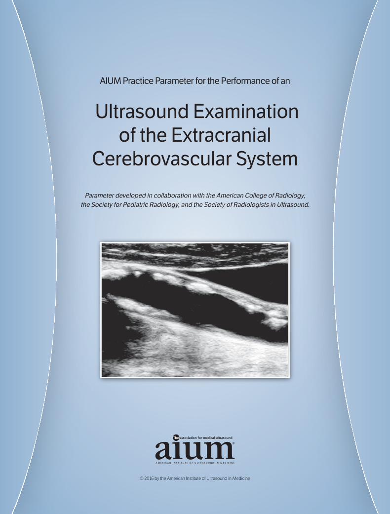
Ultrasound imaging, using grayscale imaging, Doppler spectral analysis, and color Dopplerimaging (CDI), is a proven useful procedure for evaluating the extracranial cerebrovascularsystem. Although it is not possible to detect every abnormality, adherence to the followingparameters will maximize the probability of detecting most extracranial cerebrovascular abnor-malities. Occasionally, an additional and/or specialized examination may be necessary.
II. Indications
Indications for an ultrasound examination of the extracranial carotid and vertebral arteriesinclude but are not limited to:
1. Evaluation of patients with hemispheric neurologic symptoms, including stroke, tran-sient ischemic attack, and amaurosis fugax1–4;
2. Evaluation of patients with a cervical bruit;3. Evaluation of pulsatile neck masses;4. Preoperative evaluation of patients scheduled for major cardiovascular surgical procedures;5. Evaluation of nonhemispheric or unexplained neurologic symptoms;6. Follow-up evaluation of patients with proven carotid disease;7. Evaluation of postoperative or postinterventional patients after cerebrovascular revascu-
larization, including carotid endarterectomy, stenting, or carotid-to-subclavian arterybypass graft;
8. Intraoperative monitoring of vascular surgery;9. Evaluation of suspected subclavian steal syndrome5;10. Evaluation for suspected carotid artery dissection,6 arteriovenous fistula, or
pseudoaneurysm; 11. Evaluation of patients with carotid reconstruction after extracorporeal membrane oxy-
genation bypass;12. Evaluation of patients with syncope, seizures, or dizziness;13. Screening high-risk patients: atherosclerosis elsewhere, history of head and neck radia-
tion, known fibromuscular dysplasia (FMD), Takayasu arteritis, or other vasculopathy inanother circulation;
14. Neck trauma; and15. Hollenhorst plaque visualized on retinal examination.
2016—AIUM PRACTICE PARAMETER—Extracranial Cerebrovascular Ultrasound
1
extracranial.qxp_1115 6/29/16 3:54 PM Page 1
III. Qualifications and Responsibilities of the Physician
See www.aium.org for AIUM Official Statements including Standards and Guidelines for theAccreditation of Ultrasound Practices and relevant Physician Training Guidelines.7
IV. Written Request for the Examination
The written or electronic request for an ultrasound examination should provide sufficientinformation to allow for the appropriate performance and interpretation of the examination.
The request for the examination must be originated by a physician or other appropriatelylicensed health care provider or under the provider’s direction. The accompanying clinicalinformation should be provided by a physician or other appropriate health care provider famil-iar with the patient’s clinical situation and should be consistent with relevant legal and localhealth care facility requirements.
V. Specifications of the Examination
Extracranial cerebrovascular ultrasound evaluation consists of assessment of the accessibleportions of the common carotid, external and internal carotid, and the vertebral arteries.
A. Scanning Technique
All arteries should be scanned using appropriate gray scale and Doppler techniques and prop-er patient positioning.2,3,8 The common carotid and internal carotid arteries should be scannedin grayscale and with color Doppler as completely as possible. Caudad angulation of the trans-ducerin the supraclavicular area and cephalad angulation at the level of the mandible may aidanalysis .3,4 The vertebral arteries can be evaluated between the vertebral transverse process-es in the middle neck or at their origins. Grayscale imaging of the common carotid artery, itsbifurcation, and both the internal and external carotid arteries should be performed in lon-gitudinal and transverse planes. Gain should be optimized to detect the vessel wall, plaque,and other abnormalities.
Color Doppler imaging should be used to detect areas of narrowing and abnormal flow toselect areas for spectral analysis. Color Doppler is also helpful to detect external carotidartery branches to definitively identify this vessel. Color Doppler should be used to clarifythe cause of image/pulsed Doppler mismatches and to detect narrow flow channels at sites ofstenosis.9 Power Doppler evaluation may be complementary to color Doppler to search for anarrow channel of residual flow in suspected occlusion or near occlusion.
Long axis spectral Doppler velocity measurements with angle correction should be obtained atrepresentative sites in all vessels. Additionally, scanning in and through areas of stenosis or sus-pected stenosis must be adequate to determine the maximal peak systolic velocity associatedwith the stenosis and to document disturbances in the waveform distal to the stenosis.
2016—AIUM PRACTICE PARAMETER—Extracranial Cerebrovascular Ultrasound
2
extracranial.qxp_1115 6/29/16 3:54 PM Page 2
Consistent angle correction is essential for determining blood flow velocity.2 All angle-correctedspectral Doppler waveforms must be obtained from longitudinal images.
All patients at a facility should be scanned with the same angle-correction technique (eitherparallel to the vessel wall or in line with the color lumen) to ensure consistency between serialexaminations and between patients. The angle between the direction of flowing blood and theapplied Doppler ultrasound signal (angle q [theta], the Doppler angle) should be between 45and 60 degrees whenever possible. The potential velocity error related to incorrect angleassignment increases with the Doppler angle, especially at angles above 60 degrees.3 Anglesexceeding 60 degrees should be avoided whenever possible since the calculated velocity usingoverly high angles is fallible. Techniques to obtain an appropriate angle (eg, heel and toe angu-lation of the transducer) may be necessary. Deviations from the protocol may be unavoidable(eg, it may not be possible to obtain an appropriate angle with a very tortuous vessel) butshould be minimized. Spectral Doppler gain should be appropriate for the vessel scanned.Either excessive or inadequate gain may lead to errors in diagnosis. The Doppler scale shouldbe set to maximize the size of the waveforms without resulting in aliasing to improve accuracyand reproducibility of measurement. Images must be obtained with appropriate color Dopplertechnique to demonstrate filling of the normal lumen and/or flow disturbances associated withstenoses. The color Doppler scale should be chosen to avoid aliasing at typical carotid veloci-ties, and the gain should be set to minimize artifacts.
B. Recording
1. Grayscale: At a minimum, for each normal side evaluated, grayscale images must beobtained at each of the following levels:a. Long axis of common carotid artery;b. Long axis, at the carotid artery bifurcation;c. Long axis of internal carotid artery to include its origin; andd. Short axis of proximal internal carotid artery.
If abnormalities are found, additional images must be recorded:a. If atherosclerotic plaques are present, location, extent, and characteristics should be
documented with grayscale imaging in both longitudinal and transverse planes.b. Other vascular or significant perivascular abnormalities should be documented.
2. Color Doppler: At a minimum, for each normal side evaluated, color Doppler images(using color alone or as part of the spectral Doppler image) must be obtained at each ofthe following levels:a. Long axis of distal common carotid artery;b. Long axis of proximal and midinternal carotid artery;c. Long axis of external carotid artery (with identification of a branch if possible); andd. Long axis of vertebral artery.
2016—AIUM PRACTICE PARAMETER—Extracranial Cerebrovascular Ultrasound
3
extracranial.qxp_1115 6/29/16 3:54 PM Page 3
If abnormalities are found, additional images must be recorded.a. If atherosclerotic plaques are present, extent and effect on the lumen should be recorded.b. In cases of occlusion, a color and/or power Doppler image of the abnormal vessel
should be obtained.c. Other vascular or significant perivascular abnormalities should be documented.
3. Spectral Doppler: For each normal side evaluated, spectral Doppler waveforms and max-imal peak systolic velocities must be recorded at each of the following levels:a. Proximal common carotid artery;b. Middle or distal common carotid artery (2–3 cm below the bifurcation);c. Proximal internal carotid artery;d. Mid to distal cervical internal carotid artery;e. Proximal external carotid artery; andf. Vertebral artery (in neck or near origin).
If significant stenosis is found or suspected, additional images must be recorded and the loca-tion of the stenosis determined:
a. At the site of maximum velocity due to the stenosis; andb. Distal to the site of maximal velocity to document the presence or absence of
disturbed flow.
Velocity ratios and diastolic velocities may also be calculated as warranted depending on thelaboratory interpretation criteria.
The peak systolic velocity and flow direction in each of the vertebral arteries should be recorded.
Stents require additional images. In patients with indwelling stents, grayscale, spectral, andcolor Doppler should be used to evaluate the lumen, stent deployment, and flow within eachstent, and flow proximal, and distal to each stent. The site of highest peak systolic velocity aswell as the waveforms distal to this site should be recorded.
C. Interpretation
The interpretation of cerebrovascular ultrasound images requires careful attention to protocoland interpretation criteria.
1. Each laboratory must have interpretation criteria that are used by all members of thetechnical and physician staff.
2. Diagnostic criteria must be derived from the literature or from internal validation based oncorrelation with other imaging modalities or surgical and/or pathologic correlation.2,3,6,10–15
2016—AIUM PRACTICE PARAMETER—Extracranial Cerebrovascular Ultrasound
4
extracranial.qxp_1115 6/29/16 3:54 PM Page 4
3. The report must indicate internal carotid artery stenosis categories that are clinically useful and nationally or internationally accepted and based primarily upon velocity crite-ria and waveform analysis.1–3 Stenoses above 50% should be graded to within a range (eg,50%–69% or 70% to near occlusion) or a numeric grade (eg, 60% ± 10%) to provideadequate information for clinical decision making.
4. Numerous factors may falsely increase or decrease velocities (eg, systemic disease, cardivas-cular disease, contralateral severe disease or occlusion, near occlusive stenoses).8, 16-18
Simple velocity criteria may not be valid for a younger-than-usual population. Secondary criteria such as ratios may be helpful in these circumstances.
5. The report should describe abnormal waveforms, if present.5,19
6. The report must indicate vertebral artery flow direction.7. The report may characterize plaques, depending on the laboratory inter-pretation criteria.20–24
8. The report should describe significant nonvascular abnormalities.9. The criteria for common carotid and vertebral artery stenosis differ from internal carotid
artery criteria.25,26
10. A velocity threshold that indicates an external carotid stenosis is not established. A simple description indicating a stenosis, if present, may be reported. Identification ofstenosis can be based on grayscale and/or color flow narrowing, elevated velocitythrough the stenosis, and typical poststenotic waveforms.
11. The velocity criteria for stenosis after interventions may require different criteria thannative vessels.27, 28 Stents require different velocity criteria than native vessels.29–32
VI. Documentation
Adequate documentation is essential for high-quality patient care. There should be a perma-nent record of the ultrasound examination and its interpretation. Images of all appropriateareas, both normal and abnormal, should be recorded. Variations from normal size should beaccompanied by measurements. Images should be labeled with the patient identification, facility identification, examination date, and side (right or left) of the anatomic site imaged. An official interpretation (final report) of the ultrasound findings should be included in thepatient’s medical record. Retention of the ultrasound examination should be consistent bothwith clinical needs and with relevant legal and local health care facility requirements.
Reporting should be in accordance with the AIUM Practice Parameter for Documentation of anUltrasound Examination.33
VII. Equipment Specifications
The examination should be conducted with a real-time scanner with Doppler capability,preferably using a linear transducer. The examination should use the highest clinically appro-priate frequency, realizing that there is a trade-off between resolution and beam penetration.
2016—AIUM PRACTICE PARAMETER—Extracranial Cerebrovascular Ultrasound
5
extracranial.qxp_1115 6/29/16 3:54 PM Page 5
Imaging frequencies should be 5.0 MHz or greater. Doppler flow analysis should be conduct-ed with a carrier frequency of 3.0 MHz or greater. Lower frequencies are occasionally appro-priate in patients with a large body habitus or densely calcified vessels. Examination usinglower-frequency transducers can also be useful when the vessels are not adequately imaged athigher frequencies. Color Doppler imaging can be used to localize blood flow abnormalities forrange gate placement for the Doppler spectral analysis, thus facilitating the examination.
VIII. Quality Control and Improvement, Safety, InfectionControl, and Patient Education
Policies and procedures related to quality control, patient education, infection control, andsafety should be developed and implemented in accordance with the AIUM Standards and Guidelines for the Accreditation of Ultrasound Practices.
Equipment performance monitoring should be in accordance with the AIUM Standards andGuidelines for the Accreditation of Ultrasound Practices.34
IX. ALARA Principle
The potential benefits and risks of each examination should be considered. The ALARA (as low as reasonably achievable) principle should be observed when adjusting controls thataffect the acoustic output and by considering transducer dwell times. Further details onALARA may be found in the AIUM publication Medical Ultrasound Safety, Third Edition.
Acknowledgments
This parameter was revised by the American Institute of Ultrasound in Medicine (AIUM) incollaboration with the American College of Radiology (ACR), the Society for PediatricRadiology (SPR), and the Society of Radiologists in Ultrasound (SRU) according to theprocess described in the AIUM Clinical Standards Committee Manual.
Collaborative Committee
Members represent their societies in the initial and final revisions of this parameter.
2016—AIUM PRACTICE PARAMETER—Extracranial Cerebrovascular Ultrasound
6
extracranial.qxp_1115 6/29/16 3:54 PM Page 6
ACRLaurence Needleman, MD, ChairMonica S. Epelman, MDEdward G. Grant, MD
AIUMDavid M. Paushter, MDJohn S. Pellerito, MDLeslie M. Scoutt, MD