Ultrasound Imaging for Regional Anesthesia in Infants,Children, and Adolescents
A Review of Current Literature and Its Application in the Practice of Extremity andTrunk Blocks
Ban C. H. Tsui, M.D., F.R.C.P.C.,* Santhanam Suresh, M.D., F.A.A.P.†
This article has been selected for the ANESTHESIOLOGY CME Program. Learningobjectives and disclosure and ordering information can be found in the CMEsection at the front of this issue.
ABSTRACTThe use of ultrasound guidance has provided an opportunity to
perform many peripheral nerve blocks that would have been difficultto perform in children based on pure landmark techniques due to thepotential for injection into contiguous sensitive vascular areas. Thisreview article provides the readers with techniques on ultrasound-guided peripheral nerve blocks of the extremities and trunk withcurrently available literature to substantiate the available evidence forthe use of these techniques. Ultrasound images of the blocks withcorresponding line diagrams to demonstrate the placement of theultrasound probe have been provided for all the relevant nerve blocksin children. The authors hope that this review will stimulate furtherresearch into ultrasound-guided regional anesthesia in infants, chil-dren, and adolescents and stimulate more randomized controlledtrials to provide a greater understanding of the anatomy and physi-ology of regional anesthesia in pediatrics.
ONE of the most exciting recent advances in technology inpediatric regional anesthesia has been the introduction of
anatomically based ultrasound imaging for facilitating nerve lo-calization. This is because regional anesthesia techniques in chil-dren have been considered challenging due to (1) targeting neu-ral structures that often course very close to critical structures(e.g., nerves of the brachial plexus run close to the pleura as theytraverse the supraclavicular region), and particularly during cen-tral neuraxial blocks where the safety margin is narrow for needleplacement particularly close to the spinal cord, (2) the prerequi-site for sedation or general anesthesia masking potential warningsigns (paresthesia), and (3) the need for limiting the volume oflocal anesthetic solution below toxic levels. With the possibilityof visualizing the target structures, ultrasound technology mayencourage many anesthesiologists who had previously aban-doned regional techniques to resume or increase their use ofregional anesthesia in children.
Although literature evaluating the evidence for successand safety of ultrasound in regional anesthesia has begun toemerge, a comprehensive narrative review of the literaturepertaining to techniques described and outcomes evaluatingultrasound guidance in pediatric regional anesthesia was notavailable at the time of writing this article. This review aimsto provide the pediatric anesthesiologist with an overall sum-mary of the techniques used and of the outcomes found(based on controlled or comparative studies) as described inthe literature on ultrasound guidance of peripheral nerveblocks of the extremities and trunk in pediatrics. A compan-ion article with similar objectives related to neuraxial blockswill be published in the next issue of ANESTHESIOLOGY.1 Inaddition to case series and clinical studies, descriptions from
* Professor, Department of Anesthesiology and Pain Medicine,University of Alberta. † Professor, Department of Anesthesiologyand Pediatrics, Children’s Memorial Hospital, Northwestern Univer-sity, Feinberg School of Medicine.
Received from Department of Anesthesiology and Pain Medicine,University of Alberta, Edmonton, Alberta, Canada, and the Depart-ment of Pediatric Anesthesiology, Northwestern University, Fein-berg School of Medicine, Chicago, Illinois. Submitted for publica-tion April 17, 2009. Accepted for publication September 30, 2009.Supported by a Career Scientist Award in Anesthesia (to Dr. Tsui),Canadian Anesthesiologists’ Society-Abbott Laboratories Ltd., To-ronto, Ontario, Canada, a Clinical Scholar Award (to Dr. Tsui) fromthe Alberta Heritage Foundation for Medical Research, Edmonton,Alberta, Canada, and Foundation for Anesthesia Education andResearch (FAER) Research in Education grant (to Dr. Suresh).
Mark A. Warner, M.D., served as Handling Editor for this article.
Address correspondence to Dr. Suresh: Department of PediatricAnesthesiology, Children’s Memorial Hospital, 2300 Children’s Plaza,P. O. Box 19, Chicago, Illinois 60614. [email protected] article may be accessed for personal use at no charge through theJournal Web site, www.anesthesiology.org.
Anesthesiology, V 112 • No 2 473 February 2010
case reports and correspondence may shed some light intoareas where ultrasound has been especially useful for chil-dren. Moreover, acquiring a thorough understanding of re-lated regional anatomy cannot be replaced by new technicalskills and approaches when performing regional anesthesia.Consequently, this review and the upcoming review ofneuraxial blockade include descriptions and illustrations ofthe relevant sonoanatomy of the various regions. These sono-anatomy sections will hopefully serve as a foundation for abetter understanding of the block techniques as described inliterature. This review is not intended to serve as a compre-hensive training manual. Readers are encouraged to obtaintraining through workshops and under supervision of anexpert to gain competency with this technology.
Materials and Methods
A literature search for this review was performed usingMEDLINE and EMBASE f rom 1980 to May 28, 2009. Thekeywords “ultrasound and children” and “ultrasound andpediatric” were combined with “regional anesthesia” and“nerve block.” The medical subject heading term “ultra-sonography” was also combined with “nerve block” and “re-gional anesthesia,” with the age limit of 0–18 yr. Thesearches were limited to literature in humans, and althoughthere was no limit to the English language, only those articleswith English text or abstracts were described or discussed ifrelevant. Literature relevant to blocks at the extremity andtrunk were printed in full, and their reference lists werechecked manually. We included clinical studies, case series,and reports, in addition to relevant correspondence pieceswhere institutional review board approval and patient–par-ent consents were obtained. Expert reviews and descriptionsspecific to ultrasound in pediatric regional anesthesia andother correspondence pieces were reviewed for references andadditional comments on technique, although they were notused for outcome evaluation data.
The images highlighting sonoanatomy were obtained us-ing a portable ultrasound unit (Sonosite M-turbo®, Bothell,
WA) commonly used in the institutions of the authors forperformance of pediatric ultrasound-guided regional anes-thesia. Ethics committee or institutional review board ap-proval was obtained for the ultrasound imaging, and in-formed consent was provided by the parent(s) of patientsprior to acquiring images for this review. Two linear, high-frequency probes were used (L-25, 6–13 MHz, 25-mm foot-print and HFL38, 6–13 MHz, 38-mm footprint, both fromSonosite), and the hockey stick (SLA) probe was often usefulin infants due to the small footprint. The figure legendsinclude a description of the probe and a schematic line draw-ing depicting the location of its placement.
ResultsThe search provided 32 results, with one obtained after amanual review of the reference lists. The search resulted in 26studies of ultrasound-guided peripheral nerve blockade (5randomized controlled trials, 2 large prospective studies, 13case series - reports, and 7 correspondence pieces), althoughthere was a large spectrum of blocks described, with eightreports pertaining to sciatic nerve blockade. Five expert re-views and one technical report with descriptions of techniquerelated to pediatric regional anesthesia were also obtained. Inthe ensuing sections, we will discuss in depth the use ofultrasound guidance for the extremity and trunk blocks ininfants, children, and adolescents. Although there are differ-ent terms used to describe the relative placement of the nee-dle with respect to the probe, we have used the terms “out-of-plane” and “in-plane” to describe the needle beingperpendicular (or sometimes tangential) and parallel to theprobe axis, respectively (fig. 1). The probe is typically used toview the nerves in short-axis (cross-sectional, transversely),but occasionally a long-axis (longitudinal) view is helpful.
Upper Extremity BlocksAlthough many approaches to the brachial plexus have beendescribed, the axillary block using conventional methods is stillthe most commonly performed and reported brachial plexus
Fig. 1. Needle-probe alignment. In out-of-plane alignment, only a cross-section of one point of the needle is viewed; in an in-plane alignment,the entire needle shaft and tip can be seen.
474 Ultrasound for Pediatric Nerve Blockade
Anesthesiology, V 112 • No 2 • February 2010 B. C. H. Tsui and S. Suresh
blockade in children. This may be due to the fact that otherblock sites are situated near critical structures such as the cervicalpleura (supraclavicular and infraclavicular) and the spinal cord(interscalene). Although there is a paucity of related literature, theintroduction of ultrasound imaging will likely greatly increase theperformance of brachial plexus blocks in infants and children atlocations besides the commonly described axillary approach.Techniques.Interscalene Block.
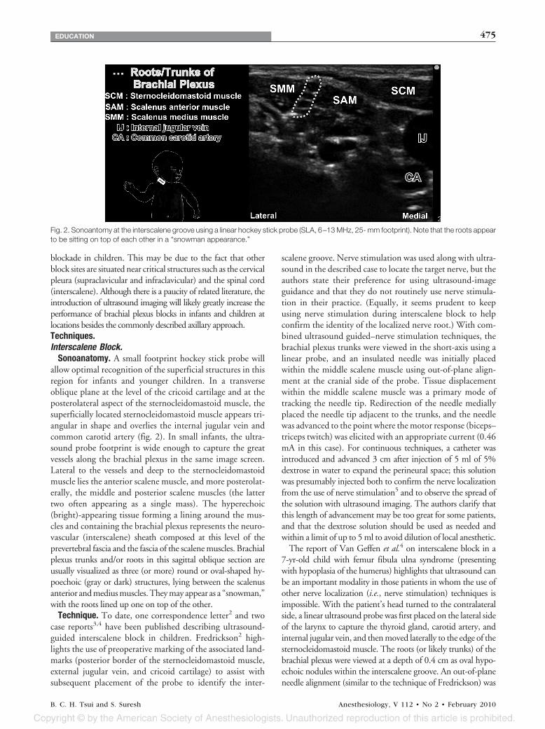
Sonoanatomy. A small footprint hockey stick probe willallow optimal recognition of the superficial structures in thisregion for infants and younger children. In a transverseoblique plane at the level of the cricoid cartilage and at theposterolateral aspect of the sternocleidomastoid muscle, thesuperficially located sternocleidomastoid muscle appears tri-angular in shape and overlies the internal jugular vein andcommon carotid artery (fig. 2). In small infants, the ultra-sound probe footprint is wide enough to capture the greatvessels along the brachial plexus in the same image screen.Lateral to the vessels and deep to the sternocleidomastoidmuscle lies the anterior scalene muscle, and more posterolat-erally, the middle and posterior scalene muscles (the lattertwo often appearing as a single mass). The hyperechoic(bright)-appearing tissue forming a lining around the mus-cles and containing the brachial plexus represents the neuro-vascular (interscalene) sheath composed at this level of theprevertebral fascia and the fascia of the scalene muscles. Brachialplexus trunks and/or roots in this sagittal oblique section areusually visualized as three (or more) round or oval-shaped hy-poechoic (gray or dark) structures, lying between the scalenusanterior and medius muscles. They may appear as a “snowman,”with the roots lined up one on top of the other.
Technique. To date, one correspondence letter2 and twocase reports3,4 have been published describing ultrasound-guided interscalene block in children. Fredrickson2 high-lights the use of preoperative marking of the associated land-marks (posterior border of the sternocleidomastoid muscle,external jugular vein, and cricoid cartilage) to assist withsubsequent placement of the probe to identify the inter-
scalene groove. Nerve stimulation was used along with ultra-sound in the described case to locate the target nerve, but theauthors state their preference for using ultrasound-imageguidance and that they do not routinely use nerve stimula-tion in their practice. (Equally, it seems prudent to keepusing nerve stimulation during interscalene block to helpconfirm the identity of the localized nerve root.) With com-bined ultrasound guided–nerve stimulation techniques, thebrachial plexus trunks were viewed in the short-axis using alinear probe, and an insulated needle was initially placedwithin the middle scalene muscle using out-of-plane align-ment at the cranial side of the probe. Tissue displacementwithin the middle scalene muscle was a primary mode oftracking the needle tip. Redirection of the needle mediallyplaced the needle tip adjacent to the trunks, and the needlewas advanced to the point where the motor response (biceps–triceps twitch) was elicited with an appropriate current (0.46mA in this case). For continuous techniques, a catheter wasintroduced and advanced 3 cm after injection of 5 ml of 5%dextrose in water to expand the perineural space; this solutionwas presumably injected both to confirm the nerve localizationfrom the use of nerve stimulation5 and to observe the spread ofthe solution with ultrasound imaging. The authors clarify thatthis length of advancement may be too great for some patients,and that the dextrose solution should be used as needed andwithin a limit of up to 5 ml to avoid dilution of local anesthetic.
The report of Van Geffen et al.4 on interscalene block in a7-yr-old child with femur fibula ulna syndrome (presentingwith hypoplasia of the humerus) highlights that ultrasound canbe an important modality in those patients in whom the use ofother nerve localization (i.e., nerve stimulation) techniques isimpossible. With the patient’s head turned to the contralateralside, a linear ultrasound probe was first placed on the lateral sideof the larynx to capture the thyroid gland, carotid artery, andinternal jugular vein, and then moved laterally to the edge of thesternocleidomastoid muscle. The roots (or likely trunks) of thebrachial plexus were viewed at a depth of 0.4 cm as oval hypo-echoic nodules within the interscalene groove. An out-of-planeneedle alignment (similar to the technique of Fredrickson) was
Fig. 2. Sonoantomy at the interscalene groove using a linear hockey stick probe (SLA, 6–13 MHz, 25- mm footprint). Note that the roots appearto be sitting on top of each other in a “snowman appearance.”
475EDUCATION
B. C. H. Tsui and S. Suresh Anesthesiology, V 112 • No 2 • February 2010
used, directing the needle slightly caudal. The injection of localanesthetic (8 ml of ropivacaine 0.75%) was noted to surroundthe plexus roots or trunks. Analgesia was effective in this patientfor 20 h after the block was performed. The authors of thisreview prefer to use a lower concentration of local anesthetic,such as 0.2–0.5% ropivacaine or bupivacaine.
Ultrasound-guided continuous interscalene blockade wasperformed for a 10-yr-old girl in the Philippines during aplastic surgery medical mission.3 Without the availability ofperineural catheters and stimulating needles, a 22- gaugeangiocatheter was used for the block, which used an in-planealignment to the posterior edge of the probe. An ultrasound unitwas fortunately available (brought by a pediatrician to performtransthoracic echocardiography) to enable the performance ofthis higher risk block in a case where there would otherwise belimited choice for providing postoperative analgesia.
Comment. Blockade of the brachial plexus at locationsabove the clavicle is necessary for shoulder girdle surgery.The interscalene block provides adequate analgesia that canlast for a period of up to 6–12 h. A catheter can be left inplace for continuous analgesia. Identification of the inter-scalene groove by palpation can be challenging in anesthetizedpatients (as is common in pediatrics), and this in addition to theshort distance of the plexus to vital structures may make it dif-ficult for some to justify performance of this block in childrenunder general anesthesia. Indeed, one recent report recom-mends that these blocks are not to be performed in any heavilysedated or anesthetized patient.6 This recommendation may becriticized though, due to a lack of support from more than a few(albeit serious) case reports and because the authors cite moreneurologic complications, resulting from large-scale studies ofawake patients than anesthetized. By allowing visualization ofvital structures and improving nerve localization, ultrasoundguidance may increase the use of this block.
Clinical pearls—interscalene block.
● Use a high- frequency linear or hockey stick probe.● The needle is placed using an in-plane approach from pos-
terior to anterior.
● Once the needle is close to the plexus, a volume of localanesthetic solution should be injected, using slight needleredirections, until it is seen to surround the brachial plexusroots or trunks. This may be accomplished with a dose aslittle as 0.1–0.2 ml/kg or approximately 5 ml in children,when there is precise needle placement.
● Aspirate intermittently to make sure that the injectate isnot placed intravascular.
Supraclavicular.Sonoanatomy. The probe is first placed in a coronal
oblique plane at the lateral end of and just above the upperborder of the clavicle. It is then moved medially until animage of the subclavian artery appears on the middle of thescreen (fig. 3). At this location, the plexus is located superiorand lateral to the artery, and the neurovascular structures arenoted to be above the first rib. The subclavian artery is gen-erally anechoic, hypodense, pulsatile, and often round; itsidentity can be further confirmed by the use of color Dopp-ler. The trunks and divisions of the brachial plexus appear asa cluster of hypoechoic grape - like structures consisting ofusually three clustered (more visualized as one moves dis-tally) nodules, all surrounded by a hyperechoic lining. Me-dial and deep to the artery, the rib may be seen as a hypere-choic line with dorsal shadowing. The subclavian vein,inferomedial to the artery, is often not visualized.
Technique. Although ultrasound-guided supraclavicularblock in adults has been described in several articles, only onereport is available in the pediatric literature.7 By using asimilar technique as described in adults, the block can beperformed using a needle (short-beveled, 22–25 gauge,35–50 mm) aligned in-plane to a high- frequency probeplaced in the coronal-oblique plane once the brachial plexustrunks or divisions (forming a cluster of hypoechoic nodules)can be visualized lateral to the subclavian artery (hypoechoic andpulsatile) and close to the underlying first rib (hyperechoic andcurvilinear).7 These authors directed the needle from lateral tomedial, to approach the plexus lateral to the artery, preventingdirect contact of the plexus. A linear probe with a 35-mm foot-
Fig. 3. Sonoanatomy at the supraclavicular block location using a linear hockey stick probe (SLA, 6–13 MHz, 25- mm footprint) placed in thecoronal oblique plane. Note the subclavian artery and that the surrounding hyperechoic plexus appears like a “cluster of grapes.”
476 Ultrasound for Pediatric Nerve Blockade
Anesthesiology, V 112 • No 2 • February 2010 B. C. H. Tsui and S. Suresh
print was used, although a smaller footprint linear or curvilinearprobe may be more suitable in some cases due to the reducedcontact area and array characteristics.8
Comment. When performing a supraclavicular block,there is a greater risk of pneumothorax as the cupola of thelung lies just medial to the first rib, not far from the plexus;the distance of the plexus from the lung being especially shortin children. It is critical to ensure that clear visibility of theneedle shaft and tip is obtained by aligning the needle in-plane to the ultrasound probe at all times. Single injectiontechniques generally suffice for the block ; however, multipleinjections of local anesthetic can be performed if needed withthe needle redirected to ensure sufficient circumferentialspread around the plexus. However, care should be taken toavoid intravascular injection of the surrounding vessels (in-cluding the transverse colli artery located cephalad to theplexus). Auscultation of the lungs should be performed be-fore and after performance of the block and before dischargeto detect the clinical signs of pneumothorax, and a chest x-rayshould be obtained when suspicion arises.
Intuitively, visualization of the needle tip and its relation tothe plexus should reduce associated complications and in-crease the use of this block significantly. Indeed, this blockoffers the most reliable blockade of the brachial plexus, withrapid onset, for analgesia and anesthesia of the entire upperextremity, such that it may be referred to it as a “spinal for theupper extremity.”
Clinical pearls—supraclavicular block.
● Recognize the structure lateral to the carotid artery andlateral to the pulsation of the subclavian artery.
● Use a linear probe or a hockey stick probe to visualize theplexus, which appears as a bunch of grapes around the artery.
● By using an in-plane technique, advance needle into theplexus, and after aspiration, inject local anesthetic untilcircumferential spread around the plexus trunks or divi-
sions is seen (0.2 ml/kg may suffice depending on the age ofthe patient and accuracy of needle tip placement).
Infraclavicular Block.Sonoanatomy. Although a lower frequency probe is often
used in adults due to the depth of the plexus, the plexus issuperficial enough in most children to use a higher frequencyprobe. In a parasagittal plane immediately medial and infe-rior to the coracoid process, a short-axis view of the brachialplexus cords and axillary vessels can be visualized (fig. 4). Thepectoralis major and minor muscles are separated by a hy-perechoic lining (perimysium); the pectoralis major musclelies superficial and lateral to the pectoralis minor muscle. Theaxillary neurovascular bundle lies deeper, with the large axil-lary vein lying medial and caudad to the artery. The lateralcord of the plexus is readily visualized as a hyperechoic ovalstructure; the medial and posterior cords can be difficult toidentify because the medial cord lies between the axillaryartery and vein, whereas the posterior cord is often hiddendeep to an axillary artery acoustic artifact. In addition, themedial cord can be posterior or even slightly cephalad to theaxillary artery.
Technique. Shortly after an initial case report describingultrasound-guided peripheral nerve blockade in children (ofthe sciatic nerve),9 Marhofer et al.10 described their use ofultrasound-guided lateral infraclavicular block for surgicalanesthesia in children. The children were placed supine withtheir arm adducted, elbow flexed, and forearm placedon their abdomen. A linear probe was placed transverselybelow the clavicle (the relative orientation to the coracoidprocess was not stated) to capture an image of the brachialplexus (presumably the cords surrounding the subclavian ar-tery). These authors report successful visualization of the plexusin all 40 patients studied. The needle was inserted out-of-plane,1 cm from the inferior aspect of the probe, and directed slightlycranially (thus at an angle approximately 60°– 70° to the skin) to
Fig. 4. Sonoantomy at the infraclavicular block location using a linear hockey stick probe (SLA, 6–13 MHz, 25- mm footprint) placed below theclavicle in a parasagittal plane. Note the pectoralis major and minor muscles and that the axillary artery is surrounded by the cords of thebrachial plexus. The pleura can be seen medially.
477EDUCATION
B. C. H. Tsui and S. Suresh Anesthesiology, V 112 • No 2 • February 2010
direct it toward the lateral border of the plexus. The local anes-thetic spread was viewed surrounding the plexus.
De Jose Maria et al.7 recently compared the efficacy ofultrasound-guided infraclavicular and supraclavicularblocks. For the infraclavicular block, these authors alsoplaced the probe below the clavicle, either parallel to theclavicle or using a slightly parasagittal plane depending onthe visibility of the plexus. A medium frequency probe (thusallowing greater visualization of deeper structures) was usedin some of the older children to improve the visibility of thepleura and vessels. In contrast to Marhofer et al., the needlewas placed immediately cephalad to the probe; both groupsuse a fairly steep angle of needle insertion. The needle wasredirected when necessary to ensure the targets of the block,i.e., the cords were adequately surrounded by local anestheticsolution. The spread of local anesthetic was seen around theaxillary artery and brachial plexus. The authors of this reviewroutinely use the paracorocoid approach with a probe that isplaced perpendicular to the clavicle. The needle is insertedfrom the superior and lateral aspect of the probe and aimedtoward the posterior cord. A total volume of 0.2 ml/kg maysuffice for providing an adequate blockade in most children ifthe needle is positioned close to the posterior cord. It is mostcritical to envision adequate spread of local anesthetic ratherthan try to inject the lowest potentially effective volume.
Comment. It is important to note that there is a great dealof individual anatomical variation in the location of the cordsaround the artery. At this location, the nerve structures nowappear hyperechoic, rather than hypoechoic as seen morecephalad, probably due to an increase in the number of fas-cicles and amount of (hyperechoic-appearing) connective tis-sue. The cervical pleura is situated closest to the brachialplexus cords at medial locations within the infraclavicularfossa, thus it may be most prudent to use a more lateralpuncture site to perform an infraclavicular blockade in chil-dren.11 Ultrasound imaging will offer visualization of therelated anatomy and the approaching needle, which may beparticularly useful for avoiding multiple punctures in chil-dren,10 and some anatomical landmarks (i.e., the coracoid
process) may be underdeveloped to enable ease of palpationusing “blind” techniques.12 Similar to the supraclavicularapproach, an in-plane needle alignment may provide supe-rior outcomes with this block, because viewing the needle tipand shaft at all times may ensure that the vessels and cervicalpleura are not punctured.13
Clinical pearls—infraclavicular block.
● Place the linear probe medial to the coracoid process andinferior to the clavicle.
● After recognizing the axillary artery, look for the cords ofthe brachial plexus surrounding the artery.
● Place a needle with an in-plane approach with the tip of theneedle aimed toward the posterior cord.
● After aspiration, inject local anesthetic solution.● Stay away from using a medially positioned needle due to
close proximity to the pleura.
Axillary Block.Sonoanatomy. With the probe placed perpendicular to the
anterior axillary fold, a short-axis view of the neurovascularbundle can be obtained (fig. 5). In this view, the bicepsbrachii and coracobrachialis muscles are seen laterally; thetriceps brachii muscle is medial and deep to the biceps brachiimuscle. The anechoic and circular axillary artery lies cen-trally, adjacent to both the biceps brachii and coracobrachia-lis muscles, and is surrounded by the nerves. Although therecan be many variations, the median nerve is typically locatedsuperficial and between the artery and biceps brachii muscle,the ulnar nerve is commonly located medial and superficialto the artery, and the radial nerve often lies deep to the arteryat the midline. At this level, the musculocutaneous nerve islocated between the biceps brachii and coracobrachialis mus-cles. This nerve may be undetectable in children.14 It is com-mon to perform a distal scan to identify the nerves along theirpaths in the upper extremity. The median nerve will con-tinue to be viewed adjacent to the artery; the ulnar nerve willremain located near the surface on its path to the ulnar nervesulcus; and the radial nerve travels posterior toward the spiralgroove of the humerus.15 Of note, the radial, median, and
Fig. 5. Sonoantomy at the axillary block location using a linear hockey stick probe (SLA, 6–13 MHz, 25- mm footprint) placed transversely tothe humerus.
478 Ultrasound for Pediatric Nerve Blockade
Anesthesiology, V 112 • No 2 • February 2010 B. C. H. Tsui and S. Suresh
ulnar nerves may appear the same size as the axillary artery onultrasound, which contradicts anatomical descriptions intextbooks.14
Technique. The literature search for this review did notresult in any original report of ultrasound-guided axillaryblock in children. Three expert reviews were found, whichdescribed the technique as performed by the respective au-thor.14–16 Intuitively, this block could be performed withsimilar techniques used in adults, using out-of-plane needlealignment to a high-frequency probe placed transversely tothe humerus and using multiple injections to surround all theterminal nerves of the plexus. Multiple punctures will benecessary to anesthetize all the relevant nerves for many sur-gical procedures (i.e., brachial cutaneous, medial antebra-chial, and possibly musculocutaneous nerves will require sep-arate blockade).
Comment. Multiple injections and needle redirections arecommonly required to ensure circumferential spread of thelocal anesthetic around each of the individual nerves. Becausethere is an abundance of vessels in this region, completeavoidance of vessel puncture can be a challenge even whenusing ultrasound imaging. It is important to understand thatthe plexus remains very close to the surface, and hence theneedle should be directed cautiously while this block is at-tempted. Smaller doses can be used to provide an adequateblockade of this plexus in infants and children.
Clinical pearls—axillary block.
● Place a hockey stick probe or a linear small footprint probein the axilla as proximal as possible.
● The needle is directed from superior to inferior using anin-plane approach.
● The structures are superficial and hence are located fairlysuperficial and can be easily identified.
● Color Doppler can be used to recognize the vascularstructures.
● Local anesthetic is injected to surround the nerves.
Other Blocks of the Terminal Nerves. Similar to axillaryblockade, there is a lack of any original report describingultrasound guidance for blocks of the terminal nerves of thebrachial plexus within the upper extremity in children. Nev-ertheless, these blocks could be performed with similartechnique to that used in adults. At the level of the wrist,one should be cautioned that there is often difficulty dis-tinguishing the nerves from the tendons due to their sim-ilar appearance.Outcome Evaluation for Upper Extremity Blocks. The firstcontrolled study related to ultrasound-guided brachial plexusblockade was performed by Marhofer et al.10 in 2004 forinfraclavicular blocks using a lateral approach. Comparingultrasound guidance to conventional nerve stimulation tech-nique for surgical anesthesia in 40 children undergoing armor forearm surgery, the authors found ultrasound to be su-perior in terms of (1) visual analog scores during block punc-ture (because of reduced discomfort from muscle contrac-tions); (2) sensory onset times (mean times of onset were 9 vs.
15 min); (3) longer duration of sensory block (384 vs. 310min); and (4) better sensory and motor block scores 10 minafter block insertion. The faster onset and longer duration ofthe blocks may have been due to the ability of ultrasound tomore accurately deposit the local anesthetic to the brachialplexus. At 30 min, all blocks whether placed by ultrasound ornerve stimulation were successful (according to the criteria ofVester-Andersen,17 which was originally used for evaluatingblocks using a large volume [40 ml] of local anesthetic andrequires at least two of four nerves to be effectively blocked)and had similar motor and sensory block quality. Althoughthis study was a prospective, randomized controlled blindedstudy, the authors did not state whether there were sufficientpower for statistical significance.
De Jose Maria et al.7 recently compared the success rate,complications, and block performance times of ultrasound-guided supraclavicular and infraclavicular blocks in 80 chil-dren scheduled for upper limb surgery. Light general anes-thesia was provided to all patients. In addition to theaforementioned outcomes, the block duration and volumesof ropivacaine (0.5%) used were recorded for the supracla-vicular approach. Both block approaches were effective (withfailure defined as any amount of supplemental analgesia re-quired during surgery or within the first 4 h after the block)and neither was associated with complications. Patients re-quiring supplemental analgesia intraoperatively had blockfailure of the radial (one with infraclavicular block) and ulnar(two from both groups) nerves. Two patients receiving infr-aclavicular block had accidental puncture of their axillaryartery. The supraclavicular block was faster to perform (9 vs.13 min; 95% confidence interval for this difference was 2–6min and was statistically significant). The sensory and motorblocks using the supraclavicular approach lasted 6.5 and 5 h,respectively. Approximately, 6 ml of ropivacaine was used.The randomization process in this study was not described,and there was no sample size calculation. Although there wasno blinding described (e.g., of the nurses analyzing postop-erative analgesia), most of the outcomes evaluated did notlend to blinding in most cases (i.e., time to perform block,complications).
Lower Extremity BlocksLumbar Plexus Block.Sonoantomy. With the exception of infants, the lumbarplexus and associated psoas major muscle are often difficultto view adequately due to their depth. In a transverse (axial)view using a linear probe in infants, bony structures such asthe spinous processes, laminae and transverse processes, andmusculature including the erector spinae, and quadratuslumborum muscles can be identified (fig. 6). Deep to thesestructures, the lumbar plexus can be seen embedded withinthe psoas muscle, which is often located posterior to thetransverse processes and lateral to the vertebral bodies. Withpotentially difficult visualization using ultrasonography inolder children, imaging may be most suitable before theblock, rather than for real-time guidance, generally for iden-
479EDUCATION
B. C. H. Tsui and S. Suresh Anesthesiology, V 112 • No 2 • February 2010
tifying the exact location and depth of the transverse pro-cesses. A curved array probe (curvilinear) is placed in thetransverse plane in the midline at the level of the L4 spinousprocess to provide an overview of the L4 vertebra. The probeshould be rotated to the longitudinal axis, parallel to thespine, which will allow a lateral scan to be performed toidentify the transverse processes. Indeed, the transverse pro-cesses (which are the primary landmarks) are still often veryvaguely delineated. Therefore, it is important to switch be-tween transverse and longitudinal planes when scanning be-tween the spinous processes and the edge of the transverseprocesses to survey the area. In the transverse scan, the spi-nous processes appear hypoechoic (likely due to dorsal shad-owing effect) and extend superficially, whereas the transverseprocesses are hyperechoic masses or lines at the lateral edge ofeach vertebra. In the longitudinal scan, the lateral edges ofthe transverse processes will be identified at the most lateralpoint where multiple hyperechoic nodules are viewed.Technique. Visualization of the lumbar paravertebral regionhas been described from sonographic imaging in 32 chil-dren.18 By using a HDI 5000 ultrasound system (ATL/Phil-ips, Bothell, WA) with a curved array probe (5–8 MHz,ATL/Philips), the authors placed the probe in longitudinaland transverse planes to view the lumbar plexus and measurethe skin-to-plexus distance. By using the longitudinal view,the vertebral level was assessed by counting transverse pro-cesses in a cephalad direction from the reflex of the cephaladportion of the sacrum. Subsequently, the transverse sono-grams were used in addition to the longitudinal view to cap-ture the plexus in relation to the paravertebral musculature(thus deep to the erector spinae and quadratus lumborummuscles and within the psoas major muscle). The plexus wasdelineated in most patients at levels L3/4 and L4/5 (fewer atL4/5 in the patients older than 8 yr) and was seen in theposterior part of the psoas major muscle. The transversesonogram showed hypoechoic dots (fascicles) enveloped byhyperechoic connective tissue (epineurium) elements, whereas the longitudinal sonogram showed hypoechoic parallel
bands bordered by hyperechoic striations. The skin-to-plexus distances were positively correlated with patientweight, age, and height, although the strongest correlationwas with weight (r � 0.68 at L3/4 and 0.64 at L4/5). Real-time ultrasound guidance, by using a portable ultrasoundsystem with a small footprint hockey-stick probe, was per-formed during blockade at L4/5 in five patients younger than2 yr undergoing inguinal herniotomy. The patients wereplaced in the lateral decubitus position, and after delineatingthe plexus as described, an insulated needle (25- gaugeSprotte, 35 mm) was aligned out-of-plane to the probeplaced in the transverse plane. The longitudinal plane wasused to verify the needle placement close to the plexus. Thelocal anesthetic solution (ropivacaine 0.33% 0.3 ml/kg) wasseen spreading within the posterior part of the psoas majormuscle. The authors report successful intra- and postopera-tive analgesia.19
Comment. Sonographic visibility of the lumbar plexus maynot be feasible in many older pediatric patients, mainly dueto poor ultrasound beam penetration at the depth requiredbut also possibly because the psoas major muscle and plexusare largely similar in echogenicity. In children, it may beeasier to directly identify the plexus because the muscle tissueappears relatively more hypoechoic when compared with thenerve structures, which contain hyperechoic connective tis-sue layers.18 Despite that ultrasound may enable a sufficientview of the bony landmarks (transverse processes) or plexusitself, lack of clear tracking of the needle tip and an adequateview of the local anesthetic spread may be considered riskyespecially if the needle is inserted in a relatively medial andcaudal location. The use of a nerve stimulator, maintaining aneedle puncture relatively lateral and cranial (see Schuepferand Johr20) and performing regular aspiration during injec-tion will help ensure the safety of this block. There must be arisk-benefit analysis when deciding to use this block; al-though it will be the most suitable block for achieving anes-thesia of the entire lumbar plexus, selective blocks of the
Fig. 6. Sonoantomy at the lumbar plexus block location using a linear probe (HFL38, 6–13 MHz, 38- mm footprint) placed transversely at L4/5.The lumbar plexus is not identified in this image.
480 Ultrasound for Pediatric Nerve Blockade
Anesthesiology, V 112 • No 2 • February 2010 B. C. H. Tsui and S. Suresh
separate nerves at more peripheral sites or a caudal block maybe more appropriate depending on the procedure.Clinical Pearls—Lumbar Plexus Block.
● Ultrasound visualization of the plexus is often limited toyoung patients, thus real-time technique may be limited tothis population. In older patients, it will be more suitable toperform a preblock scan to mark the location and depth of thebony landmarks (i.e., mainly the transverse processes).
● It is important to switch between transverse and longitu-dinal planes when scanning between the spinous processesand the edge of the transverse processes to survey the area.
● The needle should be inserted in either a cephalad orcaudad direction, rather than medially toward the spinalcanal.
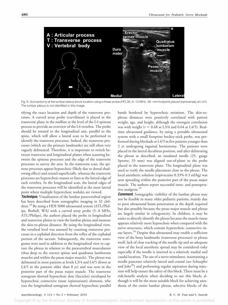
Femoral Nerve Block.Sonoanatomy. Similar to when using conventional tech-niques, the femoral artery is the key landmark when usingultrasound guidance for femoral nerve blockade (fig. 7).With the probe placed perpendicular to the nerve axis (i.e.,coronal oblique) at the level of and parallel to the inguinalcrease, the nerve appears lateral to the large, circular, andanechoic femoral artery (color Doppler may be used to iden-tify the femoral artery and vein). The nerve often appearstriangular in shape and may be of variable size due to itsirregular course; early branching above the inguinal ligamentcan increase the transverse diameter of the nerve. The fascialata (most superficial) and iliaca (immediately adjacent to thenerve and in fact separating the nerve from the artery) areseen superficial to the femoral nerve and often appear asbright and longitudinally angled echogenic signals.Technique. A comparison of ultrasound guidance to nervestimulator technique for sciatic and femoral nerve blocks wasperformed by the same group that previously described andinvestigated ultrasound guidance for infraclavicular and ilio-inguinal/iliohypogastric blockade.21 The femoral nerveblock was performed in 28 of the 46 children, because not all
children in the study required femoral block in addition toblockade of the sciatic nerve for analgesia. A linear hockeystick probe was placed parallel but inferior to the inguinalligament to view the nerve in short-axis, lateral to the femoralartery. The needle (22-gauge 40 mm with facette tip andinjection line) was aligned out-of-plane to the probe andplaced approximately 1 cm from the caudad surface of theprobe using an approximate angle of 45°. The needle tip wasviewed approaching the nerve and the local anesthetic wasinjected to surround the nerve, using redirections where nec-essary. Visualization of the nerve was possible in all cases.Comment. Although there is no direct evidence to prove thatultrasound can reduce the risk of inadvertent vessel puncture,in our experience with ultrasound guidance for this blockfewer events have been found than during our previous prac-tice. Although prevention of intraneural local anesthetic in-jection or direct needle puncture may not be possible in allcases (the view of the needle tip is often not clearly seen andthe nerve may not be adequately viewed), ultrasound imag-ing of the precise placement of the local anesthetic solutionmay reduce the practice of injecting large volumes of localanesthetic to ensure adequate spread via a blind injection.Our preference is to use an in-plane technique, which offersthe ability to visualize the needle tip as it enters the fasciailiaca.Clinical Pearls—Femoral Nerve Block.
● Place a linear probe along the inguinal–femoral crease.● Place a needle in an in-plane approach.● The local anesthetic is injected to surround the nerve.● The needle has to be placed inside the fascia iliaca com-
partment, and the local anesthetic is seen surrounding thenerve bundle.
Other Blocks of the Nerves of the Lumbar Plexus. Therehave been no clinical reports in the pediatric literature de-scribing ultrasound guidance for other commonly performedapproaches to blocking the nerves of the lumbar plexus, in-
Fig. 7. Sonoanatomy at the femoral nerve block location using a linear hockey stick probe (SLA, 6–13 MHz, 25- mm footprint) placed paralleland inferior to the inguinal ligament. The neurovascular structures from medial to lateral are the femoral vein, femoral artery, and the femoralnerve. The nerve is enveloped under the fascia iliaca and is noted to be hyperechoic as a cluster of fascicles at this point.
481EDUCATION
B. C. H. Tsui and S. Suresh Anesthesiology, V 112 • No 2 • February 2010
cluding the fascia iliaca block, 3-in-1 block, or individualblocks of the lateral femoral cutaneous or obturator nerves.There has been some mention of ultrasound-guided lateralfemoral cutaneous and 3-in-1 blocks, during which the visi-bility of the spread of local anesthetic has been possible asdeposited beneath clearly visible fascial layers.22 Despite thelack of data, these blocks could all benefit from improvedlocalization of the target nerves, or determination of theirexistence within the plane of needle entry. Furthermore, vi-sual confirmation of the spread of local anesthetic within thecorrect fascial plane will help predict the success in caseswhere the nerves may not be seen. There will likely be areduced reliance on depositing a larger than necessary vol-ume of local anesthetic in hopes of obtaining the block of allrelated nerves.22
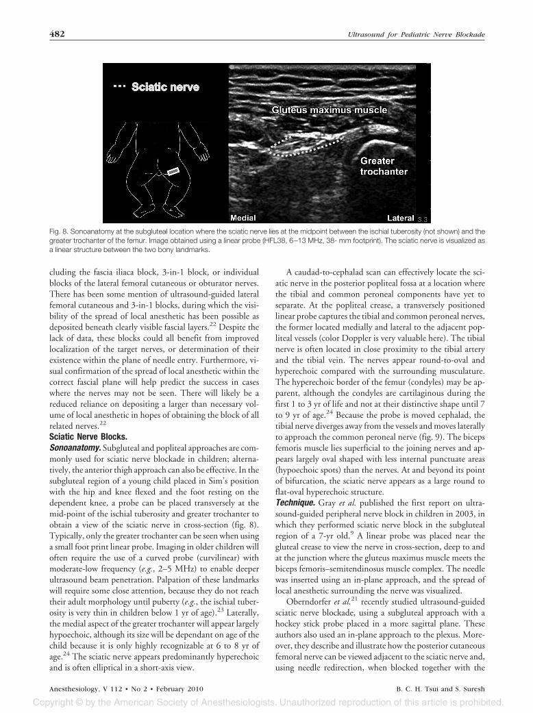
Sciatic Nerve Blocks.Sonoanatomy. Subgluteal and popliteal approaches are com-monly used for sciatic nerve blockade in children; alterna-tively, the anterior thigh approach can also be effective. In thesubgluteal region of a young child placed in Sim’s positionwith the hip and knee flexed and the foot resting on thedependent knee, a probe can be placed transversely at themid-point of the ischial tuberosity and greater trochanter toobtain a view of the sciatic nerve in cross-section (fig. 8).Typically, only the greater trochanter can be seen when usinga small foot print linear probe. Imaging in older children willoften require the use of a curved probe (curvilinear) withmoderate-low frequency (e.g., 2–5 MHz) to enable deeperultrasound beam penetration. Palpation of these landmarkswill require some close attention, because they do not reachtheir adult morphology until puberty (e.g., the ischial tuber-osity is very thin in children below 1 yr of age).23 Laterally,the medial aspect of the greater trochanter will appear largelyhypoechoic, although its size will be dependant on age of thechild because it is only highly recognizable at 6 to 8 yr ofage.24 The sciatic nerve appears predominantly hyperechoicand is often elliptical in a short-axis view.
A caudad-to-cephalad scan can effectively locate the sci-atic nerve in the posterior popliteal fossa at a location wherethe tibial and common peroneal components have yet toseparate. At the popliteal crease, a transversely positionedlinear probe captures the tibial and common peroneal nerves,the former located medially and lateral to the adjacent pop-liteal vessels (color Doppler is very valuable here). The tibialnerve is often located in close proximity to the tibial arteryand the tibial vein. The nerves appear round-to-oval andhyperechoic compared with the surrounding musculature.The hyperechoic border of the femur (condyles) may be ap-parent, although the condyles are cartilaginous during thefirst 1 to 3 yr of life and not at their distinctive shape until 7to 9 yr of age.24 Because the probe is moved cephalad, thetibial nerve diverges away from the vessels and moves laterallyto approach the common peroneal nerve (fig. 9). The bicepsfemoris muscle lies superficial to the joining nerves and ap-pears largely oval shaped with less internal punctuate areas(hypoechoic spots) than the nerves. At and beyond its pointof bifurcation, the sciatic nerve appears as a large round toflat-oval hyperechoic structure.Technique. Gray et al. published the first report on ultra-sound-guided peripheral nerve block in children in 2003, inwhich they performed sciatic nerve block in the subglutealregion of a 7-yr old.9 A linear probe was placed near thegluteal crease to view the nerve in cross-section, deep to andat the junction where the gluteus maximus muscle meets thebiceps femoris–semitendinosus muscle complex. The needlewas inserted using an in-plane approach, and the spread oflocal anesthetic surrounding the nerve was visualized.
Oberndorfer et al.21 recently studied ultrasound-guidedsciatic nerve blockade, using a subgluteal approach with ahockey stick probe placed in a more sagittal plane. Theseauthors also used an in-plane approach to the plexus. More-over, they describe and illustrate how the posterior cutaneousfemoral nerve can be viewed adjacent to the sciatic nerve and,using needle redirection, when blocked together with the
Fig. 8. Sonoanatomy at the subgluteal location where the sciatic nerve lies at the midpoint between the ischial tuberosity (not shown) and thegreater trochanter of the femur. Image obtained using a linear probe (HFL38, 6–13 MHz, 38- mm footprint). The sciatic nerve is visualized asa linear structure between the two bony landmarks.
482 Ultrasound for Pediatric Nerve Blockade
Anesthesiology, V 112 • No 2 • February 2010 B. C. H. Tsui and S. Suresh
sciatic nerve this can prevent tourniquet pain. Other de-scribed techniques place the probe in a transverse plane be-tween the greater trochanter and ischial tuberosity.
When the child cannot be placed prone or laterally, ananterior approach to the sciatic nerve can be used with thepatient lying supine. Although there has been no clinicalinvestigation published reporting this approach in children,Rapp and Grau14 comment on their technique, which uses arostral and dorsally-directed needle aligned to the cross-sec-tional view of the nerve after palpating the groove betweenthe vastus medialis and adductor muscles. Because of thedepth of the nerve, ultrasound-guided technique will likelybe augmented with nerve stimulation. Moreover, one of theauthors of this review (B.T.) has found that obtaining a lon-gitudinal view of the nerve can help confirm its identity asviewed in cross-section when uncertainty arises.25
An out-of-plane placement of the needle during ultra-sound-guided subgluteal or popliteal nerve blocks will enableeasy introduction of a catheter when continuous blockade isindicated. Continuous nerve blockade of the sciatic nerveusing ultrasound has been described in several publica-tions.3,26–30 For a subgluteal approach, the child can beplaced in the Sims position with the operative side upper-most and with the hip and knee flexed and the foot resting onthe dependent knee.28 In a descriptive study, a linear probewas placed in a transverse plane between the greater trochan-ter and ischial tuberosity to view the nerve in short-axis lat-eral to the long head of the biceps femoris muscle.28 Theneedle was introduced out-of-plane to the probe using a 45°angle to the skin, until the tip of the needle was seen lyingadjacent to the nerve. Subsequently, a catheter was intro-duced and advanced to not more than 3 cm. When usingblind catheter insertion, ultrasound imaging can be used af-ter catheter placement to confirm that the local anestheticspreads around the nerve. Alternatively, in certain patients,an infragluteal approach can be used with the child placed inthe prone position.27 In this case, the authors inserted theneedle using a 70° angle to the skin.
Ultrasound imaging may be particularly useful for allow-ing peripheral nerve blocks to be performed in cases where ablind technique would fail. In a letter, Van Geffen et al.29
described how this modality was beneficial when performingcontinuous sciatic nerve blocks in two children with venousmalformations. The sciatic nerves of these children were sur-rounded by multiple venous structures, and the imaging en-abled needle placement while avoiding vascular puncture.
Two unique case series have been published recently, de-scribing ultrasound-guided placement of peripheral nerveblock catheters.3,26 During placement of perineural cathetersin two children (mid-femoral sciatic and popliteal blocks),Koscielniak-Nielsen et al.26 were able to view the rigid epi-dural catheter exiting the needle tip and during advancementwhile using long-axis views of the nerves. After confirmingthe nerve identity using a long-axis plane, the probe wasrotated 90°, and the needle and catheter were introducedin-plane with a tangential angle. Injection of local anestheticconfirmed the needle tip and catheter location. The authorsobserved that the catheters were positioned properly with thehelp of to-and-fro movements and the rotation of the Tuohyneedle. Another recent report describes the provision of con-tinuous peripheral nerve blocks on pediatric medical mis-sions in developing countries.3 The portability of ultrasoundmachines enabled the performance of these blocks in an en-vironment where there was lack of other specialized regionalanesthesia equipment.Comment. The size of the child will determine the type ofprobe, which will produce the best image. For the subglutealblock, the depth of the sciatic nerve will vary considerably inlarger children, and deeper structures will require lower fre-quency probes to allow sufficient resolution of images forvisualization. In contrast, a high-frequency linear probe issufficient for popliteal blocks. Ultrasound will be particularlybeneficial for popliteal blocks, because it has been shown thatthe division of the sciatic nerve can be highly variant, forexample between 32 to 76 mm from the popliteal crease.31
Similar to the case of needle placement under ultrasound
Fig. 9. Sonoanatomy in the popliteal fossa just distal to where the sciatic nerve bifurcates. Image obtained using a linear hockey stick probe(SLA, 6–13 MHz, 25- mm footprint). Note the popliteal artery and its relationship to the tibial nerve; the nerve is located in close proximity to thevessels, although it moves apart as it courses cephalad. The common peroneal nerve is located lateral to the tibial nerve.
483EDUCATION
B. C. H. Tsui and S. Suresh Anesthesiology, V 112 • No 2 • February 2010
guidance, in cases where the beam axis is not in exact align-ment with the longitudinal axis of the catheter (highly likelywith the flexible material), viewing the tip of a catheter,rather than some cross-sectional point, will require some sur-rogate marking (movement or fluid injection).Clinical Pearls—Sciatic Nerve Block at the PoplitealFossa.
● Place a linear probe in the popliteal fossa at the crease at theknee.
● Look for the popliteal artery.● The popliteal vein is noted above the artery.● The tibial nerve is located superficial and often in close
proximity to the popliteal artery.● The common peroneal nerve is located lateral to the tibial
nerve.● The linear probe is gently moved cephalad until the two
nerve branches merge; the tibial nerve will diverge from thevessels.
● By using an in-plane approach, a needle is placed in closeproximity to the sciatic nerve, and local anesthetic solutionis injected to surround the nerve.
Outcome Evaluation for Lower Extremity Blocks. Obern-dorfer et al.21 compared ultrasound guidance with nervestimulator technique for sciatic and femoral nerve blocks in arandomized, blinded study of 64 children. The primary out-come in this study was block duration (defined as the timefrom injection of local anesthetic to when the child receivedtheir first postoperative analgesic medication), although localanesthetic volume was also compared. By using the blocktechniques described earlier (including multiinjection tech-nique using needle redirection in the ultrasound group tospread the local anesthetic circumferentially around thenerves), the ultrasound-guided blocks lasted longer (508 vs.335 min), using lower volumes of local anesthetic (0.2 ml/kgvs. 0.3 ml/kg of levobupivacaine, 0.5%). The ultrasound-guided sciatic nerve block included needle redirection in45% of cases to ensure that the local anesthetic injection
surrounded both the sciatic and posterior femoral cutaneousnerves. The ultrasound-guided blocks seemed to be moresuccessful than those performed using nerve stimulation, al-though this study was not powered to reach a meaningfulconclusion. A power analysis, however, was performed todetermine the primary outcome of the study, i.e., the dura-tion of blockade.
Peripheral Nerve Blocks of the Anterior TrunkAmong many blocks performed at the anterior trunk, ilioin-guinal/iliohypogastric nerve blockade is one of the mostcommonly performed blocks for surgery in the inguinal re-gion and may be one of the most common peripheral nerveblocks in children (Pediatric Regional Anesthesia Network[PRAN], personal verbal communication, SanthanamSuresh, M.D., Professor, Department of Anesthesiology andPediatrics, Children’s Memorial Hospital, Northwestern Uni-versity’s Feinberg School of Medicine, Chicago, IL, April2009). Various other nerve blocks are also becoming popularto provide analgesia for procedures in the umbilical or epi-gastric regions and for penile surgery. Ultrasonography canbe particularly beneficial for truncal blocks in children due tothe close anatomical relations between the nerves and variouscritical abdominal structures. The conventional techniquesoften include subjective detection of “pops,” “clicks,” or“scratching sensations” on penetration into the respectivefascial compartments in which the nerves are typically lo-cated. Ultrasound visibility of the musculature, related fasciaand aponeuroses, and the local anesthetic spread may im-prove the success rates and allow for administration of min-imal volumes of local anesthetic.32,33
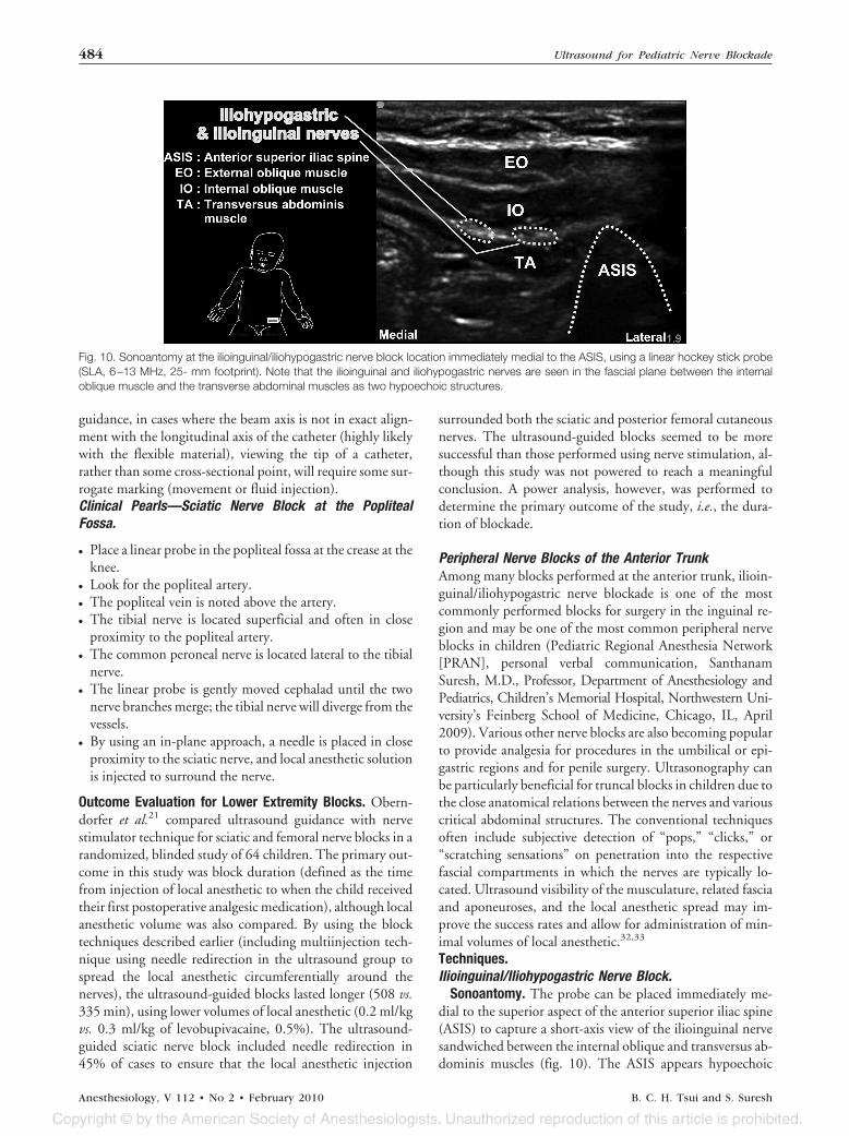
Sonoantomy. The probe can be placed immediately me-dial to the superior aspect of the anterior superior iliac spine(ASIS) to capture a short-axis view of the ilioinguinal nervesandwiched between the internal oblique and transversus ab-dominis muscles (fig. 10). The ASIS appears hypoechoic
Fig. 10. Sonoantomy at the ilioinguinal/iliohypogastric nerve block location immediately medial to the ASIS, using a linear hockey stick probe(SLA, 6–13 MHz, 25- mm footprint). Note that the ilioinguinal and iliohypogastric nerves are seen in the fascial plane between the internaloblique muscle and the transverse abdominal muscles as two hypoechoic structures.
484 Ultrasound for Pediatric Nerve Blockade
Anesthesiology, V 112 • No 2 • February 2010 B. C. H. Tsui and S. Suresh
(due to dorsal shadowing beyond the highly reflective peri-osteum) and nodular-shaped at the lateral edge of the screen.The lateral abdominal muscles will appear with multiple hy-perechoic dots within a hypoechoic background (a “starrynight” appearance); there may be hyperechoic divisions be-tween muscles representing the fascial compartments. Thenerve can be identified as an elliptical-oval shaped structurewith a hyperechoic film surrounding a hypoechoic core. Thesuperiorly and medially situated iliohypogastric nervecourses ventrally between the external and internal obliqueabdominal muscles; this nerve may not be adequately viewedin the image. Although a superficial layer consisting of theexternal oblique abdominal muscle may be present, in manypatients, this muscle has already turned into an aponeuroticsheath at this level.32
Technique. A hockey stick probe will be suitable for manyinfants and younger children, because the nerves are closelysituated beneath the skin (8 mm on average) and medial (7mm on average) to the ASIS.32 Descriptions of the techniquehave included placing the probe immediately medial to theupper aspect of the ASIS to obtain a short-axis view of thenerve(s) situated between the two abdominal muscles (inter-nal oblique and transversus abdominis), with the peritoneumimmediately beneath the internal oblique muscle.32–34 Afteraseptic preparation of the skin and probe, the needle (e.g., 22gauge, 40 mm) can be advanced using an out-of-plane tech-nique, placing the needle tip approximately 1 cm caudad tothe probe surface to allow for optimal needle tip visibilityapproximately 1 cm deep.32 Once the needle tip was viewedusually between the abdominal muscles and in close vicinityto the nerve, the local anesthetic solution was injected underreal-time control to view the solution surrounding the nerve.
The probe could be placed in a manner to use anotherneedle puncture point in a more proximal region, such as thepoint between the third and lateral quarters on a line betweenthe ASIS and umbilicus.35 As used in adults, the probe couldbe placed obliquely at a position lateral to the needle punc-ture site, and an in-plane needle alignment could be used,with the needle directed laterally and slightly caudad.36 Ourpreference is to use an in-plane technique with a linear probewith the needle placed overriding the ASIS and pointingtoward the umbilicus. This provides a clear view of the var-ious muscle layers and the nerves as they run in the planebetween the internal oblique and the transversus abdominismuscle.
Comment. Ilioinguinal/iliohypogastric blocks are com-monly used for anesthesia and analgesia for surgery involvingthe inguinal region, including inguinal hernia repair and or-chiopexy. When successfully placed, these blocks can be aseffective as a caudal block37 and are especially valuable whencaudal block is contraindicated or in larger children withpresacral fat that prevents an easy approach to the sacralhiatus. Using the conventional techniques relying on fascialclicks on penetration of the abdominal muscles for the ilio-inguinal nerve block have a reported success rate of onlyapproximately 70%.38 Moreover, with the closely situated
peritoneum, these blocks can lead to colonic or small bowelpuncture39,40 or pelvic hematomas.41
Part of the reason for block failures may be the inaccuratelocalization of the nerves when using one of the traditionallandmark-based needle insertion sites (often somewherealong a line between the ASIS and the umbilicus) and thefascial click method for determination of injection. Occa-sionally, the blocks seem to fail due to pain experienced dur-ing spermatic cord manipulation, and ideally, the genitofem-oral nerve block should be performed.36,42 The blockade ofthese nerves could benefit greatly from visualization of thecorrect plane of nerve localization (between internal obliqueabdominal and transversus abdominis muscles), the needletrajectory (adjacent to the nerves and outside the closely sit-uated peritoneum), and the local anesthetic deposition.Weintraud et al.43 used ultrasonography to determine theactual location where local anesthetic is administered whenusing the conventional fascial click or pop method for injec-tion. In 62 children, the local anesthetic solution was notedto surround the nerves in only 14% of blocks. This wasdespite the fact that the needle puncture site was determinedby an initial ultrasound scan of the region. The remainder ofthe injections (performed by an anesthesiologist blinded tothe images) were deposited within the muscles in the abdom-inal region (82% in the internal oblique abdominal, trans-versus abdominis, iliac, or external oblique abdominal mus-cle), subcutaneously (2%), or within the peritoneum (2%).This study only had a 55% success rate, based on viewing thelocal anesthetic surrounding the nerves or on clinical signsduring skin incision 15 min after the block. In a related letter,Thibaut et al.44 reported that many adult patients (12 of 14)who received injections into the internal oblique abdominalmuscle (as viewed with ultrasound) experienced successfulblockade, as determined by an anesthesia area m ore t han 25cm2. Five of 19 injections were located either subcutaneouslyor within the external oblique abdominal muscle, of whichmost were unsuccessful.
Clinical pearls—ilioinguinal nerve block.
● Place a linear probe or a hockey stick probe along the ASISwith the probe oriented toward the umbilicus.
● The three layers of the abdominal wall muscles can berecognized.
● The ilioinguinal nerve and iliohypogastric nerves are seenas two hypoechoic structures between the internal obliqueand transversus abdominis muscles.
● By using an in-plane approach, the needle (22 gauge formost children) is advanced and placed between the internaloblique abdominal and the transversus abdominis muscle.
● Use a small (1–2 ml) test injection of a dextrose solution toconfirm the spread of local anesthetic between the musclesand close to the nerves, then aspirate and inject local anes-thetic to expand the plane. Inject between 0.1 and 0.2ml/kg based on experience, duration of surgery expectedand analgesia desired, and concentration of local anestheticsolution.
485EDUCATION
B. C. H. Tsui and S. Suresh Anesthesiology, V 112 • No 2 • February 2010
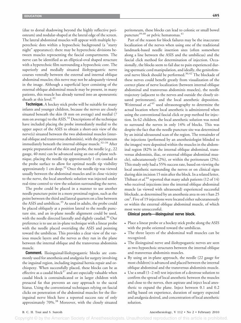
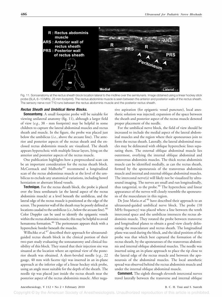
Rectus Sheath and Umbilical Nerve Blocks.Sonoantomy. A small footprint probe will be suitable for
viewing unilateral anatomy (fig. 11), although a larger fieldof view (e.g., 38 - mm footprint) may be helpful in somechildren to capture the lateral abdominal muscles and rectussheath and muscle. In the figure, the probe was placed justbelow the umbilicus (i.e., above the arcuate line). The ante-rior and posterior aspects of the rectus sheath and the en-closed rectus abdominis muscle are visualized. The sheathappears hyperechoic with multiple linear layers, lying on theanterior and posterior aspects of the rectus muscle.
One publication highlights how a preprocedural scan canbe an important consideration for the rectus sheath block.McCormack and Malherbe45 recommend a longitudinalscan of the rectus abdominus muscle at the level of the um-bilicus to exclude any anatomical variations, including bowelherniation or aberrant blood vessels.
Technique. For the rectus sheath block, the probe is placedover the linea semilunaris (at the lateral aspect of the rectusabdominis muscle) at a level beneath the umbilicus, and thelateral edge of the rectus muscle is positioned at the edge of thescreen. The posterior wall of the sheath may be poorly defined atlocations caudad to the umbilicus (i.e., below the arcuate line).46
Color Doppler can be used to identify the epigastric vesselswithin the rectus abdominis muscle; this may be helpful to avoidhematoma formation.46 The peritoneum appears thick with ahyperechoic border beneath the muscles.
Willschke et al.47 described their approach for ultrasound-guided rectus sheath block in the clinical portion of theirtwo-part study evaluating the sonoanatomy and clinical fea-sibility of this block. They stated that their injection site wassituated at the location where an optimal view of the poste-rior sheath was obtained. A short-beveled needle (e.g., 22gauge, 40 mm with facette tip) was inserted in an in-planeapproach at the inferior edge of a linear hockey stick probe,using an angle most suitable for the depth of the sheath. Theneedle tip was placed just inside the rectus sheath near theposterior aspect of the rectus abdominis muscle. After nega-
tive aspiration (for epigastric vessel puncture), local anes-thetic solution was injected; expansion of the space betweenthe sheath and posterior aspect of the rectus muscle denotedproper placement of the needle.
For the umbilical nerve block, the field of view should beincreased to include the medial aspect of the lateral abdom-inal muscles and the region where their aponeuroses join toform the rectus sheath. Laterally, the lateral abdominal mus-cles may be delineated with oblique hyperechoic lines sepa-rating them. The external oblique abdominal muscle liesoutermost, overlying the internal oblique abdominal andtransversus abdominis muscles. The thick rectus abdominismuscle can be identified medially, as can the rectus sheath,formed by the aponeurosis of the transversus abdominismuscle and internal and external oblique abdominal muscles.The intercostal nerve(s) will likely not be visualized by ultra-sound imaging. The nerves are small and run longitudinally,thus tangential, to the probe.46 The hyperechoic and linearappearance of the nerves will closely resemble the aponeuro-sis of the musculature in this region.
De Jose Maria et al.46 have described their approach to anultrasound-guided umbilical nerve block. The probe (10MHz frequency) was placed where a line between the tenthintercostal space and the umbilicus intersects the rectus ab-dominis muscle. They rotated the probe between transverseand longitudinal planes to obtain a good view clearly delin-eating the musculature and rectus sheath. The longitudinalplane was used during the block, and the ideal position of theprobe was that which best captured the formation of therectus sheath, by the aponeuroses of the transversus abdomi-nis and internal oblique abdominal muscles. The needle wasinserted using an in-plane approach to place the tip close tothe lateral edge of the rectus muscle and between the apo-neurosis of the abdominal muscles. The local anestheticspread was observed behind the rectus abdominis muscle andunder the internal oblique abdominal muscle.
Comment. The eighth through eleventh intercostal nervestravel laterally between the transverse and internal oblique
Fig. 11. Sonoanatomy at the rectus sheath block location lateral to the midline over the semilunaris. Image obtained using a linear hockey stickprobe (SLA, 6–13 MHz, 25 mm footprint). The rectus abdominis muscle is seen between the anterior and posterior walls of the rectus sheath.The sensory nerve root T10 runs between the rectus abdominis muscle and the posterior rectus sheath.
486 Ultrasound for Pediatric Nerve Blockade
Anesthesiology, V 112 • No 2 • February 2010 B. C. H. Tsui and S. Suresh
abdominal muscles and, at the level of the linea semilunaris,penetrate the rectus sheath to travel between the posteriorwall of the rectus sheath and the rectus abdominis muscle.Bilaterally, the aponeuroses of the lateral three abdominalmuscles form the rectus sheath; the sheaths join at the lineaalba and together enclose the rectus abdominis muscle. Therectus sheath block aims to place local anesthetic between theposterior aspect of the rectus sheath and the rectus abdominismuscle. The posterior wall of the sheath is not attached to themuscle and thus can be separated during the block injection(i.e., represents a potential space). The median depth of theposterior sheath in 30 children was 8.0 mm (range, 5–13.8);moreover, there was poor correlation between the depth ofthe posterior rectus sheath and weight, height, or body sur-face area.47 With the sheath’s proximity to the peritoneumand without the possibility of reliable estimations or calcula-tions of optimal needle depth, ultrasound imaging duringthe block is especially useful. It is also important to note thatthe posterior wall of the sheath is poorly defined below thearcuate line.46 Placing the needle more lateral, and thus po-tentially avoiding puncture into the peritoneum, the umbil-ical nerve block targets the region lateral to the sheath wherethe nerves exit from their course between the transversusabdominis and internal oblique abdominal muscles.
Although the techniques described for the rectus sheathand umbilical blocks used an out-of-plane needle alignment,an alternative technique using in-plane needle alignmentcould be used and is preferred by one of the authors of thisreview (S. S.). For this, a short footprint probe is preferredand direct visualization of the needle insertion can be per-formed. An out-of-plane technique may be suitable in somecases, particularly in obese children where the depth of inser-tion may be significant.
Clinical pearls—rectus sheath block.
● Place a linear high- frequency probe or a hockey stick probeat the level of the umbilicus.
● The rectus abdominis muscle is identified along with theanterior and posterior walls of the rectus sheath.
● By using an in-plane technique, a 27-gauge needle is ad-vanced until it penetrates the space between the rectusabdominis and the posterior rectus sheath.
● After a small test injection to confirm the spread of localanesthetic, 0.1–0.2 ml/kg local anesthetic solution is in-jected into the potential space between the posterior rectussheath and the rectus abdominis muscle.
● Hydrodissection can be used to find the exact plane be-cause the space is small and exact localization may be re-quired.
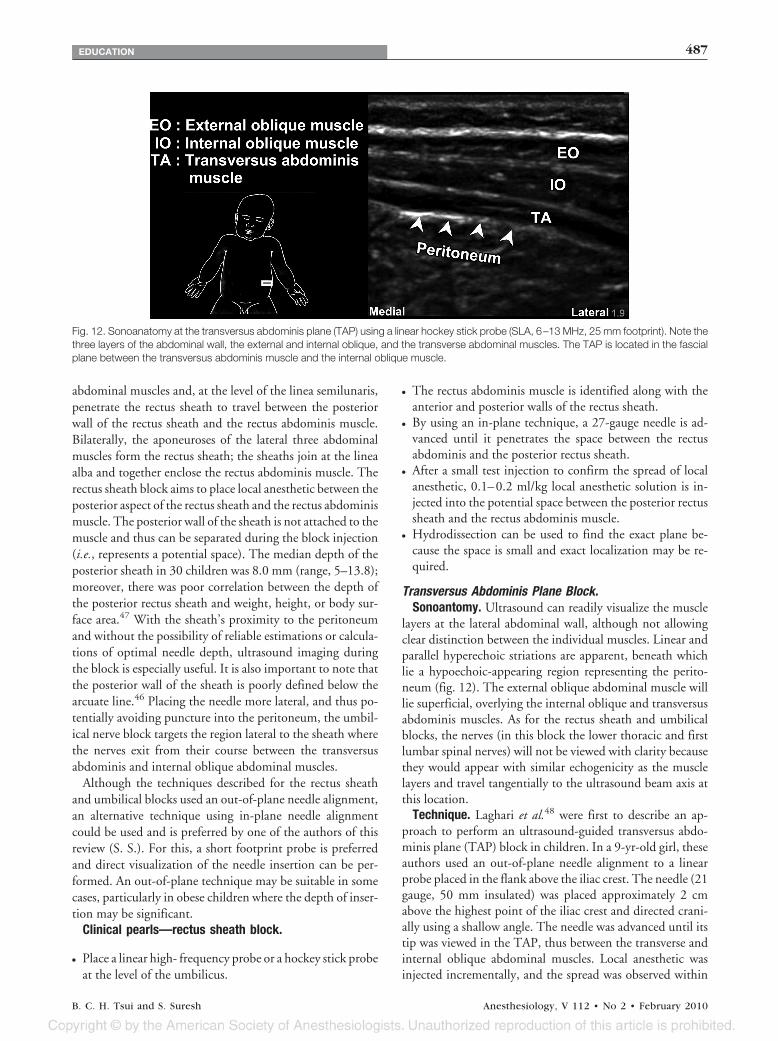
Transversus Abdominis Plane Block.Sonoantomy. Ultrasound can readily visualize the muscle
layers at the lateral abdominal wall, although not allowingclear distinction between the individual muscles. Linear andparallel hyperechoic striations are apparent, beneath whichlie a hypoechoic-appearing region representing the perito-neum (fig. 12). The external oblique abdominal muscle willlie superficial, overlying the internal oblique and transversusabdominis muscles. As for the rectus sheath and umbilicalblocks, the nerves (in this block the lower thoracic and firstlumbar spinal nerves) will not be viewed with clarity becausethey would appear with similar echogenicity as the musclelayers and travel tangentially to the ultrasound beam axis atthis location.
Technique. Laghari et al.48 were first to describe an ap-proach to perform an ultrasound-guided transversus abdo-minis plane (TAP) block in children. In a 9-yr-old girl, theseauthors used an out-of-plane needle alignment to a linearprobe placed in the flank above the iliac crest. The needle (21gauge, 50 mm insulated) was placed approximately 2 cmabove the highest point of the iliac crest and directed crani-ally using a shallow angle. The needle was advanced until itstip was viewed in the TAP, thus between the transverse andinternal oblique abdominal muscles. Local anesthetic wasinjected incrementally, and the spread was observed within
Fig. 12. Sonoanatomy at the transversus abdominis plane (TAP) using a linear hockey stick probe (SLA, 6–13 MHz, 25 mm footprint). Note thethree layers of the abdominal wall, the external and internal oblique, and the transverse abdominal muscles. The TAP is located in the fascialplane between the transversus abdominis muscle and the internal oblique muscle.
487EDUCATION
B. C. H. Tsui and S. Suresh Anesthesiology, V 112 • No 2 • February 2010
the plane. No intraoperative analgesia was required and ex-cellent pain scores (0/10) were recorded soon after the openappendectomy.
Fredrickson et al.42 have described their early experiencewith TAP block under ultrasound guidance. They placed alinear probe in the axial plane above the iliac crest or at alocation where the three lateral abdominal muscle layers weremost evident on the ultrasound screen. An in-plane needlealignment was used, with the needle (a short 22- gauge spinalneedle) directed from anterior to posterior to allow the nee-dle to be viewed along its axis. On viewing the needle tip inthe TAP, the local anesthetic was injected while observingthe spread within the plane. The blocks were successful, withthe exception of those patients who required intraoperativeopioid supplementation due to spermatic cord manipulation(genitofemoral nerve). A recent simple technique for placementof the TAP block in infants and children has been describedwith a step-by-step approach to perform this block in infantsand children by one of the authors of this review (S. S.).49
Comment. The TAP block provides analgesia to the ab-dominal wall, through blockade of the lower thoracic andfirst lumbar spinal nerves at the lateral aspect of the abdomenbefore their course between the transversus abdominis andinternal oblique abdominal muscles. In adults, there may bereliance on palpating the lumbar triangle of Petit (above thehighest point of the iliac crest and between the posteriorborder of the external oblique and the lower lateral border ofthe latissimus dorsi muscle) at the base of which lies theinternal oblique muscle.50 Because there is no distinctly pal-pable triangle of Petit in children (or one only enabling sen-sation of a tiny hole), ultrasound may be especially valuablefor determining the point of needle puncture during thisblock. Indications for the TAP block include postoperativepain control after abdominal surgery including colostomyand laparotomy especially when a central neuraxial block iscontraindicated as in children with spinal dysraphism or inchildren with coagulation abnormalities. Because this blockincludes blockade of the first lumbar nerve root, it may sub-stitute the ilioinguinal/iliohypogastric block in cases whenthe region anterior and medial to the ASIS offers suboptimal
imaging; the muscles in this region are apparently prone toanisotropy (i.e., artifactual images produced due to the beampenetration angle in relation to the muscles).42
● Place a high- frequency linear probe or a hockey stick probelateral to the umbilicus.
● Slide the probe laterally until the three muscle layers of theabdominal wall are recognized (external and internaloblique abdominal and transverse abdominal).
● At or just posterior to the midaxillary line, by using anin-plane approach, place a needle between the internaloblique and the transverse abdominal muscles.
● As local anesthetic is injected, the plane is seen to expandwith posterior movement of the transversus abdominismuscle.
● Hydrodissection can be performed to recognize the exactplane of injection of the local anesthetic solution.
Dorsal Penile Nerve Block.Sonoantomy. By placing a probe sagittally along the shaft
of the penis, the subpubic space can be located as a trianglecontaining the deep penile fascia (inferiorly), the pubic sym-physis (superiorly), and the membranous layer of the super-ficial (Scarpa’s) fascia.51 Although the pubic symphysis andthe membranous layer of the superficial fascia can generallybe visualized, one may find that it is often difficult to clearlyidentify the deep fascia (fig. 13).
Technique. In a case report, Sandeman and Dilley51 de-scribe placing a linear probe sagittally along the shaft of thepenis to view the subpubic space. Penile block can be accom-panied by bilateral injections into the subpubic space. Injec-tion deep to Scarpa’s fascia can be guided by ultrasoundimaging to confirm the spread of local anesthetic solution tothe deep fascia. The fundiform ligament, forming a midlineseptum within the deep fascia of the penis, appears fan-shaped. The needle was introduced on both sides of thefundiform ligament (thus on either side of the probe sepa-rately), to reach the region adjacent to the penile shaft struc-tures. The authors state that they avoided injecting beneaththe deep fascia to avoid nerve injury or intravascular injec-
Fig. 13. Sonoanatomy of the subpubic space using a linear probe (HFL38, 6–13 MHz, 38- mm footprint).
488 Ultrasound for Pediatric Nerve Blockade
Anesthesiology, V 112 • No 2 • February 2010 B. C. H. Tsui and S. Suresh
tion. The local anesthetic can be seen spreading throughoutthe subpubic space.
Comment. This block is indicated for children undergoingpenile procedures where a caudal block may be contraindi-cated. It can also be used in older children and adolescentswhere presacral fat can make the performance of the caudalblock difficult. The performance of this block under ultra-sound guidance is not well established at this time.
Clinical pearls—dorsal penile nerve block.
● The probe is placed sagittally along the shaft of the penis toview the subpubic space.
● The spread of local anesthetic deep to Scarpa’s fascia can beconfirmed using ultrasound.
● Performance of an ultrasound-guided penile block maylikely remain uncommon in children because ultrasoundimaging may be cumbersome to use due to the limitedcontact area for the probe.
Outcome Evaluation for Blocks at the Anterior Trunk.Willschke et al.32 performed a prospective, randomized dou-ble-blinded study with 100 children scheduled for inguinalsurgery to compare block efficacy between ultrasound-guided and conventional ilioinguinal/iliohypogastric nerveblock. The end point in the ultrasound group was obtaininga view of the local anesthetic solution surrounding thenerves, whereas detecting a fascial click was used to deter-mine injection in the traditional block group. Fewer childrenreceived intraoperative or postoperative (up to 4 h) analgesiain the ultrasound group (4% and 6%, respectively) than inthe fascial click group (26 and 40%). Furthermore, the vol-ume of local anesthetic injected was lower in the ultrasoundgroup (0.19 vs. 0.30 ml/kg).
Further evaluation of the optimal volume of local anes-thetic required for ultrasound-guided ilioinguinal/iliohypo-gastric blocks was performed by this group in a subsequentstudy.33 By using a modified step-up-step-down approach,with 10 patients in each group starting at 0.2 ml/kg, a vol-ume of 0.75 ml/kg was found to achieve 100% block successfor the first 4 h of postoperative care (longer analgesia mayrequire higher volumes). These authors state that using lowervolumes of local anesthetic could reduce the risk of localanesthetic toxicity and the incidence of adverse side effects,such as femoral nerve palsy.
Interesting findings have been published recently, whichtend to support the use of lower volumes of local anestheticwhen placing ilioinguinal blocks under ultrasound guidancewhen compared with the use of landmark-based technique.In a pharmacokinetic study, plasma levels of ropivacainewere found to be higher using ultrasound guidance whencompared with a single pop technique (Cmax: 1.78 [0.62] vs.1.23 [0.70] � g/ml). Although this finding is interesting andwill likely encourage the planning of similar studies, theplasma levels reached in this study have never been associatedwith any clinical signs of toxicity.34
There have been no controlled or comparative studiespublished related to any of the other trunk blocks described
previously. Earlier reports of the rectus sheath block, usingblind techniques, describe successful blocks as performed insmall numbers of patients.52,53 By using ultrasound guidancefor rectus sheath blocks in 20 children, Willschke et al. re-ported a 100% success rate for intra operative and postoper-ative analgesia (until time to discharge at 4 h), using (prede-termined volumes in their study) lower doses of localanesthetics (i.e., levobupivacaine 0.25% 0.1 vs. bupivacaine0.5% 0.2 ml/kg) than previously described when using blindtechnique with the same injection protocol.47,53 Similardoses of local anesthetic have been used for the umbilicalnerve block as those for the ultrasound-guided rectus sheathblock (i.e., bupivacaine 0.25% 0.1 ml/kg).46
Discussion
The performance of various peripheral nerve blocks, includ-ing single-shot techniques of the lumbar and sacral plexuses,has increased in recent years.54 Even though the success andsafety of peripheral blocks is usually good to excellent, incertain cases, improvements can be made (e.g., ilioinguinalblock success), and there are questions related to the reliabil-ity of the conventional techniques of nerve localization. Al-though much of the difficulty when performing peripheralnerve blocks in children stems from their relatively smallanatomical structures and close proximity of their nerves tocritical structures, the superficial position of many of therelevant structures enables the use of high-resolution ultra-sound imaging, which can enable excellent real-time blockguidance. Thus, real-time ultrasound imaging may improvethe ability of the anesthesiologist to identify the neural struc-tures, place the block needle in close proximity to the target,and precisely administer optimal volumes of local anesthetic.
Ultrasound may enable lower volumes of local anestheticsolution to be used in some cases where local anesthetic tox-icity is a concern (e.g., combined femoral and sciatic nerveblocks) or unwanted side effects are common (e.g., spillovercausing femoral nerve palsy during ilioinguinal blocks). Al-though some reports suggest that ultrasound should be usedto minimize volumes of local anesthetic, it should be stressedthat the volume used should maintain the desired parametersof the block, including its extent and duration. Some practi-tioners, especially those without similar expertise, may notobtain the degree of success to those reporting higher quali-ties of blockade with such low dosages. In addition, onerecent study in adults found (using Dixon and Massey up-and-down methodology) that similar doses were required forultrasound-guided supraclavicular block (minimum effectiveanesthetic volume in 50% of patients was 23 ml and calcu-lated effective volume in 95% of patients was 42 ml) to thosecommonly used without ultrasound.55 Although commonlyrecommended doses of local anesthetic56,57 will maintainplasma concentrations in ranges, which have not been shownto cause toxicity, the thresholds for toxicity are not accuratelyknown and ultrasound-guided placement of local anestheticmay have effects on plasma concentrations as recently shown
489EDUCATION
B. C. H. Tsui and S. Suresh Anesthesiology, V 112 • No 2 • February 2010
for ilioinguinal blocks.34 More studies will hopefully directus to the appropriate dose of local anesthetic to be usedduring ultrasound-guided techniques while maintaining op-timal success without risks of toxicity and unwanted sideeffects.
Although anatomical knowledge may be acquiredthrough reviewing textbooks, the fact that sonoanatomy ishighly dependant on the system used and on the plane ofview makes it particularly important to obtain hands-on im-aging experience. Nevertheless, this review includes repre-sentative images obtained from a portable ultrasound unit
(using the scanning planes commonly described) to serve asexamples of typical sonograms used during ultrasound guid-ance of extremity and trunk blocks. Bony, vascular and /orsoft tissue landmarks are often essential to help visually guidethe operator to the neural structures. Although more impres-sive during dynamic viewing, the pulsatile nature of arteriesis commonly evident and can be very helpful at many blocklocations (e.g., supra clavicular and infraclavicular, femoral,and popliteal); alternatively color or power Doppler can beused and will help with future reference to a static image.Bony structures often serve as good reference landmarks for
Table 1. Useful Landmarks for Identification of Nerves Using Ultrasound
Block Useful Ultrasound Landmark Comments
Interscalene Scalene muscles and subclavian artery At the posterolateral aspect of thesternocleidomastoid muscle, thesuperficially locatedsternocleidomastoid muscleappears triangular in shape andoverlies the internal jugular veinand common carotid artery.
Supraclavicular Subclavian artery Brachial plexus lies lateral and oftensuperior to the artery.
Infraclavicular Subclavian artery and vein Brachial plexus cords surround theartery. Posterior and medial cordcan be difficult to visualize clearly.
Axillary Axillary artery Terminal nerves surround the artery.Important to avoid the multiplevessels present.
Lumbar plexus Transverse processes Plexus lies between and just deepto the lateral tips of the processeswhen using a longitudinal view. Inpediatrics, the transverse viewcan be used to visualize thepsoas muscle.
Femoral Femoral artery Nerve lies lateral to artery (veinmost medial).
SciaticSubgluteal Greater trochanter and ischial
tuberosityNerve lies between the two bone
structures. With a linear probe,only the lateral bone (i.e., femur)is often seen.
Popliteal Popliteal artery Traceback from the popliteal creasewhere the tibial nerve is adjacentto the artery. Scanning proximallyto the sciatic bifurcation, theartery becomes deeper and at agreater distance from the tibialnerve, which is joined by theperoneal nerve.
TrunkTransversus abdominis
plane blockThree muscles layers (external and
internal oblique and transverseabdominal)
No nerve can be seen.
Rectus sheath andumbilical block
Rectus sheath and rectus abdominalmuscle
No nerve can be seen.
Ilioinguinal/iliohypogastric Two to three layers of muscle (externaloblique oftentimes aponeurosis) andanterior superior iliac spine
Nerve(s) can be seen.
490 Ultrasound for Pediatric Nerve Blockade
Anesthesiology, V 112 • No 2 • February 2010 B. C. H. Tsui and S. Suresh
many block locations, especially when the nerves may not beclearly viewed. For instance, the transverse processes can beused to mark the block site for lumbar plexus block in pa-tients older than infancy where the nerves are not clearlydelineated. If there is not a readily identifiable landmark inclose vicinity to the nerve at the traditional block location (orthe nerve remains to be identified), it may be of great help tofirst identify the nerve in relation to a prominent landmark ata “satellite” location and thereafter trace the nerve to theblock location. Table 1 provides some examples of land-marks commonly used by the authors and others for identi-fying nerves during many ultrasound-guided peripheralnerve blocks. Of course, there are many variations to the“usual” anatomy, and although not discussed here, this factgreatly supports the use of direct, real-time imaging to vieweach individual’s related anatomy. It is imperative to practicedynamic viewing while obtaining control of the block nee-dle’s trajectory and ultimately the deposition of local anes-thetic. Although a discussion of the aforementioned tech-nique is beyond this review, there are multiple publicationsand workshops (presented at various society meetings andother gatherings) describing and providing hands-on experi-ence of particular techniques, which may be of use to thenovice or experienced.
The authors only discussed those articles that were eitherpublished in English or which contained English abstracts.Of three reviews obtained, which were written in alternatelanguages,58–60 one had an English abstract58 but within thisthere was no relevant information on technique of ultra-sound-guided blockade. One of the reviews may have in-cluded some description of technique, which would be ofinterest,59 although the other60 was written by experts inultrasound guidance who have published several of the arti-cles, which have been described in this review. Another lim-itation is the inclusion of only those papers specifically per-taining to the pediatric population. This was partly due tothe vast expanse of general review articles of ultrasound inregional anesthesia, but mainly because those general reviewswhich include a section on pediatrics are often written bythose experts who have published the studies, which wereincluded in this review.
This review has focused on published descriptions of ul-trasound-guidance techniques and study outcomes based oncomparisons of ultrasound to conventional nerve localiza-tion techniques. There is an increasing amount of literaturebeing published describing ultrasound-guidance techniquesfor extremity and trunk nerve blocks in children. Althoughthe outcomes documented suggest that there may be bene-fits, there is insufficient evidence to make such claims basedfrom a few relatively small studies. More studies are neededwith particular emphasis on establishing whether superioranalgesic efficacy can be achieved with this imaging technol-ogy, particularly in relation to specific surgical procedures.However, it is reasonable to postulate that ultrasound, pri-marily used during the performance of upper extremity and
trunk blocks, may reduce complications such as mechanicalnerve injury and arterial, pleural, or peritoneal puncture.
The authors thank Jennifer Pillay, B.Sc. (Research Assistant, Depart-ment of Anesthesiology and Pain Medicine, University of Alberta,Edmonton, Alberta, Canada), for her contributions, Jenkin Tsui(Medical Illustrator, Department of Anesthesiology and Pain Medi-cine, University of Alberta) for his line drawings and assistance withthe figures, and Sonosite Inc., Bothell, Washington for its generousequipment support.
References
1. Tsui BC, Suresh S: Ultrasound imaging for regional anes-thesia in infants, children and adolescents: A review ofcurrent literature and its application in the practice ofneuraxial blockade. ANESTHESIOLOGY 2010; 112:in press
2. Fredrickson MJ: Ultrasound-assisted interscalene catheterplacement in a child. Anaesth Intensive Care 2007; 35:807– 8
3. Mariano ER, Ilfeld BM, Cheng GS, Nicodemus HF, Suresh S:Feasibility of ultrasound-guided peripheral nerve blockcatheters for pain control on pediatric medical missions indeveloping countries. Paediatr Anaesth 2008; 18:598 – 601
4. Van Geffen GJ, Tielens L, Gielen M: Ultrasound-guidedinterscalene brachial plexus block in a child with femurfibula ulna syndrome. Pediatr Anesth 2006; 16:330 –2
5. Tsui BC, Wagner A, Finucane B: Electrophysiologic effectof injectates on peripheral nerve stimulation. Reg AnesthPain Med 2004; 29:189 –93
6. Bernards CM, Hadzic A, Suresh S, Neal JM: Regional anes-thesia in anesthetized or heavily sedated patients. RegAnesth Pain Med 2008; 33:449 – 60
7. De Jose María B, Banus E, Navarro EM, Serrano S, PerelloM, Mabrok M: Ultrasound-guided supraclavicular vs infra-clavicular brachial plexus blocks in children. Paediatr An-aesth 2008; 18:838 – 44
8. Tsui BC, Twomey C, Finucane BT: Visualization of thebrachial plexus in the supraclavicular region using acurved ultrasound probe with a sterile transparent dress-ing. Reg Anesth Pain Med 2006; 31:182– 4
9. Gray AT, Collins AB, Schafhalter-Zoppoth I: Sciatic nerveblock in a child: A sonographic approach. Anesth Analg2003; 97:1300 –2
10. Marhofer P, Sitzwohl C, Greher M, Kapral S: Ultrasoundguidance for infraclavicular brachial plexus anaesthesia inchildren. Anaesthesia 2004; 59:642– 6
11. Greher M, Retzl G, Niel P, Kamolz L, Marhofer P, Kapral S:Ultrasonographic assessment of topographic anatomy involunteers suggests a modification of the infraclavicularvertical brachial plexus block. Br J Anaesth 2002; 88:632– 6
12. de Jose M aria B, Tielens LK: Vertical infraclavicular bra-chial plexus block in children: A preliminary study. Pae-diatr Anaesth 2004; 14:931–5
13. Chin KJ, Chan VW, van Geffen GJ: Ultrasound-guided in-fraclavicular block: The in-plane versus out-of-plane ap-proach. Paediatr Anaesth 2008; 18:1279 – 80
23. Scheuer L, Black S, Christie A: The pelvic girdle, Develop-mental Juvenile Osteology. Edited by Scheuer L. London,Academic Press, 2000, pp 341–73
24. Scheuer L: The lower extremity, Developmental JuvenileOsteology. Edited by Scheuer L. London, Academic Press,2000, pp 374 – 467
25. Tsui BC, Ozelsel TJ: Ultrasound-guided anterior sciaticnerve block using a longitudinal approach: “Expanding theview.” Reg Anesth Pain Med 2008; 33:275– 6
26. Koscielniak-Nielsen ZJ, Rasmussen H, Hesselbjerg L: Long-axis ultrasound imaging of the nerves and advancement ofperineural catheters under direct vision: A preliminaryreport of four cases. Reg Anesth Pain Med 2008; 33:477– 82
27. van Geffen GJ, Scheuer M, Muller A, Garderniers J, GielenM: Ultrasound-guided bilateral continuous sciatic nerveblocks with stimulating catheters for postoperative painrelief after bilateral lower limb amputations. Anaesthesia2006; 61:1204 –7
28. van Geffen GJ, Gielen M: Ultrasound-guided subglutealsciatic nerve blocks with stimulating catheters in children:A descriptive study. Anesth Analg 2006; 103:328 –33
29. van Geffen GJ, Bruhn J, Gielen M: Ultrasound-guided con-tinuous sciatic nerve blocks in two children with venousmalformations in the lower limb. Can J Anaesth 2007;54:952–3
30. van Geffen GJ, McCartney CJ, Gielen M, Chan VW: Ultra-sound as the only nerve localization technique for periph-eral nerve block. J Clin Anesth 2007; 19:381–5
31. Schwemmer U, Markus CK, Greim CA, Brederlau J, Traut-ner H, Roewer N: Sonographic imaging of the sciatic nerveand its division in the popliteal fossa in children. PediatrAnesth 2004; 14:1005– 8
33. Willschke H, Bosenberg A, Marhofer P, Johnston S, KettnerS, Eichenberger U, Wanzel O, Kapral S: Ultrasonographic-guided ilioinguinal/iliohypogastric nerve block in pediat-ric anesthesia: What is the optimal volume? Anesth Analg2006; 102:1680 – 4
34. Weintraud M, Lundblad M, Kettner SC, Willschke H, Ka-pral S, Lonnqvist PA, Koppatz K, Turnheim K, Bsenberg A,Marhofer P: Ultrasound versus landmark-based techniquefor ilioinguinal-iliohypogastric nerve blockade in children:The implications on plasma levels of ropivacaine. AnesthAnalg 2009; 108:1488 –92
35. Dalens B: Nerve blocks of the trunk, Regional Anesthesiain Infants, Children and Adolescents. Edited by Dalens B.Baltimore, Williams & Wilkins, 1995, pp 461– 88
36. Hu P, Harmon D, Frizelle H: Ultrasound guidance forilioinguinal/iliohypogastric nerve block: A pilot study. IrJ Med Sci 2007; 176:111–5
37. Markham SJ, Tomlinson J, Hain WR: Ilioinguinal nerveblock in children. A comparison with caudal block for
intra and postoperative analgesia. Anaesthesia 1986; 41:1098 –103
38. Lim SL, Ng SA, Tan GM: Ilioinguinal and iliohypogastricnerve block revisited: Single shot versus double shot tech-nique for hernia repair in children. Paediatr Anaesth 2002;12:255– 60
39. Amory C, Mariscal A, Guyot E, Chauvet P, Leon A, Poli-Merol ML: Is ilioinguinal/iliohypogastric nerve block al-ways totally safe in children? Paediatr Anaesth 2003; 13:164 – 6
40. Johr M, Sossai R: Colonic puncture during ilioinguinalnerve block in a child. Anesth Analg 1999; 88:1051–2
41. Vaisman J: Pelvic hematoma after an ilioinguinal nerveblock for orchialgia. Anesth Analg 2001; 92:1048 –9
42. Fredrickson M, Seal P, Houghton J: Early experience withthe transversus abdominis plane block in children. Paedi-atr Anaesth 2008; 18:891–2
43. Weintraud M, Marhofer P, Bosenberg A, Kapral S,Willschke H, Felfernig M, Kettner S: Ilioinguinal/iliohypo-gastric blocks in children: Where do we administer thelocal anesthetic without direct visualization? Anesth Analg2008; 106:89 –93
44. Thibaut D, de la Cuadra-Fontaine J, Bravo MP, de la FuenteR: Ilioinguinal/iliohypogastric blocks: Where is the anes-thetic injected? Anesth Analg 2008; 107:728 –9
45. McCormack JG, Malherbe S: Applications of ultrasound inpaediatric anaesthesia. Curr Anaesth Crit Care 2008; 19:302– 8
46. De Jose MB, Gotzens V, Mabrok M: Ultrasound-guidedumbilical nerve block in children: A brief description of anew approach. Paediatr Anaesth 2007; 17:44 –50
47. Willschke H, Bosenberg A, Marhofer P, Johnston S, KettnerSC, Wanzel O, Kapral S: Ultrasonography-guided rectussheath block in paediatric anaesthesia: A new approach toan old technique. Br J Anaesth 2006; 97:244 –9
49. Suresh S, Chan V: Ultrasound guided transversus abdomi-nis plane block in infants, children and adolescents: Asimple procedural guidance for their performance. Paedi-atr Anaesth 2009; 19:296 –9
50. Rafi AN: Abdominal field block: A new approach via thelumbar triangle. Anaesthesia 2001; 56:1024 – 6
51. Sandeman DJ, Dilley AV: Ultrasound guided dorsal penilenerve block in children. Anaesth Intensive Care 2007;35:266 –9
52. Courreges P, Poddevin F, Lecoutre D: Para-umbilicalblock: A new concept for regional anaesthesia in children.Paediatr Anaesth 1997; 7:211– 4
53. Ferguson S, Thomas V, Lewis I: The rectus sheath block inpaediatric anaesthesia: New indications for an old tech-nique? Paediatr Anaesth 1996; 6:463– 6
54. Lacroix F: Epidemiology and morbidity of regional anaes-thesia in children. Curr Opin Anaesthesiol 2008; 21:345–9
55. Duggan E, Beheiry H, Perlas A, Lupu M, Nuica A, Chan V,Brull R: Minimum effective volume of local anesthetic forultrasound-guided supraclavicular brachial plexus block.Reg Anesth Pain Med 2009; 34:215– 8
56. Eck JB, Ross AK: Paediatric regional anaesthesia: Whatmakes a difference? Best Pract Res Clin Anaesthesiol 2002;16:159 –74
57. Murat I: Pharmacology, Regional Anesthesia in Infants,Children, and Adolescents. Edited by Dalens B. London,Williams & Wilkins, 1995, pp 67–125
58. Hillmann R, Kretz FJ: [Risks and dangers in pediatric re-gional anesthesia]. Anaesthesist 2008; 57:165–74
59. Lacroix F: [Localization of nerves in pediatric regionalanesthesia]. Arch Pediatr 2006; 13:653–5
60. Marhofer P, Kapral S: [The use of ultrasonography forpaediatric regional anesthesia]. Anasthesiol IntensivmedNotfallmed Schmerzther 2006; 41:178 – 83
492 Ultrasound for Pediatric Nerve Blockade
Anesthesiology, V 112 • No 2 • February 2010 B. C. H. Tsui and S. Suresh