66
Ultrasound in Obstetrics and Gynecology for the Generalist Dr S.Moola Ob/gyn - Nelson BC
Ultrasound in Obstetrics andGynecology for the Generalist
Dr S.MoolaOb/gyn - Nelson BC

US for the Generalist
• Ultrasound should be viewed as an an adjunct tool to clinical exam and history (ie: like using performing an ECG for chest pain or using slit lamp)
• It should not be used as a diagnostic test (realm of radiology/perinatology)
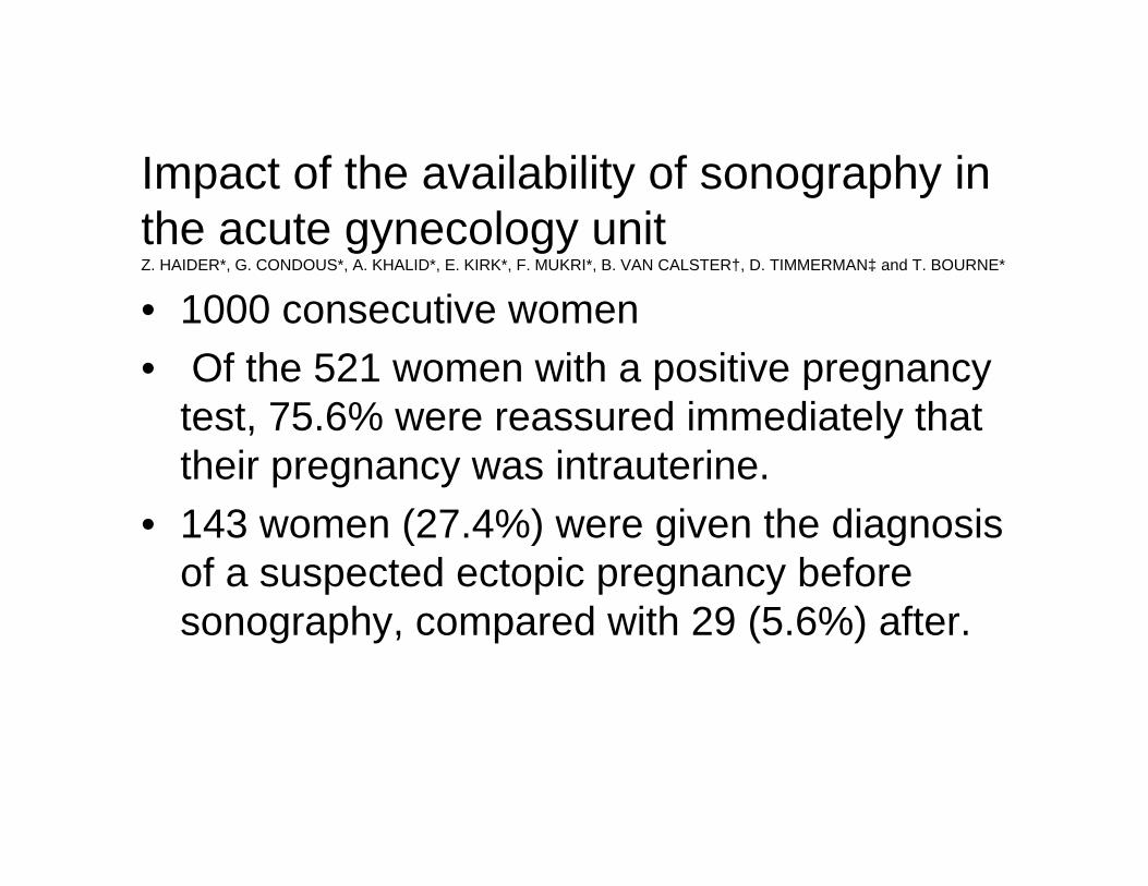
• 1000 consecutive women• Of the 521 women with a positive pregnancy
test, 75.6% were reassured immediately that their pregnancy was intrauterine.
• 143 women (27.4%) were given the diagnosis of a suspected ectopic pregnancy before sonography, compared with 29 (5.6%) after.
Impact of the availability of sonography in the acute gynecology unitZ. HAIDER*, G. CONDOUS*, A. KHALID*, E. KIRK*, F. MUKRI*, B. VAN CALSTER†, D. TIMMERMAN‡ and T. BOURNE*

• Following the ultrasound examination there was a change in clinical management in 54.1% of the women with a positive pregnancy test and a reduction in admissions (including inpatient theater admissions) (from 40.3% to 17.1%) and outpatient follow-up examinations (from 41.1% to 35.5%).
• In 90 (23.8%) non-pregnant women a significant ovarian cyst (> 5 cm) was suspected clinically; 28/90 (31.1%) were confirmed on sonography.
• Following the ultrasound examination there was a change in clinical management for 38.1% of non-pregnant women and a reduction in admissions (from 37.1% to 19.4%) and outpatient follow-up examinations (from 25.7% to 18.1%).

Ob/Gyn US
• Know how to turn machine on• Know how to turn machine off• Know how to clean probes• Know how to document observations


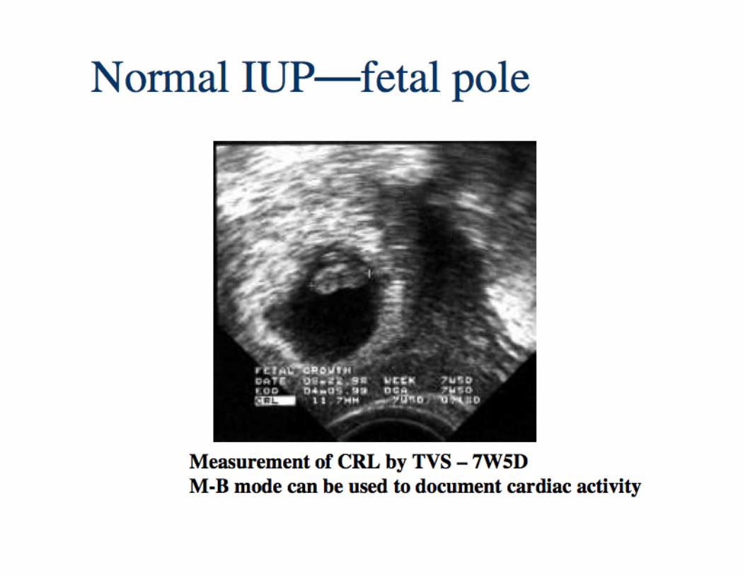
Sac Diameter/ Crown Rump Length


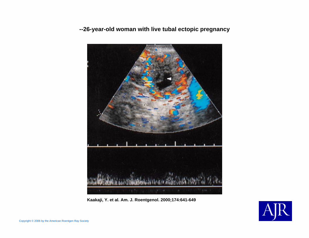
Copyright © 2006 by the American Roentgen Ray Society
Kaakaji, Y. et al. Am. J. Roentgenol. 2000;174:641-649
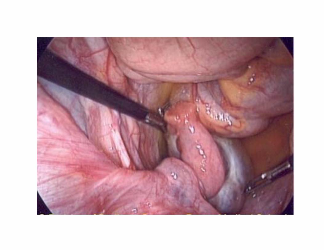
--26-year-old woman with live tubal ectopic pregnancy


Copyright © 2007 by the American Roentgen Ray Society
Kaakaji, Y. et al. Am. J. Roentgenol. 2000;174:641-649
--32-year-old woman with intra abdominal pregnancy

Copyright © 2007 by the American Roentgen Ray Society
Kaakaji, Y. et al. Am. J. Roentgenol. 2000;174:641-649
--32-year-old woman with intra abdominal pregnancy

Remember non gynecologic causes


3 tenets of OB US
• Location…where is the head• Location….where is the placenta (or
where isn’t the placenta)• Location…where is the heart• ….how many fetuses

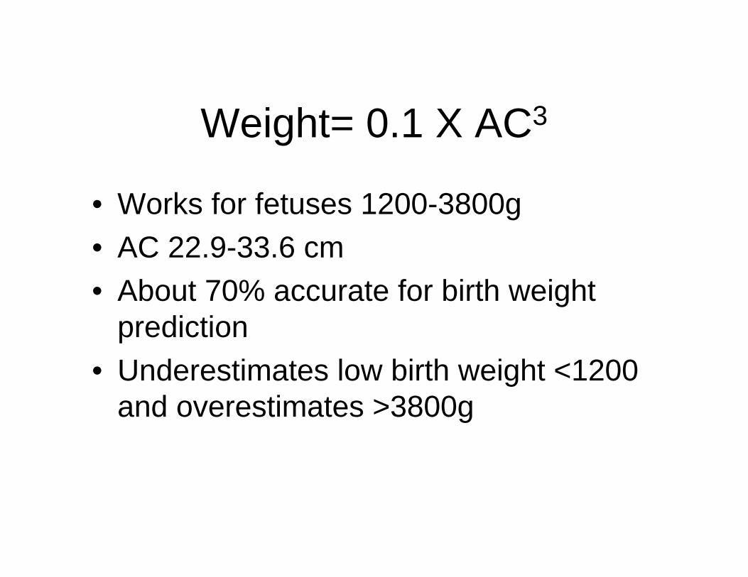
Weight= 0.1 X AC3
• Works for fetuses 1200-3800g• AC 22.9-33.6 cm• About 70% accurate for birth weight
prediction• Underestimates low birth weight <1200
and overestimates >3800g


Placenta previa
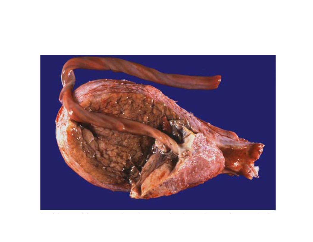

Abruptio placenta


“Miles on the ultrasound odometer will not only sharpen sonographic skills but also will
help the EP to better communicate with nonpregnant patients presenting with
abdominal pain.
There is a fairly specific barometer already in place to gauge one gynecologic ultrasound skills: a seasoned EP sonographer never
skips over the chart of a young woman with right lower quadrant pain.” - M.lambert


