Ultrasound in Obstetrics Who, Where, When and How Many? Anthony Johnson, D.O. Visiting Professor Departments of Obstetrics, Gynecology and Reproductive Sciences and Pediatric Surgery Co-Director, Texas Fetal Center
Transcript
Ultrasound in Obstetrics Who, Where, When and How Many?
Anthony Johnson, D.O.
Visiting Professor
Departments of Obstetrics, Gynecology and
Reproductive Sciences and Pediatric Surgery
Co-Director, Texas Fetal Center
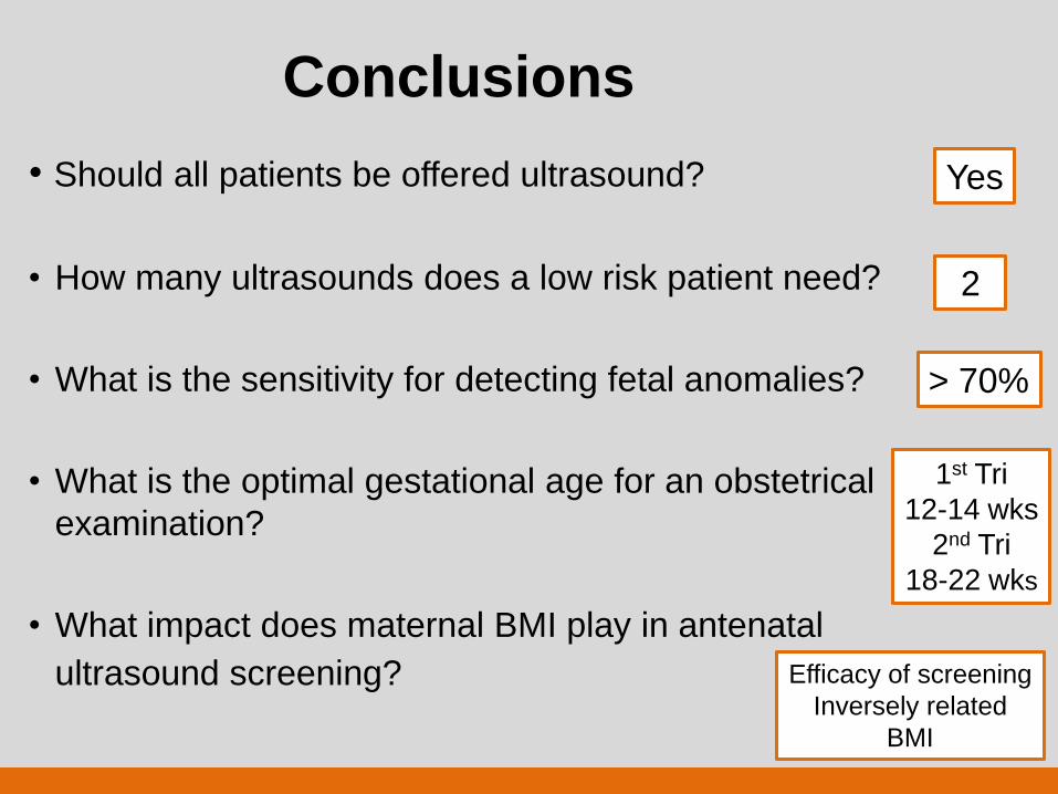
Clinical Considerations
• Should all patients be offered ultrasound?
• How many ultrasounds does a low risk patient need?
• What is the sensitivity for detecting fetal anomalies?
• What is the optimal gestational age for an obstetrical
examination?
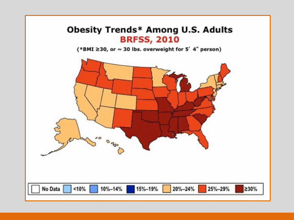
• What impact does maternal BMI play in antenatal
ultrasound screening?
Should all patients be offered
ultrasonography, and what is the
sensitivity for detecting fetal anomalies?
• 90% of fetal anomalies are born to women
considered “low risk”
• Sensitivity varies amongst studies
• Different definition of major vs. minor malformation
• Populations differences, high vs. low risk
• Expertise of imaging
• Structure imaged (DR higher with CNS vs. cardiac)
Abuhamad AZ ACOG Practice Bulletin #101, 2009
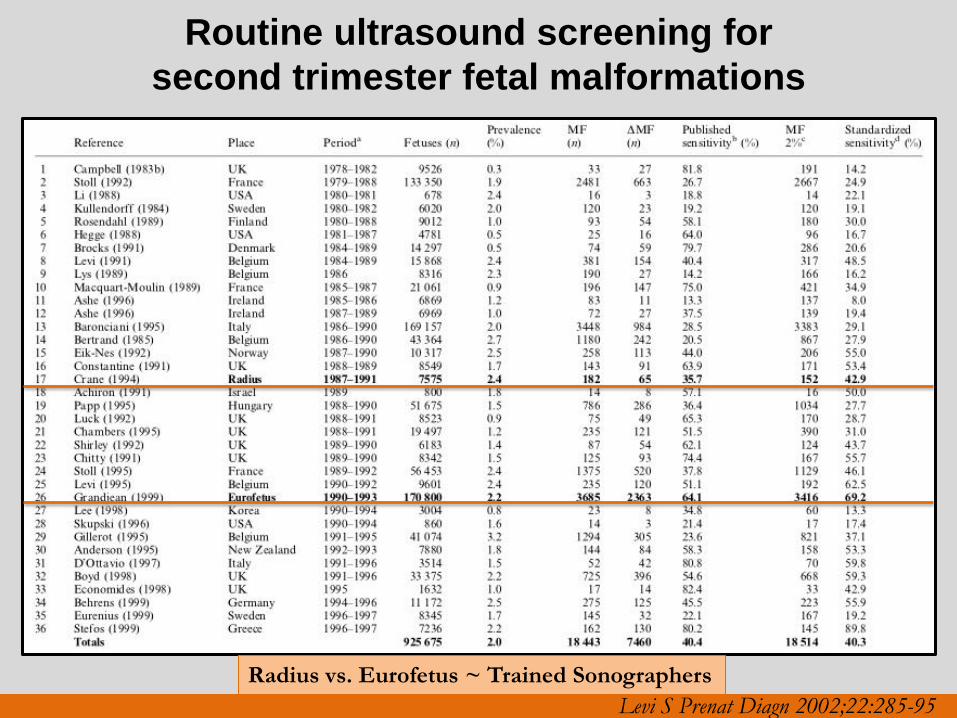
Levi S Prenat Diagn 2002;22:285-95
Routine ultrasound screening for
second trimester fetal malformations
Radius vs. Eurofetus ~ Trained Sonographers
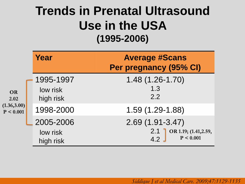
Trends in Prenatal Ultrasound
Use in the USA (1995-2006)
Year Average #Scans
Per pregnancy (95% CI)
1995-1997
low risk
high risk
1.48 (1.26-1.70) 1.3
2.2
1998-2000 1.59 (1.29-1.88)
2005-2006
low risk
high risk
2.69 (1.91-3.47) 2.1
4.2
OR
2.02
(1.36,3.00)
P < 0.001
OR 1.19; (1.41,2.59,
P < 0.001
Siddique J et al Medical Care. 2009;47:1129-1135
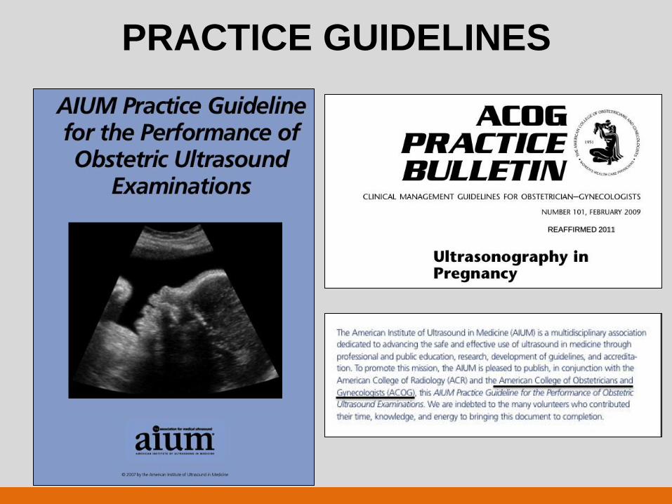
REAFFIRMED 2011
PRACTICE GUIDELINES
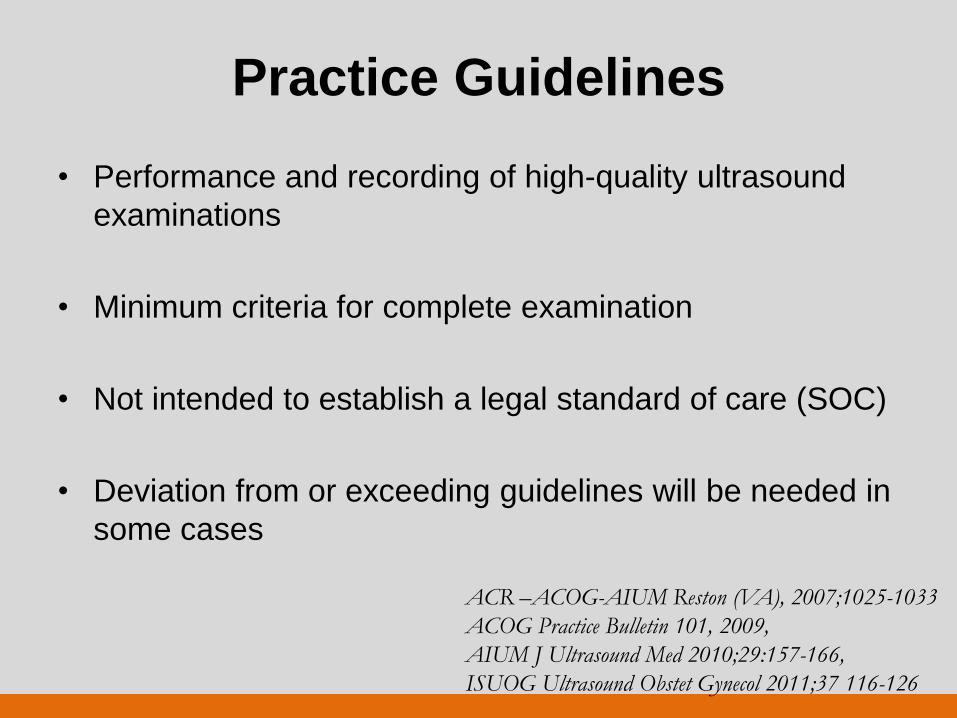
Practice Guidelines
• Performance and recording of high-quality ultrasound
examinations
• Minimum criteria for complete examination
• Not intended to establish a legal standard of care (SOC)
• Deviation from or exceeding guidelines will be needed in
some cases
ACR –ACOG-AIUM Reston (VA), 2007;1025-1033
ACOG Practice Bulletin 101, 2009,
AIUM J Ultrasound Med 2010;29:157-166,
ISUOG Ultrasound Obstet Gynecol 2011;37 116-126
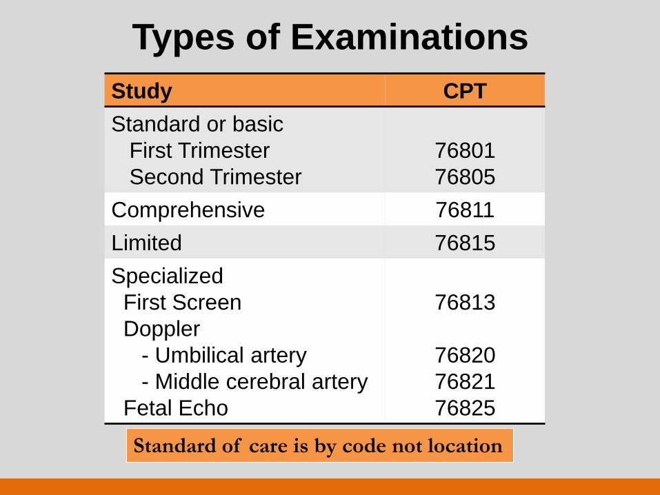
Types of Examinations
Study CPT
Standard or basic
First Trimester
Second Trimester
76801
76805
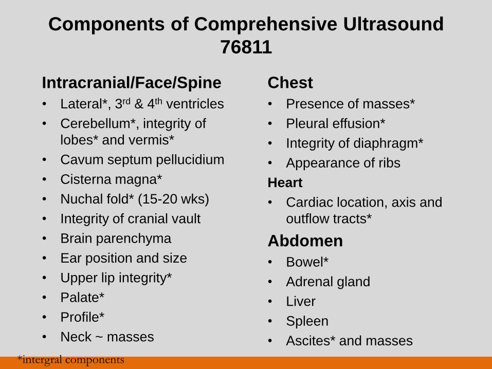
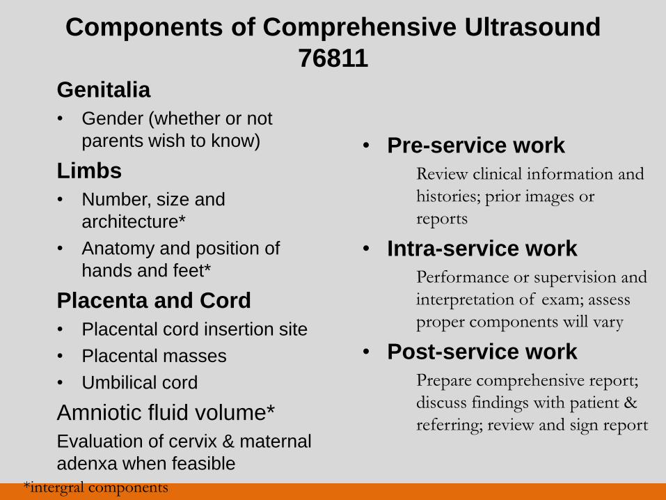
Comprehensive 76811
Limited 76815
Specialized
First Screen
Doppler
- Umbilical artery
- Middle cerebral artery
Fetal Echo
76813
76820
76821
76825
Standard of care is by code not location
Indications: 1st trimester
• Gestational dating
• Dx / evaluate mulit-fetal
• Confirm IUP
• Aneuploidy screening
• Evaluate ectopic
• Vaginal bleeding
• Assess pelvic pain
• Confirm cardiac activity
• Adjust embryo transfer
• CVS guidance
• Removal IUD
• Evaluate maternal pelvic,
uterine or adenxal
pathology
• Suspected hydatidiform
mole
ACOG Practice Bulletin 101, 2009,
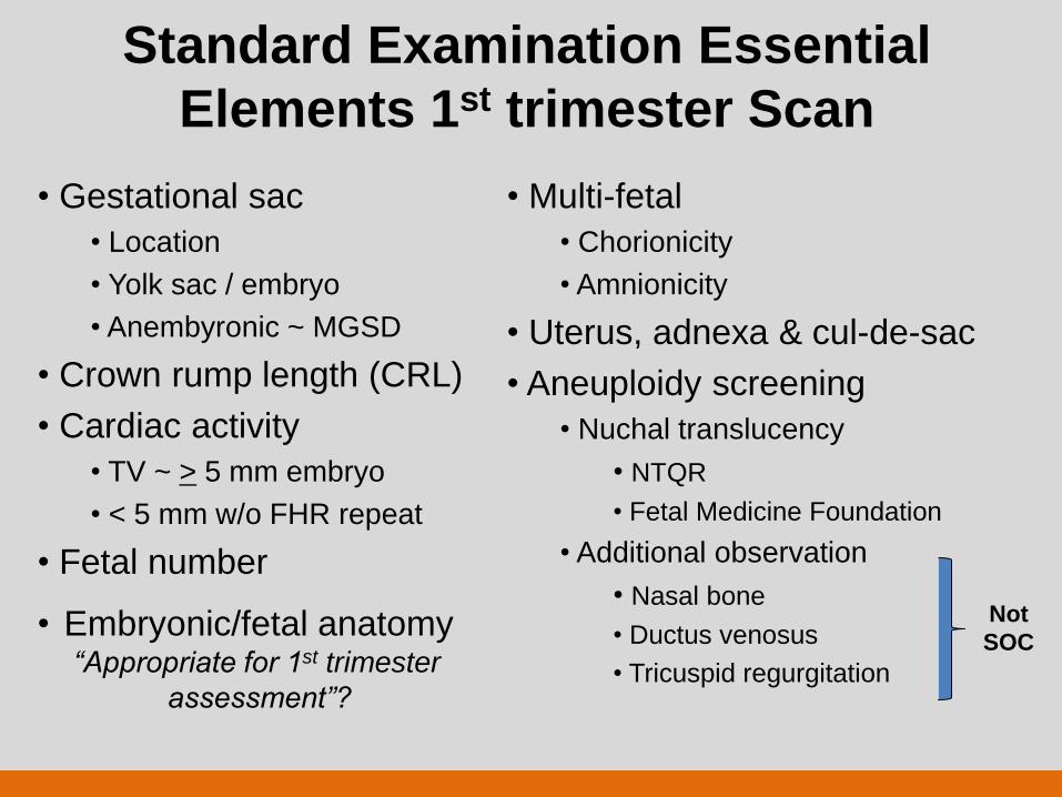
Standard Examination Essential
Elements 1st trimester Scan
• Gestational sac
• Location
• Yolk sac / embryo
• Anembyronic ~ MGSD
• Crown rump length (CRL)
• Cardiac activity
• TV ~ > 5 mm embryo
• < 5 mm w/o FHR repeat
• Fetal number
• Multi-fetal
• Chorionicity
• Amnionicity
• Uterus, adnexa & cul-de-sac
• Aneuploidy screening
• Nuchal translucency
• NTQR
• Fetal Medicine Foundation
• Additional observation
• Nasal bone
• Ductus venosus
• Tricuspid regurgitation
Not
SOC • Embryonic/fetal anatomy
“Appropriate for 1st trimester
assessment”?
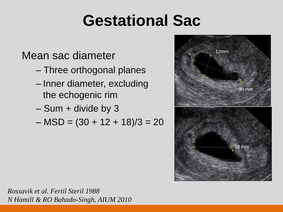
Gestational Sac
Mean sac diameter
– Three orthogonal planes
– Inner diameter, excluding
the echogenic rim
– Sum + divide by 3
– MSD = (30 + 12 + 18)/3 = 20
18 mm
12mm
30 mm
Rossavik et al. Fertil Steril 1988
N Hamill & RO Bahado-Singh, AIUM 2010
Gestational Sac
Linear growth early in
pregnancy
Rule of thumb – MSD( mm) + 30 = gestational
age (GA; days)
MSD = 20 ~ GA 50 days
Rossavik et al. Fertil Steril 1988
Dickey et al. Hum Reprod 1994
N Hamill & RO Bahado-Singh, AIUM 2010
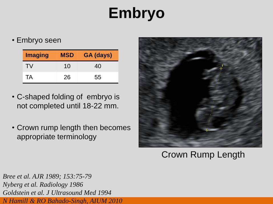
Embryo
• Embryo seen
• C-shaped folding of embryo is
not completed until 18-22 mm.
• Crown rump length then becomes
appropriate terminology
Bree et al. AJR 1989; 153:75-79
Nyberg et al. Radiology 1986
Goldstein et al. J Ultrasound Med 1994
N Hamill & RO Bahado-Singh, AIUM 2010
Imaging MSD GA (days)
TV 10 40
TA 26 55
Crown Rump Length
Cardiac Motion
Parameter + heart rate
Gestational age 37 days
MSD 18 mm
Embryo length (TV) 3-5 mm
Rempen et al. J Ultrasound Med 1990
N Hamill & RO Bahado-Singh, AIUM 2010
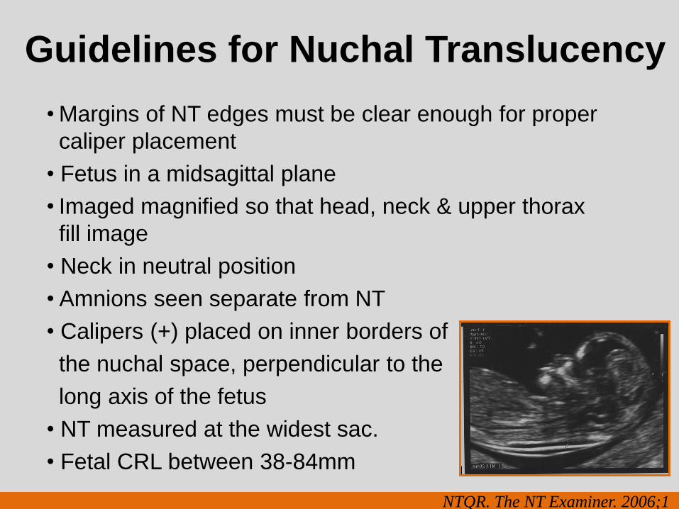
Guidelines for Nuchal Translucency
• Margins of NT edges must be clear enough for proper
caliper placement
• Fetus in a midsagittal plane
• Imaged magnified so that head, neck & upper thorax
fill image
• Neck in neutral position
• Amnions seen separate from NT
• Calipers (+) placed on inner borders of
the nuchal space, perpendicular to the
long axis of the fetus
• NT measured at the widest sac.
• Fetal CRL between 38-84mm
NTQR. The NT Examiner. 2006;1
First trimester ~ Anatomic Survey “Appropriate for 1st trimester assessment”
Nasal bone
4th ventricle CM/ICT
Orbits
Cerebellum
Falx
Choroid Plexus
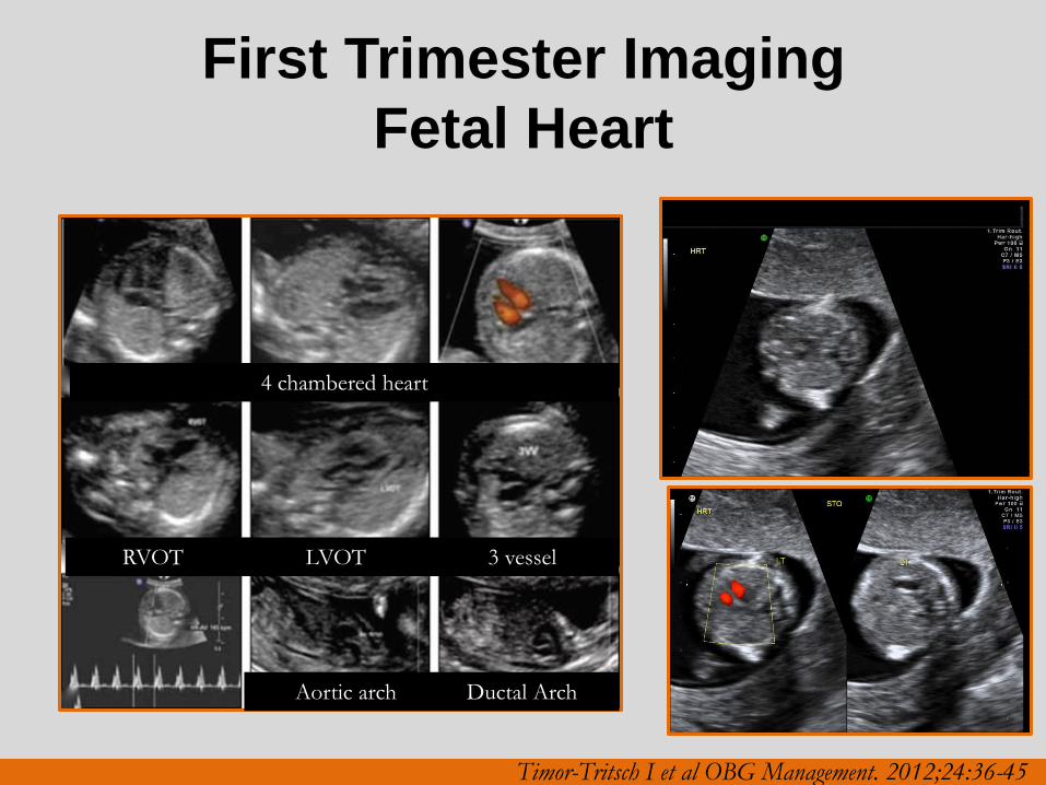
First Trimester Imaging
Fetal Heart
4 chambered heart
RVOT LVOT 3 vessel
Aortic arch Ductal Arch
Timor-Tritsch I et al OBG Management. 2012;24:36-45
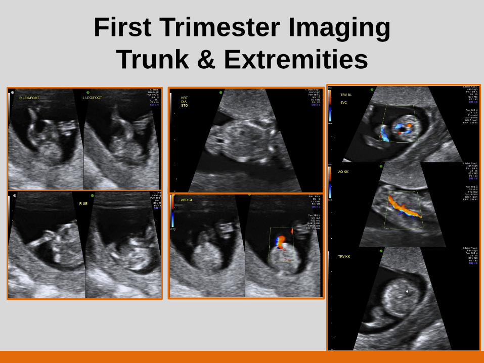
First Trimester Imaging
Trunk & Extremities
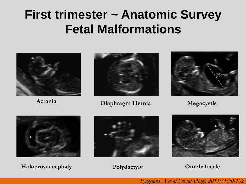
First trimester ~ Anatomic Survey
Fetal Malformations
Acrania
Holoprosencephaly
Diaphragm Hernia
Polydactyly
Megacystis
Omphalocele
Syngelaki A et al Prenat Diagn 2011;31:90-102
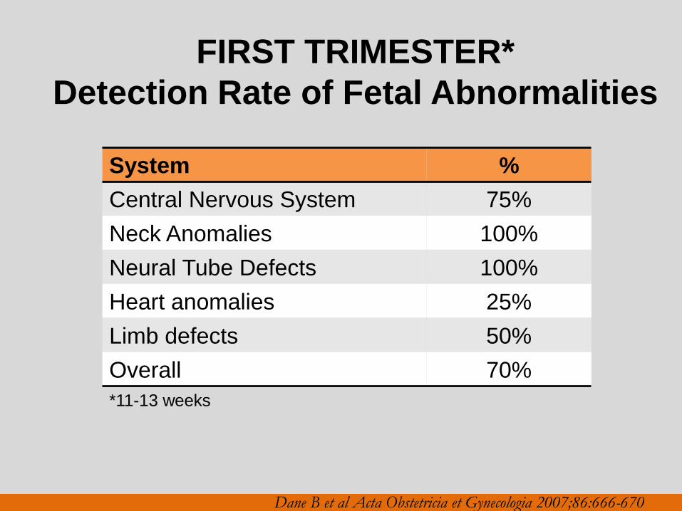
FIRST TRIMESTER*
Detection Rate of Fetal Abnormalities
System %
Central Nervous System 75%
Neck Anomalies 100%
Neural Tube Defects 100%
Heart anomalies 25%
Limb defects 50%
Overall 70%
Dane B et al Acta Obstetricia et Gynecologia 2007;86:666-670
*11-13 weeks
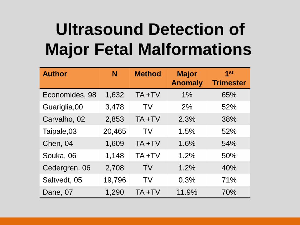
Ultrasound Detection of
Major Fetal Malformations
Author N Method Major
Anomaly
1st
Trimester
Economides, 98 1,632 TA +TV 1% 65%
Guariglia,00 3,478 TV 2% 52%
Carvalho, 02 2,853 TA +TV 2.3% 38%
Taipale,03 20,465 TV 1.5% 52%
Chen, 04 1,609 TA +TV 1.6% 54%
Souka, 06 1,148 TA +TV 1.2% 50%
Cedergren, 06 2,708 TV 1.2% 40%
Saltvedt, 05 19,796 TV 0.3% 71%
Dane, 07 1,290 TA +TV 11.9% 70%
Indications: 2nd/3rd trimester
•Gestational dating
•Fetal growth
•Vaginal bleeding
•Cervical insufficiency
•Abdominal/pelvic pain
•Fetal presentation
•Suspected multi-fetal
•PPROM or PTL
•Increase risk aneuploidy
•Fetal anomaly screening
•Adjust to procedures
•Size/dates discrepancy
•Evaluation pelvic mass
•Hydatidiform mole
•Ectopic pregnancy
•Uterine abnormality
•Fetal well-being
•Amniotic fluid abnormalities
•Placenta
•Abruption
•Location ~ Previa
•Implantation ~ previous C-sec
Standard Examination Essential Elements
2nd*/3rd trimester ultrasound (76805)
• Fetal presentation
• Amniotic fluid volume
• Cardiac activity (FHR)
• Placental position
• Fetal biometry
• Fetal number
• Anatomic survey*
• Maternal cervix and adnexa
> 18 weeks
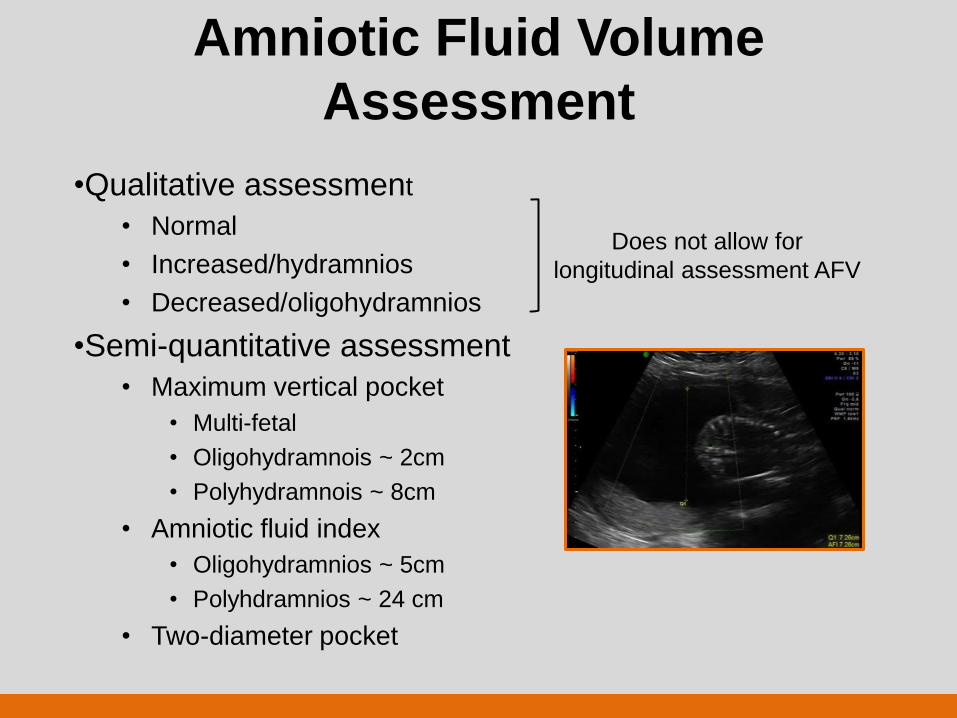
Amniotic Fluid Volume
Assessment
•Qualitative assessment
• Normal
• Increased/hydramnios
• Decreased/oligohydramnios
•Semi-quantitative assessment
• Maximum vertical pocket
• Multi-fetal
• Oligohydramnois ~ 2cm
• Polyhydramnois ~ 8cm
• Amniotic fluid index
• Oligohydramnios ~ 5cm
• Polyhdramnios ~ 24 cm
• Two-diameter pocket
Does not allow for
longitudinal assessment AFV
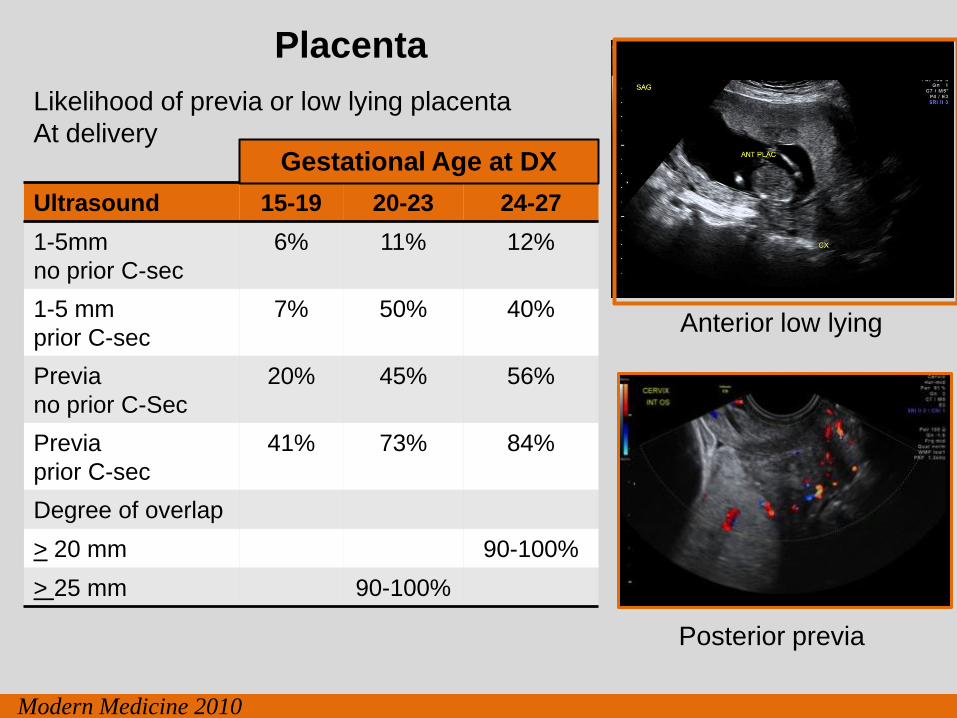
Placenta
Posterior previa
Anterior low lying
Ultrasound 15-19 20-23 24-27
1-5mm
no prior C-sec
6% 11% 12%
1-5 mm
prior C-sec
7% 50% 40%
Previa
no prior C-Sec
20% 45% 56%
Previa
prior C-sec
41% 73% 84%
Degree of overlap
> 20 mm 90-100%
> 25 mm 90-100%
Gestational Age at DX
Likelihood of previa or low lying placenta
At delivery
Modern Medicine 2010
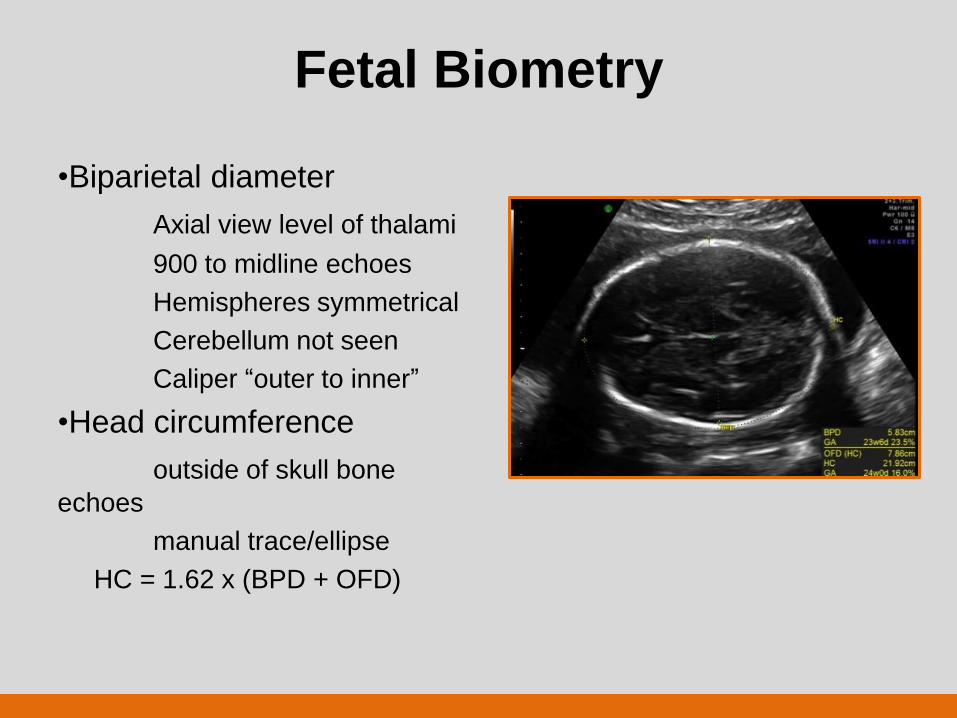
Fetal Biometry
•Biparietal diameter
Axial view level of thalami
900 to midline echoes
Hemispheres symmetrical
Cerebellum not seen
Caliper “outer to inner”
•Head circumference
outside of skull bone
echoes
manual trace/ellipse
HC = 1.62 x (BPD + OFD)
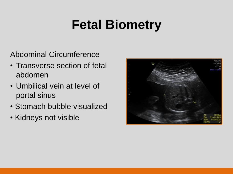
Fetal Biometry
Abdominal Circumference
• Transverse section of fetal
abdomen
• Umbilical vein at level of
portal sinus
• Stomach bubble visualized
• Kidneys not visible
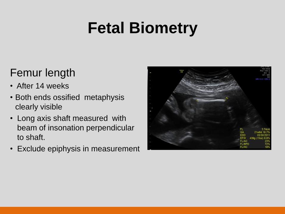
Fetal Biometry
Femur length • After 14 weeks
• Both ends ossified metaphysis
clearly visible
• Long axis shaft measured with
beam of insonation perpendicular
to shaft.
• Exclude epiphysis in measurement
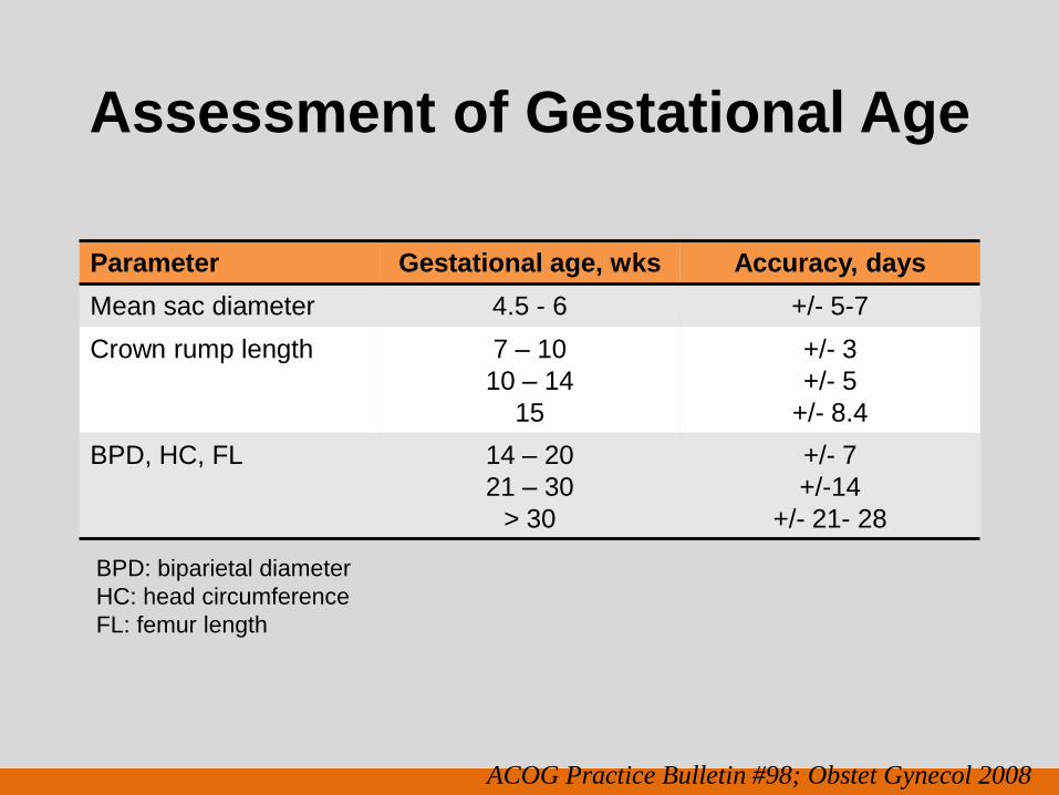
Assessment of Gestational Age
Parameter Gestational age, wks Accuracy, days
Mean sac diameter 4.5 - 6 +/- 5-7
Crown rump length
7 – 10
10 – 14
15
+/- 3
+/- 5
+/- 8.4
BPD, HC, FL 14 – 20
21 – 30
> 30
+/- 7
+/-14
+/- 21- 28
ACOG Practice Bulletin #98; Obstet Gynecol 2008
BPD: biparietal diameter
HC: head circumference
FL: femur length
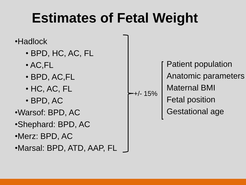
Estimates of Fetal Weight
•Hadlock
• BPD, HC, AC, FL
• AC,FL
• BPD, AC,FL
• HC, AC, FL
• BPD, AC
•Warsof: BPD, AC
•Shephard: BPD, AC
•Merz: BPD, AC
•Marsal: BPD, ATD, AAP, FL
Patient population
Anatomic parameters
Maternal BMI
Fetal position
Gestational age
+/- 15%
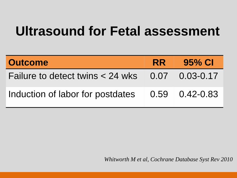
Ultrasound for Fetal assessment
Outcome RR 95% CI
Failure to detect twins < 24 wks 0.07 0.03-0.17
Induction of labor for postdates 0.59 0.42-0.83
Whitworth M et al, Cochrane Database Syst Rev 2010
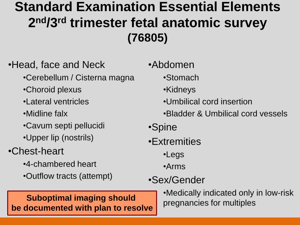
Standard Examination Essential Elements
2nd/3rd trimester fetal anatomic survey (76805)
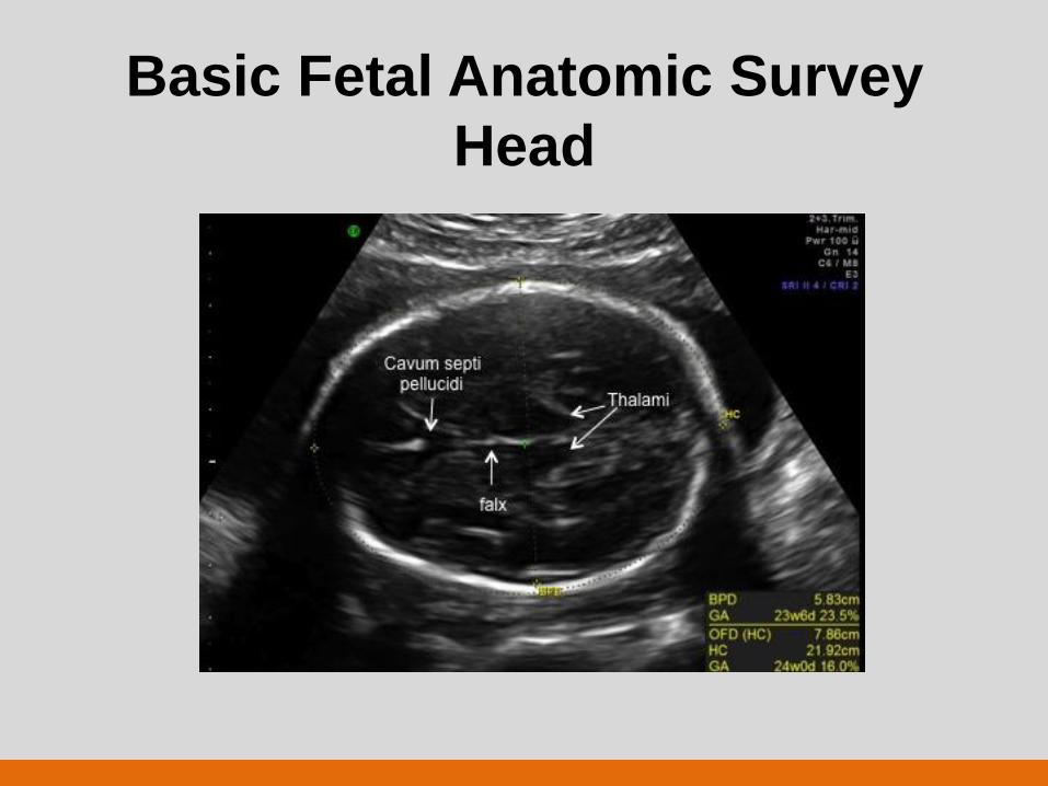
•Head, face and Neck
•Cerebellum / Cisterna magna
•Choroid plexus
•Lateral ventricles
•Midline falx
•Cavum septi pellucidi
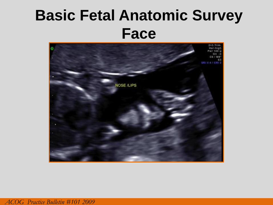
•Upper lip (nostrils)
•Chest-heart
•4-chambered heart
•Outflow tracts (attempt)
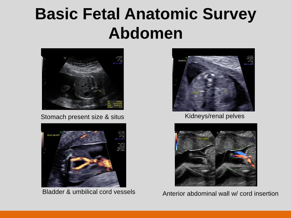
•Abdomen
•Stomach
•Kidneys
•Umbilical cord insertion
•Bladder & Umbilical cord vessels
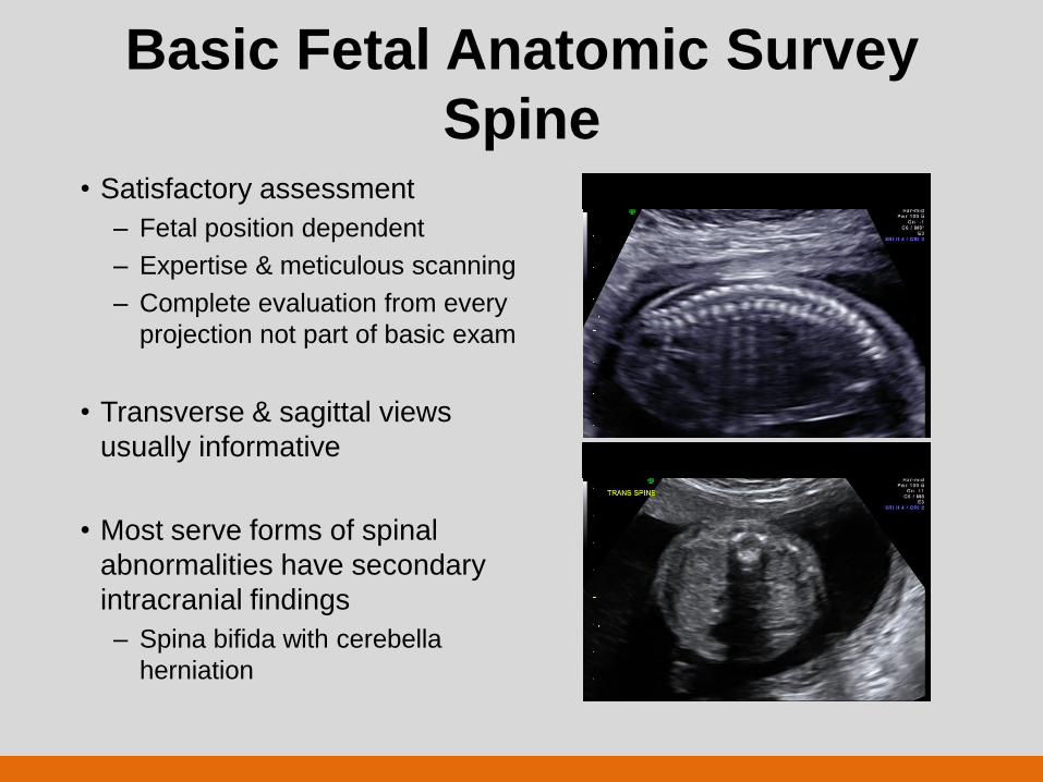
•Spine
•Extremities
•Legs
•Arms
•Sex/Gender
•Medically indicated only in low-risk
pregnancies for multiples
Suboptimal imaging should
be documented with plan to resolve
Basic Fetal Anatomic Survey
Head
Basic Fetal Anatomic Survey
Head
Basic Fetal Anatomic Survey
Face
ACOG Practice Bulletin #101 2009
Basic Fetal Anatomic Survey
Face
ISUOG Practice Guidelines Ultrasound Obstet Gynecol 2011
Basic Fetal Anatomic Survey
Face
ISUOG Practice Guidelines Ultrasound Obstet Gynecol 2011
Basic Fetal Anatomic Survey
Face
Midfacial hypoplasia
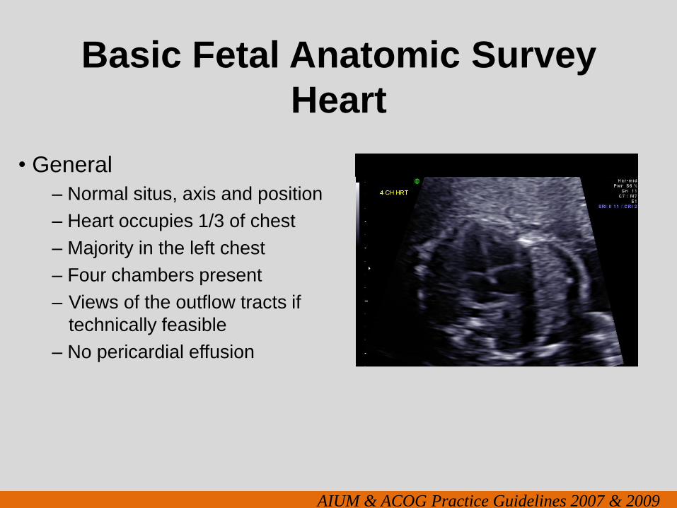
Basic Fetal Anatomic Survey
Heart
• General
– Normal situs, axis and position
– Heart occupies 1/3 of chest
– Majority in the left chest
– Four chambers present
– Views of the outflow tracts if
technically feasible
– No pericardial effusion
AIUM & ACOG Practice Guidelines 2007 & 2009
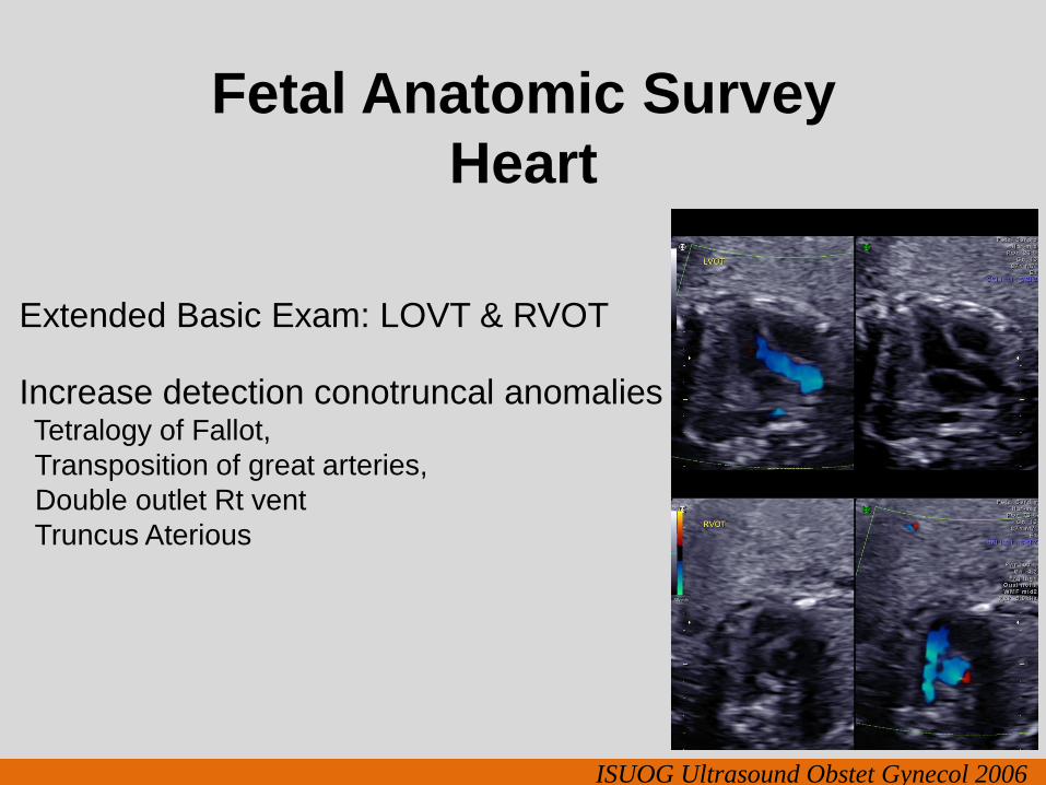
Fetal Anatomic Survey
Heart
Extended Basic Exam: LOVT & RVOT
Increase detection conotruncal anomalies Tetralogy of Fallot,