HTA Ultrasound screening in pregnancy: a systematic review of the clinical effectiveness, cost-effectiveness and women’s views L Bricker J Garcia J Henderson M Mugford J Neilson T Roberts M-A Martin Health Technology Assessment NHS R&D HTA Programme Health Technology Assessment 2000; Vol. 4: No. 16 Review
Transcript
HTA
Ultrasound screening in pregnancy:a systematic review of the clinicaleffectiveness, cost-effectivenessand women’s views
L BrickerJ GarciaJ HendersonM MugfordJ NeilsonT RobertsM-A Martin
Health Technology Assessment NHS R&D HTA Programme
Dr George Poste Chief Science & TechnologyOfficer, SmithKline Beecham
Professor Michael Rawlins Wolfson Unit of Clinical Pharmacology,University of Newcastle-upon-Tyne
Professor Martin Roland Professor of General Practice, University of Manchester
Professor Ian Russell Department of Health Sciences& Clinical Evaluation, University of York
Dr Charles Swan Consultant Gastroenterologist, North Staffordshire Royal Infirmary
* Previous Chair
Standing Group on Health Technology
Past members
Details of the membership of the HTA panels, the NCCHTA Advisory Group and the HTACommissioning Board are given at the end of this report.
Chair: Professor Kent WoodsProfessor of Therapeutics,University of Leicester
Professor Martin Buxton Director & Professor of Health Economics, Health Economics Research Group, Brunel University
Professor Shah EbrahimProfessor of Epidemiology of Ageing, University of Bristol
Professor Francis H CreedProfessor of Psychological Medicine,Manchester Royal Infirmary
Professor John Gabbay Director, Wessex Institute for Health Research & Development
Professor Sir John Grimley Evans Professor of Clinical Geratology, Radcliffe Infirmary, Oxford
Dr Tony Hope Clinical Reader in Medicine,Nuffield Department of Clinical Medicine, University of Oxford
Professor Richard Lilford Regional Director of R&D, NHS Executive West Midlands
Dr Jeremy Metters Deputy Chief Medical Officer,Department of Health
Professor Maggie PearsonRegional Director of R&D, NHS Executive North West
Mr Hugh Ross Chief Executive, The United Bristol Healthcare NHS Trust
Professor Trevor SheldonJoint Director, York HealthPolicy Group, University of York
Professor Mike SmithFaculty Dean of Research for Medicine, Dentistry,Psychology & Health, University of Leeds
Dr John Tripp Senior Lecturer in ChildHealth, Royal Devon and ExeterHealthcare NHS Trust
Professor Tom WalleyDirector, Prescribing Research Group,University of Liverpool
Dr Julie Woodin Chief Executive, Nottingham Health Authority
Current members
How to obtain copies of this and other HTA Programme reports.An electronic version of this publication, in Adobe Acrobat format, is available for downloading free ofcharge for personal use from the HTA website (http://www.hta.ac.uk). A fully searchable CD-ROM isalso available (see below).
Printed copies of HTA monographs cost £20 each (post and packing free in the UK) to both public andprivate sector purchasers from our Despatch Agents.
Non-UK purchasers will have to pay a small fee for post and packing. For European countries the cost is£2 per monograph and for the rest of the world £3 per monograph.
You can order HTA monographs from our Despatch Agents:
– fax (with credit card or official purchase order) – post (with credit card or official purchase order or cheque)– phone during office hours (credit card only).
Additionally the HTA website allows you either to pay securely by credit card or to print out yourorder and then post or fax it.
Contact details are as follows:HTA Despatch Email: [email protected]/o Direct Mail Works Ltd Tel: 02392 492 0004 Oakwood Business Centre Fax: 02392 478 555Downley, HAVANT PO9 2NP, UK Fax from outside the UK: +44 2392 478 555
NHS libraries can subscribe free of charge. Public libraries can subscribe at a very reduced cost of £100 for each volume (normally comprising 30–40 titles). The commercial subscription rate is £300 per volume. Please see our website for details. Subscriptions can only be purchased for the current orforthcoming volume.
Payment methods
Paying by chequeIf you pay by cheque, the cheque must be in pounds sterling, made payable to Direct Mail Works Ltdand drawn on a bank with a UK address.
Paying by credit cardThe following cards are accepted by phone, fax, post or via the website ordering pages: Delta, Eurocard,Mastercard, Solo, Switch and Visa. We advise against sending credit card details in a plain email.
Paying by official purchase orderYou can post or fax these, but they must be from public bodies (i.e. NHS or universities) within the UK.We cannot at present accept purchase orders from commercial companies or from outside the UK.
How do I get a copy of HTA on CD?
Please use the form on the HTA website (www.hta.ac.uk/htacd.htm). Or contact Direct Mail Works (seecontact details above) by email, post, fax or phone. HTA on CD is currently free of charge worldwide.
The website also provides information about the HTA Programme and lists the membership of the variouscommittees.
HTA
Ultrasound screening in pregnancy:a systematic review of the clinicaleffectiveness, cost-effectiveness and women’s views
L Bricker1*
J Garcia2
J Henderson2
M Mugford3
J Neilson4
T Roberts5
M-A Martin6
1Department of Obstetrics & Gynaecology, University of Liverpool, UK2National Perinatal Epidemiology Unit, Oxford, UK3University of East Anglia, UK4University of Liverpool, UK5Health Economics Facility, University of Birmingham, UK6Oxford, UK
*Corresponding author
Competing interests: none declared
Published September 2000
This report should be referenced as follows:Bricker L, Garcia J, Henderson J, Mugford M, Neilson J, Roberts T, et al. Ultrasound screening in pregnancy: a systematic review of the clinical effectiveness, cost-effectiveness and women’s views. Health Technol Assess 2000;4(16).
Health Technology Assessment is indexed in Index Medicus/MEDLINE and Excerpta Medica/EMBASE.Copies of the Executive Summaries are available from the NCCHTA website (see overleaf).
NHS R&D HTA Programme
The overall aim of the NHS R&D Health Technology Assessment (HTA) programme is to ensurethat high-quality research information on the costs, effectiveness and broader impact of health
technologies is produced in the most efficient way for those who use, manage and work in the NHS.Research is undertaken in those areas where the evidence will lead to the greatest benefits topatients, either through improved patient outcomes or the most efficient use of NHS resources.
The Standing Group on Health Technology advises on national priorities for health technologyassessment. Six advisory panels assist the Standing Group in identifying and prioritising projects.These priorities are then considered by the HTA Commissioning Board supported by the NationalCoordinating Centre for HTA (NCCHTA).
This report is one of a series covering acute care, diagnostics and imaging, methodology,pharmaceuticals, population screening, and primary and community care. It was identified as apriority by the Population Screening Panel and funded as project number 93/30/03.
The views expressed in this publication are those of the authors and not necessarily those of theStanding Group, the Commissioning Board, the Panel members or the Department of Health. Theeditors wish to emphasise that funding and publication of this research by the NHS should not betaken as implicit support for the recommendations for policy contained herein. In particular, policyoptions in the area of screening will be considered by the National Screening Committee. ThisCommittee, chaired by the Chief Medical Officer, will take into account the views expressed here,further available evidence and other relevant considerations.
Reviews in Health Technology Assessment are termed ‘systematic’ when the account of the search,appraisal and synthesis methods (to minimise biases and random errors) would, in theory, permitthe replication of the review by others.
Criteria for inclusion in the HTA monograph series Reports are published in the HTA monograph series if (1) they have resulted from work either prioritised by theStanding Group on Health Technology, or otherwise commissioned for the HTA Programme, and (2) they are ofa sufficiently high scientific quality as assessed by the referees and editors.
Series Editors: Andrew Stevens, Ken Stein and John GabbayMonograph Editorial Manager: Melanie Corris
The editors and publisher have tried to ensure the accuracy of this report but do not accept liabilityfor damages or losses arising from material published in this report. They would like to thank thereferees for their constructive comments on the draft document.
Enquiries relating to copyright should be addressed to the NCCHTA (see address given below).
Published by Core Research, Alton, on behalf of the NCCHTA.Printed on acid-free paper in the UK by The Basingstoke Press, Basingstoke.
Copies of this report can be obtained from:
The National Coordinating Centre for Health Technology Assessment,Mailpoint 728, Boldrewood,University of Southampton,Southampton, SO16 7PX, UK.Fax: +44 (0) 23 8059 5639 Email: [email protected]://www.ncchta.org
List of abbreviations .................................... i
Executive summary ..................................... iii
2 Systematic review of the clinicaleffectiveness of routine ultrasoundIntroduction ................................................... 3Systematic reviews of randomised trials ....... 3
3 Detection of fetal abnormalities by routineultrasoundIntroduction .................................................. 7Methods ......................................................... 7Results ............................................................ 8Discussion ...................................................... 20Recommendations for research ................... 28Implications for policy and practice ............ 28
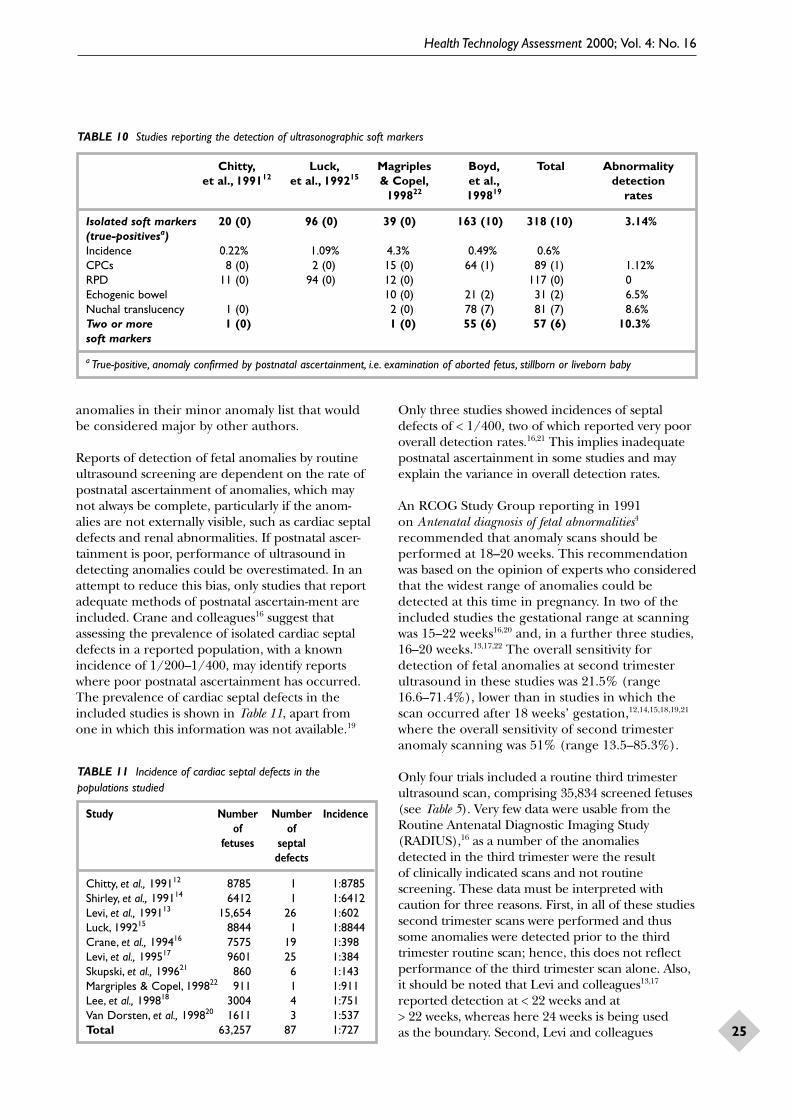
4 Other clinical aspects of routine ultrasound in pregnancyUltrasonographic soft markers ..................... 29First trimester ultrasound screening for fetalchromosomal and structural abnormalities 32
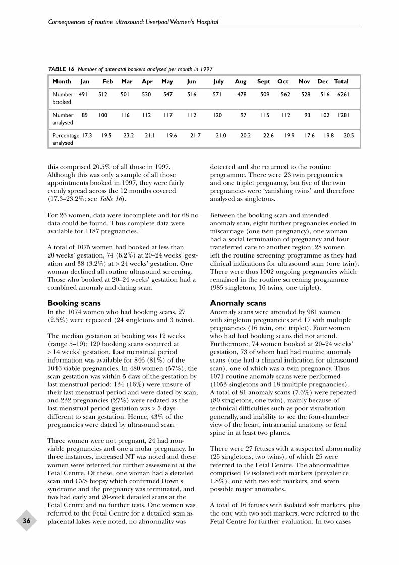
5 Consequences of routine ultrasound:Liverpool Women’s HospitalIntroduction .................................................. 35Objectives ....................................................... 35Methods ......................................................... 35Results ............................................................ 35Discussion ...................................................... 37Recommendations for research ................... 39Implications for practice .............................. 39
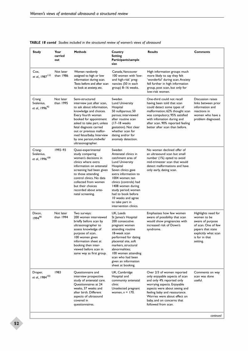
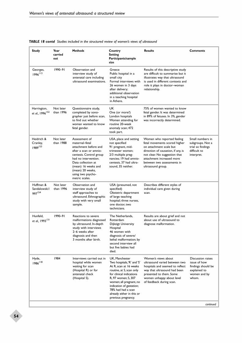
6 Women’s views of antenatal ultrasound:a structured reviewIntroduction .................................................. 41Methods ......................................................... 41Results ............................................................ 42Conclusions .................................................... 48Recommendations for research ................... 48Implications for practice ............................... 48
7 Introduction to costs and cost-effectivenessof routine ultrasound in pregnancy .......... 63
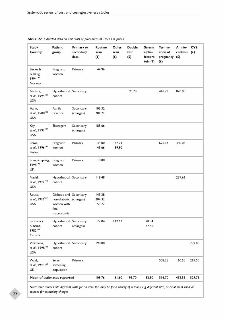
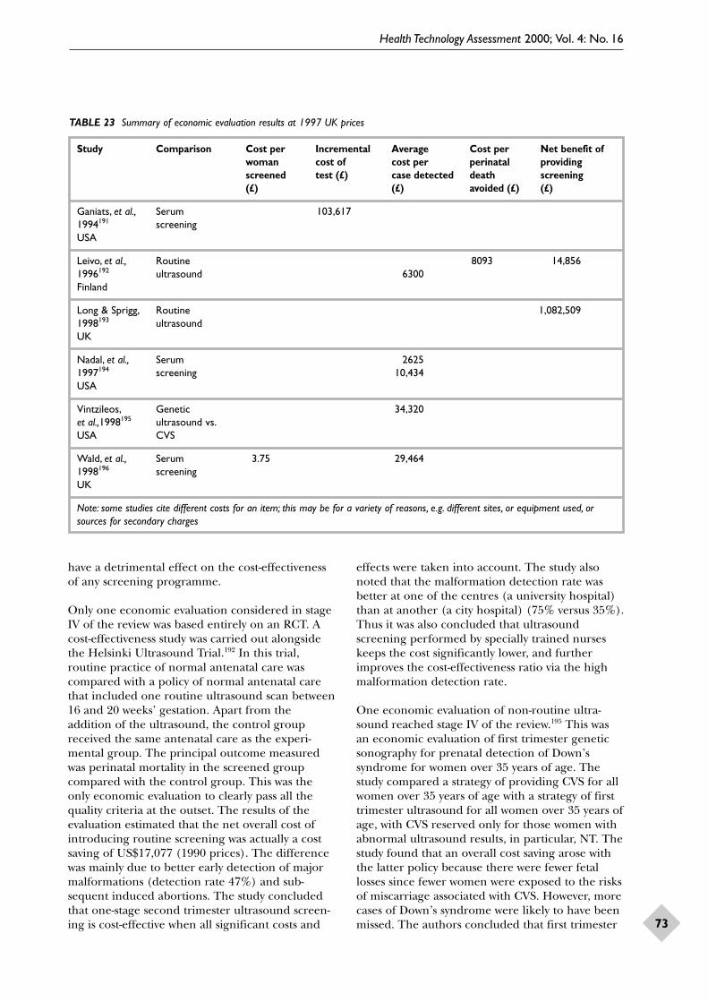
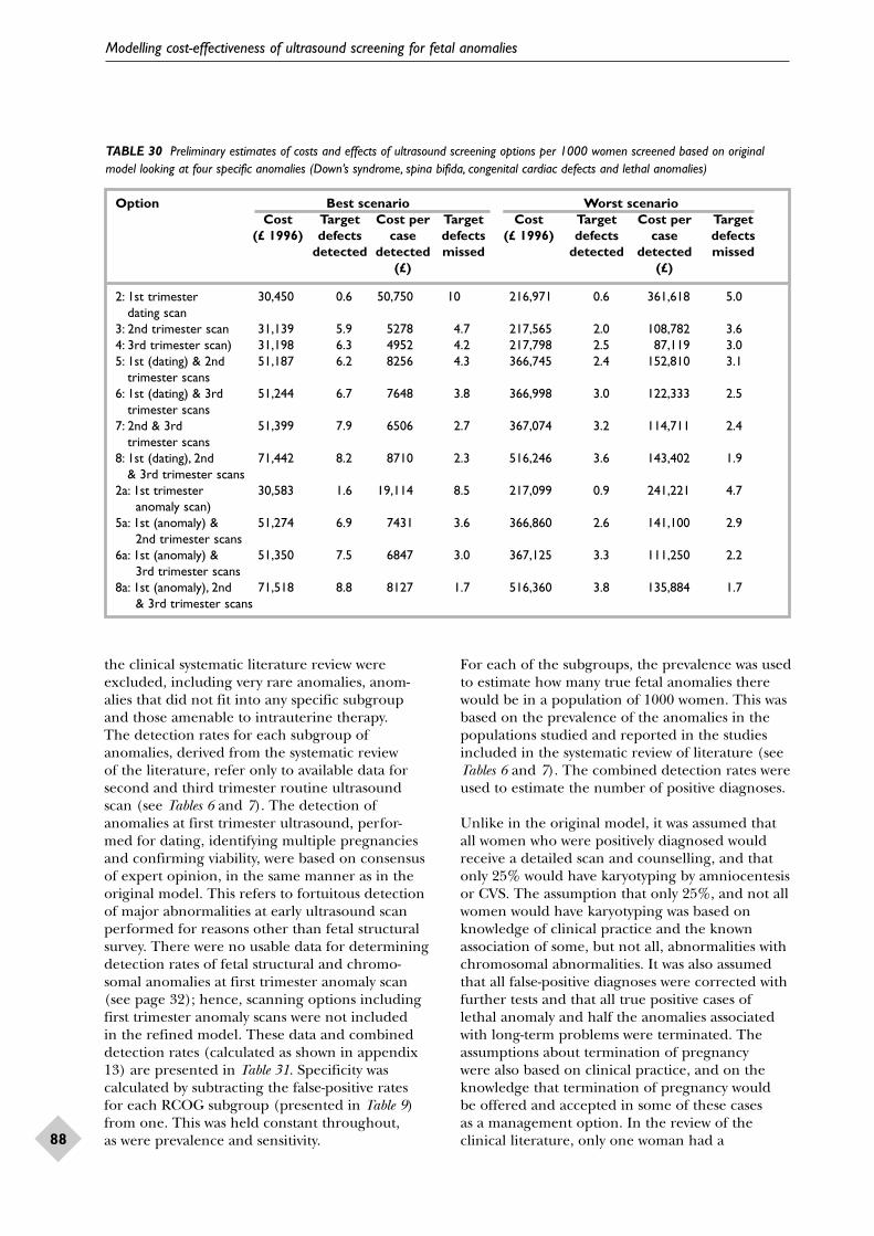
8 Systematic review of costs andcost-effectiveness studiesIntroduction .................................................. 65Methods ......................................................... 65Results ............................................................ 67Discussion ...................................................... 75Recommendations for research ................... 75Implications for policy and practice ............ 75
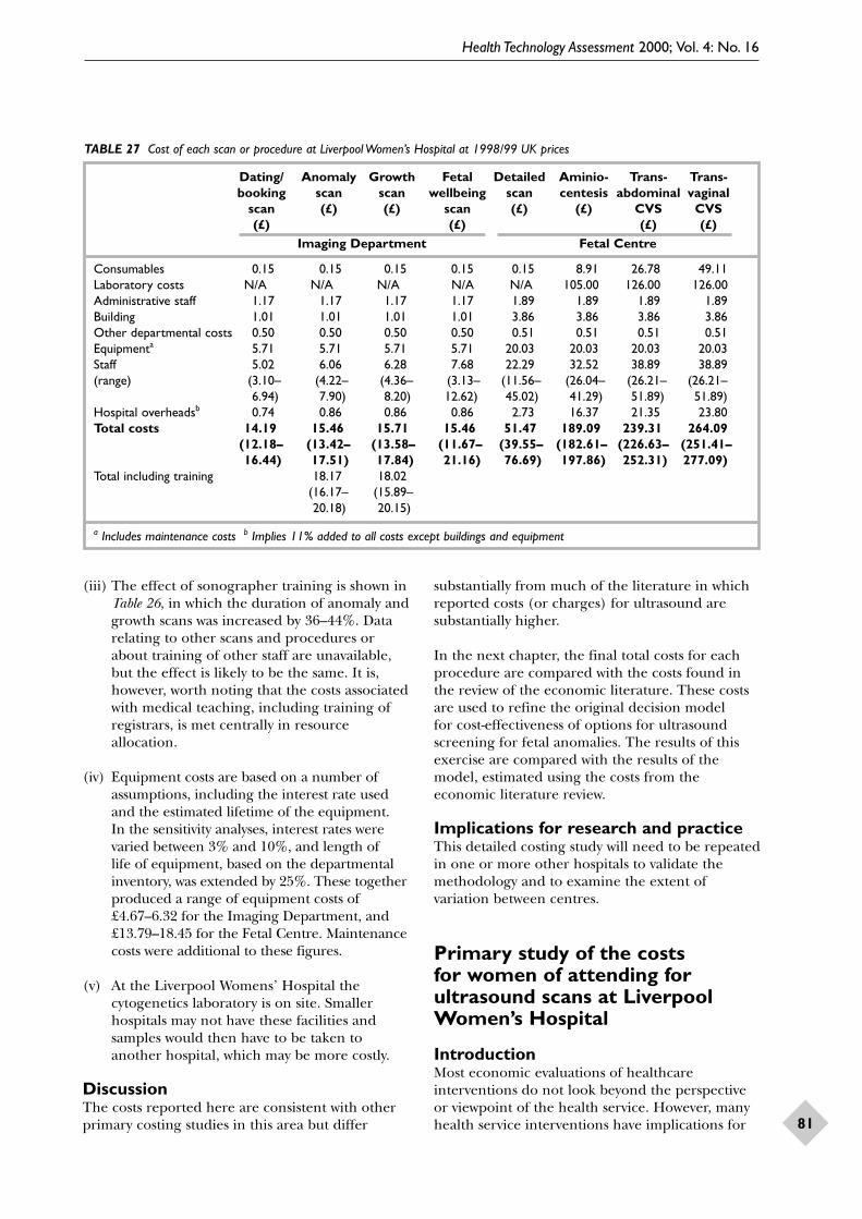
9 Primary studies of costs of ultrasoundResource use and costs of procedures associated with routine ultrasound screening carried out at Liverpool Women’s Hospital ......................................... 77Primary study of the costs for women of attending for ultrasound scans at Liverpool Women’s Hospital ........................ 81
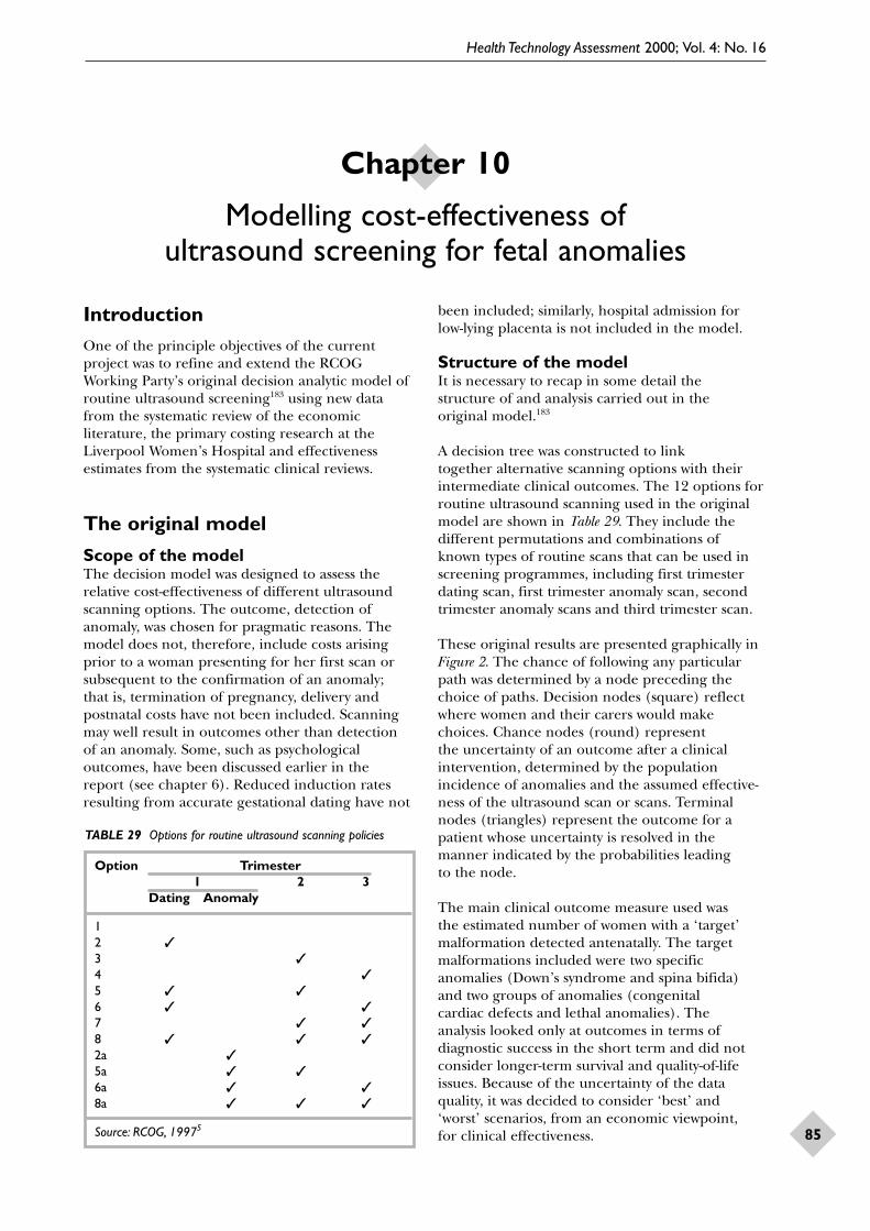
10 Modelling cost-effectiveness of ultrasoundscreening for fetal anomaliesIntroduction .................................................. 85The original model ....................................... 85Methods ......................................................... 87Results ............................................................ 91Discussion ...................................................... 97Recommendations for research ................... 102
11 Evidence for cost-effectiveness of ultrasound in pregnancy ............................. 103
12 Authors’ synthesis and comments on the reviewIntroduction .................................................. 105Overview of findings ..................................... 105Methods of the review ................................... 107Policy relevance and further research ......... 108Conclusions about further research ............ 109
Appendix 4 Search strategy for the review of the detection of fetal abnormalities by routine ultrasound .............................................. 157
Appendix 5 Data extraction sheet for the review of the detection of fetal abnormalities by routine ultrasound ......................................... 159
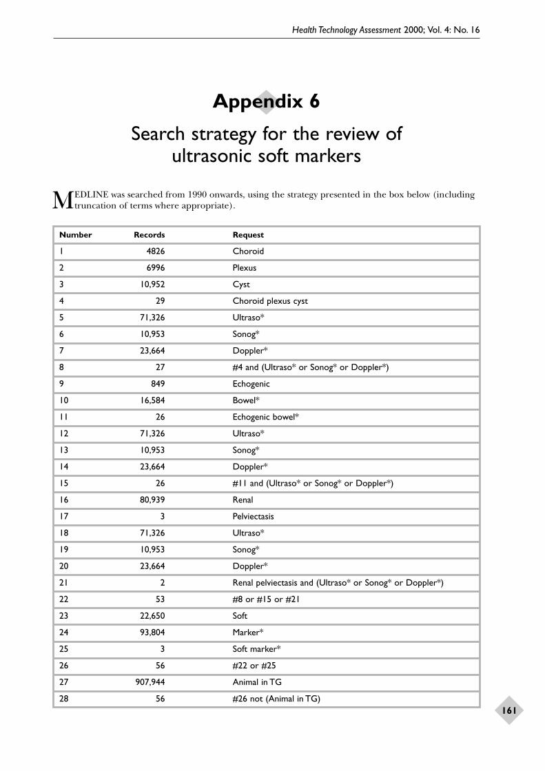
Appendix 6 Search strategy for the review of ultrasonic soft markers ................................... 161
Appendix 7 Terms for finding references onwomen’s views of ultrasound screening ............ 163
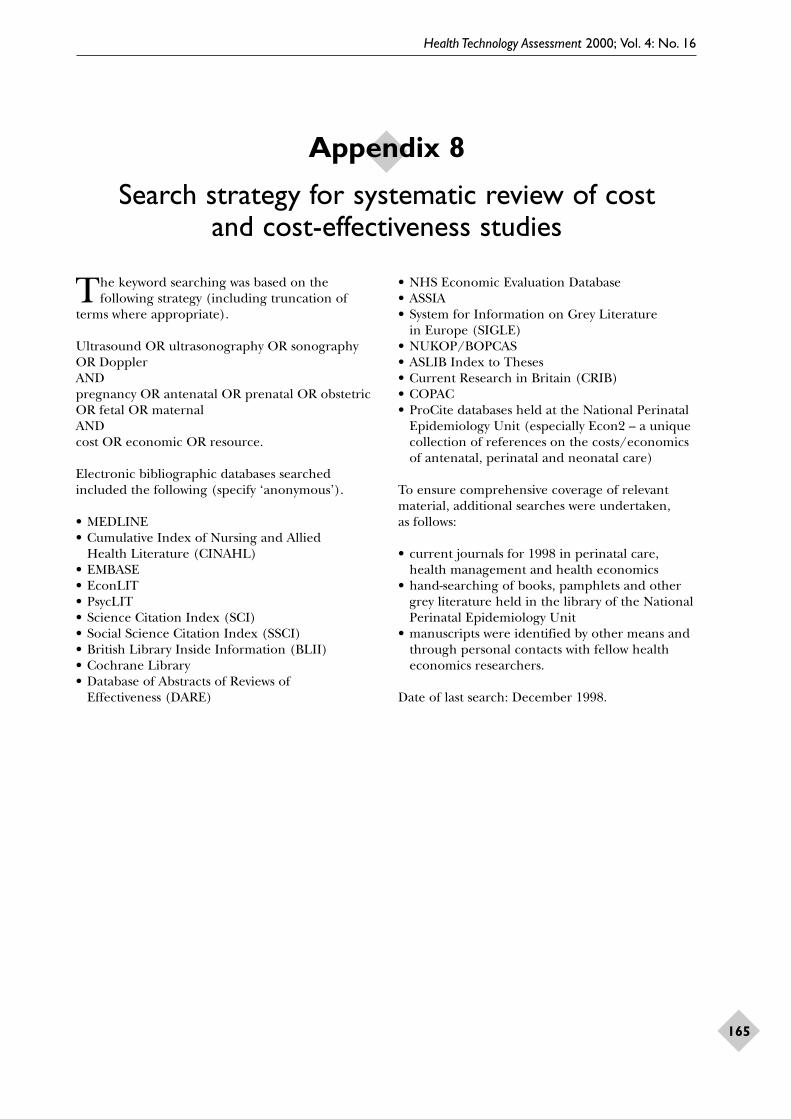
Appendix 8 Search strategy for the systematicreview of cost and cost-effectiveness studies ..... 165
Appendix 9 Data extraction sheet for systematicreview of costs and cost-effectiveness studies .... 167
Appendix 10 Staff diary for time scanning ..... 171
Appendix 11 Data sheet for scans/procedures performed at the Fetal Centre,Liverpool Women’s Hospital .............................. 173
Appendix 12 Patient information andquestionnaire for survey of women’s costs of ultrasound scans ............................................. 175
Appendix 13 Combining detection rates from scans at different times ............................. 179
Appendix 14 National Screening Committee’scriteria for appraising the viability, effective-ness and appropriateness of a screeningprogramme: with comments in the light of this study .............................................................. 181
Health Technology Assessment reportspublished to date .............................................. 185
Health Technology Assessmentpanel membership ............................................ 189
Health Technology Assessment 2000; Vol. 4: No. 16
i
List of abbreviations
ASD Arial septal defect*
AVSD atrioventricular septal defect*
CAML congenital adenomatous malformation of the lung*
CDH congenital diaphragmatic hernia*
CI confidence interval
CNS central nervous system
CPC choroids plexus cyst
CVS chorionic villus sampling
GP general practitioner
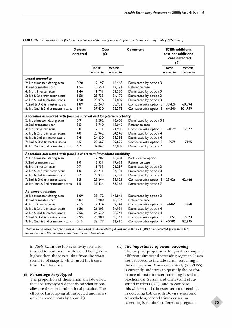
ICER incremental cost-effectiveness ratios
ICU intensive care unit*
MeSH medical subject headings
NT nuchal translucency
OR odds ratio
RADIUS Routine Antenatal Diagnostic Imaging Study
RCOG Royal College of Obstetricians and Gynaecologists
RCR Royal College of Radiologists
RCT randomised controlled trial
RPD renal pelvic dilatation
SD standard deviation
SURUSS Serum, Urine and Ultrasound Screening Study
T1, 2, 3 first, second and third trimesters*
VSD ventricular septal defect*
WMD weighted mean difference*
* Used only in tables and figures
Health Technology Assessment 2000; Vol. 4: No. 16
iii
Objectives
• To update the pre-existing Cochrane review of ultrasound for routine fetal assessment inearly pregnancy.
• To compile new Cochrane reviews of– routine ultrasound in late pregnancy– routine Doppler® ultrasound in pregnancy.
• To review the literature on the detection of fetalabnormalities by ultrasound screening exam-inations during pregnancy.
• To conduct a primary study to assess theconsequences of a routine two-stage ultra-sound regimen in pregnancy in a teachinghospital (clinical pathways).
• To compile literature reviews of (a) women’sviews on undergoing routine ultrasound exam-ination and (b) estimates of costs and cost-effectiveness of routine ultrasound examinations.
• To conduct a primary study of costs of a routinetwo-stage ultrasound regimen in early or mid-pregnancy in a UK teaching hospital.
• To refine and update a decision model of cost-effectiveness of options for routine scanning for fetal anomalies.
Methods
Full details of search strategies for systematicreviews are in the appendices. Other methods are described in individual sections of the fullreport, as are methods for the primary studies of clinical pathways and costs.
Results
Routine ultrasound before 24 weeks:• leads to earlier diagnosis of multiple
pregnancies but has not been shown to have an important positive impact on the outcome of multiple pregnancies
• is associated with fewer inductions of labour for ‘post-term’ pregnancy
• reduces perinatal mortality rate if detection offetal malformations is an important objective anda high level of diagnostic expertise exists and iftermination of pregnancy for fetal abnormality iswidely accepted in the population screened.
Routine ultrasound after 24 weeks:• has not been shown to confer any clear benefit
to mother or baby, except that assessment ofplacental appearances may, as an adjunct to fetalmeasurement, help reduce perinatal mortality.
Routine Doppler ultrasound in pregnancy:• has not been shown to be of benefit and may
even increase the risk of adverse outcome.
Detection of fetal abnormality by screeningultrasound examinations:• detection rates vary with the organ system
affected, with generally high rates of detectionof abnormalities of the CNS, and low rates forskeletal and cardiac abnormalities
• similar variations are seen at both second andthird trimester examinations
• data on the value of first trimester anomalyscreening are lacking.
Clinical pathways:• largely unrecognised consequences of routine
ultrasound examinations exist that have healthservice resource implications as well as thepotential to alarm women. Specifically:– 2.5% of booking scans are repeated– 7.6% of anomaly scans are repeated
• women present for antenatal booking atdifferent gestations; hence, the coverage of any one scan regimen may be incomplete.
Women’s views• Ultrasound is very attractive to women and
partners; this may be because it provides earlyvisual confirmation of pregnancy and contact withtheir babies, and reassures about fetal well-being.
• Such features may augment the potential foranxiety, shock and disappointment when thescan shows a problem. Recent changes in theuse of ultrasound may lead to more findings of uncertain clinical significance, which is likely to have important psychological and social consequences for women.
• Women’s earlier fears, that ultrasound mightharm the fetus, do not feature in later research,although this may be partly due to researchersnot asking about fears.
• Reports of a reduction in anxiety after ultrasound examination are likely to reflect
Executive summary
iv
increased anxiety before the scan rather than areal benefit of ultrasound.
• There is no reliable evidence of reducedsmoking or any other positive health behaviouras a consequence of routine ultrasound.
• Trials comparing ultrasound with no ultra-sound have not considered its psychological or social impact on both parents and babies.
Costs and cost-effectivenessLiterature review• There are few good quality economic
evaluations and primary cost studies ofultrasound scanning in pregnancy. Only oneeconomic evaluation conducted alongside an RCT was included in the review.
• Routine scanning in the second trimester wasshown to be relatively cost-effective.
• The skill of ultrasonographers in detectinganomalies and the time taken to perform a scan have a significant effect on the relative cost-effectiveness.
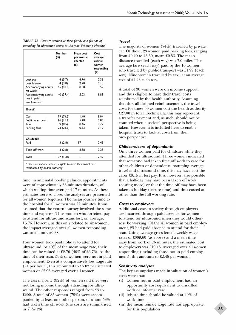
Primary costing study• Costs to women of attending ultrasound
examinations were significant compared withNHS service costs.
• It is important to include women’s costs ineconomic evaluation of routine ultrasoundscreening, particularly where cost shifting mayoccur, because any change in the provision ofroutine ultrasound may shift the costs away from the provider on to women and theirfamilies and influence attendance.
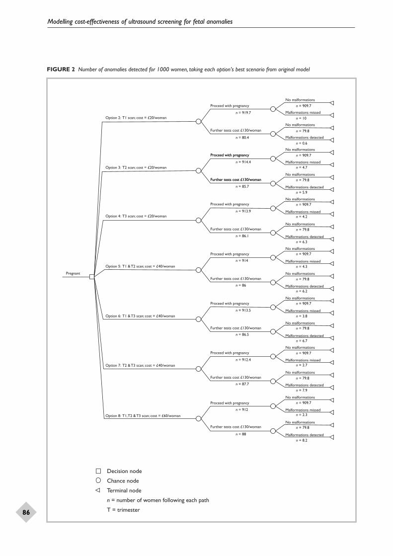
Decision-analysis modelling• The initial eight options considered were
reduced to three dominating options:– one second trimester scan alone– one third trimester scan alone– a combination of one second trimester scanfollowed by one third trimester scan.
• More representative cost data are requiredbefore precise estimates of the additional costsand benefits of alternative screening options can be determined.
• One second trimester scan emerged as a clearreference case, being one of the cheapestoptions yet still detecting a significant numberof anomalies.
• When termination is acceptable and available, a third trimester scan alone or the combinationof one second with one third trimester scan,although comparable in economic terms, maybe impractical because of the delay inidentifying anomalies.
• The interaction of an anomaly scan(s) with a first trimester scan for dating purposes was not assessed.
Conclusions
Implications for policy and practice• There is evidence that routine ultrasound in
early pregnancy provides:(i) better gestational age assessment(ii) earlier detection of multiple pregnancies(iii) detection of clinically unsuspected fetal
malformation at a time when termination of pregnancy is possible. These effects havenot been shown to improve ultimate fetaloutcome. No convincing evidence ofbenefit from routine examination in latepregnancy (> 24 weeks) was found, whetherusing imaging or Doppler ultrasound.
• Clinicians, women and health planners need to decide if these effects are sufficient to justifyroutine ultrasound. Clinicians in the UK seemconvinced of the benefits, given the very wide-spread use of the technique. As seen from thesystematic review of women’s views, imaging ispopular with women (provided the appearanceof the baby is normal). The study in Liverpoolindicates that the average cost to the hospital ofproviding a 20-week anomaly scan is £15. Thisseems modest in the UK but will be prohibitivelyhigh in many developing countries.
• If routine ultrasound is to be offered before 24 weeks, what timing is optimal? The RoyalCollege of Obstetricians and Gynaecologists’(RCOG) Working Party report of 1997recommended a two-stage regimen of bookingultrasound at about 12 weeks, followed by asecond ultrasound anomaly scan at 20 weeks –the regimen offered at Liverpool Women’sHospital. When this report was initially drafted,no comparative information was available aboutthe clinical impact of different regimens. Sincethen, an RCT comparing the two-stage regimenwith a 20-week scan alone has demonstrated less need for readjustment of dates at the mid-pregnancy scan in the two-stage group (withpossible consequences for timing serum screen-ing, if available) and less anxiety among thewomen. Again, clinicians, women and healthplanners have to decide whether such benefitsjustify the costs.
• The systematic review of the effectiveness ofanomaly detection has highlighted substantialvariation in, and limits to, detection rates ofcertain structural abnormalities. Thisinformation should be made available to
Executive summary
Health Technology Assessment 2000; Vol. 4: No. 16
v
clinicians and women, and may also be relevantto the medico-legal arena. Given these limits,the RCOG Working Party’s recommendations,that ultrasound examinations should beconducted only by appropriately trainedpersonnel and using equipment no more than 5 years old, seem appropriate. Quality controlmechanisms should be set in place to auditperformance. The system of reporting suspectedanomalies to regional fetal anomaly registersshould be encouraged where these exist.
• A number of inefficiencies in the routineultrasound screening programme wereidentified (including the need for repeat scans and that not all women book at earlygestations), some of which are unavoidable, but which have implications for both its clinicaland cost-effectiveness.
Research recommendationsWithin each category below, the researchrecommendations are prioritised.
Guidelines on research methodsAll future work evaluating uses of ultrasound inpregnancy should take account of the followingmethodological points.
• Published reports from clinical departments of detection rates of fetal abnormalities byultrasound screening may not represent general standards. General detection ratesshould be assessed by linkage with high-ascertainment fetal abnormality registers at a regional level.
• Reporting of costs and cost-effectiveness ofroutine ultrasound screening should takeaccount of recommended standards foreconomic evaluation.
• New or extended uses for pregnancy ultra-sound should be evaluated in psychological and social, as well as healthcare efficiency andclinical terms.
• Studies of women’s views of ultrasound, clinicaleffectiveness and costs of technologies shouldreport the date and place of the research anddescribe the clinical contexts and purposes for which ultrasound was used for thoseresearch participants.
Priorities for researchEffectiveness of newer applications of ultra-sound screening and alternative forms of care
Some forms of ultrasound screening are beingintroduced into routine practice without strongevidence on effectiveness; others are promising but need more evaluation.
• Nuchal translucency scanning and other types ofultrasound screening for anomalies during thefirst trimester of pregnancy are topical andcontroversial issues in obstetric care. None ofthe limited number of reports on these topicsmet our criteria for inclusion in systematicreviews and have therefore not been consideredin detail. Researchers should be encouraged tostudy rigorously not only the effectiveness ofdetection of anomalies but also adverse clinicalsequelae, psychological impact on women andtheir partners, and economic consequences.Until these data are available, the evidence doesnot support screening in the clinical service.
• More representative data are required on theclinical and psychological effects and costimplications of first trimester anomaly scanning.
• The possible value of routine mid-pregnancyuterine Doppler ultrasound to predict pre-eclampsia, intrauterine growth restriction andother adverse outcomes should be assessed inrandomised trials.
• A single trial has suggested that placentaltexture grading during the third trimester may be helpful; this merits further study.
Documenting current practice, clinicalpathways, costs and outcomes
In order to develop relevant guidance for the NHS, more needs to be known about current practice.
• Research is needed to assess the effects and costs of detection of fetal abnormalitiesamenable to in-utero intervention and neonatalsurgery on substantive outcomes, such as short-and long-term morbidity and mortality for both mother and child, including parentalpsychological consequences.
• The findings of the primary studies of costs and clinical pathways undertaken to augmentanticipated gaps in knowledge in this reviewneed to be repeated and validated in other settings.
• Further evaluation is required on the impact of changes in routine antenatal care practiceand its influence on family economy, clinicalattendance or healthcare efficiency.
Defining options for screening
Developments in ultrasound technology provideinformation with uncertain implications.
• There is continuing controversy about thesignificance of ultrasound ‘soft markers’ andtheir relationship to, in particular, chromo-somal abnormalities. There should be ongoing
Executive summary
vi
clinical research into the significance andimplications of detection of all sonographic soft markers in unselected and low-riskpopulations. These findings should beinterpreted in the light of other screeningprogrammes for chromosomal abnormalities(e.g. biochemical screening).
Ethical and cultural issues
Current practice is not based on a strong basis ofknowledge of women’s needs and understandingof ultrasound.
• Ways of improving women’s understanding ofthe information gained from ultrasound shouldbe developed and evaluated.
• There is scope for further investigation into thevalues women attach to their own time and toattending for a scan in different circumstances.
• Comparative research into the ways in whichprenatal ultrasound is carried out and exper-ienced in different countries and cultures would be valuable.
Cost-effectiveness
This is not constant over time and regularupdating of models should be based on research as recommended above.
• Further development of economic models of cost-effectiveness of ultrasound screening in pregnancy should include assessing theeffects of including a first or second trimesterdating scan, and considering longer-termconsequences and changing evidence on technologies, effectiveness and outcomes.
Health Technology Assessment 2000; Vol. 4: No. 16
1
There has been little debate, rightly or wrongly,about the usefulness of ultrasound exam-
ination in those clinical situations in which thereare clear reasons to suppose that such an investi-gation might provide important information whichwould complement clinical assessment. The manyexamples would include:
• antepartum haemorrhage, primarily to identifyor exclude placenta praevia
• clinical suspicion that a fetus was small forgestational age, because of the well-recognisedperinatal risks that can be avoided by early delivery
• polyhydramnios, because the excessive amountof amniotic fluid may result from a structuralmalformation in the fetus.
What is much more controversial is the routine useof ultrasound in all pregnancies, a procedure thathas become standard practice in many countries.The use of ultrasound in the UK is not routinelydocumented in NHS statistics and so the onlysource of evidence on current practice is fromsurveys and clinical audit. Two national studies ofpregnancy ultrasound use in the UK have beenfound.1,2 Both found that women are offered atleast one scan in pregnancy. The Royal College ofObstetricians and Gynaecologists (RCOG)/RoyalCollege of Radiologists (RCR) survey2 found that77% of hospitals offered a routine dating scan,while 82% offered a second trimester anomaly scan(at 18–20 weeks). Only 5% of departments under-took an additional routine third trimester scan.
An RCOG Working Party suggested in 19843
that, in the UK, a single routine ultrasoundexamination, ideally between 16 and 18 weeks ofpregnancy, might be beneficial. A stronger recom-mendation came from an RCOG Study Group in19914 – endorsing a routine ‘anomaly scan’ at18–20 weeks. This was an ‘expert-based’ ratherthan evidence-based recommendation. A furtherRCOG report on routine ultrasound was publishedin 1997.5 This Working Party, chaired by ProfessorMJ Whittle, reviewed the evidence available at thattime, including systematic reviews. Their report:
• supported the offer of a routine anomaly scan to women at 18–20 weeks, although it indicated
that the exact regimen would hinge on specificobjectives and financial considerations
• did not endorse nuchal translucency (NT)scanning as that was still being evaluated
• did support a ‘booking scan’ before and inaddition to the anomaly scan
• emphasised the need for women to makeinformed decisions about whether they wishedto undergo routine ultrasound
• highlighted the dearth of knowledge about costsand cost-effectiveness of routine ultrasound, and recommended economic research
• stressed the need for ultrasound to beperformed by appropriately trained personnelusing modern equipment (< 5 years old), safepractice (applying the ALARA [as low asreasonably achievable] principle) and audited performance.
The RCOG Working Party5 also listed issues thatrequired further research, including populationimpact of screening, continuing surveillance ofsafety, psychological impact of positive prenataldiagnosis, effects of prenatal invasive proceduresand neonatal surgery.
This study has sought to fill, where possible, the gaps in knowledge identified by the RCOGWorking Party. The primary tool has been thesystematic review.
All interventions in pregnancy have the capabilityof doing harm as well as good, and there are manyprocedures that have been discarded aftersystematic evaluation of their impact, for example,routine enemas and pubic shaving in labour.Routine ultrasound has been less easy to evaluatebecause it is a relatively new technology in whichthere have been vast improvements in imagingcapabilities within a short time. Thus, an individualstudy may have little relevance within a few years as technical advances expand clinical application.However, assumptions cannot be made aboutsafety, both in the sense of potential damage byultrasound energy (although relevant studies havebeen generally reassuring), or of inappropriateclinical intervention based on routine ultrasoundfindings, or of unnecessary distress produced byfindings of unclear significance. In addition, allcountries are grappling with increasing demands
Chapter 1
Background
Background
2
for limited health resources, and it is a particularresponsibility of health planners to ensure thatsuch screening procedures are cost-effective as wellas being clinically effective.
Suggested applications of routine ultrasound haveincluded (in chronological order) the following.
• Measurement of fetal NT during the late first trimester as a means of screening forDown’s syndrome and other chromosomalabnormalities6 and cardiac malformations.7
This is a recent innovation, currently beinginvestigated in an ongoing HTA-funded project(the Serum, Urine and Ultrasound ScreeningStudy – SURUSS) and will receive little attentionin this report.
• A ‘booking scan’, usually performed at about 12–14 weeks, with the primary aims of establishing gestational age, viability, and detecting multiple pregnancies (andchorionicity if a multiple pregnancy is detected).Although some gross fetal malformations willalso be detected during this investigation, this is not its primary aim.
• An ‘anomaly scan’, usually performed at about20 weeks, which does have the primary aim ofdetecting structural malformations in the fetus.This may include a deliberate search for so-called ‘soft markers’ – features that, inthemselves, have little or no functional
significance but which may indicate an increasedrisk of chromosomal abnormality, for example,choroid plexus cysts (CPCs), echogenic bowel,or echogenic cardiac foci (‘golf balls’) in thefetal heart.
• A Doppler® ultrasound study of the maternaluterine arteries at about 22 weeks to identify anyincreased risk of the subsequent development of pre-eclampsia and fetal growth restriction.
• Third trimester ultrasound measurements of thefetus or imaging of the placenta or Dopplerstudy of the umbilical arteries, primarily toinvestigate clinically unsuspected fetal growthrestriction. This option also includes detectinganomalies (whether meant to or not), whichmay also trigger interventions pre- or postnatally.
This study had three main parts: clinicaleffectiveness, women’s views and cost-effectiveness.All three rely on the concept of the systematicreview as a scientific, replicable method ofexplicitly describing objectives, the search strategyfor relevant literature, and the methods forprocessing information and deriving conclusions.8
In some areas there was a need for primaryresearch to supplement literature-based data, andthese studies were performed in the LiverpoolWomen’s Hospital, one of the largest maternityunits in the UK, where two of the research teamare based.
Health Technology Assessment 2000; Vol. 4: No. 16
3
IntroductionThe use of ultrasound imaging in pregnancy hasbecome an integral part of antenatal care in mostparts of the world. While there has been littledebate about its value in clinical situations forwhich there are specific indications, there is anincreasing realisation that initial assumptions about its value as a screening tool in low-riskpregnancy may have been optimistic, and this has led to much uncertainty and controversy. The issues are complex and include questionsabout its effect on hard outcomes such as perinatalmorbidity and mortality, the use of availableresources, and the short- and long-term psycho-logical and social consequences for individuals and society at large.
The aim of this part of the review was to assess the clinical effectiveness of routine ultrasoundscreening in pregnancy, identifying those areas in which the evidence is lacking and where further research is required, and providing clinical information for the economics section of the review. This was undertaken in three parts.
1. Updating and performing systematic reviews ofthe existing literature on randomised trials todetermine the clinical effectiveness of routineultrasound in pregnancy, using the well-established methods for systematic reviewsdeveloped by the Cochrane Collaboration. (see below).
2. Systematically reviewing all the literature onroutine ultrasound screening in pregnancy,including non-randomised studies, withparticular reference to detection, managementand outcome of the abnormal fetus. (see chapter 3).
3. Studying the patterns which emerge from theroutine two-scan regimen at the LiverpoolWomen’s Hospital, with the intention ofobtaining information about the clinicalpathways that develop as a result of routineultrasound and which may be missing from the literature review.(see chapter 4).
Systematic reviews of randomised trialsThree systematic reviews, detailed below, have undergone the peer review process of the Cochrane Collaboration’s Pregnancy andChildbirth Group. Details are available electron-ically on the Cochrane Library CD-ROM or on theInternet at http://www.update-software.com/ccweb/.
Ultrasound for fetal assessment in early pregnancyThe abstract that follows is taken from the pre-existing Cochrane review, which has recently beenupdated – see appendix 1 for the full review.
BackgroundAdvantages of early pregnancy ultrasoundscreening are considered to be more accuratecalculation of gestational age, earlier identificationof multiple pregnancies, and diagnosis of non-viable pregnancies and certain fetal malformations.
ObjectiveThe objective of this review was to assess the use of routine (screening) ultrasound compared withthe selective use of ultrasound in early pregnancy(i.e. before 24 weeks).
Search strategyThe Cochrane Pregnancy and Childbirth GroupTrials Register and the Cochrane Controlled TrialsRegister (up to July 1998) were searched.
Selection criteriaAdequately controlled trials of routine ultrasoundimaging in early pregnancy.
Data collection and analysisOne reviewer assessed trial quality and extracteddata. Study authors were contacted for additionalinformation.
Main resultsNine trials were included. The quality of the trials was generally good. Routine ultrasoundexamination was associated with earlier detection
Chapter 2
Systematic review of clinical effectiveness of routine ultrasound in pregnancy
Systematic review of clinical effectiveness of routine ultrasound in pregnancy
4
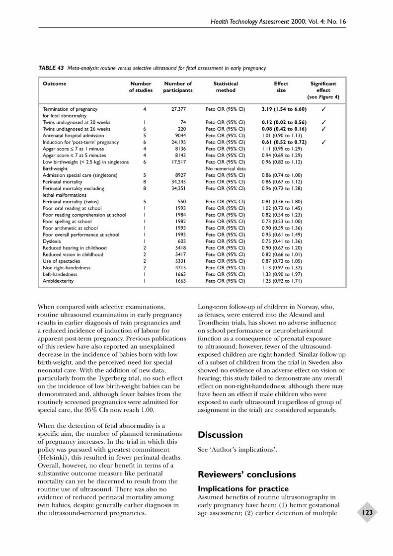
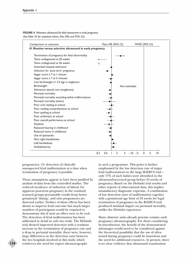
of multiple pregnancies (twins undiagnosed at 26 weeks, odds ratio (OR) 0.08, 95% confidenceinterval (CI), 0.04 to 0.16) and reduced rates ofinduction of labour for post-term pregnancy (OR, 0.61, 95% CI, 0.52 to 0.72). There were no differences detected for substantive clinicaloutcomes such as perinatal mortality (OR, 0.86,95% CI, 0.67 to 1.12). Where detection of fetalabnormality was a specific aim of the examination,the number of terminations of pregnancy for fetalanomaly increased.
Reviewers’ conclusionsRoutine ultrasound in early pregnancy appears toenable better gestational age assessment, earlierdetection of multiple pregnancies and earlierdetection of clinically unsuspected fetal malform-ation at a time when termination of pregnancy ispossible. However, the benefits for othersubstantive outcomes are less clear.
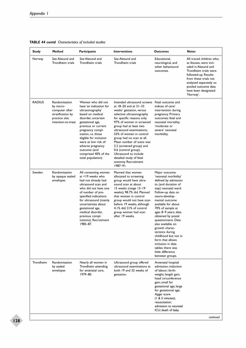
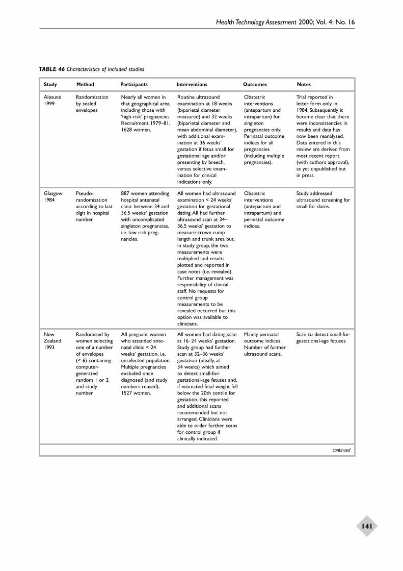
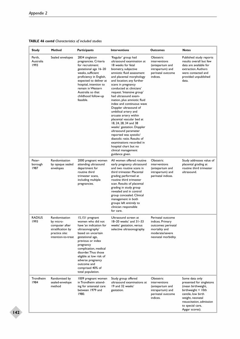
Routine ultrasound in late pregnancy (> 24 weeks’ gestation)The following abstract of a Cochrane review wasrecently published in the Cochrane Library – seeappendix 2 for the full review.
BackgroundDiagnostic ultrasound is used selectively in late pregnancy when there are specific clinicalindications. However, the value of routine latepregnancy ultrasound screening in unselectedpopulations is controversial. The rationale for such screening would be the detection of clinicalconditions which place the fetus or mother at high risk, which would not necessarily have beendetected by other means such as clinical exam-ination, and for which subsequent managementwould improve perinatal outcome.
ObjectivesTo assess the effects on obstetric practice andpregnancy outcome of routine late pregnancyultrasound, defined as greater than 24 weeks’gestation, in women with either unselected or low-risk pregnancies.
Search strategyThe Cochrane Pregnancy and Childbirth Group Specialised Register of Controlled Trialsand the Cochrane Controlled Trials Register were searched.
Selection criteriaAll acceptably controlled trials of routineultrasound in late pregnancy (defined as after 24 weeks).
Data collection and analysisThe principal reviewer assessed trial quality and extracted data, under supervision of the co-reviewer.
Main resultsSeven trials recruiting 25,036 women wereincluded. The quality of trials overall wassatisfactory. There was no difference in antenatal,obstetric and neonatal intervention or morbidity in screened versus control groups. Routine latepregnancy ultrasound was not associated withimprovements in overall perinatal mortality.Placental grading as an adjunct to a third-trimesterexamination scan was associated with a significantreduction in the stillbirth rate in the one trial thatassessed it. There is a lack of data with regard tolong-term substantive outcomes such as neuro-development. There is a lack of data on maternalpsychological effects.
Reviewers’ conclusionsBased on existing evidence, routine late pregnancyultrasound in low risk or unselected populationsdoes not confer benefit on mother or baby. There is a lack of data about the potential psychological effects of routine ultrasound in late pregnancy, and the effects on both short- and long-term neonatal and childhood outcome.Placental grading in the third trimester may be valuable but whether reported results arereproducible remains to be seen, and futureresearch into late pregnancy ultrasound should include evaluation of placental textural assessment.
Routine Doppler ultrasound inpregnancyThe following abstract is of a Cochrane reviewpublished in the Cochrane Library – see appendix 3 for the full review.
BackgroundDoppler ultrasound study of umbilical arterywaveforms helps to identify the compromised fetusin ‘high-risk’ pregnancies and, therefore, deservesassessment as a screening test in ‘low-risk’pregnancies. One of the main aims of routineantenatal care is to identify the ‘at-risk’ fetus in order to apply clinical interventions which could result in reduced perinatal morbidity and mortality.
ObjectivesTo assess the effects on obstetric practice andpregnancy outcome of routine Doppler ultrasoundin unselected and low-risk pregnancies.
Health Technology Assessment 2000; Vol. 4: No. 16
5
Search strategyThe Cochrane Pregnancy and Childbirth GroupSpecialised Register of Controlled Trials and the Cochrane Controlled Trials Register weresearched. Date of last search: September 1999.
Selection criteriaAcceptably controlled trials of routine Dopplerultrasound (umbilical circulation and/or uterinecirculation) in unselected or low-risk pregnancies.
Data collection and analysisBoth reviewers assessed trial quality and extracteddata. Authors of two trials were contacted foradditional information.
Main resultsFive trials were included which recruited 14,338women. The methodological quality of the trialswas generally good. Based on existing evidence,routine Doppler ultrasound examination in low-risk or unselected populations did not result inincreased antenatal, obstetric and neonatal
interventions, and no overall differences weredetected for substantive short-term clinicaloutcomes such as perinatal mortality. There is no available evidence to assess the effect onsubstantive long-term outcomes such as childhoodneurodevelopment. There is no available evidenceto assess maternal outcomes, particularly psycho-logical effects. In two studies there were un-expected findings suggesting possible harmfuleffects but the explanation for these is not clear, and further evaluation regarding the safety ofDoppler ultrasound is required.
Reviewers’ conclusionsBased on existing evidence, routine Dopplerultrasound in low risk or unselected populationsdoes not confer benefit on mother or baby. Future research should be powerful enough to address small changes in perinatal outcome and should include evaluation of maternalpsychological effects, long-term outcomes such as neurodevelopment, and issues of safety.
Health Technology Assessment 2000; Vol. 4: No. 16
7
IntroductionTwo members of this review group were involved inthe production of the RCOG Working Party report,Ultrasound screening for fetal anomalies (1997),5 andwere aware of the existence of a paper by Chitty(1995),9 Ultrasound screening for fetal abnormalities.This paper is a well-structured, extensive literaturereview and, acknowledging the advances in ultra-sound understanding and technology, it was decidednot to review the literature predating it but rather toupdate it. There were a number of areas that it, andthe RCOG report, did not address in detail. First,there was no mention of the methodology employedin assessing quality of studies for inclusion orexclusion in the reviews, that is, it is not clear if a systematic structured format was employed.Second, both publications concentrated on routinesecond trimester ultrasound screening for fetalabnormalities and, for the purposes of this project,data about first and third trimester routine ultra-sound fetal anomaly detection performance wasrequired. Third, there were few data at the time to assess the effect of detecting or reportingultrasonographic ‘soft’ markers of chromosomaland structural abnormalities.
Methods
Inclusion/quality criteriaFor a study to be included in the review, thefollowing criteria had to be fulfilled.
1. The study should be population-based, using anunselected or low-risk population.
2. The aim(s) should be clearly stated.3. The setting, participants and period covered
should be specified.4. The ultrasound intervention should be fully
described, including gestation at the time ofultrasound, diagnostic approach, quality control,operator(s) and skills, and equipment used.
5. It should have an adequate description of thedefinition of anomalies sought.
6. The method of postnatal ascertainment,including reporting of false-positives and false-negatives, should be described and becomprehensive (including neonatal examination
of all live-born babies; examination (preferably aformal post-mortem) of stillborn babies, babieswho die in the neonatal period and fetusesaborted after the first trimester; and post-delivery follow-up of all abnormalities suspectedat routine ultrasound examination and by otherprenatal tests; whenever possible this should also include checking available fetal anomalyregisters and databases of the genetics, neonatal,and paediatric departments).
7. Details of anomalies detected as per fetus/system should be reported.
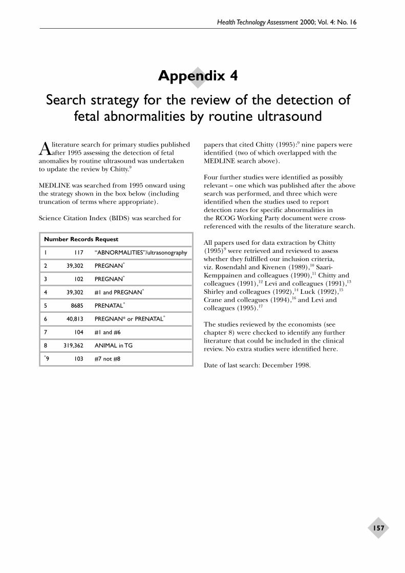
Studies were identified using the search strategypresented in appendix 4. Material was managedusing a reference manager database (Procite™).
Methodological evaluation and data extractionStage I – initial categorisation of studiesEach study was categorised on the basis of the titleand abstract when available. The following initialcriteria were used to determine the relevance ofeach study to the systematic review.
1. Primary study of routine ultrasound screening in pregnancy(a) randomised controlled trial (RCT)(b) prospective study(c) retrospective study(d) not clear whether RCT, prospective,
retrospective.2. Primary study which may be relevant, but not
clear from the title or abstract.3. Primary study which is not directly relevant but
may have some relevant information.4. Review, but not primary study.5. Foreign language:
(a) may be relevant(b) not relevant.
6. Document/letter/communication.7. Not relevant.E. Any study which might be relevant to the
economic review was flagged as ‘E’ and passedon to the economic reviewers.
Stage II – further categorisation of studiesAll studies in categories 1, 2 and 3 were consideredrelevant and were retrieved and reviewed in full.
Chapter 3
Detection of fetal abnormalitiesby routine ultrasound
Detection of fetal abnormalities by routine ultrasound
8
No foreign language papers were retrieved – forreasons see results below. Retrieved studies werefurther categorised as follows:
A1 – relevant and acceptable quality, all data to be extractedA2 – relevant and acceptable quality, some data to be excludedB1 – relevant but does not meet criteria for data extractionB2 – relevant but poor qualityC – not relevant.
This two-stage categorisation process was devisedby and agreed between the two clinical reviewers.
Stage III – data extraction and manipulationData were extracted in three stages to fulfil thefollowing aims.
1. To develop an overview of study characteristicsincluding type of study, period, setting(population studied, type of service andcountry), intervention and overall performance,in order to identify factors which affect efficacyof routine ultrasound screening in pregnancy.
2. To determine detection rates for specific anom-alies in anatomical systems and chromosomalabnormalities in each trimester of pregnancy.
3. To assign specific anomalies to pragmatic groupsgoverned by the likely interventions andoutcomes of affected pregnancies.
4. To provide clinical data based on availableevidence to be incorporated by the economistsinvolved in this project into refining a previouslydeveloped economic decision model of scanningpolicies in pregnancy.
As the overall aim was to assess the cost-effectiveness of routine ultrasound screening inpregnancy, the manner in which the data weremanaged and reported was largely governed by the structure of the decision model.
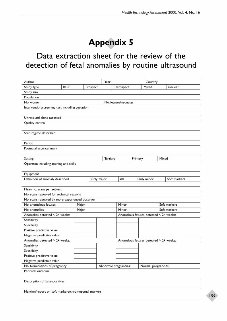
An example of the data extraction sheet ispresented in appendix 5.
Results
Stage IIn all, 110 papers were identified by our searchstrategy, with a further eight from the Chitty paper,three from the RCOG Working Party report, andone published after the literature search had beenperformed. Thus the abstracts of 122 papers werereviewed in total.
A total of 37 papers were initially categorised 1 (25), 2 (11) or 3 (3). The remaining 85 fell intocategories 4–7 and were not considered further.Seven papers flagged ‘E’ (i.e. possibly relevant tothe economics review) were cross-referenced withthe economic reviewers (TR and JH) and had beenidentified by them. Only five of the 25 foreignlanguage papers were of possible relevance(category 5a). One would have been coded 1 andfour coded 2; however, they were not pursued,mainly because, of the English language papers, all those coded 2 and more than half of thosecoded 1 were, when reviewed in full during stageIII, not suitable for the review and, hence, it wasunlikely that the ‘possibly relevant’ foreignlanguage papers would have been.
Stage IIOf the 37 papers identified in stage I, 36 werereviewed in full. One paper in category 1 was notretrieved as the journal would have been difficultto obtain and, on rereading the abstract, seemedto refer to a subset of the population of babies whowere born with anomalies and also referred forneonatal surgical treatment.
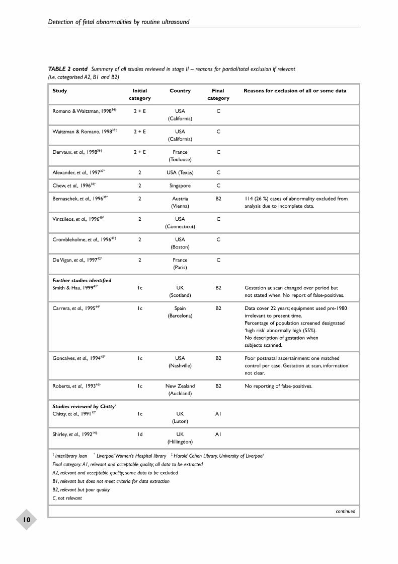
The study categorisations in stages I and II aresummarised in Table 1, and details of all studiesreviewed in stage II are summarised in Table 2,10–46
together with the reasons given if they onlyqualified for partial data extraction (that is,categorised A2), or for exclusion if they failed onquality criteria (that is, categorised B1 or B2).
Of the 24 available studies in category 1, furthercategorisation was as follows: A1 (8), A2 (3), B1 (1), B2 (11), C (1). Of the 11 available studiesin category 2, further categorisation was as follows:B2 (1), C (10). The one study in category 3 wasfurther categorised as B2. Thus, 11 papers(classified A1 or A2) entered stage III.
TABLE 1 Summary of categorisation of studies on the detection of fetal abnormalities by routine ultrasound
Initial Number Subsequent categorisation Notcategorisation of after full review retrieved(stage 1) papers (stage II)
TABLE 2 Summary of all studies reviewed in stage II – reasons for partial/total exclusion if relevant (i.e. categorised A2, B1 and B2)
Study Initial Country Final Reasons for exclusion of all or some datacategory category
Studies identified by literature searchMagriples & Copel, 199822* 1c USA A1
(Connecticut)
Lee, et al., 198818† 1c Korea A2 Only routine screening data extracted (data presented separately for routine and indicated screening).
Queisser, et al., 199826* 1d Germany B2 Gestation at anomaly detection not clear.(Mainz)
Grandjean, et al., 199827† 2 France C Only reported detection of (Toulouse) chromosomal abnormalities.
Van Dorsten, et al., 199820* 1b + E USA A2 Only data for screened population extracted.(South Carolina)
Zimmer, et al., 199728* 1d Israel B2 Gestation at which routine scan performed(Haifa) not reported. False-positives not reported.
30.6 % of population never scanned.
Dillon & Walton, 199729‡ 1c UK B2 False-positives not reported (Stockton-on-Tees) (cannot calculate specificity).
Skupski, et al., 199621* 1c USA A1(Texas)
Ashe, et al., 199630† 1c Northern Ireland B2 False-positives not reported (Belfast) (cannot calculate specificity).
Geerts, et al., 199631* 1a + E South Africa C False-positives not reported. Detection of fetal(Cape Town) anomalies not a primary aim of study.
Eurenius, et al., 199632‡ 1b Sweden B2 False-negatives not reported (Uppsala) (cannot calculate sensitivity).
Nasrat, 199833 1c – Difficult to retrieve; on reading abstract again,(not retrieved) referred to a subset of population in which babies
born with fetal anomalies were referred forneonatal surgical treatment.
Boyd, et al., 199819* 1c UK A2 Only data independent of serum screening(Oxford) to be extracted.
Skari, et al., 199825‡ 2 Norway C(Oslo)
D’Ottavio, et al., 199824† 3 Italy B2 Mentions 15 with NT but only five of those(Trieste) abnormal karyotype; no postnatal ascertain-
ment of the other ten but states ‘no falsepositives’ at TVS screening, therefore unclear.
Hernadi & Torocsik, 199723* 1b Hungary B2 No reporting of false-positives.
† Interlibrary loan * Liverpool Women’s Hospital library ‡ Harold Cohen Library, University of LiverpoolFinal category: A1, relevant and acceptable quality; all data to be extractedA2, relevant and acceptable quality; some data to be excludedB1, relevant but does not meet criteria for data extractionB2, relevant but poor qualityC, not relevant
continued
Detection of fetal abnormalities by routine ultrasound
10
TABLE 2 contd Summary of all studies reviewed in stage II – reasons for partial/total exclusion if relevant (i.e. categorised A2, B1 and B2)
Study Initial Country Final Reasons for exclusion of all or some datacategory category
Romano & Waitzman, 199834† 2 + E USA C(California)
Waitzman & Romano, 199835† 2 + E USA C(California)
Dervaux, et al., 199836† 2 + E France C(Toulouse)
Alexander, et al., 199737* 2 USA (Texas) C
Chew, et al., 199638† 2 Singapore C
Bernaschek, et al., 199639* 2 Austria B2 114 (26 %) cases of abnormality excluded from(Vienna) analysis due to incomplete data.
Vintzileos, et al., 199640* 2 USA C(Connecticut)
Crombleholme, et al., 199641† 2 USA C(Boston)
De Vigan, et al., 199742* 2 France C(Paris)
Further studies identifiedSmith & Hau, 199943* 1c UK B2 Gestation at scan changed over period but
(Scotland) not stated when. No report of false-positives.
Carrera, et al., 199544* 1c Spain B2 Data cover 22 years; equipment used pre-1980(Barcelona) irrelevant to present time.
Percentage of population screened designated‘high risk’ abnormally high (55%).No description of gestation when subjects scanned.
Goncalves, et al., 199445* 1c USA B2 Poor postnatal ascertainment: one matched(Nashville) control per case. Gestation at scan, information
not clear.
Roberts, et al., 199346† 1c New Zealand B2 No reporting of false-positives.(Auckland)
Studies reviewed by Chitty9
Chitty, et al., 199112* 1c UK A1(Luton)
Shirley, et al., 199214‡ 1d UK A1(Hillingdon)
† Interlibrary loan * Liverpool Women’s Hospital library ‡ Harold Cohen Library, University of Liverpool
Final category: A1, relevant and acceptable quality; all data to be extracted
A2, relevant and acceptable quality; some data to be excluded
B1, relevant but does not meet criteria for data extraction
B2, relevant but poor quality
C, not relevant
continued
Health Technology Assessment 2000; Vol. 4: No. 16
11
TABLE 2 contd Summary of all studies reviewed in stage II – reasons for partial/total exclusion if relevant (i.e. categorised A2, B1 and B2)
Study Initial Country Final Reasons for exclusion of all or some datacategory category
Levi, et al., 199113* 1b Belgium A1(Brussels)
Luck, 199215* 1b UK A1(Ascot)
Crane, et al., 199416* 1a USA A1(RADIUS)
Levi, et al., 199517* 1b Belgium A1
Rosendhal & Kivinen, 198910* 1d Finland B2 Two phases:phase 1: 18-week scan; phase 2: 18- and 34-week scan. However, not reported separatelyand gestation at diagnosis unclear.
Saari-Kempapainen, et al., 199011* 1a Finland B1 Anomalies detected not reported individually,(Helsinki) so no available data to extract.
† Interlibrary loan * Liverpool Women’s Hospital library ‡ Harold Cohen Library, University of LiverpoolFinal category: A1, relevant and acceptable quality; all data to be extractedA2, relevant and acceptable quality; some data to be excludedB1, relevant but does not meet criteria for data extractionB2, relevant but poor qualityC, not relevant
Stage IIIThe overall characteristics and overall results of the 11 included studies are presented in Table 3,10,13–22 including type of study, period ofstudy, country where performed, populationstudied, setting, personnel performing sono-graphy, number of fetuses scanned, gestation atscanning, prevalence of anomalous fetuses andanomalies, number of false-positives, detectionrates in each trimester including sensitivity andspecificity, overall detection rates, termination ofpregnancy rates and whether sonographic softmarkers are reported.
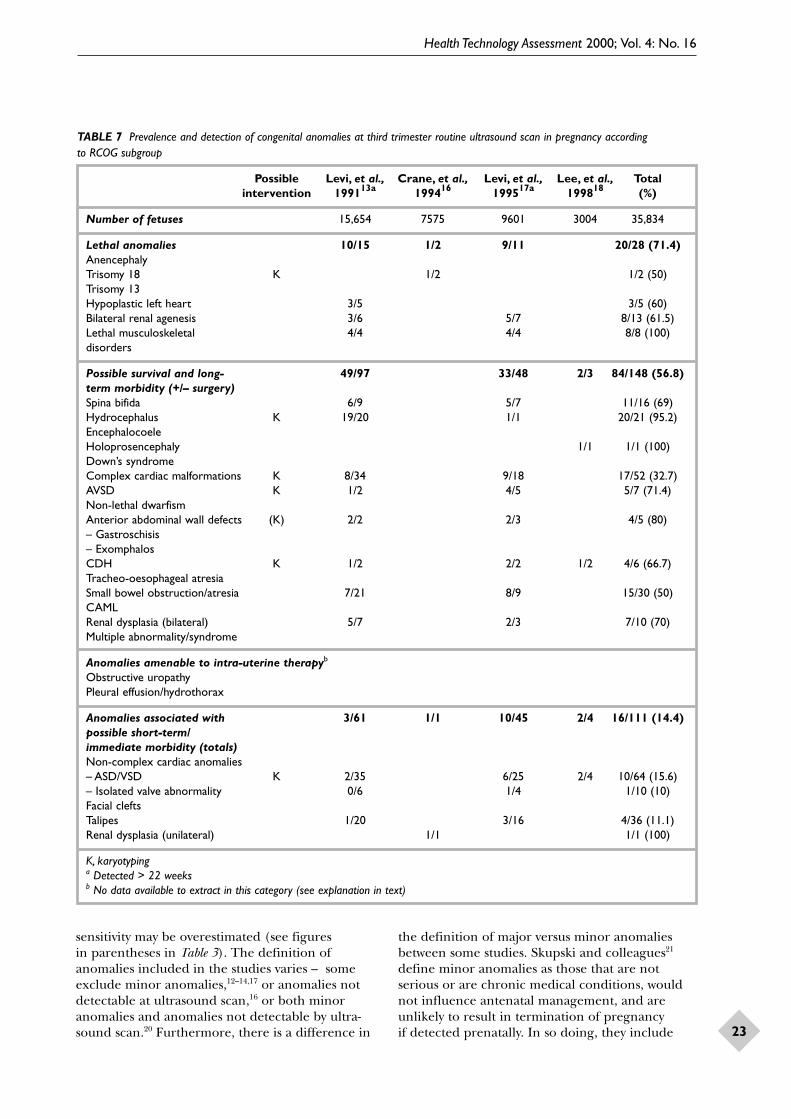
Results of second trimester routine ultrasound forfetal anomalies were reported in all the includedstudies. In only four studies13,16–18 were routinethird trimester ultrasound results reported, and in none were the results of routine first trimesteranomaly screening reported.
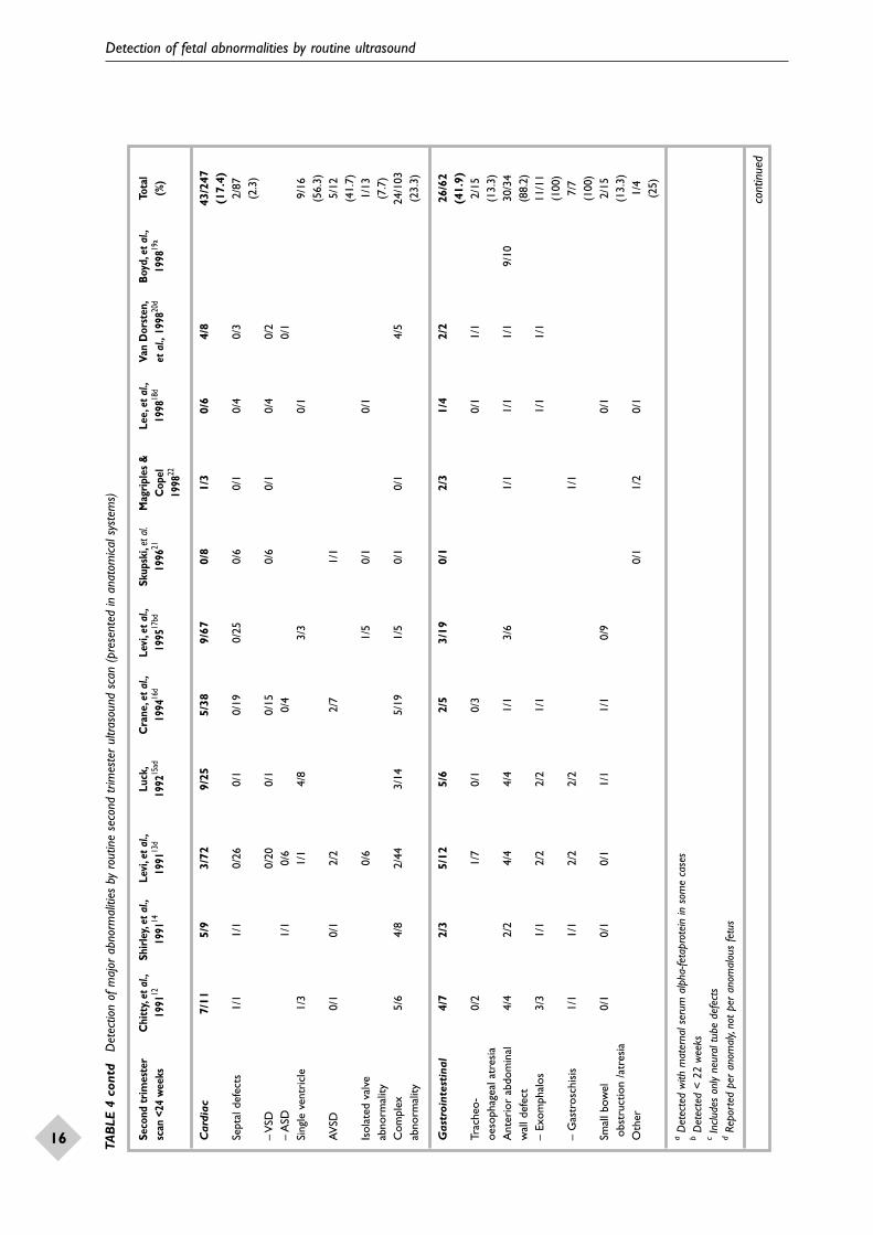
The detection rates at routine second trimesterultrasound of individual structural abnormalitiesreported in anatomical systems are shown in Table 4.
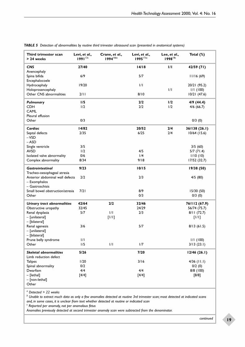
The detection rates at routine third trimesterultrasound of individual structural abnormalitiesreported in anatomical systems are shown in Table 5.
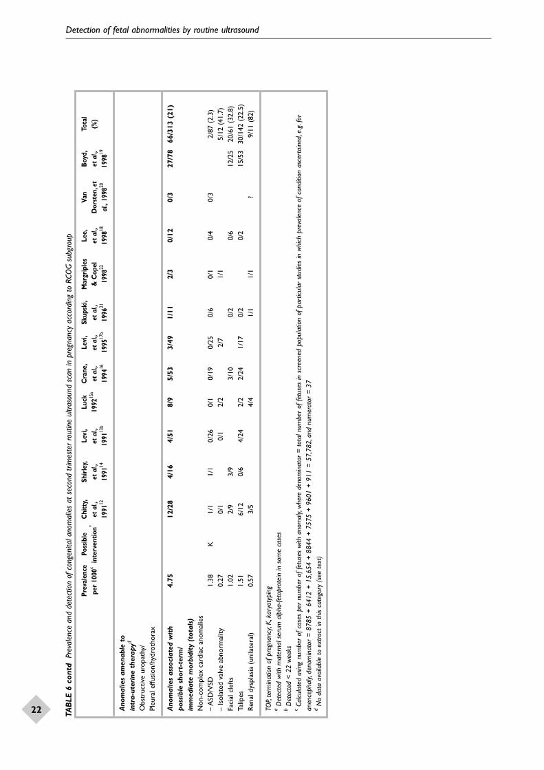
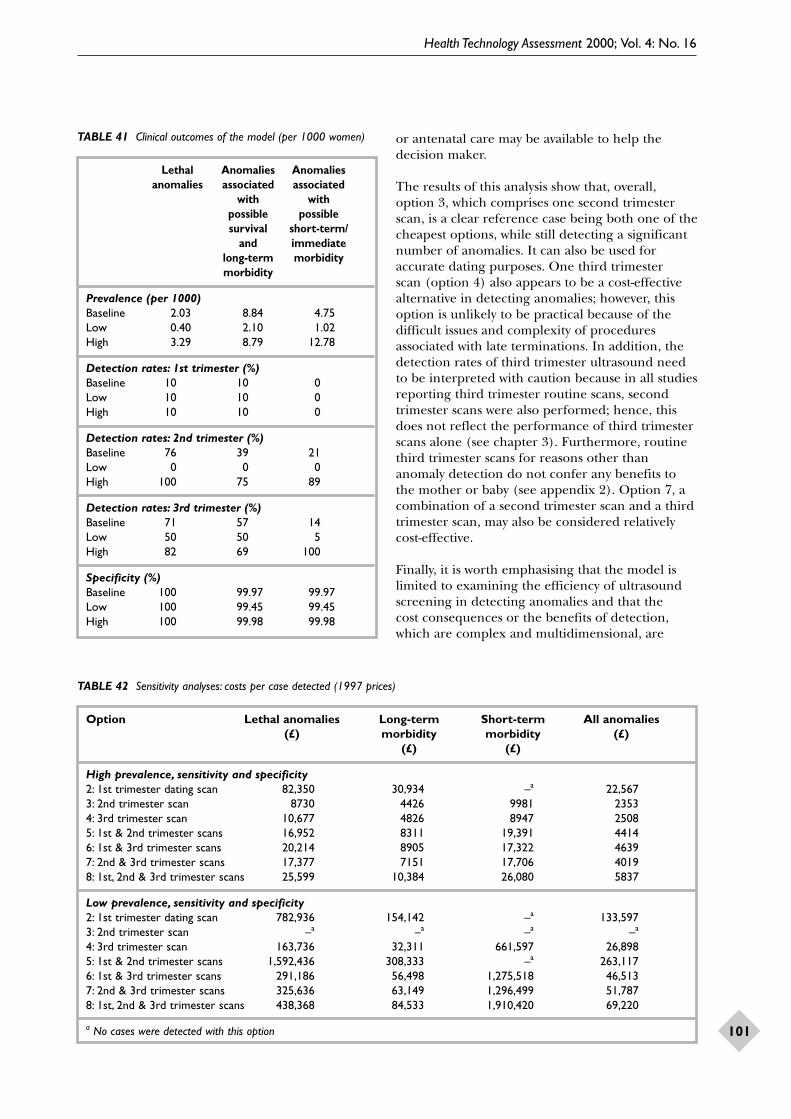
The prevalence and detection of congenitalanomalies at second trimester ultrasound scan aresummarised in Table 6, grouped according to theirlikely clinical consequences. These four pragmaticsubgroups were proposed by the RCOG in 1997,5
and include:
(i) lethal anomalies(ii) anomalies associated with possible survival and
long-term morbidity(iii) anomalies which may be amenable to intra-
uterine therapy(iv) anomalies associated with possible immediate
or short-term morbidity.
Included in this table is a column indicating theother interventions that may be offered on detec-tion of the particular abnormality. These werelisted for use in the cost-effectiveness section of this review, as previous models of cost-effectivenessassumed that all detected abnormalities would be offered interventions, and this in turn over-estimates costs. These possible interventions were based on clinical practice at the LiverpoolWomen’s Hospital and on knowledge of theassociation of some of the abnormalities withchromo-somal abnormalities and the long-termconsequences to babies born alive with the listed
Detection of fetal abnormalities by routine ultrasound
12 TAB
LE 3
O
verv
iew
of s
tudi
es in
clud
ed in
the
lite
ratu
re r
evie
w
Stud
yC
hitt
y,et
al.,
Shir
ley,
et a
l.,Le
vi,e
t al.,
Luck
,C
rane
,et a
l.,Le
vi,e
t al.,
Skup
ski,
et a
l.,M
agri
ples
&Le
e,et
al.,
Van
Dor
sten
,B
oyd,
et a
l.,To
tal
1991
1219
9114
1991
13d
1992
15cf
1994
16d
1995
17d
1996
21C
opel
1998
18d
et a
l.,19
9820
d19
9819
ab
1998
22
Type
Ret
rosp
ectiv
eR
etro
spec
tive
Pros
pect
ive
Pros
pect
ive
RC
Te
Pros
pect
ive
Ret
rosp
ectiv
eR
etro
spec
tive
Ret
rosp
ectiv
ePr
ospe
ctiv
eR
etro
spec
tive
Peri
od
1988
–89
1989
–90
1984
–89
1988
–91
1987
–91
1990
–92
1990
–199
4?
18 m
onth
s19
90–9
419
93–9
619
91–9
6
Co
untr
yU
KU
KBe
lgiu
mU
KU
SA
Belg
ium
USA
USA
Kor
eaU
SA
UK
(L
uton
)(H
illin
gdon
)(B
russ
els)
(Asc
ot)
(RA
DIU
S)(B
russ
els)
(Tex
as)
(Con
nect
icut
)(S
.Car
olin
a)(O
xfor
d)
Popu
lati
on
stud
ied
Uns
elec
ted
Uns
elec
ted
Uns
elec
ted
Uns
elec
ted
Low
ris
kU
nsel
ecte
dLo
w r
isk
Low
ris
kLo
w r
isk
Uns
elec
ted
Uns
elec
ted
Set
ting
Dis
tric
t D
istr
ict
5 ho
spita
lsD
istr
ict
Prim
ary
5 ho
spita
lsTe
rtia
ry,
Tert
iary
,Te
rtia
ry,
Mix
ed,
Tert
iary
,ge
nera
l ge
nera
lge
nera
l+
28
sing
le c
entr
esi
ngle
cen
tre
sing
le c
entr
e2
site
ssi
ngle
cen
tre
hosp
ital
hosp
ital
hosp
ital
labo
rato
ries
So
nogr
aphe
rR
adio
grap
hers
Rad
iogr
aphe
rsO
bste
tric
ians
;R
adio
grap
hers
Tech
nici
ans;
Obs
tetr
icia
ns;
Expe
rien
ced
Sono
grap
hers
Trai
ned
Reg
iste
red
Not
tech
nici
ans;
phys
icia
n;te
chni
cian
s;so
nogr
aphe
rsob
stet
ric
diag
nost
icm
entio
ned
sono
grap
hers
sono
logi
sts
sono
grap
hers
Fello
wm
edic
al
radi
olog
ists
sono
grap
her
Num
ber
of
8785
6412
15,6
5488
4475
7596
0186
091
130
0416
1133
,376
96,6
33
fetu
ses
(tw
ins)
(mul
tiple
(73
mul
tiple
(?24
0 m
ultip
le(?
num
ber
(?20
9 m
ultip
le(6
tw
ins)
(10
twin
s)(t
win
s (t
win
s(?
tw
ins)
preg
anci
espr
egna
ncie
s)pr
egna
ncie
s)m
ultip
lepr
egna
ncie
s)ex
clud
ed)
excl
uded
)no
t m
entio
ned)
preg
nanc
ies)
Num
ber
of
scan
s?
??
??
??
1.8
±0.
99?
??
(mea
n,S
D)
aSo
ft m
arke
r da
ta e
xclu
ded
(will
be p
rese
nted
sep
arat
ely)
bIn
clud
es s
ome
patie
nts
who
had
mat
erna
l ser
um a
lpha
feto
prot
ein
scre
enin
g,bu
t th
is is
not
prov
ided
rou
tinel
y by
the
NH
Sc
Repo
rted
in s
yste
ms,
not
per
fetu
sd
Ove
rall
sens
itivit
y ca
lcula
ted
per
fetu
s bu
t in
divid
ual a
nom
alie
s re
port
ed p
er s
yste
m n
ot p
er fe
tus.
Figu
res
in p
aren
thes
es r
efer
to
calcu
latio
ns b
ased
on
num
ber
of a
nom
alie
s,no
t nu
mbe
r of
ano
mal
ous
fetu
ses
eU
ltras
ound
-scr
eene
d gr
oup
only
incl
uded
fM
ater
nal s
erum
alp
ha-fe
topr
otei
n sc
reen
ing
in a
dditi
on t
o ul
traso
und
was
und
erta
ken
in t
his
popu
latio
ngTh
is fig
ure
calcu
late
d ta
king
onl
y th
ose
defe
cts
expo
sed
to s
can
at 1
2–24
wee
ks (
n =
259
),i.e
.not
on
inte
ntio
n-to
-scr
een
cont
inue
d
Health Technology Assessment 2000; Vol. 4: No. 16
13TAB
LE 3
con
td
Ove
rvie
w o
f stu
dies
incl
uded
in t
he li
tera
ture
rev
iew
Stud
yC
hitt
y,et
al.,
Shir
ley,
et a
l.,Le
vi,e
t al.,
Luck
,C
rane
,et a
l.,Le
vi,e
t al.,
Skup
ski,
et a
l.,M
agri
ples
&Le
e,et
al.,
Van
Dor
sten
,B
oyd,
et a
l.,To
tal
1991
1219
9114
1991
13d
1992
15cf
1994
16d
1995
17d
1996
21C
opel
1998
18d
et a
l.,19
9820
d19
9819
ab
1998
22
Ges
tati
ona
l18
–20
wee
ks19
wee
ks1s
t tr
imes
ter
12–1
4 w
eeks
15–2
2 w
eeks
1st
trim
este
r18
–20
wee
ks16
–20
wee
ks18
–20
wee
ks15
–22
wee
ks18
–22
wee
ksag
e/s
16–2
0 w
eeks
19 w
eeks
31–3
5 w
eeks
16–2
0 w
eeks
3rd
trim
este
r32
–34
wee
kssc
anne
d3r
d tr
imes
ter
3rd
trim
este
r
Pre
vale
nce
of
1.50
1.40
2.30
2.30
2.45
1.16
3.07
0.76
1.30
2.17
1839
/an
om
alo
us(1
30 fe
tuse
s)(8
9 fe
tuse
s)(3
81 fe
tuse
s)(1
87 fe
tuse
s)(2
35 fe
tuse
s)(2
0 fe
tuse
s)(2
8 fe
tuse
s)(2
3 fe
tuse
s)(2
1 fe
tuse
s)(7
25 fe
tuse
s)97
,789
fetu
ses
(%)
+ 2
.09%
Pre
vale
nce
of
2.66
1.90
–2.
81–
––
ano
mal
ies
(%)
(417
(164
(232
(270
(40
(37
(29
anom
alie
s)an
omal
ies)
anom
alie
s)an
omal
ies)
anom
alie
s)an
omal
ies)
anom
alie
s)
Fals
e-2
18
37
91
50
115
52po
siti
ves
Det
ecti
on
< 15
wee
ks*
Det
ecti
on
9361
(54)
(140
)31
(69)
320
3 (5
)10
298
509/
1233
< 24
wee
ksS
ensi
tivi
ty (
%)
71.5
57.3
(21.
0)g
(85.
3)16
.6(2
5.6)
15.0
71.4
13.5
(13
.5)
47.6
41.1
41.3
%S
peci
ficit
y (%
)99
.98
99.9
7(1
00.0
0)99
.90
99.9
099
.90
99.4
010
0.00
99.9
099
.90
99.9
4 %
Det
ecti
on
(135
)34
(109
)5
(6)
39/2
10>
24 w
eeks
Sen
siti
vity
(%
)(3
7.2)
18.2
(40.
4)21
.7 (
16.2
)18
.6%
Spe
cific
ity
(%)
??
100.
00
aSo
ft m
arke
r da
ta e
xclu
ded
(will
be p
rese
nted
sep
arat
ely)
bIn
clud
es s
ome
patie
nts
who
had
mat
erna
l ser
um a
lpha
feto
prot
ein
scre
enin
g,bu
t th
is is
not
prov
ided
rou
tinel
y by
the
NH
Sc
Repo
rted
in s
yste
ms,
not
per
fetu
sd
Ove
rall
sens
itivit
y ca
lcula
ted
per
fetu
s bu
t in
divid
ual a
nom
alie
s re
port
ed p
er s
yste
m n
ot p
er fe
tus.
Figu
res
in p
aren
thes
es r
efer
to
calcu
latio
ns b
ased
on
num
ber
of a
nom
alie
s,no
t nu
mbe
r of
ano
mal
ous
fetu
ses
eU
ltras
ound
-scr
eene
d gr
oup
only
incl
uded
fM
ater
nal s
erum
alp
ha-fe
topr
otei
n sc
reen
ing
in a
dditi
on t
o ul
traso
und
was
und
erta
ken
in t
his
popu
latio
ngTh
is fig
ure
calcu
late
d ta
king
onl
y th
ose
defe
cts
expo
sed
to s
can
at 1
2–24
wee
ks (
n =
259
),i.e
.not
on
inte
ntio
n-to
-scr
een
*N
one
of t
he in
clud
ed s
tudi
es r
epor
ted
dete
ctio
n of
ano
mal
ies
at <
15
wee
ks
cont
inue
d
Detection of fetal abnormalities by routine ultrasound
14 TAB
LE 3
con
td
Ove
rvie
w o
f stu
dies
incl
uded
in t
he li
tera
ture
rev
iew
Stud
yC
hitt
y,et
al.,
Shir
ley,
et a
l.,Le
vi,e
t al.,
Luck
,C
rane
,et a
l.,Le
vi,e
t al.,
Skup
ski,
et a
l.,M
agri
ples
&Le
e,et
al.,
Van
Dor
sten
,B
oyd,
et a
l.,To
tal
1991
1219
9114
1991
13d
1992
15cf
1994
16d
1995
17d
1996
21C
opel
1998
18d
et a
l.,19
9820
d19
9819
ab
1998
22
Ove
rall
dete
ctio
n93
5115
4(1
40)
6512
0 (1
78)
208
(11)
1029
882
2/18
39S
ensi
tivi
ty (
%)
71.5
57.3
40.4
85.3
34.8
51.0
(65
.9)
15.0
71.4
34.8
(29
.7)
47.6
41.1
44.7
%S
peci
ficit
y (%
)99
.98
99.9
799
.94
99.9
099
.90
99.9
099
.80
99.4
010
0.00
99.9
099
.90
Term
inat
ions
of
5229
?19
9?
2`6
34
169
293/
71,3
78pr
egna
ncie
s
Term
inat
ions
of
0.6
0.45
0.21
0.12
0.23
0.67
0.09
0.25
0.51
0.41
%pr
egna
ncie
s (%
)
Term
inat
ions
of
00
00
00
0?
02
(1 s
oft
norm
al p
regn
anci
esm
arke
r)
‘So
ft’ m
arke
rsYe
sN
oN
oYe
sN
oN
oN
oYe
sN
oN
oYe
s
aSo
ft m
arke
r da
ta e
xclu
ded
(will
be p
rese
nted
sep
arat
ely)
bIn
clud
es s
ome
patie
nts
who
had
mat
erna
l ser
um a
lpha
feto
prot
ein
scre
enin
g,bu
t th
is is
not
prov
ided
rou
tinel
y by
the
NH
Sc
Repo
rted
in s
yste
ms,
not
per
fetu
sd
Ove
rall
sens
itivit
y ca
lcula
ted
per
fetu
s bu
t in
divid
ual a
nom
alie
s re
port
ed p
er s
yste
m n
ot p
er fe
tus.
Figu
res
in p
aren
thes
es r
efer
to
calcu
latio
ns b
ased
on
num
ber
of a
nom
alie
s,no
t nu
mbe
r of
ano
mal
ous
fetu
ses
eU
ltras
ound
-scr
eene
d gr
oup
only
incl
uded
fM
ater
nal s
erum
alp
ha-fe
topr
otei
n sc
reen
ing
in a
dditi
on t
o ul
traso
und
was
und
erta
ken
in t
his
popu
latio
ngTh
is fig
ure
calcu
late
d ta
king
onl
y th
ose
defe
cts
expo
sed
to s
can
at 1
2–24
wee
ks (
n =
259
),i.e
.not
on
inte
ntio
n-to
-scr
een
Health Technology Assessment 2000; Vol. 4: No. 16
15TAB
LE 4
D
etec
tion
of m
ajor
abn
orm
aliti
es b
y ro
utin
e se
cond
trim
este
r ul
traso
und
scan
(pr
esen
ted
in a
nato
mica
l sys
tem
s)
Seco
nd t
rim
este
rC
hitt
y,et
al.,
Shir
ley,
et a
l.,Le
vi,e
t al.,
Luck
,C
rane
,et a
l.,Le
vi,e
t al.,
Skup
ski,
et a
l.M
agri
ples
&Le
e,et
al.,
Van
Dor
sten
,B
oyd,
et a
l.,To
tal
scan
<24
wee
ks19
9112
1991
1419
9113
d19
9215
ad19
9416
d19
9517
bd19
9621
Cop
el19
9818
det
al.,
1998
20d
1998
19a
(%)
1998
22
CN
S20
/21
15/1
617
/35
11/1
19/
1017
/35
3/4
2/3
7/9
45/4
7c14
6/19
1(7
6.4)
Ane
ncep
haly
6/6
10/1
06/
67/
73/
34/
41/
237
/38
(97.
4)Sp
ina
bifid
a5/
53/
32/
52/
24/
54/
112/
222
/33
(66.
7)En
ceph
aloc
oele
2/2
1/1
2/2
1/1
1/1
7/7
(100
)H
ydro
ceph
aly
3/3
1/2
4/15
5/6
1/1
4/5
18/3
2(5
6.3)
Hol
opro
senc
epha
ly2/
30/
11/
11/
10/
14/
7(5
7.1)
Oth
er C
NS
2/2
3/6
2/2
4/14
0/1
1/2
12/2
7ab
norm
aliti
es44
.4
Pul
mon
ary
7/9
4/5
2/7
3/6
2/5
0/2
1/1
0/1
0/2
1/2
5/10
25/5
0(5
0)C
DH
2/2
2/3
1/3
2/5
1/1
0/2
0/2
1/2
9/20
(45)
CA
ML
4/4
1/1
1/1
6/6
(100
)Pl
eura
l effu
sion
1/3
1/3
(33.
3)O
ther
1/1
1/4
1/4
1/1
0/1
4/11
(36.
4)
aD
etec
ted
with
mat
erna
l ser
um a
lpha
-feta
prot
ein
in s
ome
case
sb
Det
ecte
d <
22
wee
ksc
Incl
udes
onl
y ne
ural
tub
e de
fect
sd
Repo
rted
per
ano
mal
y,no
t pe
r an
omal
ous
fetu
s
cont
inue
d
Detection of fetal abnormalities by routine ultrasound
16 TAB
LE 4
con
td
Det
ectio
n of
maj
or a
bnor
mal
ities
by
rout
ine
seco
nd t
rimes
ter
ultra
soun
d sc
an (
pres
ente
d in
ana
tom
ical s
yste
ms)
Seco
nd t
rim
este
rC
hitt
y,et
al.,
Shir
ley,
et a
l.,Le
vi,e
t al.,
Luck
,C
rane
,et a
l.,Le
vi,e
t al.,
Skup
ski,
et a
l.M
agri
ples
&Le
e,et
al.,
Van
Dor
sten
,B
oyd,
et a
l.,To
tal
scan
<24
wee
ks19
9112
1991
1419
9113
d19
9215
ad19
9416
d19
9517
bd19
9621
Cop
el19
9818
det
al.,
1998
20d
1998
19a
(%)
1998
22
Car
diac
7/11
5/9
3/72
9/25
5/38
9/67
0/8
1/3
0/6
4/8
43/2
47(1
7.4)
Sept
al d
efec
ts1/
11/
10/
260/
10/
190/
250/
60/
10/
40/
32/
87(2
.3)
– V
SD0/
200/
10/
150/
60/
10/
40/
2–
ASD
1/1
0/6
0/4
0/1
Sing
le v
entr
icle
1/3
1/1
4/8
3/3
0/1
9/16
(56.
3)A
VSD
0/1
0/1
2/2
2/7
1/1
5/12
(41.
7)Is
olat
ed v
alve
0/
61/
50/
10/
11/
13ab
norm
ality
(7.7
)C
ompl
ex
5/6
4/8
2/44
3/14
5/19
1/5
0/1
0/1
4/5
24/1
03ab
norm
ality
(23.
3)
Gas
troi
ntes
tina
l4/
72/
35/
125/
62/
53/
190/
12/
31/
42/
226
/62
(41.
9)Tr
ache
o-
0/2
1/7
0/1
0/3
0/1
1/1
2/15
oeso
phag
eal a
tres
ia(1
3.3)
Ant
erio
r ab
dom
inal
4/4
2/2
4/4
4/4
1/1
3/6
1/1
1/1
1/1
9/10
30/3
4w
all d
efec
t(8
8.2)
– Ex
omph
alos
3/3
1/1
2/2
2/2
1/1
1/1
1/1
11/1
1(1
00)
– G
astr
osch
isis
1/1
1/1
2/2
2/2
1/1
7/7
(100
)Sm
all b
owel
0/1
0/1
0/1
1/1
1/1
0/9
0/1
2/15
obst
ruct
ion
/atr
esia
(13.
3)O
ther
0/1
1/2
0/1
1/4
(25)
aD
etec
ted
with
mat
erna
l ser
um a
lpha
-feta
prot
ein
in s
ome
case
sb
Det
ecte
d <
22
wee
ksc
Incl
udes
onl
y ne
ural
tub
e de
fect
sd
Repo
rted
per
ano
mal
y,no
t pe
r an
omal
ous
fetu
s
cont
inue
d
Health Technology Assessment 2000; Vol. 4: No. 16
17TAB
LE 4
con
td
Det
ectio
n of
maj
or a
bnor
mal
ities
by
rout
ine
seco
nd t
rimes
ter
ultra
soun
d sc
an (
pres
ente
d in
ana
tom
ical s
yste
ms)
Seco
nd t
rim
este
rC
hitt
y,et
al.,
Shir
ley,
et a
l.,Le
vi,e
t al.,
Luck
,C
rane
,et a
l.,Le
vi,e
t al.,
Skup
ski,
et a
l.M
agri
ples
&Le
e,et
al.,
Van
Dor
sten
,B
oyd,
et a
l.,To
tal
scan
<24
wee
ks19
9112
1991
1419
9113
d19
9215
ad19
9416
d19
9517
bd19
9621
Cop
el19
9818
det
al.,
1998
20d
1998
19a
(%)
1998
22
Uri
nary
tra
ct
21/2
57/
912
/76
105/
105
34/3
520
/66
2/2
4/4
3/3
62/7
627
0/40
1ab
norm
alit
ies
(67.
3)O
bstr
uctiv
e ur
opat
hy10
/10
7/8
2/47
99/9
928
/29
7/36
1/1
154/
230
(67)
Ren
al d
yspl
asia
5/8
0/1
4/11
4/4
7/10
1/1
2/2
1/1
24/3
8(6
3.2)
– [u
nila
tera
l][3
/5]
[4/4
][1
/1]
[1/1
]?
[9/1
1](8
2)–
[bila
tera
l][2
/3]
[0/1
][1
/1]
?[3
/5]
(60)
Ren
al a
gene
sis
5/5
3/9
2/2
3/10
13/2
6(5
0)–
[uni
late
ral]
[1/1
][1
/1]
(100
)–
[bila
tera
l][4
/4]
[2/2
]1/
1[7
/7]
(100
)Pr
une
belly
syn
drom
e3/
43/
4(7
5)O
ther
1/2
0/5
6/6
3/10
1/1
2/2
13/2
6(5
0)
Skel
etal
abn
orm
alit
ies
17/2
72/
85/
536/
84/
304/
320/
32/
30/
440
/168
(23.
8)Li
mb
redu
ctio
n de
fect
2/5
1/1
0/6
1/2
2/5
0/1
0/1
6/21
(28.
6)Ta
lipes
6/12
0/6
4/24
2/2
2/24
1/17
0/2
0/2
15/5
330
/142
(22.
5)Sp
inal
abn
orm
ality
3/3
0/2
1/1
0/1
1/1
5/8
(62.
5)
aD
etec
ted
with
mat
erna
l ser
um a
lpha
-feta
prot
ein
in s
ome
case
sb
Det
ecte
d <
22
wee
ksc
Incl
udes
onl
y ne
ural
tub
e de
fect
sd
Repo
rted
per
ano
mal
y,no
t pe
r an
omal
ous
fetu
s
cont
inue
d
TAB
LE 4
con
td
Det
ectio
n of
maj
or a
bnor
mal
ities
by
rout
ine
seco
nd t
rimes
ter
ultra
soun
d sc
an (
pres
ente
d in
ana
tom
ical s
yste
ms)
Seco
nd t
rim
este
rC
hitt
y,et
al.,
Shir
ley,
et a
l.,Le
vi,e
t al.,
Luck
,C
rane
,et a
l.,Le
vi,e
t al.,
Skup
ski,
et a
l.M
agri
ples
&Le
e,et
al.,
Van
Dor
sten
,B
oyd,
et a
l.,To
tal
scan
<24
wee
ks19
9112
1991
1419
9113
d19
9215
ad19
9416
d19
9517
bd19
9621
Cop
el19
9818
det
al.,
1998
20d
1998
19a
(%)
1998
22
Dw
arfis
m2/
21/
10/
42/
22/
67/
15(4
6.7)
– [le
thal
]`
[0/4
][2
/6]
– [n
on-le
thal
]O
ther
4/5
1/17
0/1
0/8
2/2
7/33
(21.
2)
Oth
erC
ystic
hyg
rom
a1/
12/
21/
12/
22/
21/
11/
110
/10
(100
)Fa
cial
cle
ft2/
93/
93/
100/
20/
612
/25
20/6
1(3
2.8)
Hyd
rops
2/2
0/1
3/18
1/1
0/2
1/1
7/25
(28)
Mul
tiple
abn
orm
ality
/18
/19
3/4
5/6
0/4
2/3
2/2
30/3
8sy
ndro
me
(78.
9)
Chr
omos
omal
3/21
6/13
3/25
3/16
0/5
15/8
0(1
8.8)
Tris
omy
211/
143/
100/
311
/70
15/9
7(1
5.5)
Tris
omy
181/
13/
30/
24/
6(6
6.7)
Tris
omy
131/
21/
2(5
0)O
ther
0/4
aD
etec
ted
with
mat
erna
l ser
um a
lpha
-feta
prot
ein
in s
ome
case
sb
Det
ecte
d <
22
wee
ksc
Incl
udes
onl
y ne
ural
tub
e de
fect
sd
Repo
rted
per
ano
mal
y,no
t pe
r an
omal
ous
fetu
s
Detection of fetal abnormalities by routine ultrasound
18
Health Technology Assessment 2000; Vol. 4: No. 16
19
TABLE 5 Detection of abnormalities by routine third trimester ultrasound scan (presented in anatomical systems)
Third trimester scan Levi, et al., Crane, et al., Levi, et al., Lee, et al., Total (%)> 24 weeks 199113c 199416bc 199517ac 199818c
a Detected > 22 weeksb Unable to extract much data as only a few anomalies detected at routine 3rd trimester scan; most detected at indicated scansand, in some cases, it is unclear from text whether detected at routine or indicated scanc Reported per anomaly, not per anomalous fetusAnomalies previously detected at second trimester anomaly scan were subtracted from the denominator.
continued
Detection of fetal abnormalities by routine ultrasound
20
TABLE 5 contd Detection of abnormalities by routine third trimester ultrasound scan (presented in anatomical systems)
Third trimester scan Levi, et al., Crane, et al., Levi, et al., Lee, et al., Total (%)> 24 weeks 199113c 199416bc 199517ac 199818c
a Detected > 22 weeksb Unable to extract much data as only a few anomalies detected at routine 3rd trimester scan; most detected at indicated scansand, in some cases, it is unclear from text whether detected at routine or indicated scanc Reported per anomaly, not per anomalous fetusAnomalies previously detected at second trimester anomaly scan were subtracted from the denominator.
abnormalities. For example, in the presence ofexomphalos, karyotyping would be offered as thereis a known association with trisomies; however, thepresence of gastroschisis is not an indicator ofpossible chromosomal abnormality and hencekaryotyping would not be offered.
The detection of congenital anomalies at thirdtrimester routine ultrasound scan in pregnancy are summarised in Table 7 according to theaforementioned RCOG subgroups, and the table also includes a column indicating whichinterventions may be offered.
False-positive diagnoses at second trimester ultra-sound scan are summarised in Table 8, includingthe RCOG subgroup with which they may beassociated, and the false-positive rates in eachRCOG subgroup are summarised in Table 9. A false-positive is defined as an anomaly suspected at any time prenatally which was not confirmed postnatally.
Details of ultrasonographic soft markers reportedin some studies are summarised in Table 10.
Discussion
The prevalence of anomalous fetuses ranged from 0.76% to 2.45% with an overall prevalence of2.09%. The study reporting a very low incidence offetal anomalies18 of 0.76% was undertaken in a low-risk population in Korea, and this may be due topoor postnatal ascertainment (although it fulfilled
quality assessment for inclusion) or may reflect areal difference in the type of population studied.
Overall, the sensitivity for the detection of fetalanomalies was 44.7% but varied widely, 15–85.3%.The four UK studies included12,14,15,19 showed slightlybetter overall sensitivity of 53% but still varied widelyfrom 41.1% to 85.3%. It is difficult to determinewhy the sensitivity of routine ultrasound screeningfor fetal anomalies varies so extensively but it shouldbe acknowledged that a number of factors contri-bute to successful detection of anomalies, including,the type of anomaly, gestational age at scanning, theskill of those performing the examination, the wayin which the scan is conducted, and the quality ofthe equipment being used.
Differences in the manner in which the studieswere conducted also affects the overall results. Ofthe included studies, one was an RCT, four wereprospective observational studies and six wereretrospective studies, but there was no obviousdifference in incidence of abnormalities ordetection rates according to type of study.
Studies that report data by number of anomaliesrather than by number of fetuses overestimatesensitivity. The study reporting the highestsensitivity of 85.3% reported in this manner15
and, in fact, there is no mention of the number of anomalous fetuses. Some studies report overalldetection rates per fetus but per anomaly whenpresenting the anatomical system data.13,16–18,20 Withthe exception of the study by Lee and colleagues,18
the results of these studies demonstrate how
Health Technology Assessment 2000; Vol. 4: No. 16
21TAB
LE 6
Pr
eval
ence
and
det
ectio
n of
con
geni
tal a
nom
alie
s at
sec
ond
trim
este
r ro
utin
e ul
traso
und
scan
in p
regn
ancy
acc
ordi
ng t
o RC
OG
sub
grou
p
Prev
alen
cePo
ssib
leC
hitt
y,Sh
irle
y,Le
vi,
Luck
Cra
ne,
Levi
,Sk
upsk
i,M
argr
iple
sLe
e,Va
nB
oyd,
Tota
lpe
r 10
00c
inte
rven
tion*
et a
l.,et
al.,
et a
l.,19
9215
aet
al.,
et a
l.,et
al.,
& C
opel
et a
l.,D
orst
en,e
tet
al.,
(%)
1991
1219
9114
1991
13b
1994
1619
9517
b19
9621
1998
2219
9818
al.,
1998
2019
9819
Num
ber
of f
etus
es87
8564
1215
,654
8844
7575
9601
860
911
3004
1611
33,3
7696
,633
Leth
al a
nom
alie
s (t
otal
s)2.
0313
/16
13/1
37/
1113
/17
3/3
9/13
2/3
0/3
60/7
9 (7
6)A
nenc
epha
ly0.
58TO
P6/
610
/10
6/6
7/7
3/3
4/4
1/2
37/3
8 (9
7.4)
Tris
omy
180.
33K
,TO
P1/
13/
30/
24/
6 (6
6.7)
Tris
omy
13K
,TO
P1/
21/
2 (5
0)H
ypop
last
ic le
ft h
eart
0.35
TOP
1/3
1/1
4/8
3/3
0/1
9/16
(56
.3)
Bila
tera
l ren
al a
gene
sis
0.38
TOP
4/4
2/2
1/1
7/7
(100
)Le
thal
mus
culo
skel
etal
dis
orde
rs0.
39TO
P0/
42/
62/
10 (
20)
Pos
sibl
e su
rviv
al a
nd lo
ng-t
erm
8.
8448
/68
20/3
616
/88
20/3
612
/30
11/3
80/
66/
84/
1313
/16
11/7
016
1/40
9 (3
9.4)
mor
bidi
ty (
+/-
sur
gery
) (t
otal
s)Sp
ina
bifid
a0.
56TO
P5/
53/
32/
52/
24/
54/
112/
222
/33
(66.
7)H
ydro
ceph
alus
0.71
K,T
OP
3/3
1/2
4/15
5/6
1/1
4/5
18/3
2 (5
6.3)
Ence
phal
ocoe
le0.
17TO
P2/
21/
12/
21/
11/
17/
7 (1
00)
Hol
opro
senc
epha
ly0.
19TO
P2/
30/
11/
11/
10/
14/
7 (5
7.1)
Dow
n’s
synd
rom
e1.
88K
,TO
P1/
143/
100/
311
/70
15/9
7 (1
5.5)
Com
plex
car
diac
mal
form
atio
ns1.
71K
,TO
P5/
64/
82/
443/
145/
191/
50/
10/
14/
524
/103
(23
.3)
AV
SD0.
45K
,TO
P0/
61/
50/
10/
11/
13 (
7.7)
Non
-leth
al d
war
fism
TOP
Ant
erio
r ab
dom
inal
wal
l def
ects
– G
astr
osch
isis
0.21
3/3
1/1
2/2
2/2
1/1
1/1
1/1
11/1
1 (1
00)
– Ex
omph
alos
0.17
K1/
11/
12/
22/
21/
17/
7 (1
00)
CD
H0.
33K
,TO
P2/
22/
31/
32/
51/
10/
20/
21/
29/
20 (
45)
Trac
heo-
oeso
phag
eal a
tres
ia0.
330/
21/
70/
10/
30/
11/
12/
15 (
13.3
)Sm
all b
owel
obs
truc
tion/
atre
sia
0.25
0/1
0/1
0/1
1/1
1/1
0/9
0/1
2/15
(13
.3)
CA
ML
0.25
4/4
1/1
1/1
6/6
(100
)R
enal
dys
plas
ia (
bila
tera
l)0.
312/
30/
11/
1?
3/5
(60)
Mul
tiple
abn
orm
ality
/syn
drom
e1.
32K
,TO
P18
/19
3/4
5/6
0/4
2/3
2/2
30/3
8 (7
8.9)
TOP,
term
inat
ion
of p
regn
ancy
;K,k
aryo
typi
nga
Det
ecte
d w
ith m
ater
nal s
erum
alp
ha-fe
topr
otei
n in
som
e ca
ses
bD
etec
ted
< 2
2 w
eeks
cCa
lcula
ted
usin
g nu
mbe
r of
cas
es p
er n
umbe
r of
fetu
ses
with
ano
mal
y,w
here
den
omin
ator
= t
otal
num
ber
of fe
tuse
s in
scr
eene
d po
pula
tion
of p
artic
ular
stu
dies
in w
hich
pre
vale
nce
of c
ondi
tion
asce
rtai
ned,
e.g.
for
anen
ceph
aly,
deno
min
ator
= 8
785
+ 6
412
+ 1
5,65
4 +
884
4 +
757
5 +
960
1 +
911
= 5
7,78
2,an
d nu
mer
ator
= 3
7
cont
inue
d
Detection of fetal abnormalities by routine ultrasound
22 TAB
LE 6
con
td
Prev
alen
ce a
nd d
etec
tion
of c
onge
nita
l ano
mal
ies
at s
econ
d tr
imes
ter
rout
ine
ultra
soun
d sc
an in
pre
gnan
cy a
ccor
ding
to
RCO
G s
ubgr
oup
Prev
alen
cePo
ssib
leC
hitt
y,Sh
irle
y,Le
vi,
Luck
Cra
ne,
Levi
,Sk
upsk
i,M
argr
iple
sLe
e,Va
nB
oyd,
Tota
lpe
r 10
00c
inte
rven
tion*
et a
l.,et
al.,
et a
l.,19
9215
aet
al.,
et a
l.,et
al.,
& C
opel
et a
l.,D
orst
en,e
tet
al.,
(%)
1991
1219
9114
1991
13b
1994
1619
9517
b19
9621
1998
2219
9818
al.,
1998
2019
9819
Ano
mal
ies
amen
able
to
intr
a-ut
erin
e th
erap
yd
Obs
truc
tive
urop
athy
/Pl
eura
l effu
sion
/hyd
roth
orax
Ano
mal
ies
asso
ciat
ed w
ith
4.75
12/2
84/
164/
518/
95/
533/
491/
112/
30/
120/
327
/78
66/3
13 (
21)
pos
sibl
e sh
ort-
term
/im
med
iate
mor
bidi
ty (
tota
ls)
Non
-com
plex
car
diac
ano
mal
ies
– A
SD/V
SD1.
38K
1/1
1/1
0/26
0/1
0/19
0/25
0/6
0/1
0/4
0/3
2/87
(2.
3)–
Isol
ated
val
ve a
bnor
mal
ity0.
270/
10/
12/
22/
71/
15/
12 (
41.7
)Fa
cial
cle
fts
1.02
2/9
3/9
3/10
0/2
0/6
12/2
520
/61
(32.
8)Ta
lipes
1.51
6/12
0/6
4/24
2/2
2/24
1/17
0/2
0/2
15/5
330
/142
(22
.5)
Ren
al d
yspl
asia
(un
ilate
ral)
0.57
3/5
4/4
1/1
1/1
?9/
11 (
82)
TOP,
term
inat
ion
of p
regn
ancy
;K,k
aryo
typi
nga
Det
ecte
d w
ith m
ater
nal s
erum
alp
ha-fe
topr
otei
n in
som
e ca
ses
bD
etec
ted
< 2
2 w
eeks
cCa
lcula
ted
usin
g nu
mbe
r of
cas
es p
er n
umbe
r of
fetu
ses
with
ano
mal
y,w
here
den
omin
ator
= t
otal
num
ber
of fe
tuse
s in
scr
eene
d po
pula
tion
of p
artic
ular
stu
dies
in w
hich
pre
vale
nce
of c
ondi
tion
asce
rtai
ned,
e.g.
for
anen
ceph
aly,
deno
min
ator
= 8
785
+ 6
412
+ 1
5,65
4 +
884
4 +
757
5 +
960
1 +
911
= 5
7,78
2,an
d nu
mer
ator
= 3
7d
No
data
ava
ilabl
e to
ext
ract
in t
his
cate
gory
(se
e te
xt)
Health Technology Assessment 2000; Vol. 4: No. 16
23
TABLE 7 Prevalence and detection of congenital anomalies at third trimester routine ultrasound scan in pregnancy according to RCOG subgroup
Possible Levi, et al., Crane, et al., Levi, et al., Lee, et al., Totalintervention 199113a 199416 199517a 199818 (%)
K, karyotypinga Detected > 22 weeksb No data available to extract in this category (see explanation in text)
sensitivity may be overestimated (see figures in parentheses in Table 3). The definition ofanomalies included in the studies varies – someexclude minor anomalies,12–14,17 or anomalies notdetectable at ultrasound scan,16 or both minoranomalies and anomalies not detectable by ultra-sound scan.20 Furthermore, there is a difference in
the definition of major versus minor anomaliesbetween some studies. Skupski and colleagues21
define minor anomalies as those that are notserious or are chronic medical conditions, wouldnot influence antenatal management, and areunlikely to result in termination of pregnancy if detected prenatally. In so doing, they include
Detection of fetal abnormalities by routine ultrasound
24
TABLE 8 False-positives at routine second trimester ultrasound scan: numbers, rates, descriptions and RCOG subgroup classification
Author Number False- Description of false-positives Outcome RCOGof false- positive subgroupa
positives rate
Chitty, 2 0.02% 1 tracheo-oesophageal fistula – persistent Live birth IIet al., 199112 polyhydramnios, small stomach bubble
1 CAML – echogenic lungsb Live birth IIShirley, 1 0.02% 1 exomphalos Live birth IIet al., 199114
Levi, 8 0.05% 3 microcephaly Live births IIet al., 199113 1 dolichocephalyb Live birth ?
1 clubhand Live birth IV1 intestinal mass Live birth II1 hydronephrosis (dilated renal pelvis)b Live birth IV1 cleft lip Live birth IV
Luck, 199215 3 0.03% 1 oesophageal atresia Live birth II2 abnormal four-chamber view of heart Live births II/IV
Crane, 7 0.09% 1 unilateral multicystic renal dysplasia Live birth IVet al., 199416 2 hydronephrosisb Live births IV
3 cerebral ventriculomegaly Live births II1 sacrococcygeal tumour Live birth IV
Levi, 9 0.09% 3 microcephaly Live births IIet al., 199517 4 hydronephrosis (dilated renal pelvis)b Live births IV
1 clubfoot Live birth IV1 megacystis Live birth III
Skupski, et al., 199621 1 0.12% 1 VSD Live birth IVMagriples & Copel, 5 0.55% 1 hydronephrosis + clubfoot Live birth IV199822 1 cleft lip Live birth IV
1 small stomach + small bladderb Live birth II2 abnormal four-chamber view Live births II/IV
Lee, et al., 199818 0 0%Van Dorsten, 1 0.06% 1 hydrops (non-immune)b Live birth II/IVet al., 199820
Boyd, et al., 199819 15 0.04% 5 cystic hygromasb Live births II1 ascitesb Live birth III3 clubfoot Live births IV1 intra-abdominal cyst Live birth II1 CAMLb Live birth II1 VSD Live birth IV2 renal lesions (not defined in report) Live births ?1 oesophageal atresia (absent stomach bubble) Termination II
a RCOG subgroups: I, lethal anomalies; II, anomalies associated with possible survival and long-term morbidity (+/– surgery);III, anomalies amenable to intrauterine therapy; IV, anomalies associated with possible short-term/immediate morbidityb These conditions fall into a group which may be present at anomaly scan but resolve spontaneously as pregnancy progresses,i.e. may not be true false-positives
TABLE 9 False-positive rates in RCOG subgroups
RCOG subgroup Number False-positives (%) False-positive ratea
Lethal anomalies 0 0 0Possible survival and long-term morbidity 19/39 49 0.025%(+/– surgery)Anomalies amenable to intrauterine therapy 2/39 5 0.002%Anomalies associated with possible 18/39 46 0.023%short-term/immediate morbidity
a Calculated by multiplying percentage in each pragmatic group by overall false-positive rate (0.05%; taken from Table 8)
Health Technology Assessment 2000; Vol. 4: No. 16
25
TABLE 10 Studies reporting the detection of ultrasonographic soft markers
Chitty, Luck, Magriples Boyd, Total Abnormalityet al., 199112 et al., 199215 & Copel, et al., detection
a True-positive, anomaly confirmed by postnatal ascertainment, i.e. examination of aborted fetus, stillborn or liveborn baby
anomalies in their minor anomaly list that wouldbe considered major by other authors.
Reports of detection of fetal anomalies by routineultrasound screening are dependent on the rate ofpostnatal ascertainment of anomalies, which maynot always be complete, particularly if the anom-alies are not externally visible, such as cardiac septaldefects and renal abnormalities. If postnatal ascer-tainment is poor, performance of ultrasound indetecting anomalies could be overestimated. In anattempt to reduce this bias, only studies that reportadequate methods of postnatal ascertain-ment areincluded. Crane and colleagues16 suggest thatassessing the prevalence of isolated cardiac septaldefects in a reported population, with a knownincidence of 1/200–1/400, may identify reportswhere poor postnatal ascertainment has occurred.The prevalence of cardiac septal defects in theincluded studies is shown in Table 11, apart fromone in which this information was not available.19
Only three studies showed incidences of septaldefects of < 1/400, two of which reported very pooroverall detection rates.16,21 This implies inadequatepostnatal ascertainment in some studies and mayexplain the variance in overall detection rates.
An RCOG Study Group reporting in 1991 on Antenatal diagnosis of fetal abnormalities4
recommended that anomaly scans should beperformed at 18–20 weeks. This recommendationwas based on the opinion of experts who consideredthat the widest range of anomalies could bedetected at this time in pregnancy. In two of theincluded studies the gestational range at scanningwas 15–22 weeks16,20 and, in a further three studies,16–20 weeks.13,17,22 The overall sensitivity fordetection of fetal anomalies at second trimesterultrasound in these studies was 21.5% (range16.6–71.4%), lower than in studies in which thescan occurred after 18 weeks’ gestation,12,14,15,18,19,21
where the overall sensitivity of second trimesteranomaly scanning was 51% (range 13.5–85.3%).
Only four trials included a routine third trimesterultrasound scan, comprising 35,834 screened fetuses(see Table 5). Very few data were usable from theRoutine Antenatal Diagnostic Imaging Study(RADIUS),16 as a number of the anomalies detected in the third trimester were the result of clinically indicated scans and not routinescreening. These data must be interpreted withcaution for three reasons. First, in all of these studiessecond trimester scans were performed and thussome anomalies were detected prior to the thirdtrimester routine scan; hence, this does not reflectperformance of the third trimester scan alone. Also,it should be noted that Levi and colleagues13,17
reported detection at < 22 weeks and at> 22 weeks, whereas here 24 weeks is being used as the boundary. Second, Levi and colleagues
TABLE 11 Incidence of cardiac septal defects in the populations studied
Study Number Number Incidenceof of
fetuses septaldefects
Chitty, et al., 199112 8785 1 1:8785Shirley, et al., 199114 6412 1 1:6412Levi, et al., 199113 15,654 26 1:602Luck, 199215 8844 1 1:8844Crane, et al., 199416 7575 19 1:398Levi, et al., 199517 9601 25 1:384Skupski, et al., 199621 860 6 1:143Margriples & Copel, 199822 911 1 1:911Lee, et al., 199818 3004 4 1:751Van Dorsten, et al., 199820 1611 3 1:537Total 63,257 87 1:727
Detection of fetal abnormalities by routine ultrasound
26
reported detection of individual anomalies inanatomical systems and not as anomalous fetuses;hence, those with multiple anomalies may bereported more than once, thus falsely increasingsensitivity. Third, the overall performance of thesefour studies in detection of fetal anomalies was notas good as in the UK studies reviewed, even thoughthe UK studies reported routine scanning in thesecond trimester only. Thus this is the best availableevidence but is not ideal. It is interesting to note,however, that the detection rates of routine thirdtrimester ultrasound reported by the RCOGWorking Party,5 which were based on ‘clinical hunch’(expert consensus) because of lack of available data,were similar to those found in this review.
No literature was available to address the clinicaleffectiveness of routine first trimester anomalyultrasound scan in detecting fetal structuralanomalies. Two studies identified in the search,which reported routine first trimester anomalyscreening,23,24 did not fulfil the quality require-ments to be included in the review. First trimesterroutine ultrasound screening for fetal chromo-somal and structural anomalies are discussedseparately (in chapter 4).
It is of interest that the best UK performers were in district general hospital settings.12,15 This is incontrast to the RADIUS study16 conducted in theUSA, for which a relative detection rate of 2.7 intertiary versus non-tertiary ultrasound units wasreported. In this study, however, there was widevariation in levels of skill and expertise among thesonographers, who included technicians, physiciansonologists, obstetricians and radiologists.
Levi and colleagues17 demonstrated thatimprovements in operator skill can result in betterperformance in the detection of abnormalities.Between 1984 and 1989 detection of anomalousfetuses by routine second and third trimester scanswas 40.4%.13 Following this, a programme ofeducation and training was introduced, whichresulted in an 11% improvement in detection to51.1%. The RCOG Working Party on UltrasoundScreening for Fetal Anomalies5 emphasised in itsrecommendations that ultrasound examinationsshould only be conducted by adequately trainedpersonnel using equipment no more than 5 yearsold, and that a routine screening examinationshould be conducted using an agreed protocol or check list. The RCOG has recently published a document223 detailing a suggested protocol forroutine ultrasound scanning, including minimumstandards required for provision of service and training.
In general, in this review, no patterns ofperformance could be identified according to the setting in which studies were undertaken,the expertise of the individual performing the scan or the equipment being used.
Overall detection rates for different anatomicalsystems varied considerably. At routine secondtrimester ultrasound, detection rates for differentanatomical systems were: central nervous system(CNS) 76.4%, urinary tract 67.3%, pulmonary50%, gastrointestinal 41.9%, skeletal 23.8% andcardiac 17.4% (see Table 4). At routine thirdtrimester ultrasound, detection rates for differentanatomical systems were: CNS 71%, urinary tract67.9%, gastrointestinal 50%, pulmonary 44.4%,skeletal 26.1%, cardiac 26.1% (see Table 5). Thevariation of detection within anatomical systems isalso wide. Factors which affect detection rates arecomplex but include: technical difficulties, such asmaternal habitus and fetal position at the time ofscanning; absence of or only subtle sonographicsigns associated with the anomaly; and timing ofthe scan, as some anomalies are not visibleultrasonically in the first half of pregnancy.
The reasons for variable detection rates ofindividual anomalies within each anatomicalsystem are discussed in detail in the review paperby Chitty.9
Detection of chromosomal abnormalities willdepend on whether there are associated structuralmalformations, and, in this review, the overalldetection rates for chromosomal abnormalities was18.8% in the second trimester and 10.4% in thethird trimester. The role of ultrasonographic softmarkers in improving detection of chromosomalabnormalities is discussed in chapter 4.
It is important that both clinicians and women are aware of the limitations of routine ultrasoundscreening in detecting both structural anomaliesand chromosomal anomalies.
The RCOG working party report on ultrasoundscreening for fetal anomalies5 states that infor-mation about the clinical effectiveness of routinescreening ultrasound in pregnancy is only of valueif it can be placed in the context of clinicalpractice. In view of this the RCOG recommendedthe four pragmatic subgroups for fetal abnor-malities (described above) that are governed bytheir likely consequences. Clearly, lethal congenitalabnormalities if diagnosed prenatally are amenableto termination of pregnancy. Even if the womandoes not wish to terminate the pregnancy, she may
Health Technology Assessment 2000; Vol. 4: No. 16
27
be better prepared for the inevitable death of herbaby after birth. Some argue that if congenitalabnormalities associated with possible survival andlong-term handicap are detected prenatally, theparents would have the option of consideringtermination of pregnancy in some cases but, if thepregnancy continues, knowledge of the abnor-mality may alter mode and place of delivery, whichin turn may improve the outcome of these preg-nancies. It is also argued that preparation of theparents may lessen the emotional trauma associ-ated with the birth of a congenitally abnormalbaby. Theoretically the detection of abnormalitiesthat may be amenable to intra-uterine therapy mayenable subspecialists in fetal medicine to alter theoutcome of these conditions with in-utero inter-ventions; however, this area of clinical practice is in its infancy and its advantages remain unproven.The same arguments are applicable to detection ofabnormalities that may be associated with immed-iate/short-term morbidity but, conversely, this mayhave an adverse effect on the relationship betweenparent and child. The review of women’s views (seechapter 5) seeks to answer some of these questions.
These RCOG subgroups are particularly usefulwhen economic considerations are to be taken intoaccount, as the birth of a congenitally malformedbaby has implications for the use of healthcareresources. Hence, the data extracted from theincluded studies have been grouped to reportdetection rates in each of these subgroups (seeTables 6 and 7). The overall detection rate forlethal congenital malformations was 76% in thesecond trimester and 71.4% in the third trimester.The overall detection of anomalies associated withpossible survival and long-term morbidity was39.4% in the second trimester and 56.8% in thethird trimester. The overall detection of anomaliesassociated with possible short-term or immediatemorbidity was 21% in the second trimester and 14.4% in the third trimester. There wereinsufficient data available to calculate incidence of or detection rates for the group of anomaliesamenable to intrauterine therapy. This was mainlybecause the studies did not report the obstructiveuropathies in detail, and not all obstructiveuropathies are associated with megacystis that may benefit from bladder drainage.
Of the lethal anomalies, detection of anencephalyand bilateral renal agenesis is good but trisomies 13and 18, hypoplastic left heart and lethal musculo-skeletal abnormalities, such as lethal dwarfism, less so. Both the latter abnormalities may only becomeapparent in later gestation, as is demonstrated by100% detection of lethal musculoskeletal abnor-
malities in the third trimester. However, from a purelyclinical and economic point of view, the birth of ababy with a lethal congenital abnormality will notplace as large a burden on the health service aswould the birth of a baby with a condition associatedwith possible survival and long-term handicap. Thedetection rates for these abnormalities are muchlower, particularly complex cardiac malformations,atrioventricular septal defects, tracheo-oesophagealfistula, small bowel obstruction/atresia and Down’s syndrome.
Prior knowledge of the presence of someabnormalities associated with possible survival andlong-term handicap may alter the management ofthe pregnancy, with regard to mode of delivery or place of delivery, in an attempt to improveoutcome. Evidence that the outcome of conditions,such as gastroschisis47 and cardiac abnormalities,48
may be improved by prenatal detection exists inthe literature. However, Skari and colleagues25
compared the effect of prenatal diagnosis onmanagement of labour and neonatal outcome inneonates with congenital diaphragmatic hernia,abdominal wall defects, meningomyelocele andbladder exstrophy. They found that although allprenatally diagnosed fetuses were delivered byCaesarean section in the hospital attached to the neonatal surgical unit, there was no obviousdifference in neonatal outcome. They also found a significantly lower gestational age at birth in theprenatally diagnosed group. They emphasised thatthere may be a difference in the severity of thecondition among diagnosed versus non-diagnosedabnormalities, with the prenatally diagnosed casesbeing the more severe or serious, thus makingcomparisons difficult. Of concern is the fact thatthere may be more intervention, and hence risk ofmaternal morbidity, in pregnancies in which abnorm-alities are prenatally diagnosed, without a subsequentimprovement in long-term outcome. The RADIUSstudy16 found that ultrasonographic screening didnot improve survival rates among infants with life-threatening anomalies in the screened versus thecontrol group. To address this issue in full, asystematic review of the existing literature andprimary studies reducing bias are needed.
Non-detection of anomalies associated with short-term or immediate morbidity, such as facial cleftsand talipes may not be as important, as this is un-likely to alter prenatal management and outcomeafter delivery. Furthermore, these anomalies wouldbe detected at the routine neonatal examination.
All studies had high specificity, confirmingpreviously held beliefs that normal ultrasound
Detection of fetal abnormalities by routine ultrasound
28
examinations may be reassuring. Of the 52 false-positives, none were suspected lethal anomalies, 19 were anomalies which may be associated withlong-term handicap, two were suspected anomalieswhich may be amenable to intrauterine therapy, 18 were suspected anomalies which may be associ-ated with short-term or immediate morbidity, and ten were suspected anomalies which may beassociated with either long-term or short-termmorbidity. Three of the false-positive diagnosescould not be classified. Thus false-positive rates forthe pragmatic groups were calculated on the basisof the 39 cases which were classifiable (see Table 9).Termination of pregnancy was performed againstthe clinician’s advice in one case reported by Boydand colleagues.19
Recommendations for research
Further research is needed to assess the effect ofdetection of fetal abnormalities on substantiveoutcome in terms of short- and long-termmorbidity and mortality for both mother and child. This is particularly important forconditions amenable to in-utero intervention and neonatal surgery.
Research into the efficacy of routine ultrasoundscreening for fetal abnormalities should ensure
that comprehensive postnatal ascertainment isundertaken. This includes, in the case of fetusesaborted spontaneously or iatrogenically or instillbirths, that an autopsy is undertaken, wheneverpossible, by a recognised perinatal pathologist; thatall newborn babies are examined by a paediatricianbefore discharge, and that population-based fetalanomaly registers are established and methods ofreporting to them are accessible.
Implications for policy and practiceThis review of the effectiveness of anomalydetection has highlighted substantial variation in, and limits to, detection rates of certainstructural abnormalities. It is important that both clinicians and women are made aware of this, and it may have relevance to proceedings in the medico-legal arena. Given these limits, the RCOG Working Party’s recommendations seem appropriate – that ultrasound examinationsshould be conducted only by appropriately trainedpersonnel, using equipment no more than 5 yearsold. Quality control mechanisms should be set inplace to audit performance. The system ofreporting suspected anomalies to regional fetalanomaly registers, should be encouraged wherethese exist.
Health Technology Assessment 2000; Vol. 4: No. 16
29
Ultrasonographic soft markersIntroductionUltrasonographic soft markers are structuralchanges detected at ultrasound scan which may be transient and in themselves have little or nopathological significance, but are thought to bemore commonly found in fetuses with congen-ital abnormalities, particularly karyotypicabnormalities. Markers that can be detected atsecond trimester ultrasound scan include CPCs,renal pelvic dilatation (RPD), mild cerebralventriculomegaly, echogenic bowel, echogeniccardiac foci (golf balls), odd-shaped skull, shortlimbs, talipes and nuchal thickening. NT is amarker specifically screened for in the first tri-mester of pregnancy and is discussed on page 32.With improvements in ultrasound technology and greater awareness of the existence of these markers, the frequency of their detectionis increasing.
There is disagreement about the definition of eachsoft marker, which soft markers may be clinicallysignificant and for which to offer counselling andfurther tests, particularly invasive tests for fetalkaryotyping. A questionnaire study of superin-tendent ultrasonographers in the UK North WestRegion to assess different policies regardingrecognition, reporting and subsequent manage-ment after the detection of sonographic softmarkers, highlighted the considerable variation indefinition and management policies in differenthospitals in the region.49
It was hoped that the review of primary studies in chapter 3 would provide enough general dataabout sonographic soft markers to construct theclinical pathways required for the cost-effectivenessanalysis. However, only four studies (see Table 10)mentioned soft markers.12,15,19,22 Three of thesestudies12,15,22 only alluded to the presence of thesemarkers and did not go into great detail abouttheir significance in determining fetal congenitalabnormalities. The overall incidence of softmarkers in these studies was 0.6% but varied widelyfrom 0.22–4.3%. Overall, in the presence of anisolated soft marker the abnormality rate was3.14% but in the presence of two or more soft
markers the abnormality rate was 10.3%. It hasbeen shown that in the presence of more than onesoft marker, or if a soft marker is associated withone or more major anomalies, there is more likelyto be a chromosomal abnormality. It is also clearthat the risks increase with advancing maternal age.Most of the controversy surrounds the presence ofisolated soft markers in younger women.
The study by Boyd and colleagues19 was the onlyone that assessed the effect of the detection ofultrasonographic soft markers on the efficacy ofprenatal screening programmes. The authorsreported that for a 4% increase in the detectionrate of congenital abnormalities (from 51% to55%), there was a 12-fold increase in false-positivesand, hence, a decrease in the specificity of ultra-sound screening. In the light of the relatively highfalse-positive rates associated with the detection ofultrasonograhic soft markers, concerns about theresource implications, risk of procedure-relatednormal pregnancy losses (i.e. as a result of invasiveprocedures for karyotyping), and the short- andlong-term psychological sequelae arise. Chitty andcolleagues12 reported that 4/20 (20%) of preg-nancies with isolated soft markers were karyotyped,and 3/3 (100%) of pregnancies with more thanone soft marker or a soft marker associated withother structural abnormalities were karyotyped.The other studies did not mention whether karyo-typing was offered or performed on detection of a soft marker. Only one normal pregnancy wasterminated on the basis of the detection of a soft marker.19
Thus the review in chapter 3 did not provideenough information about the detection,subsequent management and outcome ofsonographic soft markers. It was decided toconduct a sample search for studies reportingthree known soft markers, namely CPCs, RPD andechogenic bowel, with the aim of assessing whetherit would be viable to conduct a complete searchand systematically review this topic in full.
MethodsInclusion criteriaTo be included in the review, studies had to fulfilthe following criteria.
Chapter 4
Other clinical aspects of routineultrasound in pregnancy
Other clinical aspects of routine ultrasound in pregnancy
30
1. The study should be a population-based study ofan unselected or low-risk population.
2. The study aim should be clearly stated.3. The setting, participants and period of the study
should be stated.4. The ultrasound intervention should be fully
described, including gestation at the time ofultrasound, diagnostic approach, quality control,operator/s and skills, and equipment used.
5. An adequate description should be given of thedefinition of the sonographic soft marker sought.
6. The method of postnatal ascertainment, includingreporting of false-positives and false-negatives,should be described and comprehensive.
Studies were identified using the search strategydescribed in appendix 6. Material was managedusing a Procite database.
Methodological evaluationA similar approach to the methodologicalevaluation described in chapter 3 was applied.
Stage I – initial categorisation of studies Eachstudy was categorised on the basis of its title andabstract, where available. The initial criteria listedbelow were used to determine the relevance ofeach study to the systematic review (agreedbetween clinical reviewers).
1. Primary study of the detection and clinicalsignificance of the three chosen sonographicsoft markers at routine ultrasound screening in pregnancy.
2. Primary study which may be relevant but this isnot clear from the title or abstract.
3. Primary study which is not directly relevant butmay have some relevant information.
4. Review but not a primary study.5. Foreign language:
(a) may be relevant(b) not relevant.
6. Document/letter/communication.7. Not relevant.E Any study which might be relevant to the
economic review was flagged ‘E’ and passed onto the economic reviewers.
Stage II – further categorisation of studies Allstudies in categories 1, 2 and 3 were consideredrelevant and were retrieved and reviewed in full.Retrieved studies were further categorised as follows:
A, relevant and acceptable qualityB, relevant/may be relevant but does not meet
quality criteriaC, not relevant.
Stage III – data extraction Data were to have beenextracted in a manner similar to that described inchapter 3, However, for the reasons discussedbelow, this was not done.
ResultsStage IA total of 56 papers were identified. One paper wasduplicated and three had been identified in thesearch described in chapter 3, one of which wasincluded in the review. In addition, one studypublished after the literature search had beenperformed was reviewed. Hence, the abstracts of 53 papers were reviewed.
Of the 53 papers, 21 were initially categorised 1 (16), 2 (3) or 3 (2). The remaining 32 were putinto categories 4–7 and not considered further. Noforeign language papers were considered relevant.
Stage IIAll 21 papers identified in stage I were reviewed in full.
The study categorisations in stages I and II aresummarised in Table 12, and details of all studiesreviewed in stage II are summarised in Table 13,50–70
together with the reasons given for exclusion ifthey failed on quality criteria (i.e. categorised B1),or were not relevant (i.e. categorised C).
Of the 16 papers in category 1, furthercategorisation was as follows: A (4), B (3), C (9).Of the three available studies in category 2, furthercategorisation was as follows: B (1), C (2). Bothpapers in category 3 were further categorised B.Thus, four papers (classified A) would have beensuitable for review.
At this stage, this part of the systematic review was abandoned, as few of the identified studieswere suitable for the review. Of the 53 papersidentified in the sample search, only four werestudies of low-risk/unselected populations. Thiswas not surprising as it is generally accepted thatmany of the assumptions about the clinicalsignificance of sonographic soft markers arebased on studies of high-risk or selectedpopulations. In order to address the clinicaleffectiveness of the detection and management of sonographic soft markers comprehensively, itwas considered that a more extensive search wasrequired using more terms for papers reportingall soft markers, including other search strategiessuch as handsearching and searching of greyliterature. This would be a large undertaking, and beyond the scope of this project.
Health Technology Assessment 2000; Vol. 4: No. 16
31
DiscussionEven though a systematic review of sonographicsoft markers of chromosomal and other structuralabnormalities was not undertaken, there are anumber of issues worth mentioning, which presenta real dilemma in prenatal screening for a numberof reasons.
TABLE 12 Summary of categorisation of studies relating toultrasonographic soft markers
Initial Number of Subsequent categorisationcategor- papers after full review (stage II)isation(stage I) A B C
1 16 4 3 92 3 1 23 2 24 75 36 57 17
Total 53 4 6 11
TABLE 13 Summary of all studies reviewed in stage II and reasons for exclusion (categories B and C)
Study Initial Country Final Reasons for exclusioncategory category
Morcos, et al., 199850 * 1 USA (California) C High-risk population.Leonardi, et al., 199851 * 1 USA (Detroit) C Not population based: reviewed outcome of a
subgroup of fetuses with CPCs, who were karyotyped.
Deren, et al., 199852 * 1 USA (Connecticut) C High-risk population: study of fetuses at genetic amniocentesis.
Geary, et al., 199753 * 1 UK (London) ADigiovanni, et al., 199754 * 3 USA (Chicago) B Population not defined.Reinsch, 199755 * 1 USA (San Diego) AGratton, et al., 199656 * 2 USA (Pittsburgh) C Not a primary study.Slotnick & Abuhamad, 199657 * 2 USA (Norfolk) B Population not defined.Sepulveda, et al., 199658 1 UK (London) B Screened population not described.Donnenfield, 199559 * 2 USA (Philadelphia) C Not a primary study.Gonen, et al., 199560 * 3 Israel (Haifa) B Screened population not described.Hill, et al., 199461 * 1 USA (Pittsburgh) C Not population-based screening: most scans
performed for clinical indications.Kupferminc, et al., 199462 * 1 USA (Chicago) C Selected population.Bromley, et al., 199463 * 1 USA (Boston) B Population not defined.Porto, et al., 199364 ‡ 1 USA (California) B Population not defined.Nyberg, et al., 199365 ‡ 1 USA (Seattle) C High-risk population.Scioscia, et al., 199266 * 1 USA (San Diego) C High-risk population.Perpignano, et al., 199267 ‡ 1 USA (New York) C High-risk population.Chinn, et al., 199168 † 1 USA (California) APlatt, et al., 199169 * 1 USA (California) C High-risk population.Chitty, et al., 199870 † 1 UK (London) A
† Interlibrary loan* Liverpool Women’s Hospital library‡ Harold Cohen Library, University of LiverpoolFinal category:A, relevant and acceptable quality; B, relevant/may be relevant but does not meet quality criteria; C, not relevant
There is limited understanding of the biology and natural history of soft markers. While somedescribed markers are indeed transient findings andmay resolve spontaneously, for example, CPCs, thedistinction between a ‘marker’ and structuralpathology is not always simple. For example,echogenic bowel may signify prior concealedbleeding and risk of further placental accidentleading to insufficiency and intrauterine growthrestriction, and some others may represent the earlystages of more severe disease, for example renalpelviectatsis progressing to full-blown urinary tractobstruction, and ventriculomegaly progressing tosevere hydrocephaly. RPD may also identify a fetus athigher risk of urinary tract abnormalities in extra-uterine life, but whether identification of thesefetuses at risk of neonatal or childhood illnesstranslates into a decrease in morbidity and improvedoutcome remains unclear. This lack of distinction isexhibited in some studies reviewed in chapter 3, inwhich some markers were included as actual anom-alies; Luck included hydronephrosis (RPD) < 10 mmin the section of renal system abnormalities.15
Other clinical aspects of routine ultrasound in pregnancy
32
As mentioned before, most of the studies on which the original assumptions about the clinicalsignificance of soft markers report data collectedfrom high-risk or selected populations, or do notdescribe the population studied, and are there-fore biased. This was highlighted in the group of21 studies identified in our sample search, whereseven of the studies were of high-risk or selectedpopulations, six did not define the populations,two were not population-based, and only four werestudies of unselected or low-risk populations.
One of the studies identified,70 reported the resultsof a large prospective observational multicentrestudy of the significance of CPCs in an unselectedpopulation comprising 101,600 births, included areview of the literature related to CPCs, and high-lights a number of the issues mentioned above.Table 14 illustrates the difference in incidence ofCPCs, abnormal karyotype associated with thedetection of isolated CPCs, and abnormal karyo-type associated with the detection of CPCs in thepresence of other abnormalities, according to thepopulations studied. The lowest incidence andassociated karyotypic abnormalities occur in theunselected populations but there is a significantassociation between CPCs with other abnormalitiesand abnormal karyotype. This pooling of data alsoconfirmed that the most common karyotypicabnormality associated with CPCs is trisomy 18. We are unable to comment on the quality ofliterature reviewed by the authors of this publi-cation but the primary study did fulfil the qualitycriteria for our systematic review. The primarystudy confirmed the findings of their literaturereview. The incidence of CPCs was 0.65% and theoverall incidence of aneuploidy in fetuses withCPCs was 2.1% (0.5% if isolated CPCs and 20%with other sonographic abnormalities). In all,12/14 (85.7%) aneuploid fetuses had trisomy 18.When taking maternal age into account, the risk of aneuploidy in the presence of isolated CPCs was 0.36% if < 36 years of age and 2.4% if ≥ 36 years of age; and, in the presence of CPCs and other sonographic abnormalities, 16% if < 36 years of age and 50% if ≥ 36 years of age.
The authors concluded that in the presence ofisolated CPCs and with maternal age < 36 years one should be cautiously optimistic as the riskremains small; however, with increasing maternalage and identification of other sonographicabnormalities the risks increase. The authorscautioned, however, that counselling is importantand that parents should be allowed to decide forthemselves about whether to undergo karyotyping.They also concluded that in fetuses with isolatedCPCs and no other risk factor, the risk of trisomy 21is very small. Unfortunately this study did notreport on the number of invasive proceduresoffered for karyotype determination and theuptake of these procedures, making it difficult toevaluate the cost implications of such screening.
Recommendations for researchThe issues surrounding the detection, clinical significance, resource implications, andpsychological sequelae of sonographic soft markersremain complex. There should be ongoing clinicalresearch into the significance and implications ofdetection of all sonographic soft markers inunselected and low-risk populations. Thesefindings should be interpreted in the light of other screening programmes for chromosomalabnormalities (e.g. biochemical screening).
First trimester ultrasoundscreening for fetal chromosomaland structural abnormalities
In recent years there has been a move to takeprenatal screening into the first trimester. Theknown advantages of booking first trimesterultrasound screening are earlier detection of thenon-viable fetus, detection of multiple pregnanciesand determination of chorionicity, and accurategestational dating which increases the efficiency ofserum screening and is associated with reducedrate of induction of labour for post-datespregnancies.71 This is a simple examinationfocussing on measurement to establish gestational
TABLE 14 Summary of the literature relating to CPCs (adapted from Chitty, et al., 199870)
Number of studies Incidence of Abnormal Abnormal Abnormal(total population) CPCs karyotype if karyotype if karyotype
(%) isolated CPCs plus = trisomy 18CPCs another (%)
age, confirmation of viability by detection of fetalheart activity, and detection of multiple pregnancy.Fortuitous detection of gross fetal abnormalities,such as anencephaly and cystic hygroma, may also occur but this simple examination does notinclude a systematic assessment of fetal anatomy.
The aim of routine first trimester anomalyscreening is to assess fetal anatomy in order todetect fetal anomalies early enough to allow earliertermination of pregnancy when abnormalities aredetected. Such screening could include bothultrasound and biochemical screening. Theseexaminations are often time-consuming (withconsequent cost implications), and may be difficultand require transvaginal scanning to clarify thefeatures that are not clear from transabdominalultrasound examination.
Detailed first trimester screening would include measurement of fetal NT – an area ofsubcutaneous fluid behind the fetal neck that may,when excessive, indicate an increased likelihood of Down’s syndrome and other chromosomal andstructural abnormalities. The group that has most enthusiastically advocated this screeningprogramme is based in King’s College Hospital,London. The most recent publication6 on NTscreening from this group has described theoutcome of over 96,000 pregnancies screened in22 centres by one of 306 sonographers. Of womenwith normal pregnancies, 8.3% were adjudged athigh risk of having a fetus with Down’s syndrome(≥ 1:300), based on an algorithm that includesmaternal age, gestational-age related prevalence of Down’s syndrome, and NT thickness comparedwith fetal crown–rump length. In all, 82% ofDown’s syndrome pregnancies were identified bythis technique, as were 78% of other chromosomalabnormalities. However, a number-needed-to-treatcalculation showed that 30 women underwentinvasive diagnostic procedures to detect onechromosomally abnormal fetus. Furthermore,there were no data on the total number and typeof invasive tests performed, and the resultantprocedure-related pregnancy losses. Nor werethere data reported about the number of normalpregnancies terminated as a result of anxietyrelated to testing. It is likely that such screeningwill, in the future, include biochemical measure-ments to further refine risk calculations for Down’s syndrome. NT measurements may alsohelp identify fetuses with cardiac defects.7 Someother groups72,73 have not found such a strongassociation between NT screening and Down’ssyndrome detection; whether this is due to lesser technical expertise or to some other
methodological or population difference is notclear. The HTA study, SURUSS, being undertakenat 17 centres in the UK, will provide insights intothe generalisability of this technique. Its aims are:(i) to quantify the performance of first trimesterscreening (10–12 weeks) based on biochemical and ultrasound markers, and to compare this withscreening at 16 weeks of pregnancy; (ii) to developa new integrated method for Down’s syndromescreening in the first trimester; and (iii) to specifythe components of the new screening method.
It is worth stressing that the majority of anomalousfetuses that are detected by routine first trimesterultrasound have structural defects rather thanchromosomal abnormalities such as Down’ssyndrome. The questions about the potential valueof routine first trimester ultrasound are thereforesubstantially broader than arguments about the ad-vantages74 and disadvantages75,76 of implementingNT scanning as a routine service provision.
There are, however, disadvantages in routine first trimester anomaly screening. A number ofabnormal fetuses will die in utero in the late first orearly second trimester. First trimester screeningmay pre-empt this and, in pregnancies where thefetus is destined to die in utero, the parents may be unecessarily exposed to the psychologicalburden and long-term consequences of having to consciously decide whether to terminate thepregnancy. Chorionic villus sampling (CVS) is theinvasive procedure employed to karyotype fetuseswith suspected chromosomal abnormalities but, in1–2% of cases, mosaicism may present a diagnosticdilemma and it has a higher procedure-relatedpregnancy loss rate than amniocentesis, which isemployed for karyotyping in the second trimester.77
Unfortunately first trimester amniocentesis doesnot perform as well as second trimester amnio-centesis in terms of procedure-related loss rate,0.5–1% versus 1.5–2.5%.78 CVS is also moreexpensive than amniocentesis in terms oflaboratory processing.
Chitty and Pandya79 reviewed the publishedliterature on first trimester ultrasound screeningprogrammes for both aneuploidy and fetalstructural abnormalities. They noted that there arerelatively few data on screening unselected or low-risk populations, as most papers report results ofscreening in high-risk populations. Table 15 is asummary of studies reporting the use of NTmeasurement in screening for aneuploidy inunselected or low-risk populations modified fromthis paper.80–85 Some of the studies reportingscreening of unselected low-risk populations
Other clinical aspects of routine ultrasound in pregnancy
34
exhibit high failure to measure NT rates, thusquestioning the feasibility and reproducibility ofthis examination. The overall detection rate of allabnormal karyotypes in these studies is 70% butthis varies widely from 40% to 100%, with theoverall detection rate of trisomy 21 being 62% but varying widely from 33% to 100%. The overallfalse-positive rate is 4% but this varies widely from0.9% to 6.3%. We did not review these papers, socannot comment on the methodological quality.
A few authors have reported other sonographicfeatures that may be associated with chromosomalabnormalities, including variations in fetal heartrate patterns, smaller than expected crown–rumplength and the detection of structural anomaliessuch as exomphalos and urinary tract obstruction.The studies reporting sonographic features otherthan NT are small and the findings preliminary.
Chitty and Pandya79 identified five studies of firsttrimester anomaly screening. Having reviewedthese studies, mentioning some of the methodo-logical weaknesses, they concluded that “thenatural history of abnormalities (some of whichmay be physiological variants) needs to be moreaccurately defined before this technique can beconsidered for widespread application to the low-risk population, and care must be taken in inter-pretation of findings”. Furthermore, first trimester
anomaly screening may be affected by the samefactors which are problematic in second trimesteranomaly screening, such as technical difficulties,subtle or no sonographic signs for some anomalies,and features not evident at the particular gestationof scanning.
In view of the paucity of data on routine firsttrimester screening for fetal structural andchromosomal anomalies in low-risk or unselectedpopulations, it was not possible to provide datathat could be used in the cost-effectiveness analysis.
Recommendations for researchBefore the widespread introduction of firsttrimester ultrasound screening for fetal anomalies, both structural and chromosomal, its efficacy in detecting anomalies and the impli-cations for health service resources need to beestablished. Furthermore, the issue of clinicalharm which can result from such a screeningprogramme needs to be addressed, in terms ofpregnancy losses related to invasive karyotypingprocedures, termination of normal pregnanciesand psychological sequelae.
Implications for policy and practiceIf first trimester anomaly and NT screening were to be introduced, extensive training would berequired and hence significant costs incurred.
TABLE 15 Summary of studies reporting the use of NT measurement in screening for aneuploidy in unselected or low risk populations(modified from Chitty & Pandya, 199779)
Study Gestation NT Number NT All Trisomy 21 False- Failure(weeks) (mm) screened (%) aneuploidies (% positive to
IntroductionIt was expected that in the systematic review of the literature to assess the clinical effectiveness of routine ultrasound, there would be a paucity of data relating to the actual clinical pathwaysassociated with and resulting from routineultrasound screening. Papers usually reportnumbers of women and fetuses scanned, numbersof abnormalities detected and missed, and out-comes. However, details of the efficiency of thescreening programme in relation to technicalproblems, and the effect of individual clinicians’and women’s decisions need to be taken intoaccount. These factors are essential to assess thetrue costs of routine ultrasound as no screeningsystem can be 100% efficient. While it is possiblethat local factors, such as women’s cultural beliefs,ultrasonographers’ skill and equipment, play a partin the efficiency of a system, it was decided that aprimary study at Liverpool Women’s Hospitalmight elucidate some of the hidden events in theroutine screening programme, which in turn mayhave a significant impact on costs and cost-effectiveness. For the purposes of the primarycosting study reported later in this report, detailswere also needed of the obstetric ultrasoundworkload of the Imaging Department at theLiverpool Women’s Hospital.
Objectives
1. To elucidate the ‘hidden events’ and clinicalpathways that develop as a result of routineultrasound screening in pregnancy, particularlythose that are not reported in the literature.
2. To assess what proportion of obstetricultrasound scanning workload is attributable toroutine screening.
Methods
The routine ultrasound screening in pregnancyprogramme at the Liverpool Women’s Hospitalincludes a booking scan to assess fetal viability,gestational dating and identify multiple
pregnancies, and a second trimester anomaly scanat 19–20 weeks to detect fetal structural anomaliesand markers for chromosomal abnormalities. TheImaging Department ultrasound records of asample of women who attended for routineultrasound examination in 1997 were examined. A list of all those with an antenatal booking in1997 was obtained from the antenatal clinicrecords, and surnames, hospital numbers and datesof first booking appointments were entered on adatabase. This comprised data for 6261 women. All scan reports are filed in alphabetical order atthe hospital and hence the database was in alpha-betical order. The first 1281 women’s records wereperused. The following information was added tothe database: routine booking scan; booking scangestation; correlation with menstrual dating (ifrecorded); repeat booking scans and indication;clinically indicated scans performed at < 20 weeks’gestation and indication; anomaly scan andabnormal findings; repeat anomaly scan andindication; further scans and procedures per-formed as a result of findings at routine scan and indication; and clinically indicated scansperformed at > 20 weeks’ gestation and indication. If information in the records was incomplete,other sources were accessed for furtherinformation. Missing details were obtained byaccessing the hospital computer records and case-note review in some cases. The Fetal Centredatabase was accessed for details of further scansand procedures performed when a scan reportindicated that a woman had been referred to thefetal centre or when an abnormality was detected.The cytogenetics laboratory records were checkedto cross-reference cytogenetic analysis requests onspecimens from any of the analysed pregnancies;and the regional anomaly register (Unit ofPerinatal and Paediatric Epidemiology,Department of Public Health, Liverpool) waschecked for all abnormalities notified from 1 January 1997 to the end of September 1998.
Results
In all, 1281 women with antenatal appointmentswere included (surnames beginning with A–C);
Chapter 5
Consequences of routine ultrasound: LiverpoolWomen’s Hospital
Consequences of routine ultrasound: Liverpool Women’s Hospital
36
this comprised 20.5% of all those in 1997.Although this was only a sample of all thoseappointments booked in 1997, they were fairlyevenly spread across the 12 months covered(17.3–23.2%; see Table 16).
For 26 women, data were incomplete and for 68 nodata could be found. Thus complete data wereavailable for 1187 pregnancies.
A total of 1075 women had booked at less than 20 weeks’ gestation, 74 (6.2%) at 20–24 weeks’ gest-ation and 38 (3.2%) at > 24 weeks’ gestation. Onewoman declined all routine ultrasound screening.Those who booked at 20–24 weeks’ gestation had acombined anomaly and dating scan.
Booking scansIn the 1074 women who had booking scans, 27(2.5%) were repeated (24 singletons and 3 twins).
The median gestation at booking was 12 weeks(range 5–19); 120 booking scans occurred at > 14 weeks’ gestation. Last menstrual periodinformation was available for 846 (81%) of the1046 viable pregnancies. In 480 women (57%), thescan gestation was within 5 days of the gestation bylast menstrual period; 134 (16%) were unsure oftheir last menstrual period and were dated by scan,and 232 pregnancies (27%) were redated as thelast menstrual period gestation was > 5 daysdifferent to scan gestation. Hence, 43% of thepregnancies were dated by ultrasound scan.
Three women were not pregnant, 24 had non-viable pregnancies and one a molar pregnancy. Inthree instances, increased NT was noted and thesewomen were referred for further assessment at theFetal Centre. Of these, one woman had a detailedscan and CVS biopsy which confirmed Down’ssyndrome and the pregnancy was terminated, andtwo had early and 20-week detailed scans at theFetal Centre and no further tests. One women wasreferred to the Fetal Centre for a detailed scan asplacental lakes were noted, no abnormality was
detected and she returned to the routineprogramme. There were 23 twin pregnancies and one triplet pregnancy, but five of the twinpregnancies were ‘vanishing twins’ and thereforeanalysed as singletons.
Between the booking scan and intended anomaly scan, eight further pregnancies ended inmiscarriage (one twin pregnancy), one woman had a social termination of pregnancy and fourtransferred care to another region; 28 women left the routine screening programme as they hadclinical indications for ultrasound scan (one twin).There were thus 1002 ongoing pregnancies whichremained in the routine screening programme(985 singletons, 16 twins, one triplet).
Anomaly scansAnomaly scans were attended by 981 women with singleton pregnancies and 17 with multiplepregnancies (16 twin, one triplet). Four womenwho had had booking scans did not attend.Furthermore, 74 women booked at 20–24 weeks’gestation, 73 of whom had had routine anomalyscans (one had a clinical indication for ultrasoundscan), one of which was a twin pregnancy. Thus1071 routine anomaly scans were performed (1053 singletons and 18 multiple pregnancies). A total of 81 anomaly scans (7.6%) were repeated(80 singletons, one twin), mainly because oftechnical difficulties such as poor visualisationgenerally, and inability to see the four-chamberview of the heart, intracranial anatomy or fetalspine in at least two planes.
There were 27 fetuses with a suspected abnormality(25 singletons, two twins), of which 25 werereferred to the Fetal Centre. The abnormalitiescomprised 19 isolated soft markers (prevalence1.8%), one with two soft markers, and sevenpossible major anomalies.
A total of 16 fetuses with isolated soft markers, plusthe one with two soft markers, were referred to theFetal Centre for further evaluation. In two cases
TABLE 16 Number of antenatal bookers analysed per month in 1997
Month Jan Feb Mar Apr May Jun July Aug Sept Oct Nov Dec Total
the soft markers were not confirmed (may haveresolved at the time they were rescanned). Fivewith isolated soft markers and the one with two soft markers underwent fetal karyotyping(amniocentesis (4) and CVS (1)). It was not clearfrom the notes or the Fetal Centre database as towhy some were offered karyotyping and others not.One fetus with ventriculomegaly had trisomy 21but was a twin (the couple elected to continue withthe pregnancy and this fetus died in utero). Oneamniotic fluid cell culture failed and fetal bloodsampling, which was undertaken in the thirdtrimester, was reported to be normal. Some casesof RPD and CPCs had further detailed scans;however, it is not clear why some did and some did not. Both fetuses with ventriculomegaly (onetrisomy 21) had further detailed scans, presumablyto assess disease progression in the singletonpregnancy and the well-being of the normal twin in the twin pregnancy. The fetus with two softmarkers and normal karyotype had a furtherdetailed scan, presumably to assess fetal well-beingand to search for further abnormalities.
Of seven women with possible major anomalies,three were found to be normal at detailedscanning. Four major abnormalities wereconfirmed, one lethal (anencephaly) for whichtermination of pregnancy ensued. All of theremaining three major anomalies had furtherdetailed scans. Unfortunately we have no details of the neonatal outcome of these cases.
Details of fetal abnormalities, further tests andimmediate outcomes are presented in Table 17.
Third trimester scansOne woman was scanned in the third trimester as a result of an anomaly scan finding of slightlyincreased liquor volume, and this scan was normal.Two women were scanned again in the thirdtrimester to assess fetal growth since their anomaly scans were performed slightly late at 24 weeks’ gestation.
In 60 individuals (5.6%), low placentas were noted at anomaly scan. Of these, two were twinpregnancies and therefore placental assessmenttook place at the time of planned regular growthscans. It was intended that the remaining 58 wouldhave placental site scans at 34 weeks’ gestation. Of these, one delivered preterm and prior to 34 weeks’ gestation, seven had indicated scans forantepartum haemorrhage, and five had indicatedscans for other reasons but the placental site wasassessed at the same time. Two were not scanned at 34 weeks for unknown reasons. Thus 43 women
had placental site scans at 34 weeks’ gestation, ofwhich four were repeated and placenta praeviadiagnosed in three cases. However, none of thesethree experienced antepartum haemorrhage. Two women had elective Caesarean sections at38–39 weeks’ gestation, one of whom had a historyof previous Caesarean section, which was cited asthe primary reason for it. Both of these preg-nancies were characterised by high presenting parts(five-fifths palpable above the pelvic brim) and thisalone would have been a clinical indication forultrasound scan, at which time the placenta praeviawould have been identified. The other case ofplacenta praevia at 34 weeks’ gestation presented inestablished labour and subsequently had a normalvaginal delivery without complication.
Late booking scans were performed on 38 womenwho booked after 24 weeks’ gestation. One was atwin pregnancy. One fetus at 27 weeks’ gestationwas thought to have a major anomaly and wasreferred to the Fetal Centre where infantilepolycystic kidney disease was diagnosed, and thecouple elected to terminate the pregnancy a weeklater. Six women were rescanned (three once, onetwice, and two four times) for concerns about fetalsize or liquor volume at the late booking scan.
In total, there were 31 women with suspected fetalabnormalities (three at booking, 27 at anomalyscan, and one at late booking scan), 29 of whomwere referred to the Fetal Centre; 23 were softmarkers and, of these, seven had fetal karyotyping,resulting in the detection of two chromosomalabnormalities. There were eight suspected major anomalies, of which five were confirmedabnormalities; none had fetal karyotyping. Therewere three terminations of pregnancy, one in eachtrimester, for trisomy 21, anencephaly and infantilepolycystic disease. In total, there were 3276 scansperformed in these pregnancies, of which 2300(70%) were routine ultrasound scans (primary) ordirectly as a result of findings at routine ultrasoundscan (secondary). In singleton pregnancies, themedian number of routine primary/secondaryscans was 2 (range 0–7) and the mean was 2 (standard deviation (SD) 0.61). In twinpregnancies, the median number of routineprimary/secondary scans was 2 (range 1–6) and the mean was 2.4 (SD 1.01).
Discussion
The clinical effectiveness of the two-scan regimenof routine ultrasound screening at the LiverpoolWomen’s Hospital cannot be commented on
Consequences of routine ultrasound: Liverpool Women’s Hospital
38
following this work, for several reasons. First, it wasnot the primary intention of this study. Second, thenumbers are small and some data is missing. Third,there was not adequate postnatal ascertainment ofabnormal fetuses to report sensitivity and false-negatives, hence specificity.
The authors recognise that looking at records forpatients with surnames beginning with A, B or Cmay have introduced bias, for example, in relationto ethnic mix. However, this is unlikely to havemodified the conclusions.
One interesting factor that was highlighted is thepoor reporting of suspected abnormalities to the
Unit of Perinatal and Paediatric Epidemiology,Department of Public Health, Liverpool, as onlyfour of the eight suspected major anomalies, and12 of the 23 soft markers were on the register. Thisinformation was conveyed to the Unit.
Ultrasound scanning was required for accurategestational dating in 43% of cases and this hasimplications for serum screening programmes, as well as for the management of post-termpregnancies.
Although three cases of increased NT werereported at booking scans, this does not reflectthe prevalence of increased NT in our population
TABLE 17 Cases of fetal abnormality detected at second trimester anomaly scan, referral, further tests and immediate outcome
Case Suspected Referred Confirmed Karyotyping Further Outcomenumber abnormality to Fetal abnormality procedure detailed
19 Short limbs Yes No 0 Normal20 NT and CPC Yes Yes Amniocentesis 1 Normal
Suspected major anomaly21 Unilateral renal Yes Yes 2 Postnatal
dysplasia follow-up22 Anencephaly Yes Yes 0 Termination of
pregnancy23 Calcified liver cyst Yes Yes 124 Irregular heartbeat Yes No 0 Normal25 Oligohydramnios Yes No 0 Normal26 Abnormal heart Yes No 0 Normal27 Cardiac anomaly Yes Yes 4
Health Technology Assessment 2000; Vol. 4: No. 16
39
as NT screening is not the intention of thebooking scan at Liverpool Women’s Hospital. Thefact that one of the three reported cases was achromosomal abnormality may overestimate theefficiency of NT screening, as it can be arguedthat if the intention of the booking scan atLiverpool Women’s Hospital is not to measure NT, then only obvious or severe cases will be noted opportunistically.
It was of interest that 2.5% of booking scans and7.6% of anomaly scans were repeated, as thisimplies that for every 100 women included in atwo-scan regimen routine ultrasound screeningprogramme, ten extra scans are performed. Not only does this have cost implications but it may also be associated with maternal psycho-logical consequences.
It is also of note that a number of women booklate, either at 20–24 weeks’ gestation (6.2%) or > 24 weeks’ gestation (3.2%). This has implicationswith regard to accurate gestational dating and the feasibility of the introduction of routine firsttrimester anomaly screening programmes. In thisstudy, 232 (19.5%) women booked at > 14 weeksand would not be eligible for NT screening if itwere part of the programme.
Of this population, 1.8% exhibited soft markers atsecond trimester anomaly scan, of which six werekaryotyped (30%) and only two had confirmedabnormalities, one chromosomal and onestructural. Although postnatal ascertainment is not complete for this series, there seems to be a significant number of false-positive scans.Fortunately no pregnancies were terminated as a result of this.
The presence of a low placenta at second trimesteranomaly scan triggered 47 third trimester scans for placental site, only three of which were abnormal, but it is questionable as to whetherprior knowledge of placenta praevia improved orchanged the outcome of these pregnancies. Sevenwomen with low placentas at anomaly scan hadantepartum haemorrhages and, therefore, clinicalindication for repeat scanning. It may be that
routinely checking placental site at 34 weeks’gestation is not necessary as a low placenta/placenta praevia will reveal itself if there is to beassociated morbidity.
Of the 38 women who booked at > 24 weeks’gestation, six (15.8%) had further scans as datingand hence fetal size was of clinical concern. Thisemphasises the value of knowing gestational dates from early scans. One major fetal anomalyincompatible with long-term survival was detected.
Recommendations for research
This primary study identified a number ofinefficiencies in the routine ultrasound screeningprogramme at Liverpool Women’s Hospital,including the need for 10% of routine scans to berepeated and that women present for antenatalbooking at varying gestations. Some of this isunavoidable but it has implications for both itsclinical and cost-effectiveness. These findings needto be validated prospectively in other settings.
The implications and management of the findingof a low placenta at second trimester scan needs tobe addressed in prospective research (none of thestudies reviewed in chapters 2 or 3 addressed thisissue either.).
Implications for practice
As there are some women who book after 14 weeksor even after 24 weeks, the likely coverage of firsttrimester and second trimester scan regimensshould be taken into account when planning orauditing routine screening programmes. This andthe scan repeat rate should be taken into accountwhen costs and cost-effectiveness of routineultrasound screening are assessed.
Systems of reporting suspected anomalies to thepopulation-based fetal anomaly registers need tobe audited and reviewed to ensure that theyaccurately reflect existing clinical findings andanomaly prevalence.
Health Technology Assessment 2000; Vol. 4: No. 16
41
IntroductionThis topic presents some challenges for thereviewer. Ultrasound is one of many methods usedin pregnancy for screening and diagnosis. Somefeatures of ultrasound may be special – the chanceto see the baby and the immediacy of the know-ledge gained – but it is not unique, and many ofthe questions that can be asked about ultrasoundcan also be asked about other screening anddiagnostic tests. The impact of antenatalultrasound on a woman (and her partner) is likely to depend on a number of factors:
• the purpose of the scan• what women know about the purpose and
likely outcomes• how it is carried out• what follows from it.
Because the subject is potentially so wide, adecision was made to limit the review to studiesthat included direct data from women (and, in a few cases, partners) about antenatal ultrasound(used for any purpose). Studies and reviews aboutprenatal screening and diagnosis were not coveredsystematically but some key papers are referred toas background and to put the ultrasound materialin its proper context.
Two recent reviews about ultrasound wereidentified. One was a structured review of thepsychological impact of antenatal ultrasound,which was identified towards the end of theprocess.86 This review sets out to answer a numberof key questions, which will be referred to in theresults. The second review87 is listed in the mainresults table because it includes some data from alarge study of antenatal care. It posed some of thesame questions as that by Baillie and colleagues86
and also has a useful section on the role of care-givers. We are grateful to the authors of thesereviews for the detailed work that went into themand for the different questions that they posed.Both took a mainly psychological perspective. Thepresent review covers some of the same issues butputs more emphasis on questions about the socialcontext of ultrasound and on women’s views.
There is another challenge in carrying out a reviewin an area like this. The essence of systematicreviewing is to build on past work so that researchcan move on. Social and psychological studies ofultrasound are very context-specific. The way inwhich the technology is used has changed overtime and varies between and within countries.Many studies do not give much contextualinformation so, for example, it is not alwayspossible to work out when the research was done.It is necessary to be very cautious about puttingtogether the results of studies in a review like thisand, also, to be aware that review findings may notbe relevant in all settings or over time. It would be possible for two thorough, well-conductedstructured reviews in this field to appear to be verydifferent because the authors had asked differentquestions of the data, or given different weight tothe contextual factors. This is not wrong but itmeans that a lot more work and discussion areneeded when reviewing studies using non-experimental or mixed methods.
Methods
The initial search strategy was designed to findmaterial on the views and experiences of womenrelating to antenatal screening and diagnosis of alltypes. The search of databases was carried out byMarie-Anne Martin; it involved combining theterms shown in appendix 7 and searching formaterial from 1981 onwards on MEDLINE,CINAHL, EMBASE and BIDS–SSCI. About 200 publications were picked up, many of themproviding general background. Studies that werelikely to be about ultrasound were then identifiedfrom the abstracts.
All the papers about ultrasound, and the widerreviews identified, were combed for additionalrelevant publications about women’s views andexperiences of ultrasound. Many more were found this way, perhaps because literature aboutultrasound has been published in such a widerange of journals. Material was also found bycontacting UK and French researchers forinformation on new or key articles. This helped
Chapter 6
Women’s views of antenatal ultrasound:a structured review
Women’s views of antenatal ultrasound: a structured review
42
to identify some forthcoming papers and work in progress. A few publications were still beingidentified when the tabulation of studies was closedfor this report. Unpublished work and studiespublished in languages other than English aremore likely to have been missed.
Publications obtained were read by one author andsorted into three categories:
(i) relevant – including direct research aboutviews on pregnancy ultrasound and about itssocial or psychological impact
(ii) background – not falling into (i) but relevantto understanding the findings of reports in (i)(e.g. reviews, work on prenatal diagnosis moregenerally and the history of use of ultrasound)
(iii) not relevant – usually comments or studiesoutside maternity care.
Out of 102 reports that reached this stage, 76 were classified as relevant but a number of these were reporting data from the same study. Inall, 59 studies were represented in the 76 reports. A further six papers, covering five studies, werefound after the first version of the report waswritten; these have been added to the table. Paperswere initially tabulated and categorised accordingto their relevance to the questions listed below.They were not graded in terms of research quality,or removed from the review for reasons of poorquality, although many had problems of design and reporting. This was because even where themethods were unsuitable to answer the questionposed by the researcher, a study had often gener-ated useful information with which to address oneor more of the questions posed in the review. Onepaper was rejected because the methods were toopoorly described for it to be used.
Results
The studies included in this review are shown inTable 18 (see page 49). Other material is cited andlisted in the references. The data have been usedto address a series of questions.
A. What do women know about reasons for usingultrasound and what a scan can do?
B. What do women like or value about scans?C. What are women’s views about the way in which
the scan is performed?D. What is the impact of the results?E. What is the psychological impact of ultrasound?F. What might be the wider impact of ultrasound
on society?
The studies identified ranged widely in terms ofthe questions addressed and the methods used(see Table 18) and also in terms of when and wherethey were undertaken (summarised in the boxbelow). Some studies did not specify where theywere done and so we have guessed. Many did notgive the date when the work was carried out, so wehave used any information available to give a likelydate. This may mean that some of the dates givenare later than they should have been because ofdelays in publication.
Ideally, each study should have been categorised in terms of the way in which ultrasound was beingused, but this information was only available in aclear way in a handful of studies (see commentscolumn in Table 18).
What do women know about thereasons for using ultrasound and what ascan can do?A recent short article in the BMJ 88 told of theexperience of a British general practitioner (GP)who received an NT scan without being aware of itspurpose. The GP was angry because she did notwant to know if her fetus was likely to have Down’ssyndrome. The rapid changes in the way thatultrasound is being used and the fact that policiesdiffer between hospitals in the UK mean that awoman may well not know what is scan is intendedto do unless she has been told. This can leave hervulnerable to a painful shock if the scan shows aproblem when she was not aware that anomalieswere being looked for. On the other hand, lack ofinformation about what the scan can do may meanthat she believes that the absence of anomaliesdetected means that all is well.
BOX 1 Studies by country and estimated dates
Scandinavia UK USA The France Canada Israel Otherand Finland Netherlands
10 20 16 2 4 3 2 7
Not later than 1981–85 198–90 1991–95 1996 or later1980
3 13 22 21 6
Health Technology Assessment 2000; Vol. 4: No. 16
43
Researchers have addressed these issues fromseveral angles. Almost all the studies show somedeficit in women’s knowledge of the purpose oftheir scan, which tallies with studies of other aspectsof prenatal screening and diagnosis reviewed byMarteau.89 A commendably clearly reported studyof women attending for routine scanning at the UKhospital90 found that few women were aware thatone aim of the scan was to look for markersassociated with Down’s syndrome. This paperdescribes in detail what the scans were intended for but many other papers do not make his clear.This makes it hard to judge how well women haveunderstood the purpose of the scan. A recent localstudy in England91 found that two-thirds of womenwho had recently had a scan, which includedmeasurement of NT, considered that they had not been adequately prepared for the scan.
Two Swedish studies carried out in the 1990s ofwomen coming for routine mid-trimester scans,asked in different ways about their knowledge ofthe purpose of the scan.92–94 The women (and theirpartners) in Uppsala93,94 selected purposes for thescan that seemed to match well those described by the authors, although the parents put moreemphasis on the detection of malformations thanthe authors thought was appropriate, given the wayin which the aims of scanning had been describedin the hospital information leaflet. In Lund92
women seemed less well informed, with 62%thinking that the scan was compulsory, and one-third claiming that they had not been given information that the scan could detect malformations.
A French study carried out in 199095 addresses areported concern about women’s unrealistic expect-ations of ultrasound. Women were interviewed byphone after the birth. Most were well informedabout the purposes of mid-trimester scans. Only 9% thought that if no anomaly was found on ultrasound they could be sure that the fetus was normal. This view was more common, though, in women with the fewest years of education. In Santalahti and colleagues’ study in Finland,96
education levels were also linked to knowledgeabout what the scan is for and what it can detect.
Gaps in the provision of information have beenhighlighted in some UK studies. An observationstudy of routine antenatal consultations in six UK hospitals97 found that information about fetalanomaly scanning was very limited, with about two-thirds of women receiving no information in theconsultation about the purposes of scans. A surveyof midwives and obstetricians carried out by the
same team98 found gaps in staff knowledge aboutantenatal screening. A survey of UK hospitalpractice99 found that just under half of thematernity units surveyed routinely gave womeninformation about the potential of a scan to detect anomalies.
Researchers have tried to improve the informationprovided to women, although only one randomisedtrial has been identified. In a quasi-experimentalstudy in Sweden,100 women at seven clinics weregiven extra information about antenatal screeningand their take-up of tests was compared with that of women at ten control clinics. None of the women in either group declined ultrasound but 1% (11 women) attending the clinics giving extrainformation chose to have only an early ultrasoundand to avoid the mid-trimester scan for detectingmalformations. In a British study with historicalcontrols,90 two surveys were carried out. In thesecond, women had been given extra writteninformation about ultrasound. Some aspects ofknowledge were better in the group given the extra information. In a trial of the offer of add-itional information (individually, or in a group) about antenatal screening,101 the uptake of ultra-sound was not affected by the intervention and was very high in all three groups (99%). Uptake of screening for cystic fibrosis was lowered in thetwo intervention groups when compared with thecontrol group. The groups who were offered extrainformation reported increased satisfaction withinformation received and improved knowledgewhen compared with the control group. Take-up ofextra information was relatively low – 61% for thoseoffered individual sessions and 42% for classes.
Further work could be done to improve ourunderstanding of staff attitudes to information-giving relating to ultrasound (and other prenataltests). Good communication about these complexissues takes time and requires considerableknowledge and confidence on the part of staff.Procedures that are seen as routine or no longernovel may not be perceived by staff to need asmuch explanation as newer techniques. Explainingabout ultrasound may be seen as less importantbecause it is viewed as non-invasive. Women, too,may put up barriers to detailed information aboutthe possible outcome of a scan, because of thestrong attraction exerted by ultrasound, asdiscussed below.
What do women like or value aboutultrasound scans?“At the first scan I was only 11 weeks and didn’tfeel very pregnant, but it was a marvellous sight
Women’s views of antenatal ultrasound: a structured review
44
seeing this tiny thing moving about and its heartbeating. I felt pregnant then.”102
“The face, and heart beating. The closest you canimagine to seeing or meeting your baby beforeyou have him. You can ‘wave’ to him. I would have them weekly if I could, and take friends to meet baby.”103
The studies that were found show that most women react very positively to ultrasound. Some areunhappy about the way in which the scan is done(see the next question below). Some women get badnews as a result of the scan and may regret havinghad it; again this is discussed below. A very smallnumber of women choose not to be scanned at all,or avoid scans that are intended to detect anom-alies. One or two women quoted in the studies thathave been reviewed feel uncomfortable seeing theimage of the fetus during the scan because they feelit intrusive, or because they are worried that theymay feel too much for the fetus and then find ithard to cope if something goes wrong.103–105
Concerns about ultrasound are reported relativelyrarely in research studies. In one early study fromthe USA,106 some women were afraid (before the scan) that it would be painful for them; inaddition, half expressed the fear that it mightharm the baby. A British study from the early1980s102 found that over 85% of women reportedthe things they enjoyed about the scan, while 15%reported worries. Altogether, 77% mentioned onlyenjoyable aspects and 4% only worries. The typesof worries included fears of harm to the fetus andconcerns about what the scan might show. Theenjoyable aspects related to seeing the baby ordetails of the baby and seeing movements. Womenenjoyed the reassurance brought by the scan andfeeling that their pregnancy had become more real to them. They also mentioned their partner’spresence and increased involvement with the baby.Ten women in this study (from 107) said that theywould like more scans, and this is mentioned insome other studies.87,107–109 In a Swedish studycarried out in 1991,93,94 women interviewed beforea scan had anxieties about what the scan mightreveal but only 2% feared that it might harm thebaby. In Crang Svalenius and colleagues’ study,92
4% of women were apprehensive that the scanmight harm the baby.
A number of studies have asked women to describe how they felt about a scan, using a list of adjectives from which they must pick one ormore. Positive adjectives were far more likely to bechosen.92–94,107–109 Several trials have compared the
reactions of women to scans when explanations areoffered and the women can see the screen (highfeedback) with scans when only the operator cansee the screen and the women are told at the endof the scan that all is normal (low feedback). Thesetrials are discussed further below but they do showthat women in the high feedback groups are morelikely to choose very positive adjectives to describetheir feelings after the scan.110,111
What women like about the scan has been summedup by Clement and colleagues87 as having threemain elements: meeting the baby, sometimes withother family members; having a visual confirm-ation of the reality of pregnancy; and gainingreassurance about the well-being of the fetus.Ultrasound is different from other kinds of testbecause it provides the first two of these alongsidethe third.
What are women’s views about the wayin which the scan is performed?Before looking at some of the issues that womenraised about the scan procedure itself, it is worthmentioning the small number of studies thatreport direct observation of ultrasound clinics and scans. These, again, are very time and contextspecific. Several authors emphasise the extent towhich a mother’s experience is mediated throughthe person carrying out the scan. Because theimage is difficult to recognise, the doctor orultrasonographer needs to explain what is beingseen. For example, in an early French study,112 thefollowing exchange was observed:
“Doctor: ‘A single fetus, head down.’Woman: ‘Oh, I can’t see anything.’Doctor: ‘Yes, there. It’s the head.’Woman: ‘Which side? I can’t see.’Doctor: ‘Good, OK. BIP 4.4, cardiac activity noted,placenta in posterior position ...’Woman: ‘Is that the heart I can see?’Doctor: ‘What? It’s the baby. Good, there is thestomach, umbilical vein...’Woman: ‘It’s a shame. I saw nothing’.”
In another early study,106 the women’s reactions are described in detail. At first most were verytense (one thought she was going to be ‘opened-up’ for the procedure). The technician doing thescan reassured them with general phrases aboutthe baby looking fine. During the phase of thescan when the dynamic image was shown, women’sattention became fixed on the screen. When theyrecognised some part of the baby, their reactionswere strong. ‘Oh, I see it!’ The contribution of thetechnician was crucial to this recognition.
Health Technology Assessment 2000; Vol. 4: No. 16
45
In a study carried out in Greece in 1990/91,113,114
the authors observed more than 80 scans at a large teaching hospital in Athens and at a smallerhospital away from capital. In general, the doctordid not speak during the scan except to say if the fetus was male or female and to read-off thegestation from a chart. If the doctor did not saythat the baby was all right, the woman usuallyasked (no malformations were detected in the scans they observed). One of the authors,Mitchell,114 who had observed scans in Canada in1995, put more emphasis on the social assumptionsrevealed in the ways that the sonographers talkedabout the fetus. One, for example, told a fathernot to say fetus: “Your fetus? Ugh! Don’t say that.It’s your baby.”
There is a lack of more recent observation studiesof the way in which ultrasound is being used. Itwould be helpful, for example, to know whatexplanations of the purposes of the scan are givenby the person performing it. This would comple-ment the evidence referred to earlier about thelack of information given in antenatal clinics aboutthe purposes of ultrasound.97 It would also beuseful to know more about how much women aretold before the scan by the person performing it,and how any problems detected during the scanare talked about. This is mentioned in Baillie’sinterview study115 of women with potentialproblems detected at a scan. Some women in thatstudy reported that they picked up a worried orserious reaction from the ultrasonographer beforeanything had been said about a problem.
Women need to know what to expect during thescan itself, although few women now would expectthe scan to be painful.106 Barton and colleagues116
did a study of women referred for fetal echo-cardiography because of concerns or risk factors.Some women found the long silent period at thestart of the scan very unsettling and the authorsrecommended that women be told that this doesnot mean that an anomaly has been found. Inother studies women have commented about thediscomfort of a full bladder117 or uncomfortablecouches.103 Women need to know about suchpractical aspects and also to be told who canaccompany them.102
The key issue for most women, though, is the partplayed by the person doing the scan. Womenrespond badly to unspoken tensions, mutteredcomments, to lack of explanation, or dismissiveanswers.102,115,118–120 In this, as in other aspects ofcare, women appreciate being treated kindly andrespectfully.121 Ultrasound creates extra tensions
because of the immediate knowledge gained andthe possibility of worrying news. It is likely thatpractice has changed over time, so that women are given more feedback now during the scan andsonographers are more aware of how women feel.We have no evidence about this, however.
In the early days of ultrasound, some user grouprepresentatives commented on the problem ofhaving to wait for the scan results to be given by adoctor.122 This issue was also raised in a survey of a volunteer sample of National Childbirth Trustmembers.119,120 Other studies have tended not tomention this, which may be because scans wereperformed by obstetricians in many studies, orbecause ultrasonographers now provide informationabout the outcome of the scan directly to women.
What is the impact of the results?From a woman’s point of view a scan can have thefollowing outcomes.
• No adverse findings.• News – for example twins, or finding out the sex
of the baby.• Failure to see or measure what was intended,
leading to further tests or scans.• A worrying finding leading to further tests
or scans.• A clear-cut bad outcome, such as death or
serious malformation.
What is the likely impact of each of these possibleoutcomes?
No adverse findingsSome women who are told that nothing bad hasbeen found may still be worried by something theyheard or saw during the scan.102,123 In addition, aproportion of women will experience a pooroutcome of pregnancy and may feel that the scan should have picked it up. A small number ofgenuine false-negatives will also happen, so that ababy with an anomaly may be born after a negativetest result. A recent general review of the impact of false-negatives in screening programmes124
suggested that better information about thelimitations of screening programmes should beprovided so that participation in screening is morefully informed. The authors point to evidence ofgaps in public understanding of screening andlimited perceptions of risk, and recommend thedevelopment and testing of better approaches toinformation-giving. A recent study of false-negativeresults following antenatal screening for Down’ssyndrome125 showed a limited adverse impact onparental adjustment detected between 2 and
Women’s views of antenatal ultrasound: a structured review
46
6 years after the birth. These authors alsoemphasise the need for better information forparents about the limitations of screening tests.
News – for example twins, or findingout the sex of the babyExamples of individual women’s responses to news from scans, such as the presence of twins orlearning the baby’s gender, have been quoted insome studies. Some women may be upset if theirbaby’s gender is revealed to them when they didnot want to know it.87
Failure to see or measure what was intendedScans that fail to get the necessary information can be quite difficult for women.87,103 They miss thehoped-for reassurance and have to spend time onanother visit. They may also be very anxious in casesomething that is wrong with the baby was thecause of the failed scan. For example:
“They could not see all the spine. It was not fullydeveloped. We had to go back in two weeks to bechecked. I was quite worried. It would have beenshattering without my husband.”103
The findings in chapter 5 about the use ofultrasound at the Liverpool Women’s Hospitalprovide a figure of 7.6% of women having a repeatanomaly scan, mostly because some aspect of thescan could not be completed.
A worrying finding leading to further tests or scansIf the ultrasound finding indicates a possibleproblem, the woman is likely to find herselfinvolved in extra tests and scans. For example, if awoman’s placenta appears low she will be scannedagain regularly. Some low placentas will resolvespontaneously, although the woman may still be anxious about her labour.103 An early paperreporting a short case series of false-positive resultsfrom ultrasound warned of the potential costs tothe service and stress on women.126 A woman inOakley’s study in New Zealand103 had a routine scanat 18 weeks that indicated a kidney problem in thefetus. The woman commented after the scan:
“I regret having a scan. I preferred my baby theway things were.”
The woman went on to have further scans whichdid not confirm that there was an anomaly and thebaby showed no kidney problems at 6 weeks.
A recent British study looked at the experiences ofwomen who had had a false-positive result from
serum or NT screening.115 Women in this studywho had soft markers found on ultrasound hadbeen unprepared for adverse findings. Ultrasoundwas, for them, a high spot in pregnancy. One said:
“We were thinking – brilliant. We’ll be able toknow if it’s a boy or a girl and all things like that,not that anything would be wrong.”115
Parents in this study115 found it difficult tounderstand the idea that the scan findingindicated an increased risk rather than a definitefinding, and also reported their confusion anddifficulty in asking further questions. Some womenwere not fully reassured by the later test findingsthat ruled out the abnormality. They also experi-enced a more generalised anxiety – now thatsomething had gone wrong with the pregnancyother disasters might follow.
A clear-cut bad outcomeFor a small number of women the scan leads to aclear-cut bad outcome. Findings of fetal death inearly scans must be quite common but little hasbeen written about the impact on women, or theway the news is conveyed.127 Later in pregnancy,ultrasound may detect serious malformations. Theimpact on women is likely to be similar whetherultrasound is involved or some other screeningtechnique. Women may, however, be less preparedfor untoward findings when having a routine scan.
The issues facing women in these situations have been considered in reviews about prenataltesting.89,128 Parents often rely on care-givers forinformation about the conditions diagnosed,although those at increased risk or with inheritedconditions may be well-informed from their ownexperience. As Marteau89 points out, we have littleknowledge of the social and family context withinwhich decisions about termination are made.Information and counselling is crucial for parentsconsidering termination. For those who would not consider termination, the option of avoidingscreening tests altogether must be available, whichreturns to the discussion of information needs atthe start of this review.60,92
Only three of the identified studies were aboutwomen’s experiences after the detection of mal-formations.129–131 They deal mainly with the pain andgrief experienced by parents and the decision tohave a termination, and not with the process ofultrasound diagnosis. Some anomalies may betreatable after birth and it is possible that parentsmay find it helpful to know in advance so that theycan be prepared. This specific issue was not
Health Technology Assessment 2000; Vol. 4: No. 16
47
addressed in the three relevant papers just ment-ioned. A study that analysed termination decisionsafter ultrasound diagnosis of anomalies found thatthat terminations were less likely when the anomalywas classified as ‘mild’ or ‘uncertain’.132 Those in the‘uncertain’ category included abnormalities thathave a variable natural history or cases when therewere worrying but inconclusive signs on ultrasound.This study did not involve any direct data collectionfrom parents (and so is not included in Table 18).The authors speculated in their discussion thatparents with an ‘uncertain’ scan result may feelincreasingly confident in the abilities of care-giversto intervene to treat the abnormality.
What is the psychological impact of ultrasound?Two recent reviews86,87 have explored the impact ofultrasound on a range of psychological variablesusing many of the studies summarised in Table 18.Rather than repeat this process, their key findingsare summarised here.
Ultrasound and anxietyIt is likely that the reductions in anxiety following ascan reported in some studies are mainly caused byincreased anxiety just before the scan, rather thana real benefit of ultrasound. Much of the research,however, covers very small samples and no trials ofroutine ultrasound (compared with no routinescans) have measured psychological outcomes.
Ultrasound and attachmentEvidence about this is inconclusive. Earlysuggestions of improved attachment to the babyfollowing ultrasound and women’s comments inqualitative studies led to an assumption in much ofthe literature that this was a real effect. Prospectivestudies, however, show a trend to increasedattachment over the course of pregnancy. The onlyrandomised trial to consider it showed no impact of high feedback ultrasound on attachment.133
This outcome has not been assessed in trialscomparing ultrasound with no ultrasound. Studiesof pregnancy loss do raise the issue of whether theexperience of having seen an ultrasound image hasan impact on subsequent bereavement.134–136
Ultrasound and health behaviourThere is no evidence from trials of any impact ofultrasound on smoking, or of high feedback onsmoking and other aspects of health behaviour.
What might be the wider impact ofultrasound on society?Writers and researchers have raised a number ofissues relating to the potential impact of antenatalultrasound more widely.
A psychoanalytic approachThe French language literature refers to a con-cern arising from psychoanalytic theory about thepossible adverse effect of ultrasound on a woman’sown image of the fetus. The ultrasound image,seen by the woman, is thought to interfere with the ‘child of the imagination’ that she needs todevelop in the course of her pregnancy.137,138 Astudy in Quebec,139 referred to these anxieties butthe authors did not feel that their evidence, frominterviews and self-completion questionnaire,supported the psychoanalytic viewpoint. Well-designed comparisons of ultrasound with noultrasound have not looked for an impact on therelationship between the parents and the baby, orat other aspects of psychological or psychoanalyticwell-being in the short or long term.
Bonding and the pro-life agendaA survey of 50 sonographers working in anAmerican city140 suggested that their experiencewith ultrasound had made them less favourable to abortion, and all but four believed that ultra-sound with feedback ‘strengthened maternal–fetalbonding’. In Europe the possibility that ultrasoundincreases attachment to the fetus has been raisedeither as a general benefit or as a potentialproblem for parents who may have an anomalydiagnosed and then find it difficult to considertermination. The emphasis has tended to bedifferent in the USA, and some writers haveexpressed concern that ultrasound is being used as part of an anti-abortion agenda.141 The use ofultrasound pictures in the anti-abortion film, TheSilent Scream, was also discussed by Petchesky,141
who suggested that visual images of the fetus canstrengthen the emphasis on the rights of the fetusas an individual. This theme was also discussed byMitchell and Georges,114 who contrasted the NorthAmerican individualisation of the fetus with thevery different Greek perspective in which theemphasis is on the community or nation.
Other feminist concernsFeminist writers and researchers have raised anumber of interlocking issues about the impact ofultrasound. Mitchell, in her paper with Georges,114
described her impression of the scan as an opport-unity for messages to be given to pregnant womenabout appropriate behaviour and language. This fitsin with the work cited earlier, which showed howdependent the woman is on the interpretation ofthe person doing the scan. Ann Oakley142 expressedthe concern that ultrasound was a further way ofreducing the importance of women’s own know-ledge about their bodies in favour of ‘objective’measures and this is echoed by examples in Mitchell
Women’s views of antenatal ultrasound: a structured review
48
and Georges’ study. This ‘direct’ access to the fetusand the use of images of the fetus detached fromthe mother’s body is linked back to the individual-isation of the fetus and the political conflicts thathave arisen when the rights of the fetus and thewoman come into conflict.141 All these concernshave to be viewed in the light of the generalpopularity of ultrasound and the lack of evidence of widespread unhappiness among those whoexperience it. Petchesky 141 discusses these apparentdissonances in the concluding section of her article.
Conclusions
Ultrasound is very attractive to women and theirpartners and this may be because, unlike otherforms of prenatal screening, it provides them withearly visual confirmation of pregnancy and contactwith their babies, in addition to reassurance aboutfetal well-being. These features, though, mayaugment the potential for feelings of anxiety, shock and disappointment when the scan shows aproblem. Recent changes in the use of ultrasoundmay lead to more findings of uncertain clinicalsignificance and this is likely to have importantpsychological and social consequences for women.
Early studies reported that some women fearedthat ultrasound might harm the fetus. Concerns of this type are not a feature of later research,although this may be partly because researchershave not asked about fears.
Reports of a reduction in anxiety after ultrasoundexamination are likely to reflect increased anxietybefore the scan rather than a real benefit.
There is no reliable evidence of reduced smokingor any other positive health behaviour as aconsequence of routine ultrasound.
Trials comparing ultrasound with no ultrasoundhave not looked at its psychological or socialimpact on parents and babies.
Recommendations for research
New or extended uses for pregnancy ultrasoundshould be evaluated in psychological and social, aswell as clinical terms.
Ways of improving women’s understanding of theinformation gained from ultrasound should bedeveloped and evaluated.
Comparative research on the ways in whichpregnancy ultrasound is carried out andexperienced in different counties and cultureswould be valuable.
Studies of women’s views about ultrasound shouldreport the date and place of the research andshould describe the clinical contexts and purposesfor which ultrasound was used for those researchparticipants.
Implication for practice
Rapid changes in care may leave maternity staffand women behind. Better information is neededfor both.
Health Technology Assessment 2000; Vol. 4: No. 16
49
TABLE 18 Studies included in the structured review of women’s views of ultrasound
Study Year Methods Country Results Commentscarried Settingout Participants/sample
size
continued
Allen,1996143
1995 Self-administeredquestionnaire - no detailsof how or when.Womenasked to select, from alist, reasons for use ofultrasound and rankthem in order ofimportance.
UKHospital clinics in TrentregionPregnant women,n = 44.
95% of women selected ‘to seeif the baby has any abnormality’as one of their four reasons.Half these ranked this as mostimportant reason.
Very little detailgiven of methodsor findings, andsample very small.
Anderson,1995144
1994 Aim was to find outabout women’s viewsand knowledge in orderto improve theinformation provided.Short self-completionquestionnaire given atthe clinic.
UK,West MidlandsMaternity unit50 consecutivepregnant womenattending for antenatalcare.
Women’s answers aboutreasons for scan tallied fairlywell with information leafletsent out to them.They tendedto underestimate ability of scanto detect problems.
Very small sample.
Baillie,1997115
1995–96 Study of impact of being‘false-positive’ usingpsychometric tests atthree points in time(after amniocentesis butbefore result; afterresult; at about 34weeks) and qualitativeinterview at same timeas second questionnaire.
UK, LeedsFetal assessment unitPregnant womenreferred foramniocentesis becauseof triple test results orsuspicious ultrasoundscan; those with noproblem detected onfurther testing (false-positives) formedstudy sample (36 aftertriple test and 24 afterultrasound).
Women unprepared for adversefindings from scan. Manycontinued to be anxious evenafter amniocentesis found no anomaly.
One of few studiesof impact of false-positives.
Barton,et al., 1989116
Not laterthan 1988
Prospective interviewstudy with cases andcontrols.All scanned,with immediate feedback.All interviewed beforeand after scan.Topics:knowledge, views,anxiety, experience of scan.
UKSpecialist centre,probably London (notstated)Cases: 24 womenreferred for fetalechocardiographybecause identified as atincreased risk;controls: 26 womenselected at randomfrom antenatal clinics.
No abnormalities detected.‘High-risk’ group more anxiousbefore scan. State of anxietylower in both groups after scan,with ‘high-risk’ score fallingfurther, to same level ascontrols. Similar finding for attitude to baby and baby’s health.
Early use ofscanning to detectfetal abnormality.
Barton,et al., 1989116
Not laterthan 1988
Prospective interviewstudy, cases only.Pre-scan interview:psychological and socialissues and attitudes;post-scan: experience ofscan, level of informationanxiety. Follow-upquestionnaire at 2 weeks(by post): satisfaction.
UKSpecialist centre,probably London (notstated)48 women referred forfetal echocardiography(as above); no controls.
No abnormalities detected.State of anxiety lower afterscan.Anxiety and changes inanxiety varied a great dealwithin this sample.Aspects ofscan procedure commented on.
Women’s views of antenatal ultrasound: a structured review
50
TABLE 18 contd Studies included in the structured review of women’s views of ultrasound
Study Year Methods Country Results Commentscarried Settingout Participants/sample
size
-
continued
Berwick &Weinstein,1985145
Not laterthan 1984
Focus groups with eightpregnant women whohad ultrasound todiscuss valued aspects ofinformation from it.Questionnaire thendeveloped to assess‘willingness-to-pay’.Administered byinterview; analysedquantitatively.
USA, HarvardCommunity HealthPlan43 women currentlypregnant, all‘considered normal’;37 had ultrasound.
Authors emphasise valueattached to non-decisionalinformation. Information onhealth and normality of babywas valued most highly.
Method used foreconomic analyses.How do findingsrelate to realchoices?Ultrasound notnamed in question-naire. Scenariosdescribe infor-mation fromunnamed test.
Black, 1992134 1985–88 Women approached 1 month and 6 monthsafter pregnancy loss.Partly structuredtelephone interviewscarried out and tape-recorded.
USAWomen enrolled innational study of CVSand amniocentesis,who then had mis-carriage or termin-ation for abnormality(about half and half);121 women took partin one or bothinterviews; participantsrelatively well-educatedand affluent; gestationat pregnancy loss,7–27 weeks; all hadhad scan at 7–10weeks at entry to mainstudy and many hadhad further scans.
Results presented from firstinterviews with 105 women. Fornearly half (44%), seeing fetus atscan had made loss moredifficult to cope with.Authordiscusses impact of scan,positive and negative, inwomen’s own words. Onewoman reports her thoughtsabout scan image duringtermination. Other women alsofound ultrasound images helpful;e.g. in providing confirmationthat fetus had died or in givingthe woman something morereal to grieve for.
Boyer &Porret,1991138
Not laterthan 1987
Semi-structuredinterview study carriedout before women’ssecond ultrasound at 20weeks.
France, GrenoblePregnant women inthe course of antenatalcare; n = 630.
77% of women said that scanhad helped them to imaginebaby. 43% said that they haddreamed more since scan.
Comparison of trans-vaginal and trans-abdominal ultrasound.Women first had trans-abdominal scan and werethen asked if they wouldhave transvaginal scan.Then asked to completequestionnaire. Mainlylooking at discomfort.Women asked to emptybladders beforetransvaginal scan.
UKProbably Londonteaching hospital (butnot specified)160 unselected ‘low-risk’ women attendingfor routine dating scan(12–13 weeksgestation).
160 had transabdominal scan,141 had transvaginal.Womenwho had filled bladdersreported most discomfort with transabdominal scan.Transvaginal scan associatedwith more reported milddiscomfort than trans-abdominal.Women less anxiousabout transvaginal scan if theyhad had one before.
Health Technology Assessment 2000; Vol. 4: No. 16
51
TABLE 18 contd Studies included in the structured review of women’s views of ultrasound
Study Year Methods Country Results Commentscarried Settingout Participants/sample
size
continued
Brown,1988146
Not laterthan 1987
Quasi-experimentalstudy. Some fathers sawimage of baby, othersattended visit but did notsee scan. Completedrange of psychologicaltests before and afterwoman’s scan on anxiety,stress and attitudes topregnancy. For thosewho witnessed scan,some questions on whatthey had seen. Noinformation on level offeedback during scan.
USA, Los AngelesCouples attendingantenatal appointmentat office of threedoctors in privateobstetric practice35 men saw scan;32 did not.
Slightly greater reduction instress scores after procedurefor fathers who saw scan.
No mention ofwhy some fatherssaw scan and not others butimplication is thatit was the father’schoice.
Brown,et al., 1994147
1989 Questionnaire study ofall aspects of maternitycare. Postal survey sentout 6 months after birth.
Australia,VictoriaAll women giving birthin the state over fixedperiod;n = 790/1107.
Over 70% of women ratedultrasound as one of bestaspects of their antenatal care.
Cappa,et al., 1987148
Not laterthan 1987
Interview study of twogroups of pregnantwomen, one ‘normal’,other with ‘pathologicalevents’ in first 3 monthsof pregnancy; studied atabout first (10–16weeks) and second(25–30 weeks) scans.Allprimigravida. Focusmainly qualitative.
Italy?location?sample size.
This was preliminary paper andsuggested (authors’ abstract)that women in higher-riskgroup needed reassuranceabout health of fetus.Thosewith normal pregnancies moreinterested in description oftheir child.
No main reportfound
Clement,et al., 199887
1993–95 Part of trial of differentschedules of antenatalvisits. Data came fromanalysis of free-textwritten answers toquestions about best and worst aspects ofantenatal care in a postalquestionnaire completedat about 34 weeks ofpregnancy.
UK, South-eastLondonThree hospitals takingpart in trial Analysis ofdata from sub-sampleof 700 women whoreturned question-naires betweenDecember 1993 andJune 1994 and whowrote something inresponse to at leastone of the openquestions.
Among best things mentionedby women about their antenatalcare, ultrasound came second.Women liked seeing baby, likedconfirmation that they werepregnant and were reassuredthat baby was well.They likeinvolving partner and family, andhaving a picture.
This chapter mainlya review, with data from trial and women’scomments toillustrate themes.
Colluciello,
1998149Not laterthan 1998
Self-completionquestionnaire given towomen before and afterultrasound scan, duringroutine antenatal visit.Aim was to discovertheir perception of the fetus.
USA, midwestMaternal/fetal healthclinics50 pregnant womenaged 19 years oryounger.
Differences in perceptionscores before and after scanreported but no tables shownand directions of anydifferences not given, exceptthat the young women hadmore accurate perception ofbabies’ lie after scan.
Numbers small;data not fullypresented, andsignificance of any changes inperceptions notdiscussed. Did scanhave any impact onhow young womenrelate to theirbabies later?
Women’s views of antenatal ultrasound: a structured review
52
TABLE 18 contd Studies included in the structured review of women’s views of ultrasound
Study Year Methods Country Results Commentscarried Settingout Participants/sample
size
continued
Cox,
et al., 1987110Not laterthan 1986
Women randomlyassigned to high or lowinformation during scan.Tests before and after scanto look at anxiety, etc.
Canada,Vancouver100 women with ‘low-and high-risk’ preg-nancies (50 in eachgroup) 8–16 weeks.
High information groups muchmore likely to say they felt‘wonderful’ during scan.Anxietyfell further in high informationgroup, post scan, but only forlow-risk women.
CrangSvalenius,
et al., 199692
Not laterthan 1995
Semi-structuredinterview just after scan,to ask about information,knowledge and choices.Every fourth womanbooked for appointmentasked to take part, unlessfetal diagnosis carriedout or previous malfor-med fetus/baby. Interviewby one person,midwife/ultrasonographer.
One-third could not recallhaving been told that scancould detect some types ofmalformation; 62% thought scanwas compulsory; 95% satisfiedwith infomation during andafter scan. 90% reported feelingbetter after scan than before.
Discussion raiseslinks between priorinformation andreactions inwomen who have aproblem diagnosed.
Crang-Svalenius,
et al., 1996100
1992–93 Quasi-experimentalstudy comparingwomen’s decisions inclinics where extrainformation on antenatalscreening had been givento those attendingcontrol clinics. No datacollected from womenbut their choicesrecorded about ante-natal screening.
SwedenAntenatal clinics incatchment area ofLund UniversityHospitalSeven clinics gaveextra information to1004 women; tenclinics (controls) had1408 women duringstudy period; womenhad to book before 10 weeks and agree to take part inintervention clinics.
No woman declined offer of an ultrasound scan but smallnumber (1%) opted to avoidmid-trimester scan that woulddetect malformations and haveonly early, dating scan.
Dixon,
199490Not laterthan 1994
Two surveys:200 women interviewedbriefly before scan byultrasonographer toassess knowledge ofpurpose of scan.100 women giveninformation sheet atbooking then inter-viewed before scan insame way as first group.
UK, LeedsSt James’s Hospital200 consecutivepregnant womenattending routine 18-week scanperformed for datingplacental site, softmarkers, structuralabnormalities;100 women attendingscan who had beengiven an informationsheet at booking.
Emphasises how few womenaware of possibility that scanwould show pregnancies withincreased risk of Down’ssyndrome.
Highlights need forwomen to beaware of purposeof scan. One of fewpapers that stateexplicitly what scanis for in thatsetting.
Draper,
et al., 19841021983 Questionnaire and
interview prospectivestudy of antenatal care.Questionnaires at 24weeks, 37 weeks andafter birth. Differentaspects of ultrasoundcovered inquestionnaires.
UK, CambridgeHospital andcommunity antenatalclinicUnselected pregnantwomen, n = 170.
Over 2/3 of women reportedonly enjoyable aspects of scanand only 4% reported onlyworrying aspects. Enjoyableaspects were about seeing andfeeling baby and reassurance.Worries were about effect onbaby, and on concerns thatfollowed from scan.
Comments on wayscan was doneuseful.
Health Technology Assessment 2000; Vol. 4: No. 16
53
TABLE 18 contd Studies included in the structured review of women’s views of ultrasound
Study Year Methods Country Results Commentscarried Settingout Participants/sample
size
continued
Eurenius,
et al., 1996;93
199794
1991 Questionnaires given toeach woman and herpartner. One before scan(while waiting) and oneto be filled in after (athome) and sent back.Questions includedknowledge of purpose of scan, desire forinformation, smokingplans.
SwedenUppsala University393 unselected,consecutive womenand their partnerscoming for mid-trimester scan;exclusion, no Swedishlanguage; part 1completed by 299 women and 255men; part 2 by 271women and 228 men.
1996 paper deals with smokingand ultrasound; found that scandid not seem to have effect onproportion of men or womenwho thought that their abilityto stop smoking was betterthan 50%.1997 paper gives details ofviews about purpose of scanand their expectations, includingsome differences betweenwomen’s and men’s views.Anxieties before scan related to baby’s health and possiblemalformations. Only 2% ofwomen feared that scan mightharm baby. Feelings about scanfar more positive than negativefor both women and men, whenseries of adjectives offered.
Field,
et al., 1985150Not laterthan 1984
Women randomlyassigned to low or highfeedback group (n = 20per group). Ultrasoundtook place on threeoccasions in pregnancy.Assessed after each scanwith psychological tests,a fetal activity scheduleto complete at home fora half-hour period for 5nights, and a record ofsleep and dreams.Follow-up within 2 daysof birth to look at infantbehaviour.
USA (presumed butnot specified)40 pregnant womenreferred for ultrasoundassessment ofgestational age.
All results are presented asmeans, split between first-timemothers and others.Authorsconclude that birthweight andBrazelton scores better forbabies of first-time mothers in high feedback group, but very small numbers and largestandard deviations make thisless than convincing.
Fleeman &Dawson,
1995151
1994 Postal questionnairestudy of all aspects ofmaternity care 7–8weeks after birth.
UKLiverpool HealthAuthorityWomen resident inarea who had givenbirth during a fixedperiod; n = 526/701sent out.
In response to questions aboutinformation needs, 35–40% ofwomen said they had wantedmore information either before,during or after scan; 96%reported that scan had beenpleasant experience.
Garel &Franc,
1980112
1978–79 Ultrasound examinationsobserved and thenwomen interviewed.Results of analysis ofobservation datareported.
France, ParisLarge maternityhospital75 women; data usedhere refer only to 54first-time mothers;scans routine at thirdand sixth month ofpregnancy.
Woman experiences scan‘through’ doctor, who explainsand interprets image. Reactionsare varied and complex.
Women’s views of antenatal ultrasound: a structured review
54
TABLE 18 contd Studies included in the structured review of women’s views of ultrasound
Study Year Methods Country Results Commentscarried Settingout Participants/sample
size
continued
Georges,
19961131990–91 Observation and
interview study ofantenatal care includingultrasound examinations.
GreecePublic hospital in asmall cityFormal interviews with26 women in 3 daysafter delivery;additional observationin a teaching hospitalin Athens.
Results of this descriptive studyare difficult to summarise but itillustrates way that ultrasoundis used in different contexts androle it plays in doctor–womanrelationship.
Harrington,
et al., 1996152Not laterthan 1996
Questionnaire study,completed by sono-grapher just before scan,to find out whetherwoman wanted to knowfetal gender.
75% of women wanted to knowfetal gender. It was determinedin 89% of fetuses. In 3%, genderwas incorrectly determined.
Heidrich &Cranley,
1989153
Not laterthan 1988
Assessment ofmaternal–fetalattachment before andafter a scan or amnio-centesis. Control grouphad no intervention.Data collection at(mean) 16 weeks and(mean) 20 weeks,using two psycho-metric scales.
USA, place and settingnot specified91 pregnant, mid-trimester women;2/3 multiple preg-nancies; 19 had amnio-centesis, 37 had ultra-sound, 35 neither.
Women who reported feelingfetal movements scored higheron attachment scale butdirection of causation, if any, isnot clear. No suggestion thatattachment increased morebetween two assessments inultrasound group.
Small numbers insubgroups. Not atrial so findingsdifficult tointerpret.
Huffman &Sandalowski1
997154
Not laterthan 1996
Observation andinterview study of staff approaches toultrasound. Ethnographicstudy with very smallsample.
USA (presumed, notspecified)Obstetric departmentof large teachinghospital; three nurses,one doctor, twotechnicians.
Describes different styles ofindividual care given duringscan.
Hunfeld,
et al., 19931311990–91 Reactions to severe
malformations diagnosedby ultrasound. In-depthstudy with interviews2–6 weeks afterdiagnosis and then 3 months after birth.
The Netherlands,RotterdamDijkzigt UniversityHospital46 women withdiagnosis of severe/lethal malformation; bysecond interview allbut five babies haddied.
Results are about grief and notabout use of ultrasound todiagnose malformation.
Hyde,
19861181984 Interviews carried out in
hospital while womenwaiting for scan(Hospital R) or forantenatal check (Hospital S).
UK, ManchesterTwo hospitals, ‘R’ and ‘S’At R, scan at 16 weeksroutine, at S, scan onlyfor clinical indicationsR, 97 women; S, 307women; all pregnant, noindication of gestation;78% had had a scanalready either in this orprevious pregnancy.
Women’s views aboutultrasound varied between twohospitals and seemed to reflectway that ultrasound had beenpresented to them. Somewomen unhappy about level of feedback during scan.
Discussion raisesissue of howfindings should beexplained towomen and bywhom.
Health Technology Assessment 2000; Vol. 4: No. 16
55
TABLE 18 contd Studies included in the structured review of women’s views of ultrasound
Study Year Methods Country Results Commentscarried Settingout Participants/sample
size
continued
Janus &Janus,
1980155
1979 Individuals givenquestionnaires beforeand after ultrasound.
USA, NY, Mount SinaiHospitalAll patients havingpelvic/abdominalultrasound (important:not just pregnantwomen);n = 204 (134 female),number pregnant notgiven.
Generally, patients poorlyinformed about purpose of scanand way that ultrasound works.90% of pregnant womencommented favourably onexperience.
Includes non-obstetricultrasound.
Jörgensen,et al.,
1985129
1984 Interviews carried outeither at home or indepartment ofobstetrics. Semi-structured. Exploringdiagnosis of severemalformation, decision to have abortion andfeelings since.
SwedenUniversity HospitalLundWomen who had hadtermination followingdiagnosis of major fetalmalformation followingroutine scan at 17 weeks; 10 women,6–34 months aftertermination.
Discussion of difficult decisionto have termination andreactions felt afterwards,including fears that women had in some way causedmalformation. Five women had some reason to suspectproblem in pregnancy and fivedid not. Study did not find anydifference in reported reactionsto diagnosis.
Findings relating tospecific features ofultrasound notgiven. For example,it would have beenuseful (for thisreview) to havedetails of women’sexperiences ofscan and being toldabout themalformation.
Jörgensen,et al.,
1985130
1984 Semi-structuredinterviews either athome or in departmentof obstetrics aboutpregnancy after having amalformation seen onultrasound.
SwedenUniversity HospitalLundWomen who had hadfetal malformationdiagnosed late inpregnancy at routine32-week scan; 14women, 7–39 monthsafter birth of baby; allbabies alive and halfjudged to be healthy attime of follow-up.
Three women were not toldabout malformation duringpregnancy and who suspectedthat something was wrong wereupset at time of interviewabout not being told.Womenreported that remainder ofpregnancy was great strain.Some had imagined very severemalformation and experiencedsome relief after birth.
Study deals with type ofconsequences thatcan also arise fromother methods ofidentifying fetalanomalies.
Jørgensen,1995156
1988–89 Self-completionquestionnaires handedout to be filled in thenor returned by posteither at 30 weeks(those accepting alpha-fetoprotein screening) orapprox. 16–18 weeks(those, approximately10%, who declined it.Questions about routine offer of amnio-centesis/CVS and ultra-sound.
Women who had declined alphafetoprotein test less favourabletowards routine offer ofultrasound for detectingmalformation.Whereultrasound already routine,more women supportedroutine use.Women more likelyto say that they would acceptscreening than to recommendits routine offer.
Julien-Reynier,
et al., 199495
1990 Telephone interviews,mainly closed questions,?in first few days/weeksafter birth, to askreasons for use ofultrasound and what itcan do.
France, Bouches-du-RhoneRepresentative sampleof French-speakingwomen who hadnormal live-born baby;644 approached,514 interviewed.
93% said that mid-trimesterultrasound was to see if babywas normal. Only a smallproportion (9%) thought thatone could be sure that babywould be normal if no abnorm-alities seen with ultrasound.These women more likely tohave lower education level.
Women’s views of antenatal ultrasound: a structured review
56
TABLE 18 contd Studies included in the structured review of women’s views of ultrasound
Study Year Methods Country Results Commentscarried Settingout Participants/sample
size
continued
Kohn,
et al., 19801571978 Self-completion
questionnaires pre- andpost scan about views ofbaby. Scan was seen bythe women andexplained withopportunity fordiscussion. Includescomments from authorsabout women’s questionsduring scan andreactions of a fewwomen in longer term.
USAPennsylvania Hospital.Women referred forobstetric ultrasoundwho had never seenscan or X-ray image inthis or any earlierpregnancy. Possiblefetal death was reasonfor exclusion fromstudy.
Questionnaires reprinted withnumbers of responses for eachitem. Some changes areapparent after scan, e.g. indescriptions of fetus as activeand perception of space forfetus.Women particularlyinterested in seeing movementand reported that seeing heartbeating was important to them.A few said that they wereworried in case seeing fetusmade it more difficult ifsomething went wrong.Authorsreport on three women whothey considered showed betterattachment to baby after scan.All but four women said thatthey enjoyed seeing baby.
This early studyresponded to newtechnology thatprovided real-timeimages. It recordedonly immediatereactions to scan.Though authorscareful in theirconclusions, workhas been taken toshow an impact of scanning onattachment moregenerally.
Kovacevik,
1993158Not laterthan 1992
Quasi-random? 146 highfeedback, 150 lowfeedback; gave a 4-waysplit by risk status andfeedback. Psychologicaltest pre- and post-scan.Fathers also included.
Croatia, ZagrebPrimigravid, referredfor ultrasound;n = 296;at-risk, n = 150;no risk, n = 146.
Anxiety and stress fell afterscan, as in other studies. Notclear whether high feedbackassociated with sharper falls inanxiety, etc.
Krubel,
1996159Not laterthan 1996
Comparison of womenhaving and not havingtransvaginal ultrasound.Telephone interviews toassess views andsatisfaction.
USA, San DiegoLarge healthmaintenanceorganisationPregnant womenattending emergencyroom with vaginalbleeding: 44 had trans-vaginal ultrasound, 52did not; respondents33/44 and 26/52.
Women who had transvaginalultrasound more satisfied withtheir care than those who did not.
What care didcontrols get? When did phoneinterviews takeplace? What wassubsequentmanagement?
Langer, et al.,1988;160
Ringler, et al.,1985;161
Fischl, et al.,1983162
Not laterthan 1985
Given questionnairesbefore and after scan toassess views of fetus andpregnancy.
AustriaUniversity of ViennaWomen at 12–20weeks gestation,scanned for first timethis pregnancy andbefore fetalmovements felt;n = 60.
There were changes in some of ways in which the fetusdescribed.After scan, fetusmore likely to be described asactive.
Layng, 199891 Not laterthan 1998
Postal questionnaire toask about preparationfor ultrasound test andknowledge of Down’ssyndrome.
UK, Berkshire96 women within (?)one general practicewho had recently hadultrasound scan tolook at NT; 68 replied.
42/68 women replied that they felt they had not beenadequately prepared for test.24/62 rated their knowledge ofDown’s syndrome as 3 or lesson scale of 1–10.
Small local study,but little workavailable so far onuse of ultrasoundfor Down’sscreening. Reportis brief.
Health Technology Assessment 2000; Vol. 4: No. 16
57
TABLE 18 contd Studies included in the structured review of women’s views of ultrasound
Study Year Methods Country Results Commentscarried Settingout Participants/sample
size
continued
Lydon &Dunkel-Schetter,1994163
Not laterthan 1991
Interviews oncommitment to baby at four time points:1 and 2, immediatelybefore and after ultra-sound and amnio-centesis; 3, by telephone7–10 days later; 4, bytelephone 2–7 days afterresult.
Place not specified butprobably NorthAmerica41 women, all havingan amniocentesis ongrounds of maternalage; 14–20 weeks;amniocentesispreceded by scan,no abnormalitiesdetected.
Number of fetal body partsreported by woman as havingbeen seen at ultrasound is saidto be predictive of woman’sexpressed commitment at firsttelephone interview.
Michaelacci,et al., 1988164
Not laterthan 1988
Series of psychologicaltests before and aftereach scan.
Italy, Bologna20 ‘low-risk’ pregnantwomen having threescans, one in eachtrimester, sameobstetrician.
Anxiety fell after each scan. Pre-scan anxiety rather similar foreach trimester.
Milne & Rich,1981106
Not laterthan 1981
Study using observationand interview.Womenwere accompanied byresearcher from periodbefore scan, during andafter. 16/20 were inter-viewed afterwards.
USA, north-eastLarge universityhospital20 women, 20–35weeks pregnant; firstexperience of real-time scanning; aim ofscan was dating.
Detailed and illuminating dataabout how scans done and how women reacted.Womenconcerned that this novelprocedure could harm theirbaby and might be painful.Women’s pleasure atrecognition of baby’s shape or movement described.
An early study(probably carriedout before 1981).
Oakley,1997103
1994–95 Semi-structuredinterviews mainly athome, following scan.
New Zealand, Dunedin41 pregnant women,15–42 weeks; all hadreceived scan butunclear as to mainpurpose; volunteersample.
Most thought they had had scan because it was routine.Information given during scanvaried a lot. Confusion overpost-scan ‘results’ for somewomen. Impact of uncertain or‘worrying’ scan results, and‘false-positives’.
Some of theverbatim accountsand detailedcomments in thisthesis are veryuseful.
Mitchell &Georges,1998114
Not laterthan 1993
Observation of scans,‘conversations’ withcare-givers andinterviews with 49 women (and some partners).
CanadaPlace not specifiedPregnant womenexpecting first babyand labelled as ‘lowrisk’. n = 49.
Results of this qualitative studydifficult to summarise but touchon views of sonographers aboutwomen from different ethnicbackgrounds. Descriptions givenof way scan is ‘interpreted’ forparents, and way staff could besaid to use scan to put acrossmessages about appropriatebehaviour.
This chapter alsocompares Mitchell’sfindings with thoseof Georges (seeentry in this table).
Women’s views of antenatal ultrasound: a structured review
58
TABLE 18 contd Studies included in the structured review of women’s views of ultrasound
Study Year Methods Country Results Commentscarried Settingout Participants/sample
size
continued
Reading,1983;169
Reading &Cox, 1982;166
Reading,et al., 1981;133
1982;165,168
1984;167
1988;170
Campbell, etal., 1982111
Not laterthan 1981
Women randomlyallocated to either ‘highfeedback’ (n = 67) or‘low feedback’ (n = 62)group.Anxiety and othermeasures pre- and post-scan.Then again withfurther scans at 16, 32weeks, just after birthand at 3 months postbirth. High feedbackmeant seeing screen and having imageexplained.
UK, LondonKing’s College Hospital‘Obstetrically normal’first-time mothers,10–14 weeks pregnantat entry to study:n = 129.
High feedback group morepositive about scan immediatelyafterwards. No differentialimpact found on anxiety. Nolonger-term effects foundexcept that women in highfeedback group were mostlikely to rate that first scan asmost important for them.
Longitudinalelement of studyhas also been usedto look at changeover timeregardless ofallocated group.
Reading &Platt, 1985171
Not laterthan 1984
Women randomlyallocated to one of fourgroups: high feedbackultrasound (n = 11); lowfeedback ultrasound (n = 8); fetal heart ratemonitoring (n = 11);control group whoviewed video of anultrasound of a fetus thatthey knew was not theirown (n = 7).Psychological assessmentbefore and afterprocedure.
USA, Los AngelesWomen at ‘high risk’ inthird trimester.
Very small numbers, and lack ofdetail about trial proceduresand comparability, make itdifficult to know what to makeof this study.Anxiety fell for allwomen after test but appearedto fall more sharply for womenin high feedback ultrasoundgroup.
Puddifoot &Johnson,1999135
Johnson &Puddifoot,1998136
Not laterthan 1998
Study of male partnersof women who hadmiscarried, using self-completionquestionnaires andpsychological scales,within 8 weeks ofmiscarriage.Two reports of samestudy.
EnglandMen referred throughhealth services in NEEngland and WestMidlandsAll partners of womenwho had miscarriedbefore 25 weeks ofpregnancy n = 323;135
n = 158136
Men reported levels of griefcomparable with those instudies of women.First paper reports higher griefscores in men who had seenfetus at scan.Second paper reports thatvividness of men’s reportedimagery about fetus positivelyassociated with whether theyhad seen scan and, to lesserextent, whether they hadplanned to see scan.Those who had neither seen one norplanned to had lowest scores.
Taken together,two reports couldsuggest that seeingscan images mayaffect way thatfetus is imaginedand may influencegrief after loss.Alternatively, it maybe that men’s pre-disposition towardsbaby affects bothchoice to go toscan and grief.Allthese must be verytentative becauseof effect ofcollecting dataretrospectively.
Roberts,1986119,120
1983–84 Women wrote inresponse to a shortquestionnaire publishedin magazine asking basicquestions aboutexperience ofultrasound.
UKReaders of NewGeneration, magazine ofNational ChildbirthTrust.Volunteersample, n = 142.
Importance of how, when,and by whom, results werediscussed.Also discomfort andlack of reassurance during scan.
Health Technology Assessment 2000; Vol. 4: No. 16
59
TABLE 18 contd Studies included in the structured review of women’s views of ultrasound
Study Year Methods Country Results Commentscarried Settingout Participants/sample
size
continued
Sandelowski1994104,105
1987–93 Interviews at severalpoints during pregnancy.Study designed to lookat transition toparenthood, with focuson infertility.
USA (place notspecified)62 childbearingcouples, 42 of whomhad been infertile.Allhad had at least oneultrasound scan.
Contrasting reactions toultrasound and amniocentesis(experienced by a subset).Men’s views about ultrasoundand women’s views about theirpartner’s reactions. Photos andvideos: ultrasound as a ‘firstmeeting with the baby’.
Data about use ofultrasound notgathereddeliberately.
Santalahti, etal., 1996;172
1998;96
1993–94 Self-completionquestionnaire onknowledge and views ofprenatal screening,including ultrasound,handed out at clinicvisits.Two separatesurveys (a) and (b). Notclear whether identicalquestionnaire used foreach survey.
Finland(a) Ultrasound surveyPregnant women fromtown of Turkuattending for antenatalcare; 497 offeredquestionnaires;424 returned.Almostall between 15 and 22weeks pregnancy; allbut five had had scan.(b) Serum screeningsurvey1035 pregnant womenfrom towns ofJyvaskyla and Kuopioattending antenatalcare offeredquestionnaire;909 returned.
Findings cover knowledge oftests and views about whatthey can detect. Education levellinked to knowledge.Womenless aware of potential forultrasound to detectabnormalities.
Skov, 1991173 1988 Survey of whetherultrasound should beavailable routinely inDenmark.
Denmark, KoldingHospital220 pregnant women.
93% of respondents supportedroutine offer of ultrasound toall pregnant women (fromauthor’s abstract).
Smith &Marteau,199597
Not laterthan 1994
Observation study ofroutine antenatal visitsto look at how serumscreening and fetalanomaly scanning arementioned/explained to women.
UK6 hospitals215 women,28 midwives, 9 obste-tricians; women either seeing midwifeat booking (10–12 weeks) or an obstetrician at 16 weeks.
Information about serumscreening given more oftenthan information about fetalanomaly scanning. Purpose ofanomaly scanning less likely tobe mentioned. Informationabout meaning of results and possible errors very rarely given.
An important part of picture of women’sknowledge andchoices.
Sommerseth,1993174
1990 Questionnaire surveyabout information givento women in relation toroutine scan at about 17 weeks of pregnancy.
Just over half the respondentssaid that they had been givenno information about scan.Substantial number thought thatscan compulsory.Author arguesfor better information forwomen (from abstract).
Women’s views of antenatal ultrasound: a structured review
60
TABLE 18 contd Studies included in the structured review of women’s views of ultrasound
Study Year Methods Country Results Commentscarried Settingout Participants/sample
size
continued
Sparling,et al., 1988175
Not laterthan 1988
Women approachedwhen attending forultrasound between 20and 32 weeks.Psychological and otherquestionnairescompleted. Ultrasoundsession observed.Further contacts with women in laterpregnancy, just after birthand 3 months after.Interviews andobservation ofparent/child interactioncarried out.
USA, North Carolina108 pregnant womenreferred for ultrasound(? ‘high-risk’); finalsample was 80 inthree-risk strata,on basis of first ultra-sound (impaired, 16,questionable, 31,normal, 33).
Differences between three ‘risk’groups not detected in scoreson anxiety, depression andhostility (but numbers are verysmall). No differences inmother–child interactiondetected.
Teichman,et al., 1991176
Not laterthan 1990
Intervention appears to involve giving orwithholding informationon gender of fetus. Notclear how randomisationworked since it appearsthat there were threegroups; 100 giveninformation on fetalgender; 41 not giveninformation; 56 who saidin advance they wouldnot want it.Anxiety anddepression assessed10–14 days before scan,just after scan and afterbirth.
IsraelPrimigravid ‘low risk’;no previousultrasound;(25–27 weeks);n = 197; all had a scan.
Anxiety (for all 197 women)higher after scan than before,and higher still just after birth.
Thornton, etal., 1995101
1991–94 RCT assessing impact of extra information onprenatal tests, uptake oftests, and anxiety,satisfaction andunderstanding.Threegroups: (i) control; (ii)extra information givenindividually; (iii) extrainformation given in aclass. Postal question-naires at 16–18 weeks,20 weeks, 34 weeks and6 weeks post birth.
UK, Leeds andBradford1691 women bookingfor antenatal carebefore 15 weeks.
Intervention did not affectuptake of ultrasound or serum screening for Down’ssyndrome, but lower uptake ofscreening for cystic fibrosis.Anxiety lower in group offeredindividual information (at 20and 34 weeks). 99% of womentook up offer of ultrasound.
Thorpe,et al., 1993177
Not laterthan 1993
Qualitative interviewstudy exploring women’sreactions to pregnancyultrasound and tocerebral ultrasound fortheir newborn
UK (assumed Bristol)42 mothers,approached forconsent for cerebralultrasound scan ontheir new baby (notbecause of any riskfactor or indication);30/42 agreed to babyhaving scan; all butthree mothers had hadpregnancy ultrasound.
Many women voiced concernabout safety of cerebral ultra-sound for their new baby.Pregnancy ultrasoundcategorised by some women asbeing about reassurance andconfirmation of normality.Women’s comments alsohighlighted emotional appeal ofpregnancy ultrasound.
Health Technology Assessment 2000; Vol. 4: No. 16
61
TABLE 18 contd Studies included in the structured review of women’s views of ultrasound
Study Year Methods Country Results Commentscarried Settingout Participants/sample
size
continued
Tourette &Bouhard,1986137
Not laterthan 1985
Questionnaireadministrator not clear.Covered experience ofultrasound, knowledge ofpurpose, reactions toscan, information, waspartner there, etc.
France (placeunspecified)85 women at differentgestations, 25 beforeultrasound, 60 after;31 locations, hospital,private clinic ofwoman’s obstetrician,at a radiologist’s clinic.
Women generally well-informedabout purposes of ultrasound.Some aspects of scan andcommunication with staffcaused concern.
Tsoi &Hunter,1987;109
Tsoi, et al.,1987;108
Hunter,et al., 1987107
Not laterthan 1985
Pre- and post scaninterviews: anxiety;attitude to pregnancy.Then postalquestionnaire 4 weeksafter scan.
UK, LondonKing’s College Hospital30 pregnant womenwith raised alpha-fetoprotein forultrasound 30 pregnant women‘controls’ for routineultrasound.
Alpha-fetoprotein group moreanxious before scan.Anxiety fellfor both groups after scan (nodifferences then, or at follow-up).Women in both groupsreacted very positively to scan.Some wanted more informationduring and after scan.
One-third ofwomen droppedout between post-scan and follow-upassessments, sothis may underminereported finding ofrise in anxiety inboth groups atfollow-up.
Tymstra,et al., 1991178
Not laterthan 1985
Women offered fourscenarios in postalquestionnaire.Thesecovered: amniocentesisand CVS; ultrasound fortreatable abnormalities;ultrasound for untreat-able abnormalities; serumalpha-fetoprotein.Askedwhether screeningoptions should beoffered to all women inThe Netherlands andwhether they would takeup such an offer.
The NetherlandsUniversity HospitalGroningen185 women, ‘a fewmonths’ post delivery;all had had first baby;127 returnedquestionnaires.
Women most positive aboutuse of ultrasound for detectingtreatable abnormalities but evenfor CVS/amniocentesis 36% saidthey would definitely wish for it during their next pregnancy,if offered.
Valbo &Blaas, 1991179
1989 Alternate allocation trialof extra information onroutine ultrasound.Women completedquestionnaire after scan.
Norway, place notclear655 pregnant women.
Results difficult to interpretbecause some women excludedfrom analysis.Women whoreceived extra writteninformation seemed moresatisfied with information thanthose who did not.Womenvery positive about scan andabout information receivedduring it.
Villeneuve, etal., 1988139
Not laterthan 1987
Direct observation (notreported here).Interviews with pregnantwomen and partners.Some seen more thanonce. Questionnairedistributed to womenand men in clinic onselected days over 3-week period andreturned by post.
Canada, MontrealWomen and partnersattending for antenatalcare. Questionnairesreturned by: 154/207women and 64/90 men
Problems with seeing image ofbaby reported.Women saidwhat they liked best aboutseeing image of baby. Fathers aspositive as mothers about scan.
Women’s views of antenatal ultrasound: a structured review
62
TABLE 18 contd Studies included in the structured review of women’s views of ultrasound
Study Year Methods Country Results Commentscarried Settingout Participants/sample
size
Wu &Eichmann,1988180
Not laterthan 1988
Questionnaires at 18 weeks and again at 37 weeks. Self-completion, attachmentscales.Also (?) telephoneinterview at about sametime (37 weeks).
USA (assumed)No place specifies57 couples, recruitedat 18-week ultrasoundclinic; then 34-weekscan where those whoasked were told fetalgender.
Attachment scores lower (but what counts as low?) inparents who knew fetal gender,compared with those who didnot, but their scores alreadylower before being told fetal gender.
Zlotogorskietal., 1995;181
1996182
Not laterthan 1994
Women completedpsychologicalquestionnaire and thenrandomly assigned toeither high (n = ?) or lowfeedback (n = ?); 182/211reported in results butnot clear if missing 29allocated or not.
Authors conclude that feedbacklevel did not affect anxiety.Women in both areas lessanxious after the scan.Conclusions about effect offeedback limited by lack ofinformation on randomisationand likelihood that somewomen were excluded afterrandomisation.
Health Technology Assessment 2000; Vol. 4: No. 16
63
The RCOG Working Party on UltrasoundScreening for Fetal Abnormalities (1997)5 was
set up in 1995, in response to concerns raised bypurchasers, professionals in the speciality and,occasionally, patients about the justification forroutine ultra-sound screening for fetal anomaliesand the number of scans. Little real evidence existsbut anecdotal evidence suggests that the package ofantenatal screening varies widely depending on thearea of the country, health authority or antenatalclinic providing the care. For most women, theantenatal package will consist of at least oneultrasound scan.1 However, there is no consistentpolicy in hospitals or among clinicians regardingeither the optimal number of scans or when duringpregnancy the scans should be carried out.
A preliminary review of the economic evidence for routine ultrasound screening,183 was used toconstruct a decision analytical model. Thispresented the possible permutations and clinicalpathways for screening programmes to illustratethe wide range of uncertainty surrounding thecomparative cost-effectiveness of various ultra-sound screening options for the detection of fetalanomalies. It was not possible to select any singleprogramme as a clear choice for NHS purchasers,because of the uncertainty illustrated by the wideoverlapping ranges and the unknown factors thathad to be excluded in the initial model. However,
it was demonstrated by the analysis that a largeamount of resources are used in routine ultra-sound screening and it was recommended that more robust evidence should be sought, which would allow areas of uncertainty in themodel to be resolved.
In this report, the current evidence relating to thecost-effectiveness of different strategies for routineultrasound in pregnancy in the NHS is presentedin three parts.
1. The evidence available from existing researchand systematic reviews is assessed and estimatesof costs and cost-effectiveness of differentaspects of ultrasound are described in chapter 8.
2. A primary study of costs of routine ultrasoundscanning and its immediate cost consequencesin one NHS Trust is presented in chapter 9.
3. A decision model of the cost-effectiveness ofscanning for fetal anomalies in pregnancy isrefined and updated, using data from theprevious two chapters. Ranges of cost-effectiveness of alternative policies based on thismodel and existing data and the associatedincremental cost-effectiveness ratios are assessedin chapter 10.
4. Finally, the evidence for cost-effectiveness ofother aspects of ultrasound considered inchapters 8–10 is discussed in chapter 11.
Chapter 7
Introduction to costs and cost-effectiveness ofultrasound in pregnancy
Health Technology Assessment 2000; Vol. 4: No. 16
65
IntroductionSystematic reviewing and meta-analysis of clinicalstudies, particularly of RCTs, is a well-establishedresearch method. However, economic and coststudies are only beginning to be considered in thisway.184 In her thesis, Mugford185 reviewed costs andeconomic studies of neonatal care and care inlabour. A systematic review of costs and economicstudies of antenatal care has also been carried outusing the same methodology.186
Previous work also carried out by members of the project team includes a preliminary review ofthe economic evidence relating to ultrasound in pregnancy, from which it was concluded that there was very little good data on costs or effectiveness.183
The objective of this section of the project was toreview the costs and cost-effectiveness of routineultrasound screening in pregnancy based onsystematic review of the literature. The aim wasto include all information relating to costs of allaspects of routine ultrasound in pregnancy.
Methods
Inclusion criteriaTo be included in this study, reports had to meetthe following criteria.
Participants: pregnant women undergoing routineantenatal care.Interventions: primary ultrasound, secondaryscreening as appropriate.Studies: formal economic evaluations, cost studies.Cost studies include studies reporting primaryresearch on the costs and utilisation of care, andstudies that discuss economic aspects of care andcontain useful primary or secondary cost orutilisation data.
The ‘cost-generating’ events or knock-on costsinfluenced by ultrasound screening were alsoconsidered. These include: further tests/outpatientvisits/other changes in management of pregnancy/
delivery/changes in neonatal management/post-natal care for women.
Studies were identified using the search strategydescribed in appendix 8. Material was managedusing a Procite database and covered 1984–98.
Selection of papers for reviewStage I – initial categorisation of studiesEach study was categorised independently by two investigators (JH, TR) on the basis of its title,Medical Subject Heading (MeSH) and abstract,when available. Each reviewer was blind to thecategorising decision of the other throughout thestudy. Discussion and consensus resolved anydifferences.
The following initial criteria were used todetermine the relevance of each study to thesystematic review.
A. Primary research is reported on the costs orutilisation of care and formal economicevaluation included.
B. Economic aspects of care discussed and usefulprimary or secondary cost or utilisation dataincluded.
C. Contains useful information but does notobviously fall into categories A or B.
D. Economic aspects of policies for care arediscussed but study is in neither category A nor B.
E. Study does not have any relevance to theeconomic evaluation of ultrasound.
Studies in categories A, B and C were consideredrelevant to the systematic review. Those in cat-egories D and E were not considered further.
Studies were coded as C when there wasinsufficient information in the title, MeSH or abstract to be certain of its relevance to thereview. For pragmatic reasons, a decision rule wasdesigned to approach this issue. A random sampleof 25% of category C documents were retrievedand reviewed in full. If 20% of these wereconsidered useful, the remaining 75% were to be retrieved, reviewed in full and included in
Chapter 8
Systematic review of cost and cost-effectiveness studies
Systematic review of cost and cost-effectiveness studies
66
the review. However, if less than 20% weresubsequently deemed useful then the remaining75% would be rejected from the review.
Studies that were considered to relate to othersections of this review, such as the clinical section(chapters 2–5), or the women’s views section(chapter 6) were passed to the appropriatereviewers. Similarly, the reviewers of the clinicaland women’s views sections passed on any papersof potential relevance to the economics reviewers.
Stage II–further categorisation of studiesAll studies in categories A, B (or C) were furtherclassified after reading the full paper into thecategories below by type of study.
2. Other cost study3. Effectiveness study with some assessment
of implications for cost or quantity of resources used
4. Description of methods used in aspects ofeconomic evaluation of ultrasound screening
5. Review of economic aspects of care6. Other, such as, survey of resources and facilities,
survey of utilisation, estimate of economic bur-den of disease, discussion of health finance orpolicy
7. Not relevant to the economic evaluation ofantenatal ultrasound screening
8. Foreign language: to be reviewed by relevant linguist
All studies classified as A(1), A(2), B(1), B(2),C(1), or C(2) were included in the qualityassessment section of the review. Some studiesclassified as A(3) and B(3) were also included.Foreign language papers were included if relevant.All other papers and studies that did not fall intoone of the above categories were rejected.
Stage III – quality criteriaThe quality of the economic evaluations wasassessed according to the criteria used in a review of economic studies of antenatal care.186
The criteria listed below were short-listed from 35 criteria in the BMJ checklist for economicevaluations.187
The eight criteria shortlisted from the BMJ checklist of 35 were decided by discussion and consensusbetween the three economists in the review group(TR, JH, MM). These criteria were agreed by thegroup to be ‘hanging offences’ on the basis that, if
any of these eight criteria were not fulfilled, thedata available for extraction would be useless forthis review. For example, if a paper under reviewdid not present currency and price informationthen it risked misrepresentation and might dilutethe quality of the other collected data when pooled.Other BMJ criteria not included in the shortlistwere, for example, questions regarding sensitivityanalysis. While the authors regard sensitivity analysisas an extremely important aspect of economicevaluation, it was considered that the baseline datapresented in a reviewed study would be appropriatefor extraction since it would be pooled andsubjected to our own sensitivity analysis.
Economic evaluation quality criteria for paperscoded A(1), B(1), or C(1) were as follows:
• the research question is stated/implied orapparent (provision of comparative options)
• the viewpoints(s) of the analysis arestated/implied
• the source(s) of effectiveness estimates used arestated/implied or apparent
• the primary outcome measure(s) arestated/implied or apparent
• quantities of resources are reported separatelyfrom their unit costs or can be derived
• both currency and price data are recorded• details of currency or price adjustments for
inflation or currency conversion are given (if appropriate)
• the discount rate is stated/apparent andjustified (if relevant).
Cost study quality criteria for papers classified A(2),B(2), C(2) and, in certain cases, A(3) or B(3), were:
• methods for the estimation of quantities andunit costs are described (or cited)
• sources of cost data are stated/apparent• indirect costs (if included) are reported
separately from direct costs• both currency and price data are recorded• details of currency or price adjustments for
inflation or currency conversion are given (if appropriate)
• the discount rate is stated/apparent andjustified (if relevant).
If the studies fulfilled all the necessary criteria theywere considered for data extraction in Stage IV.Some studies that just missed fulfilling all thequality criteria, but which, nevertheless, containedinformation that might be relevant and might bethe only such available data, were not rejected butwere marked with a query (?).
Health Technology Assessment 2000; Vol. 4: No. 16
67
Stage IV – data extractionAn example of a data extraction sheet is presentedin appendix 9. Data were extracted then convertedfrom their respective currencies to £ (sterling)using purchasing power parities published by the Organization for Economic Cooperation andDevelopment.188 Once converted to £ (sterling),the cost data were inflated to 1997 prices using theNHS Executive Hospital and Community HealthServices Pay and Prices inflation index.
Results
Stages I, II and IIIA total of 188 papers were identified by theliterature search. Two economic reviewers (TR, JH)reviewed their titles, MeSH and abstracts independ-ently. The initial and subsequent classifications ofthese studies, together with the result of the qualityassessment, are shown in Figure 1. Four papers
could not be retrieved and two that were reviewed in full turned out to be duplicatepublications. A random sample (25%; n = 15) ofthose in category C were retrieved according to thedecision rule and reviewed in full. None of thesestudies were subsequently classified as C(1) orC(2). Consequently, no studies classified as categoryC were considered further in the review. Anadditional 11 papers were retrieved as a result offollowing-up relevant references in reviewed papers.
The two reviewers agreed on the quality of 77% of the papers. The final categorisation of the 23%of papers on which the reviewers disagreed wasreached by consensus. Agreement was tested usingthe kappa statistic; this was 0.68, which has a zvalue of 3.62, showing highly statistically significantagreement between reviewers (p < 0.01).
At the end of stage III, 24 papers were consideredfor data extraction (stage IV). Nine of these were
Figure 1 Flow chart of various stages in review showing numbers of papers at each stage (P = pass; F = fail)
A (1) = 7
A (2) = 2
A (3) = 4
A (4–8) = 3
B (1) = 2
B (2) = 11
B (3) = 7
B (4–8) = 13
C (4–8) = 15
P = 5? = 1F = 1
? = 2F = 2
P = 2
P = 1? = 1
P = 4? = 4F = 3
? = 4F = 3
A = 16(1 duplicate1 missing)
B = 37(1 duplicate3 missing)
199 papers identifiedin search
D + E = 83
C = 63
Subset = 15
24
Stage ICategorisation based on title,
MeSH & abstract
Stage IICategorisation based
on full paper
Stage IIIQuality
assessment
Stage IVData
extraction
Systematic review of cost and cost-effectiveness studies
68
economic evaluations (two of which had initiallybeen coded as cost studies); the other 15 papersreported cost studies or effectiveness studies withsome assessment of costs. Of the 24 papers, 12 passed the quality checklist and 12 were ofslightly dubious quality, but were included whenthey reported the only data available on aparticular topic. Details of the nine economicevaluations are presented in Table 19.183,189–196
Data were extracted from six papers but the data from the other three proved to be too poorfor inclusion. Data were also extracted from six ofthe 15 cost or effectiveness studies, as shown inTables 2031,35,197–203 and 21,204–209 respectively.Resource use data were not extracted because oftheir generally poor quality.
The reported costs of various procedures convertedto £UK at 1997 values are shown in Table 22. Therange of costs and charges for scans, amniocentesisand CVS was very great. Costs for routine scansranged from £18 to £204; costs for non-routine scanswere reported less frequently and ranged from £32to £113. All the studies from the USA and Canadaquoted charges (which the authors acknowledgedwere not the same as costs) and these were generallyhigher than costs estimated in the four UK or Scan-dinavian studies, all of which reported primarycosting research. The mean of the estimatesreported (see bottom line of table) is a summarymeasure of different studies reporting differenttechniques on different populations and this shouldtherefore be treated with considerable caution.
TABLE 19 Economic evaluations in stage IV of the review (i.e. coded A(1) and B(1))
Study Final Primary Viewpoint Study Data Year DataCountry classification focus design sources and type extracted
and quality ofassessment currency
DeVore,1994189
USA
A(1)(?)
Routine ultrasoundre: RADIUS
Healthservice
Cost-effectivenessanalysis
RADIUS and othersecondary data(California maternalserum alpha-fetoprotein study)
Effectiveness data fromthe literature. Cost datafrom Medirisk tables(US medical reimburse-ment cost estimates
1997US$
Yes
Wald, et al.,1998196
USA
A(1)Pass
Serum screening forDown’s syndrome
Healthservice
Cost-effectivenessanalysis
Published literature 1995£ UK
Yes
Health Technology Assessment 2000; Vol. 4: No. 16
69
Estimates of costs of various tests and measures of benefit reported in economic evaluations areshown in Table 23. Summary measures of economicbenefit (such as cost per perinatal death avoided)are clearly dependent on the quality of theeffectiveness data. Only one study192 was based on a RCT; one other study193 was based on abefore–after comparison. All the other studiescited effectiveness data from the literature andmodelled their economic evaluation on them.
In Tables 24 and 25, estimates are presented of other health service costs associated withultrasound scans and other antenatal tests. Someare presented as full costs (e.g. of equipment)others are average costs per woman tested. In twostudies,192,197 the woman’s travel costs were alsoconsidered. At 1997 prices, these amounted tobetween £1.43 and £16.65, depending on thedistance travelled.
Detailed comments on economicevaluationsFour of the economic evaluations entering stage IVhad routine ultrasound as the primary focus of thestudy (see Table 19).182,189,190,192,193 One economicevaluation195 had non-routine ultrasound screeningfor Down’s syndrome for women of advancedmaternal age as the primary focus. The primaryfocus of the remaining three studies191,194,196 wasmaternal serum screening. However, it was con-sidered important to evaluate and extract datafrom these because of their potential relevance toroutine ultrasound screening programmes in theUK, and the implications for the cost-effectivenessof any combined programme.
An earlier study by two of the project team183
presented 12 possible routine screening optionsusing different permutations of ultrasound scans atdifferent stages in pregnancy. Ranges of relativecost-effectiveness in terms of cost per targetanomaly detected were presented for eachscreening programme in terms of best and worstscenarios for costs. The study referred to the UKand the costs, which were derived from nationaland international literature, were presented in£UK at 1996 prices. However, the ranges ofuncertainty in the costs presented did not allowselection of any one programme as a clear choicefor NHS purchasers. The authors emphasised thatthe data on both costs and effectiveness were poor.They were also explicit that the cost data, inparticular, had been selected from a very inclusivesearch of the international health evaluationliterature and a low threshold for criteria of quality had been used in the review of the studies
identified. Thus no data were extracted from thisstudy but it forms the basis of chapter 10 later.
Only one other economic evaluation of routineultrasound referred specifically to the UK.193
Initially this study did not pass all the qualitycriteria and was marked with a query, butclarification was obtained from the authors. Theobjective of the study was to carry out an economicevaluation of a change in policy from selective(high-risk) ultrasound scanning to routineultrasound scanning for fetal anomalies. The study covered 24 months of data collection, fromJuly 1992 to July 1994 at one centre in Sheffield.Selective screening was monitored in the first 12 months until July 1993, when routine secondtrimester ultrasound for fetal anomalies wasintroduced and offered to all women at 18–20 weeks’ gestation after appropriatecounselling. All congenital abnormalities, mode of detection and outcome were recorded, and each cohort of women was followed-up until 24 months after delivery. The authors conductedtheir own cost analysis. The total cost, including all staff, equipment, disposables and capital costs,for the department for each year was calculatedfrom the hospital finance data. The net increase in costs of routine fetal anomaly ultrasound scanswas estimated and apportioned by the increasedworkload of resulting from the routine scans.Additional staff were employed in the second yearof the study to take on the additional workload,and additional disposables, for example, werepurchased. No additional equipment needed to be purchased as it was relatively underused in theprevious year. The proportion of the departmentalworkload represented by routine fetal anomalyscans was 49%. The final cost of introducingroutine second trimester scanning was estimated to be equivalent to £16 per pregnancy at 1993prices. The additional cost of additional tests, suchas amniocentesis, carried out as a result of theroutine scans was included in this figure. Thespecificity and sensitivity of the tests involved are not explicit in this study because the out-come data in terms of fetal anomalies detected is measured before and after the introduction of routine screening.
The authors concluded that the total cost saving ofintroducing the routine second trimester screeningprogramme was £957,973 in 1 year (1993 prices).This saving to the health service is based on theaverted lifetime costs of the anomalies detected bythe introduction of routine screening in the firstyear, for which the pregnancies were subsequentlyterminated, minus the cost of the routine ultra-
Systematic review of cost and cost-effectiveness studies
70
TABLE 20 Cost studies entering stage IV of the review (i.e. coded A(2) and B(2))
Study Final Primary Data Year and Data CommentCountry classification focus sources type of extracted
and status for costs currency
Note: A(2), initially classified as economic evaluation, subsequently coded as costs study; NTD, neural tube defect; NOK, Norwegian krone
Backe &Buhaug,1994197
Norway
B(2)Pass
Generalantenatal care
Primary 1988NOKSwedishkroner
Yes Good cost study from broad clinical andeconomic perspective. Detail provided oncost of scan, antenatal visits, travel and lostwork time.
Bolling,et al., 1998198
USA
B(2)?
Routinematernalserumscreening forNTD
Secondary Not stated? US$ but notexplicit
No Provides cost data on different tests:ultrasound scans, amniocentesis, maternalserum alpha-fetoprotein.These appear tobe quite old but not clear to which yearthey refer.
Geerts,199631
S.Africa
B(2)?
Routineultrasound
Secondary Not stated? £ UK but notexplicit
No RCT appears comprehensively carried out.Costs given but detail on them poor.
Hahn,et al.,1988199
USA
B(2)?
Routineultrasound
Primaryresourceuse andcharges
1985 impliedUS$
Yes Study estimates cost saving of GPsperforming and reading own ultrasoundscans, as opposed to referring patients tohospital and paying laboratory fee.Contains cost data on cost of machine.Scan performed by GP ($95) and scanperformed in hospital ($185).
Kay,et al.,1991200
USA
B(2)Pass
Community-basedantenatal careforadolescents
Primaryresourceuse andcharges
1987US$
Yes Not directly relevant to routineultrasound but provides some useful costdata. Provides dollar ranges for two sitesin study. Range extracted includes lowestand highest of two sites combined. Dataprovided on cost of scan, laboratory tests,initial and later antenatal visits andinpatient stay.
Rosenblatt, etal.,1995201
UK and USA
B(2)Pass
Routine andnon-routineultrasound
Primary 1993US$
Yes Gives costs for range of differentultrasound machines. In increasingsophistication: old poor resolution, mid-range basic, high-resolution basic, high-resolution Doppler and state-of-the-artcolour Doppler.
Rouse,et al.,1996202
USA
B(2)?
Non-routineultrasound
Secondary– charges
1995US$
Yes Authors state that charges are typicallysubstantially greater than costs. So, to beconservative, charges adjusted. Chargesreimbursement rates (0.59 of charges).
Sadovnick &Baird, 1982203
Canada
B(2)Pass
Non-routineultrasound(Down’ssyndromeand NTD)
Primaryresourceuse andcharges
1980Can $
Yes Some useful cost information but very old.
Waitzman &Romano,199835
USA
A(2)Pass
Routineultrasound
Secondary 1992US$
No Provided cost range for scan but this laterfound to be sourced from another study(ref 9: Pitkin, 1991) which had failed ourquality check and was not included in this review.
sound screening programme in the first year. The final result should be treated with caution asthere may be many confounding factors and thepopulation included in the study for the yearbefore and after the introduction of routinescreening may not be homogenous or
representative of a typical year. However, theauthors point out that, despite its small numbers,this study has the advantage of representingcomparative data for patients scanned in the same unit by the same experienced sonographers.A multicentre study may have the advantage of
Health Technology Assessment 2000; Vol. 4: No. 16
71
TABLE 21 Effectiveness studies with some assessment of costs or resource use (coded A(3) or B(3))
Study Final Primary Data Primary or Year and Data CommentCountry classification focus collection secondary type of extracted
and status period data currency
Note:A(3), initally coded as economic evaluation, subsequently coded as effectiveness study with some assessment of costs; B(3), initially coded as coststudy, subsequently coded as effectiveness study with some assessment of costs; B(2), coded as cost study at both stages. CHD, congenital heart disease
Bakketieg, etal., 1984204
Norway
B(3)?
Routineultrasound
1979–80 Notavailable
Not givenUS$
Cost of screening programme, $250per pregnancy. Includes two scans;increased use of hospital resources,loss of time and income for attendingwomen and cost of travel to and from hospital for ultrasound. Priceyear not specified.
No
Copel,et al., 1997205
USA
B(3)?
Non-routineultrasoundfor CHD
Notavailable
Notavailable
Notavailable
Resource use only; relevant toneonatal stay but not routineultrasound.
No
Haley, et al.,1997206
UK
B(3)?
Non-routineultrasound
Notavailable
Notavailable
Notavailable
Resource use only. Non-routineultrasound. Small-for-gestational-agebabies: days in hospital, 2.5(cardiotocograph) vs. 1.1 (Doppler).
No
Nienhuis,et al., 1991207
TheNetherlands
A(3)?
Non-routineDopplerultrasound
Notavailable
Notavailable
Notavailable
Resource use only. Non-routineultrasound. No cost data.
No
Omtzigt,et al., 1994208
TheNetherlands
B(3)?
Non-routineDopplerultrasound
Notavailable
Notavailable
Notavailable
Non-routine. Resource use only:–antenatal admissions 3.26 days (Dopplergroup) vs. 3.03 days (control group).Neonatal admissions, 37.4% vs. 39.3%.
No
Rustico,et al., 1995209
Italy
A(3)?
Non-routineultrasound
1986-92 Primary Not givenUS$
Cost of scan = $40.Takes 30 minutesto perform and includes 2-dimensionalECG. But no price year.
No
increased numbers but the disadvantage ofvariability in equipment and sonographer expertisebetween departments.
Two papers, both by De Vore,189,190 refer at least in part to the RADIUS study.210 RADIUS was anRCT in which ultrasound screening was comparedwith clinically indicated ultrasonography only, andwas designed to test the hypothesis that routinescreening ultrasonography would improve peri-natal outcome. In a related paper,211 the sameauthors claimed, in the penultimate paragaraph,that “...the routine use of ultrasound screening aspresented in the RADIUS study added on average1.6 scans per pregnancy. Thus screening more than4 million pregnant women annually in the USA at$200 per scan would increase the costs by morethan $1 billion.” No explanation of the source ofthe cost for the ultrasound scan was provided, norwere there any details of what the cost of a scanincluded (such as counselling, staff, overheads and consequential costs) or to what year the costreferred. Consequently, neither study reached anystage of our review. Both studies by De Vore189,190
used these data in subsequent economic analyses,which served to undermine their quality, leading us to mark both studies by De Vore with a query in regard to quality. In spite of this use of poorlyexplained data, both studies otherwise appear tohave been well conducted. They provide evidencefor an important message about the relationshipbetween the skill of the sonographer carrying outthe scans, the time taken to perform a scan andthe effect these can have on the relative cost-effectiveness of screening programmes. De Vore189
also compared routine second trimester ultrasoundin low risk women (as in the RADIUS study) withmaternal serum alpha-fetoprotein for low riskwomen (taken from the California maternal serumalpha-fetoprotein programme). The absolute costresults provided by De Vore in terms of cost percase detected for each programme in the com-parison are considered unreliable and have notbeen extracted. However, the conclusion drawn byDe Vore, based on the relative cost-effectivenessbetween screening programmes, shows thatsonographers who are not properly trained toundertake routine screening for anomalies will
Systematic review of cost and cost-effectiveness studies
72
TABLE 22 Extracted data on unit costs of procedures at 1997 UK prices
Study Patient Primary or Routine Other Double Serum Termin- Amnio- CVSCountry group secondary scan scan test alpha- ation of centesis (£)
data (£) (£) (£) fetopro- pregnancy (£)tein (£) (£)
Wald, Serum Primary 508.25 160.50 267.50et al., 1998196 screeningUK population
Mean of estimates reported 109.76 61.60 95.70 32.90 516.70 412.55 529.75
Note: some studies cite different costs for an item; this may be for a variety of reasons, e.g. different sites, or equipment used, orsources for secondary charges
Health Technology Assessment 2000; Vol. 4: No. 16
73
TABLE 23 Summary of economic evaluation results at 1997 UK prices
Study Comparison Cost per Incremental Average Cost per Net benefit ofwoman cost of cost per perinatal providingscreened test (£) case detected death screening(£) (£) avoided (£) (£)
Ganiats, et al., Serum 103,6171994191 screeningUSA
Leivo, et al., Routine 8093 14,8561996192 ultrasound 6300Finland
Long & Sprigg, Routine 1,082,5091998193 ultrasoundUK
Nadal, et al., Serum 26251997194 screening 10,434USA
Wald, et al., Serum 3.75 29,4641998196 screeningUK
Note: some studies cite different costs for an item; this may be for a variety of reasons, e.g. different sites, or equipment used, orsources for secondary charges
have a detrimental effect on the cost-effectivenessof any screening programme.
Only one economic evaluation considered in stageIV of the review was based entirely on an RCT. Acost-effectiveness study was carried out alongsidethe Helsinki Ultrasound Trial.192 In this trial,routine practice of normal antenatal care wascompared with a policy of normal antenatal carethat included one routine ultrasound scan between16 and 20 weeks’ gestation. Apart from theaddition of the ultrasound, the control groupreceived the same antenatal care as the experi-mental group. The principal outcome measuredwas perinatal mortality in the screened groupcompared with the control group. This was theonly economic evaluation to clearly pass all thequality criteria at the outset. The results of theevaluation estimated that the net overall cost ofintroducing routine screening was actually a costsaving of US$17,077 (1990 prices). The differencewas mainly due to better early detection of majormalformations (detection rate 47%) and sub-sequent induced abortions. The study concludedthat one-stage second trimester ultrasound screen-ing is cost-effective when all significant costs and
effects were taken into account. The study alsonoted that the malformation detection rate wasbetter at one of the centres (a university hospital)than at another (a city hospital) (75% versus 35%).Thus it was also concluded that ultrasoundscreening performed by specially trained nurseskeeps the cost significantly lower, and furtherimproves the cost-effectiveness ratio via the highmalformation detection rate.
One economic evaluation of non-routine ultra-sound reached stage IV of the review.195 This wasan economic evaluation of first trimester geneticsonography for prenatal detection of Down’ssyndrome for women over 35 years of age. Thestudy compared a strategy of providing CVS for allwomen over 35 years of age with a strategy of firsttrimester ultrasound for all women over 35 years ofage, with CVS reserved only for those women withabnormal ultrasound results, in particular, NT. Thestudy found that an overall cost saving arose withthe latter policy because there were fewer fetallosses since fewer women were exposed to the risksof miscarriage associated with CVS. However, morecases of Down’s syndrome were likely to have beenmissed. The authors concluded that first trimester
Systematic review of cost and cost-effectiveness studies
74
TABLE 25 Other health service costs incurred in ultrasound screening programmes at 1997 UK prices
Study Patient Outpatient Outpatient Outpatient First Later Inpatient Over- FloorCountry group visit visit visit after antenatal antenatal visit heads space
(£) without referral visit visit (£) (£) per m2
referral (£) (£) (£) (£)(£)
Backe & 38.83 126.70 50.78 14.61Buhaug,1994197
Norway
Kay, et al., Control 30.94 115.52 76.33 326.97 18.57 110.37 470.351991200 groupUSA Teenagers
Leivo, et al., Routine 39.90 195.78 5.47 1.731996192 ultrasoundFinland No 16.88
routineultrasound
TABLE 24 Reported unit costs specific to ultrasound scanning at 1997 UK prices
Study Patient Ultrasound machine Consumables Staff Counselling Laboratory testsCountry group Per machine Per scan Per scan
(£) (£) (£)
Hahn, et al., Hypothetical 28,2781988199 cohortUSA
Kay, et al., Teenagers 20.631991200
USA
Leivo, et al., Pregnant 6.04 0.67 7.39 121.711996192 womenFinland
Long & Sprigg, Pregnant 27,5721998193 womenUK
Nadal, et al., Hypothetical 26.931997194 cohortUSA
Rosenblatt, Comparison of 5424et al., 1995201 machines used 31,820USA & UK in Washington 47,731
State, USA and 101,971Wales, UK 151,148
Sadovnick & 155.91 319.25Baird, 1982203
Canada
Note: some studies cite different costs for an item; this may be for a variety of reasons, e.g. different sites, or equipment used, orsources for secondary charges
Health Technology Assessment 2000; Vol. 4: No. 16
75
ultrasound cannot be economically beneficialunless the overall sensitivity in detecting Down’ssyndrome is greater than 70%. Although notstrictly evaluating routine ultrasound in the firsttrimester, the implications of the results of thisstudy for first trimester routine ultrasound screen-ing for anomalies may be of potential importance.
Three other economic evaluations reached stage IVof the review191,194,196 but none of these were relevantto routine ultrasound directly as all were evaluatingdifferent strategies of maternal serum screening.However, the results may have implications for anystrategy of routine ultrasound screening whichincludes routine serum screening as part of thepackage. Cost data and some economic evaluationdata were extracted from all these studies.
Discussion
The main focus of economic evaluation has beenon the specific question of screening for fetalanomalies, rather than the general issue of the role of ultrasound in improving fetal or maternaloutcomes (with one exception).
This review has clearly shown the dearth of goodquality primary cost studies of ultrasound scanningin pregnancy. The majority of data came from theUSA and used secondary charge data that do notaccurately reflect costs. It had been hoped thatestimates of the costs of various different scan typescould be obtained but these were not available.Even less has been reported on women’s costs forattending ultrasound scans, indeed little has beenwritten about women’s costs for attending ante-natal care generally. When cost-effectiveness wasreported, the quality of the effectiveness evidencewas often dubious; only one economic evaluationwas based on a RCT.192 Longer-term cost conse-quences have not been explored at all.
In other areas, where more and better cost dataare reported, it may be possible to quantitativelyaggregate the results of different studies, as iscommonly done with systematic reviews of RCTs. In the area of ultrasound in pregnancy it has not been possible to do this.
However, some important messages did emerge asa result of the literature review. The skill of thesonographer and the time taken for a scan mayboth have an effect on the cost-effectiveness of anyultrasound screening programme. Sonographerswho are not properly trained to perform routineanomaly screening may have a detrimental effecton the efficiency, in terms of cost-effectiveness, of any screening programme. This result wasreiterated by two studies.189,192 In addition, the onlyeconomic evaluation conducted alongside a trialconcluded that one-stage second trimesterultrasound is cost-effective.192
Recommendations for research
There is a need for more published data on thecosts and cost-effectiveness of policies for use of routine ultrasound screening both for fetalanomalies and for other purposes, such as dating,and also of longer-term consequences of scanningfor anomalies. Routine scanning in the secondtrimester alone is worthy of further examination.
Implications for policy and practiceThere are some implications for policy andpractice relating to the provision of adequatetraining for ultrasonographers who choose tospecialise in anomaly screening. These skills mayneed to be assessed at frequent intervals to ensurethe maintenance of required standards.
Health Technology Assessment 2000; Vol. 4: No. 16
77
Resource use and costs ofprocedures associated withroutine antenatal ultrasoundscreening carried out at theLiverpool Women’s Hospital
BackgroundThe preliminary, unsystematic, review of the liter-ature that was undertaken for the RCOG WorkingParty report on cost-effectiveness of ultrasoundscreening for fetal anomalies183 emphasised thelack of good cost data. In particular, the authorsreported that little explanation was provided onthe methods used to estimate costs or the types of costs reported. For instance, when the cost of ascan was reported it was not clear who had carriedout the scan, the exact purpose of the scan, forexample, whether for dating or anomaly, orwhether or not counselling or other additionalcare was included in the basic cost provided.
Although the authors were aware that the currentsystematic review might prove more successful ingaining information with the required detail, therewas concern that this might prove misleading andunhelpful in the UK context if obtained frominternational studies alone, since different routineprocedures might prevail.
UK hospital finance departments often have theirown estimates of resource use and costs for differentprocedures, and can provide an estimate of a cost ofa routine antenatal ultrasound scan. However, theytypically have insufficiently detailed data on scanscarried out for different purposes by differentpersonnel, which may make such data unrepre-sentative. Thus a primary study was under-taken to estimate resource use and costs of proceduresdirectly involved and associated with routine ante-natal ultrasound screening at Liverpool Women’sHospital. This centre was chosen primarily forconvenience, as the centre of study for two authorsof this review (JN, LB). It is also one of the largestEuropean centres providing antenatal care.
ObjectiveThe objective of the primary research was toprovide a contextual, NHS-based comparison for
data collected from the systematic literature review of economic studies reported in chapter 8.This primary research also has the advantage ofcompleting gaps in information not filled by thereview but needed for confident estimation of cost-effectiveness. In particular, it would provide thenecessary resource components with a range ofcosts for scans that are carried out for differentpurposes. The authors acknowledge that thesecosts are representative of only one UK centre, andthe size and throughput of this hospital is likely topresent resulting cost data that will not necessarilybe representative of the costs and resource use ofother UK centres. These methods could, however,be applied in other centres willing to repeat the exercise.
Definition of different scans/proceduresBooking scan: a scan undertaken at first antenatalvisit, preferably before 16 weeks’ gestation, tomeasure the fetus for gestational dating, identifymultiple pregnancies and associated chorionicity,and assess viability.
Anomaly scan: a scan undertaken at 18–20 weeks’gestation to perform a fetal structural survey anddetect structural anomalies, fetal biometry toconfirm appropriate size for gestational age, andassess the placental site and liquor volume.
Growth scan: a scan performed for fetal biometryto assess fetal size and growth.
Fetal wellbeing scan: this represents a number of alternatives used to assess the condition of the fetus, and which may include fetal biometry,fetal weight estimation, liquor volume assess-ment subjectively or by measuring the amni-otic fluid index, umbilical artery Dopplervelocimetry, and biophysical profile assessment(fetal tone, fetal movements, fetal breathingmovements and amniotic fluid volume). As all, some or one of the above factors may be assessed in different combinations, they have been ‘lumped together’ because few datawere collected by staff for the various combin-ations, the time spent was not dissimilar and it was considered too complicated to present them separately.
Chapter 9
Primary studies of costs
Primary studies of cost
78
Detailed scan: a scan performed by a consultant(sub-specialist in feto–maternal medicine, specialinterest in feto–maternal medicine, radiologist) toassess all or some of the fetal structural anatomy, orother aspects of the pregnancy, such as suspectedpelvic abnormalities. Such a scan may occur as a result of an abnormality suspected at routineultrasound scan, a family or personal or obstetrichistory of congenital abnormality, a maternalcondition associated with a higher risk ofcongenital abnormality (e.g. diabetes, advancedmaternal age), or an abnormal antenatal test otherthan ultrasound (e.g. serum screening).
Amniocentesis: an invasive procedure performedtransabdominally under ultrasound guidancewhereby amniotic fluid is tapped and sent forcytogenetic or, in some cases, other analysis.
CVS: an invasive procedure performedtransabdominally or transvaginally wherebyplacental tissue is aspirated and sent forcytogenetic analysis.
MethodsSettingThis study was undertaken at the LiverpoolWomen’s Hospital, a university teaching hospitalthat provides maternity care for the local popu-lation and serves as a tertiary referral centre forhospitals in Merseyside, North Wales and theNorthwest region of the UK. At their first antenatalvisit women are offered a routine booking scan(usually at about 12 weeks’ gestation). This takesplace at the antenatal clinic, the scan being carriedout by Imaging Department staff who are alsoresponsible for gynaecological and neonatalscanning. Various other routine antenatal tests,including biochemical tests, are offered andperformed at about 16 weeks’ gestation. At 18–20 weeks, women routinely attend the ImagingDepartment for an anomaly scan. If a problem issuspected, a woman may go on to have other scansand/or invasive prenatal tests (amniocentesis orCVS) in the Fetal Centre. In the ImagingDepartment and antenatal clinic, scans areperformed by one of nine full-time or four part-time ultrasonographers. At the Fetal Centre, scansor invasive procedures are performed by one offive consultants (two feto-maternal medicine sub-specialists, two obstetricians and one radiologist).
For each type of scan (booking, anomaly, growth,placental site and fetal well being) performed inthe Imaging Department, and for scans andprocedures performed by specialist staff at theFetal Centre (detailed scans with or without fetal
echocardiography, amniocentesis, CVS), costs were estimated in consultation with the financedepartment at the Liverpool Women’s Hospital,and the administrative staff of the ImagingDepartment, the Fetal Centre and the Cytogenetics Department.
Staff costsHourly rates of pay For non-medical staff(radiographers, midwives, healthcare assistants and clerical staff), hourly rates were calculated byadding annual salaries and dividing by the totalnumber of hours of expected work per annum,taking annual leave and bank holidays intoaccount. Two radiographers had both admin-istrative and clinical duties, and thus, indetermining their hourly rates, only that per-centage of their salary apportioned to clinical work was included. The remaining portion of their salary for administrative work was included in the administrative cost calculations.
Hourly consultant rates were calculated usinginformation from the finance department thatstated the number of sessions each consultantdedicated to work in the Fetal Centre and theportion of their salary attributed to these sessions.Annual leave and bank holidays were taken into account.
All salaries were taken from 1998/99 pay budgetsand included employer’s contributions for nationalinsurance and superannuation.
Time For each type of scan performed byradiographers in the Imaging Department, stafftime was recorded using diaries designed for thispurpose over a period of 1 week in November 1998(see appendix 10). The week represented a typicalweek of work in the Imaging Department, and nomember of staff was on annual or sick leave at thetime. If staff training was involved, this wasindicated in the diaries.
For each type of scan or procedure performed inthe Fetal Centre over 5 weeks in November andthe beginning of December 1998, details of theprocedure, staff time and other staff assisting or present were recorded using datasheets (seeappendix 11). Training during scans or procedureswas also documented. Because of the wide range of procedures and scans undertaken at the FetalCentre, as well as the fact that they are customisedto suit the individual clinical situation, the timescalculated for scans and procedures at the FetalCentre were supplemented by observation ofevents and discussions with staff.
Health Technology Assessment 2000; Vol. 4: No. 16
79
All these calculations included time spent coun-selling, documenting and arranging follow-up forthe scan or procedure. Clerical time related to eachscan was estimated to be 5 minutes in each case.
ConsumablesConsumable costs were calculated using differentmethods. For the cost of gel and tissue paper usedper scan, the radiographers reported how muchwas used in a particular day and the cost was thenapportioned to the number of scans performed inthat day using those consumables (i.e. costed up).Costs of other consumables were obtained bylisting those used in an average scan/procedureand then calculating the costs from the purchasingdocuments in the Imaging Department and the Fetal Centre.
Laboratory costsLaboratory costs for processing and reporting onsamples for karyotyping were obtained from theCytogenetics Department who had undertaken anindependent costing exercise.
Administrative costsAdministrative staff costs for the two departmentswere calculated by adding together the admin-istrative salary portion of the two radiographerswith administrative duties, the Imaging Depart-ment office manager’s salary and, in the case of the two clinical directors, the extra paymentreceived for their work related to clinical director-ship; the total was then divided by the number oftests/procedures performed in each department(Imaging or Fetal Centre) in the year 1997. In thecase of the Imaging Department, the calculationsincluded non-obstetric tests/procedures, includingthose performed in other areas of the hospital(e.g. gynaecology urgency room, neonatal unit) asthe department also provides gynaecological andneonatal services, and if these were not accountedfor the costs would have been overestimated.
Other departmental costsOther departmental costs (i.e. costs not includedin the other calculations and overheads), such aspostage, computer consumables and furniturereplacement, were obtained from the financedepartment. These used 1998/99 costing based on year-end expenditure documents for each ofthe two departments, apportioned by dividing the total by the number of tests/proceduresperformed in each department in the year 1997.Some of the costs were shared between depart-ments and some were specific to individualdepartments; hence the unit costs are slightly different.
OverheadsHospital overhead costs were estimated, by thefinance department, to be 11% of recurrent costs;they include the cost of management and admin-istration, IT services, cleaning, electricity and water.
Capital chargesAnnual equipment costs were estimated based onthe net present value of the equipment used, usingstraight line depreciation at 7% per annum, usingthe discount factor recommended by HM Treasury,and length of life of equipment as recorded in thedepartmental inventory. Cost of equipment per testwas estimated by dividing the annual cost by thenumber of scans performed in either the ImagingDepartment or Fetal Centre. Equipment costs foreach department were calculated separately; FetalCentre equipment costs were higher as relativelyfewer tests/procedures were performed in thisdepartment per annum, and the equipment itselfis technologically more complex. Maintenancecosts were estimated from the maintenancecontracts for each machine and then added to the annual equipment cost.
Building costs were calculated by apportioningannuatised building costs for the year end 1997/98(from the finance department) according to floorspace of each department, and dividing by thenumber of scans/procedures performed in eachdepartment separately.
All capital charges had 6% added for return oncapital employed, which is an external chargeimposed by the government.
It was assumed that all the different scans had the same costs for consumables; also thatadministrative staff, buildings, other departmentalcosts and equipment costs differed between theImaging Department and the Fetal Centre but notwithin these departments. Administrative, otherdepartmental, equipment and building costs wereapportioned per scan/procedure. This assumesthat non-obstetric scans did not consume dispro-portionately more or less of these resources. Allcosts are presented in £ sterling at 1998/99 price levels.
ResultsDuring one week in November 1998, 317 obstetricscans were performed and noted in the diary byImaging Department staff, of which only five(1.6%) were excluded from analysis because ofincomplete data. In the Fetal Centre, over the 5 weeks in November and December 1998, 200datasheets were completed, of which 12 (6%) were
Primary studies of cost
80
excluded from analysis because of incomplete data. These datasheets varied greatly in terms ofindication for the scan/procedure, time spent onthe scan/procedure and staff involved.
Details of the staff present and the averageduration of each different type of scan or pro-cedure are presented in Table 26; this is based onthe staff diaries and datasheets, with the maximumand minimum recorded times being given inparentheses. The table also shows the increase induration of scans when a sonographer was beingtrained. No booking scans involved training in theperiod studied but training increased proceduretime for growth and anomaly scans by 36–44%.
For each procedure presented, the variation intime was reflected only in the key person(s)carrying out the scan, but typically 5 minutes of clerical time were associated with each pro-cedure, for such tasks as filing and arrangingappointments. At the Fetal Centre, detailed scans were performed by a consultant. Duringother secondary procedures, such as amnio-centesis, or transabdominal or transvaginal CVS, a midwife was usually present also. In the FetalCentre, a healthcare assistant usually prepared the room for the next consultation and clearedaway and, for invasive procedures, set up andcleared the trolley.
All elements of the costs for the different scans and procedures are presented in Table 27. Booking,anomaly, growth and fetal wellbeing scans typicallycost between £14 and £16. This contrasts withdetailed scans, for which the estimated cost was
£51, and with amniocentesis, transabdominal andtransvaginal CVS, for which the costs were £189,£239 and £264, respectively.
Sensitivity analysisThe key variables in this analysis were:(i) the staff present(ii) the duration of the different scans and
procedures(iii) the effect of training(iv) equipment costs(v) on-site cytogenetics.
(i) The status and grade of staff present has thepotential to influence the results. The baselineanalysis used data from the hospital financedepartment relating to the staff employed. Ifstaff of a lower grade were employed, the costswould be proportionately lower. Similarly, atthe Liverpool Women’s Hospital, consultantscarried out the detailed scans, amniocentesesand CVS. Registrars typically get paid abouthalf the salary of consultants212 and, thus, costswould be commensurately lower. However,registrars are not generally trained in scanning,amniocentesis or CVS, so the vast majority areundertaken or supervised by consultants.
(ii) The range of durations of different scans andprocedures is shown in Table 26. For dating,anomaly, growth and fetal wellbeing scans, therange was quite small and only made a differ-ence of £3–£4. For detailed scans, however, the range was large, from 17 to 70 minutes,representing a range in cost for this procedureof £12–£45.
(iii) The effect of sonographer training is shown inTable 26, in which the duration of anomaly andgrowth scans was increased by 36–44%. Datarelating to other scans and procedures orabout training of other staff are unavailable,but the effect is likely to be the same. It is,however, worth noting that the costs associatedwith medical teaching, including training ofregistrars, is met centrally in resourceallocation.
(iv) Equipment costs are based on a number ofassumptions, including the interest rate usedand the estimated lifetime of the equipment. In the sensitivity analyses, interest rates werevaried between 3% and 10%, and length of life of equipment, based on the departmentalinventory, was extended by 25%. These togetherproduced a range of equipment costs of£4.67–6.32 for the Imaging Department, and£13.79–18.45 for the Fetal Centre. Maintenancecosts were additional to these figures.
(v) At the Liverpool Womens’ Hospital thecytogenetics laboratory is on site. Smallerhospitals may not have these facilities andsamples would then have to be taken toanother hospital, which may be more costly.
DiscussionThe costs reported here are consistent with otherprimary costing studies in this area but differ
substantially from much of the literature in whichreported costs (or charges) for ultrasound aresubstantially higher.
In the next chapter, the final total costs for eachprocedure are compared with the costs found inthe review of the economic literature. These costsare used to refine the original decision model for cost-effectiveness of options for ultrasoundscreening for fetal anomalies. The results of thisexercise are compared with the results of themodel, estimated using the costs from theeconomic literature review.
Implications for research and practiceThis detailed costing study will need to be repeatedin one or more other hospitals to validate themethodology and to examine the extent ofvariation between centres.
Primary study of the costs for women of attending forultrasound scans at LiverpoolWomen’s Hospital
IntroductionMost economic evaluations of healthcareinterventions do not look beyond the perspectiveor viewpoint of the health service. However, manyhealth service interventions have implications for
TABLE 27 Cost of each scan or procedure at Liverpool Women’s Hospital at 1998/99 UK prices
Total including training 18.17 18.02(16.17– (15.89–20.18) 20.15)
a Includes maintenance costs b Implies 11% added to all costs except buildings and equipment
Primary studies of cost
82
the users of the service and for society as a whole.It is therefore recommended that a societalviewpoint be adopted when there is doubt aboutthe extent of the impact of a policy beyond thehealth sector.213 From previous work undertaken at the National Perinatal Epidemiology Unit, theauthors were aware that little work had been done on women’s costs of attending for care inpregnancy and childbirth.186 This primary studywas thus undertaken with the aim of estimatingcosts to women, their friends and family and coststo society of attending for ultrasound in pregnancy.
MethodsWomen attending the Liverpool Women’s Hospitalfor ultrasound scans in one week in January 1999were asked to complete a short questionnaire, inwhich they were asked about costs incurred bythemselves or their friends and family to attend fortheir scan. The questionnaire was adapted fromone used in a WHO trial of antenatal care.214 Thelocal ethics committee approved the patientinformation leaflet and questionnaire (seeappendix 12). Women were asked about lost payfor themselves or anyone accompanying them, the length of time they spent at the hospital and intravelling, what their travel costs were and whetherthey had any additional costs for childcare or careof other dependents. Separate, slightly different,questionnaires were used for women attending the Imaging Department or Fetal Centre and forwomen having a scan in the course of their ante-natal booking appointment (i.e. first antenatalvisit). This was to accurately estimate the timespent on ultrasound, without confusing it with time spent in other departments; it was assesseddifferently purely for logistical reasons. Scans donein the course of a booking appointment were todetermine dates, fetal viability and multiplepregnancies. These were all performed at theantenatal clinic. All other scans (anomaly, growth,fetal wellbeing) were performed at the ImagingDepartment or Fetal Centre.
The opportunity cost of time lost from work was estimated from the mean gross weekly wagerate for women in Great Britain at April 1998(£309.60).215 Minus tax, pension and nationalinsurance contributions (estimated at 35% of gross salary), the mean hourly rate (assuming a 37.5-hour week) was £5.37. This rate was usedwhen a woman lost pay to attend for care. If shetook part of her annual holiday allowance or herappointment was outside working time, her timewas valued at 40% of the mean female wage rate(i.e. £2.15 per hour). This valuation of leisure time is somewhat arbitrary and was varied in the
sensitivity analysis. Women who were not in paidemployment were considered in the base casescenario to have an opportunity cost approxi-mating to that of cleaning work or informal care(£4 per hour). This also was varied in the sensitivityanalysis. When women were accompanied for theduration of their visit, the female wage rate couldnot be assumed and the mean adult wage rate wasused for those who would otherwise have beenworking (i.e. £384.50 gross215 or £6.66 net per hourusing the same assumptions as above). It was alsoassumed that all accompanying persons who wouldhave otherwise been working had taken unpaidleave and that they experienced the mean traveltime. If a woman was accompanied by someonewho would not have been working otherwise, their time was costed at the informal wage rate (£4 per hour).
Although, in general, women attending hospitalfor a booking appointment had an obstetric historytaken and various tests as well as their scan, thesecould equally well have been carried out in thecommunity. Thus the full travel costs are attributedto ultrasound in this analysis. Liverpool healthauthority reimburses travel expenses to women on income support at the rate of £0.10 per mile or their bus or train fare (not taxis), and this same rate was used for cost of travel. All costs are expressed in £UK at 1998/99 prices.
ResultsThe response rate for questionnaires from womenattending the Imaging Department or Fetal Centrewas 72 out of 89 (81%). For women being scannedat the antenatal booking clinic, the response ratewas 35/36 (97%). The total number of womenwho completed a questionnaire was 107. Responserates to individual questions (or sets of questions)was > 99% with one exception; only 42% of womenattending the Imaging Department or Fetal Centreresponded to the question on the duration of theirappointment. The results are summarised in Table 28.
Lost payIf not attending for a scan, 41 women (38%) would have been in paid employment. Of these, 25 took paid absence, the cost being borne by theemployer, six took unpaid absence or intended tomake the time up – the cost therefore being borneby themselves, and four took holiday. The other sixcame outside work time.
The mean duration of an appointment at theImaging Department/Fetal Centre was 29 minutes,which included approximately 10 minutes waiting
Health Technology Assessment 2000; Vol. 4: No. 16
83
time; in antenatal booking clinics, appointmentswere of approximately 33 minutes duration, ofwhich waiting time averaged 17 minutes. As theseestimates were so close, the analyses are presentedfor all women together. The mean journey time tothe hospital for all women was 22 minutes. It wasassumed that the return journey involved the sametime and expense. Thus women who forfeited payto attend for ultrasound scans lost, on average,£6.76. However, as this only related to six women,the impact averaged over all women respondingwas small, only £0.38.
Four women took paid holiday to attend forultrasound. At 40% of the mean wage rate, theirtime can be valued at £2.70 (40% of £6.76). At thetime of their scan, 59% of women were not in paidemployment. Even at a comparatively low wage rate(£4 per hour), this amounted to £5.03 per affectedwoman or £2.96 averaged over all women.
The vast majority (92%) of women said they werenot losing income through attending for ultra-sound. The other responses ranged from £5 to£200. A total of 85 women (79%) were accom-panied by at least one other person, of whom 53%had taken time off work (the costs are summarisedin Table 28).
TravelThe majority of women (74%) travelled by privatecar. Of these, 23 women paid parking fees, rangingfrom £0.20 to £5.50, mean £0.53. The meandistance travelled (each way) was 7.0 miles. Theaverage fare (each way) paid by the 16 women who travelled by public transport was £1.99 (eachway). Nine women travelled by taxi, at an averagecost of £4.23 each way.
A total of 30 women were on income support, and thus eligible to have their travel costsreimbursed by the health authority. Assuming that they all claimed reimbursement, the travelcosts for these 30 women cost the health authority£27.80 in total. Technically, this may represent a transfer payment and, as such, should not becounted when a societal perspective is being taken. However, it is included here to enablehospital trusts to look at costs from their own perspective.
Childcare/care of dependantsOnly three women paid for childcare while theyattended for ultrasound. Three women indicatedthat someone had taken time off work to care forother children or dependents. Assuming averagetravel and ultrasound time, this may have cost thecarer £8.15 in lost pay. It is, however, also possiblethat a half-day may have been taken off work(costing more) or that the time off may have beentaken as holiday (leisure time) and thus costed atother than the full working rate.
Costs to employersAdditional costs to society through employers are incurred through paid absence for women to attend for ultrasound when they would other-wise be working. Of the 41 women in paid employ-ment, 25 had paid absence to attend for their scan. Using average gross female weekly wage rates of £309.60 (as above) and a mean time away from work of 76 minutes, the estimated costto employers was £10.46. Averaged over all womenresponding (including those not in paid employ-ment), this amounts to £2.45 per woman.
Sensitivity analysesThe key assumptions made in valuation of women’scosts were that:(i) women not in paid employment had an
opportunity cost equivalent to unskilled work or informal care
(ii) leisure time should be valued at 40% of work time
(iii) the mean female wage rate was appropriatefor this population
TABLE 28 Costs to women or their family and friends ofattending for ultrasound scans at Liverpool Women’s Hospital
Car 79 (74.5) 1.40 1.04Public transport 16 (15.1) 5.48 0.83Taxi 9 (8.5) 8.46 0.72Parking fees 23 (21.9) 0.53 0.12
ChildcarePaid 3 (2.8) 17 0.48
Time off work 3 (2.8) 8.38 0.23
Total 107 (100) – 12.42
a Does not include women eligible to have their travel costreimbursed by health authority
Primary studies of cost
84
(iv) all women entitled to reimbursement oftravel costs claimed them
(v) there were seasonal effects on costs.
(i) Of the women in the sample, 59% were not inpaid employment. If their time were costed atzero, it would decrease the costs to women by£5.03 per woman affected or £2.96 averagedover all women responding.
(ii) Leisure time can be valued at anything from0% to 150% of the usual wage rate.213 This wasonly applicable to four women but, for them,150% would amount to £10.14. However,averaged over all women responding thisamounted to only £0.38.
(iii) The detailed occupational informationrequired to accurately estimate costs toemployers and employees of time off workwere not collected. Therefore, average femalewage rates were used. However, wages have a skewed distribution and the mean may be a poor measure of central tendency. If themedian female wage rate was used instead (i.e. £270.00 instead of £309.60 per week),minus tax, national insurance and pensioncontributions (estimated at 35% of grosssalary), and an average working week of 37.5 hours was assumed (i.e. £4.68 per hour),the lost pay per woman affected declined by£0.87 to £5.89. However, because so fewwomen actually lost pay to attend for a scan,the effect is very small, less than £0.50 aver-aged over all women. Costs to employersdeclined by £1.40 through using the medianfemale wage instead of the mean but, again,averaged over all women, this was a differenceof only £0.33.
(iv) In many cases people entitled to benefits donot claim them. If none of the women entitledto reimbursement of travel costs claimed themthen these women would be, on average, £0.93worse off.
(v) This survey was undertaken in January and it is possible that some factors, such as travelcosts, may be greater at this time of year. Forexample, women may choose to travel by caror bus when in summer they might walk or cycle.
DiscussionFor women to attend for ultrasound scans coststhem about £12.42 (95% CI, £11.58 to £13.26). The majority of the women sampled were not in
paid employment. This finding may not be general-isable to other cities because Liverpool has relativelyhigh rates of unemployment. The majority of thewomen in paid employment took paid absence fromwork, and their employers and society thereforebore the cost of these absences. A small proportionof women took unpaid leave, intended to make thetime up or took annual holiday to attend for theirscan; however, averaged over all women attending,the impact of this was small. Of the women attend-ing for a scan, 81% were accompanied by at leastone other adult and approximately half of theseaccompanying adults took time off work to do so.These costs made up a substantial proportion of thetotal costs incurred by the women and their friendsand family. Women’s costs were sensitive to assump-tions about the valuation of unpaid work. Otherwisethe findings were robust.
Because of employment rights for pregnantwomen, for employed women most of the cost oflost working time is met by their employer. Theemployers’ costs estimated for a scan are only a small part of the much larger cost to them of providing for maternity rights, part of which are repaid by the Department of Social Securitythrough the Statutory Maternity Pay scheme.
Most women welcome the opportunity to ‘see’their baby via an ultrasound scan92,94 and would not perceive time spent in this way as a ‘cost’.Nevertheless, such costs may be an importantproportion of household resources in low-incomehouseholds. The impact of changes in health-care practice may therefore influence either the family economy or clinic attendance as aresult, but this requires further evaluation. Thesimilarity between women’s costs and NHS costsis consistent with that found by Meldrum inAberdeen in 1989.216 Costs to families aresensitive to assumptions about the value ofwomen’s time given to this activity. There is scope for further investigation of the values women attach to their own time and to attending for a scan in different circumstances.
Implications for research and practiceCosts to the NHS are not as great as suggested inthe majority of the literature. Further costing ofultrasound services in other settings is, however,necessary to validate this study.
Women’s costs associated with attendance forultrasound scans can clearly be substantial.Economic evaluations should attempt to include users’ costs, particularly where cost shifting may occur.
Health Technology Assessment 2000; Vol. 4: No. 16
85
IntroductionOne of the principle objectives of the currentproject was to refine and extend the RCOGWorking Party’s original decision analytic model ofroutine ultrasound screening183 using new datafrom the systematic review of the economicliterature, the primary costing research at theLiverpool Women’s Hospital and effectivenessestimates from the systematic clinical reviews.
The original model
Scope of the modelThe decision model was designed to assess therelative cost-effectiveness of different ultrasoundscanning options. The outcome, detection ofanomaly, was chosen for pragmatic reasons. Themodel does not, therefore, include costs arisingprior to a woman presenting for her first scan orsubsequent to the confirmation of an anomaly;that is, termination of pregnancy, delivery andpostnatal costs have not been included. Scanningmay well result in outcomes other than detectionof an anomaly. Some, such as psychologicaloutcomes, have been discussed earlier in thereport (see chapter 6). Reduced induction ratesresulting from accurate gestational dating have not
been included; similarly, hospital admission forlow-lying placenta is not included in the model.
Structure of the modelIt is necessary to recap in some detail the structure of and analysis carried out in the original model.183
A decision tree was constructed to link together alternative scanning options with theirintermediate clinical outcomes. The 12 options forroutine ultrasound scanning used in the originalmodel are shown in Table 29. They include thedifferent permutations and combinations of known types of routine scans that can be used inscreening programmes, including first trimesterdating scan, first trimester anomaly scan, secondtrimester anomaly scans and third trimester scan.
These original results are presented graphically inFigure 2. The chance of following any particularpath was determined by a node preceding thechoice of paths. Decision nodes (square) reflectwhere women and their carers would makechoices. Chance nodes (round) represent the uncertainty of an outcome after a clinicalintervention, determined by the populationincidence of anomalies and the assumed effective-ness of the ultrasound scan or scans. Terminalnodes (triangles) represent the outcome for apatient whose uncertainty is resolved in themanner indicated by the probabilities leading to the node.
The main clinical outcome measure used was the estimated number of women with a ‘target’malformation detected antenatally. The targetmalformations included were two specificanomalies (Down’s syndrome and spina bifida)and two groups of anomalies (congenital cardiac defects and lethal anomalies). The analysis looked only at outcomes in terms ofdiagnostic success in the short term and did notconsider longer-term survival and quality-of-lifeissues. Because of the uncertainty of the dataquality, it was decided to consider ‘best’ and ‘worst’ scenarios, from an economic viewpoint, for clinical effectiveness.
Chapter 10
Modelling cost-effectiveness of ultrasound screening for fetal anomalies
TABLE 29 Options for routine ultrasound scanning policies
Option Trimester1 2 3
Dating Anomaly
12 ✓
3 ✓
4 ✓
5 ✓ ✓
6 ✓ ✓
7 ✓ ✓
8 ✓ ✓ ✓
2a ✓
5a ✓ ✓
6a ✓ ✓
8a ✓ ✓ ✓
Source: RCOG, 19975
Modelling cost-effectiveness of ultrasound screening for fetal anomalies
86
FIGURE 2 Number of anomalies detected for 1000 women, taking each option’s best scenario from original model
No malformations
Malformations missed
No malformations
Malformations detected
No malformations
Malformations missed
No malformations
Malformations detected
No malformations
Malformations missed
No malformations
Malformations detected
No malformations
Malformations missed
No malformations
Malformations detected
No malformations
Malformations missed
No malformations
Malformations detected
No malformations
Malformations missed
No malformations
Malformations detected
No malformations
Malformations missed
No malformations
Malformations detected
n = 909.7
n = 10
n = 79.8
n = 0.6
n = 909.7
n = 4.7
n = 79.8
n = 5.9
n = 909.7
n = 4.2
n = 79.8
n = 6.3
n = 909.7
n = 4.3
n = 79.8
n = 6.2
n = 909.7
n = 3.8
n = 79.8
n = 6.7
n = 909.7
n = 2.7
n = 79.8
n = 909.7
n = 2.3
n = 79.8
n = 8.2
n = 7.9
n = 919.7
n = 80.4
n = 914.4
n = 85.7
n = 913.9
n = 86.1
n = 914
n = 86
n = 913.5
n = 86.5
n = 912.4
n = 87.7
n = 912
n = 88
Proceed with pregnancy
Further tests cost £130/woman
Proceed with pregnancy
Further tests cost £130/woman
Proceed with pregnancy
Further tests cost £130/woman
Proceed with pregnancy
Further tests cost £130/woman
Proceed with pregnancy
Further tests cost £130/woman
Proceed with pregnancy
Further tests cost £130/woman
Proceed with pregnancy
Further tests cost £130/woman
Proceed with pregnancy
Further tests cost £130/woman
Option 8: T1, T2 & T3 scan; cost = £60/woman
Option 7: T2 & T3 scan; cost = £40/woman
Option 6: T1 & T3 scan; cost = £40/woman
Option 5: T1 & T2 scan; cost = £40/woman
Option 4: T3 scan; cost = £20/woman
Option 3: T2 scan; cost = £20/woman
Option 2: T1 scan; cost = £20/woman
Pregnant
Decision node
Chance node
Terminal node
n = number of women following each path
T = trimester
Health Technology Assessment 2000; Vol. 4: No. 16
87
Clinical effectiveness data used in the originalmodel (i.e. the prevalence of the target anomalies,and ultrasound sensitivity and specificity) wasbased on the work of the RCOG Working Party onultrasound screening for fetal abnormalities5 and isdocumented in their report. They combined botha non-systematic literature review and expertconsensus to provide these data.
Combined sensitivities were used to estimate thenumber of positive diagnoses in an option thatincluded more than one routine scan (i.e. options5, 6, 7, 8, 5a, 6a and 8a from Table 29). Combinedsensitivities were used rather than simply thehighest detection rate of the scans performedbecause each scan has a detection rate additionalto the previous one (see appendix 13 for a fuller explanation and an illustrated example of the calculation).
Obviously, for each option, a proportion of womenwould have been given a false-negative diagnosis,because the combined sensitivity of the test for anyoption was not 100%. These women were assumedto continue with their pregnancy unaware that theyhad a malformed fetus. The specificity of the testwas used to estimate the number of women whowould be given a ‘false-positive’ diagnosis (i.e. 1 minus the specificity).
The best scenario was that with highest prevalencefor the target anomalies, highest likely sensitivity ofthe test for each anomaly and highest specificity(lowest false-positive rate) for each anomaly. Theworst scenario was that with lowest prevalence forthe target anomalies, lowest sensitivity of the testsand lowest specificity (highest false-positive rate)for the detection of anomalies.
For each of the four target anomalies, theoutcomes of each of the screening options wereestimated for both scenarios using the upper andlower limits of the data as appropriate. For each ofthe target anomalies the prevalence was used toestimate how many true fetal anomalies therewould be in a population of 1000 women.
Cost data used in the original model were derivedfrom sources found in a very inclusive search ofinternational health evaluation literature, and a low threshold for criteria of quality of costevidence was adopted. A generic cost of a scan was used for each different option. For example,option 2, which consisted of a first trimester datingscan, was given the same cost as option 3, whichconsisted of a second trimester anomaly scan. This was because it was not possible to ascribe a
particular cost to a particular scan due to a paucity of good data.
In the initial study, it was also assumed that allwoman for whom an anomaly was detected wouldundergo a package of further tests, including a serum test, a detailed scan and amniocentesis. It was assumed that these tests would conclusivelydetermine an anomaly and rule out any false-positives. In the initial study, the generic cost of a scan was also used in this package of further tests.
Cost-effectiveness for the best and worst scenarioswas estimated by attaching the lower limit ofavailable costs to the best scenario and the upperlimit of available costs to the worst scenario. Thecost of the initial package of scans, which definedeach option path, was multiplied by 1000 (women)to give the initial cost of that programme. The costof the further non-routine tests encountered bywomen who would have fetal anomalies detected,including the false-positives, was added to theinitial cost of the programme to give the total cost of that programme.
The results of this original model are shown inTable 30 and Figure 2. Costs per case detected varied widely between the scenarios and thedifferent options.
Methods
Refining the modelThere were three stages to assessing the impact ofdifferent cost sources. All three stages includednew clinical data from chapter 3 on detection of fetal abnormalities by routine ultrasound.
1. Using costs from the systematic review of theeconomic literature (chapter 8).
2. Using costs from the primary costing study atLiverpool Women’s Hospital (chapter 9).
3. Extending the viewpoint of the model resultingfrom Stage 2, from that of the health service,which considers only direct health service costs,to a wider, societal perspective to include coststo women, their families, friends and employers.Data for this were from chapter 9 (page 81).
New clinical effectiveness dataThe model was also extended to include almost allanomalies which may be detectable by ultrasound,grouped pragmatically into RCOG subgroups asdescribed in chapter 3 and shown in Tables 6 and 7.Anomalies that were not reported consistently in
Modelling cost-effectiveness of ultrasound screening for fetal anomalies
88
TABLE 30 Preliminary estimates of costs and effects of ultrasound screening options per 1000 women screened based on originalmodel looking at four specific anomalies (Down’s syndrome, spina bifida, congenital cardiac defects and lethal anomalies)
Option Best scenario Worst scenarioCost Target Cost per Target Cost Target Cost per Target
(£ 1996) defects case defects (£ 1996) defects case defectsdetected detected missed detected detected missed
the clinical systematic literature review wereexcluded, including very rare anomalies, anom-alies that did not fit into any specific subgroup and those amenable to intrauterine therapy. The detection rates for each subgroup ofanomalies, derived from the systematic review of the literature, refer only to available data forsecond and third trimester routine ultrasound scan (see Tables 6 and 7). The detection ofanomalies at first trimester ultrasound, perfor-med for dating, identifying multiple pregnanciesand confirming viability, were based on consensusof expert opinion, in the same manner as in theoriginal model. This refers to fortuitous detectionof major abnormalities at early ultrasound scanperformed for reasons other than fetal structuralsurvey. There were no usable data for determiningdetection rates of fetal structural and chromo-somal anomalies at first trimester anomaly scan(see page 32); hence, scanning options includingfirst trimester anomaly scans were not included in the refined model. These data and combineddetection rates (calculated as shown in appendix13) are presented in Table 31. Specificity wascalculated by subtracting the false-positive rates for each RCOG subgroup (presented in Table 9)from one. This was held constant throughout, as were prevalence and sensitivity.
For each of the subgroups, the prevalence was usedto estimate how many true fetal anomalies therewould be in a population of 1000 women. This wasbased on the prevalence of the anomalies in thepopulations studied and reported in the studies included in the systematic review of literature (seeTables 6 and 7). The combined detection rates wereused to estimate the number of positive diagnoses.
Unlike in the original model, it was assumed thatall women who were positively diagnosed wouldreceive a detailed scan and counselling, and thatonly 25% would have karyotyping by amniocentesisor CVS. The assumption that only 25%, and not allwomen would have karyotyping was based onknowledge of clinical practice and the knownassociation of some, but not all, abnormalities withchromosomal abnormalities. It was also assumedthat all false-positive diagnoses were corrected withfurther tests and that all true positive cases oflethal anomaly and half the anomalies associatedwith long-term problems were terminated. Theassumptions about termination of pregnancy were also based on clinical practice, and on theknowledge that termination of pregnancy would be offered and accepted in some of these cases as a management option. In the review of theclinical literature, only one woman had a
Health Technology Assessment 2000; Vol. 4: No. 16
89
termination of a normal pregnancy (i.e. termin-ation resulting from a false-positive diagnosis).Women given a negative diagnosis were assumed to continue with their pregnancy.
This new clinical effectiveness data from thesystematic review of the clinical literature was usedfor all the stages of refining the model. When notavailable, consensus of expert opinion was used.This was reached by the RCOG Working Partymembers, who undertook at least two rounds ofrepeated estimation with different people present.
New cost dataComparison of cost estimates from the originalmodel, the literature review (see chapter 8) andthe primary costing research (see chapter 9) arepresented in Table 32. All costs are presented in £ sterling (1997 prices). Other currencies wereconverted to sterling using the Organization forEconomic Coordination and Development con-version rates,188 inflated to 1997 prices using NHSpay and prices indices. The range of cost estimatesfrom the literature review is wide – wider than therange initially used by Roberts and colleagues.183
However, to reach this stage of the review, all thedata had been through quality checks unlike thedata used in the initial study. Furthermore, there is no evidence to suggest that any cost presented is more accurate than another, as it will depend on the context in which the routine screening iscarried out. The mean of the estimates reportedfor the cost of a routine scan based on the liter-ature search was £109.76. This is much higher
than the cost of any of the routine scans estimatedin the costing study carried out at the LiverpoolWomen’s Hospital; these averaged between £14and £16, depending on the exact purpose of the scan. However, only three studies presentedprimary data on the cost of ultrasound;192,193,197 theaverage cost of a scan based on these three studiesalone was £35.42. Furthermore, primary costingwas based on UK data in only one study,193 and it isnoteworthy that their cost of a routine scan (£18.08)is closest to our own estimates. The estimated costreported by Long and Sprigg193 refers to that of a second trimester scan (carried out at 18–20 weeks).This corresponds to the Liverpool routine anomaly scan that was estimated to cost £15.46.Long and Sprigg estimated that this scan would take between 15 and 20 minutes, which falls withinthe range of timings for this scan estimated in Liverpool. The mean reported estimate based onsecondary data was £142.
The average cost of the ‘other scans’ extractedfrom the literature was £61.60. Only two studiespresented a cost for this procedure.197,203 Leivo andcolleagues192 presented the costs based on primarydata, while Sadovnick and Baird’s203 cost is basedon charges from 1982. It is not clear from theliterature, although it is implied, that this ‘otherscan’ refers to a specialised, non-routine scancarried out to confirm diagnosis of an anomaly or other complications. If this is correct, then itcorresponds most closely to the ‘detailed scan’ inthe Liverpool estimates, carried out by a consult-ant. The primary cost estimates presented by Leivo
TABLE 31 Prevalence, detection rates and combined detection rates for grouped anomalies
a Method for combining detection rates illustrated in appendix 13b Based on data derived from review described in chapter 2
Modelling cost-effectiveness of ultrasound screening for fetal anomalies
90
and colleagues, and the average for this procedure,are all within the range for this procedureestimated at the Liverpool Women’s Hospital.
A cost for amniocentesis was presented in fourstudies.191,192,194,196 Only the costs presented by Leivoand colleagues192 and Wald and colleagues196 werebased on primary costing. The study by Wald andcolleagues was the only one presenting UK primarydata and their estimate of £160.50 is very close tothe estimate of £189.09 calculated at the LiverpoolWomen’s Hospital.
Two studies presented cost estimates for CVS. Inthe study by Wald and colleagues,196 primary costswere based on UK data and, once again, thisestimate is close to the cost estimated in theprimary costing study at Liverpool.
Cost-effectiveness calculationBecause of the wide variation in costs reported, twoscenarios were used: a ‘best’ scenario using lowcosts and a ‘worst’ scenario using high costs. Allother factors remained constant. The average costper case detected was estimated by taking the costof the scan or scans, which defined each optionpath, multiplied by 1000 (women) to give the
initial cost of that programme. The cost of thefurther non-routine tests encountered by womenwho had fetal anomalies detected, including thefalse-positives, were added to the initial cost of theprogramme. Costs of terminations of pregnancywere not included nor were the costs associatedwith delivery.
Incremental cost-effectiveness ratios (ICERs) were estimated for each RCOG subgroup, bothseparately and for the aggregated results, using thecosts collected at each stage of the review. TheICER is important because it makes explicit theextra cost that would have to be paid for detectingthe additional anomalies, over and above whatwould be achieved from current practice or someother reference programme. ICERs are estimatedby comparing the difference in costs (C) with thedifference in effects (E) for the programmes (a and b) being compared; that is:
ICER = (Ca–Cb)/(Ea–Eb)
For each subgroup and the aggregated group, a reference option was chosen and used as the benchmark with which to compare othercompeting options. Typically the reference option
TABLE 32 Comparison of average costs of procedures (£ sterling, 1997 prices)
Costs used by Roberts Economic literature Primary costing fromand colleagues183 review Liverpool
Best – worst scenario Combined average Average (range)(£) (range) (£) (£)
(transvaginal)Serum alpha-fetoprotein 16.00 – 26.00 32.90 (28.34–37.46)s N/ADouble test N/A 95.70s N/ATermination of pregnancy N/A 516.70 (416.76–625.14)
p Average based on primary datas Average based on secondary data (charges)
Health Technology Assessment 2000; Vol. 4: No. 16
91
was one of the least costly options but also one thatdetected a significant number of anomalies perthousand women, and so some degree of dis-cretion was necessary. In some cases an alternativeoption proved slightly cheaper but detected toofew anomalies to be a serious competitor. ICERswere calculated for all those options which wereboth more costly and detected more anomaliesthan the reference case. They were also calculatedfor options which proved less costly and detectedslightly fewer anomalies than the reference case.Options were considered ‘dominated’ if they weremore costly and detected fewer anomalies than acompeting option. However, in some cases, anoption was also described as ‘dominated’ if it was £10,000 more costly, and detected fewer than0.5 anomalies per thousand women, than the nextbest option, to avoid over-precision of the estimatefor the detection and insignificant differencesproviding misleading results with regard to relativecost-effectiveness.
Results
Stage 1Costs from the systematic review of the literaturefailed to provide separate costs for routine scansperformed at different times in pregnancy. Hence,a low primary cost and a high charge fromsecondary sources were used in the two scenariosdescribed above. The review of the literature didreveal a separate cost for a detailed scan and this was included in follow-up costs of positivediagnoses. The number of options was reduced toeight because data of a satisfactory quality were notretrievable on the detection rates of first trimesteranomaly scanning.
The estimated costs per case detected arepresented for each RCOG subgroup in Table 33.This shows substantially higher costs than theoriginal model, reflecting the greater unit costs ofscans and other tests on which they are based. Forall anomaly groups, average cost per case detectedwas highest with a first trimester dating scanreflecting the poor sensitivity of this type of scan.Lowest cost per case detected for lethal and short/medium-term anomalies was associated with thesecond trimester anomaly scan but for the long-term morbidity group and all anomalies combineda third trimester scan was associated with lowestcosts per case detected.
The ICERs for the relevant options are presentedin Table 34, using the costs from the review of the economic literature for each of the RCOG
subgroups and the aggregated detection rates foranomalies. The results for the individual subgroupsdo show some variation compared with theaggregated group.
Stage 2The data collected from the primary cost studycarried out at the Liverpool Women’s Hospital (see chapter 7) provided substantially moredetailed information with which to refine themodel. Thus different levels of costs were appliedfor options incorporating different types of scans. For example, for option 8, the cost of a dating scan was applied to a first trimester scan, the costof an anomaly scan to a second trimester scan andthe cost of a growth/fetal well-being scan to a third trimester scan. In addition, a separate costwas applied for a detailed scan required in thepackage of consequential tests for confirmation of diagnosis. The cost of counselling was includedin the scan cost.
The results of this model are presented in Tables 35and 36. The total costs and cost per case detected(Table 35) are substantially lower than for eitherthose based on the literature review or the originalmodel. As before, the highest cost per case detectedwas associated with a first trimester scan and thelowest with either a second or third trimester scan.Detection rates, missed cases and false-positivediagnoses were the same as for stage 1.
The ICERs for the relevant options are presented in Table 36, using the costs from the primary costingsurvey for each of the RCOG subgroups and usingthe aggregated detection rates for anomalies.Compared with stage 1, the costs from the primarystudy lead to more consistent results throughouteach of the subgroups and for the aggregatedresults, and the reference case was always onesecond trimester scan (option 3). However, whenthe results were aggregated, one third trimester scan (option 4) was relatively cost-effective for the best but not for the worst scenario. Option 7,which comprises one second trimester scan and one third trimester scan, detected more anomaliesin any 1000 women at an additional cost of between £23,426 and £42,466 per additionalanomaly detected.
Stage 3The results of the primary research at the Liver-pool Women’s Hospital into cost for women andtheir families and employers of attending for ultra-sound (see page 81) were included at this stage.The results are shown in Tables 37 and 38. The unitcost of an ultrasound scan was substantially
Modelling cost-effectiveness of ultrasound screening for fetal anomalies
92
TABLE 33 Estimates of cost-effectiveness based on prevalence, sensitivity and specificity from clinical review of literature and costsfrom economic review of literature (costs per 1000 women screened, 1997 prices)
Best scenario Worst scenarioOption Cost Cost per case Cost Cost per case
increased with the inclusion of societal costs, which is reflected in the costs shown in Table 37.Relative costs per case detected were the same as for previous iterations of the model. Figure 3shows the number of malformations correctlydiagnosed, cases missed (false-negatives) and false-positive diagnoses. Compared with Figure 2(based on ‘expert opinion’), Figure 3 shows fewer malformations detected with each option. Figure 3 also shows fewer false-positives than Figure 2. The clinical outcomes of the model are summarised in Table 39. The high specificityof all the scans led to very few false-positivediagnoses. Iatrogenic loss (miscarriage resultingfrom invasive testing) was also very low.
The ICERs for the relevant options are presented in Table 38 for the societal perspective for each ofthe RCOG subgroups and based on the aggregatedgroup. Option 3 (second trimester scan) andoption 4 (third trimester scan) appear to be themost potentially cost-effective options. Theadditional cost per additional case detected per1000 women is in the range of just £271–477 foroption 4, which comprises one third trimester scan compared with one second trimester scan(option 3). Option 7 (one second trimester andone third trimester scan) also appears to berelatively competitive, costing approximately£7369–8509 more per additional anomaly detectedthan option 3.
Health Technology Assessment 2000; Vol. 4: No. 16
93
TABLE 34 Incremental cost-effectiveness ratios calculated using cost data from the systematic review of the literature (1997 prices)
Defects Cost Comment ICER: additionaldetected (£) cost per additional
case detected(£)
Best Worst Best Worstscenario scenario scenario scenario
The cost per case detected in the three iterationsof the model, using costs from the review of theeconomic literature, costs from the primary costing research at the Liverpool Women’sHospital and, lastly, extending the perspective to include women’s, families’ and employers’ costs,are summarised in Table 40.
Sensitivity analysesAll variables presenting a range of possible valueswere varied in the sensitivity analyses to test theeffect on the results. The key assumptions tested in the sensitivity analyses were:
(i) the unit cost inputs to the model
(ii) the prevalence, sensitivity and specificity of scans
(iii) that 25% of detected anomalies would be karyotyped
(iv) the importance of serum screening.
(i) Unit costs It was demonstrated in chapters 6 and 7 that awide range of cost estimates for routine scansand other tests could be used in the model.An attempt has been made to show the effectsof this by going through several stages in themodel and using ‘best’ and ‘worst’ scenarios.The results are clearly highly sensitive to this factor.
Modelling cost-effectiveness of ultrasound screening for fetal anomalies
94
TABLE 35 Estimates of cost-effectiveness based on prevalence, sensitivity and specificity from clinical review of literature and costsfrom primary costing study (costs per 1000 women screened, 1997 prices)
Best scenario Worst scenarioOption Cost Cost per case Cost Cost per case
(ii) Prevalence, sensitivity and specificityPrevalence, sensitivity and specificity werederived from the clinical studies that passed a stringent quality threshold defined inchapter 2. However, if the studies did not have complete postnatal ascertainment of the anomalies detected, prevalence may behigher than reported. It is also possible thatthese clinical studies are not representative of the quality of scanning generally, which may have lower sensitivity and specificity. The effect was tested on average cost per case detected of using extremes of preva-lence, sensitivity and specificity from thestudies included. The values used are shown
in Table 41. The results of this sensitivity analysis are summarised in Table 42. Withhigh prevalence, high sensitivity and highspecificity and baseline costs, a secondtrimester scan produced the lowest cost percase detected for all anomalies combined(£2353). With low prevalence, low sensitivityand low specificity the highest cost per casedetected for all anomalies was associated with one second trimester plus one thirdtrimester scan (£263,117). It is worth notingthat, although there was a narrow range ofprevalence and specificity in the differentstudies, the range of sensitivity was very wide– from 0% to 100% in some cases, as shown
Health Technology Assessment 2000; Vol. 4: No. 16
95
TABLE 36 Incremental cost-effectiveness ratios calculated using cost data from the primary costing study (1997 prices)
Defects Cost Comment ICER: additionaldetected (£) cost per additional
case detected(£)
Best Worst Best Worstscenario scenario scenario scenario
*NB: In some cases, an option was also described as ‘dominated’ if it cost more than £10,000 and detected fewer than 0.5anomalies per 1000 women more than the next best option
in Table 42. In the low sensitivity scenario, this led to cost per case detected being evenhigher than those resulting from the worstscenario of stage 1, which used high costsfrom the literature.
(iii) Percentage karyotypedThe proportion of those anomalies detectedthat are karyotyped depends on what anom-alies are detected and on local practice. Theeffect of karyotyping all suspected anomaliesonly increased costs by about 2%.
(iv) The importance of serum screeningThe original project was designed to comparedifferent ultrasound screening regimes. It wasnot proposed to include serum screening inthe comparison. Moreover, a study (SURUSS)is currently underway to quantify the perfor-mance of first trimester screening based onbiochemical (serum and urine) and ultra-sound markers (NT), and to compare this with second trimester serum screening, in detecting babies with Down’s syndrome.Nevertheless, second trimester serumscreening is routinely offered to pregnant
Modelling cost-effectiveness of ultrasound screening for fetal anomalies
96
TABLE 37 Estimates of cost-effectiveness from a societal perspective based on prevalence, sensitivity and specificity from clinical reviewof literature and costs from primary costing study (costs per 1000 women screened, 1997 prices)
Best scenario Worst scenarioOption Cost Cost per case Cost Cost per case
women in many hospitals in the UK, and so anattempt has been made to incorporate this intothe model. It should be noted, however, thatserum screening is used to detect only Down’ssyndrome, albeit one of the commonest con-genital abnormalities. While routine ultra-sound in the second and third trimester is notclinically effective for detecting chromosomalabnormalities, it may be employed to detect a multitude of structural abnormalities (inchromosomally normal and abnormal fetuses).In an HTA report on antenatal screening forDown’s syndrome,196 the authors showed thatsecond trimester screening performance variesaccording to the choice of markers used and
whether ultrasound is used to estimategestational age. When ultrasound is used to date the pregnancy, the second trimesterserum screening detection rate is 59%, 69%,and 76% for the double, triple and quadrupletests, respectively, for a 5% false-positiverate.196 Therefore if the package of antenatalcare includes serum screening a dating scanwill be desirable to optimise the efficiency ofserum screening. Costs for serum screeningwere taken from the review of the economicliterature (see Table 22). Sensitivity andspecificity for serum screening were takenfrom the earlier HTA report.196 Costs per case detected appeared to be lower for serum
Health Technology Assessment 2000; Vol. 4: No. 16
97
TABLE 38 Incremental cost-effectiveness ratios calculated using cost data that includes the societal perspective (1997 prices)
Defects Cost Comment ICER: additionaldetected (£) cost per additional
case detected(£)
Best Worst Best Worstscenario scenario scenario scenario
*NB: In some cases, an option was also described as ‘dominated’ if it cost more than £10,000 and detected fewer than 0.5anomalies per 1000 women more than the next best option
screening without a dating scan (£53,333)than for serum screening with a dating scan(£69,773). These appear to compare favour-ably with a first trimester ultrasound scan for Down’s syndrome (£103,139 per casedetected) based on data from the RCOGreport.5 There is clearly a need for furthermore rigorous evaluation of the cost-effectiveness of the various options andcombination of options. Furthermore, Waldand colleagues196 emphasise that multiplestepwise uncoordinated screening of Down’s
syndrome (i.e. more than one method ofscreening of the same women at differentstages of pregnancy) should be avoided, and methods of screening should not beintroduced into clinical practice before theyhave been fully evaluated.
Discussion
Decision analysis is an approach to modelling thathas the advantage of transparency. Each stage is set
Modelling cost-effectiveness of ultrasound screening for fetal anomalies
98
FIGURE 3 Number of anomalies detected for 1000 women taking each option’s best scenario, cost data from primary cost study, societalperspective and clinical data from clinical review
No malformations
Malformations missed
No malformations
Malformations detected
No malformations
Malformations missed
No malformations
Malformations detected
No malformations
Malformations missed
No malformations
Malformations detected
No malformations
Malformations missed
No malformations
Malformations detected
No malformations
Malformations missed
No malformations
Malformations detected
No malformations
Malformations missed
No malformations
Malformations detected
No malformations
Malformations missed
No malformations
Malformations detected
n = 14.5
n = 1.1
n = 0.7
n = 983.7
n = 9.6
n = 0.7
n = 6.0
n = 983
n = 8.5
n = 0.6
n = 7.2
n = 983.7
n = 9.1
n = 0.6
n = 6.6
n = 983.7
n = 8.1
n = 0.6
n = 7.6
n = 983.7
n = 5.7
n = 0.6
n = 983.7
n = 5.6
n = 0.6
n = 10.1
n = 10.0
n = 998.2
n = 1.8
n = 993.3
n = 6.7
n = 992.2
n = 7.8
n = 992.8
n = 7.2
n = 991.8
n = 8.2
n = 989.4
n = 10.6
n = 989.3
n = 10.7
Proceed with pregnancy
Further tests cost £131/woman
Proceed with pregnancy
Further tests cost £131/woman
Proceed with pregnancy
Proceed with pregnancy
Further tests cost £131/woman
Proceed with pregnancy
Further tests cost £131/woman
Proceed with pregnancy
Further tests cost £131/woman
Proceed with pregnancy
Further tests cost £131/woman
Proceed with pregnancy
Further tests cost £131/woman
Option 8: T1, T2 & T3 scan; cost = £84/woman
Option 7: T2 & T3 scan; cost = £57/woman
Option 6: T1 & T3 scan; cost = £56/woman
Option 5: T1 & T2 scan; cost = £55/woman
Option 4: T3 scan; cost = £28/woman
Option 3: T2 scan; cost = £29/woman
Option 2: T1 scan; cost = £27/woman
Pregnant
Decision node
Chance node
Terminal node
n = number of women following that path
T = trimester
n = 983.7
Health Technology Assessment 2000; Vol. 4: No. 16
99
TABLE 39 Clinical outcomes of the model (per 1000 women)
out and the assumptions and data used are madeexplicit. This decision model, originally developedby Roberts and colleagues,183 has been refinedusing effectiveness evidence from the clinicalreview of the literature and costs from thesystematic review of the economic literature andfrom the primary costing research carried out atthe Liverpool Women’s Hospital. The results showlarge variation in costs, especially when the costswere from the economic literature. The clinicalliterature produced detection rates that weresomewhat lower than those of the RCOG WorkingParty2 that formed the basis for the original model.The model using new clinical data shows fewermalformations detected with each option than inthe original model, which was based on a mixtureof ‘expert opinion’ and a non-systematic literaturereview. It is worth noting that anomaly detectionrates for first trimester scans performed for reasonsother than anomaly detection are not availablefrom the literature, so this figure was still based on consensus of expert opinion. This new modelalso showed fewer false-positives than the original,which reflects the higher specificity reported in the clinical studies reviewed. This difference mayalso be due to differences in definition and timing.
A false-positive may not be considered as such if itis subsequently corrected in a detailed scan andonly considered a false-positive if checked at a post-mortem. The effects of ultrasonographic softmarkers were discussed in chapter 4. They have the potential to increase sensitivity by up to 4%but, also, to increase the number of false-positivesgenerated. Thus, on both counts, costs would beexpected to increase.
Miscarriages are a potentially important factor notincluded in the model. However, the vast majorityof miscarriages occur prior to the date at whichmost women would present for their first scan andthus fall outside the scope of the model. Iatrogenicloss resulting from amniocentesis or CVS wasincluded (see Table 40). Reassuringly, numberswere very low. Psychological effects cannot,however, be quantified.
At the Liverpool Women’s Hospital, the routineoption used is first trimester dating scan followedby second trimester anomaly scan (option 5). Itwas noted that 2.5% of dating scans and 7.6% of anomaly scans had to be repeated because of uncertainty in findings, also that a small
Modelling cost-effectiveness of ultrasound screening for fetal anomalies
100
TABLE 40 Summary of estimates of cost per case detected according to each stage of refining the original decision model (1997 prices)
Option Best scenario: cost per case Worst scenario: cost per casedetected detected
Stage of refining the original model1 2 3 1 2 3
Literature Primary Women’s Literature Primary Women’sreview costing costs review costing costscosts (£) included costs (£) included(£) (£) (£) (£)
proportion of the karyotyping failed. This suggeststhat costs may be 5–10% higher than documented,and points to areas of inefficiency in the systemthat are probably not limited to the LiverpoolWomen’s Hospital.
The sensitivity analyses confirmed that the costs ofthe various options are highly sensitive to variationsin unit costs and to variations in scan sensitivity.Other factors had less of an impact but the findings cannot be considered robust.
ICERs, which present results in terms of additionalcost per additional case detected, made explicit the additional resources that would be required to detect additional anomalies in 1000 women.Whether a competing option is considered cost-effective is a subjective judgement, and will depend on whether a decision maker has a certain limit on additional funds, or a notionallimit on the extra resources that can be afforded to detect the additional anomalies. Alternatively,benchmark figures from other areas of screening
or antenatal care may be available to help thedecision maker.
The results of this analysis show that, overall,option 3, which comprises one second trimesterscan, is a clear reference case being both one of thecheapest options, while still detecting a significantnumber of anomalies. It can also be used foraccurate dating purposes. One third trimester scan (option 4) also appears to be a cost-effectivealternative in detecting anomalies; however, thisoption is unlikely to be practical because of thedifficult issues and complexity of proceduresassociated with late terminations. In addition, thedetection rates of third trimester ultrasound needto be interpreted with caution because in all studiesreporting third trimester routine scans, secondtrimester scans were also performed; hence, thisdoes not reflect the performance of third trimesterscans alone (see chapter 3). Furthermore, routinethird trimester scans for reasons other thananomaly detection do not confer any benefits tothe mother or baby (see appendix 2). Option 7, acombination of a second trimester scan and a thirdtrimester scan, may also be considered relativelycost-effective.
Finally, it is worth emphasising that the model islimited to examining the efficiency of ultrasoundscreening in detecting anomalies and that the cost consequences or the benefits of detection,which are complex and multidimensional, are
Health Technology Assessment 2000; Vol. 4: No. 16
101
TABLE 41 Clinical outcomes of the model (per 1000 women)
Modelling cost-effectiveness of ultrasound screening for fetal anomalies
102
not considered. Also, other screening methods,such as serum screening, have not been evaluatedexcept in a very limited manner. NT screening hasnot been evaluated, for the reasons highlighted inthe introduction. Furthermore, first trimester scanshave been included in the model but they have not been evaluated in terms of their assistance inconfirming and dating a pregnancy, which is mostoften their main purpose.
Recommendations for research
Improvement of the model requires data about theproportions of women having different types ofscans at different times. This data is not collectednationally and would require a nationallyrepresentative survey.
To improve the model would also require morerepresentative cost data, as well as data on the
effects of first trimester anomaly scanning whichwere not included here.
Many of the options analysed were shown to bedominated by option 3 (one second trimesterscan), option 4 (one third trimester scan), andoption 7 (one second trimester and one thirdtrimester scan). It may be appropriate for futureresearch, if based on the outcome of cost per casedetected, to concentrate on further assessment ofthis smaller number of options. The interaction of these options with a first trimester dating scancarried out specifically for dating purposes, alsorequires further analysis.
It must be emphasised, however, that if futureeconomic evaluations are carried out based onother outcomes, that take the cost of treatmentand/or other longer-term costs into account, then this restricted number of options will be inappropriate.
Health Technology Assessment 2000; Vol. 4: No. 16
103
Decision makers seek economic evidence,sometimes before good evidence about costs
and effectiveness is available. It has been illustratedhow the simple question of the cost of usingultrasound screening for detecting specific fetalanomalies has a relatively different answer basedon different sources of evidence. Although to someextent the review of evidence of both costs andeffectiveness actually broadened the range ofuncertainty about cost-effectiveness, a consistentmessage about which options were likely to bemost cost-effective did emerge. The primary studyof costs provides a point estimate but is applicablein fewer settings. The literature review of costs alsoillustrates that very little is known about the coststo women of routine ultrasound examinations. Theprimary research suggests that including women’scosts might substantially increase the cost of a scan. However, in all three analyses, the range was narrowed to three principal options, namely one second trimester scan (option 3), one thirdtrimester scan (option 4) or a combination of both(option 7). Almost all other options are dominatedby one or more of these three options.
The authors of the Helsinki Ultrasound trial192 alsorecommended a second trimester routine scan asbeing the most cost-effective option.
However, the results from this review, which hasnarrowed the model to three principal options,
should be considered with caution. It is not simplythe aim of ultrasound screening to detect fetalanomalies. Furthermore, the model is limited tothe antenatal period. Ultrasound is also used toconfirm fetal viability, the dates of the pregnancy,identify multiple pregnancies (and chorionicitythereof) and to check fetal growth, placentalposition and assess liquor volume. It may havebeneficial effects but it may also lead to higherintervention rates in labour and generate anxiety,even if the results of the scan are reassuring. Theinformation presented in chapter 6 suggests thatwomen themselves may not be entirely aware ofthe reason for a scan, and may not have the sameobjectives as caregivers. It is likely that womenmight make different choices, depending on thenature of the anomaly or problem sought, thepossible options if a problem should be found atdifferent stages of pregnancy, the length of timethey may be faced with uncertainty, what theyunderstand about the risks and performance ofsubsequent tests and investigations, and manyother factors. Women may have different viewsabout their experience at different stages inpregnancy and postnatally. Thus, to identify theoptimal programme will require a great deal moredata about women’s experiences in general andwith respect to specific anomalies, or types ofanomaly, as well as more evidence about clinicaloptions, costs and outcomes at and after the end of pregnancy.
Chapter 11
Evidence for the cost-effectivenessof ultrasound in pregnancy
Health Technology Assessment 2000; Vol. 4: No. 16
105
IntroductionThe evidence relating to the clinical effectiveness,cost-effectiveness and women’s views of ultrasoundscreening in pregnancy has been reviewed. Thereview has been conducted by a multidisciplinaryteam and has considered different decision-makers’ viewpoints. In this chapter, the relation-ship between the findings from the different areasand the overall implications for the NHS and forfurther research are considered. The methods ofthe review are also discussed and the possibleimplications for methodological research.
Overview of findings
The evidence that has been reviewed suggestsseveral general findings for specific decisionmakers. From the clinical point of view, theresearch highlights the wide variation in theperformance of scans, whenever they are done,and it is important that both clinicians and womenare aware of this and the resultant limitations ofroutine ultrasound screening. It is also clear thatthe scanning procedure cannot be expected tosignificantly reduce perinatal mortality, particularlywith less than optimum service provision.
From the point of view of users of the service, thereview confirms that ultrasound is very attractive towomen but that this very fact may make the shockof unexpected results of a scan greater. There isevidence that women’s views of ultrasound havechanged over time, as the technology has becomemore widespread, from earlier fear to currententhusiasm. However, the rapid change in thetechnology is difficult to assimilate and there isevidence from this review that women, and theirhealthcare providers, may not be fully aware of the potential and limitations of differentscanning modalities.
Costs to women have been found to be importantand these should be taken into account whendecisions are made about incorporation ofscanning into antenatal care. The number of cost-effective options has been reduced, with the specific objective of detecting anomalies using
routine ultrasound, subject to the limitations of the model discussed in chapter 10, and the datadiscussed in chapters 2–4. The decision analysis for the cost-effectiveness of detecting anomalieswith ultrasound has ruled out options for a firsttrimester scan only, and a first trimester scan incombination with one or more of second or third trimester scans. These options are alwaysdominated by the second trimester and thirdtrimester scans, alone or together.
Although these seem the most cost-effectiveoptions for routine scanning for fetal anomalies in terms of detection rates, the analysis does notdetermine what would be acceptable to womenand also does not take account of the otherbenefits of routine ultrasound, for example,accurate dating of pregnancy. This is particularlyimportant to note, since, as has been shown for the RCOG groupings of anomalies, it is clear thatscanning has different performance for differenttypes of anomaly at different trimesters. Eachbroad grouping of anomalies is likely to havewidely differing consequences for healthcare andsocial costs and benefits in the longer term.
Several issues emerged which were common to the reviews from the different viewpoints. Firstly,newly emerging applications and rapid diffusion of ultrasound in pregnancy mean that informationfor decision makers, care providers and users doesnot keep up with what is available. This raises the question of whether such changes can berestrained and introduced in the context ofevaluations. A further common finding from allthe reviews was that the level of training and skill,and the type of equipment used, is important, andinvestment in higher levels of training wouldimprove cost-effectiveness. A general observationwas that, with few exceptions, the quality ofreporting was not adequate to derive evidenceabout the nature and context of the scanning conducted.
The original intention of this review was toconsider optimum frequency and timing ofscanning in pregnancy. Adopting an ultrasoundscreening strategy relies on the fundamental beliefthat screening rather than selective scanning is the
Chapter 12
Authors’ synthesis and comments on the review
Authors’ synthesis and comments on the review
106
only reliable way to identify fetal abnormalities and date pregnancies accurately. The provision ofscreening depends on the availability of resources,screening objectives, the planned package ofantenatal care and acceptability to women,including that of termination of pregnancy. Theoptimal performance of the screening programmerelies on appropriate training and technicalexpertise and use of the best available technology.In their 1997 report,5 the RCOG Working Partyrecommended a two-stage regimen of bookingultrasound, principally to date the pregnancy,followed by a second ultrasound anomaly scan at18–20 weeks. This is the regimen offered at theLiverpool Women’s Hospital, where the primarycost studies were carried out.
The advantages of accurate dating are reducedinduction for presumed post-term pregnancy,increased efficacy of serum screening pro-grammes196 (4–10% increased efficacy) andoptimal timing of second trimester anomaly scan.
The primary study at the Liverpool Women’sHospital (chapter 5) has shown that care providedin 43% of pregnancies relied on accurate dating byultrasound scan. At the time of the initial draftingof this report, no comparative information wasavailable on the clinical impact of differentregimens. Since then, an RCT comparing the two-stage regimen with a 20-week scan alone hasbeen reported.217 This showed less need for re-adjustment of dates at the mid-pregnancy scan inthe two-stage group (with possible consequencesfor timing serum screening if available) and lessanxiety among women. Accurate dating could beundertaken at the time of the second trimester(18–20 weeks) anomaly scan but, if serumscreening is part of the antenatal care package, therequirement for accurate dates to calculate thereliable risk estimates would not be fulfilled.
Other presumed advantages of a first trimesterdating scan include earlier detection of non-viable pregnancies and multiple pregnancies. Theprimary study at the Liverpool Women’s Hospital(see chapter 5) has shown that 2.2% of womenhave non-viable pregnancies at booking scan.However, in the Australian trial,217 ultrasound scanat first visit did not result in significantly earlierdiagnosis of non-viable pregnancies (8 versus 9.4 weeks, p = 0.91). Earlier detection of twinpregnancies does not appear to result inimprovement in substantive outcomes such asperinatal mortality. However, the earlier the scan,the easier and more accurate the assessment ofchorionicity.218 This has implications for practice if
complications requiring intervention arise at alater stage in pregnancy.
On current evidence, a scan undertaken between18 weeks’ and 20 weeks’ gestation is the mosteffective method available to detect a wide range offetal abnormalities and, at this stage in pregnancy,there remains time to perform invasive diagnostictests and offer termination of pregnancy ifindicated. Even at this stage in pregnancy, thesensitivity of detection of fetal anomalies varieswidely according to the anatomical system affectedand the type of anomaly. There is some evidence(from one RCT)11 that if termination of pregnancyis widely accepted by the population screened,anomaly screening in the second trimester willresult in a reduction in perinatal mortality.
The potential advantages and disadvantages of first trimester NT and anomaly screening werediscussed in chapter 4. It is clear that furtherresearch is required before this option is intro-duced into routine practice. Preliminary reportssuggest that even if this option were introduced, an additional second trimester scan to detect fetalabnormalities would be required.219
No convincing evidence was found of clinicalbenefit from routine examination in latepregnancy (> 24 weeks) whether imaging orDoppler ultrasound was used. Assessment ofplacental appearances may, as an adjunct to fetalmeasurement, help reduce perinatal mortality, butthis needs to be evaluated in a large trial.
Timing and frequency were considered in theeconomic model, to the extent that the optionscompared consisted of combinations of one ormore scan in different trimesters performed to detect fetal structural and chromosomalabnormalities. This model did not address thepotential advantage of accurate dating. Althoughthe options have been narrowed down to secondand third trimester scanning options, there is notenough evidence from the review, and a need formore research, about the relative preferenceswomen have in the choices between timing ofdetection and subsequent decisions aboutdiagnosis and possible termination.
This review relates to the UK at present, and will be relevant in many parts of the world wheresimilar forms of ultrasound are being included inantenatal care programmes. The question ofultrasound versus no ultrasound has not beenconsidered explicitly in this review, because nearlyall centres in the UK will offer at least one scan per
Health Technology Assessment 2000; Vol. 4: No. 16
107
woman and the RCOG guidelines propose twoscans (first trimester dating scan and secondtrimester anomaly scan).
Because this review covers a rapidly changingtechnology, it inevitably has a ‘shelf life’. Alreadythere is new information about earlier screening,217
and there will be published data from SURUSS by early 2001. Data from controlled trials oneffectiveness of scanning options will continue to be updated regularly in the Cochrane Databaseof Systematic Reviews.220
Methods of the review
Health technology assessment asks ‘does the technology work, for whom, at what cost, and how does it compare with the alternatives?’The emphasis is on a comprehensive approach,considering users’ views, clinical effectiveness andcost, taking into consideration relevance andefficiency within specific economic and socialcontexts. Very few studies of the studies reviewedmet this demanding brief. Instead, good studies of clinical effectiveness were more likely than notto exclude questions of psychological outcome or cost. Economic studies did not use the bestevidence of effectiveness or women’s preferences.Studies of women’s views were not focussed onspecific uses of ultrasound in pregnancy. Onestudy11,192 was included in both the clinicaleffectiveness and economics reviews, and none ofthe others were included in more than one review.
Although the approach to systematic review wasthe same in each of our reviews, different criteriawere set for the inclusion of studies in each review.For example, systematic reviews of clinical effective-ness that were based on the criteria for Cochranereviews, sought and included adequately controlledstudies. This criterion was not applicable to thereviews designed to inform other issues such as test efficacy, cost, or women’s views.
Methods for the review of clinical effectivenessThe review of clinical effectiveness used the well-established methods of the Cochrane Pregnancyand Childbirth Group to assess RCTs. Explicitcriteria for inclusion of studies and pre-specifiedoutcome measures were stated. When there wasnot enough evidence from RCTs, primary studiesfulfilling pre-stated quality criteria were reviewed(as in chapter 3 – detection of fetal abnormalitiesby routine ultrasound). The NHS Executive hasendorsed a scheme whereby, in the absence of
evidence from RCTs, other research may be usedprovided that the methodology of the research hasbeen robust. The main aim of reviewing primarystudies in chapter 3 was to report detection rates of abnormalities in anatomical systems, andrandomised trials are not necessarily the mostappropriate study design to assess in detail theperformance of diagnostic or screening tests. Forthe review of primary studies, foreign languagestudies were excluded as there were timeconstraints but, more importantly, the quality of most of the English language studies that werereviewed in full was poor and it was judged unlikelythat any of the foreign language papers, if reviewedin full, would have been suitable for inclusion.
In addition, a primary study was undertaken(chapter 5 – consequences of routine ultrasound:Liverpool Women’s Hospital) to identify anyinefficiencies or hidden events in the ultrasoundscreening programme, as it was anticipated thatthis information would not be available in theliterature reviewed. The primary study of pathwaysachieved this purpose, and informed the economicmodelling exercise, and can thus be seen as anessential part of the methodology of this review.
Methods for the review of women’s viewsThe review of women’s views necessarily haddifferences in method from what is commonlyunderstood in a systematic review. It differed inthree main ways, which arose from the exploratorynature of this review.
First, the structure of the review was different.Unlike some quantitative reviews, this one did notstart out with a clear question. The questions to beasked of the material were developed by a processof reading and re-reading. The questions chosenwere to some extent idiosyncratic, and otherreviewers might well come to this material with adifferent agenda. On the other hand, the work isof value at least in so far as it tabulates the studiescovered and allows another team to build on whathas been done.
Second, no quality criterion was used for thereview but there are often comments on the extent to which the study was able to answer thequestions posed by the researchers. Studies of poormethodological quality in terms of the aims of theresearchers often still had valuable informationwithin them. It was noted that conclusions fromearlier studies were quite often repeated in laterwork by other authors but without any check onwhether the conclusions were supported by the
Authors’ synthesis and comments on the review
108
evidence. In many cases, the studies lacked keyinformation about time, place and type of ultra-sound scan being performed.
Third, this review used quite wide boundaries. It was taken as far back in time as possible andcaught some interesting studies from the early days of obstetric ultrasound. There was nolanguage criterion but probably some studies inlanguages other than English were missed. Somestudies of miscarriage were on the borderline, and some have been included in the review,perhaps not in a fully systematic way. Studies onscreening that did not refer to ultrasound were nottabulated but some key studies and reviews wereused in the writing.
Methods for the review of economicstudies and modellingEconomic studies were included if they evaluatedcurrently-used prenatal screening technologies,including use of ultrasound. BMJ criteria187 wereused for inclusion of the chosen papers. The wayin which these criteria were applied was discussedearlier (see chapter 8). There have now beenseveral studies using these criteria for this purpose,although it is not what they were originallyintended for. The authors will contribute theexperience from this exercise for analysis in anempirical review of the use of BMJ criteria to beundertaken within the Cochrane EconomicsMethods Group.
Primary research on costs in Liverpool was con-ducted jointly between the clinical and economicsresearchers. The use of this source gave weight tothe interpretation of the literature on costs andallowed cost-effectiveness to be modelled in a local setting.
Interdisciplinary issuesInterdisciplinary issues arose in comparing the results of the reviews and in conducting thereviews. It is clear from the results of the reviewsthat different disciplines ask different questionsabout technologies. This was illustrated in thereviews. Studies of clinical effectiveness and of women’s views considered different uses of the technology, were concerned with differentoutcomes and used different research methods.The authors do not, therefore, have a clear ideawhether the best clinical options would suit womenbest, or whether women were concerned about the clinical effectiveness of ultrasound. Clinicaleffectiveness results are used in economic studiesbut only one economic study was based onevidence from an RCT.
The research group represented clinicalresearchers, social scientist, economists, and aninformation specialist. All had worked previously ina multidisciplinary environment. Nevertheless, alllearned from each other during the review andfound challenges in framing key questions andterminology. For example, the interpretation andimplications of false-positive results has differentmeaning for each discipline. Clinicians are con-cerned about making the correct diagnosis butavoiding unnecessary intervention; hence, if aninitial scan shows a possible abnormality, and this is not confirmed at a subsequent detailed scan, the clinical problem is solved. If the significance of a finding is unclear, the clinician is faced withmanagement dilemmas. However, for the womenconcerned this may cause considerable distressand, for the economist, this has resulted in the use of further resources.
It was essential to meet and communicate with thewhole group regularly and, because the team wasdispersed geographically, this was facilitated bygood e-mail links. The exercise developed thepotential for closer linkage between disciplines infuture reviews and has informed the process thatwill be followed in the Cochrane Pregnancy andChildbirth Review Group, to ensure reviewersconsider wider issues in their reviews. Lessons for other such reviews include the need for teammeetings to allow time for interpretation of andreflection on the findings from each other’sperspectives. This in turn has costs in terms of time and travel of reviewers.
Policy relevance and further researchThe objectives of this review were inevitablylimited. The review was not a comparison betweenultrasound and other antenatal diagnosticmethods. This remains a question: although the review has narrowed down the cost-effectiveoptions for anomaly screening with ultrasound,there are some more complex questions anddecisions to be made by women and healthcareproviders about the interactions betweenultrasound and serum screening. Our findingsprovide a necessary building block for those decisions.
Setting the review in context within the NHS is difficult, because there are few national data about the use of ultrasound in the UK. There is aneed for a more up-to-date picture of the use ofultrasound. The most recent national survey, done
Health Technology Assessment 2000; Vol. 4: No. 16
109
under the aegis of the RCOG and the RCR, wasundertaken in 1995.2 Given the speed of change inultrasound technology and its application, muchmay be different already. Diagnostic tests are notrecorded routinely in NHS data systems, such asthe Hospital Episode System.221,222
The economic model was necessarily limited in scope to consider technologies and options,which had been defined and were in use in 1996.Although comprehensive data are not availableabout the current provision of services, it is knownthat the nature and uses of ultrasound arechanging fast but the published (and availableunpublished) evidence does not keep up. In orderto define options for future models, a horizonscanning approach to defining the emerging uses of ultrasound could be used.
Although it was not the aim of this review, the efficiency of ultrasound departments is animportant question for radiography/radiologymanagers, and needs further examination. Thiswould be relevant to our review, as the cost of ascan in any setting is not only determined by thetime it takes but is likely to be dependent on thedistribution of fixed costs between uses, and thevolume of activity. Our review found very littleinformation about the types of equipment in use and the protocols for screening.
The study of pathways has illustrated the practicalissues facing service providers attempting toimplement an evidence-based programme. Thiswork resulted in the conclusion (page 39) that “Asthere are some women who book after 14 weeks oreven after 24 weeks, the likely coverage of firsttrimester and second trimester scan regimesshould be taken into account when planning orauditing routine screening programmes. This andthe scan repeat rate should be taken into accountwhen costs and cost-effectiveness of routineultrasound screening are assessed.”
Conclusions about furtherresearchIn each part of this study, there were questionsabout ultrasound in pregnancy which could not be answered, and for which further research wouldbe needed. Clinical effectiveness of fetal NT andanomaly screening in the first trimester has notbeen evaluated in RCTs and, although becomingwidespread, still could feasibly be evaluated in this way. If this were done, cost-effectiveness andwomen’s views should be considered, as well as
clinical outcomes. The lack of clarity about what ‘soft markers’ predict needs furtherepidemiological investigation. Further research isalso needed to assess the effect of detection of fetalabnormalities on substantive outcome in terms ofshort- and long-term morbidity and mortality forboth mother and child. Placental grading in thethird trimester may be valuable but whetherreported results are reproducible remains to beseen. Future research into routine Dopplerultrasound needs to be powerful enough toaddress small changes in perinatal outcome and continued vigilance with regard to its safety is required.
Whether it is a research question or not, the clearevidence that quality of ultrasound scanning is less good in non-specialist centres needs to beaddressed. Given the lack of national data aboutultrasound, there is a place for a study of a rep-resentative sample of departments to documentcurrent provision and use, and to test hypothesesabout variations in performance of scans. Theinteraction between use of ultrasound forgestational age assessment and for screening foranomalies needs to be further considered from theclinical and economic points of view. Such a studymight also consider the role of staff training andqualifications in the provision of routine scans.
The lack of awareness of many women, andpossibly some caregivers, of the clinical purposes of ultrasound, and the strong effects it may haveon women and their partners, should be also befurther investigated. This would provide a betterbasis for communication at the time of the scan,and also would inform development of outcomemeasures for trials of ultrasound. A better under-standing of how decisions are made by women whoare having ultrasound would help improve care,and would also advance understanding of how to measure utility of healthcare, especially therelationship between anticipated and experiencedbenefits. Given the emerging uses of ultrasoundwith enhanced imaging this seems a current andrelevant research priority.
Other gaps in knowledge were found aboutwomen’s views, including potential differencesbetween different cultures, the role of ultrasoundscans in women’s experience of miscarriage, andthe effect on women of the need for repeat scans.
Further research on the economics of scanningwould include the need for long-term data onpathways, costs, effects and quality of life, followingdetection of anomalies up to delivery and in the
Authors’ synthesis and comments on the review
110
following years for the child and parents. Inaddition, representative national data on costs ofscanning would need replication of our primary
costing study in a sample of centres. Both of thesewould inform the extension of the models of cost-effectiveness described in chapter 10.
Health Technology Assessment 2000; Vol. 4: No. 16
111
This study was commissioned under the NHSHealth Technology Assessment programme.
Our thanks are due to many individuals andgroups at the Liverpool Women’s Hospital,including:
Keith Navin, David Ednay and all theultrasonographers of the Imaging DepartmentDavid Pilling, Steve Walkinshaw, Zarko Alfirevic,Donna Kirwin, Mary Goodchild and Betty Douglasof the Fetal CentreMargaret Sutton and the clerical staff of theImaging Department and Antenatal ClinicJan Nolan, IT midwifeDavid Young, Jackie Dexter and Patrick Ecclestonof the Finance DepartmentPeter Howard, Cytogenetics Department
We would also like to thank:
Grace Edwards, Regional CESDIcoordinator/Perinatal Surveys Manager, Unit ofPerinatal Epidemiology (Department of PublicHealth), University of LiverpoolLynne Hampson, Claire Winterbottom and SonjaHenderson of the Cochrane Pregnancy andChildbirth GroupMembers of the Health Economics Study Group, inparticular, James Piercy and colleagues at theNational Perinatal Epidemiology Unit, for theirconstructive commentsLorraine Evans, Sally Marchant, Lesley Kumiega,Catherine Baillie, Jo Green and Sue Hall.
Our thanks are also due to the referees for theirperseverance in reading the report and for thequality of their comments.
Finally, our thanks are due to the women ofLiverpool.
Acknowledgements
Health Technology Assessment 2000; Vol. 4: No. 16
113
1. Hay S, McLean E. The timing and content ofroutine obstetric ultrasound in the UnitedKingdom. 1994 Survey on behalf of the College of Radiographers. London: RCOG; 1994.
2. Standing Joint Committee of the RCOG/RCR.Survey of the use of obstetric ultrasound in the UK [unpublished]. London: Royal College ofObstetricians and Gynaecologists/Royal College of Radiologists; 1995.
3. Royal College of Obstetricians and Gynaecologists.Routine ultrasound examination in pregnancy: report of the RCOG Working Party. London:RCOG; 1984.
4. Drife JO, Donnai D. Antenatal diagnosis of fetalabnormalities. London: Springer-Verlag; 1991. p.354.
5. Royal College of Obstetricians and Gynaecologists.Ultrasound screening for fetal abnormalities: reportof the RCOG Working Party. London: RCOG; 1997.
6. Snijders RJ, Noble P, Sebire N, Souka A, Nicolaides KH. UK multicentre project on assess-ment of risk of trisomy 21 by maternal age and fetalnuchal-translucency thickness at 10–14 weeks ofgestation [see comments]. Lancet 1998;352:343–6.
7. Hyett J, Perdu M, Sharland G, Snijders R,Nicolaides KH. Using fetal nuchal translucencyto screen for major congenital cardiac defects at
10–14 weeks of gestation: population based cohortstudy [see comments]. BMJ 1999;318:81–5.
8. Egger M, Smith GD. Meta-analysis: potentials andpromise [review]. BMJ 1997;315:1371–4.
9. Chitty LS. Ultrasound screening for fetalabnormalities. Prenat Diagn 1995;15:1241–57.
10. Rosendahl H, Kivenen S. Antenatal detection of congenital malformations by routineultrasonography. Obstet Gynecol 1989;73:947–51.
11. Saari Kemppainen A, Karjalainen O, Ylostalo P,Heinonen OP. Ultrasound screening and perinatalmortality: controlled trial of systematic one-stagescreening in pregnancy. The Helsinki UltrasoundTrial. Lancet 1990;336:387–91.
12. Chitty LS, Hunt GH, Moore J, Lobb MO.Effectiveness of routine ultrasonography indetecting fetal structural abnormalities in a low riskpopulation. BMJ 1991;303:1165–9.
13. Levi S, Hyjazi Y, Schaaps JP, Defoort P, Coulon R,Buekens P. Sensitivity and specificity of routineantenatal screening for congenital anomalies byultrasound: the Belgian multicentric study.Ultrasound Obstet Gynecol 1991;1:366–71.
14. Shirley IM, Bottomley F, Robinson VP. Routineradiographer screening for fetal abnormalities byultrasound in an unselected low risk population. Br J Radiol 1992;65:564–9.
15. Luck CA. Value of routine ultrasound scanning at19 weeks: a four year study of 8849 deliveries [see comments]. BMJ 1992;304:1474–8.
16. Crane JP, LeFevre ML, Winborn RC, Evans JK,Ewigman BG, Bain RP, et al. A randomized trial ofprenatal ultrasonographic screening: impact on thedetection, management, and outcome of anom-alous fetuses. Am J Obstet Gynecol 1994;171:392–9.
17. Levi S, Schaaps JP, De Havay P, Coulon R, Defoort P. End-result of routine ultrasoundscreening for congenital anomalies: the Belgianmulticentric study 1984–92 [see comments]. Ultrasound Obstet Gynecol 1995;5:366–71.
18. Lee K, Kim SY, Choi SM, Kim JS, Lee BS, Seo K, et al. Effectiveness of prenatal ultrasonography indetecting fetal anomalies and perinatal outcome ofanomalous fetuses. Yonsei Med J 1998;39:372–82.
19. Boyd P, Chamberlain P, Hicks N. 6-year experienceof prenatal diagnosis in an unselected populationin Oxford, UK. Lancet 1998;352:1577–81.
20. Van Dorsten JP, Hulsey TC, Newman RB, Menard MK. Fetal anomaly detection by second-trimester ultrasonography in a tertiary center. Am J Obstet Gynecol 1998;178:742–9.
21. Skupski DW, Newman S, Edersheim T, Hutson JM,Udom-Rice I, Chervenak FA, et al. The impact ofroutine obstetric ultrasonographic screening in a low-risk population. Am J Obstet Gynecol 1996;175:1142–5.
22. Magriples U, Copel JA. Accurate detection ofanomalies by routine ultrasonography in anindigent clinic population. Am J Obstet Gynecol1998;179:978–81.
23. Hernadi L, Torocsik M. Screening for fetalanomalies in the 12th week of pregnancy bytransvaginal sonography in an unselectedpopulation. Prenat Diagn 1997;17:753–9.
24. D’Ottavio G, Mandruzzato G, Meir YJ, Rustico MA,Fischer-Tamaro L, Conoscenti G, et al. Comparisonsof first and second trimester screening for fetalanomalies. Ann N Y Acad Sci 1998;847:200–9.
25. Skari H, Bjornland K, Bjornstad Ostensen A,Haugen G, Emblem R. Consequences of prenatalultrasound diagnosis: a preliminary report onneonates with congenital malformations. Acta Obstet Gynecol Scand 1998;77:635–42.
References
References
114
26. Queisser Luft A, Stopfkuchen H, Stolz G, Schlaefer K,Merz E. Prenatal diagnosis of major malformations:quality control of routine ultrasound examinationsbased on a five-year study of 20,248 newborn fetusesand infants. Prenat Diagn 1998;18:567–76.
27. Grandjean H, Larroque D, Levi S. Sensitivity ofroutine ultrasound screening of pregnancies in the Eurofetus database Ann N Y Acad Sci1998;847:118–24.
28. Zimmer EZ, Avraham Z, Sujoy P, Goldstein I,Bronshtein M. The influence of prenatalultrasound on the prevalence of congenitalanomalies at birth. Prenat Diagn 1997;17:623–8.
29. Dillon E, Walton SM. The antenatal diagnosis offetal abnormalities: a 10 year audit of influencingfactors. Br J Radiol 1997;70:341–6.
30. Ashe RG, Dornan JC, Patterson CC, Thompson W.Evaluation of routine ultrasound in the prenataldiagnosis of structural anomalies of the fetus. Ir Med J 1996;89:180–2.
31. Geerts LT, Brand EJ, Theron GB. Routine obstetricultrasound examinations in South Africa: cost andeffect on perinatal outcome – a prospectiverandomised controlled trial. Br J Obstet Gynaecol1996;103:501–7.
32. Eurenius K, Axelsson O, Eriksson L. Secondtrimester routine ultrasound and abnormalfindings. Acta Obstet Gynecol Scand 1996;75:235–40.
33. Nasrat HA. Prenatal diagnosis, its potential impacton the prevalence and management of congenitalanomalies. Saudi Med J 1998;19:130–5.
34. Romano PS, Waitzman NJ. Can decision analysishelp us decide whether ultrasound screening forfetal anomalies is worth it? Ann N Y Acad Sci1998;847:154–72.
35. Waitzman NJ, Romano PS. Reduced costs ofcongenital anomalies from fetal ultrasound: are they sufficient to justify routine screening in the United States? Ann N Y Acad Sci1998;847:141–53.
36. Dervaux B, Leleu H, Lebrun T, Levi S, Grandjean H.Sensitivity of fetal anomaly detection as a function of time. Ann N Y Acad Sci 1998;847:125–35.
37. Alexander JM, Ramus R, Cox SM, Gilstrap LC.Outcome of twin gestations with a single anomalousfetus. Am J Obstet Gynecol 1997;177:849–52.
38. Chew S, Anandakumar C, Jayanthi V, Wong YC,Chia D, Arulkumaran S, et al. Incidence ofchromosomal abnormalities in 153 pregnancieswith ultrasound detected fetal abnormalities.Singapore Med J 1996;37:595–7.
39. Bernaschek G, Stuempflen I, Deutinger J. The influence of the experience of the investigatoron the rate of sonographic diagnosis of fetalmalformations in Vienna. Prenat Diagn1996;16:807–11.
40. Vintzileos AM, Campbell WA, Rodis JF, Guzman ER,Smulian JC, Knuppel RA. The use of second-trimester genetic sonogram in guiding clinicalmanagement of patients at increased risk for fetaltrisomy 21. Obstet Gynecol 1996;87:948–52.
41. Crombleholme TM, d’Alton M, Cendron M, Alman B, Goldberg MD, Klauberg GT, et al. Pre-natal diagnosis and the pediatric surgeon: theimpact of prenatal consultation on perinatalmanagement. J Pediatr Surg 1996;31:156–62;discussion 162–3.
42. De Vigan C, Goujard J, Vodovar V, Uzan S.Management of the fetus with a correctablemalformation in Paris maternity units: evolution1985–1994. Fetal Diagn Ther 1997;12:216–20.
43. Smith NC, Hau C. A six year study of the antenataldetection of fetal abnormality in six Scottish healthboards. Br J Obstet Gynaecol 1999;106:206–12.
44. Carrera JM, Torrents M, Mortera C, Cusi V, Munoz A. Routine prenatal ultrasound screeningfor fetal abnormalities: 22 years’ experience. Ultrasound Obstet Gynecol 1995;5:174–9.
45. Goncalves LF, Jeanty P, Piper JM. The accuracy ofprenatal ultrasonography in detecting congenitalanomalies [see comments]. Am J Obstet Gynecol 1994; 171:1606–12.
46. Roberts AB, Hampton E, Wilson N. Ultrasounddetection of fetal structural abnormalities inAuckland 1988–9. N Z Med J 1993;106:441–3.
47. Roberts JP, Burge DM. Antenatal diagnosis ofabdominal wall defects: a missed opportunity?Arch Dis Child 1990;65:687–9.
48. Cohen DM. Surgical management of congenitalheart disease in the 1990s [review]. Am J Dis Child1992;146:1447–52.
49. Roberts D, Alfirevic Z. Ultrasound ‘soft’ markers for aneuploidy at the routine fetal anatomy scan[abstract]. In: Proceedings Fourth Annual Conf-erence of the British Maternal and Fetal MedicineSociety. J Obstet Gynaecol 1999;19 suppl.1:S40.
50. Morcos CL, Platt LD, Carlson DE, Gregory KD,Greene NH, Korst LM. The isolated choroid plexuscyst. Obstet Gynecol 1998;92:232–6.
51. Leonardi MR, Wolfe HM, Lanouette JM, LandwehrJB, Johnson MP, Evans MI. The apparently isolatedchoroid plexus cyst: importance of minor abnorm-alities in predicting the risk for aneuploidy. Fetal Diagn Ther 1998;13:49–52.
52. Deren O, Mahoney MJ, Copel JA, Bahado SinghRO. Subtle ultrasonographic anomalies: do theyimprove the Down syndrome detection rate? Am J Obstet Gynecol 1998;178:441–5.
53. Geary M, Patel S, Lamont R. Isolated choroidplexus cysts and association with fetal aneuploidy in an unselected population. Ultrasound ObstetGynecol 1997; 10:171–3.
Health Technology Assessment 2000; Vol. 4: No. 16
115
54. Digiovanni LM, Quinlan MP, Verp MS. Choroidplexus cysts: infant and early childhood develop-mental outcome. Obstet Gynecol 1997;90:191–4.
55. Reinsch RC. Choroid plexus cysts – association withtrisomy: prospective review of 16,059 patients. Am J Obstet Gynecol 1997;176:1381–3.
56. Gratton RJ, Hogge WA, Aston CE. Choroid plexuscysts and trisomy 18: risk modification based onmaternal age and multiple-marker screening. Am J Obstet Gynecol 1996;175:1493–7.
58. Sepulveda W, Leung KY, Robertson ME, Kay E,Mayall ES, Fisk NM. Prevalence of cystic fibrosismutations in pregnancies with fetal echogenicbowel. Obstet Gynecol 1996;87:103–6.
59. Donnenfeld AE. Prenatal sonographic detection ofisolated fetal choroid plexus cysts: should we screenfor trisomy 18? J Med Screen 1995;2:18–21.
60. Gonen R, Dar H, Degani S. The karyotype offetuses with anomalies detected by second trimesterultrasonography. Eur J Obstet Gynecol Reprod Biol1995;58:153–5.
61. Hill LM, Fries J, Hecker J, Grzybek P. Second-trimester echogenic small bowel: an increased riskfor adverse perinatal outcome. Prenat Diagn1994;10:845–50.
62. Kupferminc MJ, Tamura RK, Sabbagha RE, Parilla BV, Cohen LS, Pergament E. Isolatedchoroid plexus cyst(s): an indication for amnio-centesis. Am J Obstet Gynecol 1994;171:1068–71.
63. Bromley B, Doubilet P, Frigoletto FD Jr, Krauss C,Estroff JA, Benacerraf BR. Is fetal hyperechoicbowel on second-trimester sonogram an indicationfor amniocentesis? Obstet Gynecol 1994;83:647–51.
64. Porto M, Murata Y, Warneke LA, Keegan KA Jr.Fetal choroid plexus cysts: an independent riskfactor for chromosomal anomalies. J Clin Ultrasound1993;21:103–8.
65. Nyberg DA, Dubinsky T, Resta RG, Mahony BS,Hickok DE, Luthy DA. Echogenic fetal bowelduring the second trimester: clinical importance.Radiology 1993;188:527–31.
66. Scioscia AL, Pretorius DH, Budorick NE, Cahill TC,Axelrod FT, Leopold GR. Second-trimesterechogenic bowel and chromosomal abnormalities.Am J Obstet Gynecol 1992;167:889–94.
67. Perpignano MC, Cohen HL, Klein VR, Mandel FS,Streltzoff J, Chervanak FA, et al. Fetal choroidplexus cysts: beware the smaller cyst. Radiology 1992; 182:715–17.
68. Chinn DH, Miller EI, Worthy LM, Towers CV.Sonographically detected fetal choroid plexus cysts.Frequency and association with aneuploidy [seecomments]. J Ultrasound Med 1991;10:255–8.
69. Platt LD, Carlson DE, Medearis AL, Walla CA. Fetalchoroid plexus cysts in the second trimester of preg-nancy: a cause for concern [see comments]. Am JObstet Gynecol 1991;164:1652–5; discussion 1655–6.
70. Chitty LS, Chudleigh P, Wright E, Campbell S,Pembrey M. The significance of choroid plexuscysts in an unselected population: results of amulticenter study. Ultrasound Obstet Gynecol1998;12:391–7.
71. Neilson JP. Routine ultrasound in early pregnancy.In: Neilson JP, Crowther CA, Hodnett ED, HofmeyrGJ, editors. Pregnancy and childbirth module of theCochrane database of systematic reviews (CochraneLibrary). Oxford: Update Software, 1998.
72. Haddow JE. Antenatal screening for Down’s synd-rome: where are we and where next [comment]?Lancet 1998;352:336–7.
73. Taipale P, Hiilismaa V, Salonen R. Increased nuchaltranslucency and fetal chromosomal defects. N Engl J Med 1998;338:1228–30.
74. Soothill P, Kyle P. Fetal nuchal translucency test forDown’s syndrome [letter; comment]. Lancet1997;350:1629; discussion 1631–2.
75. Neilson JP. Assessment of fetal nuchal translucencytest for Down’s syndrome [letter]. Lancet1997;350:1631–2.
76. Neilson JP. Assessment of fetal nuchal translucencytest for Down’s syndrome [see comments]. Lancet 1997;350:754–5.
77. Alfirevic Z, Gosden C, Neilson JP. Chorion villussampling versus amniocentesis for prenatal diagnosis.In: Neilson JP, Crowther CA, Hodnett ED, HofmeyrGJ, editors. Pregnancy and childbirth module of theCochrane database of systematic reviews (CochraneLibrary). Oxford: Update Software, 1999.
78. Alfirevic Z. Early amniocentesis versus transabdom-inal chorion villus sampling for prenatal diagnosis.In: Neilson JP, Crowther CA, Hodnett ED, HofmeyrGJ, editors. Pregnancy and childbirth module of theCochrane database of systematic reviews (CochraneLibrary). Oxford: Update Software, 1999.
79. Chitty LS, Pandya PP. Ultrasound screening for fetalabnormalities in the first trimester. Prenat Diagn1997;17:1269–81.
80. Bewley S, Roberts IJ, Mackinson AM, Rodeck CH.First trimester fetal nuchal translucency: problemswith screening the general population: 2. Br J Obstet Gynaecol 1995;102:386–8.
81. Szabo J, Gellen J, Szemere G. First trimester ultra-sound screening for aneuploidies in woment over35 and under 35 years of age. Ultrasound ObstetGynaecol 1995;5:161–3
82. Hafner E, Schuchter K, Philipp K. Screening forchromosomal abnormalities in an unselectedpopulation by fetal nuchal translucency. Ultrasound Obstet Gynecol 1995;6:330–3.
References
116
83. Kornman LH, Morssink LP, Beekhuis JR, de Wolf BTHM, Heringa MP, Mantingh A. Nuchaltranslucency can’t be used as a screening test forchromosomal abnormalities in the first trimester of pregnancy in a routine ultrasound practice. Prenat Diagn 1996;16:797–805.
84. Pandya PP, Goldberg H, Walton B, Riddle A,Shelley S, Snijders RJM, et al. The implementationof 1st trimester scanning at 10–13 weeks’ gestationand the measurement of fetal nuchal translucencythickness in two maternity units. Ultrasound ObstetGynecol 1995;5:20–5.
85. Bower SJ, Chitty L, Bewley S, Robert LJ, Clark T,Fisk NM, et al. First trimester nuchal translucencyscreening of the generl population: data from 3 centres. In: Proceedings British Congress ofObstetrics and Gynaecology: Dublin; 1995. p.3.
86. Baillie C, Hewison J, Mason J. Should ultrasoundscanning in pregnancy be routine? J Reprod Infant Psychol 1999;17:149–57.
87. Clement S, Wilson J, Sikorski J. Women’s experiencesof antenatal ultrasound scans. In: Clement S. Psycho-logical perspectives on pregnancy and child-birth.Edinburgh: Churchill Livingstone; 1998. p.7–24.
88. Venn-Treloar J. Nuchal translucency – screeningwithout consent. BMJ 1998;316:1027.
89. Marteau TM. Towards informed decisions aboutprenatal testing: a review. Prenat Diagn1995;15:1215–26.
90. Dixon AM. The need for greater patient awarenessin routine antenatal ultrasound. Radiography Today1994;60 (684):9–11.
91. Layng J. Counselling should be considered anintegral part of screening programmes [letter].BMJ 1998;317:749.
92. Crang Svalenius E, Dykes AK, Jorgensen C.Organized routine ultrasound in the secondtrimester – one hundred women’s experiences. J Matern Fetal Invest 1996;6:219–22.
93. Eurenius K, Axelsson O, Sjoden PO. Pregnancy,ultrasound screening and smoking attitudes.Gynecol Obstet Invest 1996;42:73–6.
94. Eurenius K, Axelsson O, Gallstedt Fransson I,Sjoden PO. Perception of information, expectationsand experiences among women and their partnersattending a second-trimester routine ultrasoundscan. Ultrasound Obstet Gynecol 1997;9:86–90.
95. Julian Reynier C, Macquart Moulin G, Aurran Y,Chabal F, Ayme S. [Prenatal diagnosis: perceptionsof women concerning their pregnancies. InFrench]. J Gynecol Obstet Biol Reprod (Paris)1994;23:691–5.
96. Santalahti P, Aro AR, Hemminki E, Helenius H,Ryynanen M. On what grounds do womenparticipate in prenatal screening? Prenat Diagn1998;18:153–65.
97. Smith DK, Marteau T. Detecting fetal abnormality:serum screening and fetal anomaly scans. Br J Midwifery 1995;3:133–6.
98. Smith DK, Shaw RW, Marteau TM. Informedconsent to undergo serum screening for Down’ssyndrome: the gap between policy and practice.BMJ 1994;309:776.
99. Proud J, Murphy-Black T. Choice of a scan: howmuch information do women receive beforeultrasound? Br J Midwifery 1997;5:144–7.
101. Thornton JG, Vail A, Lilford RJ, Hewison J. Arandomised trial of three methods of givinginformation about prenatal testing. BMJ 1995;311:1127–30.
102. Draper J, Field S, Thomas H. The early parenthoodproject: an evaluation of a community antenatalclinic. Cambridge: Hughes Hall; 1984.
103. Oakley ME. Women’s subjective experience of theultrasound examination during pregnancy [thesis].Dunedin: University of Otago; 1997.
104. Sandelowski M. Channel of desire: fetalultrasonography in two-use contexts. Qual Health Res 1994;4:262–80.
105. Sandelowski M. Separate but less unequal fetal ultrasonography and the transformation of expectant fatherhood. Gender Soc1994;8:230–45.
106. Milne LS, Rich OJ. Cognitive and affective aspectsof the responses of pregnant women to sonography.Matern Child Nurs J 1981;10:15–39.
107. Hunter MS, Tsoi MM, Pearce M, Chudleigh P,Campbell S. Ultrasound scanning in women withraised serum alpha fetoprotein: long termpsychological effects. J Psychosom Obstet Gynaecol1987;6:25–31.
108. Tsoi MM, Hunter M, Pearce M, Chudleigh P,Campbell S. Ultrasound scanning in women withraised serum alpha fetoprotein: short termpsychological effect. J Psychosom Res 1987;31:35–9.
109. Tsoi MM, Hunter M. Ultrasound scanning inpregnancy: Consumer reactions. J Reprod InfantPsychol 1987;5:43–8.
110. Cox DN, Wittmann BK, Hess M, Ross AG, Lind J,Lindahl S. The psychological impact of diagnosticultrasound. Obstet Gynecol 1987;70:673–6.
111. Campbell S, Reading AE, Cox DN, Sledmere R,Mooney P, Chudleigh J, et al. Ultrasound scanningin pregnancy: the short-term psychological effectsof early real-time scans. J Psychosom Obstet Gynaecol1982;1:57–61.
Health Technology Assessment 2000; Vol. 4: No. 16
117
112. Garel M, Franc M. [Women reactions to ultrasoundscanning during pregnancy. In French]. J GynecolObstet Biol Reprod (Paris) 1980;9:347–54.
113. Georges E. Fetal ultrasound imaging and theproduction of authoritative knowledge in Greece.Med Anthropol Q 1996;10:157–75.
114. Mitchell LM, Georges E. Baby’s first picture: thecyborg fetus of ultrasound imaging. In: Davis-FloydR, Dumit J, editors. Cyborg babies: from techno-sexto techno-tots. London: Routledge; 1998.
115. Baillie C. Lay explanations of positive screening testresults and their psychological consequences [PhDthesis]. Leeds: Leeds University; 1997.
116. Barton T, Harris R, Weinman J, Crawford D, Allan L.Psychological effects of prenatal diagnosis: theexample of fetal echocardiography. In: Johnston M,Marteau T, editors. Applications in health psych-ology. New Brunswick: Transaction Publishers;1989. p.87–98.
117. Braithwaite JM, Economides DL. Acceptability bypatients of transvaginal sonography in the electiveassessment of the first-trimester fetus. UltrasoundObstet Gynecol 1997;9:91–3.
118. Hyde B. An interview study of pregnant women’sattitudes to ultrasound scanning. Soc Sci Med1986;22:587–52.
119. Roberts EJ. Aspects of patient care: the consumer’sview of ultrasound in pregnancy. Radiography1986;52:293–4.
120. Roberts J. The consumer’s viewpoint on ultrasoundin pregnancy. Bull Br Med Ultrasound Soc1986;(Feb/Mar):18–19.
121. Garcia J, Redshaw M, Fitzsimons B, Keene J. Firstclass delivery: a national survey of women’s views of maternity care. Audit Commission, NationalPerinatal Epidemiology Unit. London: StationeryOffice; 1998.
122. Stewart N. Women’s views of ultrasonography inobstetrics. Birth 1986;13:39–43.
123. Lumley J. Through a glass darkly: ultrasound andprenatal bonding. Birth 1990;17:214–17.
124. Pettigrew M, Sowden A, Lister-Sharp D. Falsenegative results in screening programmes: impactand implications. Health Technol Assess 2000;4 (5).
125. Hall S, Bobrow M, Marteau TM. Psychologicalconsequences for parents of false negative resultson prenatal screening for Down’s syndrome:retrospective interview study. BMJ 2000;320:407–12.
127. Moulder C. Understanding pregnancy loss: perspectives and issues in care. Basingstoke:Macmillan; 1998.
128. Green JM, Statham H, Snowdon C. Screening forfetal abnormalities: attitudes and experiences. In:Chard T, Ricjards MPM, editors. Obstetrics in the1990s: current controversies. London: McKeithPress; 1992. p.65–89.
129. Joergensen C, Uddenberg N, Ursing I. Diagnosis of fetal malformation in the 32nd week of gestation: a psychological challenge to thewoman and the doctor. J Psychosom Obstet Gynaecol1985;4:73–82.
130. Joergensen C, Uddenberg N, Ursing I. Ultrasounddiagnosis of fetal malformation in the secondtrimester: the psychological reactions of thewomen. J Psychosom Obstet Gynaecol 1985;4:31–40.
131. Hunfeld JA, Wladimiroff JW, Passchier J, Venemavan Uden MU, Frets PG, Verhage F. Emotionalreactions in women in late pregnancy (24 weeks orlonger) following the ultrasound diagnosis of asevere or lethal fetal malformation. Prenat Diagn1993;13:603–12.
132. Pryde PG, Isada NB, Hallak M, Johnson MP, OdgersAE, Evans MI. Determinants of parental decision toabort or continue after non-aneuploid ultrasound-detected fetal abnormalities. Obstet Gynecol1992;80:52–6.
133. Reading AE, Sledmere CM, Campbell CM, MooneyR, Cox D, Chudleigh P, et al. The psychologicaleffects on the mother of real time ultrasound inantenatal clinics. Br J Radiol 1981;54:546.
134. Black RB. Seeing the baby: the impact ofultrasound technology. J Genet Counselling1992;1:45–54.
135. Puddifoot JE, Johnson MP. Active grief, despair anddifficulty coping: some measured characteristics ofmale response following their partner’s miscarriage.J Reprod Infant Psychol 1999;17:89–93.
136. Johnson MP, Puddifoot JE. Miscarriage: is vividnessof visual imagery a factor in the grief reaction ofthe partner? Br J Health Psychol 1998;3:137–46.
137. Tourette C, Bouhard V. L’influence del’echographie sur le vecu psychologique de lagrossesse. Rev Fr Gynecol Obstet 1986;81:149–56.
138. Boyer JP, Porret P. L’echographie et l’attent d’unenfant: mise en question du concept de deuil del’enfant imaginaire et ses utilisations.Neuropsychiatrie enfance 1991;39:72–7.
139. Villeneuve C, Laroche C, Lippman A, Marrache M.Psychological aspects of ultrasound imaging duringpregnancy. Can J Psychiatry 1988;33:530–6.
141. Petchesky RP. Fetal images: the power of visualculture in the politics of reproduction. Feminist Studies 1987;13:263–92.
142. Oakley A. The history of ultrasonography inobstetrics. Birth 1986;13 suppl:5–10.
143. Allen A. Sonographers’ scene: a question ofperception. Synergy 1996;p.14.
144. Anderson, JE. Do pregnant women know why theyare having an obstetric scan? Br Med Ultrasound SocBull 1995;3:36–8.
145. Berwick DM, Weinstein MC. What do patientsvalue? Willingness to pay for ultrasound in normalpregnancy. Med Care 1985;23:881–93.
146. Brown GF. Short-term impact of fetal imaging on paternal stress and anxiety. Prenat Perinat Psychol J 1988;3:25–40.
147. Brown S, Lumley J, Small R, Astbury J. Missingvoices: the experience of motherhood. Oxford:Oxford University Press; 1994.
148. Cappa F, Marianiella E, Marianiella M, MascarettiG, Nusiner MP, Pozzi V. [Reaction to ultrasonicdiagnosis in the adjustment to anticipated image ofthe child. In Italian]. Minerva Ginecol 1987; 39:15–8.
149. Collucciello ML. Pregnant adolescents’ perceptionsof their babies before and after real time ultrasound.J Psychosoc Nurs Ment Health Serv 1998;36(11):12–19.
150. Field T, Sandberg D, Quetel TA, Garcia R, RosarioM. Effects of ultrasound feedback on pregnancyanxiety, fetal activity, and neonatal outcome. Obstet Gynecol 1985;66:525–8.
151. Fleeman N, Dawson J. Survey of women’s views andexperiences of maternity services and care in Liver-pool. Observatory Report Series no. 26. Liverpool:Liverpool Public Health Observatory; 1995.
152. Harrington K, Armstrong V, Freeman J, Aquilina J,Campbell S. Fetal sexing by ultrasound in the sec-ond trimester: maternal preference and profes-sional ability. Ultrasound Obstet Gynecol 1996;8:318–21.
153. Heidrich SM, Cranley MS. Effect of fetal move-ment, ultrasound scans, and amniocentesis onmaternal–fetal attachment. Nurs Res1989;38 (Mar/Apr):81–4.
154. Huffman C, Sandelowski M. The nurse–technologyrelationship: the case of ultrasonography. J ObstetGynecol Neonatal Nurs 1997;26:673–82.
156. Jorgensen FS. Attitudes to prenatal screening,diagnosis and research among pregnant womenwho accept or decline an alpha-fetoprotein test.Prenat Diagn 1995;15:419–29.
157. Kohn CL, Nelson A, Weiner S. Gravidas’ responsesto realtime ultrasound fetal image. J Obstet GynecolNeonatal Nurs 1980;9:77–80.
158. Kovacevic M. The impact of fetus visualization onparents’ psychological reactions. Prenat PerinatPsychol J 1993;8:83–93.
159. Krubel R, Freedman D, Bursch B. Effects oftransvaginal sonography on pregnant patients’perceptions of care in the ED. Am J Emerg Med1996;14:232–3.
160. Langer M, Ringler M, Reinold E. Psychologicaleffects of ultrasound examinations: changes of body perception and child image in pregnancy. J Psychosom Obstet Gynaecol 1988;8:199–208.
161. Ringler M, Langer M, Reinold E. [Sonography inearly pregnancy from the viewpoint of the pregnantpatient. In German]. Geburtshilfe Frauenheilkd1985;45:724–6.
162. Fischl F, Huber JC, Ringler M, Reinhold E. Thepsychological influence of ultrasound investigationson pregnant women. Arch Gynecol 1983;235:446–7.
163. Lydon J, Dunkel-Schetter C. Seeing is committing: alongitudinal study of bolstering commitment inamniocentesis patients. Personality Soc Psychol Bull1994;20:218–27.
164. Michelacci L, Fava GA, Grandi S, Bovicelli L,Orlandi C, Trombini G. Psychological reactions toultrasound. Examination during pregnancy.Psychother Psychosom 1988;50:1–4.
165. Reading AE, Campbell S, Cox DN, Sledmere CM.Health beliefs and health care behaviour inpregnancy. Psychol Med 1982;12:379–83.
166. Reading AE, Cox DN. The effects of ultrasoundexamination on maternal anxiety levels. J Behav Med 1982;5:237–47.
167. Reading AE, Cox DN, Sledmere CM, Campbell S.Psychological changes over the course ofpregnancy: a study of attitudes toward thefetus/neonate. Health Psychol 1984;3:211–21.
168. Reading AE, Cox DN, Campbell S. Ultrasound scan-ning in pregnancy: the psychological efforts of fetalfeedback [letter]. Ultrasound Med Biol 1982;8:323–4.
169. Reading AE. The influence of maternal anxiety onthe course and outcome of pregnancy: a review.Health Psychol 1983;2:187–202.
170. Reading AE, Cox DN, Campbell S. A controlled,prospective evaluation of the acceptability ofultrasound in prenatal care. J Psychosom ObstetGynaecol 1988;8:191–8.
171. Reading AE, Platt LD. Impact of fetal testing onmaternal anxiety. J Reprod Med 1985;30:907–10.
172. Santalahti P, Latikka AM, Ryynanen M, HemminkiE. Women’s experiences of prenatal serumscreening. Birth 1996;23:101–7.
173. Skov RV. [The attitude of pregnant women toultrasound screening. A questionnaire study. InDanish]. Ugeskr Laeger 1991;153:283–4.
174. Sommerseth E. [Experiences of pregnant womenwith information procedures in routine ultrasoundexaminations. In Norwegian]. Tidsskr Nor Laegeforen1993;113:1218–20.
References
118
Health Technology Assessment 2000; Vol. 4: No. 16
119
175. Sparling JW, Seeds JW, Farran DC. The relation ofobstetric ultrasound to parent and infant behaviour.Obstet Gynecol 1988;72:902–7.
176. Teichman Y, Rabinovitz D, Rabinovitz Y. Emotionalreactions of pregnant women to ultrasound scanningand postpartum. In: Spielberger CD, Sarason IG,Strelau J, Brebner JMT, editors. Stress and anxiety.New York: Hemisphere Publishing; 1991. p.103–15.
177. Thorpe K, Harker L, Pike A, Marlow N. Women’sviews of ultrasonography. A comparison of women’sexperiences of antenatal ultrasound screening withcerebral ultrasound of their newborn infant. Soc Sci Med 1993;36:311–15.
178. Tymstra TJ, Bajema C, Beekhuis JR, Mantingh A.Women’s opinions on the offer and use of prenataldiagnosis. Prenat Diagn 1991;11:893–8.
179. Valbo A, Blaas HG. [Experiences of pregnantwomen with ultrasonic examination. In Nor-wegian]. Tidsskr Nor Laegeforen 1991;111:320–1.
181. Zlotogorski Z, Tadmor O, Duniec E, Rabinowitz R,Diamant Y. Anxiety levels of pregnant womenduring ultrasound examination: coping styles,amount of feedback and learned resourcefulness.Ultrasound Obstet Gynecol 1995;6:425–9.
182. Zlotogorski Z, Tadmor O, Duniec E, Rabinowitz R,Diamant Y. The effect of the amount of feedbackon anxiety levels during ultrasound scanning. J Clin Ultrasound 1996;24:21–4.
183. Roberts T, Mugford M, Piercy J. Choosing optionsfor ultrasound screening in pregnancy andcomparing cost-effectiveness: a decision analysisapproach. Br J Obstet Gynaecol 1998;105:960–70.
184. Mugford M. Using systematic reviews for economicevaluation. In: Systematic reviews. 2nd ed. London:BMJ Publications. In press.
185. Mugford M. How does the method of costestimation affect the assessment of cost-effectivenessin health care? [DPhil thesis]. Oxford: OxfordUniversity; 1996.
186. Roberts T, Henderson J, Petrou S, Martin M. Thequality of economic evidence available to decisionmakers [abstract]. London: 2nd InternationalConference on Priority Setting; October 1998.
187. Drummond MF, Jefferson TO. Guidelines for authors and peer reviewers of economicsubmissions to the BMJ. BMJ 1996;313:275–83.
188. Organization for Economic Cooperation andDevelopment. OECD Health Data 99: a comparativeanalysis of 29 countries [monograph on CD-ROM].Paris: OECD; 1999
189. DeVore GR. The routine antenatal diagnosticimaging with ultrasound study: another perspective.Obstet Gynecol 1994;84:622–6.
190. DeVore GR. Influence of prenatal diagnosis on con-genital heart defects. Ann N Y Acad Sci 1998;847:46–52.
191. Ganiats TG, Halverson AL, Bogart MH. Incrementalcost-effectiveness of incorporating oestriolevaluation in Down syndrome screeningprogrammes. Prenat Diagn 1994;14:527–35.
192. Leivo T, Tuominen R, Saari Kemppainen A, Ylostalo P, Karjalainen O, Heinonen OP. Cost-effectiveness of one-stage ultrasound screening inpregnancy: a report from the Helsinki ultrasoundtrial. Ultrasound Obstet Gynecol 1996;7:309–14.
193. Long G, Sprigg A. A comparative study of routineversus selective fetal anomaly ultrasound scanning.J Med Screen 1998;5:6–10.
194. Nadel AS, Norton ME, Wilkins Haug L. Cost-effectiveness of strategies used in the evaluation ofpregnancies complicated by elevated maternalserum alpha-fetoprotein levels. Obstet Gynecol1997;89:660–5.
195. Vintzileos AM, Ananth CV, Fisher AJ, Smulian JC.An economic evaluation of first-trimester geneticsonography for prenatal detection of Downsyndrome. Obstet Gynecol 1998;91:535–9.
196. Wald NJ, Kennard A, Hackshaw A, McGuire A.Antenatal screening for Down’s syndrome. Health Technol Assess 1998;2(1).
197. Backe B, Buhaug H. Societal costs of antenatal and obstetrical care. Acta Obstet Gynecol Scand1994;73:688–94.
198. Bolling RO, Schipul AH Jr, Barnhill DR, Chaney S,Beam TL. Prevalence of neural tube defects inUnited States Army treatment facilities, 1975–1985;cost analysis of routine screening. Mil Med1988;153:293–5.
199. Hahn RG, Ho S, Roi LD, Bugarin Viera M, DaviesTC, Rodney WM. Cost-effectiveness of officeobstetrical ultrasound in family practice:preliminary considerations. J Am Board Fam Pract1988;1:33–8.
200. Kay BJ, Share DA, Jones K, Smith M, Garcia D, Yeo SA. Process, costs, and outcomes of community-based prenatal care for adolescents. Med Care1991;29:531–42.
201. Rosenblatt RA, Dawson AJ, Larson EH, Tressler CG,Jones A, Hart LG, et al. A comparison of theinvestment in hospital-based obstetrical ultrasoundin Wales and Washington State. Int J Technol AssessHealth Care 1995;11:571–84.
202. Rouse DJ, Owen J, Goldenberg RL, Cliver SP. The effectiveness and costs of elective Caesareandelivery for fetal macrosomia diagnosed byultrasound. JAMA 1996;276:1480–6.
203. Sadovnick AD, Baird PA. Maternal age-specific costsof detecting Down syndrome and neural tubedefects. Can J Public Health 1982;73:248–50.
References
120
204. Bakketeig LS, Jacobsen G, Brodkorb CJ, EriksenBC, Eik Nes SH, Ulstein MK, et al. Randomisedcontrolled trial of ultrasonographic screening inpregnancy. Lancet 1984;ii:207–11.
205. Copel JA, Tan AS, Kleinman CS. Does a prenataldiagnosis of congenital heart disease alter short-term outcome? Ultrasound Obstet Gynecol1997;10:237–41.
206. Haley J, Tuffnell DJ, Johnson N. Randomisedcontrolled trial of cardiotocography versusumbilical artery Doppler in the management ofsmall for gestational age fetuses. Br J Obstet Gynaecol1997;104:431–5.
208. Omtzigt AMWJ, Reuwer PJHM, Bruinse HW. Arandomized controlled trial on the clinical value ofumbilical Doppler velocimetry in antenatal care.Am J Obstet Gynecol 1994;170:625–34.
209. Rustico MA, Benettoni A, D’Ottavio G, Maieron A,Fischer-Tamaro I, Conoscenti G, et al. Fetal heartscreening in low-risk pregnancies. Ultrasound ObstetGynecol 1995;6:313–19.
210. LeFevre ML, Bain RP, Ewigman BG, Frigoletto FD,Crane JP, McNellis D, et al. A randomized trial ofprenatal ultrasonographic screening: impact onmaternal management and outcome. Am J ObstetGynecol 1993;169:483–9.
211. Ewigman BG, Crane JP, Frigoletto FD, LeFevre ML,Bain RP, McNellis D, et al. Effect of prenatalultrasound screening on perinatal outcome. N Engl J Med 1993;329:821–7.
212. Review body on doctors’ and dentists’ remun-eration: 23rd report. London: HMSO, 1994.
213. Drummond MF, O’Brien B, Stoddart GL, TorranceGW. Methods for the economic evaluation of healthcare programmes. 2nd ed. Oxford: OxfordUniversity Press; 1997.
214. Mugford M, Hutton G, Fox-Rushby J. Methods foreconomic evaluation alongside a multicentre trialin developing countries: a case study from theWHO antenatal care randomised controlled trial.Paediatr Perinat Epidemiol 1998;12 suppl. 2:75–97.
215. Office of National Statistics. New earnings survey.London: ONS; 1999.
216. Meldrum P. Costing routine antenatal visits.Aberdeen: University of Aberdeen, HealthEconomics Research Unit; 1989.
217. Crowther CA, Kornman L, O’Callaghan S, George K, Furness M, Willson K. Is an ultrasoundassessment of gestational age at the first antenatalvisit of value? A randomised clinical trial. Br J Obstet Gynaecol 1999;106:1273–9.
218. Monteagudo A, Timor-Tritsch IE, Sharma S. Earlyand simple determination of chorionic andamniotic type in multifetal gestations in the firstfourteen weeks by high-frequency transvaginalultrasonography. Am J Obstet Gynecol 1994;170:824.
219. Economides DL, Whitlow BJ, Braithwaite JM.Ultrasonography in the detection of fetal anomaliesin early pregnancy. Br J Obstet Gynaecol1999;106:516–23
220. The Cochrane library. Oxford: Update Software.
221. Macfarlane A, Mugford M. Birth counts: statistics ofpregnancy and childbirth; vol. 1. Text. 2nd edition.London: Stationery Office; 2000.
222. Macfarlane A, Mugford M, Henderson J, Furtado A,Stevens J, Dunn A. Birth counts: statistics ofpregnancy and childbirth: vol. 2. Tables. 2nd ed.London: Stationery Office; 2000.
223. Royal College of Obstetricians and Gynaecologists.Routine ultrasound screening in pregnancy – proto-col, standards and training; supplement to report ofthe RCOG Working Party. London: RCOG; 2000.
Health Technology Assessment 2000; Vol. 4: No. 16
121
This review has undergone the CochraneCollaboration peer review process.
Cover sheet
Title: Ultrasound for routine fetal assessment inearly pregnancyReviewer: Neilson JPDate edited: 23/02/00Date of last substantive update: 11/07/98Contact:Professor James P Neilson
Professor of Obstetrics and GynaecologyDepartment of Obstetrics and GynaecologyUniversity of LiverpoolLiverpoolUKL69 3BX
Intramural sources of support University ofLiverpool, UK.
Extramural sources of support None.
Acknowledgements Professor SH Eik-Nes, Drs KASalvesen, LJ Vatten, O Okland have providedunpublished results from the Alesund trial. Thereview was modified following the publication of acritique by the British Columbia Office of HealthTechnology Assessment, Canada.
Potential conflict of interest None known.
Abstract
Background Advantages of early pregnancyultrasound screening are considered to be moreaccurate calculation of gestational age, earlieridentification of multiple pregnancies, anddiagnosis of non-viable pregnancies and certainfetal malformations.
Objective The objective of this review was to assessthe use of routine (screening) ultrasoundcompared with the selective use of ultrasound inearly pregnancy (i.e. before 24 weeks).
Search strategy The Cochrane Pregnancy andChildbirth Group trials register and the CochraneControlled Trials Register (up to July 1998) were searched.
Selection criteria Adequately controlled trials ofroutine ultrasound imaging in early pregnancy.
Data collection and analysis One reviewer assessedtrial quality and extracted data. Study authors werecontacted for additional information.
Main results Nine trials were included. The qualityof the trials was generally good. Routine ultra-sound examination was associated with earlierdetection of multiple pregnancies (twins undiag-nosed at 26 weeks, OR 0.08; 95% CI, 0.04 to 0.16)and reduced rates of induction of labour for post-term pregnancy (OR 0.61; 95% CI, 0.52 to 0.72).There were no differences detected for substantiveclinical outcomes such as perinatal mortality (OR0.86; 95% CI, 0.67 to 1.12). When detection offetal abnormality was a specific aim of the exam-ination, the number of terminations of pregnancyfor fetal anomaly increased.
Reviewers’ conclusions Routine ultrasound in earlypregnancy appears to enable better gestational ageassessment, earlier detection of multiple pregnanciesand earlier detection of clinically unsuspected fetalmalformation at a time when termination ofpregnancy is possible. However, the benefits forother substantive outcomes are less clear.
Background
Diagnostic ultrasound examination may beemployed in a number of specific circumstancesduring pregnancy, such as after clinical complica-
Appendix 1
Ultrasound for routine fetal assessment in earlypregnancy (< 24 weeks’ gestation):
updated Cochrane review
Appendix 1
122
tions (e.g. bleeding) or when the fetus is perceivedto be at a particularly high risk of malformation orof being inappropriately grown. Because adverseoutcome may also occur in pregnancies withoutclear risk features, assumptions have been made thatthe routine use of ultrasound in all pregnancieswould prove beneficial. Such screening examina-tions may be planned for early pregnancy, or for lategestation, or for both. The focus of this review is onroutine early pregnancy ultrasound and will not dis-cuss late pregnancy screening. The main theoreticaladvantages of early pregnancy screening are moreaccurate calculation of gestational age, earlier iden-tification of multiple pregnancies, and diagnosis ofnon-viable pregnancies and certain fetal malforma-tions. However, the quality of ultrasound imaging isdependent not only on the technical capabilities ofthe ultrasound equipment but also on the experi-ence and expertise of the operator, and standardsare variable. Mistakes certainly occur in the prenataldiagnosis of fetal structural abnormalities (bothfalse-positive and false-negative) and it is essentialthat a rigorous assessment of routine ultrasound isachieved before any confident recommendationthat, in practice, it does more good than harm.
Objectives
To assess whether routine early pregnancyultrasound (i.e. its use as a screening technique)influences the diagnosis of fetal malformations and of multiple pregnancies, the rate of clinicalinterventions, and the incidence of adverse fetaloutcome (including perinatal death) comparedwith its selective use (for specific indications).
Criteria for considering studiesfor this reviewTypes of studiesAll acceptably controlled trials of routineultrasound in early pregnancy were considered. Inlight of the shortage of information, studies thatemployed quasi-random allocation (e.g. date ofbirth or hospital number) have been included.Trials have either compared routine versusselective performance of ultrasound, or routineversus selective reporting of ultrasound findings.
Types of participantsAll participants were women with early pregnancies(usually less than 20 weeks). Their group charac-teristics varied between trials as some trialistsattempted to recruit all women (e.g. Trondheimtrial), while others excluded the recruitment of
women with risk features, previous complications,medical problems, and any clinical uncertaintyabout gestational age (e.g. RADIUS trial).
Types of interventionsUltrasound examination. Only two trials (Helsinki;RADIUS) included, as an important priority, adetailed examination to detect anatomicalmalformations in the fetus.
Types of outcome measuresSee ‘Outcomes’ under ‘Characteristics of included studies’.
Search strategy for identificationof studiesThis review has drawn on the search strategydeveloped for the Pregnancy and ChildbirthGroup as a whole.
Relevant trials were identified in the Group’sSpecialised Register of Controlled Trials. SeeReview Group’s details for more information.
The CENTRAL/CCTR database was searched inJuly 1998 (CCTR 1998).
Methods of the review
See ‘Search strategy’.
Description of studiesSee ‘Characteristics of included studies’ below.
In only two trials (Helsinki; RADIUS) was thestated aim a detailed study of fetal anatomy toallow detection of fetal structural malformations.
Methodological quality of includedstudiesThe methodological quality was, on the whole,good. The London trial was ‘quasi-randomized’with allocation effected by hospital case recordnumber, with a consequent risk of biasedallocation; this study was further weakened by the revelation of 30% of results in the ‘blind’control group, because of clinical concern in later pregnancy.
Results
See meta-analysis table (Table 43) and metaview(Figure 4).
Health Technology Assessment 2000; Vol. 4: No. 16
123
TABLE 43 Meta-analysis: routine versus selective ultrasound for fetal assessment in early pregnancy
Outcome Number Number of Statistical Effect Significantof studies participants method size effect
(see Figure 4)
Termination of pregnancy 4 27,377 Peto OR (95% CI) 3.19 (1.54 to 6.60) ✓
for fetal abnormalityTwins undiagnosed at 20 weeks 1 74 Peto OR (95% CI) 0.12 (0.02 to 0.56) ✓
Twins undiagnosed at 26 weeks 6 220 Peto OR (95% CI) 0.08 (0.42 to 0.16) ✓
Antenatal hospital admission 5 9044 Peto OR (95% CI) 1.01 (0.90 to 1.13)Induction for ‘post-term’ pregnancy 6 24,195 Peto OR (95% CI) 0.61 (0.52 to 0.72) ✓
Apgar score ≤ 7 at 1 minute 4 8136 Peto OR (95% CI) 1.11 (0.95 to 1.29)Apgar score ≤ 7 at 5 minutes 4 8143 Peto OR (95% CI) 0.94 (0.69 to 1.29)Low birthweight (< 2.5 kg) in singletons 6 17,517 Peto OR (95% CI) 0.96 (0.82 to 1.12)Birthweight No numerical dataAdmission special care (singletons) 5 8927 Peto OR (95% CI) 0.86 (0.74 to 1.00)Perinatal mortality 8 34,245 Peto OR (95% CI) 0.86 (0.67 to 1.12)Perinatal mortality excluding 8 34,251 Peto OR (95% CI) 0.96 (0.72 to 1.28)lethal malformationsPerinatal mortality (twins) 5 550 Peto OR (95% CI) 0.81 (0.36 to 1.80)Poor oral reading at school 1 1993 Peto OR (95% CI) 1.02 (0.72 to 1.45)Poor reading comprehension at school 1 1984 Peto OR (95% CI) 0.82 (0.54 to 1.23)Poor spelling at school 1 1982 Peto OR (95% CI) 0.73 (0.53 to 1.00)Poor arithmetic at school 1 1993 Peto OR (95% CI) 0.90 (0.59 to 1.36)Poor overall performance at school 1 1993 Peto OR (95% CI) 0.95 (0.61 to 1.49)Dyslexia 1 603 Peto OR (95% CI) 0.75 (0.41 to 1.36)Reduced hearing in childhood 2 5418 Peto OR (95% CI) 0.90 (0.67 to 1.20)Reduced vision in childhood 2 5417 Peto OR (95% CI) 0.82 (0.66 to 1.01)Use of spectacles 2 5331 Peto OR (95% CI) 0.87 (0.72 to 1.05)Non right-handedness 2 4715 Peto OR (95% CI) 1.13 (0.97 to 1.32)Left-handedness 1 1663 Peto OR (95% CI) 1.33 (0.90 to 1.97)Ambidexterity 1 1663 Peto OR (95% CI) 1.25 (0.92 to 1.71)
When compared with selective examinations,routine ultrasound examination in early pregnancyresults in earlier diagnosis of twin pregnancies and a reduced incidence of induction of labour forapparent post-term pregnancy. Previous publicationsof this review have also reported an unexplaineddecrease in the incidence of babies born with lowbirth-weight, and the perceived need for specialneonatal care. With the addition of new data,particularly from the Tygerberg trial, no such effecton the incidence of low birth-weight babies can bedemonstrated and, although fewer babies from theroutinely screened pregnancies were admitted forspecial care, the 95% CIs now reach 1.00.
When the detection of fetal abnormality is aspecific aim, the number of planned terminationsof pregnancy increases. In the trial in which thispolicy was pursued with greatest commitment(Helsinki), this resulted in fewer perinatal deaths.Overall, however, no clear benefit in terms of asubstantive outcome measure like perinatalmortality can yet be discerned to result from theroutine use of ultrasound. There was also noevidence of reduced perinatal mortality amongtwin babies, despite generally earlier diagnosis inthe ultrasound-screened pregnancies.
Long-term follow-up of children in Norway, who, as fetuses, were entered into the Alesund andTrondheim trials, has shown no adverse influence on school performance or neurobehaviouralfunction as a consequence of prenatal exposure to ultrasound; however, fewer of the ultrasound-exposed children are right-handed. Similar follow-upof a subset of children from the trial in Sweden alsoshowed no evidence of an adverse effect on vision orhearing; this study failed to demonstrate any overalleffect on non-right-handedness, although there mayhave been an effect if male children who wereexposed to early ultrasound (regardless of group ofassignment in the trial) are considered separately.
Discussion
See ‘Author’s implications’.
Reviewers’ conclusions
Implications for practiceAssumed benefits of routine ultrasonography inearly pregnancy have been: (1) better gestationalage assessment; (2) earlier detection of multiple
Appendix 1
124
FIGURE 4 Metaview: ultrasound for fetal assessment in early pregnancy(See Table 43 for statistical values, Peto ORs and 95% CIs)
0.2 0.5 1 2 5 –10 –5 0 5 10
Termination of pregnancy for fetal abnormalityTwins undiagnosed at 20 weeksTwins undiagnosed at 26 weeksAntenatal hospital admissionInduction for 'post-term' pregnancyApgar score ≤ 7 at 1 minuteApgar score ≤ 7 at 5 minutesLow birthweight (< 2.5 kg) in singletonsBirthweightAdmission special care (singletons)Perinatal mortalityPerinatal mortality excluding lethal malformationsPerinatal mortality (twins)Poor oral reading at schoolPoor reading comprehension at schoolPoor spelling at schoolPoor arithmetic at schoolPoor overall performance at schoolDyslexiaReduced hearing in childhoodReduced vision in childhoodUse of spectaclesNon right-handednessLeft-handednessAmbidexterity
Not estimable
01 Routine versus selective ultrasound in early pregnancy
Comparison or outcome Peto OR (95% CI) WMD (95% CI)
pregnancies; (3) detection of clinicallyunsuspected fetal malformation at a time whentermination of pregnancy is possible.
These assumptions appear to have been justified byanalysis of data from the controlled studies. Thereduced incidence of induction of labour forapparent post-term pregnancy in the routinelyscanned groups presumably results from bettergestational ‘dating’, and twin pregnancies aredetected earlier. Neither of these effects has beenshown to improve fetal outcome but much largernumbers of participants would be required todemonstrate this if such an effect were to be real.The detection of fetal malformation has beenaddressed in detail in only two trials. The Helsinkitrial showed improved detection with a resultantincrease in the termination of pregnancy rate anda drop in perinatal mortality; there were, however,large differences in the detection rates between the two hospitals involved in this study, whichreinforces the need for expert ultrasonography
in such a programme. This point is furtheremphasised by the low detection rate of major fetal malformations in the large RADIUS trial –only 17% of such babies were identified in theultrasound-screened group before 24 weeks ofpregnancy. Based on the Helsinki trial results andother reports of observational data, this impliesunsatisfactory diagnostic expertise. A combinationof low detection rates of malformation togetherwith a gestational age limit of 24 weeks for legaltermination of pregnancy in the RADIUS trialproduced minimal impact on perinatal mortality,unlike the Helsinki experience.
Many obstetric units already practise routine earlypregnancy ultrasonography. For those consideringits introduction, the benefit of the demonstratedadvantages would need to be considered againstthe theoretical possibility that the use of ultra-sound during pregnancy could be hazardous andthe need for additional resources. At present, thereis no clear evidence that ultrasound examination
Health Technology Assessment 2000; Vol. 4: No. 16
125
during pregnancy is harmful. The findings fromthe follow-up of schoolchildren, exposed as fetusesto ultrasound in the Norwegian and Swedish trials(Norway; Sweden) are generally reassuring; thefinding that fewer children in the Norwegian ultra-sound groups were right-handed was not confirmedby intention-to-treat analysis of long-term follow-updata from the Swedish trial. The Norwegian findingis difficult to interpret. This may have been a chanceobservation that emanated from the large numberof outcome measures assessed, or from the methodof ascer-tainment; alternatively, if it was a real conse-quence of ultra-sound exposure, then it could implythat the effect of diagnostic ultrasound on the devel-oping brain may alter developmental pathways. Nofirm conclusion can be reached from available dataand there is a need to study these children formallyrather than to rely on a limited number of question-naire responses obtained from the parents (Paneth, 1998).
The financial costs also need to be considered.Calculations by the authors of the RADIUS reportindicate that screening 4 million pregnant womenin the USA at $200 per scan would increase costsby $1 billion per year. While costs might be less in other countries, economic issues will still berelevant. Clinicians, health planners and pregnantwomen need to decide if these results justify theexpense of providing routine ultrasound exam-ination in early pregnancy.
Ultrasound scans are, however, popular – thepotential enjoyment that parents can receive fromseeing the image of their baby in utero is discussedelsewhere (Neilson, 1995).
Implications for research1. Other benefits which could result from better
gestational age assessment, e.g. better manage-ment of pregnancies complicated by fetalgrowth retardation, need to be assessed in muchlarger studies than have been reported so far.
2. Earlier detection of twin pregnancies has notbeen translated into an improvement in fetaloutcome. The continuing high perinatalmortality rate from multiple pregnancies is alegitimate cause for concern and requires to bestudied on a number of fronts; at least, earlydetection improves the potential for the properscientific study of other, potentially useful,interventions in multiple pregnancies.
3. There is a lack of useful data about the value ofdetection of fetal malformations as part of rou-tine ultrasound examination programmes. Mostinformation comes from ‘centres of excellence’.That these results may not be representative of
those obtained in primary level obstetric unitshas been confirmed by Rosendahl and Kivinen(1989) and indeed by the RADIUS trial.
4. The optimal timing of the examination, ifadopted, could be addressed by an RCT. Earlierexamination provides (theoretically) moreaccurate assessment of gestational age; laterexamination (e.g. between 18 and 22 weeks)allows more full inspection of fetal anatomy but is more complex and time-consuming.
5. The desirability, or not, of implementing aprogramme of routine ultrasound screening willbe influenced by whether or not a screeningprogramme for fetal Down’s syndrome (e.g. bybiochemical testing) is in operation. Analysis ofsuch test results relies strongly on accurateknowledge of gestational age.
Characteristics of included studies
See Table 44 for details of all included studies.
References to studies
Included studiesAlesund (published and unpublished data)Eik-Nes SH, Okland O, Aure JC, Ulstein M. Ultrasoundscreening in pregnancy: a randomised controlled trial.Lancet 1984;i:1347.
Eik-Nes SH, Salvesen KA, Okland O, Vatten LJ. Routine ultrasound fetal examination in pregnancy: the ‘Alesund’ randomized controlled trial. UltrasoundObstet Gynecol. In press.
Salvesen KA. Routine ultrasonography in utero anddevelopment in childhood – a randomized control-ledfollow-up study [thesis]. Trondheim: University ofTrondheim, Faculty of Medicine; 1993.
Helsinki (published data only)Saari-Kemppainen A, Karjalainen O, Ylostalo P,Heinonen OP. Ultrasound screening and perinatalmortality: controlled trial of systematic one-stagescreening in pregnancy. Lancet 1990;336:387–91.
Saari-Kemppainen A, Karjalainen O, Ylostalo P,Heinonen OP. Fetal anomalies in a controlled one-stageultrasound screening trial. A report from the HelsinkiUltrasound Group J Perinat Med 1994;22:279–89.
Saari-Kemppainen A. Use of antenatal care services in acontrolled ultrasound screening trial. Acta Obstet Gynecol Scand 1995;74:12–14.
London (published data only)Bennett MJ, Little G, Dewhurst J, Chamberlain GVP.Predictive value of ultra-sound measurement in earlypregnancy: a randomized controlled trial. Br J ObstetGynaecol 1982;89:338–41.
Appendix 1
126
Missouri (published data only)Ewigman B, LeFevre M, Hesser J. A randomized trial ofroutine prenatal ultrasound. Obstet Gynecol 1990;76:189–94.
Norway (published data only)Salvesen KA, Bakketeig LS, Eik-Nes SH, Undheim JO,Okland O. Routine ultrasonography in utero and schoolperformance at age 8–9 years. Lancet 1992;339:85–9.
Salvesen KA, Jacobsen G, Vatten LJ, Eik-Nes SH,Bakketeig LS. Routine ultrasonography in utero andsubsequent growth during childhood. Ultrasound ObstetGynecol 1993;3:6–10.
Salvesen KA, Vatten LJ, Jacobsen G, Eik-Nes SH, OklandO, Molne K, et al. Routine ultrasonography in utero andsubsequent vision and hearing at primary school age. Ultrasound Obstet Gynecol 1992;2:243–7.
Salvesen KA, Vatten LJ, Eik-Nes SH, Hugdahl K,Bakketeig LS. Routine ultrasonography in utero andsubsequent handedness and neurological development.BMJ 1993;307:159–64.
RADIUS (published data only)Ewigman BG, Crane JP, Frigoletto FD, LeFevre ML, BainRP, McNellis D,et al. Effect of prenatal ultrasoundscreening on perinatal outcome. N Engl J Med1993;329:821–7.
LeFevre ML, Bain RP, Ewigman BG, Frigoletto FD, CraneJP, McNellis D, et al. A randomized trial of prenatalultrasonographic screening: impact on maternalmanagement and outcome. Am J Obstet Gynecol1993;169:483–9.
LeFevre ML, Evans JK, Ewigman B, and the RADIUSstudy group. Is smoking an indication for prenatalultrasonography? Arch Fam Med 1995;4:120–3.
Sweden (published and unpublished data)Kieler H, Axelsson O, Haglund B, Nilsson S, SalvesenKA. Routine ultrasound screening in pregnancy and thechildren’s subsequent handedness. Early Hum Dev1998;50:233–45.
Kieler H, Haglund B, Waldenstrom U, Axelsson O.Routine ultrasound screening in pregnancy and thechildren’s subsequent growth, vision and hearing. Br JObstet Gynaecol 1997;104:1267–72.
Waldenstrom U, Axelsson O, Nilsson S, Eklund G, FallO, Lindeberg S, et al. Effects of routine one-stageultrasound screening in pregnancy: a randomisedcontrolled trial. Lancet 1988;ii:585–8.
Trondheim (published data only)Bakketeig LS, Jacobsen G, Brodtkorb CJ, Eriksen BC,Eik-Nes SH, Ulstein MK, et al. Randomised controlledtrial of ultrasonographic screening in pregnancy. Lancet1984;ii:207–10.
Tygerberg (published data only)Geerts LTGM, Brand EJ, Theron GB. Routine ultrasoundexaminations in South Africa: cost and effect onperinatal outcome – a prospective randomisedcontrolled trial. Br J Obstet Gynaecol 1996;103:501–7.
Excluded studiesNone
Studies awaiting assessmentOxford (unpublished data sought but not used)Wald N. Randomized controlled trial of routine datingultrasound in pregnancy. Oxford: personalcommunication, 21 March, 1988.
Ongoing studiesAdelaideCrowther CA. Trial to assess whether ultrasoundexamination at the booking antenatal visit reduces thenumber of repeat screenings and results in earlierdiagnosis of non-viable pregnancy/congenitalabnormality. Adelaide, Australia: personalcommunication, 1999.
Neilson JP. High vs. low feedback to mother at fetalultrasound. [revised 12 May 1994]. In: Enkin MW, Keirse MJNC, Neilson JP, Crowther CA, editors.Pregnancy and childbirth module. In: The CochranePregnancy and Childbirth Database [database on diskand CDROM]. The Cochrane Collaboration; Issue 2.Oxford: Update Software; 1995.
Paneth N. Prenatal sonography – safe or sinister? Lancet1998;352:5–6.
Rosendahl H, Kivinen S. Antenatal detection ofcongenital malformations by routine ultrasonography.Obstet Gynecol 1989;73:947–51.
Health Technology Assessment 2000; Vol. 4: No. 16
127
TABLE 44 Characteristics of included studies
Study Method Participants Interventions Outcomes Notes
continued
Alesund Randomisationby sealedenvelopes
Nearly all women inthat geographical area,including those with‘high-risk pregnancies.Recruitment 1979–81,1628 women.
Primary outcome:induction of labourfor ‘post-term’pregnancy;secondaryoutcomes: indices ofperinatal mortalityand morbidity.
Trial first reported inletter form only in1984. It subsequentlybecame clear that therewere inconsistencies inresults and it wastherefore temporarilywithdrawn from thisreview. Data have nowbeen re-analysed byauthors. Data enteredinto this version ofreview are derived fromonly those pregnanciesthat were singleton,except for perinatalmortality rates whichare calculated from allpregnancies.
Helsinki Randomisationby sealedenvelopes
All women attendingone of 64 healthcentres. Recruitment1986–87.
Routine ultrasoundexamination at 16–20weeks, versus selectivescanning for specificreasons; 77% of women inthe control groupunderwent at least oneultrasound scan duringpregnancy. Mean scans perpregnancy: 2.1 (studygroup), 1.8 (control group).
Fetal outcome andclinicalinterventions.
London Allocation by lastdigit of hospitalnumber
All women attendingone of three consul-tant antenatal clinics.
Ultrasound on all atapproximately 16 weeks:results revealed (studygroup) or concealed(controls). Recruitmentstarted 1977.
Value of this studyweakened by revelationof 30% of results in‘blind’ concealed group,because of clinicalconcern in laterpregnancy.
Routine ultrasoundexaminations at 18(biparietal diametermeasured) and 32 weeks(biparietal diameter andmean abdominal diameter)with additional exam-ination at 36 weeks if fetussmall for gestational age orpresenting by the breech –versus selective exam-ination for specific clinicalindications only. In thecontrol group 77% ofwomen did not haveultrasound examination.Ultrasound examinationsperformed by one of twoexperienced doctors.
Women who did nothave ‘an indication forultrasonography’based on medicaldisorder, uncertaingestational age,previous or currentpregnancy comp-lication, i.e. thoseeligible for inclusionwere at low risk ofadverse pregnancyoutcome.They alsohad to be < 18 weeksgestation. Only 42% ofwomen fulfilledcriteria and wereeligible for recruit-ment (1984–86).
Routine ultrasound -optimally at 10–12 weeksbut permissible up to 18weeks in the study group.Ultrasound for specificindications allowed at anytime in either group.
Major outcomes:induction of labourfor post-termpregnancy; earlydetection of multiplepregnancy (< 24 weeks);adverse perinataloutcome (perinataldeath, admission toneonatal ICU;5 minutes Apgar < 6).
Appendix 1
128
TABLE 44 contd Characteristics of included studies
Study Method Participants Interventions Outcomes Notes
Women who did nothave ‘an indication forultrasonography’based on medicaldisorder, uncertaingestational age,previous or currentpregnancy compl-ication, i.e. thoseeligible for inclusionwere at low risk ofadverse pregnancyoutcome (andcomprised 40% of thetotal population).
Intended ultrasound screensat 18–20 and at 31–33weeks’ gestation, versusselective ultrasonographyfor specific reasons only.97% of women in screenedgroup had at least twoultrasound examinations;55% of women in controlgroup had no scan at all.Mean number of scans was2.2 (screened group) and0.6 (control group).Ultrasound to includedetailed study of fetalanatomy. Recruitment1987–91.
All traced children who,as fetuses, were incl-uded in Alesund andTrondheim trials werefollowed-up. Resultsfrom these trials notanalysed separately sopooled outcome datahave been designated‘Norway’.
Sweden Randomisationby opaque sealedenvelopes
All consenting womenat <19 weeks whohad not already hadultrasound scan andwho did not have oneof number of pre-specified indicationsfor ultrasound (mainlyuncertainties aboutgestational age,medical disorder,previous compl-ications). Recruitment1985–87.
Planned that womenallocated to screeninggroup would have ultra-sound scan at about 15 weeks (range 13–19weeks); 98.7% did. Plannedthat women in controlgroup would not have scanbefore 19 weeks, although4.1% did; 31% of controlgroup women had scanafter 19 weeks.
Major outcome:‘neonatal morbidity’defined by admissionto (and duration ofstay) neonatal ward.Follow-up data onneuro-develop-mental outcomeavailable for about70% of sample atages 8–9 years; dataobtained by postalquestionnaire. Dataalso available ongrowth charac-teristics duringchildhood but not inform that allowsinclusion in datatables; there waslittle differencebetween groups.
Trondheim Randomisationby sealedenvelopes
Nearly all women inTrondheim attendingfor antenatal care,1979–80.
Ultrasound group offeredultrasound examinations atboth 19 and 32 weeks ofgestation.
Antenatal hospitaladmission; inductionof labour; birth-weight; length gain;head circumferencegain; small forgestational age; largefor gestational age;Apgar score (1 & 5 minutes);resuscitation;admission to neonatalICU; death of baby.
Health Technology Assessment 2000; Vol. 4: No. 16
129
TABLE 44 contd Characteristics of included studies
Study Method Participants Interventions Outcomes Notes
Tygerberg Randomisationby sealed, opaqueenvelopes
Women at less than24 weeks ofpregnancy.Exclusions: alreadyhad ultrasoundexamination, diabetes,rhesus sensitisation,increased risk of fetalmalformation.Recruitment 1991–92.
Study group: ‘level 1’ultrasound examination byobstetric registrar ormedical officer. Controlgroup: ultrasoundexamination only forspecific clinical indication(25% of women didundergo ultrasoundexamination at some time).
This review has undergone the CochraneCollaboration peer review process.
Cover sheet
Title: Routine ultrasound in late pregnancy (> 24 weeks’ gestation)Reviewer(s): Bricker L, Neilson JPDate edited: 07/02/00Date of last substantive update: 01/10/99Protocol first published: Issue 2, 1999Review first published: Issue 1, 2000Contact:Dr Leanne Bricker
Clinical Research FellowUniversity Department of Obstetrics and GynaecologyLiverpool Women’s HospitalCrown StLiverpool, UKL69 3BXTelephone 1: 0151 702 4158Facsimile: 0151 702 4024
Intramural sources of support NHS R&D HealthTechnology Assessment Programme, Grant number93/30/03, UK. University of Liverpool, UK.
Extramural sources of support None.
Acknowledgements Professor A M Weindling(Professor of Perinatal Medicine, University of Liver-pool): for advice on neonatal outcome measures;Professor SH Eik-Nes, Drs KA Salvesen, LJ Vattenand O Okland: for unpublished results from theAlesund trial; Professor J Newnham and Dr SharonEvans: for unpublished results from the Perth trial.
Potential conflict of interest JP Neilson wasprincipal investigator in a trial that was consideredfor inclusion.
Synopsis
Routine screening with ultrasound in late preg-nancy (24 weeks onwards) does not appear to haveany major impact on physical outcomes or care.
Ultrasound can be used in late pregnancy to find the cause of complications such as bleeding.However, screening all women for possible problemslate in pregnancy is controversial. Routine lateultrasound aims to detect growth or other problemsin the baby, abnormalities in the amount of fluidaround the baby, problems with the placenta, andthe baby’s position. Potential problems with suchscreening include increasing major interventions(such as caesarean sections) without benefit. Thereview found that there is as yet no evidence thatroutine late ultrasound improves outcome orincreases interventions. There is no evidence about the psychological impact.
Abstract
Background Diagnostic ultrasound is usedselectively in late pregnancy where there arespecific clinical indications. However, the value of routine late pregnancy ultrasound screening in unselected populations is controversial. Therationale for such screening would be the detec-tion of clinical conditions that place the fetus ormother at high risk, which would not necessarilyhave been detected by other means such as clinical examination, and for which subsequentmanagement would improve perinatal outcome.
Objectives To assess the effects on obstetricpractice and pregnancy outcome of routine latepregnancy ultrasound, defined as greater than 24 weeks’ gestation, in women with eitherunselected or low-risk pregnancies.
Search strategy The Cochrane Pregnancy and Childbirth Group Specialised Register ofControlled Trials and the Cochrane ControlledTrials Register were searched.
Selection criteria All acceptably controlled trials ofroutine ultrasound in late pregnancy (defined asafter 24 weeks).
Data collection and analysis The principalreviewer assessed trial quality and extracted data,under supervision of the co-reviewer.
Appendix 2
Routine ultrasound in late pregnancy (> 24 weeks gestation): Cochrane review
Appendix 2
132
Main results Seven trials recruiting 25,036 womenwere included. The quality of trials overall wassatisfactory. There was no difference in antenatal,obstetric and neonatal intervention or morbidity in screened versus control groups. Routine latepregnancy ultrasound was not associated withimprovements in overall perinatal mortality.Placental grading as an adjunct to a third trimesterexamination scan was associated with a significantreduction in the stillbirth rate in the one trial thatassessed it. There are few data relating to long-term substantive outcomes such as neuro-development. There are few data on maternalpsychological effects.
Reviewers’ conclusions Based on existingevidence, routine late pregnancy ultrasound in low-risk or unselected populations does not confer benefit on mother or baby. There are fewdata about the potential psychological effects ofroutine ultrasound in late pregnancy, and theeffects on both short- and long-term neonatal and childhood outcome. Placental grading in the third trimester may be valuable but whetherreported results are reproducible remains to beseen, and future research of late pregnancyultrasound should include evaluation of placental textural assessment.
Background
Diagnostic ultrasound is a sophisticated electronictechnology, which utilises pulses of high frequencysound. The transducer that is moved across the area to be examined emits the pulses of ultrasoundthat propagate through the tissues and some arereflected back to the transducer, which convertsthese returning echoes into electronic signals. Tissue interface characteristics determine thestrength of the returning echo. Signals are pro-cessed by a computer, which displays each echo inboth strength and position as an image on a screen.
Diagnostic ultrasound is used selectively in late pregnancy when there are specific clinicalindications, such as antepartum haemorrhage orclinical concern that the fetus may be poorlygrown. However, the value of routine latepregnancy ultrasound screening in unselectedpopulations is controversial. The rationale for such screening would be the detection of clinicalconditions that place the fetus or mother at high risk, which would not necessarily have beendetected by other means such as clinical examin-ation, and for which subsequent managementwould improve perinatal outcome.
Fetal growth/sizeSmall-for-gestational-age fetuses are at greater risk ofstillbirth, birth hypoxia, neonatal complications inthe perinatal period, impaired neuro-developmentand cerebral palsy in childhood, and non-insulindependent diabetes and hypertension in adult life(Barker, et al., 1993). The majority of these smallinfants are not diagnosed until delivery (Leeson &Aziz, 1997), and detecting these fetuses prenatallyremains a priority of antenatal care. Methods ofdetecting such fetuses include antenatal clinicalexamination, measurement of symphysis–fundalheight, fetal anthropometry and ultrasound-estimated fetal weight. Harding and colleagues(1995) demonstrated that symphysis–fundal heightmeasurements perform relatively poorly comparedwith ultrasound abdominal circumference meas-urements. A combined approach of screening with symphysis–fundal height measurement,complemented by ultrasound derived fetalabdominal circumference if failing growth issuspected has been advocated. Holmes and Soothill(1996) caution that small size should be viewed as aclinical sign and not as a diagnosis, as a number ofsmall fetuses are not at risk of adverse outcome.Furthermore, the use of ultrasound to detect thesmall-for-gestational-age fetus is dogged by anumber of complicating factors, including the lack of defined thresholds for normality versusabnormality, its dependence on accurate gestationaldating, the fact that the assessment of growthvelocity (serial measurements) may be morevaluable clinically than a single estimate of size, anddifferences due to other factors, namely, maternalethnicity and parity, fetal gender and environmentalfactors (Altman & Hytten, 1989). A previoussystematic review of routine late pregnancyanthropometry concluded that despite increasedintervention (admission to hospital and induction oflabour), there was no identifiable benefit in fetaloutcome (Neilson, 1995).
Another clinical concern is with the large-for-gestational-age fetus. These babies are at increasedrisk of perinatal morbidity and mortality, whicharises mainly from birth injury and asphyxia; theirmothers are at increased risk of cephalo-pelvicdisproportion and its sequelae, and operativedelivery and the associated morbidity. Our abilityto detect fetal macrosomia antenatally by clinicalexamination remains limited (Lurie, et al., 1995),and the antenatal prediction of fetal macrosomia isassociated with a marked increase in Caesareanbirths without a significant reduction in theincidence of shoulder dystocia or fetal injury(Weeks, et al., 1995). This is because most cases ofshoulder dystocia and birth trauma occur in non-
Health Technology Assessment 2000; Vol. 4: No. 16
133
macrosomic infants (Gonen, et al., 1996). Hence,the value of detecting these fetuses by routineultrasound in late pregnancy is questionable.
Amniotic fluidFetal urine is the major source of amniotic fluid in the latter half of pregnancy (Brace & Wolf, 1989). Decreased amniotic fluid volume(oligohydramnios) in the absence of rupturedmembranes or fetal anomalies is considered to be associated with chronic fetal compromise andredistribution of regional blood flow leading toreduction in fetal renal blood flow, fetal oliguriaand, thus, less amniotic fluid. Increased amnioticfluid volume (polyhydramnios) occurs as a resultof overproduction (polyuria in fetuses of diabeticmothers, rare placental tumours), decreasedturnover (congenital anomalies affecting fetalswallowing), or unknown aetiology. Both oligohyd-ramnios and polyhydramnios can be diagnosed byultrasound measurement of maximum pool depth,two-diameter amniotic fluid pocket or amnioticfluid index (the sum of the vertical maximum pooldepths in four quadrants), and applying the resultto normal reference ranges. While in high-riskpregnancies, such as post-dates pregnancies, themeasurement of amniotic fluid volume may havebearing on management decisions, there is somedebate about the best measurement method andthe clinical significance of the available referenceranges, which compounds the uncertainty aboutthe effect on perinatal outcome of detectingamniotic fluid abnormalities.
PlacentaPlacenta praevia occurs in 0.5% of pregnanciesand is associated with considerable risk to bothmother and fetus. Ultrasound is the best availablemethod of locating the placental position (Neilson& Grant, 1989). Only 10% of low placentas atsecond trimester scan remain low at term (Rizos, et al., 1979). However, in most pregnancies withplacenta praevia, a clinical indication fordiagnostic ultrasound, such as antepartumhaemorrhage and fetal malpresentation, will ariseand hence the role of screening for placentapraevia is debatable.
Grannum and colleagues (1979) described aclassification system to grade the placental textureappearances on ultrasound imaging and suggesteda correlation between maturational changes of theplacenta as seen on ultrasound and fetal pulmonicmaturity. This was not confirmed in further studybut an association between ‘mature’ appearancesat earlier gestations with maternal smoking andplacental dysfunction was postulated. Thus, the
knowledge of placental appearances in latepregnancy could, in theory, result in care leading to improved perinatal outcome.
Structural fetal abnormalitiesA number of structural fetal abnormalities may manifest later in pregnancy. These includecraniospinal abnormalities (microcephaly andhydrocephaly), gastrointestinal abnormalities(intestinal obstruction and atresia), urinary tractabnormalities and some skeletal abnormalities(Chitty, 1995). It has been suggested that the valueof detecting fetal structural abnormalities beforebirth allows for the optimal timing and mode ofdelivery, leading to improved management andoutcome. However, a report of a Working Party of the RCOG on Ultrasound Screening for FetalAbnormalities (RCOG, 1997) stated that furtherresearch is required to evaluate whether prioridentification of an abnormality before birth,particularly those amenable to intrauterineprocedures and neonatal surgery, is advantageousin both the short and long term.
Fetal presentationSome fetal malpresentations (e.g. breech) goundetected during routine antenatal care butwould be identified by routine ultrasound in late pregnancy. In a retrospective case review,Nwosu and colleagues (1993) showed that babiesundiagnosed as a breech were not subject toincreased morbidity and mortality compared with abreech diagnosed prior to labour. This highlightsthe uncertainty about the clinical value of routineultrasound screening for fetal malpresentations.
SafetyThe use of routine pregnancy ultrasound needs tobe considered in the context of potential hazards.Theoretically, some ultrasonic energy propagatedthrough tissue is converted to heat, and biologicaleffects of ultrasound have been observed inlaboratory experiments. However, these effectshave been produced using continuous waveultrasound with long ‘dwell’ time (time insonatingone area) and high power output. Diagnosticultrasound is pulsed wave (short pulses of soundpropagation) and most modern machines haveinbuilt safety features so that safe power outputlimits cannot be exceeded. Operators are advisedto apply the ALARA principle (As Low AsReasonably Achievable) to the ultrasound poweroutput used (EFSUMB, 1995) and to ensure timetaken for an examination, including the ‘dwell’time over a specific target, is kept to a minimum.At present, there is no clear epidemiologicalevidence that ultrasound examination during
Appendix 2
134
pregnancy is harmful but no firm conclusion hasbeen reached from available data (see Neilson,1999); hence, continual vigilance is necessary.
Objectives
To assess the effects on obstetric practice andpregnancy outcome of routine late pregnancyultrasound, defined as greater than 24 weeks’gestation, in women with either unselected or low-risk pregnancies.
Criteria for considering studiesfor this reviewTypes of studiesAll acceptably controlled trials of routineultrasound in late pregnancy (after 24 weeks).Because of an anticipated paucity of RCTs, quasi-randomised trials were considered for inclusion.Routine ultrasound in early pregnancy (Neilson,1999) was considered in a previous Cochranereview. Routine Doppler ultrasound in pregnancywill be considered in a separate review.
Types of participantsWomen in late pregnancy (after 24 weeks’gestation) in both unselected populations anddesignated low-risk populations.
Types of interventionsRoutine ultrasound examination in late pregnancy(after 24 weeks’ gestation) to assess one/some/allof the following: fetal size; amniotic fluid volume;placental site; placental grading; fetal structuralanatomy; fetal presentation.
Types of outcome measures
Pre-specified outcome measureInterventions– antenatal admission to hospital– antenatal fetal monitoring
– intention to deliver– induction of labour– operative delivery
elective Caesarean sectionemergency Caesarean sectioninstrumental vaginal deliveryCaesarean section for distress
Caesarean section for distress antepartumCaesarean section for distress intrapartum
Perinatal outcome– gestational age at birth– birthweight (mean and standard deviation)– birthweight < tenth percentile– birthweight < third percentile– preterm delivery < 37 weeks– preterm delivery < 34 weeks– low birth weight (< 2.5 kg)– very low birth weight (< 1.5 kg)– need for resuscitation– need for ventilation– admission to special care baby unit and
average length of stay– low Apgar score (< 7 at 5 minutes)– perinatal deathNeonatal outcome– acute neonatal problems
– early neonatal death (in first week of life)– late neonatal death (from 1 to 4 weeks)– infant death (1 month to 1 year)– neurodevelopment at age 2 yearsMaternal outcome– psychological effects
(including stress, anxiety, depression,quality of life, satisfaction)
Detection of– major anomaly before birth– malpresentation before labour.
Primary outcome measuresOnly six of the above outcome measures will beused for subgroup analysis, as follows:
– induction of labour– Caesarean section– all deaths (perinatal, neonatal and infant)– preterm delivery < 34 weeks– neurodevelopment at age 2 years– maternal psychological effects.
Furthermore, the following non-prespecifiedoutcome measures were used:– post-term delivery > 42 weeks– birthweight < fifth percentile– moderate neonatal morbidity (includes any of
the following: presumed neonatal sepsis, oxygenrequired > 48 hours, necrotising enterocolitiswithout perforation, grade I or II intraventricularhaemorrhage, fracture of clavicle or other bones,
Health Technology Assessment 2000; Vol. 4: No. 16
135
facial nerve injury, brachial plexus injury, stay > 5days in the special care nursery)
– severe neonatal morbidity (includes any of thefollowing: grade IV retinopathy of prematurity,bronchopulmonary dysplasia, mechanicalventilation > 48 hours, intestinal perforation dueto necrotising enterocolitis, grade III or IVintraventricular haemorrhage, subdural orcerebral haemorrhage, spinal cord injury,neonatal seizures, placement of chest tube,documented neonatal sepsis, stay > 30 days in the special care nursery)
– perinatal mortality of twins.
Search strategy for identification of studiesThis review drew on the search strategy developedfor the Pregnancy and Childbirth Group as a whole.
Relevant trials were identified in the Group’sSpecialised Register of Controlled Trials. SeeReview Group’s details for more information.In addition, the Cochrane Controlled Trials Registerwas searched. Date of last search: Issue 3, 1999.
Methods of the review
All potential studies were identified as outlined inthe search strategy. The two reviewers independ-ently assessed the trials for methodological qualityand appropriateness for inclusion. The reason forexclusion of any trial was clearly stated. The trialswere not assessed blinded and the reviewers knewthe author’s name, institution, source of the pub-lication and results when applying inclusion criteria.Disagreements were resolved by discussion untilconsensus was reached. Additional information was sought from some trialists by personal contact.
Data extraction was performed by the principalreviewer and double-checked for discrepancies withthe co-reviewer. Statistical analysis was performedusing Review Manager (RevMan) software.
If appropriate, stratified analyses for all outcomemeasures were performed as follows.
Routine ultrasound after 24 weeks gestation versus no routine ultrasound/concealed routineultrasound (caregivers not aware of results) in thefollowing categories:
(i) all participants(ii) unselected populations
(iii) designated low risk populations(iv) single ultrasound examination after
24 weeks’ gestation(v) serial ultrasound examinations (2 or more)
after 24 weeks’ gestation.
If appropriate, subgroup analyses for the primaryoutcome measures were performed for each of the above categories according to the type ofultrasound performed, as follows:
(i) to detect small-for-gestational-age orintrauterine growth retardation
(ii) to detect large-for-gestational-age(iii) to detect amniotic fluid abnormalities(iv) to detect fetal malpresentation(v) to assess placental site(vi) for placental grading/maturity.
If the findings of any of the above subgroupanalyses for the primary outcome measures weresignificant, the analysis was extended to include all outcome measures.
It was conceivable that some studies would haveincorporated a combination of the abovediagnostic approaches.
If appropriate, sensitivity analyses were performedon the basis of quality of randomisation.
Ultrasound examination options differed betweentrials, with some offering no routine scans at anytime in pregnancy to the control group, someoffering routine scans to all participants earlier in pregnancy (before 24 weeks’ gestation), andsome offering routine scans at all stages of the trialbut only revealing results of late pregnancy ultra-sound (after 24 weeks’ gestation) for the studygroups. Three trials (Alesund 1999 (Norway);RADIUS 1993 (USA); Trondheim 1984 (Norway))offered routine ultrasound in the second and thirdtrimesters versus selective ultrasound. In the NewZealand 1993 trial, all women had second trimesterultrasound scans and only the study group under-went further third trimester ultrasound. In theGlasgow 1984 trial, all women were offered second
Appendix 2
136
and third trimester ultrasound but the results ofthird trimester ultrasound were revealed only forthe study group. In the Peterborough 1987 trial, all women had routine second and third trimesterultrasound but placental grading at third trimesterultrasound was revealed only for the study group.In the Perth (Australia) 1996 trial, all women hadroutine second trimester ultrasound scans and onlythe study group were offered serial ultrasoundscreening thereafter.
The trials evaluated different aspects of thirdtrimester ultrasound. Two trials (Glasgow 1984;New Zealand 1993) addressed ultrasoundscreening for small-for-dates. The Peterborough1987 trial addressed the value of placental gradingas an adjunct to routine third trimester ultrasoundscan. The RADIUS 1993 trial in the USA was theonly study which reported in detail the detectionof fetal abnormalities at routine third trimesterultrasound scan. The Perth (Australia) 1996 trialcombined repeated ultrasound scan for fetalbiometry and amniotic fluid assessment withDoppler ultrasound; the data were thereforeanalysed in a separate comparison (serial ultra-sound and Doppler ultrasound versus selectiveultrasound) and were also included in anotherCochrane review, Routine Doppler ultrasound in pregnancy.
The results of the review should be considered inthe light of these different factors, as the specificnature of the ultrasound regimens may have hadsome effect on the outcome measures.
Methodological quality of included studiesThe methodological quality in general was good.The Glasgow 1984 study was ‘quasi-randomised’with allocation according to hospital number, andthis had the potential to introduce bias.
Results
See meta-analysis table (Table 45) and metaview(Figure 5).
There were no or few data available for some ofthe prespecified outcome measures, particularlymaternal outcomes, neonatal outcomes and long-term neurodevelopmental outcome.
Five outcome measures that were not pre-specifiedwere included, namely post-term delivery, birth
weight < 5th centile (< 3rd centile was pre-specifiedbut there were no data), moderate neonatalmorbidity, severe neonatal morbidity and perinatalmortality specifically of twin babies.
Stratified and subgroup analyses were notperformed due to the small number of includedstudies and, hence, limited data.
Routine ultrasound after 24 weeksgestation versus no/concealed/selectiveultrasound after 24 weeks’ gestationThe screened group were less likely to have furtherultrasound scans (Peto OR, 0.70; 95% CI, 0.57 to0.85). There were no differences in antenataladmissions or other tests of fetal well-being, norwere there significant differences in obstetricinterventions, such as induction of labour,instrumental deliveries and Caesarean section.
Screened groups were less likely to deliver post-term (after 42 weeks’ gestation) (Peto OR, 0.69;95% CI, 0.58 to 0.81). Pre-term delivery rates andbirth-weight data were similar in study and controlgroups. Overall, perinatal mortality, including ornot including, congenital abnormalities was nodifferent. There was also no difference in peri-natal mortality of twins. Only two studies reportedseparate data for stillbirths and neonatal deaths incongenitally normal fetuses/neonates (Glasgow1984; Peterborough 1987). The Peterborough1987 data suggested a reduction in the stillbirthrate if placental grading is incorporated intoroutine third trimester ultrasound scan (Peto OR, 0.13; 95% CI, 0.04 to 0.50).
Neonatal interventions such as resuscitation,ventilation, admission to special care, were nodifferent in screened/revealed versus controlgroups; nor was there a difference in 5-minuteApgar scores. The only study in which moderateand severe neonatal morbidity was reported(RADIUS 1993) showed no differences betweenstudy groups.
Psychological and other maternal outcomes werenot reported in any of the included studies.
Serial ultrasound and Dopplerultrasound versus selective ultrasoundThis comparison includes published andunpublished data from the Perth (Australia) 1996trial. The results showed no significant differencesbetween the two groups in antenatal admissions orother tests of fetal well-being; nor was there asignificant difference in obstetric interventions,such as induction of labour, instrumental deliveries
Health Technology Assessment 2000; Vol. 4: No. 16
137
TABLE 45 Meta-analysis: routine ultrasound in late pregnancy (> 24 weeks gestation)
Outcome Number Number of Statistical Effect Significantof studies participants method size effect
(see Figure 5)
Comparison 1: routine versus no/concealed/selective ultrasound at > 24 weeksAntenatal admission 4 5396 Peto OR (95% CI) 1.03 (0.90 to 1.17)Number of days in hospital (mean, SD) 2 2877 WMD (fixed) (95% CI) 0.17 (0.16 to 0.18)Cardiotocograph 1 2000 Peto OR (95% CI) 1.09 (0.87 to 1.36)Further ultrasound scan/s 2 2536 Peto OR (95% CI) 1.70 (0.57 to 0.85) ✓
Induction of labour 5 20,665 Peto OR (95% CI) 0.98 (0.92 to 1.05)Instrumental delivery 3 3886 Peto OR (95% CI) 1.06 (0.89 to 1.28)Caesarean section 4 19,037 Peto OR (95% CI) 1.05 (0.97 to 1.14)Elective Caesarean section 3 3886 Peto OR (95% CI) 0.99 (0.73 to 1.34)Emergency Caesarean section 3 3886 Peto OR (95% CI) 1.06 (0.82 to 1.36)Pre-term delivery < 37 weeks’ gestation 2 17,151 Peto OR (95% CI) 0.95 (0.84 to 1.08)Post-term delivery > 42 weeks’ gestation 2 17,151 Peto OR (95% CI) 0.68 (0.58 to 0.81) ✓
Gestation at delivery (mean, SD) 2 2877 WMD (fixed) (95% CI) –0.13 (–0.13 to –0.12)Birthweight (mean, SD) 4 19,710 WMD (fixed) (95% CI) –0.47 (–15.49 to 14.54)Birthweight < 10th centile 3 18,295 Peto OR (95% CI) 1.07 (0.91 to 1.26)Birthweight < 5th centile 2 2404 Peto OR (95% CI) 1.19 (0.80 to 1.78)Low birth weight < 2.5 kg 3 4510 Peto OR (95% CI) 0.91 (0.70 to 1.19)Neonatal resuscitation 3 4541 Peto OR (95% CI) 0.92 ( 0.75 to 1.13)Neonatal ventilation 2 3004 Peto OR (95% CI) 0.86 (0.63 to 1.16)Admission to special care baby unit 3 4541 Peto OR (95% CI) 0.97 (0.78 to 1.21)Apgar score < 7 at 5 minutes 3 3891 Peto OR (95% CI) 0.89 (0.56 to 1.42)Moderate neonatal morbidity 1 15,281 Peto OR (95% CI) 0.97 (0.80 to 1.16)Severe neonatal morbidity 1 15,281 Peto OR (95% CI) 1.03 (0.78 to 1.37)Perinatal mortality 6 22,278 Peto OR (95% CI) 1.03 (0.75 to 1.41)Stillbirths 4 19,710 Peto OR (95% CI) 1.15 (0.74 to 1.79)Neonatal deaths 4 19,710 Peto OR (95% CI) 1.04 (0.58 to 1.86)Perinatal mortality (excluding 4 19,736 Peto OR (95% CI) 1.12 (0.74 to 1.68)congenital abnormalities)Stillbirths (excluding 2 2902 Peto OR (95% CI) 0.13 (0.04 to 0.49) ✓
congenital abnormalities)Neonatal deaths (excluding 2 2902 Peto OR (95% CI) 1.94 (0.20 to 18.7)congenital abnormalities)Perinatal mortality (twins) 3 314 Peto OR (95% CI) 0.60 (0.20 to 1.76)
Comparison 2: Serial and Doppler ultrasound versus selective ultrasoundCardiotocograph 1 2834 Peto OR (95% CI) 1.01 (0.87 to 1.17)Induction of labour 1 2834 Peto OR (95% CI) 1.03 (0.88 to 1.21)Caesarean section 1 2834 Peto OR (95% CI) 0.86 (0.72 to 1.04)Elective Caesarean section 1 2834 Peto OR (95% CI) 0.94 (0.74 to 1.19)Emergency Caesarean section 1 2834 Peto OR (95% CI) 0.80 (0.61 to 1.05)Gestation at delivery (mean, SD) 1 2834 WMD (fixed) (95% CI) –0.1 (–1.20 to 1.00)Birthweight (mean, SD) 1 2834 WMD (fixed) (95% CI) –25 (–67.53 to 17.53)Birthweight < 10th centile 1 2834 Peto OR (95% CI) 1.41 (1.11 to 1.78) ✓
Birthweight < 3rd centile 1 2834 Peto OR (95% CI) 1.67 (1.11 to 2.53) ✓
Low birthweight < 2.5 kg 1 2834 Peto OR (95% CI) 1.15 (0.84 to 1.56)Very low birthweight < 1.5 kg 1 2834 Peto OR (95% CI) 1.27 (0.65 to 2.50)Neonatal resuscitation 1 2834 Peto OR (95% CI) 0.96 (0.83 to 1.11)Neonatal ventilation 1 2834 Peto OR (95% CI) 0.67 (1.41 to 1.09)Admission to special care baby unit 1 2834 Peto OR (95% CI) 0.94 (0.67 to 1.33)Apgar score < 7 at 5 minutes 1 2834 Peto OR (95% CI) 0.76 (0.46 to 1.27)Neonatal intraventricular haemorrhage 1 2834 Peto OR (95% CI) 0.80 (0.22 to 2.97)Perinatal mortality 1 2834 Peto OR (95% CI) 0.59 (0.30 to 1.16)Stillbirths 1 2834 Peto OR (95% CI) 0.83 (0.36 to 1.93)Neonatal deaths 1 2834 Peto OR (95% CI) 0.34 (0.11 to 1.01)Neonatal deaths (excluding 1 2834 Peto OR (95% CI) 0.42 (0.10 to 1.87)congenital abnormalities)
Appendix 2
138
FIGURE 5 Metaview: routine ultrasound in late pregnancy (> 24 weeks)(See Table 45 for statistical values, Peto ORs or WMDs and 95% CIs)
0.2 0.5 2 5 –10 –5 0 5 10
Antenatal admissionNumber of days in hospital (mean, SD)CardiotocographFurther ultrasound scan/sInduction of labourInstrumental deliveryCaesarean sectionElective Caesarean sectionEmergency Caesarean sectionPre-term delivery < 37 weeks’ gestationPost-term delivery > 42 weeks’ gestationGestation at delivery (mean, SD)Birthweight (mean, SD)Birthweight < 10th centileBirthweight < 5th centileLow birthweight < 2.5 kgNeonatal resuscitationNeonatal ventilationAdmission to special care baby unitApgar score < 7 at 5 minutesModerate neonatal morbiditySevere neonatal morbidityPerinatal mortalityStillbirthsNeonatal deathsPerinatal mortality (excluding congenital abnormalities)Stillbirths (excluding congenital abnormalities)Neonatal deaths (excluding congenital abnormalities)Perinatal mortality (twins)
01 Routine versus no/concealed/selective ultrasound > 24 weeksComparison or outcome Peto OR (95% CI) WMD (95% CI)
CardiotocographInduction of labourCaesarean sectionElective Caesarean sectionEmergency Caesarean sectionGestation at delivery (mean, SD)Birthweight (mean, SD)Birthweight < 10th centileBirthweight < 3rd centileLow birthweight (< 2.5 kg)Very low birthweight (< 1.5 kg)Neonatal resuscitationNeonatal ventilationAdmission to special care baby unitApgar score < 7 at 5 minutesNeonatal intraventricular haemorrhagePerinatal mortalityStillbirthsNeonatal deathsNeonatal death (excluding congenital abnormalities)
02 Serial and Doppler versus selective ultrasound
1
Health Technology Assessment 2000; Vol. 4: No. 16
139
and Caesarean section. More babies in thescreened group had a birthweight < 10th centile,(Peto OR, 1.41; 95% CI, 1.11 to 1.78) and < 3rdcentile (Peto OR, 1.67; 95% CI, 1.11 to 2.53).There were no differences in other indices ofperinatal or neonatal outcome.
Discussion
The ultrasound scan protocols in each trial varied.It is difficult to assess the effect of scans before 24 weeks’ gestation on the outcome measures. Forexample, the finding of a reduction in post-termdelivery in the screened group of the RADIUS1993 study is probably due to better gestational age assessment at the 18–20 week scan. Further-more, the reason for routine ultrasound scan after24 weeks’ gestation differed between trials. Ideally,subgroup analyses according to reason for the scanwould resolve the possible difference in outcomesaccording to diagnostic approach but there are notenough studies to perform meaningful subgroupanalyses. The results of the meta-analysis should be viewed in this light.
While the most accurate approach to assessment ofthe effect of routine late pregnancy ultrasoundwould be trials in which the intervention is latepregnancy ultrasound alone, no such trials exist.The fact that assessment of most parameters at latepregnancy ultrasound are based on gestationalreference data, which, in turn, rely on accurategestational dating in early pregnancy, furthercompounds this issue. It is neither realistic,therefore, nor pragmatic to consider routine late pregnancy ultrasound in isolation, and theincluded studies probably reflect existing practice.
Meta-analysis of the data shows no differencebetween groups in antenatal, obstetric and neo-natal interventions. Overall perinatal mortality wasno different for all fetuses/neonates and twinpregnancies. Although there was nonsignificantheterogeneity in perinatal mortality overall (chi-squared 10.73, p > 0.05), there was significantheterogeneity in perinatal mortality corrected forabnormality (chi-squared 8.31, p < 0.025). This was due to the data from the Peterborough 1987trial that suggested a significant reduction in thenumber of congenitally normal stillbirths. This trialis unique in that it is an evaluation of placentalgrading as an adjunct to routine late pregnancyultrasound. The authors state that this observationwas not a formal prior hypothesis and may be anoverestimate of the true effect of the test. In viewof the nature of the trial, that is, single centre
and limited power to assess perinatal outcome(2000 participants), and that it was performed over a decade ago (1987), this finding needs to be revisited in future research.
In the Perth (Australia) 1996 trial, there was an unexpected finding of significantly higherintrauterine growth restriction in the serial ultra-sound and Doppler examination group (i.e. theintensive group). The authors state that while thismay have been a chance finding, it is possible thatfrequent exposure to ultrasound may haveinfluenced fetal growth. This finding was notassociated with increased perinatal morbidity andmortality, and follow-up of these children at 1 yearof age found that the difference was no longerdiscernible (Newnham, et al., 1996). The authorsstress the need for further investigation of theeffects of frequent ultrasound exposure on fetalgrowth. Furthermore, if this were a true effect, themodality responsible (Doppler ultrasound versusreal-time ultrasound) would need to be elucidated.
The only study that addressed detection of fetalanomalies in the third trimester was the 1993RADIUS trial. The overall fetal anomaly detectionrate in this trial was poor, at 35%. After 24 weeks’gestation, 34/156 (22%) anomalous fetuses weredetected in the screened group and 10/155 (6.5%)anomalies were detected in the control group.However, the better detection rate in the screenedgroup did not translate into an improvement ininfant survival (relative survival rate 1.4, 95% CI,0.9 to 2.3).
None of the trials addressed long-termneurodevelopmental outcome or maternalpsychological outcome, and it is arguable thatthese are the most important outcomes. Exposureof the expectant mother to uncertainty andpossible anxiety about the health of her baby hasimplications of which may be far reaching. Inaddition, perinatal survival does not automaticallytranslate into long-term success, as little is knownabout the long-term prognosis of the in-uterocompromised fetus.
Reviewer’s conclusions
Implications for practiceThere is no evidence that routine ultrasound in late pregnancy improves perinatal outcome.However, its use does not appear to increase the rate of interventions, and hence the risk of iatrogenic morbidity. As a result of this review, it is not clear what aspects of late pregnancy ultra-
Appendix 2
140
sound may be valuable in centres where it isundertaken. However, placental grading appearsto be useful and, perhaps, should be consideredin late pregnancy ultrasound, whether routine or selective.
Implications for researchThere are few data on the potential psychologicaleffects of routine ultrasound in late pregnancy, andthe effects on both short and long-term neonataland childhood outcome. Future studies shouldaddress these issues.
Based on the available data about the value of placental grading, future research into latepregnancy ultrasound should include assessmentof placental texture.
Characteristics of included studies
See Table 46 for details of all included studies.
Characteristics of excludedstudiesSee Table 47 for details of excluded studies.
References to studies
Included studiesAlesund 1999 (unpublished data only)Eik-Nes SH, Okland O, Aure JC, Ulstein M. Ultrasoundscreening in pregnancy: a randomised controlled trial[letter]. Lancet 1984;i:1347.
Eik-Nes SH, Salvesen KA, Okland O, Vatten LJ. Routineultrasound fetal examination in pregnancy: the Alesundrandomised controlled trial. Ultrasound Obstet Gynecol.In press.
Glasgow 1984 (published data only)Neilson JP, Munjanja SP, Whitfield CR. Screening forsmall for dates fetuses: a controlled trial. BMJ1984;289:1179–82.
New Zealand 1993 (published data only)Duff GB. A randomised controlled trial in a hospitalpopoulation of ultrasound measurement screening forthe small for dates baby. Aust N Z J Obstet Gynaecol1993;33:374–8.
Perth (Australia) 1993 (published and unpublished data)Newnham J, MacDonald W, Gurrin L, Evans S, LandauL, Stanley F. The effect of frequent prenatal ultrasoundon birthweight: follow-up at one year of age [abstract].In: Proceedings of Australian Perinatal Society inconjunction with the New Zealand Perinatal Society,March 1996.
Newnham JP, Evans SF, Michael CA, Stanley FJ, LandauLI. Effects of frequent ultrasound during pregnancy: arandomised controlled trial. Lancet 1993;342:887–91.
Peterborough 1987 (published data only)Proud J, Grant AM. Third trimester placental grading byultrasonography as a test of fetal well-being. BMJ1987;294:1641–4.
RADIUS 1993 (published data only)Crane JP, LeFevre ML, Winborn RC, Evans JK, EwigmanBG, Bain RP, et al. A randomized trial of prenatalultrasonographic screening: impact on the detection,management and outcome of anomalous fetuses. Am JObstet Gynecol 1994;171:392–9.
Ewigman BG, Crane JP, Frigoletto FD, LeFevre ML, BainRP, McNellis D, et al. Effect of prenatal ultrasoundscreening on perinatal outcome. N Engl J Med1993;329:821–7.
LeFevre ML, Bain RP, Ewigman BG, Frigoletto FD, CraneJP, McNellis D, et al. A randomised trial of prenatalultrasonographic screening: impact on maternalmanagement and outcome. Am J Obstet Gynecol1993;169:483–9.
Trondheim 1984 (published data only)Bakketeig LS, Jacobsen G, Brodtkorb CJ, Eriksen BC,Eik-Nes SH, Ulstein MK, et al. Randomised controlledtrial of ultrasonographic screening in pregnancy. Lancet1984;ii:207–10.
Excluded studies(Published data only)Secher NJ, Kern Hansen P, Lenstrup C, Sindberg EriksenP. Controlled trial of ultrasound screening for light forgestational age (LGA) infants in late pregnancy. Eur JObstet Gynecol Reprod Biol 1986;23:307–13.
Secher NJ, Kern Hansen P, Lenstrup C, Sindberg EriksenP, Morsing G. A randomized study of fetal abdominaldiameter and fetal weight estimation for detection oflight-for-gestation infants in low-risk pregnancies. Br JObstet Gynaecol 1987;94:105–9.
Wladimiroff JW, Laar J. Ultrasonic measurement of fetalbody size. A randomized controlled trial. Acta ObstetGynecol Scand 1980;59:177–9.
Studies awaiting assessment(Published data only)Ellwood D, Peek M, Curren J. Predicting adversepregnancy outcomes with ultrasound. A random-isedcontrolled trial. Personal communication, 1997.
Other referencesAdditional referencesAltman DG, Hytten F. Assessment of fetal size and fetalgrowth. In: Chalmers I, Enkin M, Keirse MJNC, editors.Effective care in pregnancy and childbirth. Oxford:Oxford University Press; 1989. p.411–18.
Barker DJP, Gluckman PD, Godfrey KM, Harding JE,Owens JA, Robinson JS. Fetal nutrition and cardio-vascular disease in adult life. Lancet 1993;341:938–41.
Health Technology Assessment 2000; Vol. 4: No. 16
141
TABLE 46 Characteristics of included studies
Study Method Participants Interventions Outcomes Notes
continued
Alesund1999
Randomisationby sealedenvelopes
Nearly all women inthat geographical area,including those with‘high-risk’ pregnancies.Recruitment 1979–81,1628 women.
Routine ultrasoundexamination at 18 weeks(biparietal diametermeasured) and 32 weeks(biparietal diameter andmean abdominal diameter),with additional exam-ination at 36 weeks’gestation if fetus small forgestational age and/orpresenting by breech,versus selective exam-ination for clinicalindications only.
Obstetricinterventions(antepartum andintrapartum) forsingletonpregnancies only.Perinatal outcomeindices for allpregnancies(including multiplepregnancies).
Trial reported in letter form only in1984. Subsequently itbecame clear that therewere inconsistencies inresults and data hasnow been reanalysed.Data entered in thisreview are derived frommost recent report(with authors approval),as yet unpublished butin press.
Glasgow1984
Pseudo-randomisationaccording to lastdigit in hospitalnumber
887 women attendinghospital antenatalclinic between 34 and36.5 weeks’ gestationwith uncomplicatedsingleton pregnancies,i.e. low risk preg-nancies.
All women had ultrasoundexamination < 24 weeks’gestation for gestationaldating.All had furtherultrasound scan at 34–36.5 weeks’ gestation tomeasure crown rumplength and trunk area but,in study group, the twomeasurements weremultiplied and resultsplotted and reported incase notes (i.e. revealed).Further management wasresponsibility of clinicalstaff. No requests forcontrol groupmeasurements to berevealed occurred but thisoption was available toclinicians.
All women had dating scanat 16–24 weeks’ gestation.Study group had furtherscan at 32–36 weeks’gestation (ideally, at34 weeks) which aimed to detect small-for-gestational-age fetuses and,if estimated fetal weight fellbelow the 20th centile forgestation, this reportedand additional scansrecommended but notarranged. Clinicians wereable to order further scansfor control group ifclinically indicated.
Mainly perinataloutcome indices.Number of furtherultrasound scans.
Scan to detect small-for-gestational-age fetuses.
Appendix 2
142
TABLE 46 contd Characteristics of included studies
Study Method Participants Interventions Outcomes Notes
Perth,Australia1993
Sealed envelopes 2834 singletonpregnancies. Criteriafor recruitment:gestational age 16–20weeks, sufficientproficiency in English,expected to deliver athospital, intention toremain in WesternAustralia so thatchildhood follow-upfeasible.
‘Regular’ group hadultrasound examination at18 weeks for fetalbiometry, subjectiveamniotic fluid assessmentand placental morphologyand location; any furtherscans in pregnancyconducted at clinicians’request. ‘Intensive group’had ultrasound exam-ination, plus amniotic fluidindex and continuous waveDoppler ultrasound ofumbilical artery andarcuate artery withinplacental vascular bed at18, 24, 28, 34 and 38weeks’ gestation. Dopplerultrasound parameterreported was systolic/diastolic ratio. Results ofexaminations recorded inhospital chart but noclinical managementguidance given.
All women offered routineearly pregnancy ultrasoundand two routine scans inthird trimester. Placentalgrading performed atroutine third trimesterscan. Results of placentalgrading in study grouprevealed and in controlgroup concealed. Clinicalmanagement in bothgroups left entirely toclinician responsible for care.
15,151 pregnantwomen who did nothave ‘an indication forultrasonography’based on uncertaingestational age,previous or indexpregnancycomplication, medicaldisorder.Thus thoseeligible at low risk ofadverse pregnancyoutcome andcomprised 40% oftotal population.
Ultrasound screen at18–20 weeks’ and 31–33weeks’ gestation, versusselective ultrasonography.
Some data onlypresented for singletons(mean birthweight,birthweight < 10thcentile, low birthweight, neonatalresuscitation, admissionto special care,Apgar scores).
Health Technology Assessment 2000; Vol. 4: No. 16
143
TABLE 47 Characteristics of excluded studies
Study Reasons for exclusion
Secher 1986
Methodology unclear as all suspected light-for-gestational age fetuses to be referred to obstetrician for furtherevaluation. However, 26 suspected light-for-gestational-age fetuses included in final analysis not reported to clinicians,primarily because they were part of another randomised study.The other randomised trial (Secher 1987) also notincluded (see below).
Secher 1987
In this study, third trimester ultrasound used to identify group of uncomplicated pregnancies when there wasultrasound suspicion but no clinical suspicion of poor intrauterine growth. Only these pregnancies randomised.Revealed group underwent serial tests of fetal well-being (non-stress cardiotocograph and serum oestriol and placentallactogen) and fetal growth and management planned depending on results of tests.Thus, study assesses value of varioustests of fetal well-being if fetal growth retardation is suspected rather than the value of routine third trimesterultrasound alone.
Wladimiroff1980
Primary aim of study was to assess ability of third trimester ultrasound in detecting small- and large-for-dates infants,and no clinical outcomes were evaluated.
Brace RA, Wolf EJ. Characterisation of normalgestational changes in amniotic fluid volume. Am J Obstet Gynecol 1989;161:382–8.
Chitty LS. Ultrasound screening for fetal abnormalities.Prenat Diagn 1995;15:1241–57.
EFSUMB (European Federation of Societies forUltrasound in Medicine and Biology) Watchdogcommittee, 1994 clinical safety statement. Eur J Ultrasound 1995;2:77.
Gonen R, Spiegal D, Abend M. Is macrosomiapredictable, and are shoulder dystocia and birth traumapreventable? Obstet Gynecol 1996;88:526–9.
Grannum PA, Berkowitz RL, Hobbins JC. The ultrasonicchanges in the maturing placenta and their relation tofetal pulmonary maturity. Am J Obstet Gynecol1979;133:915–22.
Harding K, Evans S, Newnham J. Screening for the smallfetus: a study of the relative efficacies of ultrasoundbiometry and symphysiofundal height. Aust N Z ObstetGynaecol 1995;35:160-4.
Leeson S, Aziz N. Customised fetal growth assessment. BrJ Obstet Gynaecol 1997;104:648–51.
Lurie S, Yalel Y, Hagay ZJ. The evaluation of acceleratedfetal growth. Curr Opin Obstet Gynecol 1995;7:477–81.
Neilson JP, Grant A. Ultrasound in pregnancy. In:Chalmers I, Enkin M, Keirse MJNC, editors. Effectivecare in pregnancy and childbirth. Oxford: OxfordUniversity Press; 1989. p.419–39.
Neilson JP. Ultrasound for fetal assessment in earlypregnancy (Cochrane review). In: Cochrane Library,Issue 1, 1999. Oxford: Update Software; 1999.
RCOG Working Party. Ultrasound screening for fetalabnormalities. London: RCOG; 1997.
Rizos N, Miskin M, Benzie RJ, Ford JA. Natural history ofplacenta praevia ascertained by diagnostic ultrasound.Am J Obstet Gynecol 1979;133:287–91.
Weeks JW, Pitman T, Spinnato JA II. Fetal macrosomia:does antenatal prediction affect delivery route and birthoutcome? Am J Obstet Gynecol 1995;173:1215–19.
Other published versions of this reviewNeilson JP. Routine fetal anthropometry in latepregnancy [revised 12 May 1994]. In: Enkin MW, Keirse MJNC, Renfrew MJ, Neilson JP, Crowther C,editors. Pregnancy and childbirth module. In: Cochrane Pregnancy and Childbirth Database[database on disk and CD–ROM]. Cochrane Library; Issue 2. Oxford: Update Software; 1995.
Health Technology Assessment 2000; Vol. 4: No. 16
145
This review has undergone the CochraneCollaboration peer review process.
Cover sheet
Title: Routine Doppler ultrasound in pregnancyReviewer(s): Bricker L, Neilson JPDate edited: 12/03/00Date of last substantive update: 14/01/00Protocol first published: Issue 2, 1999Review first published: Issue 2, 2000Contact:Dr Leanne Bricker
Clinical Research FellowUniversity Department of Obstetrics and GynaecologyLiverpool Women’s HospitalCrown StreetLiverpoolUKL69 3BXTelephone 1: 0151 702 4158Facsimile: 0151 702 4024
Intramural sources of support NHS R&D HealthTechnology Assessment Programme, Grantnumber 93/30/03, UK. University of Liverpool, UK.
Extramural sources of support None.
Contribution of reviewers Both reviewers assessedstudies and extracted data. Agreement about studyinclusion and exclusion was reached and dataextraction cross-checked. L Bricker entered dataand wrote the review under the supervision of JP Neilson.
Acknowledgements Professor AM Weindling(Professor of Perinatal Medicine, University ofLiverpool): for advice on neonatal outcomes. Dr JADavies: for further data from the London trial.Professor J Newnham and Dr Sharon Evans: forunpublished results from the Perth trial.
Potential conflict of interest JP Neilson was a co-investigator in the Glasgow trial that was includedin the review.
SynopsisRoutine Doppler ultrasound in pregnancy does nothave health benefits for women or babies and maydo some harm.
Doppler ultrasound uses sound waves to detect the movement of blood. It is used in pregnancy tostudy blood circulation in the baby, uterus andplacenta. Using it in high-risk pregnancies, inwhich there is concern about baby’s condition,reduces the risk of the baby dying and the needfor interventions around birth, such as Caesareansection. However, its value as a screening tool inall pregnancies is limited by complications beingrare, and the greater possibility of unnecessaryintervention and adverse effects. The review oftrials of routine Doppler ultrasound in pregnancyfound that it does not improve the health ofeither the woman or baby, and it may do some harm.
Abstract
Background Doppler ultrasound study ofumbilical artery waveforms helps identify thecompromised fetus in ‘high-risk’ pregnancies andtherefore deserves assessment as a screening test in ‘low-risk’ pregnancies.
One of the main aims of routine antenatal care is to identify the ‘at-risk’ fetus in order to applyclinical interventions that could result in reducedperinatal morbidity and mortality.
Objectives To assess the effects on obstetric practiceand pregnancy outcome of routine Doppler ultra-sound in unselected and low-risk pregnancies.
Search strategy The Cochrane Pregnancy and Childbirth Group Specialised Register ofControlled Trials and the Cochrane ControlledTrials Register were searched. Date of last search:September 1999.
Routine Doppler ultrasound in pregnancy:Cochrane review
Appendix 3
146
and/or uterine circulation) in unselected or low-risk pregnancies.
Data collection and analysis Both reviewers assessedtrial quality and extracted data. Authors of two trialswere contacted for additional information.
Main results Five trials were included whichrecruited 14,338 women. The methodologicalquality of the trials was generally good. Based on existing evidence, routine Doppler ultrasoundexamination in low-risk or unselected populationsdid not result in increased antenatal, obstetric andneonatal interventions, and no overall differenceswere detected for substantive short-term clinicaloutcomes, such as perinatal mortality. There is no available evidence to assess the effect on sub-stantive long-term outcomes, such as childhoodneurodevelopment. There is no available evidenceto assess maternal outcomes, particularly psycho-logical effects. In two studies there were unex-pected findings suggesting possible harmful effects but the explanation for this is not clear, and further evaluation regarding the safety ofDoppler ultrasound is required.
Reviewers’ conclusions Based on existing evi-dence, routine Doppler ultrasound in low-risk orunselected populations does not confer benefit onmother or baby. Future research should be power-ful enough to address small changes in perinataloutcome, and should include evaluation of mater-nal psychological effects, long-term outcomes suchas neurodevelopment, and issues of safety.
Background
One of the main aims of routine antenatal care is to identify the ‘at-risk’ fetus in order to applyclinical interventions that could result in reducedperinatal morbidity and mortality. The routine useof a screening test should be based on provenclinical effectiveness, without subjecting a large group of normal women to anxiety andinappropriate intervention, and to subsequent risk of iatrogenic morbidity and mortality.
Doppler ultrasound is a non-invasive techniquewhereby the movement of blood (usually in a vessel)is studied by detecting the change in frequency ofreflected sound. Doppler ultrasound has been usedin obstetrics since 1977 to study the feto-placental(umbilical) circulation (Fitzgerald & Drumm, 1977)and, since the 1980s, to study the utero-placental(uterine) circulation (Campbell, et al., 1983) andfetal circulation (Eik-Nes, et al., 1980).
Doppler ultrasound of the umbilical and/or theuterine circulation has been the subject of morerandomised trials than any other test of fetal well-being, despite its relatively recent introduction toobstetrics. The use of Doppler ultrasound of theumbilical artery in high-risk pregnancies (particu-larly in association with intra-uterine growth retard-ation and hypertensive disorders of pregnancy) isassociated with a reduction in perinatal mortalityand obstetric interventions, including antenataladmission, elective delivery and induction oflabour (Neilson & Alfirevic, 1999). Furthermore, it has been shown in a longitudinal, observationalstudy that Doppler ultrasound of the umbilicalartery is more helpful than other tests of fetal well-being (namely, heart rate variability and bio-physical profile score) in distinguishing betweenthe normal small fetus and the ‘sick’ small fetus(Soothill, et al., 1993). However, its exact role inoptimising management, particularly timing ofdelivery, remains unclear and is currently beinginvestigated in a multicentre trial (GrowthRestriction Intervention Trial (GRIT); coordinator,Dr Jim Thornton, University of Leeds, UK).
The relationship between abnormal uterine arteryDoppler velocimetry and pre-eclampsia, intra-uterine growth retardation and adverse pregnancyoutcome is well established. However, whether itsuse as a routine screening test ultimately results ina decrease in maternal and perinatal morbidityand mortality remains questionable. This is due to a number of factors, including the low positivepredictive value of the test, the lack of an acceptedstandardised analysis of the uterine artery wave-form, and the dependence on operator skill(Aquilina & Harrington, 1996). Furthermore,screening is only worthwhile if an effectivepreventive treatment is available. The debate aboutthe benefit of prophylactic treatment with low-doseaspirin may not be completely resolved but theresults of the two largest multicentre randomisedtrials (CLASP, 1994; ECPPA, 1996) were not encouraging.
Theoretically, the use of routine Dopplerultrasound in unselected or low-risk pregnancieswould be to detect those pregnancies in whichthere has been failure to establish or maintain the normal low-resistance umbilical and uterinecirculations (a pathological process leading toplacental dysfunction and associated withintrauterine growth retardation and pre-eclampsia)before there is clinical evidence of fetalcompromise. In practice, observational andlongitudinal studies of Doppler ultrasound inunselected or low-risk pregnancies have raised
Health Technology Assessment 2000; Vol. 4: No. 16
147
doubts about its application as a routine screeningtest and some authors have cautioned against itsintroduction into obstetric practice withoutsupportive evidence from randomised trials(Sijoms, et al., 1989; Beattie & Dornan, 1989;Goffinet, et al., 1997). The relatively low incidenceof significant, poor perinatal outcomes in low-riskand unselected populations presents a challenge in evaluating the clinical effectiveness of routineDoppler ultrasound, as large numbers are requiredto test the hypothesis.
The safety of Doppler ultrasound remains ofconcern. In particular, the use of pulsed Doppler(RCOG, 1997) involves the use of higherintensities compared with diagnostic ultrasoundand, hence, may cause significant tissue heatingand thermal effects. However, these thermal effectsdepend on the presence of a tissue/air interfaceand may not, therefore, be clinically significant inobstetric ultrasound examinations (Barnett, 1995).Clearly, while there is continuing concernregarding the safety of Doppler ultrasound, itshould only be used if of proven value.
Objectives
To assess the effects on obstetric practice andpregnancy outcome of routine Doppler ultrasoundin unselected and low-risk pregnancies.
A low-risk population is defined as a population inwhich those considered at risk have been excluded.Criteria of ‘at risk’ are variously defined and this istaken into consideration when the features ofincluded studies are described.
Criteria for considering studiesfor this reviewTypes of studiesAll acceptably controlled trials of routine Dopplerultrasound (umbilical circulation and/or uterinecirculation) in unselected or low-risk pregnancies.Because of an anticipated paucity of RCTs, quasi-randomised trials were considered for inclusion.
Types of participantsPregnant women in both unselected and low-risk populations.
Types of interventionsRoutine Doppler ultrasound of the umbilical arteryand/or uterine circulation in pregnancy inunselected or low-risk populations.
Types of outcome measures
All outcome measuresPerinatal outcome– gestational age at birth– birth-weight– birth-weight < 10th percentile– birth-weight < 3rd percentile– preterm delivery < 28 weeks– preterm delivery < 34 weeks– preterm delivery < 37 weeks– low birth-weight (< 2.5 kg)– very low birth-weight (< 1.5 kg)– need for resuscitation – use of ventilation – admission to a special care baby unit
and length of stay– low Apgar score (< 7 at 5 minutes)– perinatal deathDetection of– pre-eclampsia– intrauterine growth retardationObstetric interventions– antenatal admission– antenatal fetal monitoring
– neonatal death– early neonatal death (first week of life)– late neonatal death (1–4 weeks)– infant death (1 month–1 year)– neurodevelopment at age 2 years
Maternal outcome– psychological effects
(including stress, anxiety, depression, quality of life, reassurance)
Appendix 3
148
Primary outcome measuresOnly seven of the above outcome measures will beused for subgroup analyses as follows:
– induction of labour– Caesarean section– preterm delivery < 28 weeks– preterm delivery < 34 weeks– all deaths (perinatal, neonatal, and infant)– neurodevelopment at 2 years of age– maternal psychological effects.
Search strategy for identificationof studiesThis review drew on the search strategy developedfor the Pregnancy and Childbirth Group as a whole.
Relevant trials were identified in the group’sSpecialised Register of Controlled Trials. SeeReview Group’s details for more information.
In addition, the Cochrane Controlled TrialsRegister was searched. Date of last search: Issue 3,September 1999.
Methods of the review
All potential studies to be considered wereidentified as outlined in the search strategy. Thetwo reviewers independently assessed the trials formethodological quality and appropriateness forinclusion. The reason for exclusion of any trial wasclearly stated. The trials were not assessed blindedand the reviewers knew the author’s name,institution, source of the publication and resultswhen applying inclusion criteria. There were no disagreements but, if there had been, theywould have been resolved by discussion untilconsensus was reached. Additional information was sought from the authors of two trials bypersonal communication.
Data extraction was performed by two reviewersand double-checked for discrepancies. Statisticalanalysis was performed using RevMan software.
If appropriate, stratified analyses of all outcome measures were performed in thefollowing categories:
(i) all routine Doppler versus noDoppler/concealed Doppler examinations(i.e. caregivers not aware of results)
(ii) single Doppler versus no Doppler/concealedDoppler examinations
(iii) multiple Doppler versus no Doppler/concealed Doppler examinations.
If appropriate, subgroup analyses of the primaryoutcomes were only performed for the following categories:
(i) umbilical artery Doppler versus no Doppler/concealed Doppler examinations
(ii) uterine artery Doppler versus no Doppler/concealed Doppler examinations
(iii) umbilical and uterine artery Doppler versusno Doppler/concealed Doppler examinations.
Sensitivity analyses were performed if sufficienttrials existed to make these appropriate, focusingon methodological quality, especially on allocationconcealment.
Description of studies
See ‘Characteristics of included studies’ below.
Five trials comprising 14,338 women wereincluded: France 1997; Glasgow 1994; Leeds 1993;London 1992; Perth (Australia) 1993.
Two trials (London 1992; Glasgow 1994) studiedunselected populations, and three (Leeds 1993;France 1997; Perth (Australia) 1993 studied low-risk populations.
Only two trials evaluated both uterine andumbilical artery waveforms by Doppler ultrasound(London 1992; Perth (Australia) 1993) and bothwere different in design compared with the otherthree included trials, which evaluated umbilicalartery Doppler ultrasound alone. The Perth(Australia) 1993 trial was unusual in that itcombined intensive repeated ultrasoundassessment of the fetus plus Doppler study of the umbilical and uterine arteries versus selectiveultrasound. The data from this trial were thereforeanalysed in a separate comparison (serial ultra-sound and Doppler ultrasound versus selectiveultrasound), and have been included in anotherCochrane review (Bricker & Neilson, 2000). TheLondon 1992 trial was also unusual in that twodifferent protocols were used for high- and low-riskpopulations, with the high-risk group (15% of thepopulation) having serial Doppler examinationsand the low-risk group having Doppler exam-inations on two occasions (19–22 weeks and 32 weeks). The data for each population were
Health Technology Assessment 2000; Vol. 4: No. 16
149
not reported separately and, hence, could not beanalysed separately. The Glasgow 1994 trial offeredDoppler ultrasound of the umbilical artery to allparticipants but revealed results for the studygroup only. The intention of the London trial wasfor no Doppler ultrasound to be performed in thecontrol group at any time during pregnancy.Doppler ultrasound was allowed, if clinicallyindicated, for the control group in the other trials(France 1997; Leeds 1993; Perth (Australia) 1993).
The trials studied, and therefore reported,different parameters of Doppler ultrasound. Three trials only studied umbilical artery Doppler(France 1997; Glasgow 1994; Leeds 1993), andreported parameters were resistance index,maximum A:B ratio and A:B ratio, respectively. The two trials which studied both umbilical arteryand uterine artery Doppler (London 1992; Perth(Australia) 1993) reported pulsatility index andA:B ratio for the umbilical artery, respectively, and lowest resistance index and A:B ratio of anarcuate artery within the placental vascular bed,respectively. Furthermore, reference ranges used to determine whether normal or abnormal weredifferent. For more details see ‘Characteristics ofincluded studies’ below.
The results of the review should be considered in the light of these different factors, as the specific nature of the Doppler techniques andinterpretation of reported parameters may havehad some effect on the outcome measures.
No trials included a standardised managementprotocol for abnormal Doppler results.
Methodological quality of includedstudiesThe methodological quality of all included studieswas good in terms of allocation concealment. TheGlasgow study (1994) included, for various logisticreasons (one operator who was, at times, notavailable; women who declined to participate; non-attendance at hospital-based antenatal clinics),only 40% of potential participants. However, the authors state that they believe this wasrepresentative of the total population, andcompared the availability (revealed group) versusunavailability (concealed group) of Doppler ultra-sonography. Furthermore, the characteristics of the two study groups were similar. Although therandomisation procedure in the Glasgow study(1994) was acceptable (sealed opaque envelopescontaining groups generated by random-number
tables), clerical error in preparation producedunbalanced numbers in the two groups. For detailsabout losses to follow-up, see ‘Characteristics ofincluded studies’ below.
Results
See meta-analysis table (Table 48) and metaview(Figure 6).
Few or no data were available for some of theprespecified outcome measures, particularly acuteneonatal problems, long-term neurodevelopmentand maternal psychological effects. There were notrials in which the use of routine mid-pregnancyuterine Doppler ultrasound to predict pre-eclampsia, intrauterine growth restriction oradverse pregnancy outcome was evauluated.
Due to the small number of included studies, nostratified or subgroup analyses were performed.
Routine Doppler ultrasound versusno/concealed/selective DopplerultrasoundThe results of meta-analysis of four trials (France1997; Glasgow 1994; Leeds 1993; London 1992)showed no differences between groups in ante-natal admissions, obstetric interventions, neonatalinterventions and overall perinatal mortality. In the French trial (France 1997), the screenedgroup were more likely to have further Dopplerultrasound examinations (Peto OR, 1.57; 95% CI,1.30 to 1.90). Among the three trials (France 1997;Glasgow 1994; London 1992) that reportedperinatal mortality for fetuses/neonates withoutcongenital abnormalities separately, there isheterogeneity of the results (chi-squared 10.44, p < 0.025). This is because the data from theLondon trial (1992) found increased perinatalmortality in the screened group (Peto OR, 3.31;95% CI, 1.37 to 7.97).
Serial ultrasound and Dopplerultrasound versus selective ultrasoundThis comparison includes published andunpublished data from Perth (Australia) 1993. The results of this trial showed no differencesbetween the two groups in antenatal admissions or other tests of fetal well-being, use of obstetricinterventions (such as induction of labour,instrumental deliveries and Caesarean section) orperinatal and neonatal outcome. More babies inthe screened group were of birthweight < 10thcentile (Peto OR, 1.41; 95% CI, 1.11 to 1.78) and < 3rd centile (Peto OR, 1.67; 95% CI, 1.11 to 2.53).
Outcome Number Number of Statistical Effect Significantof studies participants method size effect
(see Figure 7)
Comparison 1: routine Doppler versus no/concealed/selective Doppler ultrasound Antenatal admission 3 9359 Peto OR (95% CI) 1.05 (0.95 to 1.15)Cardiotocograph 1 3898 Peto OR (95% CI) 0.98 (0.80 to 1.97)Further Doppler ultrasound 1 3898 Peto OR (95% CI) 1.57 (1.30 to 1.90) ✓
Further ultrasound scan 2 6373 Peto OR (95% CI) 1.16 (1.02 to 1.32)(other than Doppler)Number of outpatient attendances 1 2475 WMD (fixed) (95% CI) 0.1 (–0.11 to 0.31)Induction of labour 4 11,375 Peto OR (95% CI) 1.03 (0.93 to 1.13)Instrumental delivery 2 6373 Peto OR (95% CI) 1.01 (0.89 to 1.15)Caesarean section 2 6373 Peto OR (95% CI) 1.00 (0.86 to 1.15)Elective Caesarean section 4 11,375 Peto OR (95% CI) 1.02 (0.87 to 1.19)Emergency Caesarean section 2 5461 Peto OR (95% CI) 1.01 (0.84 to 1.23)Caesarean section for distress 1 3898 Peto OR (95% CI) 1.00 (0.32 to 3.10)antepartumCaesarean section for distress 2 6884 Peto OR (95% CI) 0.91 (0.67 to 1.24)intrapartumPre-term delivery < 37 weeks’ gestation 3 9359 Peto OR (95% CI) 1.09 (0.89 to 1.33)Gestation at delivery (mean, SD) 1 2016 WMD (fixed) (95% CI) –0.02 (–0.19 to 0.15)Birthweight (mean, SD) 1 2016 WMD (fixed) (95% CI) –27 (–74.23 to 20.23)Birthweight < 10th centile 1 2475 Peto OR (95% CI) 0.97 (0.74 to 1.25)Birthweight < 3rd centile 1 3898 Peto OR (95% CI) 1.17 (0.79 to 1.73)Neonatal resuscitation 2 6373 Peto OR (95% CI) 1.02 (0.83 to 1.26)Neonatal ventilation 1 2986 Peto OR (95% CI) 0.99 (0.54 to 1.83)Admission to special care baby unit 3 7477 Peto OR (95% CI) 0.99 (0.82 to 1.19)Apgar score < 7 at 5 minutes 4 11,375 Peto OR (95% CI) 0.88 (0.56 to 1.40)Neonatal hypoxic ischaemic 1 2016 Peto OR (95% CI) 0.99 (0.06 to 15.78)encephalopathyPerinatal mortality 4 11,375 Peto OR (95% CI) 0.89 (1.52 to 1.50)Stillbirths 3 9359 Peto OR (95% CI) 0.83 (0.42 to 1.63)Neonatal deaths 3 9359 Peto OR (95% CI) 1.38 (0.45 to 4.30)Perinatal mortality (excluding 3 9359 Peto OR (95% CI) 1.10 (0.59 to 2.07)congenital abnormalities)Stillbirths (excluding 3 9359 Peto OR (95% CI) 0.93 (0.46 to 1.87)congenital abnormalities)Neonatal deaths (excluding 3 9359 Peto OR (95% CI) 2.33 (0.53 to 10.28)congenital abnormalities)
Comparison 2: serial ultrasound and Doppler ultrasound versus selective ultrasoundCardiotocograph 1 2834 Peto OR (95% CI) 1.01 (0.87 to 1.17)Induction of labour 1 2834 Peto OR (95% CI) 1.03 (0.88 to 1.21)Caesarean section 1 2834 Peto OR (95% CI) 0.86 (0.72 to 1.04)Elective Caesarean section 1 2834 Peto OR (95% CI) 0.94 (0.74 to 1.19)Emergency Caesarean section 1 2834 Peto OR (95% CI) 0.80 (0.61 to 1.05)Gestation at delivery (mean, SD) 1 2834 WMD (fixed) (95% CI) –0.1 (–1.20 to 1.00)Birthweight (mean, SD) 1 2834 WMD (fixed) (95% CI) –25 (–67.53 to 17.53)Birthweight < 10th centile 1 2834 Peto OR (95% CI) 1.41 (1.11 to 1.78) ✓
Birthweight < 3rd centile 1 2834 Peto OR (95% CI) 1.67 (1.11 to 2.53) ✓
Low birth weight < 2.5 kg 1 2834 Peto OR (95% CI) 1.15 (0.84 to 1.56)Very low birth weight < 1.5 kg 1 2834 Peto OR (95% CI) 1.27 (0.65 to 2.50)Neonatal resuscitation 1 2834 Peto OR (95% CI) 0.96 (0.83 to 1.11)Neonatal ventilation 1 2834 Peto OR (95% CI) 0.67 (1.41 to 1.09)Admission to special care baby unit 1 2834 Peto OR (95% CI) 0.94 (0.67 to 1.33)Apgar score < 7 at 5 minutes 1 2834 Peto OR (95% CI) 0.76 (0.46 to 1.27)Neonatal intraventricular haemorrhage 1 2834 Peto OR (95% CI) 0.80 (0.22 to 2.97)Perinatal mortality 1 2834 Peto OR (95% CI) 0.59 (0.30 to 1.16)Stillbirths 1 2834 Peto OR (95% CI) 0.83 (0.36 to 1.93)Neonatal deaths 1 2834 Peto OR (95% CI) 0.34 (0.11 to 1.01)Neonatal deaths (excluding 1 2834 Peto OR (95% CI) 0.42 (0.10 to 1.87)congenital abnormalities)
Health Technology Assessment 2000; Vol. 4: No. 16
151
FIGURE 6 Metaview: routine Doppler ultrasound in pregnancy (See Table 45 for statistical values, Peto ORs or WMDs and 95% CIs)
0.2 0.5 2 5 –10 –5 0 5 10
Antenatal admissionCardiotocographFurther Doppler ultrasoundFurther ultrasound scan (other than Doppler)Number of outpatient attendancesInduction of labourInstrumental deliveryCaesarean sectionElective Caesarean sectionEmergency Caesarean sectionCaesarean section for distress antepartumCaesarean section for distress intrapartumPreterm delivery < 37 weeks’ gestationGestation at delivery (mean, SD)Birthweight (mean, SD)Birthweight < 10th centileBirthweight < 3rd centileNeonatal resuscitationNeonatal ventilationAdmission to special care baby unitApgar score < 7 at 5 minutesNeonatal hypoxic ischaemic encephalopathyPerinatal mortalityStillbirthsNeonatal deathsPerinatal mortality (excluding congenital abnormalities)Stillbirths (excluding congenital abnormalities)Neonatal deaths (excluding congenital abnormalities)
01 Routine Doppler ultrasound versus no / concealed / selective Doppler ultrasoundComparison or outcome Peto OR (95% CI) WMD (95% CI)
CardiotocographInduction of labourCaesarean sectionElective Caesarean sectionEmergency Caesarean sectionGestation at delivery (mean, SD)Birthweight (mean, SD)Birthweight < 10th centileBirthweight < 3rd centileLow birthweight (< 2.5 kg)Very low birthweight (< 1.5 kg)Neonatal resuscitationNeonatal ventilationAdmission to special care baby unitApgar score < 7 at 5 minutesNeonatal intraventricular haemorrhagePerinatal mortalityStillbirthsNeonatal deathsNeonatal deaths (excluding congenital abnormalities)
02 Serial and Doppler ultrasound versus selective ultrasound
1
Appendix 3
152
There was no difference in other indices ofperinatal or neonatal outcome.
Discussion
The higher perinatal mortality of normalfetuses/neonates in the screened group of theLondon trial (1992) suggests that routine Dopplerultrasound in unselected pregnancies may do more harm than good. However, the authorsacknowledge that this was an unexpected findingwhich may have occurred by chance. Furthermore,they state that the study was not designed to testthe ability of routine Doppler ultrasound examin-ations to reduce perinatal mortality, as a muchlarger number of women would need to beincluded in such a trial to test this hypothesis. In this review, meta-analysis of perinatal out-come in normal fetuses includes data from 9359 pregnancies from three trials (France 1997;Glasgow 1994; London 1992). No differences inperinatal mortality are demonstrated but thisnumber of participants remains too small to detectsmall but significant changes in perinatal outcome.
In the London trial (1992), there was only onesingle case of absent umbilical artery end-diastolicflow, a worrying feature, but this was associatedwith pregnancy-induced hypertension, resulting in intra-uterine death of a fetus with birthweight < 10th centile.
In the Perth study (Australia 1993), there was anunexpected finding of a greater risk of intrauterinegrowth restriction in the serial ultrasound andDoppler examination group (i.e. the intensivegroup). The authors state that while this may havebeen a chance finding, it is possible that frequentexposure to ultrasound may have influenced fetalgrowth. This finding was not associated withincreased perinatal morbidity and mortality, andfollow-up of these children at 1 year of age foundthat the difference in growth was no longerdiscernible (Newnham, et al., 1996). This is,however, a further finding that suggests more harm than good, and the authors stress the needfor further investigation of the effects of frequentultrasound exposure on fetal growth.
No trial addressed long-term neurodevelopmentaloutcome or maternal psychological outcome and it is arguable that these are the important outcomes.The finding of increased further Doppler examin-ations in the screened group of the French study(France 1997), implies exposure of the expectantmother to uncertainty and possible anxiety about
the health of her baby, the implications of whichmay be far-reaching. In addition, perinatal survivaldoes not automatically translate into long-termsuccess, as little is known about the long-termprognosis of the compromised fetus in utero.
Reviewers’ conclusions
Implications for practiceExisting evidence suggests that the use of routineDoppler ultrasound in low-risk or unselectedpopulations does not benefit either mother orbaby, and may be harmful. At present, Dopplerultrasound examination should be reserved for usein high-risk pregnancies (see Cochrane review:Neilson & Alfirevic, Cochrane Library, Issue 3, 2000).
Implications for researchIf there is to be future research into Dopplerultrasound examination in low-risk or unselectedpopulations, a large trial with adequate power totest hypotheses related to perinatal outcome isrequired. Trials should include assessment ofmaternal outcomes, particularly psychologicaleffects, neonatal morbidity, long-term neuro-development and safety. The value of routine mid-pregnancy uterine Doppler ultrasound to predictpre-eclampsia, intrauterine growth restriction andother adverse outcomes should be assessed inrandomised trials.
Characteristics of included studies
See Table 49 for characteristics of included studies.
Characteristics of excludedstudiesSee Table 50 for characteristics of excluded studies.
References to studies
Included studiesFrance 1997 (published data only)Doppler French Study Group. A randomised controlledtrial of Doppler ultrasound velocimetry of the umbilicalartery in low risk pregnancies. Br J Obstet Gynaecol1991;14:419–22.
Glasgow (UK) 1994 (published data only)Whittle MJ, Hanretty KP, Primrose MH, Neilson JP.Screening for the compromised fetus: a randomised trialof umbilical artery velocimetry in unselectedpregnancies. Am J Obstet Gynaecol 1994;170:555–9.
Health Technology Assessment 2000; Vol. 4: No. 16
153
Leeds (UK) 1993 (published data only)Mason GC, Lilford RJ, Porter J, Nelson E, Tyrell S.Randomised comparison of routine versus highlyselective use of Doppler ultrasound in low riskpregnancies. Br J Obstet Gynaecol 1993;100:130–3.
London (UK) 1992 (published and unpublished data)Davies J, Spencer J, Gallivan S. Randomised trial ofDoppler screening in a general obstetric population[abstract]. British Congress 1992.
Davies JA, Gallivan S, Spencer JAD. Randomisedcontrolled trial of Doppler ultrasound screening ofplacental perfusion during pregnancy. Lancet1992;340:1299–1303.
Spencer JAD, Davies JA, Gallivan S. Randomised trial of routine Doppler screening during pregnancy[conference abstract]. J Matern Fetal Invest 1992;1:126.
Perth (Australia) 1993 (published and unpublished data)Newnham JP, Evans SF, Michael CA, Stanley FJ, LandauLI. Effects of frequent ultrasound during pregnancy: arandomised controlled trial. Lancet 1993;342:887–91.
Newnham J, MacDonald W, Gurrin L, Evans S, LandauL, Stanley F. The effect of frequent prenatal ultrasoundon birthweight: follow up at one year of age [conferenceabstract]. In: Proceedings of Australian Perinatal Society,in conjunction with the New Zealand Perinatal Society;March 1996.
Excluded studies (Published data only)Gonsoulin. Umbilical artery Doppler waveform analysis:a randomised study on effect on outcome [abstract]. AmJ Obstet Gynaecol 1991;164:370.
Schneider KTM, Amberg-Wendland D, Renz S, FurstenauU. Prospective randomised study of the clinical value ofDoppler sonography as a screening procedure [trans-lation]. Gynakol Rundsch 1991;31:139–40.
Schneider KTM, Renz S, Furstenau U, Amberg-Wendland D, Prochaska D, Graeff H. Doppler flowmeasurements as a screening method during pregnancy:is it worth the effort [conference abstract]? J Matern FetalInvest 1992;1:125.
Studies awaiting assessmentEllwood D, Peek M, Curren J. Predicting adversepregnancy outcomes with ultrasound [unpublisheddata]. A randomised controlled trial. Personalcommunication, 1997.
Scholler J, Putz M, Sainz HG, Altrichter R, Philipp K.Value of Doppler sonography in management of non-risk pregnancies at term [translation]. Gynakol Rundsch 1993;33 (1 suppl):118–19.
Other referencesAdditional referencesAquilina J, Harrington K. Pregnancy hypertension anduterine artery Doppler ultrasound. Curr Opin ObstetGynecol 1996;8:435–40.
Barnett SB. Ultrasound safety in obstetrics: what are theconcerns? Ultrasound Q 1995;13:228–39.
Campbell S, Diaz-Recasens J, Griffin DR, Cohen-Overbeek TE, Pearce JM, Wilson K, et al. New Dopplertechnique for assessing utero–placental blood flow.Lancet 1983;i:675–7.
CLASP (Collaborative Low-Dose Aspirin Study inPregnancy) Collaborative Group. CLASP: a randomisedtrial of low-dose aspirin for the prevention andtreatment of pre-eclampsia among 9364 pregnantwomen. Lancet 1994;343:619–29.
ECPPA (Estudo Colaborativo para Provencao da Pre-eclampsia com Aspirina) Collaborative Group.ECPPA: randomised trial of low dose aspirin for theprevention of maternal and fetal complications in high-risk pregnant women. Br J Obstet Gynaecol1996;103:39–47.
Fitzgerald DE, Drumm JE. Non-invasive measurement ofthe human circulation using ultrasound: a new method.BMJ 1977;2:1450–1.
Goffinet F, Paris J, Heim N, Nisand I, Breart G.Predictive value of Doppler umbilical artery velocimetryin a low risk population with normal fetal biometry. Aprospective study of 2016 women. Eur J Obstet GynecolReprod Biol 1997;71:11–19.
Neilson JP, Alfirevic Z. Doppler ultrasound in high riskpregnancies (Cochrane review). In: Cochrane library,Issue 3, 1998. Oxford: Update Software; 1998.
RCOG Working Party. Ultrasound screening for fetalabnormalities. London: RCOG; 1997.
Sijoms EA, Reuwer PJHM, Van Beek E, Bruinse HW. Thevalidity of screening for small-for-gestational-age and low-weight-for-length infants by Doppler ultrasound. Br JObstet Gynaecol 1989;96:557–61.
Soothill PW, Ajayi RA, Campbell S, Nicolaides KH.Prediction of morbidity in small and normally grownfetuses by fetal heart rate variability, biophysical profilescore and umbilical artery Doppler studies. Br J Obstet Gynaecol 1993;100:742–5.
Zelen M. A new design for the randomised clinical trial.N Engl J Med 1979;300:1242–5.
Appendix 3
154
TABLE 49 Characteristics of included studies
Study Method Participants Interventions Outcomes Notes
continued
France1997
Centrallyprepared,consecutivelynumbered, sealedenvelopes.Protocolviolation at 3centres; hence115 womenexcluded fromanalysis.
3898 pregnant womenrecruited, at 20centres throughoutFrance, with normalroutine ultrasound at28–34 weeks’gestation: fetalbiometry > 10thpercentile. Low-riskpopulation, excludedwomen with clinicalindication for Dopplerultrasound, listed as:medical history ofhypertension ordiabetes; obstetrichistory of fetal death,intrauterine growthretardation, orhypertensivedisorders ofpregnancy;hypertension ordiabetes in indexpregnancy; and thosewho had Dopplerbefore 28 weeks’gestation for anyreason. 174 womenlost to follow-up(4.5%).
Single Doppler ultra-sound examination at28–34 weeks’ gestation.Continuous wave orpulsed wave (according tocentre) Dopplerultrasound of umbilicalartery to calculateresistence index, derivedfrom mean of six velocitywaveforms, and fetal heartrate. No managementprotocol if abnormalresults. Doppler allowed incontrol group if developedclinical indication.
Obstetricinterventions(antenatal andintrapartum);occurrence ofpregnancycomplications/disorders(hypertensivedisorders,antepartumhaemorrhage, fetalgrowth and amnioticfluid abnormalities);perinatal outcome.
Glasgow(UK) 1994
Sealed opaqueenvelopescontaining groupgenerated byrandom-numbertables (thoughclerical error inpreparationproducedunbalancednumbers in thetwo groups)
2986 singletonpregnancies less than26 weeks’ gestation atfirst antenatal visit.Only exclusioncriterion was multiplepregnancy. Nocomment on losses tofollow-up – assumenone.
Umbilical artery Dopplerultrasound at 26–30 and34–36 weeks’ gestation inall women, who wererandomised into revealedand concealed groups. Nomanagement protocol ifabnormal result. Norequests to reveal Dopplerin concealed group.Parameter calculated wasmaximum A:B ratio of fiveanalysed waveforms(positive result – A:B ratio> 95th centile forgestation).Absent enddiastolic flow specified if present.
Weakness of study wasthat, for various logisticreasons (one operatorwas sometimes notavailable; womendeclined to participate;non-attendance athospital-based antenatalclinics), only 40% ofpotential participantsincluded. However,authors consider thiswas representative oftotal population, andtrial comparedavailability (revealedgroup) versusunavailability (concealedgroup) of Dopplerultrasonography.Characteristics of studygroups similar.
Health Technology Assessment 2000; Vol. 4: No. 16
155
TABLE 49 contd Characteristics of included studies
Study Method Participants Interventions Outcomes Notes
Leeds(UK)1993
Numberedopaqueenvelopes;randomised atbooking as permethoddescribed byZelen, 1979
2145 primigravidwomen withnegative medicaland gynaecologicalhistories andphysical examin-ations attending onecentre for antenatalbooking; 120 (5.6%)either miscarried ormoved away (i.e.lost to follow-up),and nine twinpregnanciesexcluded fromanalysis.
Continuous wave umbilical artery Dopplerultrasound at 28 and 34 weeks’ gestation.Calculated mean A:B ratio over 5 wave-forms (positive result defined as mean A:Bratio > 4.5). Positive results further cat-egorised according to severity, as reducedor no end diastolic flow. No managementprotocol if abnormal result, clinical teaminformed of positive results, and decidedfurther management. Doppler ultrasoundavailable for control population if clinicallyindicated, e.g. if developed intrauterinegrowth retardation and/or hypertensivedisorders of preg-nancy; 42 (3.9%) ofcontrol group underwent Dopplerultrasound for clinical indications.
A few perinataloutcome indices.Two obstetricinterventionsmentioned –induction oflabour andelectiveCaesareansection.
Little usableoutcome datacompared withother includedtrials.
London(UK)1992
Sealed opaqueenvelopes;randomisationin four blocksof 500 andthree blocksof 200
Two protocols.High-risk protocol: monthly umbilical anduterine artery Doppler ultrasound. High-risk pregnancies identified by presence ofone or more of the following factors: pre-existing medical condition (e.g. diabetes,hypertension), previous small-for-gestational-age live birth, previousstillbirth or neonatal death, hypertensionin previous pregnancy or at booking, orsmoking > 10 cigarettes per day.Low risk protocol: umbilical and uterineartery Doppler ultrasound at 19–22 and32 weeks’ gestation and, if resultabnormal, managed according to high-riskprotocol but transferred back to low-riskprotocol if subsequent examinationnormal.Women in control group notintended to have Doppler ultrasound atany time during pregnancy but 15 (1.2%)did so (one in error, two at clinician’srequest and 12 as part of intensiveinvestigation in fetal medicinedepartment). Parameters used werelowest resistance index from uterinearteries, and pulsatility index fromumbilical artery, reported as normal,raised (> 95th centile for gestation), orshowing absent end diastolic flow. If initialuterine artery resistance index wasabnormal, test repeated at 24 weeksbefore being deemed abnormal.
More detail onsome data obtainedby contacting firstauthor. Of studypopulation, 15%were ‘high risk’.Data for high-riskpopulation notavailable forseparate analysis.Number of high-risk pregnanciessimilar in Dopplerand control groups(192 vs. 189). Onlyone case of absentumbilical arteryend-diastolic flow,associated withpregnancy-inducedhypertension,resulting in intra-uterine fetal deathof a fetus with birth weight < 10th centile.
Perth(Austra-lia) 1993
Sealedenvelopesprepared inblocks of 20,withcomputer-generatedrandomnumbers
2834 singletonpregnancies.Criteria forrecruitment:gestational age16–20 weeks,sufficientproficiency inEnglish, expected todeliver at hospitaland intention toremain in WesternAustralia so thatchildhood follow-upwould be feasible.Losses to follow-up:33 (1.2%)
‘Regular’ group had ultrasoundexamination at 18 weeks for fetalbiometry, subjective amniotic fluidassessment and placental morphology andlocation; any further scans in pregnancyconducted on clinician’s request. ‘Intensivegroup’ had ultrasound examination, plusamniotic fluid index and continuous waveDoppler ultrasound of umbilical arteryand arcuate artery within placentalvascular bed, at 18, 24, 28, 34 and 38 weeks’ gestation. Doppler ultra-sound parameter reported was A:B ratio.Results recorded in hospital chart. Nomanagement protocol if results abnormal.
Published studyreports resultsoverall but fewdata available forextraction.Authorscontacted andprovidedunpublished data.
Appendix 3
156
TABLE 50 Characteristics of excluded studies
Study Reasons for exclusion
Gonsoulin1991
Conference abstract: not clear whether high-risk/low-risk/unselected pregnancies and no data suitable for inclusion.Number of women included in study was small (147) and would not affect meta-analysis. Further details sought fromauthor, without success.
Schneider1992
Conference abstract in English language identified: unexplained difference in numbers (250 vs. 329) in Doppler vs.control groups suggesting allocation bias. Definitive publication after translation from German did not explain thisdifference and failed to outline trial methodology.
Health Technology Assessment 2000; Vol. 4: No. 16
157
Aliterature search for primary studies publishedafter 1995 assessing the detection of fetal
anomalies by routine ultrasound was undertaken to update the review by Chitty.9
MEDLINE was searched from 1995 onward usingthe strategy shown in the box below (includingtruncation of terms where appropriate).
Science Citation Index (BIDS) was searched for
papers that cited Chitty (1995):9 nine papers wereidentified (two of which overlapped with theMEDLINE search above).
Four further studies were identified as possiblyrelevant – one which was published after the abovesearch was performed, and three which wereidentified when the studies used to reportdetection rates for specific abnormalities in the RCOG Working Party document were cross-referenced with the results of the literature search.
All papers used for data extraction by Chitty(1995)9 were retrieved and reviewed to assesswhether they fulfilled our inclusion criteria, viz. Rosendahl and Kivenen (1989),10 Saari-Kemppainen and colleagues (1990),11 Chitty andcolleagues (1991),12 Levi and colleagues (1991),13
Shirley and colleagues (1992),14 Luck (1992),15
Crane and colleagues (1994),16 and Levi andcolleagues (1995).17
The studies reviewed by the economists (seechapter 8) were checked to identify any furtherliterature that could be included in the clinicalreview. No extra studies were identified here.
Date of last search: December 1998.
Appendix 4
Search strategy for the review of the detection offetal abnormalities by routine ultrasound
Number Records Request
1 117 “ABNORMALITIES”/ultrasonography
2 39,302 PREGNAN*
3 102 PREGNAN*
4 39,302 #1 and PREGNAN*
5 8685 PRENATAL*
6 40,813 PREGNAN* or PRENATAL*
7 104 #1 and #6
8 319,362 ANIMAL in TG
*9 103 #7 not #8
Health Technology Assessment 2000; Vol. 4: No. 16
159
Appendix 5
Data extraction sheet for the review of thedetection of fetal anomalies by routine ultrasound
Author Year Country
Study type RCT Prospect Retrospect Mixed Unclear
Study aim
Population
No. women No. fetuses/neonates
Intervention/screening test including gestation
Ultrasound alone assessed
Quality control
Scan regime described
Period
Postnatal ascertainment
Setting Tertiary Primary Mixed
Operator, including training and skills
Equipment
Definition of anomaly described Only major All Only minor Soft markers
No. terminations of pregnancy Abnormal pregnancies Normal pregnancies
Perinatal outcome
Description of false-positives
Mention/report on soft markers/chromosomal markers
Appendix 5
160
System Notes Ultrasound detected Prevalence Detected, %
< 24 weeks > 24 weeks Total
CNS
Anencephaly
Spina bifida
Encephalocele
Hydrocephaly
Holoprosencephaly
Other
Pulmonary
Congenital diaphragmatic hernia
Congenital adenomatous
malformation of the lung
Pleural effusion
Other
Cardiac
Ventricular septal defect
Atrial septal defect
Atrioventricular septal defect
Single ventricle
Valve abnormalities
Outflow tract abnormalities
Other complex
Other
Gastrointestinal tract
Tracheo-oesophageal fistula
Exomphalos
Gastroschisis
Small bowel obstruction/atresia
Other
Urinary tract
Obstructive uropathy
Renal dysplasia
(Unilateral)
(Bilateral)
Renal agenesis
(Unilateral)
(Bilateral)
Prune belly
Other
Skeletal
Limb reduction defect
Talipes
Spinal abnormality
Dwarfism
Other
Other
Cystic hygroma
Facial cleft
Hydrops
Multiple abnormality/syndrome
Chromosomal
Trisomy 21
Trisomy 18
Trisomy 13
Turners
Other
Soft markers Number True-positives False-positives
CPCs
Renal pelviectatsis
Echogenic bowel
NT
Golf balls
Ventriculomegaly
Short limbs
Odd-shaped skull
Other
Other
2 soft markers
3 soft markers
4 soft markers
Total
Health Technology Assessment 2000; Vol. 4: No. 16
161
MEDLINE was searched from 1990 onwards, using the strategy presented in the box below (includingtruncation of terms where appropriate).
Appendix 6
Search strategy for the review ofultrasonic soft markers
Number Records Request
1 4826 Choroid
2 6996 Plexus
3 10,952 Cyst
4 29 Choroid plexus cyst
5 71,326 Ultraso*
6 10,953 Sonog*
7 23,664 Doppler*
8 27 #4 and (Ultraso* or Sonog* or Doppler*)
9 849 Echogenic
10 16,584 Bowel*
11 26 Echogenic bowel*
12 71,326 Ultraso*
13 10,953 Sonog*
14 23,664 Doppler*
15 26 #11 and (Ultraso* or Sonog* or Doppler*)
16 80,939 Renal
17 3 Pelviectasis
18 71,326 Ultraso*
19 10,953 Sonog*
20 23,664 Doppler*
21 2 Renal pelviectasis and (Ultraso* or Sonog* or Doppler*)
22 53 #8 or #15 or #21
23 22,650 Soft
24 93,804 Marker*
25 3 Soft marker*
26 56 #22 or #25
27 907,944 Animal in TG
28 56 #26 not (Animal in TG)
Health Technology Assessment 2000; Vol. 4: No. 16
163
Appendix 7
Some terms for finding references on women’sviews of ultrasound
A. Person B. Procedure C. Feelings D. Ultrasound E. Prenatal
Woman/women
Mother/s
Maternal
Patient/s
Consumer/sFetus/fetal
Scan(ning)/s
Screen(ing)/s
Monitor(ing)
Test(ing)/s
Diagnosis/diagnostic
Experience/s
Expectations/s
Satisfaction
View(point)/s
Acceptance/ability
Inform(ing/ation)
Belief/s
Attitude/s
Response/s
Preference/s
Emotion(al)/s
Feel(ing)/s
Anxiety/anxious
Stress(ful)
Behavio(u)r(al)/s
Mood/s
Well(-)being
Psychology/psychological
Psych(-)social
SocialFeedback
Ultrasound
Ultrasonography
Sonography
Doppler
Pregnant/cy
Ante(-)natal
Pre(-)natal
Obstetric
Fetus/fetalMaternal
Health Technology Assessment 2000; Vol. 4: No. 16
165
The keyword searching was based on thefollowing strategy (including truncation of
terms where appropriate).
Ultrasound OR ultrasonography OR sonographyOR DopplerANDpregnancy OR antenatal OR prenatal OR obstetricOR fetal OR maternalANDcost OR economic OR resource.
Electronic bibliographic databases searchedincluded the following (specify ‘anonymous’).
• MEDLINE• Cumulative Index of Nursing and Allied
Health Literature (CINAHL)• EMBASE• EconLIT• PsycLIT• Science Citation Index (SCI)• Social Science Citation Index (SSCI)• British Library Inside Information (BLII)• Cochrane Library• Database of Abstracts of Reviews of
Effectiveness (DARE)
• NHS Economic Evaluation Database• ASSIA• System for Information on Grey Literature
in Europe (SIGLE)• NUKOP/BOPCAS• ASLIB Index to Theses• Current Research in Britain (CRIB)• COPAC• ProCite databases held at the National Perinatal
Epidemiology Unit (especially Econ2 – a uniquecollection of references on the costs/economicsof antenatal, perinatal and neonatal care)
To ensure comprehensive coverage of relevantmaterial, additional searches were undertaken, as follows:
• current journals for 1998 in perinatal care,health management and health economics
• hand-searching of books, pamphlets and othergrey literature held in the library of the NationalPerinatal Epidemiology Unit
• manuscripts were identified by other means andthrough personal contacts with fellow healtheconomics researchers.
Date of last search: December 1998.
Appendix 8
Search strategy for systematic review of cost and cost-effectiveness studies
Health Technology Assessment 2000; Vol. 4: No. 16
167
Study type, methods and key results
Appendix 9
Example of data extraction sheets for systematicreview of costs and cost-effectiveness studies
Antepartum haemorrhageSuspected small for gestational ageSuspected large for gestational ageSuspected oligohydramniosSuspected polyhydramniosPainDiabetesPremature rupture of membranesHistory: intrauterine growth retard’nKnown anomalyViabilityRec miscHigh alpha fetoproteinSmokerReduced fetal movementOther (specify)
Patient information and questionnaire for survey ofwomen’s costs of ultrasound scans
PATIENT INFORMATION LEAFLET
An evaluation of what it costs to attend for antenatal tests
Before you fill in the questionnaire it is important that you read this leaflet. It outlines why we areasking you (and many other women) to fill in the questionnaire. Please ask if you do not understandor would like more information. You do not have to agree to fill in the questionnaire and your carewill not be affected in any way.
The aim of the questionnairePregnant women are offered a number of tests, including ultrasound scan, blood tests, and so on.However, very little is known about the hidden costs of these tests to the health service, to womenand to society at large. An evaluation of the costs would be incomplete if we did not consider the costto the women when attending for a test. By doing this we can find out if the service we provide isvaluable for each individual and society as a whole. The information we get from this questionnairewill be part of a larger study evaluating the cost-effectiveness of ultrasound scanning in pregnancy.This does not mean we will stop performing ultrasound scans in pregnancy but that we will have abetter idea of how and when is the best time to do them.
What you need to doWe would appreciate it if you would take the time to fill in the questionnaire. It shouldn’t takelonger than 5–10 minutes. You do not have to put your name on the questionnaire and therefore theinformation you provide remains anonymous.
How this will benefit youIn the short term this will not benefit you as nothing will change in the way you are cared for duringyour pregnancy. However, it will help us plan and hopefully improve certain aspects of antenatal carein the future, which will ultimately benefit you and other pregnant women.
Contact person for the study
Leanne Bricker (Clinical Research Fellow) Liverpool Women’s HospitalExtension 4149or bleep via switchboard
Appendix 12
176
Questionnaire for measuring women’s costs of attending for antenatal tests
Thinking about your visit today1. What would you have been doing today if you were not attending the clinic?
Paid employment ❏Looking after children or relatives ❏Studying at school/college ❏Other Please specify __________________________________
If you are in paid employment, please answer question 2, if not go to question 3.
2. What arrangements did you make to take time off work? (Please tick one box)Paid absence from work ❏Unpaid absence from work ❏Will make the time up ❏Came to clinic outside work time ❏Took holiday ❏Other arrangements Please specify __________________________________
3 (a) Did you travel here today by:Walking ❏Bicycle ❏Private car ❏Public transport ❏Other Please specify __________________________________
(b) If you came by private car, were you given a lift by someone else?Yes ❏ No ❏
(c) If you came by private car, how much was paid in car park fees? £______ p______
(d) If you came by public transport how much did it cost? £_____ p_____(write down the single fare; if a return fare, halve it)
4. How long did the whole journey take? ________ minutes
5. Did anyone come with you to the hospitaland wait for you while you received your care? Yes ❏ No ❏
If yes, did they take time off work? Yes ❏ No ❏If more than one person, please specify __________________________________________________
6. How long did you spend at the hospital today from arrival to departure?(don’t count the time spent filling in this questionnaire) ________ minutes
Some women having more detailed tests may be advised to take time off work7. Have you been advised to take time off work after today’s visit? Yes ? No ?
If yes, how long? ________ minutes
8. What money income, if any, are you losing today by coming to hospital?None ❏ £______ p______
Health Technology Assessment 2000; Vol. 4: No. 16
177
9. If you have children or other dependantsHave you paid someone to look after them? Yes ❏ No ❏ Not applicable ❏If yes, how much has it cost? £______ p______ORHas someone taken time off work to look after them? Yes ❏ No ❏
If you have any comments about your costs for attending the hospital or anything else about thisstudy please write them below.
Thank you for your co-operation and help
To be filled in by staff member before giving out questionnaire
Date: ____________________
Attended:Ultrasound Department ❏Fetal Centre ❏Obstetric Day Unit ❏Other (please specify) ______________________________
❏ amniocentesis❏ chorionic villus sampling❏ cardiotocograph❏ other (please specify) _____________________________________
Health Technology Assessment 2000; Vol. 4: No. 16
179
Detection rates from scans done at differenttimes are combined because the scans are not
independent of each other.
Suppose women are scanned in the first trimester;there is a 10% chance of a lethal anomaly beingdetected. That 10% of women would go on to havefurther tests. Only the 90% remaining would go on
to have a routine anomaly scan with a 76% chanceof detecting a lethal anomaly.
Thus, the combined detection rate for both thesescans is:
0.1 + (0.9 x 0.76) = 0.784
Appendix 13
Combining detection rates from scans done at different times
Health Technology Assessment 2000; Vol. 4: No. 16
181
The criteria, which are set out below, are based on the classic criteria first promulgated in a
WHO report in 1966 but take into account boththe more rigorous standards of evidence required to improve effectiveness and the greater concernabout the adverse effects of healthcare; regrettablysome people who undergo screening will sufferadverse effects without receiving benefit from the programme.
These criteria have been prepared taking intoaccount international work on the appraisal ofscreening programmes, particularly that in Canada and the USA. It is recognised that not all of the criteria and questions raised in theformat will be applicable to every proposedprogramme, but the more that are answered will obviously assist the NSC to make betterevidence-based decisions.
All of the following criteria should be met beforescreening for a condition is initiated.
The condition
1. The condition should be an important health problem.
Ultrasound screening during pregnancy does notaim to detect a single health problem. Rather itaims at establishing gestational age to assistsubsequent clinical care (if complications occur),detect multiple pregnancies and fetuses withstructural malformations. The incidence of one ormore major anomalies at birth is about 2% and theresponses below concentrate on this issue.
2. The epidemiology and natural history of thecondition, including development from latent todeclared disease, should be adequately understoodand there should be a detectable risk factor, ordisease marker and a latent period or earlysymptomatic stage.
The purpose of screening for fetal anomalies isprimarily to allow the woman the option oftermination of pregnancy. Occasionally, prenataldiagnosis allows intrauterine therapy or pre-planning of neonatal medical and surgical care.
3. All the cost-effective primary preventioninterventions should have been implemented as faras practicable.
There are few proven measures that prevent fetalmalformations (e.g. folic acid to prevent neuraltube defects). Health promotion informationabout folic acid is routinely available.
The test4. There should be a simple, safe, precise andvalidated screening test.
Ultrasound is an operator-dependent diagnostictechnique and accurate detection of anomaliesrequires experience and expertise. As discussed in this report, studies of safety have been broadlyreassuring, but little is known about the safety ofnewer technologies, for example, transvaginalultrasound which uses higher frequencies andpulsed Doppler, and continued vigilance is advised.Furthermore, while some anomalies are detectedwith a high sensitivity (e.g. CNS abnormalities),others are frequently missed (e.g. cardiac andskeletal abnormalities).
5. The distribution of test values in the targetpopulation should be known and a suitable cut-offlevel defined and agreed.
Not applicable.
6. The test should be acceptable to the population.
As discussed in this report, ultrasound is popularwith pregnant women – as long as the findings arereassuring.
Appendix 14
National Screening Committee’s criteria forappraising the viability, effectiveness and
appropriateness of a screening programme:with comments in the light of this study
Appendix 14
182
7. There should be an agreed policy on the furtherdiagnostic investigation of individuals with a positive test result and on the choices available tothose individuals.
There is no single appropriate response to thismulti-dimensional investigation.
The treatment
8. There should be an effective treatment orintervention for patients identified through early detection, with evidence of early treatment leading to better outcomes than late treatment.
See above.
9. There should be agreed evidence-based policiescovering which individuals should be offered treat-ment and the appropriate treatment to be offered.
See above.
10. Clinical management of the condition andpatient outcomes should be optimised by allhealthcare providers prior to participation in a screening programme.
See above.
The screening programme
11. There must be evidence from high qualityRCTs that the screening programme is effective inreducing mortality or morbidity.
Where screening is aimed solely at providinginformation to allow the person being screened tomake an ‘informed choice‘ (e.g. Down’s syndrome,cystic fibrosis carrier screening), there must beevidence from high quality trials that the testaccurately measures risk. The information that isprovided about the test and its outcome must be ofvalue and readily understood by the individualbeing screened.
Only one randomised trial has shown a reductionin perinatal mortality as a result of ultrasoundscreening. This Finnish study was the only trial to target detection of anomalies and detectanomalies effectively and take place in acommunity with high acceptance of termin-ation of pregnancy.
12. There should be evidence that the completescreening programme (test, diagnostic procedures,treatment/ intervention) is clinically, socially andethically acceptable to health professionals and the public.
See report. Appears acceptable.
13. The benefit from the screening programmeshould outweigh the physical and psychologicalharm (caused by the test, diagnostic proceduresand treatment).
This is discussed at some length in the report.
14. The opportunity cost of the screeningprogramme (including testing, diagnosis,treatment, administration, training and qualityassurance) should be economically balanced in relation to expenditure on medical care as a whole (i.e. value for money).
We have not compared the costs of ultra-sound screening and diagnostic sequelae andtermination of pregnancy, with the costs of care of surviving children with anomalies, nor have we compared combinations of ultrasoundscreening with other screening options inantenatal care.
15. There must be a plan for managing andmonitoring the screening programme and anagreed set of quality assurance standards.
In the UK, this is patchy to say the least. Where regional fetal abnormality registers exist(e.g. Northern Region, Mersey, West Midlands),there is potential for such monitoring.
16. Adequate staffing and facilities for testing,diagnosis, treatment and programme managementshould be made available prior to thecommencement of the screening programme.
The adequacy of staff and facilities throughout the UK is unknown. This is, in part, the conse-quence of ‘creeping’ rather than strategicintroduction.
17. All other options for managing the conditionshould have been considered (e.g. improvingtreatment, providing other services), to ensure that no more cost-effective intervention could be introduced or current interventions increasedwithin the resources available.
Not applicable.
Health Technology Assessment 2000; Vol. 4: No. 16
183
18. Evidence-based information, explaining the consequences of testing, investigation and treatment, should be made available to potential participants to assist them in making an informed choice.
While we are aware of individual hospitals thatsupply such material, we know of no way ofassessing the national situation.
19. Public pressure for widening the eligibilitycriteria for reducing the screening interval, and for increasing the sensitivity of the testing process, should be expected. Decisions about these parameters should be scientifically justifiable to the public.
Not applicable.
ReferencesDepartment of Health. Screening of pregnant women forhepatitis B and immunisation of babies at risk. Health Ser-vice Circular: HSC 1998/127. Department of Health; 1998.
Wilson JMG, Jungner G. Principles and practice ofscreening for disease. Public Health Paper Number 34.Geneva: WHO; 1968.
Cochrane AL, Holland WW. Validation of screeningprocedures. Br Med Bull 1971;27:3.
Sackett DL, Holland WW. Controversy in the detectionof disease. Lancet 1975;ii:357–9.
Wald NJ, editor. Antenatal and neonatal screening.Oxford: Oxford University Press; 1984.
Holland WW, Stewart S. Screening in healthcare. TheNuffield Provincial Hospitals Trust, 1990.
Gray JAM. Dimensions and definitions of screening.Milton Keynes: NHS Executive Anglia and Oxford,Research and Development Directorate; 1996.
Health Technology Assessment 2000; Vol. 4: No. 16
189
Health Technology Assessment panel membership
This report was identified as a priority by the Population Screening Panel.
Professor John Farndon*
University of Bristol
Professor Senga Bond University of Newcastle-upon-Tyne
Professor Ian Cameron Southeast Thames Regional Health Authority
Ms Lynne Clemence Mid-Kent Health Care Trust
Professor Cam Donaldson University of Aberdeen
Professor Richard Ellis St James’s University Hospital,Leeds
Mr Ian Hammond Bedford & Shires Health & Care NHS Trust
Professor Adrian Harris Churchill Hospital, Oxford
Dr Gwyneth Lewis Department of Health
Mrs Wilma MacPherson St Thomas’s & Guy’s Hospitals,London
Dr Chris McCall General Practitioner, Dorset
Professor Alan McGregorSt Thomas’s Hospital, London
Professor Jon Nicholl University of Sheffield
Professor John NormanUniversity of Southampton
Professor Michael SheppardQueen Elizabeth Hospital,Birmingham
Professor Gordon Stirrat St Michael’s Hospital, Bristol
Dr William Tarnow-MordiUniversity of Dundee
Professor Kenneth TaylorHammersmith Hospital, London
Acute Sector Panel
continued
Past members
Chair: Professor Francis H CreedUniversity of Manchester
Professor Clifford BaileyUniversity of Leeds
Ms Tracy BuryChartered Society of Physiotherapy
Professor Collette CliffordUniversity of Birmingham
Dr Katherine Darton M.I.N.D.
Mr John Dunning Papworth Hospital, Cambridge
Mr Jonathan EarnshawGloucester Royal Hospital
Mr Leonard Fenwick Freeman Group of Hospitals, Newcastle-upon-Tyne
Professor David Field Leicester Royal Infirmary
Ms Grace Gibbs West Middlesex UniversityHospital NHS Trust
Dr Neville Goodman Southmead Hospital Services Trust, Bristol
Professor Mark Haggard MRC Institute of Hearing Research, University of Nottingham
Professor Robert Hawkins University of Manchester
Dr Duncan Keeley General Practitioner, Thame
Dr Rajan Madhok East Riding Health Authority
Dr John Pounsford Frenchay Hospital, Bristol
Dr Mark Sculpher University of York
Dr Iqbal Sram NHS Executive, North West Region
Mrs Joan Webster Consumer member
Current members
* Previous Chair
190
Health Technology Assessment panel membership
continued
Professor Anthony Culyer*
University of York
Professor Michael Baum Royal Marsden Hospital
Dr Rory Collins University of Oxford
Professor George Davey SmithUniversity of Bristol
Professor Stephen FrankelUniversity of Bristol
Mr Philip Hewitson Leeds FHSA
Mr Nick Mays King’s Fund, London
Professor Ian Russell University of York
Professor David Sackett Centre for Evidence Based Medicine, Oxford
Dr Peter Sandercock University of Edinburgh
Dr Maurice Slevin St Bartholomew’s Hospital,London
Professor Charles WarlowWestern General Hospital,Edinburgh
Methodology Group
Past members
Chair: Professor Martin BuxtonHealth Economics Research Group, Brunel University
Professor Doug Altman ICRF/NHS Centre for Statistics in Medicine, University of Oxford
Dr David Armstrong Guy’s, King’s & St Thomas’sSchool of Medicine & Dentistry, London
Professor Nicholas Black London School of Hygiene & Tropical Medicine
Professor Ann BowlingUniversity College LondonMedical School
Dr Mike Clarke UK Cochrane Centre, Oxford
Professor Paul Dieppe MRC Health Services Research Collaboration,University of Bristol
Professor Mike DrummondCentre for Health Economics,University of York
Dr Vikki Entwistle University of Aberdeen
Professor Ewan Ferlie Imperial College, London
Professor Ray FitzpatrickUniversity of Oxford
Mrs Jenny Griffin Department of Health
Professor Jeremy GrimshawUniversity of Aberdeen
Dr Stephen Harrison University of Leeds
Mr John Henderson Department of Health
Professor Richard Lilford R&D, West Midlands
Professor Theresa MarteauGuy’s, King’s & St Thomas’sSchool of Medicine & Dentistry, London
Dr Henry McQuay University of Oxford
Dr Nick Payne University of Sheffield
Professor Maggie Pearson NHS Executive North West
Dr David Spiegelhalter Institute of Public Health,Cambridge
Professor Joy TownsendUniversity of Hertfordshire
Ms Caroline WoodroffeStanding Group on Consumersin NHS Research
Current members
* Previous Chair
Professor Michael Maisey*
Guy’s & St Thomas’s Hospitals,London
Professor Andrew Adam Guy’s, King’s & St Thomas’sSchool of Medicine & Dentistry,London
Dr Pat Cooke RDRD, Trent Regional Health Authority
Ms Julia Davison St Bartholomew’s Hospital,London
Professor MA Ferguson-SmithUniversity of Cambridge
Dr Mansel Haeney University of Manchester
Professor Sean Hilton St George’s Hospital Medical School, London
Mr John Hutton MEDTAP International Inc.,London
Professor Donald Jeffries St Bartholomew’s Hospital,London
Dr Ian Reynolds Nottingham Health Authority
Professor Colin Roberts University of Wales College of Medicine
Miss Annette Sergeant Chase Farm Hospital, Enfield
Professor John Stuart University of Birmingham
Dr Ala Szczepura University of Warwick
Mr Stephen Thornton Cambridge & Huntingdon Health Commission
Dr Jo Walsworth-Bell South Staffordshire Health Authority
Diagnostics and Imaging Panel
Past members
Chair: Professor Mike SmithUniversity of Leeds
Dr Philip J Ayres Leeds Teaching Hospitals NHS Trust
Dr Paul Collinson St George’s Hospital, London
Dr Barry Cookson Public Health Laboratory Service, Colindale
Professor David C CumberlandUniversity of Sheffield
Professor Adrian Dixon University of Cambridge
Mr Steve Ebdon-JacksonDepartment of Health
Mrs Maggie FitchettAssociation of Cytogeneticists,Oxford
Dr Peter Howlett Portsmouth Hospitals NHS Trust
Professor Alistair McGuire City University, London
Dr Andrew Moore Editor, Bandolier
Dr Peter Moore Science Writer, Ashtead
Professor Chris Price London Hospital Medical School
Dr William RosenbergUniversity of Southampton
Mr Tony Tester South BedfordshireCommunity Health Council
Dr Gillian Vivian Royal Cornwall Hospitals Trust
Dr Greg Warner General Practitioner,Hampshire
Current members
Health Technology Assessment 2000; Vol. 4: No. 16
191
Dr Sheila Adam*
Department of Health
Professor George FreemanCharing Cross & WestminsterMedical School, London
Dr Mike GillBrent & Harrow Health Authority
Dr Anne Ludbrook University of Aberdeen
Professor Theresa Marteau Guy’s, King’s & St Thomas’s School ofMedicine & Dentistry, London
Professor Catherine PeckhamInstitute of Child Health,London
Dr Connie Smith Parkside NHS Trust, London
Ms Polly ToynbeeJournalist
Professor Nick Wald University of London
Professor Ciaran WoodmanCentre for Cancer Epidemiology,Manchester
Population Screening Panel
Past members
Chair: Professor Sir John Grimley EvansRadcliffe Infirmary, Oxford
Dr Sarah Stewart-Brown Health Service Research Unit,University of Oxford
Current members
continued
* Previous Chair
Professor Michael Rawlins*
University of Newcastle-upon-Tyne
Dr Colin Bradley University of Birmingham
Professor AlasdairBreckenridge RDRD, Northwest Regional Health Authority
Ms Christine Clark Hope Hospital, Salford
Mrs Julie Dent Ealing, Hammersmith &Hounslow Health Authority,London
Mr Barrie Dowdeswell Royal Victoria Infirmary, Newcastle-upon-Tyne
Dr Tim Elliott Department of Health
Dr Desmond Fitzgerald Mere, Bucklow Hill, Cheshire
Professor Keith Gull University of Manchester
Dr Keith Jones Medicines Control Agency
Dr John Posnett University of York
Dr Tim van Zwanenberg Northern Regional Health Authority
Dr Kent Woods RDRD, Trent RO, Sheffield
Pharmaceutical Panel
Past members
Chair: Professor Tom WalleyUniversity of Liverpool
Dr Felicity Gabbay Transcrip Ltd
Dr Peter Golightly Drug Information Services,NHS Executive Trent
Dr Alastair Gray Health Economics Research Centre, University of Oxford
Professor Rod Griffiths NHS Executive West Midlands
Mrs Jeanette Howe Department of Health
Professor Trevor Jones ABPI, London
Ms Sally Knight Lister Hospital, Stevenage
Dr Andrew MortimoreSouthampton & SW HantsHealth Authority
Mr Nigel Offen NHS Executive Eastern
Dr John Reynolds The Oxford Radcliffe Hospital
Mrs Marianne Rigge The College of Health, London
Mr Simon Robbins Camden & Islington Health Authority, London
Dr Frances Rotblat Medicines Control Agency
Dr Eamonn Sheridan St James’s University Hospital,Leeds
Mrs Katrina Simister National Prescribing Centre,Liverpool
Dr Ross Taylor University of Aberdeen
Current members
192
Health Technology Assessment panel membership
Primary and Community Care Panel
Chair: Dr John TrippRoyal Devon & ExeterHealthcare NHS Trust
Mr Kevin Barton East London & City Health Authority
Professor John Bond University of Newcastle-upon-Tyne
Dr John Brazier University of Sheffield
Ms Judith Brodie Cancer BACUP
Mr Shaun Brogan Ridgeway Primary Care Group,Aylesbury
Mr Joe Corkill National Association for Patient Participation
Dr Nicky Cullum University of York
Professor Pam EnderbyUniversity of Sheffield
Dr Andrew FarmerInstitute of Health Sciences,Oxford
Dr Jim FordDepartment of Health
Professor Richard HobbsUniversity of Birmingham
Professor Allen HutchinsonUniversity of Sheffield
Dr Aidan MacFarlaneIndependent Consultant
Professor David Mant Institute of Health Sciences,Oxford
Dr Chris McCall General Practitioner, Dorset
Dr Robert Peveler University of Southampton
Professor Jennie PopayUniversity of Salford
Dr Ken Stein North & East Devon Health Authority
Current members
continued
Professor Angela Coulter*
King’s Fund, London
Professor Martin Roland*
University of Manchester
Dr Simon Allison University of Nottingham
Professor Shah Ebrahim Royal Free Hospital, London
Ms Cathy Gritzner King’s Fund, London
Professor Andrew Haines RDRD, North Thames Regional Health Authority
Dr Nicholas Hicks Oxfordshire Health Authority
Mr Edward Jones Rochdale FHSA
Professor Roger Jones Guy’s, King’s & St Thomas’sSchool of Medicine & Dentistry, London
Mr Lionel Joyce Chief Executive, Newcastle City Health NHS Trust
Professor Martin Knapp London School of Economics & Political Science
Dr Phillip Leech Department of Health
Professor Karen Luker University of Liverpool
Dr Fiona Moss Thames Postgraduate Medical& Dental Education
Professor Dianne Newham King’s College London
Professor Gillian Parker University of Leicester
Dr Mary Renfrew University of Oxford
Ms Hilary Scott Tower Hamlets Healthcare NHS Trust, London
Past members
* Previous Chair
Health Technology Assessment 2000; Vol. 4: No. 16
193
National Coordinating Centre for Health Technology Assessment, Advisory Group
Chair: Professor John GabbayWessex Institute for HealthResearch & Development
Dr Sheila Adam Department of Health
Professor Nicholas Black London School of Hygiene and Tropical Medicine
Professor Martin BuxtonHealth Economics Research Group, Brunel University
Mr Harry CaytonAlzheimer’s Disease Society
Professor Angela CoulterThe King’s Fund, London
Professor Paul DieppeMRC Health Services Research Collaboration,University of Bristol
Professor MikeDrummond Centre for Health Economics, University of York
Professor Shah EbrahimMRC Health Services Research Collaboration,University of Bristol
Ms Lynn Kerridge Wessex Institute for HealthResearch & Development
Professor Jos Kleijnen NHS Centre for Reviews and Dissemination, University of York
Dr Ruairidh Milne Wessex Institute for HealthResearch & Development
Ms Kay Pattison Research &Development Directorate, NHS Executive
Professor James Raftery Health Economics Unit, University of Birmingham
Professor Ian RussellDepartment of Health Sciences & Clinical Evaluation, University of York
Dr Ken Stein North & East Devon Health Authority
Professor Andrew Stevens Department of Public Health & Epidemiology, University of Birmingham
Professor Kent WoodsDepartment of Medicine & Therapeutics, University of Leicester
Current members
Dr Paul Roderick Wessex Institute for HealthResearch & Development
Past member
Professor Ian Russell*Department of Health Sciences & Clinical Evaluation, University of York
Professor Charles Florey*
Department of Epidemiology & Public Health, Ninewells Hospital & Medical School, University of Dundee
Professor David Cohen Professor of Health Economics, University of Glamorgan
Mr Barrie Dowdeswell Chief Executive, Royal Victoria Infirmary,Newcastle-upon-Tyne
Dr Michael Horlington Head of Corporate Licensing,Smith & Nephew GroupResearch Centre
Professor Sir Miles Irving Professor of Surgery, University of Manchester, Hope Hospital, Salford
Professor Martin Knapp Director, Personal Social ServicesResearch Unit, London School of Economics & Political Science
Professor Theresa Marteau Director, Psychology & Genetics Research Group, Guy’s, King’s & St Thomas’sSchool of Medicine & Dentistry, London
Professor Sally McIntyre MRC Medical Sociology Unit,Glasgow
Professor David Sackett Centre for Evidence Based Medicine, Oxford
Dr David Spiegelhalter MRC Biostatistics Unit, Institute of Public Health,Cambridge
Professor David Williams Department of Clinical Engineering, University of Liverpool
Dr Mark Williams Public Health Physician, Bristol
* Previous Chair
HTA Commissioning Board
Past members
Chair: Professor Shah EbrahimProfessor of Epidemiology of Ageing, University of Bristol
Professor Doug Altman Director, ICRF Medical Statistics Group, Centre forStatistics in Medicine, University of Oxford
Professor John BondDirector, Centre for HealthServices Research, University ofNewcastle-upon-Tyne
Mr Peter Bower General Manager andIndependent Health Advisor,Thames Valley Primary Care Agency
Ms Christine Clark Honorary Research Pharmacist, Hope Hospital, Salford
Professor Martin Eccles Professor of Clinical Effectiveness, University of Newcastle-upon-Tyne
Dr Mike Gill Regional Director of Public Health, NHS Executive South East
Dr Alastair Gray Director, Health EconomicsResearch Centre, University of Oxford
Professor Mark HaggardDirector, MRC Institute of Hearing Research, University of Nottingham
Dr Jenny Hewison Senior Lecturer, Department of Psychology,University of Leeds
Professor Alison Kitson Director, Royal College of Nursing Institute
Dr Donna Lamping Senior Lecturer, Department of Public Health,London School of Hygiene &Tropical Medicine
Professor Alan MaynardJoint Director, York HealthPolicy Group, University of York
Professor David Neal Joint Director, York HealthPolicy Group, University of York
Professor Jon Nicholl Director, Medical Care Research Unit, University of Sheffield
Professor Gillian Parker Nuffield Professor ofCommunity Care, University of Leicester
Dr Tim Peters Reader in Medical Statistics,Department of Social Medicine,University of Bristol
Professor Martin SeversProfessor in Elderly Health Care, University of Portsmouth
Dr Sarah Stewart-BrownHealth Service Research Unit,University of Oxford
Professor Ala Szczepura Director, Centre for Health Services Studies, University of Warwick
Dr Gillian Vivian Consultant, Royal CornwallHospitals Trust
Professor Graham WattDepartment of GeneralPractice, University of Glasgow
Professor Kent WoodsProfessor of Therapeutics,University of Leicester
Dr Jeremy Wyatt Senior Fellow, Health Knowledge Management Centre, University College London
Current members
Copies of this report can be obtained from:
The National Coordinating Centre for Health Technology Assessment,Mailpoint 728, Boldrewood,University of Southampton,Southampton, SO16 7PX, UK.Fax: +44 (0) 23 8059 5639 Email: [email protected]://www.ncchta.org ISSN 1366-5278
Health Technology Assessm
ent 2000;Vol.4:No.16
Ultrasound screening in pregnancy
FeedbackThe HTA programme and the authors would like to know
your views about this report.
The Correspondence Page on the HTA website(http://www.ncchta.org) is a convenient way to publish
your comments. If you prefer, you can send your comments to the address below, telling us whether you would like