Ultrastructural demonstration of antigen presenting cells in human uterine tube ORIGINAL ARTICLE Eur. J. Anat. 18 (4): 253-260 (2014) Suganthy Rabi 1, *, Jessie Lionel 2 and Inbam Indrasingh 1 1 Department of Anatomy, Christian Medical College, Vellore, India and 2 Department of Obstetrics and Gynaecology, Christian Medical College, Vellore, India SUMMARY Langerhans cells (LCs) are the predominant anti- gen-presenting cells distributed in the mucosa of various organs with high antigenic exposure. They capture antigens, process and present them to the T lymphocytes. LCs are known to be present in the human female reproductive tract. Very few studies have demonstrated the presence of LCs in human uterine tubes. The aim of the present study was to demonstrate the morphology and distribution of LCs in the normal and postpartum human uterine tube by electron microscopy. Tissues from two normal and three postpartum uterine tubes were studied under electron microscopy. The epithelium of the uterine tube varied from simple ciliated co- lumnar epithelium to stratified ciliated columnar epithelium. LCs with a single dendritic process could be identified in the epithelium. The dendritic process displayed the unique Birbeck granules in the cytoplasm. Close apposition of LCs with the intraepithelial lymphocytes was noted. In addition, there were M cells in the epithelium of the normal uterine tube. In the lamina propria, LCs with two or three processes were present which displayed Birbeck granules. They were in close association with lymphocytes as well as with the endothelial cells of the capillaries. A few high endothelial ven- ules (HEVs) were present in the lamina propria of the postpartum uterine tube. The presence of LCs, M cells and HEVs in the uterine tube indicates that the uterine tube is an integral part of mucosa- associated lymphoid tissue. Key words: Langerhans cell – M cell – HEV – In- traepithelial lymphocyte – MALT INTRODUCTION Dendritic cells are a system of antigen-presenting cells that is distributed in various tissues with high antigenic exposure where they capture antigens, process it and present them to the T lymphocytes. They serve as platforms for initiating T cell re- sponses for both immunity and tolerance (Chung et al., 2007). Langerhans cells (LCs), a subtype of dendritic cells are the predominant antigen pre- senting cells in the epithelial tissues. They are known to be major histocompatibility complex (MHC) class II expressing bone-marrow derived epidermal dendritic cell (Hamrah et al., 2003). The female reproductive tract is considered to be part of the mucosa-associated lymphoid tissue (MALT). The local immune system in the female reproductive tract encounters both commensal and pathological microorganisms that multiply in the mucosa (Johansson et al., 1999). The epithelium offers a physical barrier for infection (Iijuma et al., 2008). In addition to this, defense at mucosa sur- face is mediated through humoral and cell- mediated immunity. Langerhans cells are known to be present in the female reproductive organs in- cluding the human vagina (Weiser et al., 2001), the uterine cervix (Figureo and Caorsi, 1980; Mor- ris et al., 1983; Poppe et al., 1996) and the uterus (Hachisuga et al., 1997). Their presence in human uterine tubes has been demonstrated by zinc io- dide osmium method (Suganthy et al. 2006). The definite way of identifying Langerhans cell is by showing Birbeck granules by electron microscopy (Rodriguez and Caorsi, 1978; Valladeau et al., 2000). The aim of the present study was to posi- tively identify LCs and other antigen-presenting cells in the uterine mucosa and study their mor- 253 Submitted: 10 December, 2013. Accepted: 7 March, 2014. Corresponding author: Suganthy Rabi. Department of Ana- tomy, Christian Medical College, Vellore 632 002, India. E-mail: [email protected]
Transcript
Ultrastructural demonstration of antigen presenting cells in human
uterine tube
ORIGINAL ARTICLE Eur. J. Anat. 18 (4): 253-260 (2014)
Suganthy Rabi1,*, Jessie Lionel2 and Inbam Indrasingh1 1Department of Anatomy, Christian Medical College, Vellore, India and 2Department of
Obstetrics and Gynaecology, Christian Medical College, Vellore, India
SUMMARY Langerhans cells (LCs) are the predominant anti-
gen-presenting cells distributed in the mucosa of various organs with high antigenic exposure. They capture antigens, process and present them to the T lymphocytes. LCs are known to be present in the human female reproductive tract. Very few studies have demonstrated the presence of LCs in human uterine tubes. The aim of the present study was to demonstrate the morphology and distribution of LCs in the normal and postpartum human uterine tube by electron microscopy. Tissues from two normal and three postpartum uterine tubes were studied under electron microscopy. The epithelium of the uterine tube varied from simple ciliated co-lumnar epithelium to stratified ciliated columnar epithelium. LCs with a single dendritic process could be identified in the epithelium. The dendritic process displayed the unique Birbeck granules in the cytoplasm. Close apposition of LCs with the intraepithelial lymphocytes was noted. In addition, there were M cells in the epithelium of the normal uterine tube. In the lamina propria, LCs with two or three processes were present which displayed Birbeck granules. They were in close association with lymphocytes as well as with the endothelial cells of the capillaries. A few high endothelial ven-ules (HEVs) were present in the lamina propria of the postpartum uterine tube. The presence of LCs, M cells and HEVs in the uterine tube indicates that the uterine tube is an integral part of mucosa-associated lymphoid tissue.
Key words: Langerhans cell – M cell – HEV – In-traepithelial lymphocyte – MALT
INTRODUCTION Dendritic cells are a system of antigen-presenting
cells that is distributed in various tissues with high antigenic exposure where they capture antigens, process it and present them to the T lymphocytes. They serve as platforms for initiating T cell re-sponses for both immunity and tolerance (Chung et al., 2007). Langerhans cells (LCs), a subtype of dendritic cells are the predominant antigen pre-senting cells in the epithelial tissues. They are known to be major histocompatibility complex (MHC) class II expressing bone-marrow derived epidermal dendritic cell (Hamrah et al., 2003).
The female reproductive tract is considered to be part of the mucosa-associated lymphoid tissue (MALT). The local immune system in the female reproductive tract encounters both commensal and pathological microorganisms that multiply in the mucosa (Johansson et al., 1999). The epithelium offers a physical barrier for infection (Iijuma et al., 2008). In addition to this, defense at mucosa sur-face is mediated through humoral and cell-mediated immunity. Langerhans cells are known to be present in the female reproductive organs in-cluding the human vagina (Weiser et al., 2001), the uterine cervix (Figureo and Caorsi, 1980; Mor-ris et al., 1983; Poppe et al., 1996) and the uterus (Hachisuga et al., 1997). Their presence in human uterine tubes has been demonstrated by zinc io-dide osmium method (Suganthy et al. 2006). The definite way of identifying Langerhans cell is by showing Birbeck granules by electron microscopy (Rodriguez and Caorsi, 1978; Valladeau et al., 2000). The aim of the present study was to posi-tively identify LCs and other antigen-presenting cells in the uterine mucosa and study their mor-
MATERIALS AND METHODS Ethical approval was obtained from the Institu-
tional Review Board and informed consent was obtained from all patients who participated in the study. Two normal uterine tube samples were ob-tained from the ampullary region from patients who underwent abdominal hysterectomy for fibroid uter-us, and three postpartum uterine tube samples from patients who underwent puerperal steriliza-tion. Tissues were processed for both light micros-copy and electron microscopy. For light microsco-py, tissues were fixed in neutral formalin and then processed for immunohistochemistry, embedded in paraffin; 4 µm thick sections were cut. The sec-tions were deparaffinised, antigen retrieval done and stained with polyclonal Rabbit anti-human S100 (dilution 1:100; Dako, CA, USA) and counter-stained with Harris haematoxylin. For electron mi-croscopy, tissues were fixed in 3% glutaraldehyde for 3 hours, washed thrice in 0.1M sodium caco-dylate buffer at half hourly intervals and post-fixed in 1% osmium tetroxide for 2 hours. After thorough washing in 0.1M sodium cacodylate buffer, the tissue was dehydrated in ascending grades of ethyl alcohol. After dehydration, clearing was done by two 15 minutes changes in propylene oxide and infiltrated with epoxy resin and embedded in the resin mixture. One-micrometer sections were cut using an ultramicrotome (LKB Nova), stained with 1% toluidine blue and viewed under light microsco-py. Areas with epithelium were selected for elec-tron microscopy. Ultrathin sections (60-90 nm) were cut from the selected areas on an ultramicro-tome (Leica Ultracut UCT, UC7) with a diamond
knife (Diatome), and sections mounted on the cop-per grids. Sections were stained with freshly pre-pared saturated aqueous uranyl acetate for 1 hour and counterstained with Reynolds lead citrate for 3 minutes. The grids were examined under a Philips EM 201C electron microscope at 40KV.
RESULTS
Immunostaining with S100 showed S100 positive
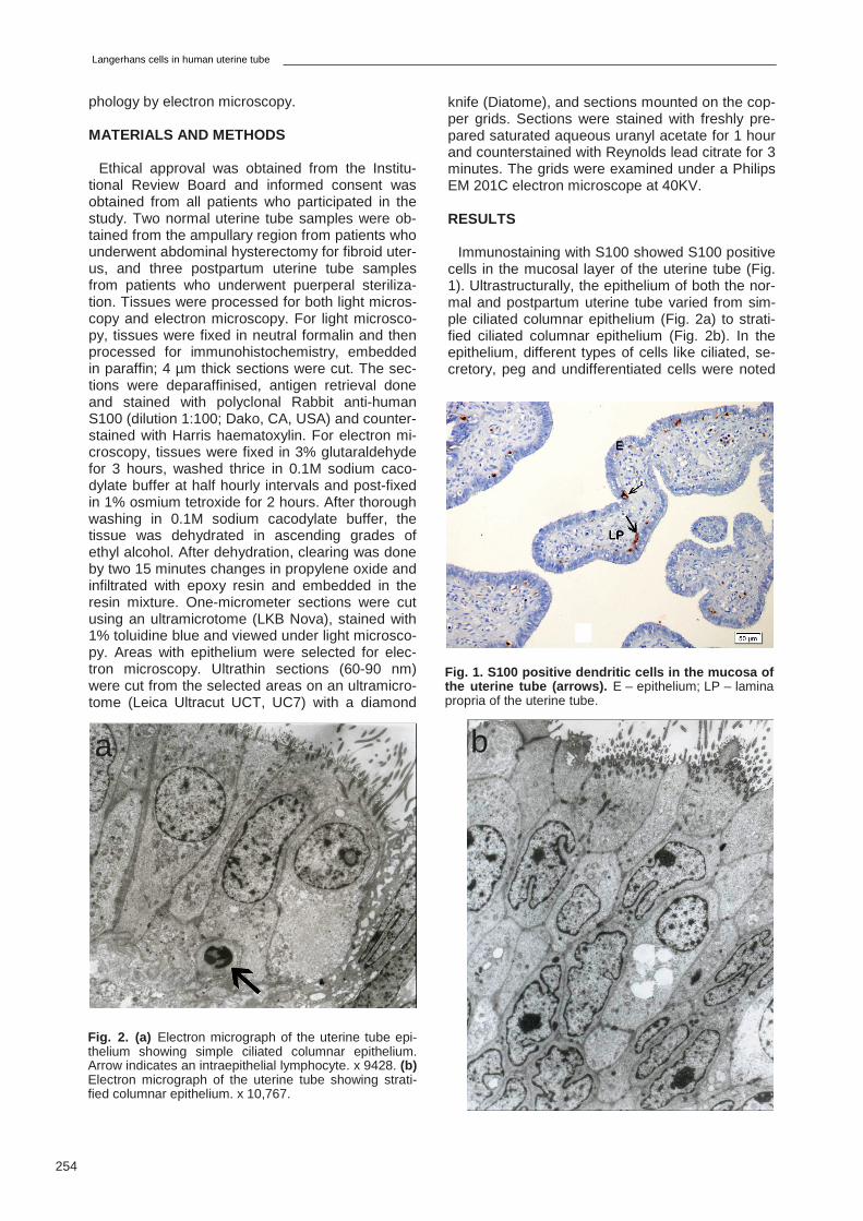
cells in the mucosal layer of the uterine tube (Fig. 1). Ultrastructurally, the epithelium of both the nor-mal and postpartum uterine tube varied from sim-ple ciliated columnar epithelium (Fig. 2a) to strati-fied ciliated columnar epithelium (Fig. 2b). In the epithelium, different types of cells like ciliated, se-cretory, peg and undifferentiated cells were noted
Fig. 1. S100 positive dendritic cells in the mucosa of the uterine tube (arrows). E – epithelium; LP – lamina propria of the uterine tube.
Fig. 2. (a) Electron micrograph of the uterine tube epi-thelium showing simple ciliated columnar epithelium. Arrow indicates an intraepithelial lymphocyte. x 9428. (b) Electron micrograph of the uterine tube showing strati-fied columnar epithelium. x 10,767.
a b
S. Rabi et al.
255
Fig. 3. (a) Pale ciliated cells (Ci) with euchromatic nucleus in the epithelium of the normal uterine tube. x 16,151. (b) A postpartum uterine tube showing a dark ciliat-ed columnar cell (Ci). x 14,303. (c) Epithelium of normal uterine tube showing secretory cells. Note the secretory granules in the supranuclear region. x 25,666. (d) The epithelium of uterine tube showing peg cells (Pg). x 6357. (e) A small undifferentiated cell (Ud) in the epithelium of a uterine tube. x 9535.
b
Fig. 4. (a) A dendritic cell (DC) with a single process extending towards the basement membrane in the uter-ine tube epithelium. x 9536. (b) dendritic cell (DC) in close apposition with a lymphocyte (L) in the uterine tube epithelium. x 25,500. (c) Arrow indicates a tennis racket-shaped Birbeck granule present in the process of a DC present in the uterine tube epithelium. Note the process is in close contact with a lymphocyte (L). x 36,428.
a
c
Langerhans cells in human uterine tube
256
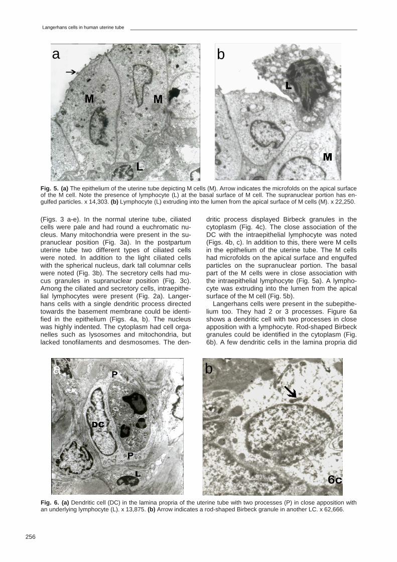
Fig. 5. (a) The epithelium of the uterine tube depicting M cells (M). Arrow indicates the microfolds on the apical surface of the M cell. Note the presence of lymphocyte (L) at the basal surface of M cell. The supranuclear portion has en-gulfed particles. x 14,303. (b) Lymphocyte (L) extruding into the lumen from the apical surface of M cells (M). x 22,250.
a b
a b
Fig. 6. (a) Dendritic cell (DC) in the lamina propria of the uterine tube with two processes (P) in close apposition with an underlying lymphocyte (L). x 13,875. (b) Arrow indicates a rod-shaped Birbeck granule in another LC. x 62,666.
(Figs. 3 a-e). In the normal uterine tube, ciliated cells were pale and had round a euchromatic nu-cleus. Many mitochondria were present in the su-pranuclear position (Fig. 3a). In the postpartum uterine tube two different types of ciliated cells were noted. In addition to the light ciliated cells with the spherical nucleus, dark tall columnar cells were noted (Fig. 3b). The secretory cells had mu-cus granules in supranuclear position (Fig. 3c). Among the ciliated and secretory cells, intraepithe-lial lymphocytes were present (Fig. 2a). Langer-hans cells with a single dendritic process directed towards the basement membrane could be identi-fied in the epithelium (Figs. 4a, b). The nucleus was highly indented. The cytoplasm had cell orga-nelles such as lysosomes and mitochondria, but lacked tonofilaments and desmosomes. The den-
dritic process displayed Birbeck granules in the cytoplasm (Fig. 4c). The close association of the DC with the intraepithelial lymphocyte was noted (Figs. 4b, c). In addition to this, there were M cells in the epithelium of the uterine tube. The M cells had microfolds on the apical surface and engulfed particles on the supranuclear portion. The basal part of the M cells were in close association with the intraepithelial lymphocyte (Fig. 5a). A lympho-cyte was extruding into the lumen from the apical surface of the M cell (Fig. 5b).
Langerhans cells were present in the subepithe-lium too. They had 2 or 3 processes. Figure 6a shows a dendritic cell with two processes in close apposition with a lymphocyte. Rod-shaped Birbeck granules could be identified in the cytoplasm (Fig. 6b). A few dendritic cells in the lamina propria did
S. Rabi et al.
257
not have Birbeck granules. The lamina propria of the postpartum uterine tube had lot of blood ves-sels. Dendritic cells were seen in apposition with the endothelial cells of the capillaries (Fig. 7). High endothelial venules lined by cuboidal endothelium were present in the lamina propria (Fig. 8).
DISCUSSION
The immune system of the female reproductive
system is under the influence of ovarian hormones oestradiol and progesterone, which prepare the reproductive tract for successful fertilization, im-plantation and pregnancy, while the epithelial cells of the female reproductive tract confer protection against potential pathogens by secreting bacteri-cidal agents that inhibit the growth of micro-organisms (Wira and Fahey, 2004). The epithelial lining of the uterine tube is composed of at least four types of cells: the ciliated columnar, secretory, peg and undifferentiated cells. Their ratio varies with hormone levels and position. Ciliated cells are much reduced after the menopause. Secretory cells are interspersed among ciliated cells. Peg cells are narrow columnar elements with oval nu-clei. They are thought to represent secretory cells in the non-secretory phase of their cycle. Undiffer-entiated cells are small cells restricted to the epi-thelial base. They are probably mainly stem cells for the ciliated and secretory cell populations (Williams et al., 1995).
Langerhans cells in uterine mucosa
Langerhans cells (LCs) are immature dendritic cells playing a sentinel role through their special-ized function in antigen capture, and their capacity to migrate to secondary lymphoid tissue to initiate specific immunity (Valladeau et al., 2003). Very
few studies have demonstrated the presence of LCs in the uterine tube (Hagiwara et al., 1996; Suganthy et al., 2006). In the present study, LCs were identified both in the epithelium and lamina propria of the uterine tube both by light microscopy and electron microscopy. It has been described that markers including CD1a (Krenácse et al., 1993), Langerin (Lau et al., 2008), S100, ATPase, MHC Class II (Tay et al., 1987) and ZIO (Niebauer et al., 1969) have been used to demonstrate the different subpopulations of LCs. S100 protein was first isolated from the bovine brain and later identi-fied in glial and Schwann cells of the nervous sys-tem, epidermal LCs and in other cells like myoepi-thelial cells of the salivary and mammary glands (Turusov, 1990). Most of the S100-positive cells of the lymphoreticular system are dendritic cells in-volved in the immune response and they belong to the mononuclear/phagocytic system. Although there is lack of specificity with S100 protein, it has been used as an immunohistological diagnostic marker for the malignacicies of the immune sys-tem, as it is specifically related to dendritic cell mi-croenvironment (Carbone et al., 1986). S100 pro-tein has been used to identify LCs in the vulva (Taube, 2007) and in uterine endometrium (Coppola, 1998). In the present study, the pres-ence of LCs in the uterine tube mucosa was con-firmed by staining the sections with S100. S100 positive cells were identified both in the epithelium and subepithelium. Ultrastructurally, LCs were identified by the presence of intended nucleus and lack of tonofilaments in the cytoplasm and desmo-somes. In the epithelium, the cells had a single process, which is directed towards the basement membrane. This is in accordance with the previous study of dendritic cells in uterine the tube using Zinc Iodide Osmium technique (Suganthy et al.,
Fig. 7. Arrow indicates an endothelial cell (En) lining the capillary. Note the LCs in close association with the ca-pillary. P – dendritic processes of the LCs. x 9709.
Fig. 8. A high endothelial venule in the lamina propria of a postpartum uterine tube. Arrows indicate the cuboidal endothelial cells. RBC – red blood corpuscle; N – neu-trophil. x 16,392.
Langerhans cells in human uterine tube
258
2006). The dendritic processes displayed the unique Birbeck granules. They are composed of Langerin and are part of endosomal recycling pathway (Romani et al., 2012). Although the Birbeck granules are thought to be important in antigen processing (Stossel et al., 1990), their functions are poorly understood. Birbeck granules could allow internalization of antigens and possibly their storage to delay presentation to T-lymphocytes (Valladeau et al., 2003).
In the subepithelium, LCs had two or more pro-cesses and they too displayed the Birbeck gran-ules. Close association of LCs and blood vessels was demonstrated. It has been suggested that the DCs may not be able to pass the endothelium of blood vessels, but with their long processes they form the right microenvironment to retain T cells and B cells during the process of antigen presenta-tion (Kraal et al., 1989).
M cells in the uterine tube
M cells are exclusively present in the dome areas that associated with the submucosal lymphoid folli-cles. Usually they are present in the small and large intestines. But they have been identified in other lymphoid organs like the palatine tonsils (Indrasingh et al., 1999) and adenoids (Claeys et al., 1996). The interactions of antigens with the apical surface of M cells play an important role in the initial step of intestinal and systemic immune responses or tolerance (Gebert et al., 1996). In the present study, M cells were identified electron mi-croscopically in the uterine tube. The apical sur-face was characterized by microfolds. The pres-ence of engulfed particles in the M cells confirmed their role in endocytotic uptake at the apical mem-brane, transcytotic transport and exocytotic re-lease of luminal substances to the intercellular space of the epithelium. M cells were seen in rela-tion to the intraepithelial lymphocytes at the base or in basolateral position. There is evidence that M cells express MHC class II molecules and are ca-pable of presenting antigens to lymphoid cells (Allan et al., 1993). Therefore it can be concluded that M cells are not only involved in the passive antigen transport, but also process and present antigens to the adjacent intraepithelial lympho-cytes. In the present study, the surface of M cell ruptured and lymphocytes were liberated into the lumen of uterine tube. The bursting of M cells was previously reported in inflamed mucosa and it was suggested that it could be either due to the hyper-plasia of the underlying lymphoid tissue which re-sulted in stretching of the M cell or due to increase in endosomes or the combination of both (Cuvelier, 1994).
Intraepithelial lymphocytes
T-Lymphocytes interspersed in the epithelium are called intraepithelial lymphocytes. They are components of the MALT and are predominantly
CD8+ (Cesta, 2006). They are important in cell-mediated immune responses, in the regulation of secretory immunity and in the mediation of system-ic tolerance (Gebert, 1996). They have been ex-tensively studied in the intestines (Mahadeva et al., 2002; Chang et al., 2005). The presence of intraepithelial lymphocytes in the uterine tube has been previously demonstrated by immunoelectron-microscopic study (Otsuki et al., 1989) and they have proposed that they migrate via the basal lam-ina from the underlying follicles. In the present study, not only the presence of intraepithelial lym-phocytes in the uterine tube epithelium but also their close association with dendritic cells and M cells has been demonstrated.
High endothelial venules
High endothelial venules (HEVs) are specialized postcapillary venules found in lymphoid tissues that support lymphocyte extravasation from the blood (Girard and Springer, 1995). They are lined by cuboidal epithelium. Recirculating lymphocytes migrate into peripheral lymphoid tissue through HEV (Duijvestijn and Hamann,1989). HEVs had been described in lymph nodes (Gowans and Knight, 1964; Farr, 1951; Farr and De Bruyn, 1975), Peyer’s patches (Schoefl, 1972) and tonsils (Sordat et al., 1971; Indrasingh et al., 2002). Otsu-ki et al. (1989) have reported that HEVs are absent in the uterine tube. But in the present study, HEVs could be identified in the subepithelium of the post-partum uterine tube by electron microscopy. As HEVs are recognized to be the selective site for lymphocytic migration, their presence in the human uterine tube confirms the uterine tube as part of MALT.
In conclusion, the demonstration of various com-ponents of the mucosal immune system like Lang-erhans cells, M cells, intraepithelial lymphocytes and HEVs in the human uterine tube by electron microscopy indicates that the uterine tube is an integral part of MALT. The antigen-presenting cells in the human uterine tube involve both in im-munostimulation while counteracting pathogens, and in immunotoleration in allowing successful fertilization. Our identification of the components of MALT in human uterine tube would result in further studies on the role of these cells in the female re-productive organs.
ACKNOWLEDGEMENTS
The authors gratefully acknowledge the Indian Council of Medical Research for funding this pro-ject.
REFERENCES
ALLAN CH, MENDRICK DL, TRIER JS (1993) Rat intes-
tinal M cells contain acidic endosomal-lysosomal com-
S. Rabi et al.
259
partments and express class II major histocompatibility complex determinants. Gastroenterology, 104: 698-708.
CESTA MF (2006) Normal structure, function, and his-tology of mucosa-associated lymphoid tissue. Toxicol Pathol, 34: 599-608.
CARBONE A, MANCONI R, POLETTI A, VOLPE R (1986) Significance of S-100 protein immunostaining in the immunohistological analysis of normal and neo-plastic lymphoid tissues – an appraisal. Int J Biol Markers, 1: 57-66.
CHANG F, MAHADEVA U, DEERE H (2005) Pathologi-cal and clinical significance of increased intraepithelial lymphocytes (IELs) in small bowel mucosa. APMIS, 113: 385-399.
CHUNG Y, CHANG J, KIM B, LEE J, KIM H, KANG C (2007) Anatomic location defines antigen presentation by dendritic cells to T cells in response to intravenous soluble antigens. E J Immunol, 37: 1453-1562.
CLAEYS S, CUVELIER C, QUATACKER J, VAN CAU-WENBERGE P (1996) Ultrastructural investigation of M-cells and lymphoepithelial contacts in naso-pharyngeal associated lymphoid tissue (NALT). Acta Otolaryngol Suppl, 523: 40-42.
COPPOLA D, FU L, NICOSIA SV, KOUNELIS S, JONES M (1998) Prognostic significance of p53, bcl-2, vimentin and s100 protein-positive Langerhans cells in endometrial carcinoma. Pathol, 29: 455-462.
CUVELIER CA, QUATACKER J, MIELANTS H, DE VOS M, VEYS E, ROELS HJ (1994) M-cells are dam-aged and increased in number in inflamed human ileal mucosa. Histopathology, 24: 417-426.
DUIJVESTIJN A, HAMANN A (1989) Mechanisms and regulation of lymphocyte migration. Immunol Today, 10: 23-28.
FARR AG, DE BRUYN PP (1975) The mode of lympho-cyte migration through postcapillary venule endotheli-um in lymph node. Am J Anat, 143: 59-92.
FARR RS (1951) Experiments on the fate of the lympho-cyte. Anat Rec, 109: 515-533.
FIGUEROA CD, CAORSI I (1980) Ultrastructural and morphometric study of the Langerhans cell in the nor-mal human exocervix. J Anat, 131: 669-682.
GEBERT A, ROTHKÖTTER HJ, PABST R (1996) M cells in Peyer’s patches of the intestine. Int Rev Cytol, 167: 91-159.
GIRARD JP, SPRINGER TA (1995) High endothelial venules (HEVs): specialized endothelium for lympho-cyte migration. Immunol Today, 16: 449-457.
GOWANS JL, KNIGHT EJ (1964) The route of re-circulation of lymphocytes in the rat. Proc R Soc Lond B Biol Sci, 159: 257-282.
HACHISUGA T, FUKUDA K, NAKAMURA S, IWASAKA T, SUGIMORI H (1997) Local immune response in endometrial carcinomas. Br J Obstet Gynaecol, 104: 110-114.
HAGIWARA H, OHWADA N, AOKI T, FUJIMOTO T (1998) Langerhans cells in the human oviduct muco-sa. Ital J Anat Embryol, 103: 253-258.
HAMRAH P, LIU Y, ZHANG Q, DANA MR (2003) Altera-tions in corneal stromal dendritic cell phenotype and distribution in inflammation. Arch Ophthalmol, 121: 1132-1140.
IIJIMA N, THOMPSON JM, IWASAKI A (2008) Dendritic cells and macrophages in the genitourinary tract. Mu-cosal Immunol, 1: 451-459.
INDRASINGH I, CHANDI G, ABRAHAM S (1999) Ultra-structural heterogeneity in the morphology of M cell in the human palatine tonsil. Ann Natl Acad Med Sci, 35: 83-95.
INDRASINGH I, CHANDI G, VETTIVEL S (2002) Route of lymphocyte migration through the high endothelial venule (HEV) in human palatine tonsil. Ann Anat, 184: 77-84.
JOHANSSON EL, RUDIN A, WASSÉN L, HOLMGREN J (1999) Distribution of lymphocytes and adhesion molecules in human cervix and vagina. Immunology, 96: 272-277.
KRAAL G (1989) Immunocytochemistry of dendritic cells. A clue to their function? Res Immunol, 140: 891-895.
KRENÁCS L, TISZALVICZ L, KRENÁCS T, BOUMSELL L (1993) Immunohistochemical detection of CD1A antigen in formalin-fixed and paraffin-embedded tissue sections with monoclonal antibody 010. J Pathol, 171: 99-104.
LAU SK, CHU PG, WEISS LM (2008) Immunohisto-chemical expression of Langerin in Langerhans cell histiocytosis and non-Langerhans cell histiocytic disor-ders. Am J Surg Pathol, 32: 615-619.
MAHADEVA S, WYATT JI, HOWDIE PD (2002) Is raised intraepithelial lymphocyte count with normal duodenal villous architecture clinically relevant? J Clin Pathol, 55: 424-428.
MORRIS HH, GATTER KC, STEIN H, MASON DY (1983) Langerhans’ cells in human cervical epithelium: an immunohistological study. Br J Obstet Gynaecol, 90: 400-411.
OTSUKI Y, MAEDA Y, MAGARI S, SUGIMOTO O (1989) Lymphatics and lymphoid tissue of the Fallopi-an tube: immunoelectronmicroscopic study. Anat Rec, 225: 288-296.
POPPE WA, DRIJKONINGEN M, IDE PS, LAU-WERYNS JM, VAN ASSCHE FA (1996) Langerhans’ cells and L1 antigen expression in normal and abnor-mal squamous epithelium of the cervical transfor-mation zone. Gynecol Obstet Invest, 41: 207-213.
RODRÍGUEZ EM, CAORSI I (1978) A second look at the ultrastructure of the Langerhans cell of the human epidermis. J Ultrastruct Res, 65: 279-295.
ROMANI N, BRUNNER PM, STINGL G (2012) Chang-ing views of the role of Langerhans cells. J Invest Der-matol, 132: 872-881.
SCHOEFL GI (1972) The migration of lymphocytes across the vascular endothelium in lymphoid tissue. A reexamination. J Exp Med, 136: 568-588.
Langerhans cells in human uterine tube
260
SORDAT B, HESS MW, COTTIER H (1971) IgG immu-noglobulin in the wall of post-capillary venules: possi-ble relationship to lymphocyte recirculation. Immunolo-gy, 20: 115-118.
STOSSEL H, KOCH F, KIMPGEN E, STOGER P, LENZ A, HEUFLER C, ROMANI N, SCHULER G (1990) Dis-appearance of certain acidic organelles (endosomes and Langerhans cell granules) accompanies loss of antigen processing capacity upon culture of epidermal Langerhans cells. J Exp Med, 172: 1471-1482.
SUGANTHY J, INDRASINGH I, LIONEL J (2006) Demonstration of ZIO positive Langerhans cells in the normal and the postpartum human Fallopian tubes. J Anat Soc India, 55: 1-4.
TAUBE JM, NICHOLS AD, BORNMAN LS, BORNMAN DM, JACKSON JB (2007) Langerhans cell density and high-grade vulvar intraepithelial neoplasia in women with human immunodeficiency virus infection. J Cutan Pathol, 34: 565-570.
TAY SK, JENKINS D, MADDOX P, CAMPION M, SING-ER A (1987) Subpopulations of Langerhans’ cells in cervical neoplasia. Brit J Obstet Gynaecol, 94: 10-15.
TURUSOV VS (1990) Protein S-100 in the histological diagnosis of tumours. Arkh Patol, 52: 71-80.
VALLADEAU J, RAVEL O, DEZUTTER-DAMBUYANT C, MOORE K, KLEIJMEER M, LIU Y, DUVERT-FRANCES V, VINCENT C, SCHMITT D, DAVOUST J, CAUX C, LEBECQUE S, SAELAND S (2000) Langerin, a novel C-type lectin specific to Langerhans cells, is an endocytic receptor that induces the for-mation of Birbeck granules. Immunity, 12: 71-81.
VALLADEAU J, DEZUTTER-DAMBUYANT C, SAELAND S (2003) Langerin/CD207 sheds light on formation of Birbeck granules and their possible func-tion in Langerhans cells. Immunol Res, 28: 93-107.
WIESER F, HOSMANN J, TSCHUGGUEL W, CZER-WENKA K, SEDIVY R, HUBER JC (2001) Progester-one increases the number of Langerhans cells in hu-man vaginal epithelium. Fertil Steril, 75: 1234-1235.
WILLIAMS PL, BANNISTER LH, BERRY MM, COLLINS P, DYSON M, DUSSEK JE, FERGUSON MWJ (1995) Reproductive organs of the female. In: Bannister LH, Dyson M (eds). Gray’s Anatomy. The Anatomical basis of Medicine and Surgery. 38th ed. Churchill Living-stone, Great Britain, pp 1867.
WIRA CR, FAHEY JV (2004) The innate immune sys-tem: gatekeeper to the female reproductive tract. Im-munology, 1: 13-15.