Ultrastructure within the Lateral Plexus of the Limulus Eye MOSHE GUR, RICHARD L. PURPLE, and RUSSELL WHITEHEAD From the Laboratory of Neurophysiology, Department of Physiology, University of Minnesota Medical School, Minneapolis, Minnesota 55455, and the Department of Biology, Macalester College, St. Paul, Minnesota 55104 ABSTRACT The ultrastructure of the lateral plexus in the compound eye of Limulus is investigated by serial section technique. "Cores" of tissue containing the axons, lateral plexus, and neuropile associated with one sensory ommatidium show the following features: (a) collateral branches from retinular cells do not contribute to the lateral plexus proper, but do form "retinular neuropile" by contacting collaterals of a self-contained cluster of retinular axons; (b) col- lateral branches from eccentric cell axons always branch repeatedly upon leaving the parent axon, and compose the bulk of the lateral plexus; (c) the most distal collateral branches from an eccentric cell axon appear to form neuropile and synaptic contacts with each other, whereas more proximal branches form synaptic contacts with collaterals from eccentric cell axons of neighboring ommatidia. We conclude that the ribbon synapses and associated transmitter substance in eccentric cell collaterals must be inhibitory, and that two pathways for self-inhibition may exist. We suggest, as a working hypothesis for the structure of the lateral plexus, a branching pattern with depth that mirrors the horizontal spread of lateral inhibition measured physiologically. INTRODUCTION Function of the lateral plexus of the Limulus eye has been studied extensively (for recent reviews see Hartline, 1969 and Knight et al., 1970), but little direct information is available on the fine structure of the plexus that under- lies its function. Miller's excellent light microscopy (in Hartline et al., 1956 and Ratliff et al., 1958) reveals extensive branching between ommatidia, but the thickness of the sections and the nonselectivity of the silver stain make difficult an interpretation of relative contributions made by retinular and eccentric cells to the plexus. Moreover, Miller's electron micrographs (in Ratliff et al., 1958; Hartline et al., 1961; and Ratliff et al., 1963) show that the dense pattern of branching within the lateral plexus, and more particularly within the neuropile areas where synaptic contacts have been described THE JOURNAL OF GENERAL PHYSIOLOGY VOLUME 59, 1972 pages 285-304 285 on April 5, 2019 jgp.rupress.org Downloaded from http://doi.org/10.1085/jgp.59.3.285 Published Online: 1 March, 1972 | Supp Info:
Transcript
Ultrastructure within the Lateral
Plexus of the Limulus Eye
MOSHE GUR, RICHARD L. PURPLE,
and RUSSELL WHITEHEAD
From the Laboratory of Neurophysiology, Department of Physiology, University of MinnesotaMedical School, Minneapolis, Minnesota 55455, and the Department of Biology, MacalesterCollege, St. Paul, Minnesota 55104
ABSTRACT The ultrastructure of the lateral plexus in the compound eye ofLimulus is investigated by serial section technique. "Cores" of tissue containingthe axons, lateral plexus, and neuropile associated with one sensory ommatidiumshow the following features: (a) collateral branches from retinular cells do notcontribute to the lateral plexus proper, but do form "retinular neuropile" bycontacting collaterals of a self-contained cluster of retinular axons; (b) col-lateral branches from eccentric cell axons always branch repeatedly uponleaving the parent axon, and compose the bulk of the lateral plexus; (c) themost distal collateral branches from an eccentric cell axon appear to formneuropile and synaptic contacts with each other, whereas more proximalbranches form synaptic contacts with collaterals from eccentric cell axons ofneighboring ommatidia. We conclude that the ribbon synapses and associatedtransmitter substance in eccentric cell collaterals must be inhibitory, and thattwo pathways for self-inhibition may exist. We suggest, as a working hypothesisfor the structure of the lateral plexus, a branching pattern with depth thatmirrors the horizontal spread of lateral inhibition measured physiologically.
INTRODUCTION
Function of the lateral plexus of the Limulus eye has been studied extensively(for recent reviews see Hartline, 1969 and Knight et al., 1970), but littledirect information is available on the fine structure of the plexus that under-lies its function. Miller's excellent light microscopy (in Hartline et al., 1956
and Ratliff et al., 1958) reveals extensive branching between ommatidia, butthe thickness of the sections and the nonselectivity of the silver stain makedifficult an interpretation of relative contributions made by retinular andeccentric cells to the plexus. Moreover, Miller's electron micrographs (inRatliff et al., 1958; Hartline et al., 1961; and Ratliff et al., 1963) show thatthe dense pattern of branching within the lateral plexus, and more particularlywithin the neuropile areas where synaptic contacts have been described
THE JOURNAL OF GENERAL PHYSIOLOGY VOLUME 59, 1972 pages 285-304 285
on April 5, 2019jgp.rupress.org Downloaded from http://doi.org/10.1085/jgp.59.3.285Published Online: 1 March, 1972 | Supp Info:
286 THE JOURNAL OF GENERAL PHYSIOLOGY VOLUME 59 ' 1972
(Whitehead and Purple, 1970), are beyond the resolving power of the lightmicroscope.
The lateral plexus is "large",' but a limited, ultrastructural, serial sectionstudy of the lateral plexus appears worthwhile because of the importance ofthe lateral eye in neurophysiology and because of the potential help this studycould bring to understanding the neuropile in general; meaningful questionsof neuronal specificity and genetic determination of branching patterns aremore easily formulated and answered if the normal end product is known.
Our approach has been to study "cores" of tissue each containing at least asingle ommatidium, its axons, and associated plexus and neuropile. The coresare approximately 150 A in diameter and extend some 300-500 A proximalfrom the eccentric cell body. In addition to studying adult animals (15 cmprosomal width), young, intermolt animals and developing eyes of embryosare being studied, although this report will deal almost exclusively with adults. 2
METHODS
1. Material Adult, intermolt animals of Limulus polyphemus from Cortez,Florida, were maintained in "Instant Ocean" aquaria (Aquarium Systems, Inc.,Eastlake, Ohio), exposed to a 12 hr light, 12 hr dark regime. Methods of fixation andembedding have been published (Whitehead and Purple, 1970). Although goodfixation has been obtained using a number of different combinations of bufferedglutaraldehyde and postfixing in osmium, virtually all methods employed (see, forinstance, Fahrenbach, 1969) suffer from "spottiness." Even within the same block oftissue some ommatidia fix well while neighboring ommatidia show poor fixation.Subsequent to most of the work reported here, the general take of the fixative wasmuch improved by killing and fixing immediately upon receipt of the animals, thusindicating that storage, even in well-maintained aquaria, may work physical changeson Limulus.
2. Procedure To obtain cross-sections perpendicular to the cornea, blocks oftissue (about I mm square) were oriented and sectioned from the cornea proximallyto the level of the eccentric cell body. The tissue blocks were then trimmed to about0.7 X 0.6 mm, oriented a final time, and serial sectioning was begun. Serial sectionsaveraged 90.0 nm (900 A) in thickness and the number of sections multiplied by theaverage thickness served as the primary measure of distance. The sections werefloated onto Formvar-coated (Belden Mfg. Co., Chicago, Ill.), carbon-stabilized
1 In adult animals, the lateral plexus is a sheet of branching axon collaterals some 500 thick andgreater than 1 cm2 in area. Within the lateral plexus are regions of neuropile that surround ec-centric cell axons, extending out as much as 30 pA and ranging in depth along the axons from 20to 300 p.2 Patten (1912) and Waterman (1954) have suggested that the lateral eye of the adult Limulus,which has received the brunt of neurophysiological studies on the species, may be a degeneratingorgan. To correlate structure and function, the larger animals have thus been studied, although itenhances the difficulties of the serial section approach. Reports on the younger animals and onembryological material will follow later.
GUR, PURPLE, AND WHITEEAD Lateral Plexus of Limulus Eye 287
grids, and were observed and photographed using a Hitachi HU-11 or an RCA-EMU3G electron microscope.
A typical core of tissue included the axons from the primary ommatidium in ad-dition to parts of the neighboring ommatidia on each side. Each core required some2000-3000 sections to follow the axons of an ommatidium through most of the plexusarea. The diameter of the ommatidia studied ranged between 150-200 pa at the distalportion, and 60-100 ji proximal to the sensory portion of the ommatidium in theregion of the lateral plexus. About five to seven picutres at a magnification of 3000were required to construct a montage covering the whole central region of a section.Where fine details of collateral branches or of synaptic regions were desired, ad-ditional pictures of magnification 15,000 or greater were obtained. Because economyof time and resources dictated that photographs and montages of each section couldnot be made on adult tissue, we resorted to the following procedure. Nearly 100%of sections available were observed and charted using the electron microscope. Thequality (see next Section) of each section was estimated as well as the possible useablearea if photographs were desired later. The quality of the sections, and the extent ofgross changes observed, dictated the spacing of photographs for careful evaluation.Photography was often done in a second round of section scanning.
3. Quality Control The percentage of "publication perfect" photographsobtained in a study such as this is small compared to the number of sections andphotographs that, while technically defective, may still be used for tracing andreconstruction. As often as not, an important change, worthy of illustrating thenature of the branching pattern, will occur in a section whose photographic qualityis not good. As a guide to our terminology, we indicate for each of the figures il-lustrated the quality of the section, using the terms good, fair, and useable. Theseterms indicate the general condition of the section for tracing purposes.
4. Lost Sections The number of unuseable sections and of sections destroyed inhandling before electron microscope inspection were recorded along with the useablesections. For tracing purposes, 6-10 consecutive sections can be lost without inter-rupting the ability to follow the course of the larger collaterals, or of a major struc-tural change in the neuropile. Conversely, the loss of just one or two sections makesit impossible to trace perfectly the very fine collaterals. This latter problem makesparticularly frustrating the task of producing a detailed, ultrastructural reconstruc-tion of the core tissue.
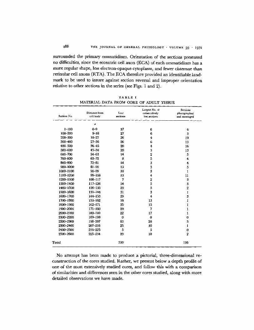
5. Nomenclature Distal refers to the direction towards the eccentric cell bodyand the periphery. Proximal denotes away from the eccentric cell body and towardsthe optic nerve proper. Table I summarizes the essential material data at hand fromthe core of adult tissue presented in detail in this report. The data are typical foreach of the three cores studied in most detail.
RESULTS
Three cores of adult material were studied in some detail. Areas from eightother cores were studied in less detail, including areas from ommatidia which
288 THE JOURNAL OF GENERAL PHYSIOLOGY · VOLUME 59 1972
surrounded the primary ommatidium. Orientation of the sections presentedno difficulties, since the eccentric cell axon (ECA) of each ommatidium has amore regular shape, less electron-opaque cytoplasm, and fewer cisternae thanretinular cell axons (RTA). The ECA therefore provided an identifiable land-mark to be used to insure against section reversal and improper orientationrelative to other sections in the series (see Figs. 1 and 2).
TABLE I
MATERIAL DATA FROM CORE OF ADULT TISSUE
Largest No. of SectionsDistance from Lost consecutively photographed
Section No. cell body sections lost sections and montaged
No attempt has been made to produce a pictorial, three-dimensional re-construction of the cores studied. Rather, we present below a depth profile ofone of the most extensively studied cores, and follow this with a comparisonof similarities and differences seen in the other cores studied, along with moredetailed observations we have made.
GUR, PURPLE, AND WHITEHEAD Lateral Plexus of Limulus Eye 289
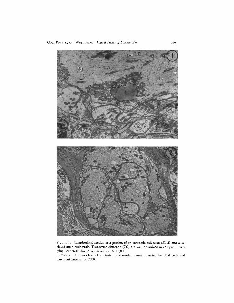
FIGURE 1. Longitudinal section of a portion of an eccentric cell axon (ECA) and asso-ciated axon collaterals. Transverse cisternae (TC) are well organized in compact layerslying perpendicular to neurotubules. X 16,000.FIGURE 2. Cross-section of a cluster of retinular axons bounded by glial cells andbasement lamina. X 7500.
THE JOURNAL OF GENERAL PHYSIOLOGY · VOLUME 59 I1972
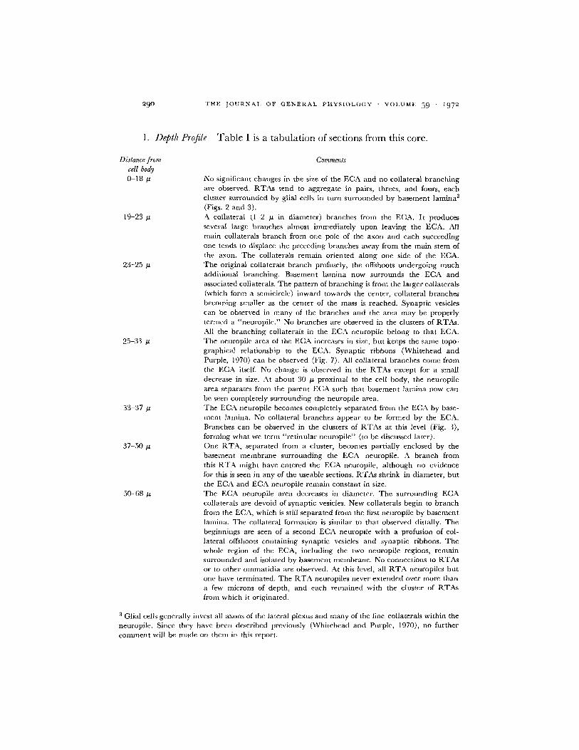
1. Depth Profile Table I is a tabulation of sections from this core.
Distance from Commentscell body0-18 g/ No significant changes in the size of the ECA and no collateral branching
are observed. RTAs tend to aggregate in pairs, threes, and fours, eachcluster surrounded by glial cells in turn surrounded by basement lamina3
(Figs. 2 and 3).19-23 pa A collateral (1-2 u in diameter) branches from the ECA. It produces
several large branches almost immediately upon leaving the ECA. Allmain collaterals branch from one pole of the axon and each succeedingone tends to displace the preceding branches away from the main stem ofthe axon. The collaterals remain oriented along one side of the ECA.
23-25t The original collaterals branch profusely, the offshoots undergoing muchadditional branching. Basement lamina now surrounds the ECA andassociated collaterals. The pattern of branching is from the larger collaterals(which form a semicircle) inward towards the center, collateral branchesbecoming smaller as the center of the mass is reached. Synaptic vesiclescan be observed in many of the branches and the area may be properlytermed a "neuropile." No branches are observed in the clusters of RTAs.All the branching collaterals in the ECA neuropile belong to that ECA.
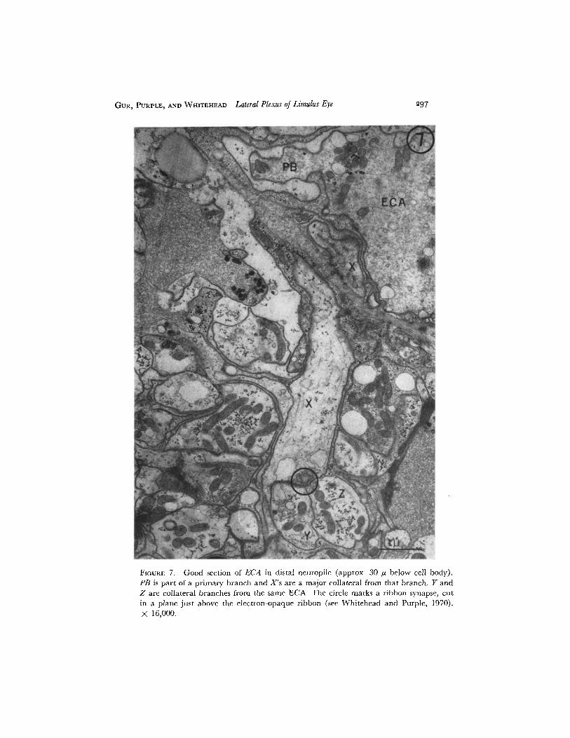
25-33 p The neuropile area of the ECA increases in size, but keeps the same topo-graphical relationship to the ECA. Synaptic ribbons (Whitehead andPurple, 1970) can be observed (Fig. 7). All collateral branches come fromthe ECA itself. No change is observed in the RTAs except for a smalldecrease in size. At about 30 pA proximal to the cell body, the neuropilearea separates from the parent ECA such that basement lamina now canbe seen completely surrounding the neuropile area.
33-37 u The ECA neuropile becomes completely separated from the ECA by base-ment lamina. No collateral branches appear to be formed by the ECA.Branches can be observed in the clusters of RTAs at this level (Fig. 3),forming what we term "retinular neuropile" (to be discussed later).
37-50 u One RTA, separated from a cluster, becomes partially enclosed by thebasement membrane surrounding the ECA neuropile. A branch fromthis RTA might have entered the ECA neuropile, although no evidencefor this is seen in any of the useable sections. RTAs shrink in diameter, butthe ECA and ECA neuropile remain constant in size.
50-68 p The ECA neuropile area decreases in diameter. The surrounding ECAcollaterals are devoid of synaptic vesicles. New collaterals begin to branchfrom the ECA, which is still separated from the first neuropile by basementlamina. The collateral formation is similar to that observed distally. Thebeginnings are seen of a second ECA neuropile with a profusion of col-lateral offshoots containing synaptic vesicles and synaptic ribbons. Thewhole region of the ECA, including the two neuropile regions, remainsurrounded and isolated by basement membrane. No connections to RTAsor to other ommatidia are observed. At this level, all RTA neuropiles butone have terminated. The RTA neuropiles never extended over more thana few microns of depth, and each remained with the cluster of RTAsfrom which it originated.
3 Glial cells generally invest all axons of the lateral plexus and many of the fine collaterals within theneuropile. Since they have been described previously (Whitehead and Purple, 1970), no furthercomment will be made on them in this report.
290
GUR, PURPLE, AND WHITEHEAD Lateral Plexus of Limulus Eye
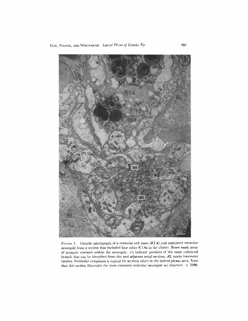
FIGURE 3. Useable micrograph of a retinular cell axon (RTA) and associated retinularneuropile from a section that included four other RTAs in the cluster. Boxes mark areasof synaptic contacts within the neuropile. x's indicate portions of the same collateralbranch that can be identified from this and adjacent serial sections. BL marks basementlamina. Retinular cytoplasm is typical for sections taken in the lateral plexus area. Notethat this section illustrates the most extensive retinular neuropile we observed. X 7000.
29I
292 THE JOURNAL OF GENERAL PHYSIOLOGY - VOLUME 59 1972
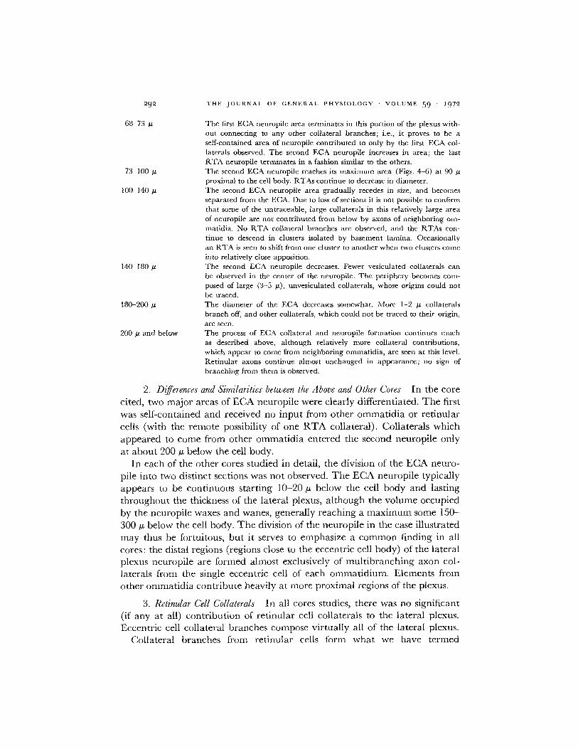
68-73
73-100
100-140 .
140-180 u
180-200
200 /s and below
The first ECA neuropile area terminates in this portion of the plexus with-out connecting to any other collateral branches; i.e., it proves to be aself-contained area of neuropile contributed to only by the first ECA col-laterals observed. The second ECA neuropile increases in area; the lastRTA neuropile terminates in a fashion similar to the others.The second ECA neuropile reaches its maximum area (Figs. 4-6) at 90 Aproximal to the cell body. RTAs continue to decrease in diameter.The second ECA neuropile area gradually recedes in size, and becomesseparated from the ECA. Due to loss of sections it is not possible to confirmthat some of the untraceable, large collaterals in this relatively large areaof neuropile are not contributed from below by axons of neighboring om-matidia. No RTA collateral branches are observed, and the RTAs con-tinue to descend in clusters isolated by basement lamina. Occasionallyan RTA is seen to shift from one cluster to another when two clusters comeinto relatively close apposition.The second ECA neuropile decreases. Fewer vesiculated collaterals canbe observed in the center of the neuropile. The periphery becomes comn-posed of large (3-5 u), unvesiculated collaterals, whose origins could notbe traced.The diameter of the ECA decreases somewhat. More 1-2 ,u collateralsbranch off, and other collaterals, which could not be traced to their origin,are seen.The process of ECA collateral and neuropile formation continues muchas described above, although relatively more collateral contributions,which appear to come from neighboring ommatidia, are seen at this level.Retinular axons continue almost unchanged in appearance; no sign ofbranching from them is observed.
2. Differences and Similarities between the Above and Other Cores In the corecited, two major areas of ECA neuropile were clearly differentiated. The firstwas self-contained and received no input from other ommatidia or retinularcells (with the remote possibility of one RTA collateral). Collaterals whichappeared to come from other ommatidia entered the second neuropile onlyat about 200 o below the cell body.
In each of the other cores studied in detail, the division of the ECA neuro-pile into two distinct sections was not observed. The ECA neuropile typicallyappears to be continuous starting 10-20 tu below the cell body and lastingthroughout the thickness of the lateral plexus, although the volume occupiedby the neuropile waxes and wanes, generally reaching a maximum some 150-300 , below the cell body. The division of the neuropile in the case illustratedmay thus be fortuitous, but it serves to emphasize a common finding in allcores: the distal regions (regions close to the eccentric cell body) of the lateralplexus neuropile are formed almost exclusively of multibranching axon col-laterals from the single eccentric cell of each ommatidium. Elements fromother ommatidia contribute heavily at more proximal regions of the plexus.
3. Retinular Cell Collaterals In all cores studies, there was no significant(if any at all) contribution of retinular cell collaterals to the lateral plexus.Eccentric cell collateral branches compose virtually all of the lateral plexus.
Collateral branches from retinular cells form what we have termed
GUR, PURPLE, AND WHITEHEAD Lateral Plexus of Limulus Eye
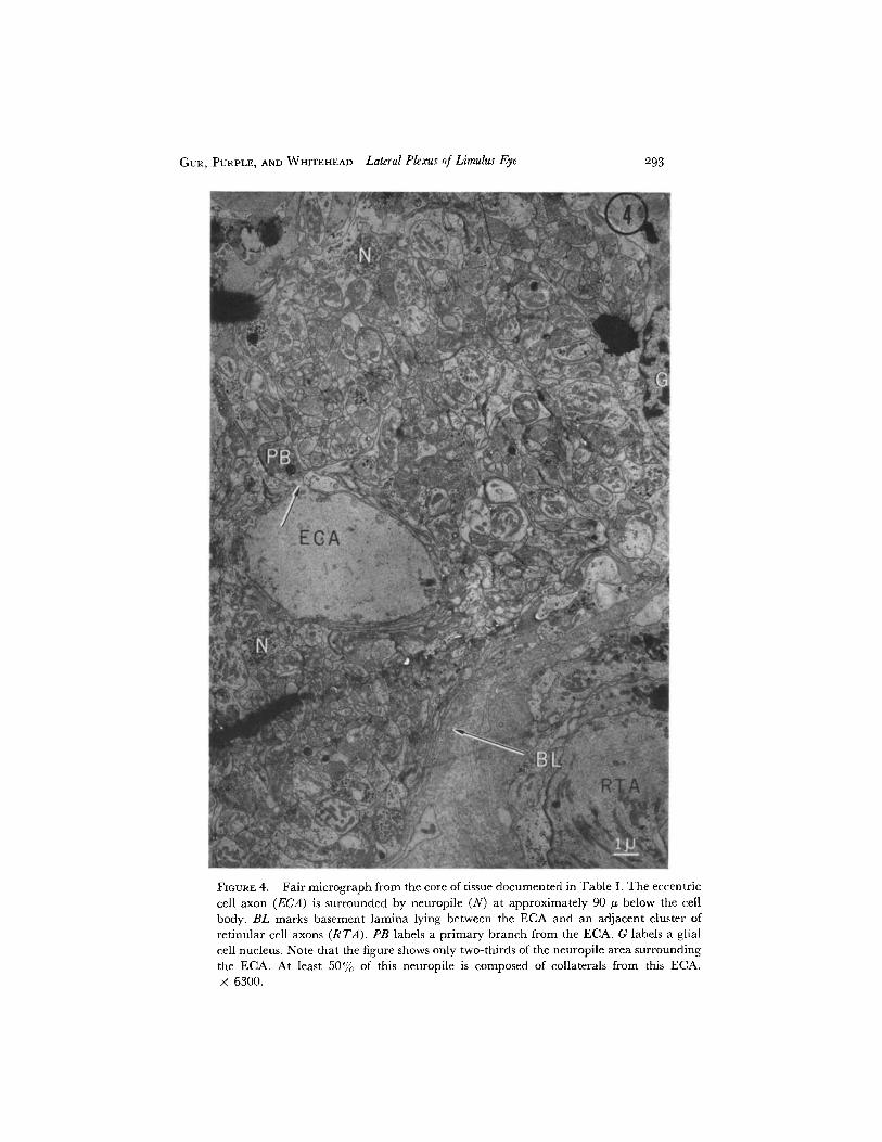
FIGURE 4. Fair micrograph from the core of tissue documented in Table I. The eccentriccell axon (ECA) is surrounded by neuropile (N) at approximately 90 A below the cellbody. BL marks basement lamina lying between the ECA and an adjacent cluster ofretinular cell axons (RTA). PB labels a primary branch from the ECA. G labels a glialcell nucleus. Note that the figure shows only two-thirds of the neuropile area surroundingthe ECA. At least 50% of this neuropile is composed of collaterals from this ECA.X 6300.
293
294 THE JOURNAL OF GENERAL PHYSIOLOGY - VOLUME 59 - 1972
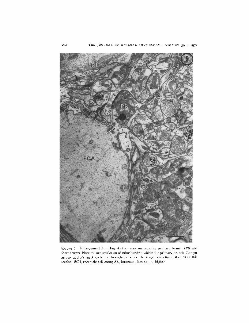
FIGURE 5. Enlargement from Fig. 4 of an area surrounding primary branch (PB and
short arrow). Note the accumulation of mitochondria within the primary branch. Longerarrows and x's mark collateral branches that can be traced directly to the PB in this
section. ECA, eccentric cell axon; BL, basement lamina. X 16,000.
GUR, PURPLE, AND WHITEHEAD Lateral Plexus of Limulus Eve 295
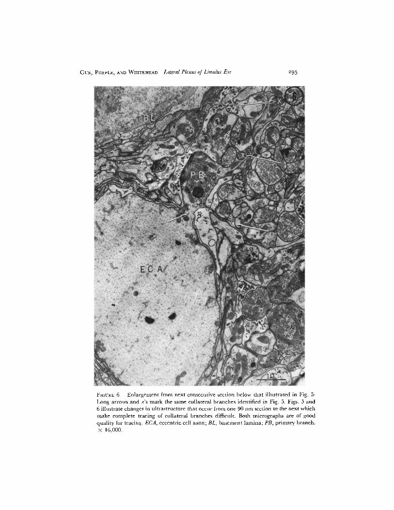
FIGURE 6. Enlargement from next consecutive section below that illustrated in Fig. 5.
Long arrows and x's mark the same collateral branches identified in Fig. 5. Figs. 5 and
6 illustrate changes in ultrastructure that occur from one 90 nm section to the next which
make complete tracing of collateral branches difficult. Both micrographs are of good
296 THE JOURNAL OF GENERAL PHYSIOLOGY · VOLUME 59 · 1972
"retinular neuropile" (see Fig. 3). Retinular neuropile is composed exclu-sively of branches from retinular cells of a cluster, which after traversingdistances of less than 50 y terminate without making contact with structuresother than collaterals from the same cluster of retinular cells. Retinularneuropile is small in cross-sectional area, short, and lacks the very fine branch-ing pattern seen in eccentric cell neuropile. The retinular collaterals do con-tain some synaptic vesicles and synaptic ribbons which appear similar tothose of eccentric cell collaterals (see Whitehead and Purple, 1970).
4. Collateral Branching Pattern and Structure of the ECA Neuropile Themost distal ECA collaterals terminate after giving rise to the initial neuropile.In all cores studied, each ECA collateral repeatedly sends out clusters ofsmaller branches within 2-3 yt of leaving the ECA. The neuropile near theECA is therefore composed of at least 50% by volume of its own collateralbranches. Close to the cell body, this figure approaches 100%. Our observa-tions appear to rule out the possibility that any significant number of ECAcollaterals proceed directly without branching to neighboring ommatidia.Figs. 5-8 illustrate the above points.
The large collaterals remain grouped about the periphery of the neuropile,and the pattern of branching is from the outside in. In the neuropile there isa wide spectrum of collateral diameters. The smallest, usually located in thecenter of the mass, are less than 0.3 , while the larger collaterals at theperiphery range up to 5 t in diameter. The rate of change with distance inneuropile structure appears to depend upon fiber size. As noted in theMethods section, a major collateral and its large subbranches leave the ECA(what we call a major structural change), over 10-15 u of the axon length(approximately 100-150 thin sections). Thus, in large collaterals, a loss of6-10 consecutive sections did not interrupt our ability to trace these collateralsor to note major branching from them. A landmark which aided in tracingthe branching pattern was the clustering of mitochondria at branch points.Within the ECA, that portion, or "pole," of the axon where branches leftwas always marked by an accumulation of mitochondria around the brancharea (Fig. 5).
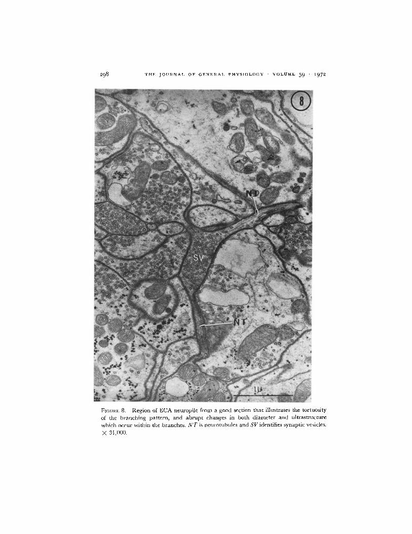
The finer collaterals within the neuropile have very tortuous courses,branch repeatedly, and are almost impossible to trace for distances of morethan a few microns. The loss of one thin section makes certain identificationand tracing of the fine collaterals a tenuous thing indeed. This difficulty intracing was due as much to the relatively abrupt changes in the ultrastructureof the collaterals as to their abrupt changes in course. Portions of the finecollaterals would be packed with synaptic vesicles, other portions relativelydevoid of all electron-opaque material except for neurotubules (Figs. 5-8).Synaptic ribbons appeared scattered along the course of the collaterals withno hint of placement according to any ordered pattern, except for their
GUR, PURPLE, AND WHITEHEAD Lateral Plexus of Limulus Eye 297
FIGURE 7. Good section of ECA in distal neuropile (approx. 30 /z below cell body).PB is part of a primary branch and X's are a major collateral from that branch. Y andZ are collateral branches from the same ECA. The circle marks a ribbon synapse, cutin a plane just above the electron-opaque ribbon (see Whitehead and Purple, 1970).X 16,000.
THE JOURNAL OF GENERAL PHYSIOLOGY - VOLUME 59 1972
FIGURE 8. Region of ECA neuropile from a good section that illustrates the tortuosity
of the branching pattern, and abrupt changes in both diameter and ultrastructure
which occur within the branches. NT is neurotubules and SV identifies synaptic vesicles.
X 31,000.
298
GUR, PURPLE, AND WHITEHEAD Lateral Plexus of Limulus Eye 299
tendency to occur where three branches came into close apposition (Whiteheadand Purple, 1970). While counts of synaptic ribbons tended to be higher infine collateral branches (less than 1 u in diameter), the ribbons occurred inlarger collateral branches as well.
5. Unidentified Collaterals In each of the cores studied, the number ofunidentified collaterals increased with distance from the eccentric cell body.We attribute most of these to input from neighboring ommatidia, althoughwe could not trace the collaterals to their origins. In one core, we also notedtwo small axons that appeared at the level of the eccentric cell body whereserial sectioning was commenced. It was possible to trace these axons toapproximately 35 u below the eccentric cell body, where their relative positionat this level was displaced outside the section. These small axons did notbranch, although they did run inside the basement lamina surrounding thedistal neuropile for approximately 15 u. We cannot say whether they areafferent or efferent, although they bore a close resemblance to axon structuresin the Limulus lateral eye which Fahrenbach (1970) has labeled as neuro-secretory efferents. We did not observe any other type of cell which could belabeled as an interneuron in the plexus, although pigment cell processes fromcell bodies located in the ommatidia did accompany the axon bundles forseveral microns below the sensory portion of the ommatidia.
DISCUSSION
Our results are based mainly on serial section studies from three cores of adultmaterial. Areas of eight other cores were studied in less detail. Conclusionsdrawn must be tempered by the relatively small sample size. We feel, how-ever, that the sample is large enough to warrant a number of conclusions,inferences, and even frank speculations. From a macroscopic view, the lateraleye of Limulus is a homogeneous receptor complex and retina (Ratliff, 1965).Individual variations in ommatidial physiology have been noted often,however, and it appears reasonable to assume that some of these variationsmay have a structural basis that might correlate with differences noted in oursample.
1. Retinular Cells and the Lateral Plexus Collaterals from RTAs contributelittle, if any, material to the lateral plexus proper. The one possible exceptionnoted by us is listed as a possible exception because we could not trace com-pletely the extent of that one RTA's penetration into the ECA neuropile. Ofthe 200 thin sections covering this region, 40 were lost or unusable, but thelargest number of consecutive sections lost was only four (see Table I). Weare relatively confident that, had the RTA collateral branched and con-tributed significantly to the ECA neuropile, we would have detected it.
Otherwise, the organization of RTA collaterals is so distinctly different
3oo00 THE JOURNAL OF GENERAL PHYSIOLOGY - VOLUME 59 I1972
from that of ECA collaterals that we ascribe the term "retinular neuropile"to the organization of the RTA clusters and their collaterals. It remains to beshown: (a) if the RTA neuropile in Limulus is of functional significance and(b) if in other species (or in immature Limulus) the terminology can be useful.From a comparative viewpoint, the retinular neuropile in Limulus could beeither a primitive or a degenerate anlage.
2. Eccentric Cells and the Lateral Plexus Four points will be discussed,followed by a "working hypothesis" for the structure of the lateral plexus.The first point is that the neuronal elements of the lateral plexus proper arealmost exclusively eccentric cells. We conclude that synaptic ribbons observedpreviously (Whitehead and Purple, 1970), including ribbon synapses oppositeeach other, were eccentric cell in origin. Heretofore, two hypotheses existed toaccount for the spread of lateral inhibition (see, for example, Barlow, 1967):(a) that it was eccentric cell to eccentric cell, and that the synaptic transmitteragent within eccentric cell branches was inhibitory; (b) that a possibleinternuncial pathway featuring either RTA collaterals or another neuron(Graziadie, as quoted by Schwartz, 1971) existed-the synaptic transmitterof eccentric cells being excitatory and the transmitter agent of retinular cellsor another internuncial being inhibitory. We reject the second hypothesis infavor of the first.
The second point is that eccentric cell collaterals make synaptic contactswith collaterals from the same ECA. For self-inhibition in the eye of Limulus(Stevens, 1964; Purple and Dodge, 1966), we now hold that two possiblepathways may exist which are not mutually exclusive. We have reported herethe pathway of collateral to collateral from the same cell. Previously(Whitehead and Purple, 1970), the demonstration of ribbon synapses directlyopposite each other was interpreted as morphological evidence favoring thehypothesis of a synapse whose transmitter agent could affect both the pre- andpostsynaptic endings (two-way chemical transmission as postulated by Purpleand Dodge, 1966). The existence of collateral-to-collateral connections fromthe same cell does not alter the status of the "two-way chemical synapse"pathway. Indeed one can contend that this relatively primitive neuronalsystem in which neurons appear incapable of discriminating between branchesfrom themselves and another cell, might have just the biochemical substratumrequired for a nonselective, two-way synapse.
The third point is that the branching pattern of the lateral plexus involvesECA collaterals which literally produce a shower of subbranches near theECA. The same large collaterals must make up the lateral plexus whichfunctionally connects the ommatidia. M. Behrens of the Masonic MedicalResearch Laboratory has kindly allowed us to view her slides of sections fromeccentric and retinular cells that were injected with Procion yellow. Thefluorescent pattern of stain in her sections and in those of Schwartz (1971)
GUR, PURPLE, AND WHITEHEAD Lateral Plexus of Limulus Eye 301
show a clustering of points in neuropile regions, and also indicate only shortRTA collaterals. Thus, results of Procion yellow staining are in general agree-ment with our findings, although the resolution of the fluorescent stain is toolimited to be taken as direct confirmation of the ultrastructural patterns re-ported here.
Given the three points above, we conclude that the physiological mech-anisms of self-inhibition (Stevens, 1964; Purple and Dodge, 1966), of synapticdelay and threshold for lateral inhibition (Hartline et al., 1956; Hartline andRatliff, 1957), and also of the depolarizing and hyperpolarizing componentsof lateral inhibition (Tomita et al., 1960; Purple, 1964; Knight et al., 1970)must be resolved within the structural context of the ribbon synapses in thefine collateral branches of the neuropile. Finally, Purple and Dodge (1965)have proposed a cable model for interpreting intracellular recordings fromthe eccentric cell. As part of this cable model, the requirement exists for alength of passive axon and/or axon collaterals between the cell body and boththe site of impulse encoding and the site of postsynaptic inhibition. We sug-gest that a major site of the passive cable resides in the fine collaterals of theneuropile. Propagated action potentials must be generated somewhere in thecollaterals giving rise to the fine fiber system of the neuropile, since virtuallyall of these larger collaterals (except the distal ones which end on each other)are the only available links to more distant ommatidia. The dimensions of thesmall collaterals within the neuropile interior, together with their abundanceof synaptic vesicles and ribbons, suggest their function to be primarily non-electrogenic (see Grundfest, 1965, 1971).
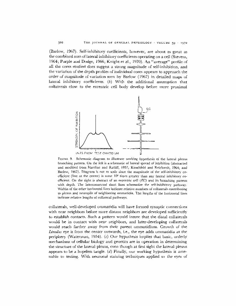
3. A Working Hypothesis for the Structure of the Lateral Plexus We suggestthat the depth profile of ECA collaterals and neuropile topographicallymirrors the functional organization of lateral inhibition that has been eluci-dated by Hartline and his colleagues (for a review, see Hartline, 1969). Inessence, we suggest that the most distal collaterals make contact with them-selves, and those collaterals lying successively proximal make contacts withommatidia successively farther away (Fig. 9). We term this suggested arrange-ment a working hypothesis because the present study confirms the first partof it but only hints at the successive distribution of the collateral system withdepth in the lateral plexus.
The hypothesis has the following merits. (a) It can account for the generalprofiles of lateral inhibitory magnitude vs. distance, and is flexible enough toaccount exactly for individual variations. Averaged maps of inhibitory co-efficients for lateral inhibition (Hartline and Ratliff, 1957; Kirschfeld andReichardt, 1964; Barlow, 1967, 1969) show a gradual reduction with distancein the strength of inhibition exerted by one cell upon its neighbors. Consider-able variation exists for the strength of lateral inhibition exerted by any onecell on neighbors when compared to a large population that has been averaged
302 THE JOURNAL OF GENERAL PHYSIOLOGY · VOLUME 59 1972
(Barlow, 1967). Self-inhibitory coefficients, however, are about as great asthe combined sum of lateral inhibitory coefficients operating on a cell (Stevens,1964; Purple and Dodge, 1966; Knight et al., 1970). An "average" profile ofall the cores studied does suggest a strong magnitude of self-inhibition, andthe variation of the depth profiles of individual cores appears to approach theorder of magnitude of variation seen by Barlow (1967) in detailed maps oflateral inhibitory coefficients. (b) With the additional assumption thatcollaterals close to the eccentric cell body develop before more proximal
EC
GUR, PURPLE, AND WHITEHEAD Lateral Plexus of Limulus Eye 303
developing embryos, it should be possible to trace the maturation stages ofthe lateral plexus.
We thank Mr. Kenneth Hopper for technical assistance, and Doctors Richard Poppele and CarloTerzuolo for their helpful comments on the manuscript.
This work was supported by United States Public Health Service Grants No. EY00293, EY00526,and GM00572.
Received for publication 23 August 1971.
BIBLIOGRAPHY
BARLOW, R. B. JR. 1967. Inhibitory fields in the Limulus lateral eye. Ph.D. Thesis. The Rocke-feller University, New York.
BARLOW, R. B. JR. 1969. Inhibitory fields in the Limulus lateral eye. J. Gen. Physiol. 54:383.FAHRENBACH, W. H. 1969. The morphology of the eyes of Limulus. II. Ommatidia of the
compound eye. Z. Zellforsch. Mikrosk. Anat. 93:451.FAHRENBACH, W. H. 1970. The morphology of the Limulus visual system. III. The lateral
rudimentary eye. Z. Zellforsch. Mikrosk. Anat. 105:303.GRUNDFEST, H. 1965. Electrophysiology and pharmacology of different components of bio-
electric transducers. Cold Spring Harbor Symp. Quant. Biol. 30:1.GRUNDFEST, H. 1971. The general electrophysiology of input membrane in electrogenic ex-
citable cells. In Handbook of Sensory Physiology. Principles of Receptor Physiology. W. R.Lowenstein, editor. Springer-Verlag, Berlin. 1:135.
HARTLINE, H. K. 1969. Visual receptors and retinal interaction. Science (ashington). 164:270.HARTLINE, H. K., and F. RATLIFF. 1957. Inhibitory interaction of receptor units in the eye of
Limulus. J. Gen. Physiol. 40:357.HARTLINE, H. K., F. RATLIFF, and W. H. MILLER. 1961. Inhibitory interaction in the retina
and its significance in vision. In Nervous Inhibition. E. Florey, editor, Pergamon Press, Ltd.,Oxford. 241.
HARTLINE, H. K., H. G. WAGNER, and F. RATLIFF. 1956. Inhibition in the eye of Limulus. J.Gen. Physiol. 39:651.
KIRSCHFELD, K., and W. REICHARDT. 1964. Die Verarbeitung stationarer optischer Nachtrich-ten in Komplexauge von Limulus. Kybernetik. 2:43.
KNIGHT, B. W., J. I. TOYODA, and F. A. DODGE, JR. 1970. A quantitative description of thedynamics of excitation and inhibition in the eye of Limulus. J. Gen. Phvsiol. 56:421.
PATTEN, W. 1912. The Evolution of the Vertebrates and Their Kin. Blakiston Div., McGraw-Hill Book Co., Inc., New York.
PURPLE, R. L. 1964. Integration of excitation and inhibition in the eccentric cell in the eyeof Limulus. Ph.D. Thesis. The Rockefeller University, New York.
PURPLE, R. L., and F. A. DODGE. 1965. Interaction of excitation and inhibition in the eccentriccell in the eye of Limulus. Cold Spring Harbor Symp. Quant. Biol. 30:529.
PURPLE, R. L., and F. A. DODGE, JR. 1966. Self-inhibition in the eye of Limulus. In FunctionalOrganization of the Compound Eye. C. G. Bernhard, editor. Pergamon Press Ltd., Oxford.451.
RATLIFF, F. 1965. Mach Bands: Quantitative Studies on Neural Networks in the Retina.Holden-Day Inc., San Francisco, Calif. 105.
RATLIFF, F., H. K. HARTLINE, and W. H. MILLER. 1963. Spatial and temporal aspects ofretinal inhibitory interaction. J. Opt. Soc. Amer. 53:110.
RATLIFF, F., W. H. MILLER, and H. K. HARTLINE. 1958. Neural interaction in the eye andintegration of receptor activity. Ann. N. Y. Acad. Sci. 74:210.
SCHWARTZ, E. A. 1971. Retinular and eccentric cell morphology in the neural plexus ofLimulus lateral eye. J. Neurobiol. 2:129.
304 THE JOURNAL OF GENERAL PHYSIOLOGY VOLUME 59 ' 1972
STEVENS, C. F. 1964. A quantitative theory of neural interactions: theoretical and experi-mental investigations. Ph.D. Thesis. The Rockefeller University, New York.
TOMITA, T., R. KIKUCHI, and T. TANAKA. 1960. Excitation and inhibition in lateral eye ofhorseshoe crab. In Electrical Activity of Single Cells. Y. Katsuki, editor. Igaku Shoin Ltd.,Tokyo.
WATERMAN, T. H. 1954. Relative growth and the compound eye in Xiphosura. J. Morphol.95:125.
WHITEHEAD, R., and R. L. PURPLE. 1970. Synaptic organization in the neuropile of the lateraleye of Limulus. Vision Res. 10:129.