50
Undisputed applications for TMJ Surgery • Ankylosis • Growth disorders • Recurrent subluxation • Infections • Neoplasms • These make up the minority of TMJ cases

Undisputed applications for TMJ
Surgery
• Ankylosis
• Growth disorders
• Recurrent subluxation
• Infections
• Neoplasms
• These make up the minority of TMJ cases
Relative Indications for TMJ
Surgery
• TMD is refractory to appropriate non-surgical
therapies
• TMJ is the source of pain and/or dysfunction that
results ina significant impairment to the patient in
day to day acitivity
– Pain localized to the TMJ
– Pain on loading of the TMJ
– Pain on movement in the TMJ
– Mechainical interferences in the TMJ
Surgical Procedures for
Temporomandibular disorders
• Arthrocentesis and lavage
• Arthroscopy
• Arthrotomy
• Modified condylotomy
• Adjunctive procedures for TMJ
– Botox
– Coronoidectomy
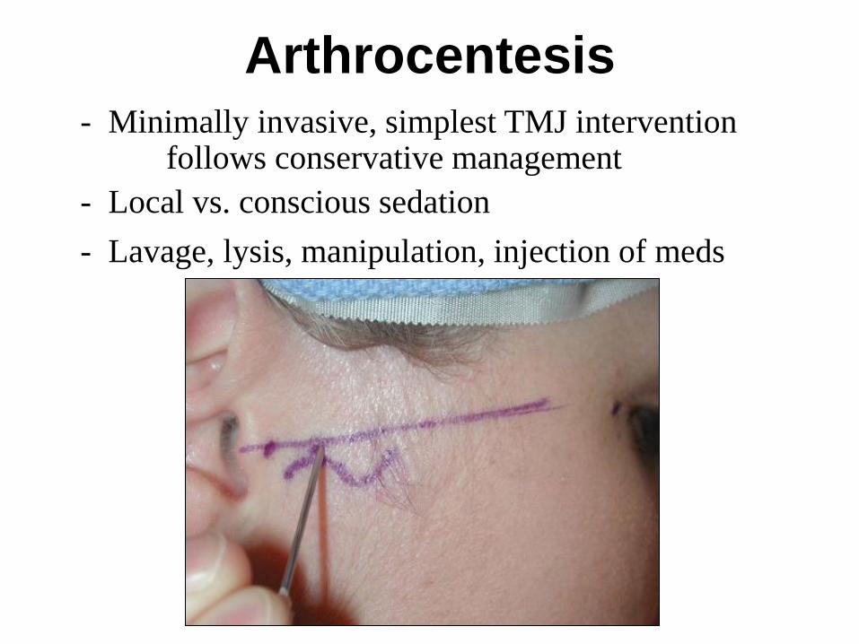
Arthrocentesis - Minimally invasive, simplest TMJ intervention follows conservative management
- Local vs. conscious sedation
- Lavage, lysis, manipulation, injection of meds
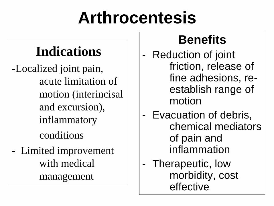
Arthrocentesis
Benefits
- Reduction of joint friction, release of fine adhesions, re- establish range of motion
- Evacuation of debris, chemical mediators of pain and inflammation
- Therapeutic, low morbidity, cost effective
Indications
-Localized joint pain,
acute limitation of
motion (interincisal
and excursion),
inflammatory
conditions
- Limited improvement
with medical
management

Arthrocentesis Technique - Auriculotemporal nerve block
- Needle positioned at 10-2 point
anterior to tragus
- Identify arch and periosteum
- Superior joint space confirmed
with vacuum after
insufflation, return of joint
fluid, mandible motion
- Additional port placed
immediately anterior
- Lavage joint with 100-200 cc
- Steroid and anesthetic infiltrated
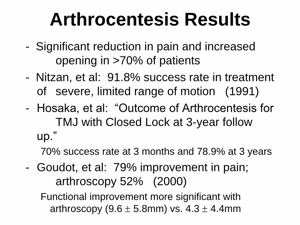
Arthrocentesis Results
- Significant reduction in pain and increased
opening in >70% of patients
- Nitzan, et al: 91.8% success rate in treatment
of severe, limited range of motion (1991)
- Hosaka, et al: “Outcome of Arthrocentesis for
TMJ with Closed Lock at 3-year follow
up.”
70% success rate at 3 months and 78.9% at 3 years
- Goudot, et al: 79% improvement in pain;
arthroscopy 52% (2000)
Functional improvement more significant with
arthroscopy (9.6 5.8mm) vs. 4.3 4.4mm
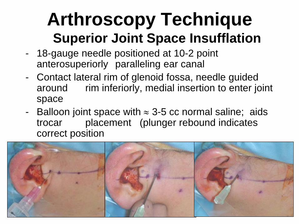
Arthroscopy Technique Superior Joint Space Insufflation
- 18-gauge needle positioned at 10-2 point anterosuperiorly paralleling ear canal
- Contact lateral rim of glenoid fossa, needle guided around rim inferiorly, medial insertion to enter joint space
- Balloon joint space with 3-5 cc normal saline; aids trocar placement (plunger rebound indicates correct position
and adequate insufflation)
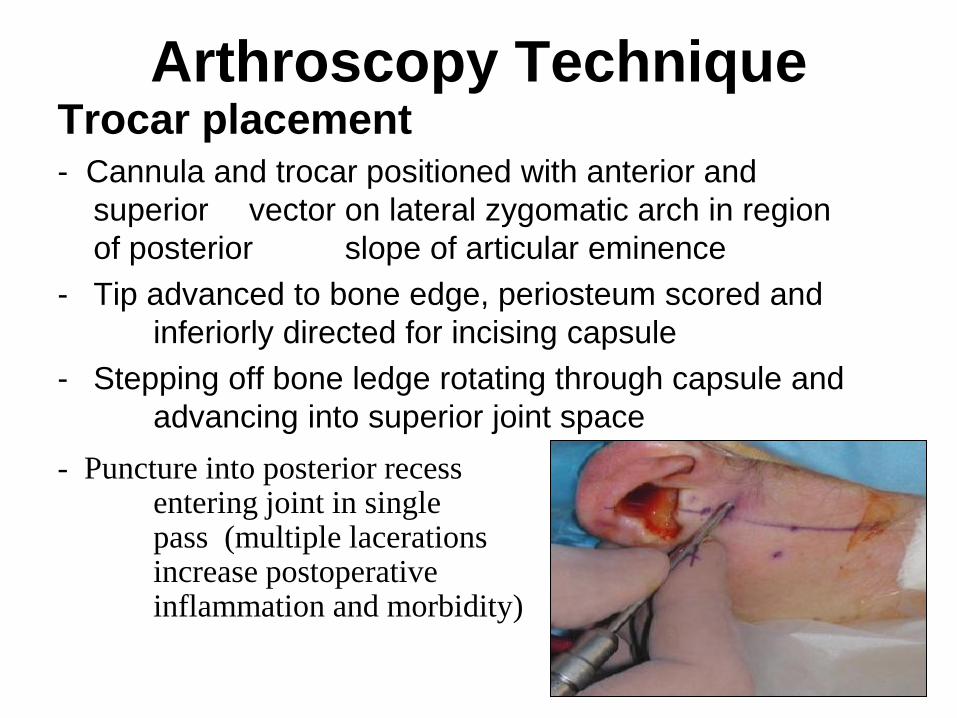
Arthroscopy Technique Trocar placement - Cannula and trocar positioned with anterior and
superior vector on lateral zygomatic arch in region
of posterior slope of articular eminence
- Tip advanced to bone edge, periosteum scored and
inferiorly directed for incising capsule
- Stepping off bone ledge rotating through capsule and
advancing into superior joint space
- Puncture into posterior recess entering joint in single pass (multiple lacerations increase postoperative inflammation and morbidity)

Arthroscopy Technique

Arthroscopy Technique
- Arthroscope advanced through lateral recess to
visualize anterior aspect of articular eminence,
anterior disk and anterodiskal tissue
- Access to anterior recess provides visualization for
placement of second working port

Arthroscopy Technique Triangulation
Working port placed after stab incision
at 25-10 point (minimum of 15 mm
separation between ports)
Second portal in eminence region placed
under direct visualization allows
instrumentation of joint contents

Arthroscopy Technique Instrumentation
- Blunt trocar, radiofrequency probe, motorized shaver, and/or laser utilized
- Treatment of adhesions, pathology, internal derangements and removal of tissues
- Depth roughly 20 – 25 mm from skin to center of joint
- Lavage of joint with irrigation expands joint space, allows visualization during instrumentation and flushes irritants (inflammatory and pain mediators)
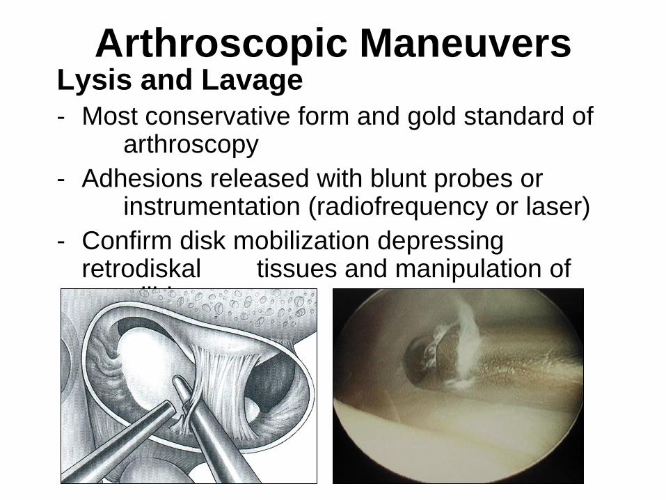
Arthroscopic Maneuvers Lysis and Lavage
- Most conservative form and gold standard of arthroscopy
- Adhesions released with blunt probes or instrumentation (radiofrequency or laser)
- Confirm disk mobilization depressing retrodiskal tissues and manipulation of mandible

Arthroscopic Maneuvers
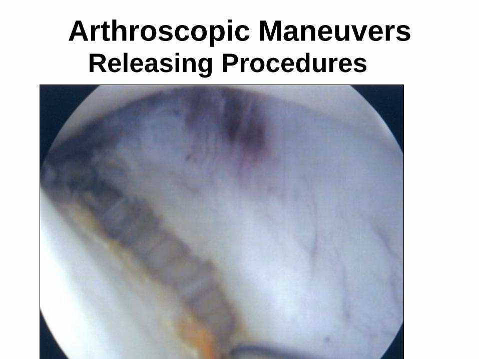
Arthroscopic Maneuvers Releasing Procedures

Arthroscopic Maneuvers
radiofrequency
fibrillations
Ablation laser
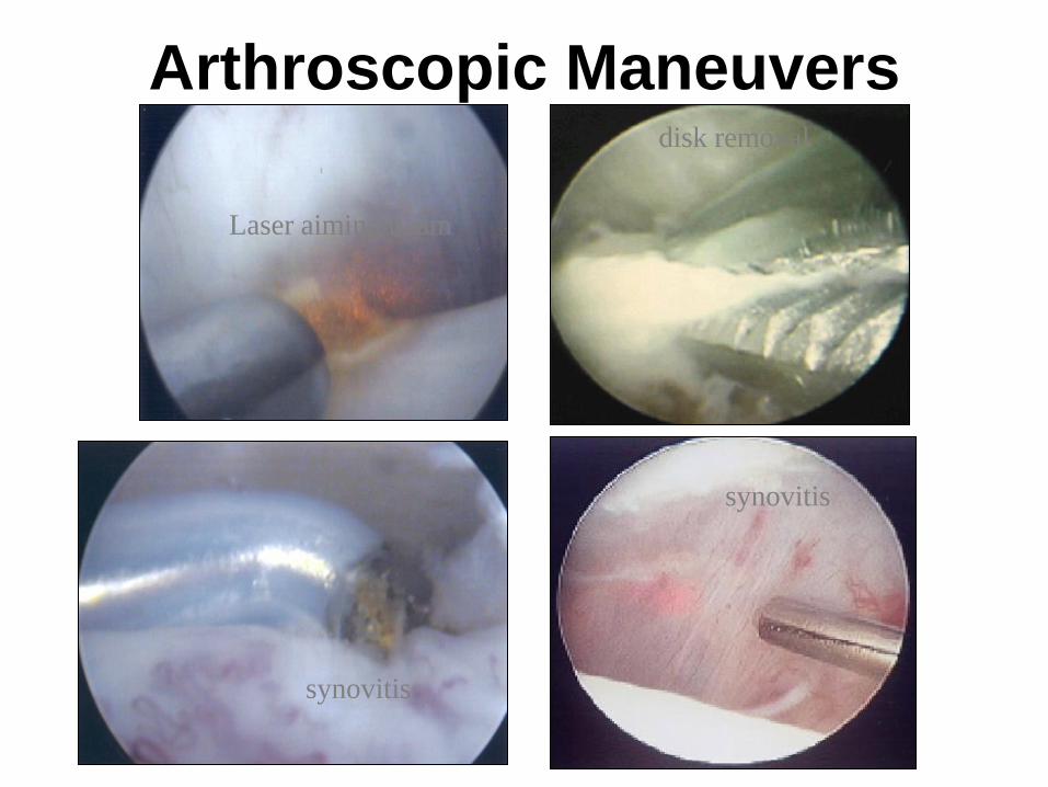
Arthroscopic Maneuvers
synovitis
disk removal
synovitis
Laser aiming beam

Condylotomy • Condylar sag aids range of
motion and internal
derangement
• Complications include
malocclusion and sensory
disturbances

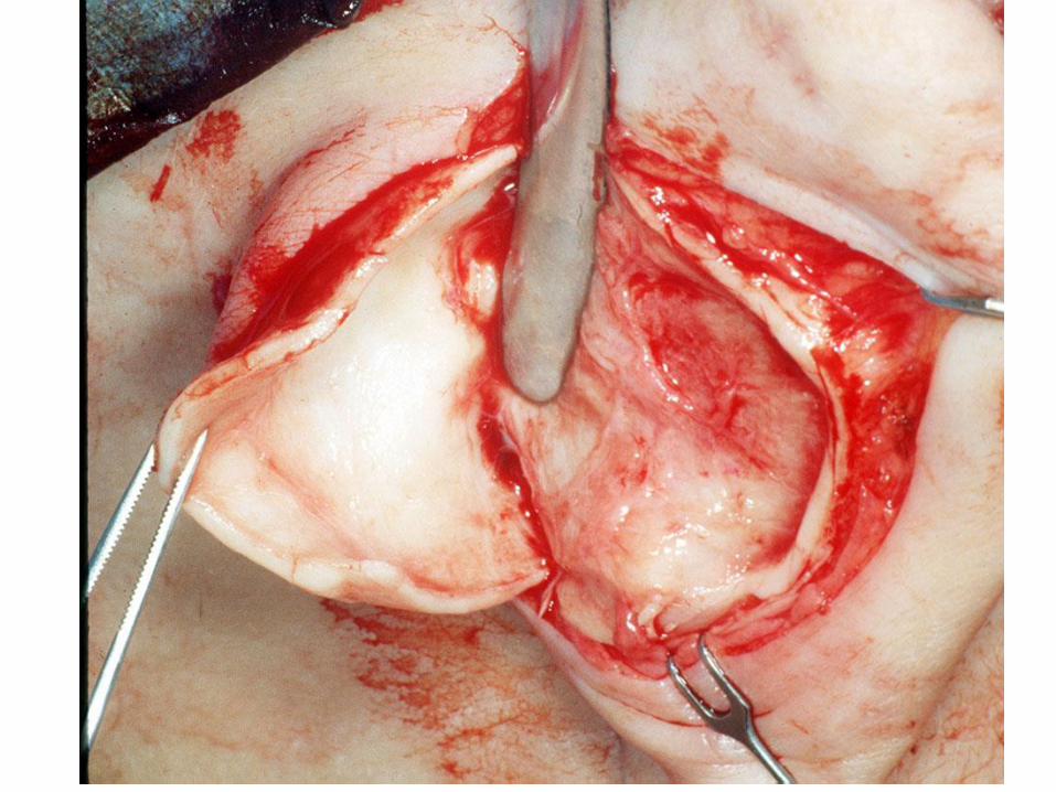
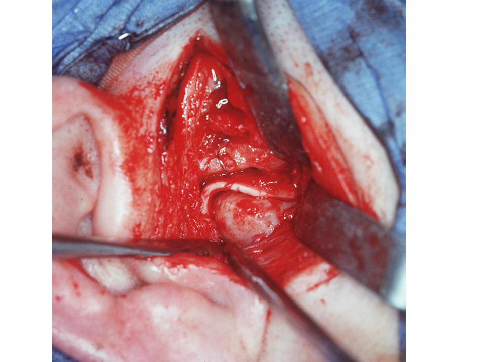
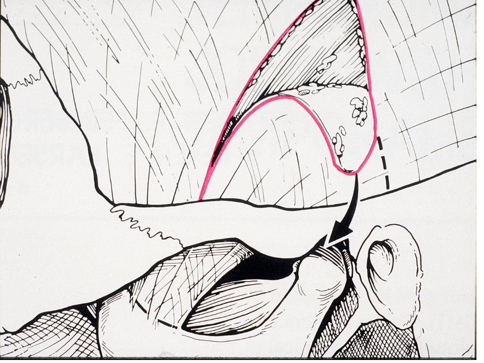
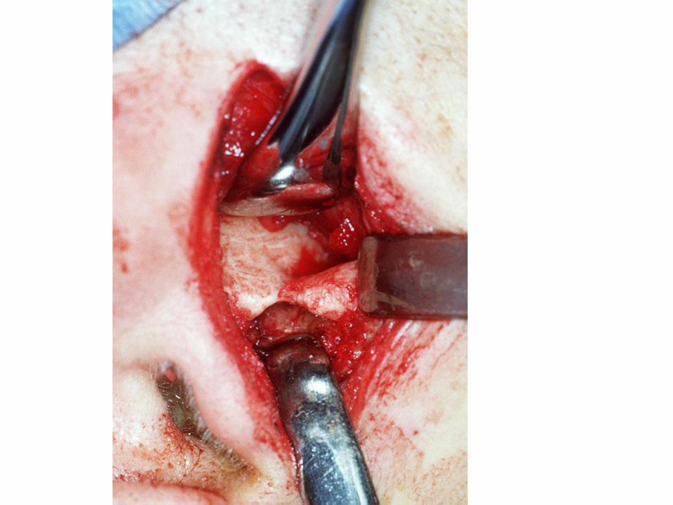
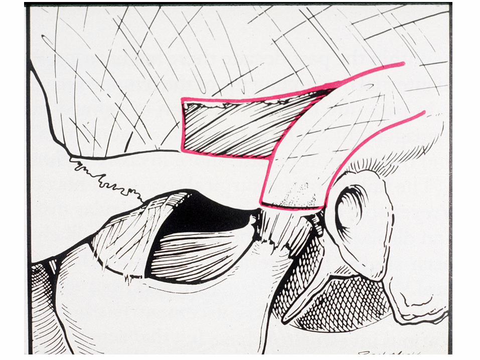
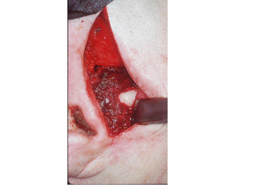
Arthrotomy – Total Joint
Reconstruction
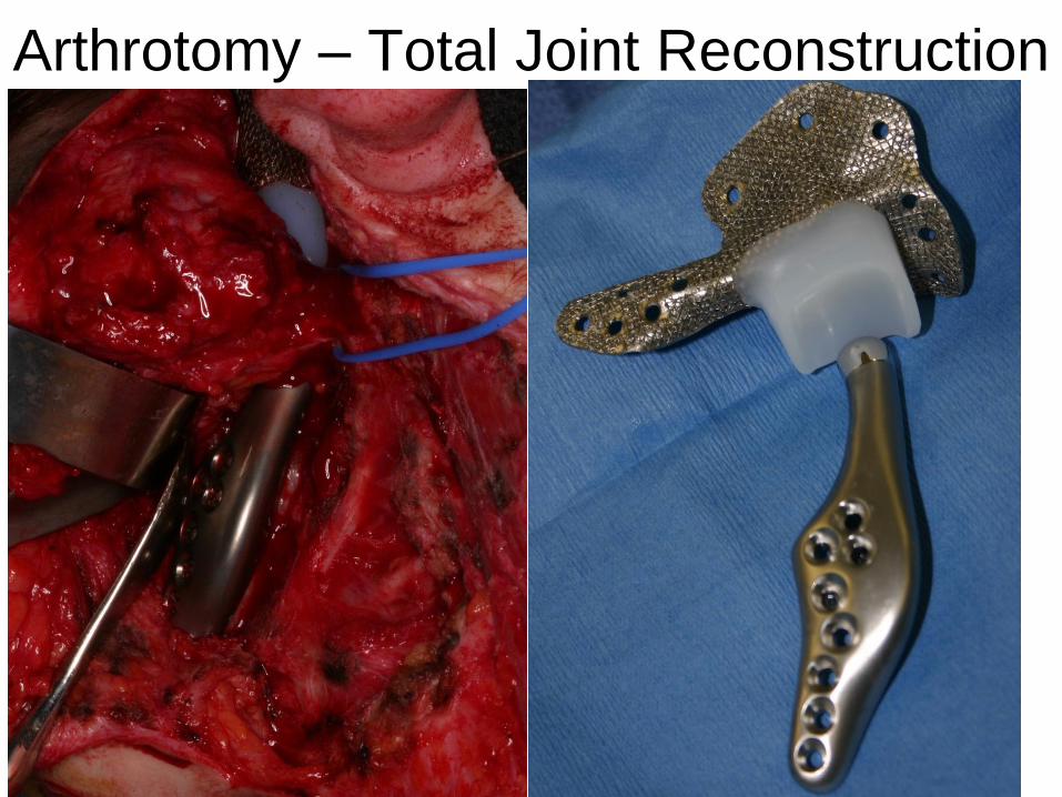
Arthrotomy – Total Joint Reconstruction

Adjunctive Measures
Distraction Osteogenesis
Condyle recreated post-condylectomy or
prosthetic joint failure

AURICULAR CARTILAGE
• Witsenburg 1984, Matukas 1990, Kent
and Widner 1990
• Somewhat operative technique
dependent
• Stabilization varies
• Early complication minimal
• Fun procedure - otoplasty effect
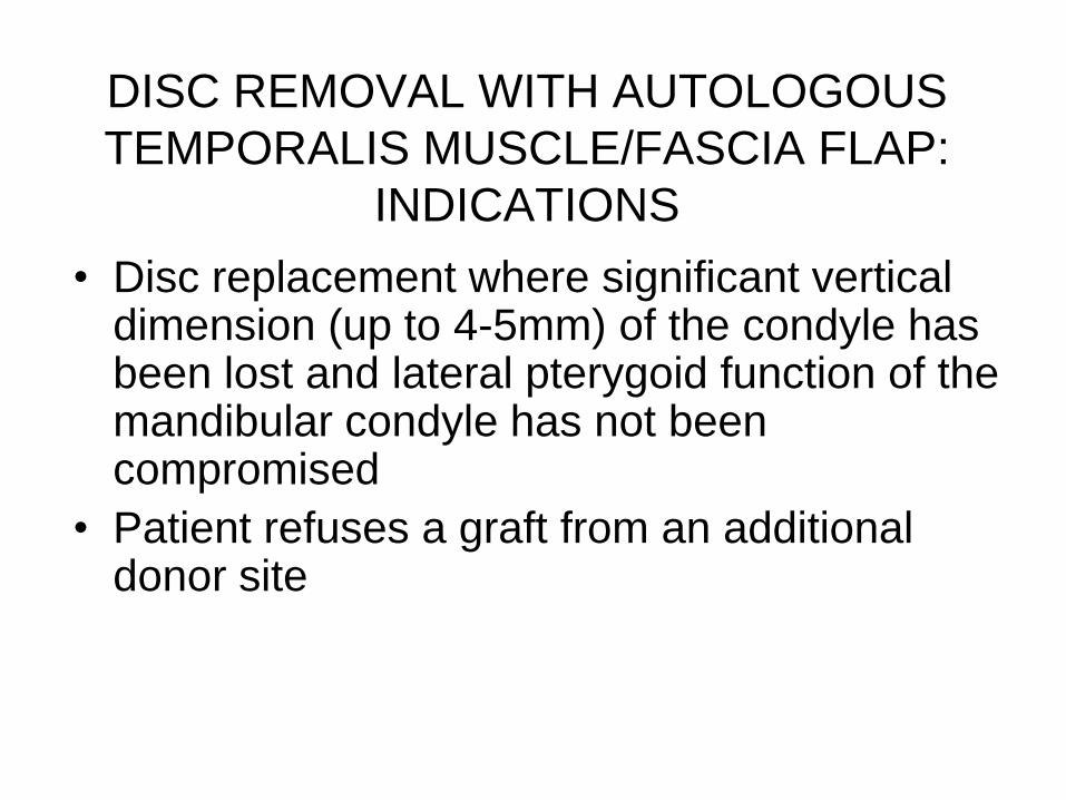
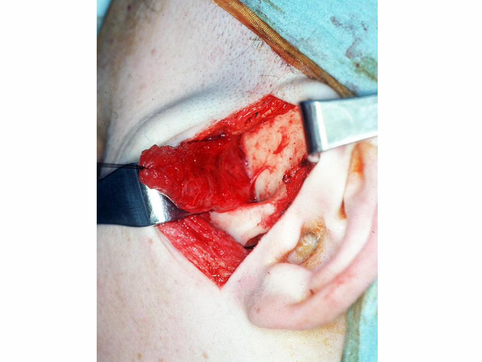
DISC REMOVAL WITH AUTOLOGOUS
TEMPORALIS MUSCLE/FASCIA FLAP:
INDICATIONS
• Disc replacement where significant vertical dimension (up to 4-5mm) of the condyle has been lost and lateral pterygoid function of the mandibular condyle has not been compromised
• Patient refuses a graft from an additional donor site


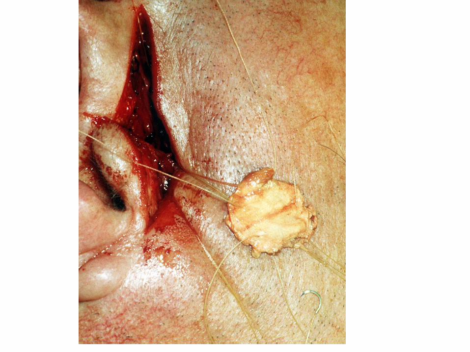
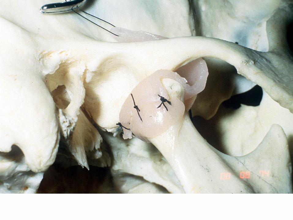
DERMIS GRAFTS Clinical-Georgiade 1957, Zetz and Irby
1984, Meyer 1988
• Disc repair
• Disc replacement
• Ankylosis cases - thickness of dermis
depends on gap
• With costochondral grafting
• Resembles a disc when used as a
patch in perforations
• Reported superior ability to
withstand joint loading compared to
other tissues
DERMIS GRAFT

De-epithelializing prior to dermis harvest
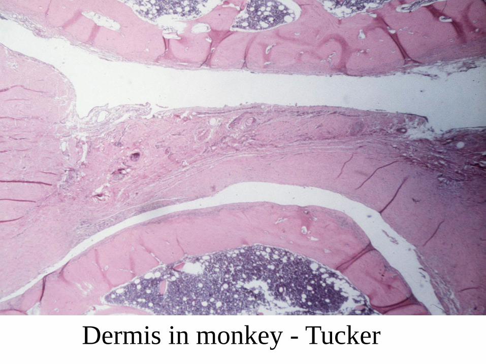
Dermis in monkey - Tucker
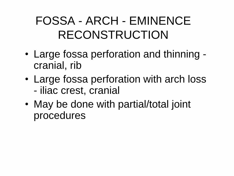
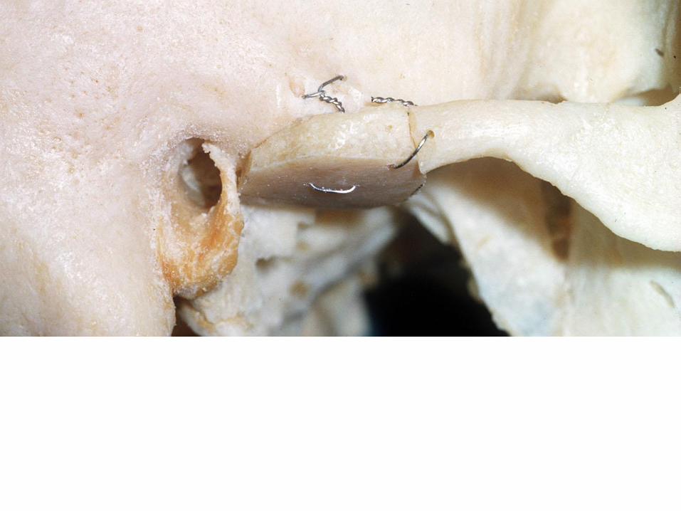
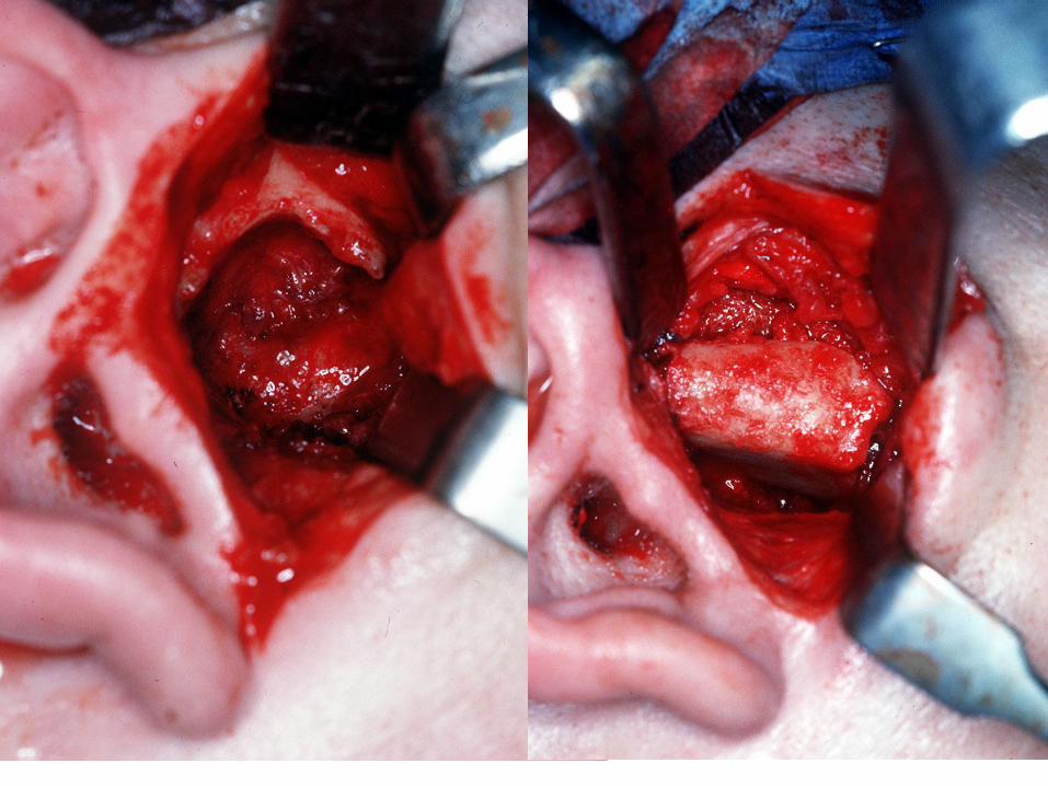
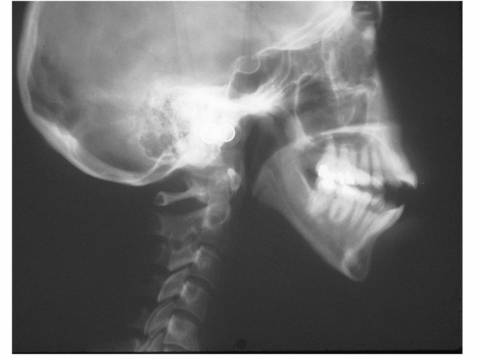
FOSSA - ARCH - EMINENCE
RECONSTRUCTION
• Large fossa perforation and thinning - cranial, rib
• Large fossa perforation with arch loss - iliac crest, cranial
• May be done with partial/total joint procedures

INDICATIONS • Condylar height loss greater than 7-8 mm
• Loss of lateral pterygoid muscle
• Trauma
• Multiple joint surgery
• Advanced rheumatoid-disease and DJD
• Ankylosis
• Hypoplasia

15
Yr
Post
op


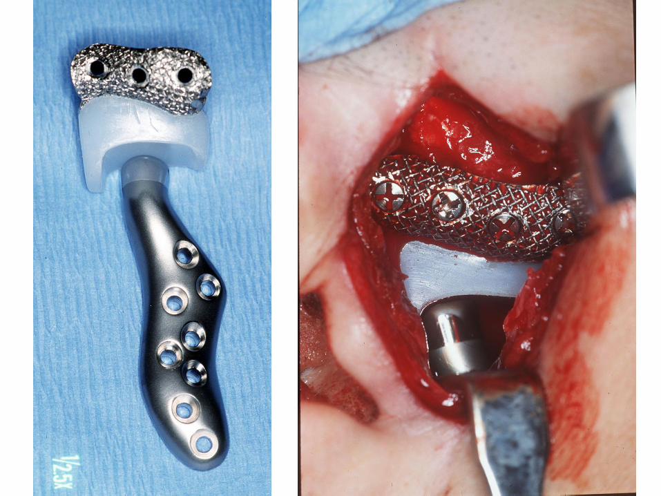
TECHMEDICA - TMJ
CONCEPTS • Custom CAD/CAM design based on CT,
computer generated plastic model, and
surgeon imput