Application of a Quantitative Method for Arch Form Evaluation in Complete Unilateral Cleft Lip and Palate PAUL W. STOCKLI, D.M.D., M.S. Zirich, Switzerland Arch form has become one of the major considerations in the treatment of the infant with a complete cleft lip and palate. The controversies regard- ing early orthopedic procedures, early bone grafting and many operative techniques originated mainly from aspects related to alignment of the segments of the upper jaw. However, only few methods are available for analyzing arch form in cleft lip and palate infants. Isolated measurements of the intertuberosity width and measurements of the posterior and/or alveolar cleft width (6, 8, 11) do not portray arch form. Descriptive criteria like "approximation without contact", "approximation with con- tact", and "overlap" (8) are too vague for comparative assessments. Evaluation of occlusion (6, 8, 9, 10) does not reveal the reaction of the upper arch during the early stages. In an analysis of the changes following presurgical orthopedic treat- ment, Huddart (4) presented a method which is applicable to the unoper- ated upper arch in infants with cleft lip and palate. For broader applica- tion, however, the method reveals two major inadequacies: (1) it is lim- ited to studies before palatal closure, since one of the reference structures is the nasal septum, and (2) it does not describe the configuration of the alveolar cleft. Because of the lack of a simple, but specific method for arch form evaluation in complete unilateral cleft lip and palate cases, an attempt was made to design a measurement system which would be suitable espe- cially for longitudinal and comparative studies. It was of primary concern that the measurement system express, in numerical terms, the spatial relationship of the two maxillary segments in the frontal and sagittal planes, particularly in the region of the alveolar cleft. The measurement system was tested in a comparative study as a means of establishing its scope. Dr. Stockli is affiliated with the University of Zurich, Zurich, Switzerland. This work was conducted at Northwestern University Dental School, in partial fulfillment of the requirements for the degree of Master of Science. Presented in part at the 2nd International Symposium on Early Treatment of Cleft Lip and Palate, Chicago, April 19-20, 1969. 322
Transcript
Application of a Quantitative Method for
Arch Form Evaluation in Complete
Unilateral Cleft Lip and Palate
PAUL W. STOCKLI, D.M.D., M.S.
Zirich, Switzerland
Arch form has become one of the major considerations in the treatment
of the infant with a complete cleft lip and palate. The controversies regard-
ing early orthopedic procedures, early bone grafting and many operative
techniques originated mainly from aspects related to alignment of the
segments of the upper jaw. However, only few methods are available for
analyzing arch form in cleft lip and palate infants. Isolated measurements
of the intertuberosity width and measurements of the posterior and/or
alveolar cleft width (6, 8, 11) do not portray arch form. Descriptive
criteria like "approximation without contact", "approximation with con-
tact", and "overlap" (8) are too vague for comparative assessments.
Evaluation of occlusion (6, 8, 9, 10) does not reveal the reaction of the
upper arch during the early stages.
In an analysis of the changes following presurgical orthopedic treat-
ment, Huddart (4) presented a method which is applicable to the unoper-
ated upper arch in infants with cleft lip and palate. For broader applica-
tion, however, the method reveals two major inadequacies: (1) it is lim-
ited to studies before palatal closure, since one of the reference structures
is the nasal septum, and (2) it does not describe the configuration of the
alveolar cleft.
Because of the lack of a simple, but specific method for arch form
evaluation in complete unilateral cleft lip and palate cases, an attempt
was made to design a measurement system which would be suitable espe-
cially for longitudinal and comparative studies. It was of primary concern
that the measurement system express, in numerical terms, the spatial
relationship of the two maxillary segments in the frontal and sagittal
planes, particularly in the region of the alveolar cleft.
The measurement system was tested in a comparative study as a means
of establishing its scope.
Dr. Stockli is affiliated with the University of Zurich, Zurich, Switzerland.This work was conducted at Northwestern University Dental School, in partial
fulfillment of the requirements for the degree of Master of Science.Presented in part at the 2nd International Symposium on Early Treatment of
Cleft Lip and Palate, Chicago, April 19-20, 1969.
322
ARCH FORM EVALUATION 323
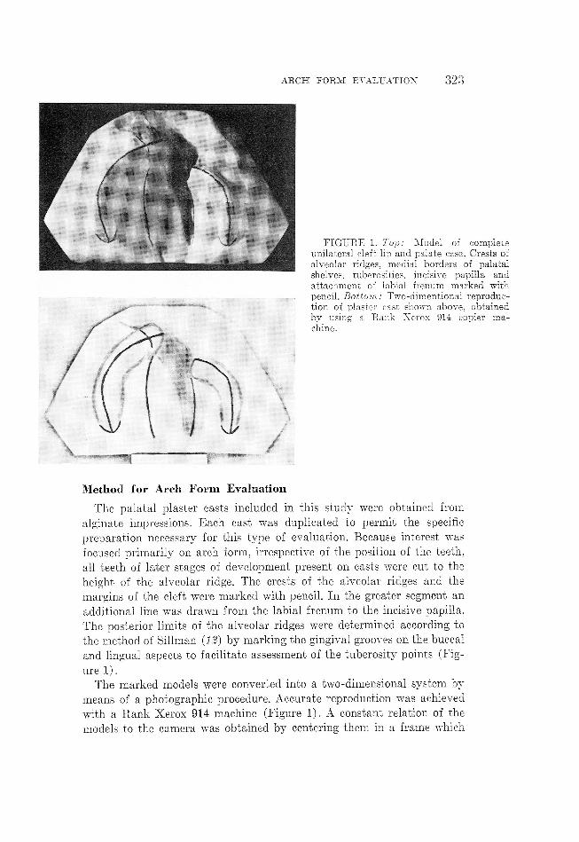
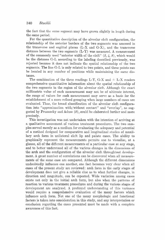
FIGURE 1. Top: Model of completeunilateral cleft lip and palate case. Crests ofalveolar ridges, medial borders of palatalshelves, tuberosities, incisive papilla andattachment of labial frenum marked withpencil. Bottom: Two-dimentional reproduc-tion of plaster cast shown above, obtainedby using a Rank Nerox 914 copier ma-chine.
Method for Arch Form Evaluation
The palatal plaster casts included in this study were obtained from
alginate impressions. Each cast was duplicated to permit the specific
preparation necessary for this type of evaluation. Because interest was
focused primarily on arch form, irrespective of the position of the teeth,
all teeth of later stages of development present on casts were cut to the
height of the alveolar ridge. The crests of the alveolar ridges and the
margins of the cleft were marked with pencil. In the greater segment an
additional line was drawn from the labial frenum to the incisive papilla.
The posterior limits of the alveolar ridges were determined according to
the method of Sillman (12) by marking the gingival grooves on the buccal
and lingual aspects to facilitate assessment of the tuberosity points (Fig-
ure 1).
The marked models were converted into a two-dimensional system by
means of a photographic procedure. Accurate reproduction was achieved
with a Rank Xerox 914 machine (Figure 1). A constant relation of the
models to the camera was obtained by centering them in a frame which
324 Stocklh
al
z l <
T (f t* 1!
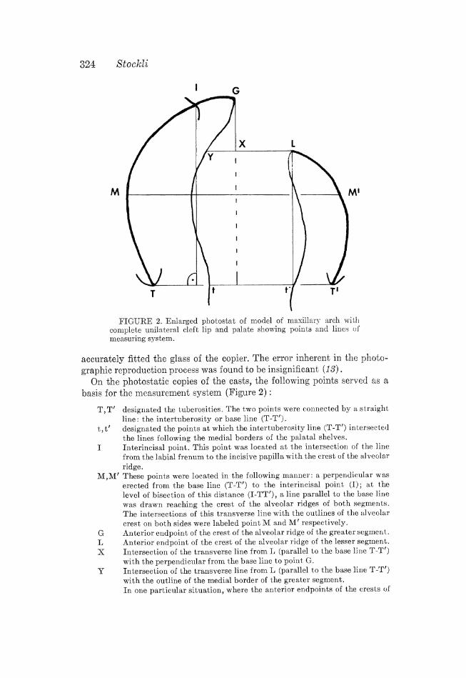
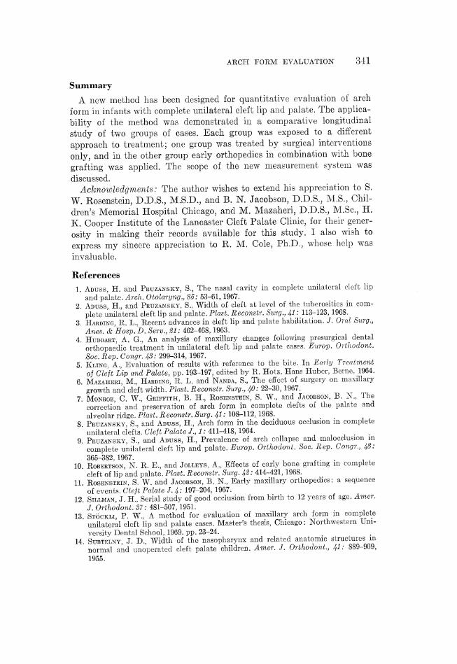
FIGURE 2. Enlarged photostat of model of maxillary arch withcomplete unilateral cleft lip and palate showing points and lines ofmeasuring system.
accurately fitted the glass of the copier. The error inherent in the photo-
graphic reproduction process was found to be insignificant (13).
On the photostatic copies of the casts, the following points served as a
basis for
T,T
t,t"
<_{MBQ
the measurement system (Figure 2) :
designated the tuberosities. The two points were connected by a straight
line: the intertuberosity or base line (T-I").designated the points at which the intertuberosity line (T-T') intersectedthe lines following the medial borders of the palatal shelves.Interincisal point. This point was located at the intersection of the linefrom the labial frenum to the incisive papilla with the crest of the alveolar
ridge.These points were located in the following manner: a perpendicular was
erected from the base line (T-T') to the interincisal point (I); at thelevel of bisection of this distance (I-TT'), a line parallel to the base line
was drawn reaching the crest of the alveolar ridges of both segments.The intersections of this transverse line with the outlines of the alveolar
crest on both sides were labeled point M and M' respectively.Anterior endpoint of the crest of the alveolar ridge of the greater segment.Anterior endpoint of the crest of the alveolar ridge of the lesser segment.Intersection of the transverse line from L (parallel to the base line T-T')
with the perpendicular from the base line to point G.Intersection of the transverse line from L (parallel to the base line T-I')
with the outline of the medial border of the greater segment.
In one particular situation, where the anterior endpoints of the crests of
ARCH FORM EVALUATION 325
G G
\} \ [ ]
FIGURE 3. Relation of the anterior endpoints of the crest of thealveolar ridges of the greater (point (G) and of the lesser segment (point L).Left: "Open" configuration, Positive L-X reading. Right: "Overlapping"configuration, Negative L-X reading.
the alveolar ridges are in an ideal end to end relation, G L X and Y arelocated at the same point.
From these points, the following measurements were carried out (Figure
2) :
Transverse measurements of the upper arch:
T-T' Intertuberosity width or posterior width of the upper arch.
M-M' Middle width of the upper arch.t-t' Width of the cleft width at the tuberosities or posterior cleft width.Antero-posterior measurements of the upper arch:
I-TT' Sagittal length of the upper arch as determined by the length of the per-pendicular from the base line (T-TI") to the interincisal point (I).
Total length, however, might well be greater in some instances as G is
positioned anterior to I. This is often the case before lip closure has been
performed.L-TT' Sagittal length of the lesser segment as determined by the length of the
~ . Aperpendicular from the base line (T-I') to point L.
Transverse and antero-posterior measurements in the region of the al-
veolar cleft:L-Y_- Transverse anterior width of the cleft* or transverse width of the alveolar
cleft.L-X Transverse relation of the lesser to the greater segment.
In an "open" situation the reading is positive, in an "overlapping"
situation the reading is negative (Figure 3).
(G-X_ Antero-posterior relation of the lesser to the greater segment.Since, in the present samples, the alveolar border of the lesser segment(L) was never positioned anterior to the alveolar border of the greater
segment (G) all readings were positive.Measurements were carried out with a caliper gauge reading to 0.1 mm. Each
measurement was taken twice. The average of the two readings was then resolved
to 0.5 mm.
* "Transverse" is added for the distinction from the commonly used "anterior widthof the cleft" which is defined as the shortest distance between the cleft seg-ments at the level of the alveolar borders. According to the labeling described above,this would be the distance between G and L. In the present investigation, however,this G-L measurement was omitted for reasons discussed later.
326 Stockl
Subjects
Longitudinal series of maxillary models of 18 patients with complete
unilateral cleft lip and plate were studied. The records derived from two
sources, each with a different treatment approach.
One group of cases was obtained from the H. K. Cooper Institute of the Lan-
caster Cleft Palate Clinic, and consisted of 8 patients. Surgically, all these patientshad been treated by a three step procedure: (1) lip closure, (2) vomer flap andrepair of the hard palate (closure of the anterior palate), and (8) closure of the
posterior palate. The advantages of performing the closure of the hard palate asa separate operation subsequent to lip closure were indicated by Harding (8).
No early orthopedic, orthodontic, or bone grafting procedures had been under-
taken in any of the cases in this group. All operations were performed by the
same surgeon. A triangular flap procedure was used for lip closure. The hardpalate was repaired in basically the same manner in all cases. The vomer flapprocedure performed simultaneously with the repair of the hard palate, variedin cases #105 and #108, where a bilateral vomer flap was carried out. Posterior
palatal closure, a median suture procedure, was the same in all cases.
Since three purely surgical interventions were applied to these patients,
the group was labeled "PSI".
The other group of cases was made available by the cleft lip and palate team
of the Children's Memorial Hospital in Chicago. In the 10 patients in this group,an orthopedic appliance without any expansion device had been inserted at, or
immediately before, lip closure. The anterior third of the greater segment re-mained uncovered by the appliance to allow for the molding effect which usually
followslip closure. After better aligmnent had been achieved, generally 7-8 months
later, a bone graft of rib was inserted as an onlay graft over the alveolar defect.The palatal appliance was worn for approximately another six months after thegraft procedure, or until the third operative step, closure of the palate, was per-
formed. The design of the appliance used, the general procedure, and the se-
quence of events have been published by Rosenstein and Jacobson (11), and
Monroe et al. (7). Two surgeons belonging to the same team, and following thesame basic surgical procedures, performed all operations in this group. Usually,
the rotation advancement approach was carried out for lip closure. In case #508and #509 the lip was closed according to technique of Wang, and in case #506
according to the technique of Cronin-Brauer. The bone grafting procedure andthe palate closure (modification of von Langenbeck) were basically the same in
all cases.
This group of cases, which were treated with the combination of early
orthopedic procedures and bone grafting, was labeled
The sex of the patients and the side of the alveolar cleft are represented
in Table 1.
The maxillary models of both groups were selected by criteria relating
to the greatest congruence in timing. The "PSI" group was represented by
36 models and the "EOG" group by 45 models, which were organized
according to the following scheme:
record a: Before lip closure.record b: In the period between lip closure and anterior palate closure in the
ARCH FORM EVALUATION 327
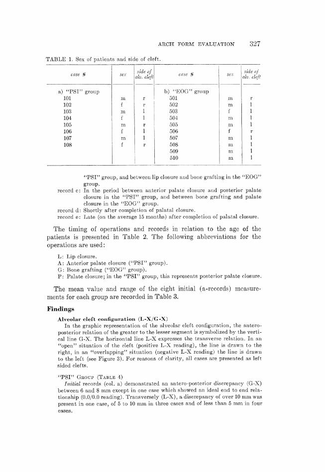
TABLE 1. Sex of patients and side of cleft.
case K sex aifeclzjjr’t case K sex akzzfiiecféét
a) "PSI" group b) "EOG" group
101 m. r 501 m. r
102 { r 502 m 1
103 m 1 503 i 1
104 f 1 504 m 1
105 m r 505 m 1
106 f 1 506 f 1
107 m 1 507 m 1
108 f r 508 m 1
509 m I
510 m I
"PSI" group, and between lip closure and bone grafting in the "EOG"
group.
record c: In the period between anterior palate closure and posterior palate
closure in the "PSI" group, and between bone grafting and palate
closure in the "EOG" group.
record d: Shortly after completion of palatal closure.
record e: Late (on the average 15 months) after completion of palatal closure.
The timing of operations and records in relation to the age of the
patients is presented in Table 2. The following abbreviations for the
operations are used:
L: Lip closure.
A: Anterior palate closure ("PSI" group).
G: Bone grafting ("EOG group).
P: Palate closure; in the "PSI" group, this represents posterior palate closure.
The mean value and range of the eight initial (a-records) measure-
ments for each group are recorded in Table 3.
Findings
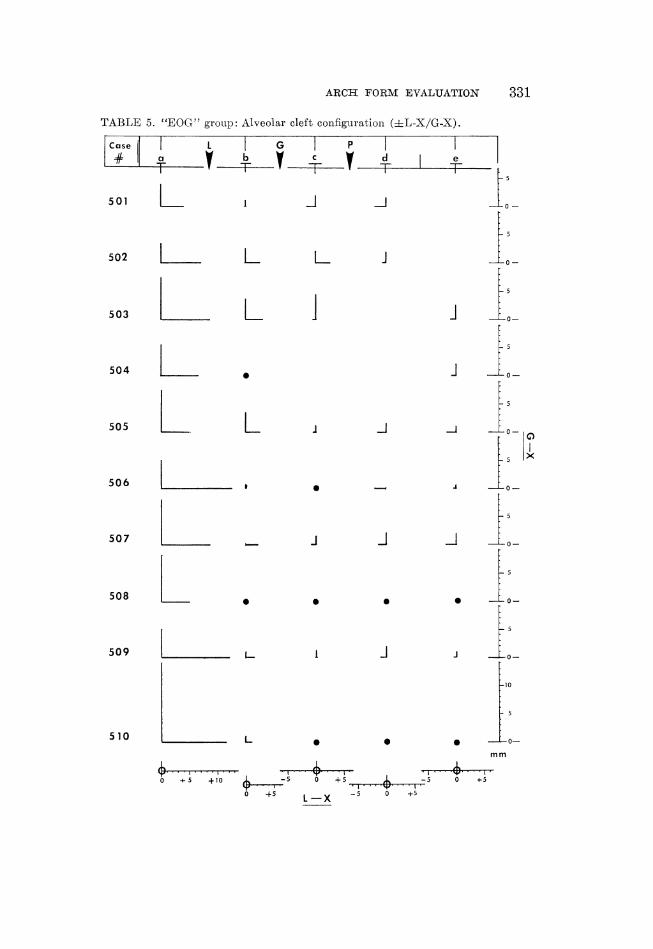
Alveolar cleft configuration (L-X/G-X)
In the graphic representation of the alveolar cleft configuration, the antero-
posterior relation of the greater to the lesser segment is symbolized by the verti-
cal line G-X. The horizontal line L-X expresses the transverse relation. In an
"'open'" situation of the cleft (positive L-X reading), the line is drawn to the
right, in an "overlapping" situation (negative L-X reading) the line is drawn
to the left (see Figure 3). For reasons of clarity, all cases are presented as left
sided clefts.
"PSI" Grour (Tarsus 4)
Initial records (col. a) demonstrated an antero-posterior discrepancy (G-X)
between 6 and 8 mm except in one case which showed an ideal end to end rela-
tionship (0.0/0.0 reading). Transversely (L-X), a discrepancy of over 10 mm was
present in one case, of 5 to 10 mm in three cases and of less than 5 mm in four
cases.
328 Stockl
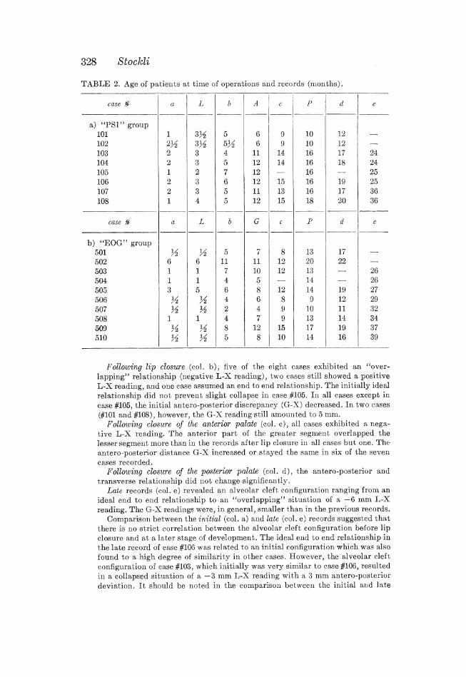
TABLE 2. Age of patients at time of operations and records (months).
Following lip closure (col. b), five of the eight cases exhibited an "over-
lapping" relationship (negative L-X reading), two cases still showed a positiveL-X reading, and one case assumed an end to end relationship. The initially idealrelationship did not prevent slight collapse in case #105. In all cases except in
case #105, the initial antero-posterior discrepancy (G-X) decreased. In two cases(#101 and #108), however, the G-X reading still amounted to 5 mm.
Following closure of the anterior palate (col. c), all cases exhibited a nega-
tive L-X reading. The anterior part of the greater segment overlapped thelesser segment more than in the records after lip closure in all cases but one. Theantero-posterior distance G-X increased or stayed the same in six of the seven
cases recorded.Following closure of the posterior palate (col. d), the antero-posterior and
transverse relationship did not change significantly.Late records (col. e) revealed an alveolar cleft configuration ranging from an
ideal end to end relationship to an "overlapping" situation of a -6 mm L-Xreading. The G-X readings were, in general, smaller than in the previous records.
Comparison between the initial (col. a) and late (col. e) records suggested thatthere is no strict correlation between the alveolar cleft configuration before lipclosure and at a later stage of development. The ideal end to end relationship inthe late record of case #106 was related to an initial configuration which was also
found to a high degree of similarity in other cases. However, the alveolar cleft
configuration of case #103, which initially was very similar to case #106, resultedin a collapsed situation of a -3 mm L-X reading with a 3 mm antero-posterior
deviation. It should be noted in the comparison between the initial and late
ARCH FORM EVALUATION 35209
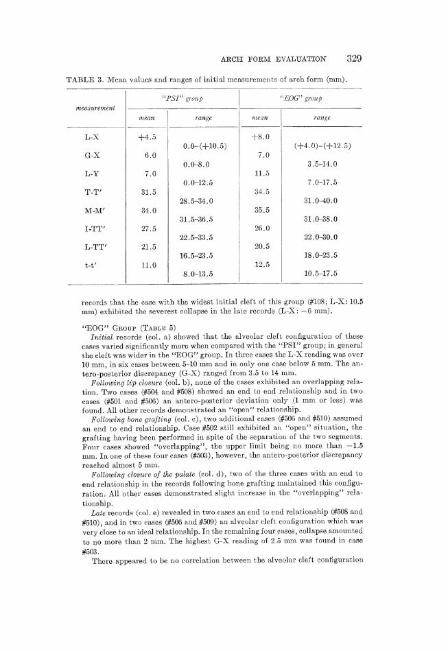
TABLE 3. Mean values and ranges of initial measurements of arch form (mm).
"PSI group "EOG" group
measurement
mean range mean range
L-X -+8.00.0-(+10.5) (+4.0)-(+12.5)
G-X 6.0 7.00.0-8.0 3 .5-14.0
L-Y 7.0 11.50.0-12.5 7.0-17.5
T-I" 31.5 34.528. 5-34.0 31l.0-40.0
M-M' 34.0 35.531.5-36.5 31.0-38.0
I-TT' 27.0 26.022.5-33.5 22.0-30.0
L-TT' 21.5 20.516.5-23.5 18.0-23.5
t-t' 11.0 12.58.0-183.5 10.5-17.5
records that the case with the widest initial cleft of this group (#108; L-X : 10.5
mm) exhibited the severest collapse in the late records (L-X : -6 mm).
"EOG" Grour (Tasur 5)Initial records (col. a) showed that the alveolar cleft configuration of these
cases varied significantly more when compared with the "PSI" group; in general
the cleft was wider in the "EOG" group. In three cases the L-X reading was over10 mm, in six cases between 5-10 mm and in only one case below 5 mm. The an-
tero-posterior discrepancy (G-X) ranged from 3.5 to 14 mm.Following lip closure (col. b), none of the cases exhibited an overlapping rela-
tion. Two cases (#504 and #508) showed an end to end relationship and in two
cases (#501 and #506) an antero-posterior deviation only (1 mm or less) wasfound. All other records demecnstrated an "open'" relationship.
Following bone grafting (col. c), two additional cases (#506 and #510) assumedan end to end relationship. Case #502 still exhibited an "open" situation, the
grafting having been performed in spite of the separation of the two segments.
Four cases showed "overlapping'', the upper limit being no more than -1.5mm. In one of these four cases (#503), however, the antero-posterior discrepancy
reached almost 5 mm.Following closure of the palate (col. d), two of the three cases with an end to
end relationship in the records following bone grafting maintained this configu-
ration. All other cases demonstrated slight increase in the "overlapping" rela-
tionship.Late records (col. e) revealed in two cases an end to end relationship (#508 and
#510), and in two cases (#506 and #509) an alveolar cleft configuration which was
very close to an ideal relationship. In the remaining four cases, collapse amounted
to no more than 2 mm. The highest G-X reading of 2.5 mm was found in case
#503.
There appeared to be no correlation between the alveolar cleft configuration
before lip closure and the alveolar cleft configuration at a later stage of develop-ment.
At the time of the investigation, intraoral radiographs had been taken of thealveolar cleft area. The time lapse between bone grafting and the latest intraoralfilms averaged 19 months, with a range of 10 to 31 months. In all ten cases a bonybridge was exhibited.Transverse width of the alveolar cleft (L-Y, Table 6)"PSI" Grour
Initial records (col. a) revealed that the L-Y distance ranged in seven cases
from 5.5 to 12.5 mm. One case of this group (#105) showed a 0.0 L-Y reading
before lip closure.Following lip closure (col. b), five of the eight cases exhibited anterior contact
between the two segments.Following closure of the anterior palate (col. c), all cases demonstrated contact.
"EOG" GrouPrInitial L-Y readings (col. a) ranged from 7.0 to 17.5 mm.Following lip closure (col. b), only four of the ten cases exhibited anterior
contact between the segments.Following bone grafting (col. c), no contact was present in three cases.Following closure of the palate (col. d), however, all cases manifested a 0.0
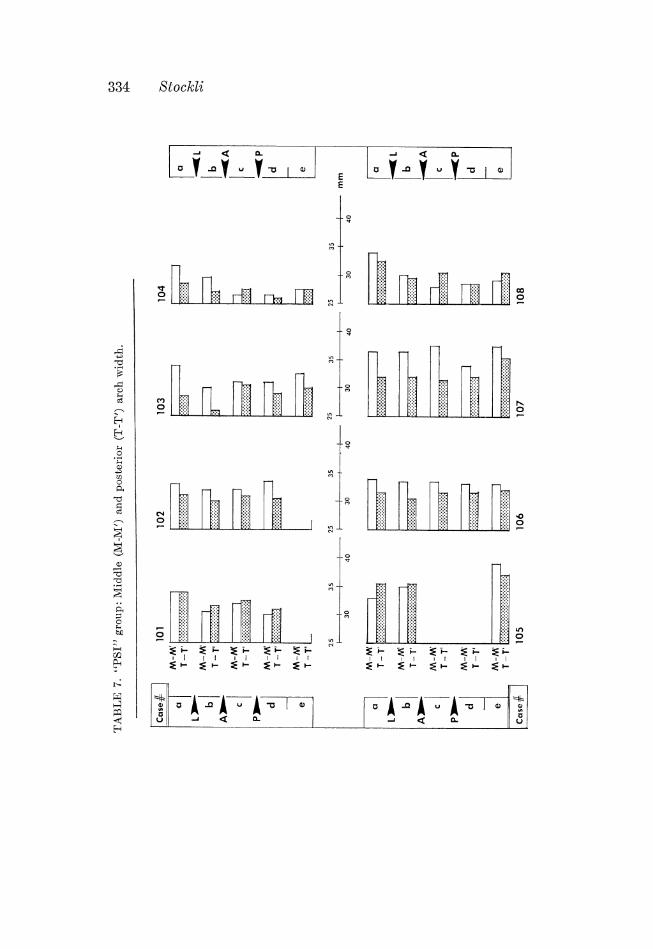
L-Y reading.Transverse arch measurements (M -M' and T-1")
"PSI" Grour (Tarsus 7)Initial records (col. a) revealed that the middle width (M-M') exceeded the
posterior width (T-I") in six of the eight cases, the M-M' reading being 1.5 to
5.5 mm higher than the T-T' reading. One case (#101) exhibited equal M-M' and
T-T' readings and in one case (#105) the posterior width was greater.Following lip closure (col. b), five of the eight cases showed simultaneous de-
crease in the middle and posterior width, the largest decrease being 4 mm in the
M-M' measurements of cases #103 and #108. The remaining cases demonstrated
only slight or no changes in the transverse arch measurements (#105, #106 and#107). With respect to the alveolar cleft configuration of these cases (see Table
4), an end to end relationship was exhibited by case #105 before lip closure and
by case #107 after lip closure. In case #106, the interrelation between the trans-
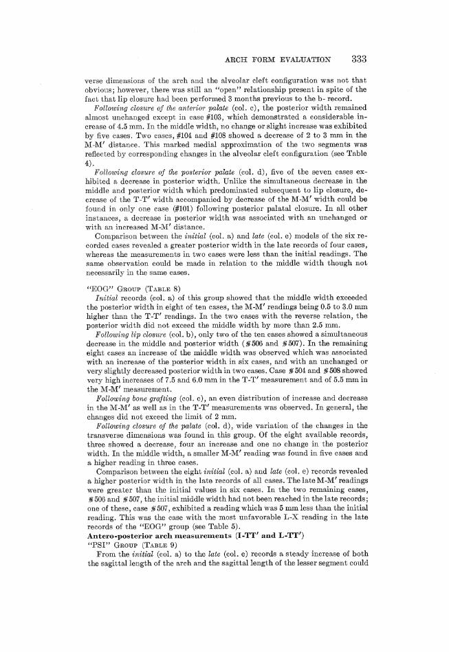
ARCH FORM EVALUATION 339
verse dimensions of the arch and the alveolar cleft configuration was not thatobvious; however, there was still an "open"" relationship present in spite of the
fact that lip closure had been performed 3 months previous to the b- record.Following closure of the anterior palate (col. c), the posterior width remained
almost unchanged except in case #103, which demonstrated a considerable in-
crease of 4.5 mm. In the middle width, no change or slight increase was exhibitedby five cases. Two cases, #104 and #108 showed a decrease of 2 to 3 mm in the
M-M' distance. This marked medial approximation of the two segments wasreflected by corresponding changes in the alveolar cleft configuration (see Table
4).Following closure of the posterior palate (col. d), five of the seven cases ex-
hibited a decrease in posterior width. Unlike the simultaneous decrease in the
middle and posterior width which predominated subsequent to lip closure, de-crease of the T-T" width accompanied by decrease of the M-M' width could befound in only one case (#101) following posterior palatal closure. In all other
instances, a decrease in posterior width was associated with an unchanged or
with an increased M-M' distance. _Comparison between the initial (col. a) and late (col. e) models of the six re-
corded cases revealed a greater posterior width in the late records of four cases,whereas the measurements in two cases were less than the initial readings. Thesame observation could be made in relation to the middle width though not
necessarily in the same cases.
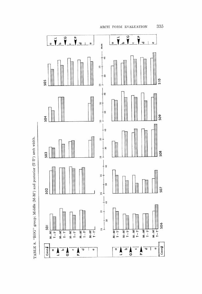
"EOG" Grour (Tasur 8)Initial records (col. a) of this group showed that the middle width exceeded
the posterior width in eight of ten cases, the M-M' readings being 0.5 to 3.0 mm
higher than the T-T' readings. In the two cases with the reverse relation, theposterior width did not exceed the middle width by more than 2.5 mm.
Following lip closure (col. b), only two of the ten cases showed a simultaneous
decrease in the middle and posterior width (#506 and #507). In the remainingeight cases an increase of the middle width was observed which was associatedwith an increase of the posterior width in six cases, and with an unchanged or
very slightly decreased posterior width in two cases. Case #504 and #508 showedvery high increases of 7.5 and 6.0 mm in the T-I" measurement and of 5.5 mm in
the M-M' measurement.Following bone grafting (col. c), an even distribution of increase and decrease
in the M-M' as well as in the T-T' measurements was observed. In general, the
changes did not exceed the limit of 2 mm.Following closure of the palate (col. d), wide variation of the changes in the
transverse dimensions was found in this group. Of the eight available records,three showed a decrease, four an increase and one no change in the posterior
width. In the middle width, a smaller M-M' reading was found in five cases anda higher reading in three cases.
Comparison between the eight initial (col. a) and late (col. e) records revealeda higher posterior width in the late records of all cases. The late M-M' readings
were greater than the initial values in six cases. In the two remaining cases,506 and £507, the initial middle width had not been reached in the late records;
one of these, case #507, exhibited a reading which was 5 mm less than the initialreading. This was the case with the most unfavorable L-X reading in the late
records of the "EOG" group (see Table 5).Antero-posterior arch measurements (I-TT' and L-TT)
"PSI" Grour (TaBur 9)From the initial (col. a) to the late (col. e) records a steady increase of both
the sagittal length of the arch and the sagittal length of the lesser segment could
TABLE
7."PSI"
group:Middle
(M-M')and
post
erio
r(T-T')
archwidth.
Case#
i101
102
2535
2535 q
}mm
--if
L
-allA P
Case#
I105
106
107
108
334 StockLh
TABLE
8."EOG"
group:Middle
(M-M')
and
posterior
(T-I")
archwidth.
Case#
|501
502
503
504
505
506
507
508
509
510
mm
ARCH FORM EVALUATION 330
TABLE
9."PSI"
group:
Sagi
ttal
length
ofthearch
(I-T
T')and
ofthe
less
ersegment
(L-TT').
25T
20
-+
35.
304
254
20+
15L
Hi;
iLLfJ
Liitiijg
Lb};“A;
ULU'LQ
104 r
||||_'
LoL
CLOL
oLog
'bCb
CaLog
ga
Caam
10
5106
107
108
336 Stockl
TABLE
10.
beffaid
Viffaid
|¥afFard
l¥affard
¥ar«
35J
25+
204
mm
354+
30+
25-+
20+
15-
"EOG"
group:
Sagittallength
ofthe
arch
(I-TT')
and
ofthe
lessersegment
(L-TT").
505
Loo
oodoJ,
34T-
cs
510
ARCH FORM EVALUATION 337
338 Stock
be observed. This increase was temporarily interrupted in three cases at thetime of posterior palatal closure (#103, #104 and #107).
Cases showing a continuous approximation of the L-TT' distance to the I-TTdistance from the initial to the late records, exhibited the best relationship of
the segments in the region of the alveolar cleft (see Table 4). Case #106 demon-strated this pattern very clearly: the initial difference between the L-TT' and
I-TT' measurement amounted to 5.5 mm and was reduced to half at the timethe late records were taken. At the alveolar cleft, this case finally assumed an
ideal end to end relationship. On the other hand, the I-TT"/L-TT' differenceincreased significantly in cases ¥104 and #108 from the initial to the lata records:;
both cases manifested in the late records the gresiest deviations in the regionof the alveolar cleft.
"EOG" Grour (Taur 10)
The same interrelation as described for the previous group, was observed.The approximation after lip closure, related to a shortening of the I-TT' distanceor to a relatively greater increase of the L-TT' distance, or to a combination
of both, was maintained in case #506, $508 and #510 to the late records. Onthe other hand, the small I-TIT/L-TT' difference of case #504 and #507 in therecords following lip closure was not preserved. Thus, the alveolar cleft configura-
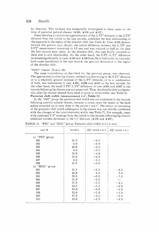
tion after lip closure altered from ideal or good to unfavorable (see Table 5).Posterior cleft width (measurement t-t', Table 11)
In the "PSI" group the posterior cleft width was not evaluated in the records
following anterior palatal closure, because in some cases the repair of the hardpalate extended up or very close to the points t and t'. The extent of narrowing
of the posterior cleft width subsequent to lip closure was not strictly correlatedwith the changes of the intertuberosity width (see Table 7). For example, caseswith unaltered T-T" readings from the initial to the records following lip closureexhibited notable decreases in the t-t' distance (#105 and #107).
TABLE 11. "PSI" and "EOG" group: Posterior cleft width (t-t') in mm.
case # record a diff . record a to b 5 diff. record a to c