Unit 2: Microbiology – Pre-Reading Sylvan Kaufman, Ph.D. Slide 1 Neither Plant Nor Animal: Microbiology Sylvan Kaufman, Ph.D. www.SylvanGreenEarth.com (most fungi photos by Wallace Kaufman unless otherwise noted) In this unit we will learn about some non-living organisms; viruses, viroids, and prions, and representatives of four Kingdoms, the Archaea, Bacteria, Protista and Fungi. It may seem strange to learn about non-living organisms that you can’t see without a powerful microscope, but they play important roles in ecosystems, and you might be surprised how often they are in the news!
Transcript
Unit 2: Microbiology – Pre-Reading Sylvan Kaufman, Ph.D.
Slide 1
Neither Plant Nor Animal:
Microbiology
Sylvan Kaufman, Ph.D.
www.SylvanGreenEarth.com(most fungi photos by Wallace
Kaufman unless otherwise noted)
In this unit we will learn about some non-living organisms; viruses, viroids, and prions, and representatives of four Kingdoms, the Archaea, Bacteria, Protista and Fungi. It may seem strange to learn about non-living organisms that you can’t see without a powerful microscope, but they play important roles in ecosystems, and you might be surprised how often they are in the news!
Unit 2: Microbiology – Pre-Reading Sylvan Kaufman, Ph.D.
Slide 2
Viruses
Image from the Center for Disease Control depicting an RNA virus with its protein coat.
Viruses were discovered in 1898 when researchers studying foot and mouth disease and tobacco mosaic virus discovered they were caused by an infectious particle smaller than a bacteria. Viruses are not considered to be “alive” because they cannot reproduce on their own. Instead they insert their genetic material into a host cell, taking over the host cell’s functions. The virus uses the host to make new copies of itself. The genetic material of a virus, DNA or RNA, is contained within a protein coat. Sometimes the protein coat is surrounded by a lipid envelope. Viruses are able to inherit genetic mutations and they are subject to natural selection. Viruses are used in genetic engineering to insert pieces of DNA into plant or animal cells. For example, a virus could be used to insert a gene coding for herbicide resistance into a soybean plant.
Unit 2: Microbiology – Pre-Reading Sylvan Kaufman, Ph.D.
Slide 3
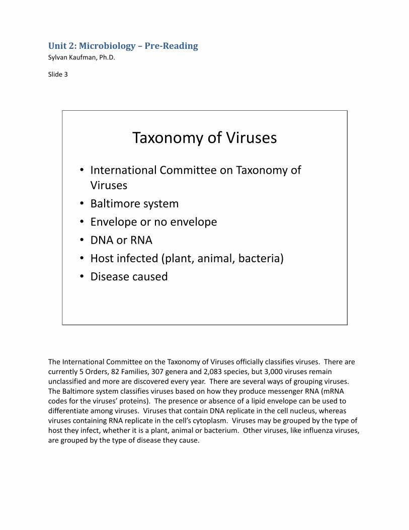
Taxonomy of Viruses
• International Committee on Taxonomy of Viruses
• Baltimore system
• Envelope or no envelope
• DNA or RNA
• Host infected (plant, animal, bacteria)
• Disease caused
The International Committee on the Taxonomy of Viruses officially classifies viruses. There are currently 5 Orders, 82 Families, 307 genera and 2,083 species, but 3,000 viruses remain unclassified and more are discovered every year. There are several ways of grouping viruses. The Baltimore system classifies viruses based on how they produce messenger RNA (mRNA codes for the viruses’ proteins). The presence or absence of a lipid envelope can be used to differentiate among viruses. Viruses that contain DNA replicate in the cell nucleus, whereas viruses containing RNA replicate in the cell’s cytoplasm. Viruses may be grouped by the type of host they infect, whether it is a plant, animal or bacterium. Other viruses, like influenza viruses, are grouped by the type of disease they cause.
Unit 2: Microbiology – Pre-Reading Sylvan Kaufman, Ph.D.
Slide 4
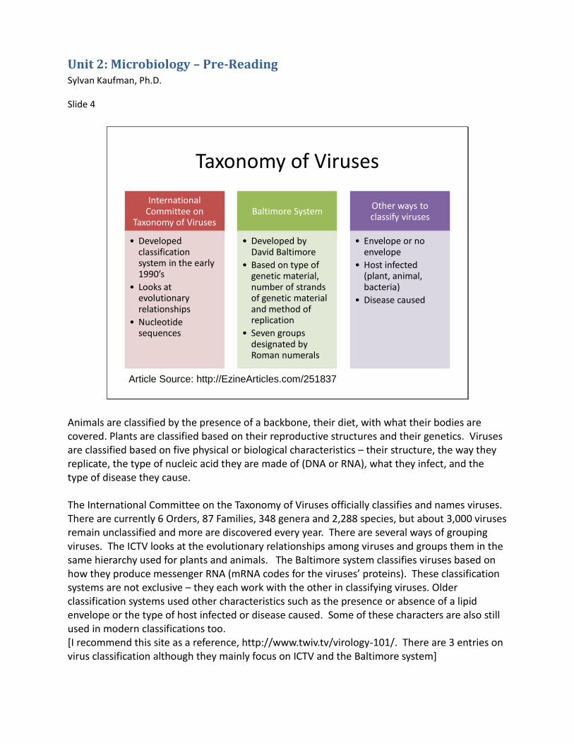
Taxonomy of Viruses
International Committee on
Taxonomy of Viruses
• Developed classification system in the early 1990’s
• Looks at evolutionary relationships
• Nucleotide sequences
Baltimore System
• Developed by David Baltimore
• Based on type of genetic material, number of strands of genetic material and method of replication
• Seven groups designated by Roman numerals
Other ways to classify viruses
• Envelope or no envelope
• Host infected (plant, animal, bacteria)
• Disease caused
Article Source: http://EzineArticles.com/251837
Animals are classified by the presence of a backbone, their diet, with what their bodies are covered. Plants are classified based on their reproductive structures and their genetics. Viruses are classified based on five physical or biological characteristics – their structure, the way they replicate, the type of nucleic acid they are made of (DNA or RNA), what they infect, and the type of disease they cause. The International Committee on the Taxonomy of Viruses officially classifies and names viruses. There are currently 6 Orders, 87 Families, 348 genera and 2,288 species, but about 3,000 viruses remain unclassified and more are discovered every year. There are several ways of grouping viruses. The ICTV looks at the evolutionary relationships among viruses and groups them in the same hierarchy used for plants and animals. The Baltimore system classifies viruses based on how they produce messenger RNA (mRNA codes for the viruses’ proteins). These classification systems are not exclusive – they each work with the other in classifying viruses. Older classification systems used other characteristics such as the presence or absence of a lipid envelope or the type of host infected or disease caused. Some of these characters are also still used in modern classifications too. [I recommend this site as a reference, http://www.twiv.tv/virology-101/. There are 3 entries on virus classification although they mainly focus on ICTV and the Baltimore system]
Unit 2: Microbiology – Pre-Reading Sylvan Kaufman, Ph.D.
Slide 5
Some Viruses Every Naturalist Should Know
• Rabies
• West Nile virus
• Influenza
• Viruses and algal blooms
Even though we can’t directly see viruses, they have important consequences for us and for the plants and animals around us. Here are a few examples of viruses that could be important for a Master Naturalist to know about. Rabies mostly infects raccoons, bats, foxes and skunks. It is transmitted to dogs, cats and humans through a bite that breaks the skin. The rabies virus causes damage to the brain and spinal cord. West Nile virus is usually passed from mosquitoes to birds, but can also infect humans, horses and other animals through mosquito bites. It was first discovered in the U.S. in 1999 when large numbers of dead crows were found and many people developed the virus in New York. It has since spread across North America and is known to infect more than 300 species of birds. Influenza, including the H1N1 flu, is in the news every year. The virus mutates rapidly making it necessary to formulate a new flu shot every year. Many influenza viruses can switch from one animal host to another. Viruses aren’t all bad though. Some viruses in the Chesapeake Bay are responsible for destroying harmful algal blooms and may reduce bacteria levels in the Bay.
Unit 2: Microbiology – Pre-Reading Sylvan Kaufman, Ph.D.
Slide 6
Prions
• An abnormally folded mammalian protein
• Causes chronic wasting disease in white-tailed deer
In the early 1980s, Dr. Stanley Prusiner, a neurologist at the University of California San Francisco, won the Nobel Prize for his discovery of prions. Prions are an infectious agent even simpler than viruses. They consist of just a protein (more specifically a sialoglycoprotein called PrP 27-30). Prions are produced when the mammalian PrP gene misfolds the protein it produces into a toxic form, the prion. Prions can adapt to new conditions (types of cells) and can develop drug resistance. They are transferred in blood or saliva. Prions cause neuro-degenerative diseases such as mad cow and chronic wasting disease, and are associated with Creutzfeldt-Jakob disease, Down’s syndrome and Alzheimer’s. Chronic wasting disease has recently infected white-tailed deer populations in Maryland.
Unit 2: Microbiology – Pre-Reading Sylvan Kaufman, Ph.D.
Slide 7
Viroids
• RNA strands that inhibit the expression of certain genes
Researchers at the Agricultural Research Service discovered viroids while studying potato spindle disease in 1971. Viroids are composed of RNA strands and they lack the protective protein coat of a virus. Viroids inhibit the expression of certain genes, usually in plants. They cause some plant diseases and can stunt growth. Hepatitis D is also caused by a viroid that is enclosed within the Hepatitis B virus.
Unit 2: Microbiology – Pre-Reading Sylvan Kaufman, Ph.D.
Slide 8
Eukaryotes vs.Prokaryotes
Image from http://www.ncbi.nlm.nih.gov/About/primer/genetics_cell.html
We will now move on to living organisms! Living organisms have one of two cell types. Prokaryotes are the simplest celled organisms. They lack a nuclear membrane and intracellular organelles (mitochondria, chloroplasts, golgi bodies). Prokaryotes do not develop into multicellular organisms. We will start by discussing prokaryotes.
Unit 2: Microbiology – Pre-Reading Sylvan Kaufman, Ph.D.
Slide 9
Prokaryotes
Archaea and Bacteria
Archaea
• Often found in extreme environments, but abundant in soils, oceans
• Bacillus thuringensis (Bt) –for organic insect control
• Use in bioremediation
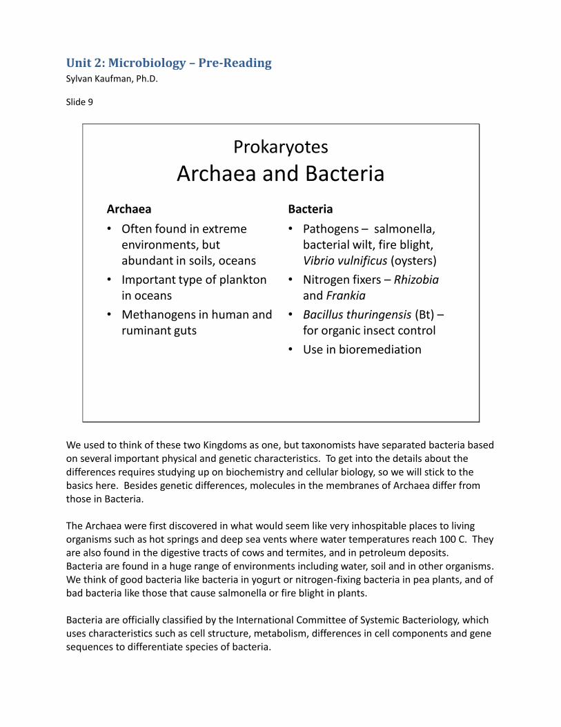
We used to think of these two Kingdoms as one, but taxonomists have separated bacteria based on several important physical and genetic characteristics. To get into the details about the differences requires studying up on biochemistry and cellular biology, so we will stick to the basics here. Besides genetic differences, molecules in the membranes of Archaea differ from those in Bacteria. The Archaea were first discovered in what would seem like very inhospitable places to living organisms such as hot springs and deep sea vents where water temperatures reach 100 C. They are also found in the digestive tracts of cows and termites, and in petroleum deposits. Bacteria are found in a huge range of environments including water, soil and in other organisms. We think of good bacteria like bacteria in yogurt or nitrogen-fixing bacteria in pea plants, and of bad bacteria like those that cause salmonella or fire blight in plants. Bacteria are officially classified by the International Committee of Systemic Bacteriology, which uses characteristics such as cell structure, metabolism, differences in cell components and gene sequences to differentiate species of bacteria.
Unit 2: Microbiology – Pre-Reading Sylvan Kaufman, Ph.D.
Slide 10
Eukaryotes - Protista
Protozoa
• 4 groups – amoeba-like, flagella-bearing, cilia-bearing and apicomplexans (tiny parasites)
• Dysentery – caused by an amoeba
• Malaria – caused by an apicomplexan
• Giardiasis - caused by flagella-bearing protist
Within the Kingdom Protista we will look at the ecological roles of protozoa and algae. Protozoa are single-celled eukaryotic organisms. They are able to move and they use organic carbon for growth. You may remember looking at protists in samples of pond water under a microscope in school. There are four groups of protists differentiated by their means of locomotion. Protists eat algae, microfungi and bacteria and are an important food source for microinvertebrates. Protists can also increase plant growth by releasing nutrients from the bacteria they have consumed. Other protists are responsible for diseases such as malaria and dysentery.
Unit 2: Microbiology – Pre-Reading Sylvan Kaufman, Ph.D.
Slide 11
Eukaryotes - Protista
AlgaeExamples of Algae
• Seaweeds – large marine algae
• Rock snot – invasive colony-forming diatom found in streams
• Red tide – dinoflagellates that release toxins
• Coral symbionts – dinoflagellates called zooxanthellae
• Diotamaceous earth – fossilized diatoms
• Edible algae – nori, wakame, carrageenan
Algae are also in the kingdom Protista. Algae may be single-celled or multi-cellular. They are distinguished from other protists in that they conduct photosynthesis in chloroplasts. Algae are grouped based on their pigmentation, red, brown or green. There are also diatoms with silica shells that float in water (planktonic) and dinoflagellates, algae with flagella. Algae are an important food source near the base of the food chain and they produce oxygen in aquatic systems. People also eat numerous species of algae. Nori, used in making sushi rolls, is from a red marine algae. Wakame, used to make a seaweed salad, is a brown kelp. Carrageenan is an extract from red algae used as a thickener. Plants probably evolved from green algae.
Unit 2: Microbiology – Pre-Reading Sylvan Kaufman, Ph.D.
Slide 12
Algal blooms
Algae can sometimes grow so densely that they block sunlight to underwater grasses and impede organisms that are filter feeders. As the algae begin to die they are consumed by bacteria. The rapid increase in bacterial levels leads to a decrease in dissolved oxygen. Aquatic insects and fish begin to die to the lack of oxygen. Some algal species also release toxins into the water, but less than 2% of algal species in the Chesapeake Bay produce toxins.
Unit 2: Microbiology – Pre-Reading Sylvan Kaufman, Ph.D.
Slide 13
Eukaryotes
Fabulous Fungi
The study of fungi is called mycology. Fungi might look something like a plant, but they often act more like an animal. Fungi can export enzymes to break down food similar to animals. They tend to live in a food supply and migrate to new food sources when the old one runs out. Fungi reproduce using spores just as primitive plants do. New research shows that fungi may communicate with each other using pheromones, chemicals produced to elicit a sexual response in a partner. Before, insects were thought to be the primary users of pheromones.
Unit 2: Microbiology – Pre-Reading Sylvan Kaufman, Ph.D.
Slide 14
Ecological Role of Fungi
• Decomposition (saprophytes)
• Pathogen
• Parasite
• Predator
• Symbiont
• Food source
Fungi perform many important roles in the environment. Some decompose dead plants and animals. Others are pathogens causing plant and animal diseases. Other fungi parasitize living plants and animals. Some fungi actually attract and trap prey. Others live symbiotically with other organisms. And of course, fungi act as a food source for many different species, including humans. We will learn more about these ecological roles after looking at micro- versus macro fungi.
Unit 2: Microbiology – Pre-Reading Sylvan Kaufman, Ph.D.
Slide 15
Microfungi
• Yeast for bread and beer but also cause food spoilage and human blood and urological infections
• Food source for bacteria, microinvertebrates
• Important decomposers
• Penicillium – for antibiotic and blue cheese production
• Plant diseases –mildews, rusts, Dutch elm disease, potato blight, and sudden oak death
Fungi that are too small to be seen except under a microscope are grouped under microfungi. These include yeasts, molds, mildews, water molds, slime molds and rusts. Some of the microfungi you might be familiar with include yeast that are used for making bread or beer, cedar apple rust that produces rusty spots on apple trees and strange slimy growths on junipers. Microfungi have also been responsible for several important plant diseases such as Dutch elm disease and the potato blight which caused the Irish potato famine.
Unit 2: Microbiology – Pre-Reading Sylvan Kaufman, Ph.D.
Slide 16
Parts of a Macro Fungi
mushroom
mycelium
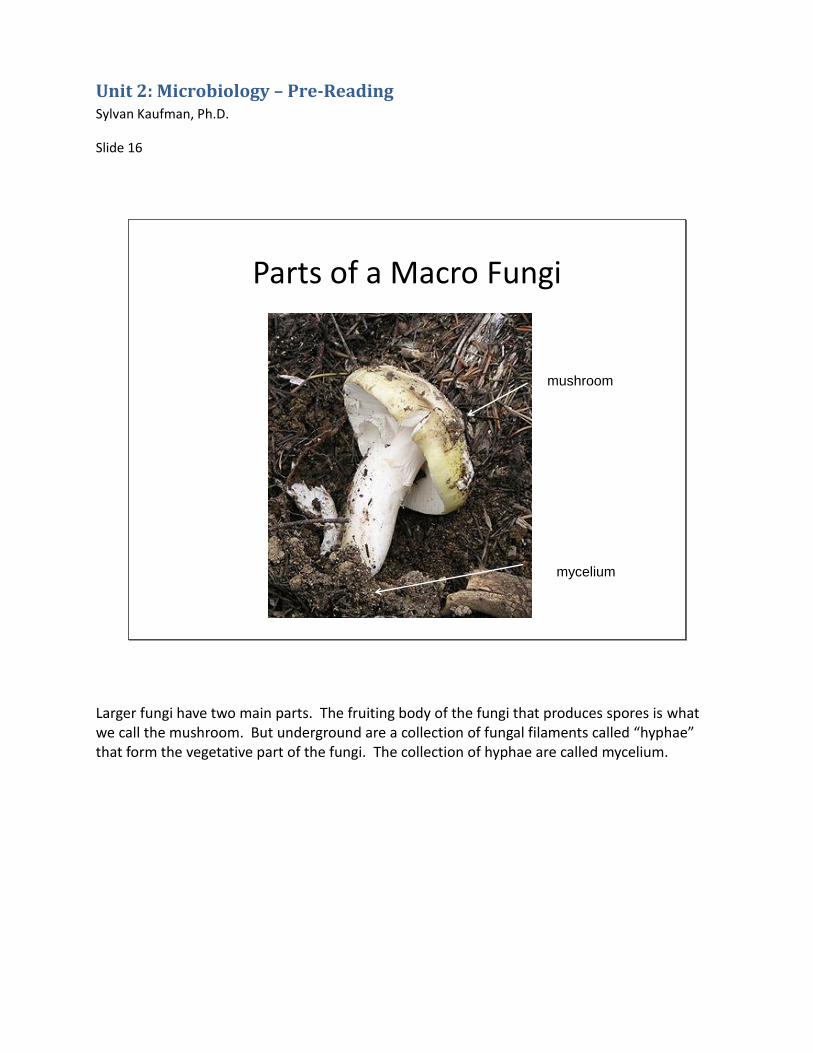
Larger fungi have two main parts. The fruiting body of the fungi that produces spores is what we call the mushroom. But underground are a collection of fungal filaments called “hyphae” that form the vegetative part of the fungi. The collection of hyphae are called mycelium.
Unit 2: Microbiology – Pre-Reading Sylvan Kaufman, Ph.D.
Slide 17
Fungi and Decomposition
• White rot – produces enzymes to degrade lignin
• Brown rot – degrade cellulose and sugars
• Fairy rings in lawns – decomposes dead grass
• Sulphur shelf –
decomposes stumps
Sulphur shelf, Laetiporussulphureus
Fungi are extremely important for decomposition. Some specialize in decomposing certain types of plant material. Look at the fruiting bodies to see where they are located because it can help with identification.
Unit 2: Microbiology – Pre-Reading Sylvan Kaufman, Ph.D.
Slide 18
Mycorrhizae
• Fungi associated with plant roots
• 80 - 90% of all plant species mycorrhizal
• Fungus supplies N and P, plant supplies C
Fly agaric, Amanita muscaria
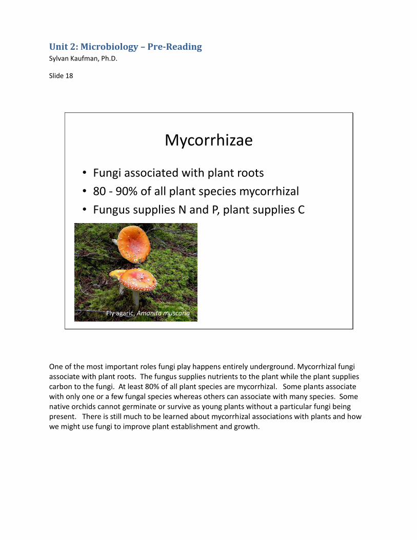
One of the most important roles fungi play happens entirely underground. Mycorrhizal fungi associate with plant roots. The fungus supplies nutrients to the plant while the plant supplies carbon to the fungi. At least 80% of all plant species are mycorrhizal. Some plants associate with only one or a few fungal species whereas others can associate with many species. Some native orchids cannot germinate or survive as young plants without a particular fungi being present. There is still much to be learned about mycorrhizal associations with plants and how we might use fungi to improve plant establishment and growth.
Unit 2: Microbiology – Pre-Reading Sylvan Kaufman, Ph.D.
Slide 19
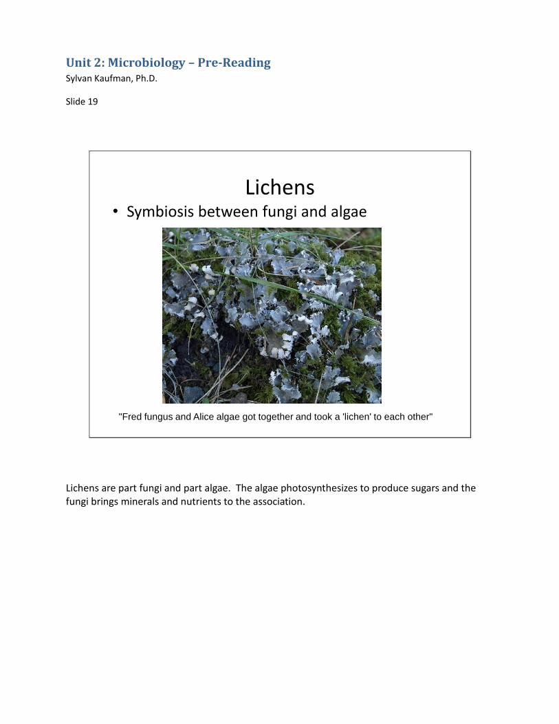
Lichens• Symbiosis between fungi and algae
"Fred fungus and Alice algae got together and took a 'lichen' to each other"
Lichens are part fungi and part algae. The algae photosynthesizes to produce sugars and the fungi brings minerals and nutrients to the association.
Unit 2: Microbiology – Pre-Reading Sylvan Kaufman, Ph.D.
Slide 20
Fungal Pathogens and Parasites
• Pathogens
– Shoestring rot
– Dutch elm disease
– Chestnut blight
• Parasites
– “summit disease” in insects
– Lobster mushroom
One fungi parasitizing another
Other fungi act as pathogens and parasites. Some fungi attack the living tissues of trees. For example, the honey mushroom, Armillaria mellea, causes shoestring rot in the interior of a tree. In our area oaks are often infected with shoestring rot. Chestnut blight was also caused by a fungus accidentally introduced on imported Chinese chestnut trees, Cryphonectria parasitica. The fungi that causes Dutch elm disease, Ophiostoma spp., are spread from tree to tree by the elm bark beetle. Fungi can parasitize insects and other fungi. Insects parasitized by a species of Cordyceps fungus crawl up to the top of a blade of grass or other plant as their tissues are replaced by fungal mycelium. The insect dies clinging to a high point and as its body decays spores are released and carried to other host insects. The lobster mushroom, considered a delicacy by some, is actually a Lactarius or Russula mushroom parasitized by a species of Hypomyces.
Unit 2: Microbiology – Pre-Reading Sylvan Kaufman, Ph.D.
Slide 21
Virtual mushroom foray
Professional and amateur mycologists often conduct forays, or mushroom hunts. Below is a list of some characteristics they might look at to distinguish one species of mushroom from another. •Shape and size of cap •Cap edge, color, texture •Spore bearing surface – gills, teeth, pores •Gill color, attachment •Spore color •Stem – size, color, ring •Veil presence or absence •Color changes when flesh is bruised or broken •Odor and taste •Habitat
Unit 2: Microbiology – Pre-Reading Sylvan Kaufman, Ph.D.
•Growth pattern •Month or season Slide 22
Fungi Groups
• Basidiomycetes
• Ascomycetes
• Gasteromycetes
There are three major groups of fungi. The basidiomycetes have the classic toadstool shape. Spores are forcibly discharged from the “basidium”, sexual cells on which spores develop. Spore prints can be helpful in distinguishing species of basidiomycetes. Ascomycetes produce spores in a sac or “ascus”. These include morels, truffles and cup fungi. Gasteromycetes use structural or physiological means to discharge spores rather than having forcible discharge. Stinkhorns, earthstars, puffballs and bird’s nest fungi are examples of gasteromycetes.
Unit 2: Microbiology – Pre-Reading Sylvan Kaufman, Ph.D.
Slide 23
Agarics
Pluteus
Agaricus
Amanita muscaria
Coprinus
The basidiomycetes can be further broken down into several groups. The Agarics are fleshy mushrooms that usually have a stalk and always have gills. The common button mushroom you buy at the grocery is an Agaric.
Unit 2: Microbiology – Pre-Reading Sylvan Kaufman, Ph.D.
Slide 24
capgills
veil
stalk
When identifying agarics look closely at the cap, gills, veil and stalk. The color, shape and texture of the cap are important. The color and way that the gills attach to the stem distinguish among species. The veil may or may not be present. It covers the gills until the cap expands fully. The stalk holds the cap above the ground and it may be thick or thin, the same or a different color from the cap, and some agarics won’t have a stem at all. Sometimes at the base of the stem there will be a cup from which the fruiting body emerged.
Unit 2: Microbiology – Pre-Reading Sylvan Kaufman, Ph.D.
Slide 25
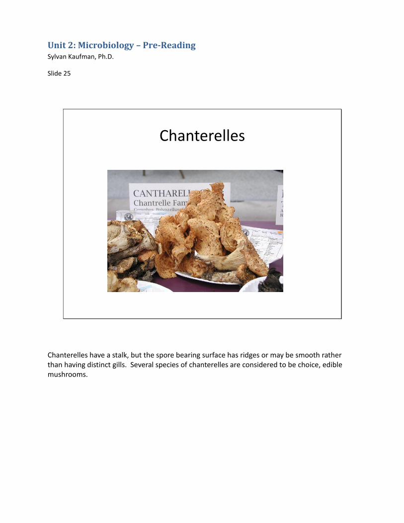
Chanterelles
Chanterelles have a stalk, but the spore bearing surface has ridges or may be smooth rather than having distinct gills. Several species of chanterelles are considered to be choice, edible mushrooms.
Unit 2: Microbiology – Pre-Reading Sylvan Kaufman, Ph.D.
Slide 26
Boletes
The boletes have pores on the spore bearing surface rather than gills. If you are familiar with Italian cooking, porcini are a species of bolete.
Unit 2: Microbiology – Pre-Reading Sylvan Kaufman, Ph.D.
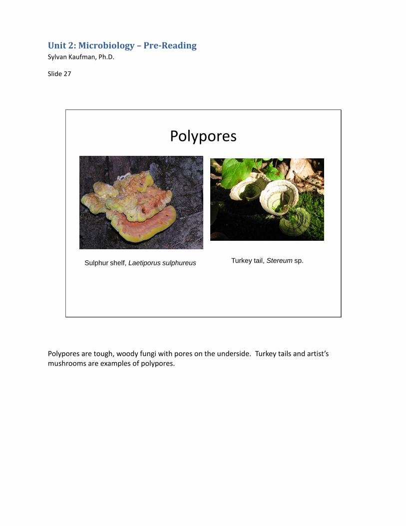
Polypores are tough, woody fungi with pores on the underside. Turkey tails and artist’s mushrooms are examples of polypores.
Unit 2: Microbiology – Pre-Reading Sylvan Kaufman, Ph.D.
Slide 28
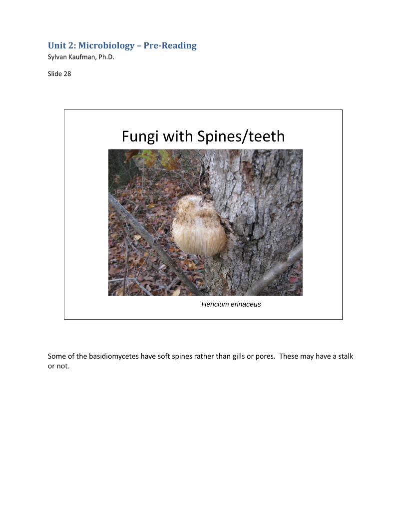
Fungi with Spines/teeth
Hericium erinaceus
Some of the basidiomycetes have soft spines rather than gills or pores. These may have a stalk or not.
Unit 2: Microbiology – Pre-Reading Sylvan Kaufman, Ph.D.
Slide 29
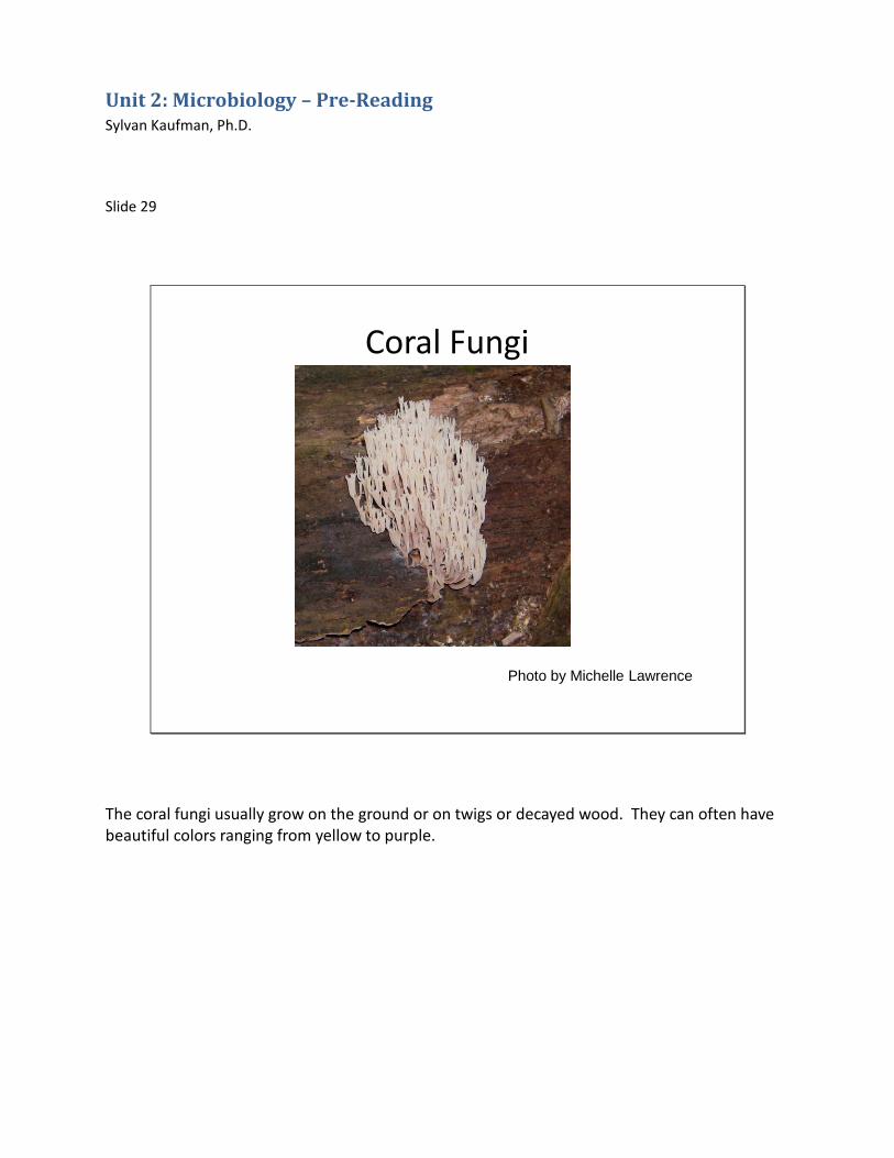
Coral Fungi
Photo by Michelle Lawrence
The coral fungi usually grow on the ground or on twigs or decayed wood. They can often have beautiful colors ranging from yellow to purple.
Unit 2: Microbiology – Pre-Reading Sylvan Kaufman, Ph.D.
Slide 30
Jelly fungi
Tremella mesenterica
The last group of Basidiomycetes look very different from the others. They are often brightly colored and the spore bearing surface is usually smooth. You often find them growing on stumps, logs and twigs in wet weather. The bright yellow species in the photo is called “witches’ butter”.
Unit 2: Microbiology – Pre-Reading Sylvan Kaufman, Ph.D.
Slide 31
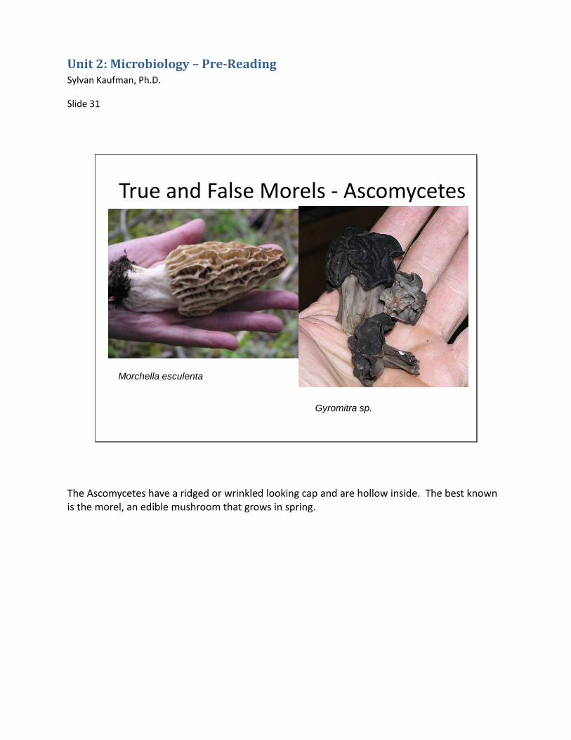
True and False Morels - Ascomycetes
Morchella esculenta
Gyromitra sp.
The Ascomycetes have a ridged or wrinkled looking cap and are hollow inside. The best known is the morel, an edible mushroom that grows in spring.
Unit 2: Microbiology – Pre-Reading Sylvan Kaufman, Ph.D.
Slide 32
Puffballs and Earthstars -Gasteromycetes
The Gasteromycetes come in a range of shapes and sizes. Puffballs are rounded and if you cut one open it is a solid mass of spores inside. Earthstars open up in a star shape. The spores are usually dispersed by rain drops or wind.
Unit 2: Microbiology – Pre-Reading Sylvan Kaufman, Ph.D.
Slide 33
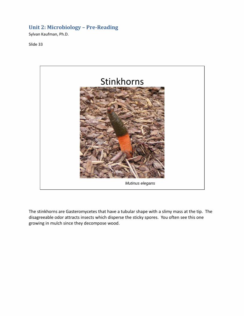
Stinkhorns
Mutinus elegans
The stinkhorns are Gasteromycetes that have a tubular shape with a slimy mass at the tip. The disagreeable odor attracts insects which disperse the sticky spores. You often see this one growing in mulch since they decompose wood.


![Sylvan [Issue: July 2011]](https://static.documents.pub/doc/80x56/568c38261a28ab02359dfab6/sylvan-issue-july-2011.jpg)

