52
General and Medical Microbiology Lab ■ 147 Unit 6 Physiological Testing
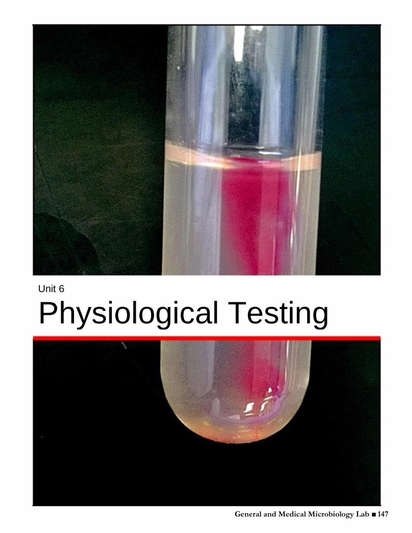
General and Medical Microbiology Lab ■ 147
Unit 6
Physiological Testing
General and Medical Microbiology Lab ■ 148
Lecture 13 Tasks for the day:
Cover Lecture 13 material. Vodcast found at: http://youtu.be/SvjRxALqykA Perform Experiment 13A: Throat Swab for Gram-positive Cocci, Lab Period One Perform Experiment 13B: Gram-positive Cocci, Lab Period One Perform Experiment 13C: Gram-negative Rod, Lab Period One
I. Unknown identification A. ______________________________________
1. ___________________________________________ from the environment of interest (this will vary depending on the researcher’s interest. It may come from soil, spoiled food, or it may be a patient sample: urine, feces, sputum, throat or skin swab, blood, etc.) 2. Obtain the organism in a _________________________ using selective and differential media.
B. ___________________________________________ 1. Perform a _______________________________ 2. Determine the organism’s unique biochemical properties:
a. Nutrient utilization b. Resistance to _________________________________________(i.e. salts, antibiotics, etc.) c. _________________________ production (catalase, coagulase, hemolysins, oxidase, etc.) d. _________________________ e. Fermentation _______________________________
f. Growth properties (temperature, O2 concentration/utilization, CO2,etc.) You will be expected to know the basis for each test and the KEY physiological characteristics of each
bacterial genera and species that we will use in the following laboratory periods! The culmination of the next few labs will enable you to take a bacterial unknown and identify its genus and species based on these tests.
You may want to make a flashcard for each test telling you the positive reaction/color, negative reaction/color, and any biochemical reaction taking place in that specific test. In addition, flashcards relating information for different bacterial organisms will also come in handy. Another useful tool for this section lab is your picture atlas — it has everything in there you could ever want to know about any of the biochemical tests, media, and positive/negative reactions. The online summary of biochemical tests also serves as a good resource and one that is more specific to the experiments performed in this class II. Bacterial groups
A. _______________________________________ 1. Genus: Staphylococcus
Key characteristics i. Arranged in grape-like clusters ii. _______________________________ (this is the most important test to distinguish between Gram-positive cocci — staphylococci and streptococci) iii. ________________________________ (will tolerate salt) iv. Facultative anaerobes v. Non-motile
Representative species: — S. ___________________ — S. saprophyticus — S. ____________________ All three of these staphylococcus species are found as parasites on the skin and mucous membranes of humans and other animals.
General and Medical Microbiology Lab ■ 149
>Only S. aureus is considered to be a ________________, causing ________________, abscesses, food poisoning, scalded skin syndrome, and _______________. The virulence of S. aureus is due to a number of toxins and enzymes that it can produce, for example:
__________________: clots blood plasma Leukocidins: kill ___________________ (WBC) _________________: damage red blood cells (RBCs) ______________________ toxid: Exfoliative dermatitis Enterotoxins: ___________________________
>S. epidermidis and S. saprophyticus have been implicated in some infectious diseases but have low virulence. Therefore, they are considered nonpathogenic.
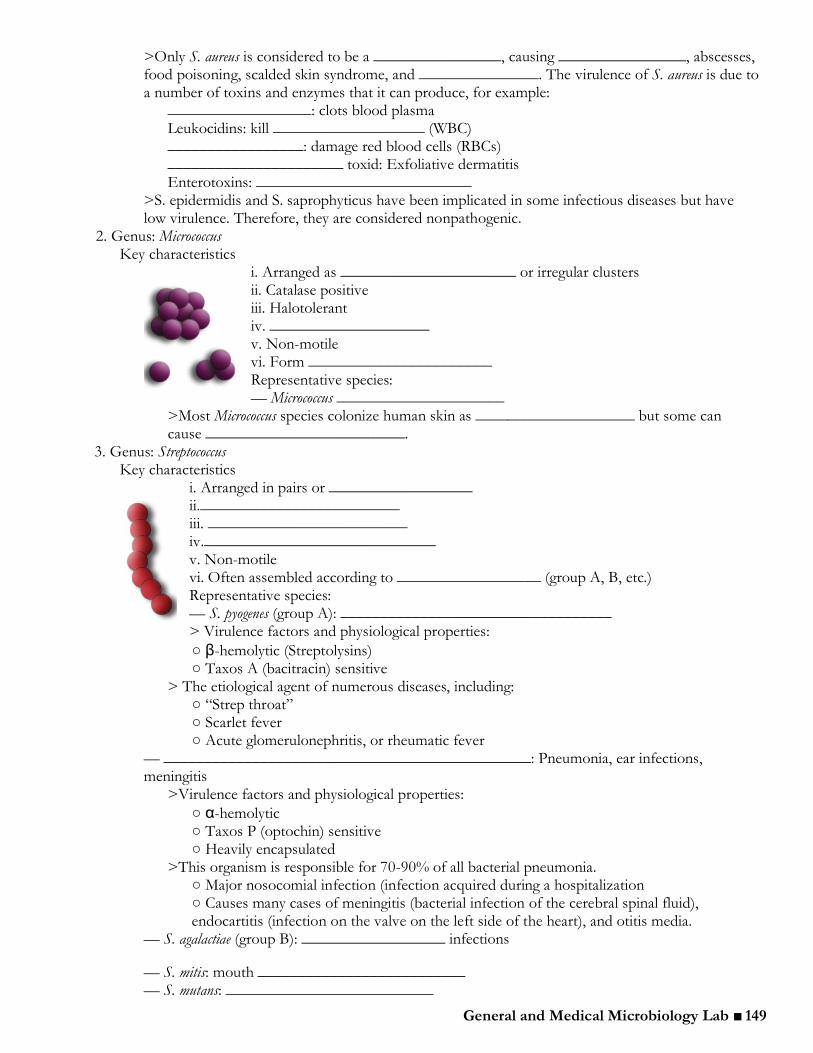
2. Genus: Micrococcus Key characteristics
i. Arranged as ______________________ or irregular clusters ii. Catalase positive iii. Halotolerant iv. ____________________ v. Non-motile vi. Form _______________________ Representative species: — Micrococcus _____________________
>Most Micrococcus species colonize human skin as ____________________ but some can cause _________________________.
3. Genus: Streptococcus Key characteristics
i. Arranged in pairs or __________________ ii._________________________ iii. _________________________ iv._____________________________ v. Non-motile vi. Often assembled according to __________________ (group A, B, etc.) Representative species: — S. pyogenes (group A): __________________________________ > Virulence factors and physiological properties:
○ β-hemolytic (Streptolysins) ○ Taxos A (bacitracin) sensitive
> The etiological agent of numerous diseases, including: ○ “Strep throat” ○ Scarlet fever ○ Acute glomerulonephritis, or rheumatic fever
— ______________________________________________: Pneumonia, ear infections, meningitis
>Virulence factors and physiological properties:
○ α-hemolytic ○ Taxos P (optochin) sensitive ○ Heavily encapsulated
>This organism is responsible for 70-90% of all bacterial pneumonia. ○ Major nosocomial infection (infection acquired during a hospitalization ○ Causes many cases of meningitis (bacterial infection of the cerebral spinal fluid), endocartitis (infection on the valve on the left side of the heart), and otitis media.
— S. agalactiae (group B): __________________ infections
— S. mitis: mouth __________________________ — S. mutans: __________________________
General and Medical Microbiology Lab ■ 150
>The virulence of the Streptococcus species is due to a number of toxins and enzymes that they can produce. For example:
_____________: protects bacteria from phagocytosis Erythrogenic toxin: scarlet fever __________________ ___________________: strep throat Streptokinase and Hyaluronidase: “_____________________”
4. Genus: Enterococcus Key characteristics
i. Arranged in pairs or ____________________________ ii. Catalase negative iii. ________________________________ iv. _________________________________ v. Non-motile vi. Localized to _____________________________
Representative species — ____________________________ — E. faecium
>Enterococci have a ____________________________ as they lack defense systems against phagocytic cells. However, Enterococci commonly cause _____________________, urinary tract infections, bacteremia, and wound infections.
Practice: A scientist is trying to identify an unknown bacterial isolate. She finds that the unknown is a Gram-positive cocci. She sees both chains and irregular clusters on the Gram stain. What test would she
do next? ___________________________________________
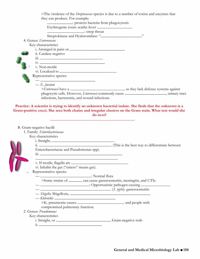
B. Gram-negative bacilli 1. Family: Enterobacteriaceae
Key characteristics i. Straight, ________________________________ ii. _____________________________________ (This is the best way to differentiate between Enterobacteriacae and Pseudomonas spp). iii. __________________________________________ __________________________________________ v. If motile, flagella are _____________________________ vi. Inhabit the gut (“entero” means gut).
Representative species — __________________________: Normal flora
>Some strains of _______ can cause gasteroenteritis, meningitis, and UTIs. — _________________________: Opportunistic pathogen causing _______________ — ____________________________________ (S. typhi): gasteroenteritis — Shigella: Shigellosis, _________________________________________ — Klebsiella: _______________________________
>K. pneumonia causes ________________________ and people with compromised pulmonary function.
2. Genus: Pseudomonas Key characteristics
i. Straight, or ____________________________ Gram-negative rods ii. ________________________________
General and Medical Microbiology Lab ■ 151
iii. ___________________________________ iv. ________________________ v. Motile via _________________________ vi. Often produce _________________________________ and colonies appear mucoid
Representative species — _____________________: Opportunistic pathogen that causes disease in _______________________________ (e.g. cystic fibrosis patients, AIDS patients, and burn victims) — P. fluorescens: Psychrophile
> P. aeruginosa produces several enzymes and toxins that account for its virulence. The most important toxin __________________________________ in eukaryotic cells. Even the water-soluble pigment pyocyanin causes _______________________ to a host.
Practice: A student has a bacterial unknown that is a Gram-negative rod. What test would the student do first to determine if his unknown is a Pseudomonas species?
____________________________________
III. Media and Biochemical Tests
A. Blood Agar Plate (BAP)
1. This is a very rich medium that allows for the growth of most organisms but is also able to differentiate organisms based on their ______________________________________.
i. Gamma (γ)—______________________________ (no clearing zones)
ii. Alpha (α)—partial hemolysis (___________________________________surround the colonies)
iii. Beta (β)—complete hemolysis (______________________________ surround the colonies)
Practice: Which bacterial species discussed above are β-hemolytic? ______________________________ and ______________________________
Practice: Which species are α-hemolytic? ______________________________ and ______________________________
B. Mannitol Salt Agar (7.5 % NaCl) 1. Selects for organisms that can live in the ____________________________________________________________ and differentiates organisms that can ______________________________________________________. The agar will turn __________________________ if the organism is able to ferment mannitol due to the acidic byproducts of the fermentation.
C. Glucose Fermentation Broth tubes 1. These tubes contain the simple ______________________________________ in addition to a ______________________________, and a __________________________________. A positive reaction will turn the broth _____________________________________. The Durham tube will measure ____________________________________ by the bacteria (gas produced by an enzyme called ____________________________________________________).
D. SIM Tube (Sulfur, Indole, Motility) 1. __________________________________ production will turn the agar _________________. Adding
Kovac’s reagent, and ______________________________________________________ determine indole production. Indole is produced from the breakdown of the amino acid _________________________. Motility is determined by observing the organism’s ability to move through the _______________________________________________________.
General and Medical Microbiology Lab ■ 152
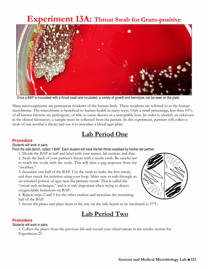
Once a BAP is inoculated with a throat swab and incubated, a variety of growth and hemolysis can be seen on the plate
Many microorganisms are permanent residents of the human body. These residents are referred to as the human microbiome. The microbiome is beneficial to human health in many ways. Only a small percentage, less than 10%, of all known bacteria are pathogenic, or able to cause disease in a susceptible host. In order to identify an unknown in the clinical laboratory, a sample must be collected from the patient. In this experiment, partners will collect a swab of one another’s throat and use it to inoculate a blood agar plate.
Lab Period One Procedure Students will work in pairs. From the side bench, collect 1 BAP. Each student will have his/her throat swabbed by his/her lab partner.
1. Divide the BAP in half and label with your names, lab section, and date. 2. Swab the back of your partner’s throat with a sterile swab. Be careful not to touch the uvula with the swab. This will elicit a gag response from the “swabbee.” 3. Inoculate one half of the BAP. Use the swab to make the first streak, and then streak for isolation using your loop. Make sure to stab through an un-streaked portion of agar near the primary streak. This is called the “streak-stab technique,” and it is only important when trying to detect oxygen-labile hemolysis on BAP. 4. Repeat steps 2 and 3 for the other student and inoculate the remaining half of the BAP. 5. Invert the plates and place them in the tray on the side bench to be incubated at 37°C.
Lab Period Two Procedure Students will work in pairs.
1. Collect the plates from the previous lab and record your observations in the results section for Experiment 25.
Experiment 13A: Throat Swab for Gram-positive Cocci
General and Medical Microbiology Lab ■ 153
Experiment 13A: Results
1. Draw an illustration of your throat swab plate. Describe the morphology and type of hemolysis surrounding two different colonies.
Colony #1: ____________________________________________________________________________ Colony #2: ____________________________________________________________________________ 2. Were the organisms on your throat swab plate simply residents of the microbiome? How can you be certain? ______________________________________________________________________________________ ______________________________________________________________________________________ ______________________________________________________________________________________

General and Medical Microbiology Lab ■ 154
Hemolysis on blood agar plates is one of the many tests that can help determine the identity of unknown Gram-positive cocci.
Because Streptococcus, Enterococcus, Micrococcus, and Staphylococcus spp. are all Gram-positive cocci, and because the four genera are comprised of both normal flora and pathogenic species, it is essential to distinguish between these organisms in the clinical microbiology laboratory. In the next couple of experiments, the key biochemical tests used to identify the species of these genera will be introduced.
Lab Period One A. Growth on BAP and MSA Procedure Students will work in groups of 4 From the side bench, each group should collect 2 BAP and 2 MSA plates.
1. Divide each of the four plates in half and label them with the names of the organisms as shown in the diagram below. Be sure to include your initials, lab section number, and the date.
2. Using sterile technique, inoculate each half of the blood agar plates as shown below. Try to streak for isolation on each half of the plates; look carefully at the below diagram. After streaking, DO NOT flame your inoculating loop. Instead, stab through an un-streaked portion of the agar near the primary streaks.
Experiment 13B: Gram-positive Cocci

General and Medical Microbiology Lab ■ 155
3. Inoculate the two MSA plates in the same manner as above, but DO NOT stab.
4. Invert both the BAP and MSA plates and place them in the tray on the side bench to be incubated at 37°C.
Lab Period Two A. Growth on BAP and MSA Procedure Students will work in groups of 4.
1. Collect the BAP and MSA plates from the previous lab period and record the results in the results section for Experiment 13B. If your results differ from the expected results described in the results section, include a possible explanation for the discrepancy. When examining hemolysis patterns and other results, have your TA or instructor check your conclusions.
B. Catalase Test Procedure
Students will work in group of four. 1. Go to the catalase station and perform the catalase test as described below:
a. Place a clean glass slide on the bench. b. Put 2 small drops of 3% hydrogen
peroxide (H2O2) on the slide. c. Pick up a colony of Staphylococcus epidermidis from your BAP plate using a wooden applicator stick. Stir this
colony into the first drop of H2O2. Repeat this procedure with the negative control, Streptococcus mitis, which is available at the catalase station. If bubbles appear, the organism is catalase positive.
3. Record any observations in the results section following this experiment.
General and Medical Microbiology Lab ■ 156
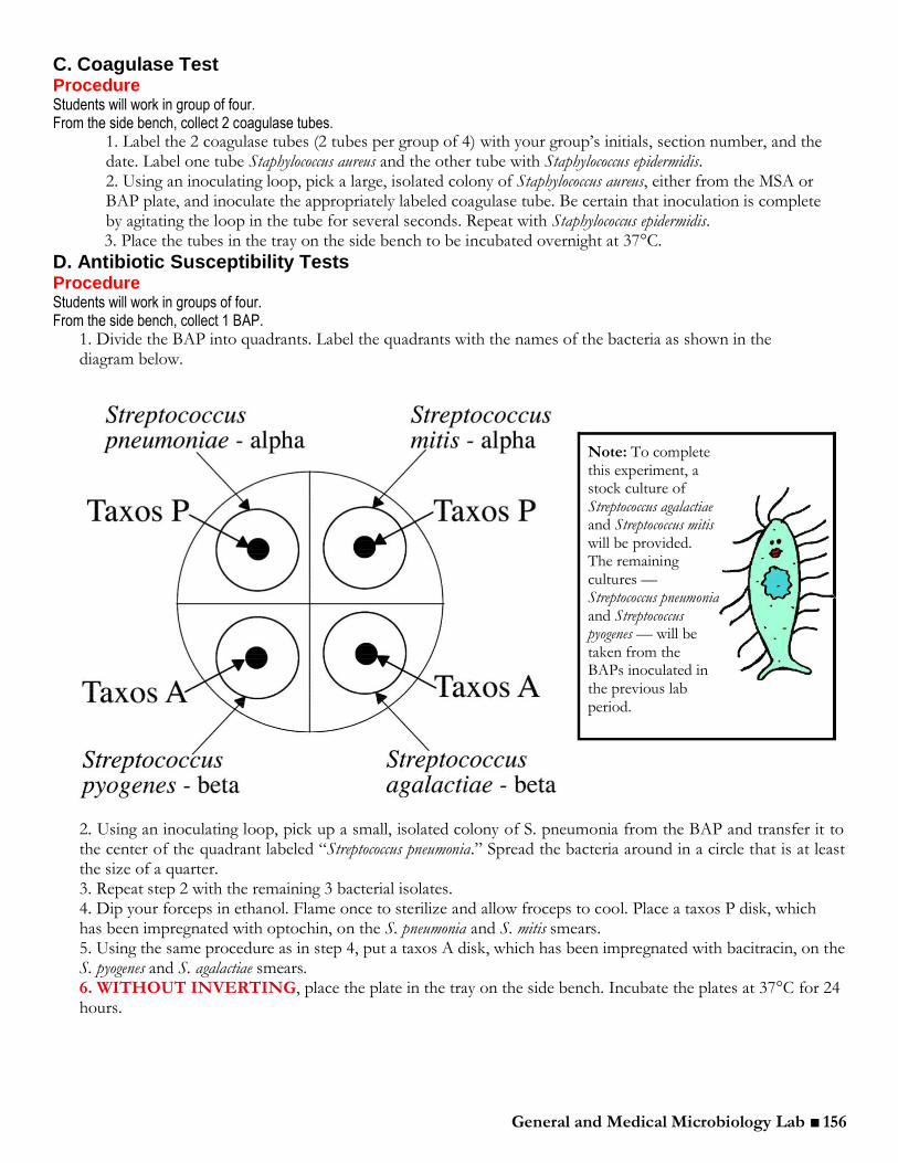
C. Coagulase Test Procedure Students will work in group of four. From the side bench, collect 2 coagulase tubes.
1. Label the 2 coagulase tubes (2 tubes per group of 4) with your group’s initials, section number, and the date. Label one tube Staphylococcus aureus and the other tube with Staphylococcus epidermidis. 2. Using an inoculating loop, pick a large, isolated colony of Staphylococcus aureus, either from the MSA or BAP plate, and inoculate the appropriately labeled coagulase tube. Be certain that inoculation is complete by agitating the loop in the tube for several seconds. Repeat with Staphylococcus epidermidis. 3. Place the tubes in the tray on the side bench to be incubated overnight at 37°C.
D. Antibiotic Susceptibility Tests Procedure Students will work in groups of four. From the side bench, collect 1 BAP.
1. Divide the BAP into quadrants. Label the quadrants with the names of the bacteria as shown in the diagram below.
2. Using an inoculating loop, pick up a small, isolated colony of S. pneumonia from the BAP and transfer it to the center of the quadrant labeled “Streptococcus pneumonia.” Spread the bacteria around in a circle that is at least the size of a quarter. 3. Repeat step 2 with the remaining 3 bacterial isolates. 4. Dip your forceps in ethanol. Flame once to sterilize and allow froceps to cool. Place a taxos P disk, which has been impregnated with optochin, on the S. pneumonia and S. mitis smears. 5. Using the same procedure as in step 4, put a taxos A disk, which has been impregnated with bacitracin, on the S. pyogenes and S. agalactiae smears. 6. WITHOUT INVERTING, place the plate in the tray on the side bench. Incubate the plates at 37°C for 24 hours.
Note: To complete this experiment, a stock culture of Streptococcus agalactiae and Streptococcus mitis will be provided. The remaining cultures — Streptococcus pneumonia and Streptococcus pyogenes — will be taken from the BAPs inoculated in the previous lab period.
General and Medical Microbiology Lab ■ 157
E. Nitrate test The nitrate test is important for the identification of both Gram-positive and Gram-negative organisms. For this portion of the nitrate test, we will be using the Gram-positive Staphylococcus epidermidis and Streptococcus pneumoniae from the previous lab period’s BAP.
Procedure Students will work in groups of 4. From the side bench, collect 2 nitrate tubes.
1. Label 2 nitrate tubes with your group’s initials, lab section number, and the date. Label each tube with one of the following organisms: Staphylococcus epidermidis and Streptococcus pneumoniae. 2. With a sterile inoculating loop, pick an isolated colony of Staphylococcus epidermidis and inoculate the appropriately labeled nitrate tube. Repeat with Streptococcus pneumoniae. 3. Place these tubes in the tray on the side bench to be incubated at 37°C.
Lab Period Three C. Coagulase Test Procedure Students will work in groups of four.
1. Observe the previous lab’s coagulase tubes. Coagulase is an enzyme produced by Staphylococcus aureus that effectively clots blood plasma. Coagulase-positive Staphylococcus spp. form a clot around themselves to protect against the host’s immune defenses. A positive coagulase test is indicated by solidification of the rabbit plasma following inoculation and incubation.
D. Antibiotic Susceptibility Tests Procedure Students will work in groups of four.
1. Measure the zone of inhibition around the Taxos A and Taxos P disks on the BAP. Use the table at the end of the results section to determine whether the organism is susceptible/sensitive to that antibiotic.
E. Nitrate test Procedure Students will work in groups of 4.
1. Collect the nitrate broth tubes that were inoculated during last lab period. 2. Perform a nitrate test on the nitrate broth tubes by following this procedure:
a. Nitrite (NO2-, a product of this reaction) is detected by adding 7 drops of sulfanilic acid (Nitrate and 7 drops of dimethyl-alpha-naphthylamine (Nitrate II) to the nitrate broth tubes. The immediate
development of a red color indicates that the organism is positive for nitrate reduction (NO3- is reduced
to NO2-). b. If the tube remains a cloudy beige color — no color change — it might be a negative test. However,
the organism might still be positive for nitrate reduction; it may simply be producing another reduced
product of nitrate, such as NO, NH4+, or N2O. Therefore, the second step in the test looks for the
depletion of nitrate, which would also indicate a positive nitrate reduction reaction. c. Use forceps to add a few grains of elemental zinc to the tube. Mix and allow at least 1 minute for the
reaction to occur. Zinc will reduce the NO3-, if it is still present, to NO2- and the medium will turn a red color.
After the addition of zinc, a change of color to red indicates a negative test, because the nitrate is still present
in the medium (nitrate has NOT been used by the organism). If the medium remains a cloudy beige color — no color change — nitrate has been depleted by reduction to the products other than nitrite and the organism is positive for nitrate reduction.
*Note: The addition of zinc will eventually cause the broth to turn slightly pink. Be sure to read your results within
the first 1-2 minutes of adding zinc.
General and Medical Microbiology Lab ■ 158
Experiment 13B: Results
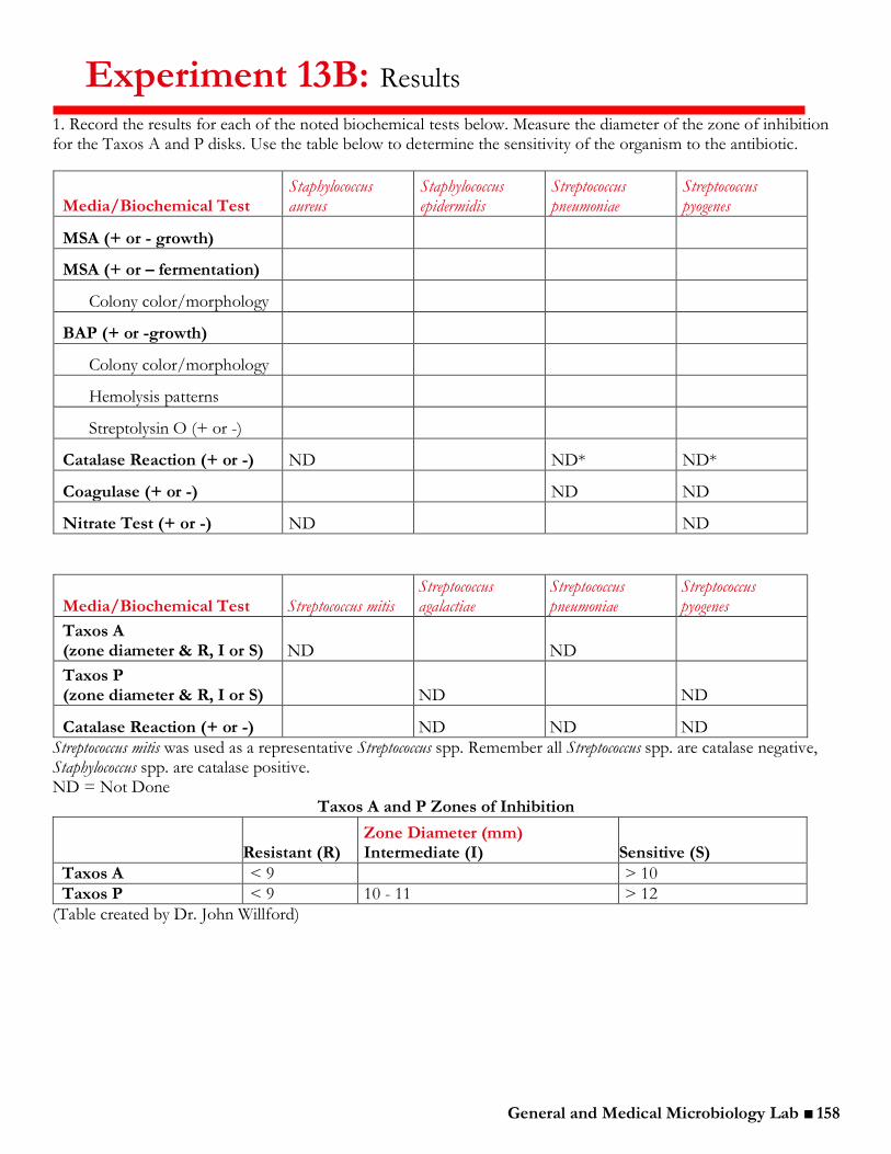
1. Record the results for each of the noted biochemical tests below. Measure the diameter of the zone of inhibition for the Taxos A and P disks. Use the table below to determine the sensitivity of the organism to the antibiotic.
Media/Biochemical Test Staphylococcus aureus
Staphylococcus epidermidis
Streptococcus pneumoniae
Streptococcus pyogenes
MSA (+ or - growth)
MSA (+ or – fermentation)
Colony color/morphology
BAP (+ or -growth)
Colony color/morphology
Hemolysis patterns
Streptolysin O (+ or -)
Catalase Reaction (+ or -) ND ND* ND*
Coagulase (+ or -) ND ND
Nitrate Test (+ or -) ND ND
Media/Biochemical Test Streptococcus mitis Streptococcus agalactiae
Streptococcus pneumoniae
Streptococcus pyogenes
Taxos A (zone diameter & R, I or S) ND ND
Taxos P (zone diameter & R, I or S) ND ND
Catalase Reaction (+ or -) ND ND ND
Streptococcus mitis was used as a representative Streptococcus spp. Remember all Streptococcus spp. are catalase negative, Staphylococcus spp. are catalase positive. ND = Not Done
Taxos A and P Zones of Inhibition
Resistant (R) Zone Diameter (mm) Intermediate (I) Sensitive (S)
Taxos A < 9 > 10
Taxos P < 9 10 - 11 > 12
(Table created by Dr. John Willford)
General and Medical Microbiology Lab ■ 159
2. Note any discrepancies from the expected results that were observed in the above data set. What are the possible explanations for these discrepancies? ______________________________________________________________________________________ ______________________________________________________________________________________ ______________________________________________________________________________________ 3. Which organism(s) grew best on MSA? Explain. ______________________________________________________________________________________ ______________________________________________________________________________________ ______________________________________________________________________________________ 4. Would you predict that all the organisms would grow on BAP? Explain. ______________________________________________________________________________________ ______________________________________________________________________________________ ______________________________________________________________________________________
5. Which genus of Gram-positive coccus is catalase positive? ______________________________________________________________________________________ 6. Which organism is coagulase positive? ______________________________________________________________________________________ 7. List two biochemical properties that differentiate Staphylococcus aureus from Staphylococcus epidermidis.
1. _____________________________________________________________________________ ________________________________________________________________________________ 2. _____________________________________________________________________________ ______________________________________________________________________________
8. Are there any Staphylococcus spp. capable of hemolysis (α or β)? If yes, which one? ______________________________________________________________________________________
9. How might an experimenter distinguish between Streptococcus pyogenes and other β-hemolytic Streptococcus spp.? ______________________________________________________________________________________ ______________________________________________________________________________________ ______________________________________________________________________________________ 10. Did Taxos P inhibit the growth of either organism tested? If yes, which one? ______________________________________________________________________________________ ______________________________________________________________________________________ 11. What is Taxos P? ______________________________________________________________________________________ ______________________________________________________________________________________ 12. What is Taxos A? ______________________________________________________________________________________ ______________________________________________________________________________________

General and Medical Microbiology Lab ■ 160
13. List three biochemical properties that would distinguish between Staphylococcus aureus and Streptococcus pyogenes.
1. ______________________________________________________________________________ ________________________________________________________________________________ 2. _____________________________________________________________________________ ______________________________________________________________________________ 3. ______________________________________________________________________________ ________________________________________________________________________________
14. What biochemical property is unique to Streptococcus agalactiae? ______________________________________________________________________________________ ______________________________________________________________________________________ ______________________________________________________________________________________ 15. What biochemical property is unique to Enterococcus faecalis? ______________________________________________________________________________________ ______________________________________________________________________________________ ______________________________________________________________________________________
Expected results
Staphylococcus aureus
■ halotolerant ■ferments mannitol
■ coagulase+
■ β-hemolytic ■ nitrate+
Streptococcus agalactiae
■ β-hemolysis ■ Taxos A resistant ■ CAMP+ ■ nitrate-
Staphylococcus epidermidis
■ halotolerant ■ does not ferment
mannitol ■ coagulase-
■ nitrate+
Streptococcus mitis
■ α-hemolytic ■ Taxos P resistant ■ nitrate-
Streptococcus pneumoniae
■ α-hemolytic ■ Taxos P sensitive ■ nitrate-
Streptococcus pyogenes
■ β-hemolytic ■ Taxos A sensitive
■ nitrate-
General and Medical Microbiology Lab ■ 161
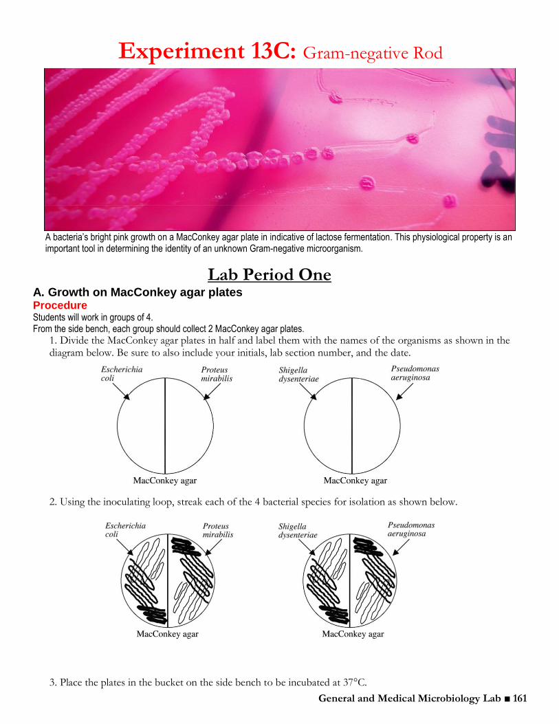
A bacteria’s bright pink growth on a MacConkey agar plate in indicative of lactose fermentation. This physiological property is an important tool in determining the identity of an unknown Gram-negative microorganism.
Lab Period One A. Growth on MacConkey agar plates Procedure Students will work in groups of 4. From the side bench, each group should collect 2 MacConkey agar plates.
1. Divide the MacConkey agar plates in half and label them with the names of the organisms as shown in the diagram below. Be sure to also include your initials, lab section number, and the date.
2. Using the inoculating loop, streak each of the 4 bacterial species for isolation as shown below.
3. Place the plates in the bucket on the side bench to be incubated at 37°C.
Experiment 13C: Gram-negative Rod

General and Medical Microbiology Lab ■ 162
B. Growth in Glucose Broth Procedure Students will work in groups of 4. From the side bench, each group should collect 4 glucose broth tubes.
1. Using an inoculating loop, inoculate each of the 4 following bacterial species into separate glucose broth tubes: Escherichia coli, Proteus mirabilis, Shigella dysenteriae, and Pseudomonas aeruginosa. Specifically, pick up a colony or piece of colony with your loop. Insert your loop into the broth and briskly stir back and forth. With a piece of tape, label the tubes with your initials, section number, and the name of the microorganism. 2. Taking care not to distrub the Durham tube, place the tubes in the bucket on the side bench to be incubated at 37°C.
C. SIM media Procedure Students will work in groups of 4. From the side bench, each group should collect 4 SIM (Sulfur, Indole, Motility) tubes.
1. Using an inoculating needle, inoculate the SIM tubes with each of the 4 following bacterial species: Escherichia coli, Proteus mirabilis, Shigella dysenteriae, and Pseudomonas aeruginosa. To inoculate SIM media, flame the inoculating needle and allow it to cool, then pick up a small amount of a bacterial colony from the stock plate. Inoculate the SIM media by making a single, straight stab down the middle of the tube to the bottom. 2. Place the tubes in the bucket on the side bench to be incubated at 37°C.
Lab Period Two A. Growth on MacConkey agar plates Procedure Students will work in groups of 4.
1. Collect the MacConkey agar plates from the previous lab period and record the results in the results section for Experiment 27. The selective and differential MacConkey agar will inhibit the growth of Gram-positive bacteria, while differentiating on the basis of lactose fermentation. Colonies of lactose-fermenting bacteria will appear dark pink/red, and non-lactose fermenting bacteria will appear colorless. If your results differ from the expected results described in the results section, include a possible explanation for the discrepancy.
B. Growth in Glucose Broth Procedure Students will work in groups of 4.
1. Examine the growth in the glucose broth tubes inoculated in the last lab period and record your observations in the results section for this experiment. Glucose fermentation broth tubes test for the ability of the organism to ferment glucose. Organic acids are the products of fermentation. These acids lower the pH of the medium causing the pH indicator, phenol red, to change from red to yellow. Fermentation can occur with or without the production of gas. If gas is produced, it is trapped in the inverted Durham tube and can be seen as a bubble. Gas cannot be produced without fermentation of the sugar.
General and Medical Microbiology Lab ■ 163
C. SIM media Procedure Students will work in groups of 4.
1. Observe the SIM media for H2S production, which is indicated by the presence of a black precipitate. 2. Examine the turbidity of the line of inoculation to determine motility. Motility is determined by observing growth/turbidity away from the line of inoculation, where non-motile bacteria will not show growth far from this line. *Look to see if the stab appears clear or cloudy. 3. Take your SIM media to the SIM station and add 6-8 drops of Kovac’s reagent to the top of the SIM tube. Mix the tubes very gently and observe the top layer for any notable color change. One of the properties the SIM tube tests is tryptophanase activity, also known as indole production, tryptophanase is an enzyme that cleaves the amino acid tryptophan into pyruvic acid, ammonia, and indole. This reaction is determined by adding Kovac’s reagent to the SIM tube following incubation. Kovac’s reagent combines with indole to produce a rose or red color. 4. Record your observations in the results section of this experiment.
D. KIA Procedure Students will work in groups of 4. From the side bench, collect 4 KIA slants.
1. Label 4 KIA tubes with your group’s initials, lab section number, and each of the 4 Gram-negative bacilli: Escherichia coli, Proteus mirabilis, Shigella dysenteriae, and Pseudomonas aeruginosa. 2. Using an inoculating needle, pick up a colony of E. coli from your MacConkey plate and inoculate the appropriately labeled KIA tube by making a single, straight stab to the bottom of the tube. As you remove the needle from the tube bottom, streak the surface of the slant. Repeat with Proteus mirabilis, Shigella dysenteriae, and Pseudomonas aeruginosa. 3. Place the tubes in the tray to be incubated at 37°C.
E. Oxidase test When performing the oxidase test on an unknown bacterium, both
a positive and negative control should be used side-by side with the unknown on the Whatman filter paper. It is only by comparison that the results of this unknown can be accurately determined.
Procedure Students will work individually.
1. Place a piece of Whatman paper in the petri dish. With a wooden stick, collect a large colony of Escherichia coli from the provided plate and smear it onto the piece of Whatman paper. Repeat with Pseudomonas aeruginosa. Make certain the growth is visible on the paper. If the growth is not visible, it is very difficult to interpret the test results. 2. Place 2-3 drops of oxidase reagent onto the culture smears. If the culture smear turns purple, the organism is oxidase-positive. If the smear remains colorless, the organism is oxidase-negative
Before you leave ... Before leaving the lab today, use the results from the previous lab period to predict the results for the four Gram-negative bacilli used to inoculate the KIA tubes. During the previous lab, we performed biochemical tests that indicated ability to ferment glucose, lactose,
and to produce H2S.
A. Escherichia coli: __________________________
__________________________________________ B. Proteus mirabilis: _________________________
__________________________________________ C. Shigella dysenteriae: ______________________ __________________________________________ D. Pseudomonas aeruginosa: _________________
__________________________________________
Note: Because the oxidase test is very important and might be included on a lab practical, each student should perform this test. Plates with Escherichia
coli and Pseudomonas
aeruginosa will be provided at the oxidase test station
General and Medical Microbiology Lab ■ 164
F. Nitrate test The nitrate test is important for the identification of both Gram-positive and Gram-negative organisms. For this portion of the nitrate test, we will be using the Gram-negative Pseudomonas aeruginosa and Escherichia coli from the previous lab period’s MacConkey agar plates.
Procedure Students will work in groups of 4. From the side bench, collect 2 nitrate tubes.
1. Label 2 nitrate tubes with your group’s initials, lab section number, and the date. Label each tube with one of the following organisms: Pseudomonas aeruginosa and Escherichia coli. 2. With a sterile inoculating loop, pick an isolated colony of Pseudomonas aeruginosa and inoculate the appropriately labeled nitrate tube. Repeat with Escherichia coli. 3. Place these tubes in the tray on the side bench to be incubated at 37°C.
Lab Period Three D. KIA
Procedure Students will work in groups of 4.
1. Collect the KIA tubes from the previous lab period. Examine the tubes for yellow or red color on the slants and butts. Note any bubbles or movement that would indicate gas production. Record your observations in the results section of this experiment.
F. Nitrate test Procedure Students will work in groups of 4.
1. Collect the nitrate broth tubes that were inoculated during last lab period. 2. Perform a nitrate test on the nitrate broth tubes by following this procedure:
a. Nitrite (NO2-, a product of this reaction) is detected by adding 7 drops of sulfanilic acid (Nitrate and 7 drops of dimethyl-alpha-naphthylamine (Nitrate II) to the nitrate broth tubes. The immediate
development of a red color indicates that the organism is positive for nitrate reduction (NO3- is reduced
to NO2-). b. If the tube remains a cloudy beige color — no color change — it might be a negative test. However,
the organism might still be positive for nitrate reduction; it may simply be producing another reduced
product of nitrate, such as NO, NH4+, or N2O. Therefore, the second step in the test looks for the depletion of nitrate, which would also indicate a positive nitrate reduction reaction.
c. Use forceps to add a few grains of elemental zinc to the tube. Mix and allow at least 1 minute for the
reaction to occur. Zinc will reduce the NO3-, if it is still present, to NO2- and the medium will turn a red
color. After the addition of zinc, a change of color to red indicates a negative test, because the nitrate is still
present in the medium (nitrate has NOT been used by the organism). If the medium remains a cloudy beige color — no color change — nitrate has been depleted by reduction to the products other than nitrite and the organism is positive for nitrate reduction.
*Note: The addition of zinc will eventually cause the broth to turn slightly pink. Be sure to read your results within
the first 1-2 minutes of adding zinc.
G. Bile eschulin and Urease Procedure Students will work individually.
1. These tests have already been done. Please observe what the results look like on the west bench.

General and Medical Microbiology Lab ■ 165
Experiment 13C: Results
1. Record the results for each of the noted biochemical tests below.
Media/Biochemical Test
Escherichia coli Proteus mirabilis Shigella dysenteriae Pseudomonas aeruginosa
MacConkey Agar (+ or - growth)
Colony color/morphology
Lactose fermentation (+ or -)
Glucose Broth (+ or - growth)
Glucose fermentation (+ or -)
Gas production
SIM Tubes (+ or -growth)
H2S production (+ or -)
Indole production (+ or -)
Motility (+ or -)
KIA (+ or - growth)
Glucose fermentation (+ or -)
Lactose fermentation (+ or -)
H2S production (+ or -)
Gas production (+ or -)
Oxidase (+ or -)
ND ND
Nitrate Test (+ or -)
ND ND
ND = Not Done
General and Medical Microbiology Lab ■ 166
2. List the organism(s) that can ferment lactose (lactose-positive). ______________________________________________________________________________________ ______________________________________________________________________________________ ______________________________________________________________________________________ 3. Are all members of Enterobacteriaceae (ex. Escherichia coli, P. mirabilis, and S. dysenteriae) glucose positive? ______________________________________________________________________________________ ______________________________________________________________________________________ ______________________________________________________________________________________ 4. List two physiological properties that could be easily used to distinguish Pseudomonas spp. from members of Enterobacteriaceae (ex. Escherichia coli, P. mirabilis, and S. dysenteriae).
1. ________________________________________________________________________________ ________________________________________________________________________________ 2. _______________________________________________________________________________ ______________________________________________________________________________
5. Indole is the product of what reaction? ______________________________________________________________________________________ ______________________________________________________________________________________ ______________________________________________________________________________________ 6. List the organism(s) that demonstrate a strong indole positive reaction. ______________________________________________________________________________________ ______________________________________________________________________________________ ______________________________________________________________________________________ 7. A black precipitate in the SIM tube indicates what biochemical property? Which organism is positive for this property? ______________________________________________________________________________________ ______________________________________________________________________________________ ______________________________________________________________________________________ 8. Which of the organisms tested are motile? ______________________________________________________________________________________ ______________________________________________________________________________________ ______________________________________________________________________________________ 9. Describe the nitrate test. ______________________________________________________________________________________ ______________________________________________________________________________________ ______________________________________________________________________________________

General and Medical Microbiology Lab ■ 167
Expected results
Enterococcus faecalis
■ bile esculin hydrolysis
■ nitrate-
■ catalase- Escherichia coli
■ glucose+
■ lactose+
■ indole+
■ nitrate+
■ oxidase- Proteus mirabilis
■ glucose+
■ H2S+
■ urease+
■ motile
■ oxidase-
Shigella dysenteriae
■ glucose+
■ H2S-
■ urease-
■ nonmotile
■ nitrate+
■ oxidase- Pseudomonas aeruginosa
■ nonfermentative
■ H2S-
■ urease +/-
■ motile
■ nitrate+
■ oxidase+
General and Medical Microbiology Lab ■ 168
Lecture 14 Tasks for the day:
Cover Lecture 14 material. Vodcast found at: https://youtu.be/iF_Ry0fqzjs Perform Experiment 13A: Throat Swab for Gram-positive Cocci, Lab Period Two Perform Experiment 13B: Gram-positive Cocci, Lab Period Two Perform Experiment 13C: Gram-negative Rod, Lab Period Two
Biochemical tests for Gram-positive cocci: I. Blood Agar Plates (BAP)
Review: What three types of hemolysis can be observed on a BAP plate? __________________________________________________________________ __________________________________________________________________
A. Throat swabs (such as those taken during the last lab period) might contain: 1. Streptococcus mitis
i. _______________________________________, ______________________________ in the throat and mouth.
2. _____________________________________________, “oral streptococci” 3. Streptococcus pyogenes
i. ______________________________________________________________________ 4. Staphylococcus aureus
i. ____________________________________ harbored by some in the pharangeal area. *Further differentiation between bacterial seen on the swabs is necessary to determine disease-causing potential and ___________________________________________.
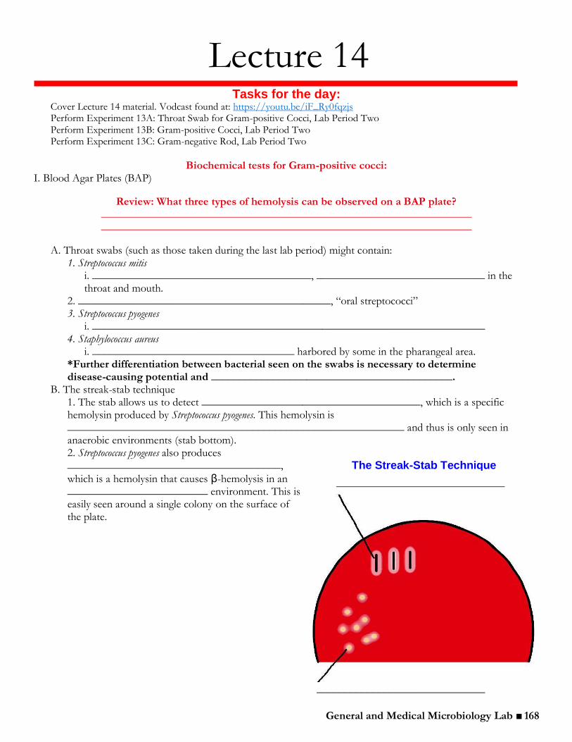
B. The streak-stab technique 1. The stab allows us to detect _______________________________________, which is a specific hemolysin produced by Streptococcus pyogenes. This hemolysin is ____________________________________________________________ and thus is only seen in anaerobic environments (stab bottom). 2. Streptococcus pyogenes also produces ______________________________________,
which is a hemolysin that causes β-hemolysis in an _________________________ environment. This is easily seen around a single colony on the surface of the plate.
The Streak-Stab Technique
______________________________
______________________________
General and Medical Microbiology Lab ■ 169
II. Mannitol Salt Agar (MSA) plate
Review: Which organism, Staphylococcus aureus or Streptococcus mitis, would grow on an MSA plate?
___________________________________________________________
Review: What color would an MSA plate be if the organism streaked on it was able to ferment mannitol?
___________________________________________________________
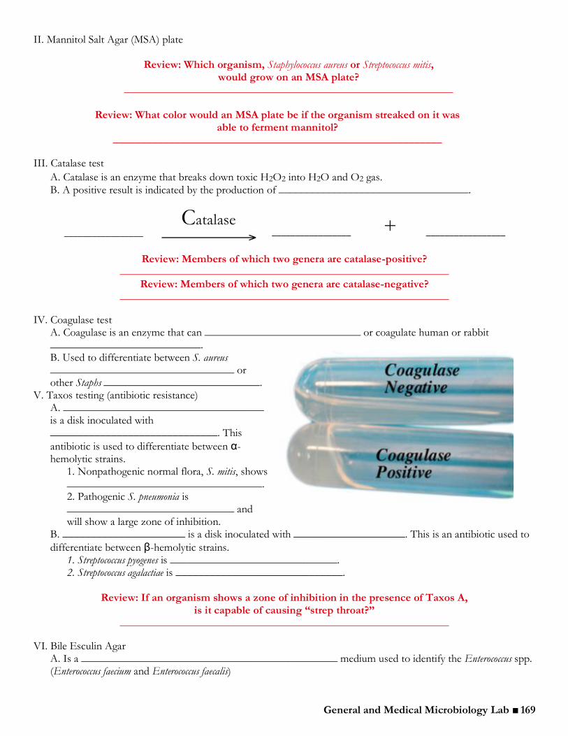
III. Catalase test
A. Catalase is an enzyme that breaks down toxic H2O2 into H2O and O2 gas.
B. A positive result is indicated by the production of __________________________________.
Review: Members of which two genera are catalase-positive? ___________________________________________________________
Review: Members of which two genera are catalase-negative? ___________________________________________________________
IV. Coagulase test A. Coagulase is an enzyme that can ____________________________ or coagulate human or rabbit ___________________________. B. Used to differentiate between S. aureus _________________________________ or other Staphs ____________________________.
V. Taxos testing (antibiotic resistance) A. ____________________________________ is a disk inoculated with ______________________________. This
antibiotic is used to differentiate between α-hemolytic strains.
1. Nonpathogenic normal flora, S. mitis, shows ___________________________________. 2. Pathogenic S. pneumonia is ______________________________ and will show a large zone of inhibition.
B. ______________________ is a disk inoculated with ____________________. This is an antibiotic used to
differentiate between β-hemolytic strains. 1. Streptococcus pyogenes is ______________________________. 2. Streptococcus agalactiae is ______________________________.
Review: If an organism shows a zone of inhibition in the presence of Taxos A,
is it capable of causing “strep throat?” ___________________________________________________________
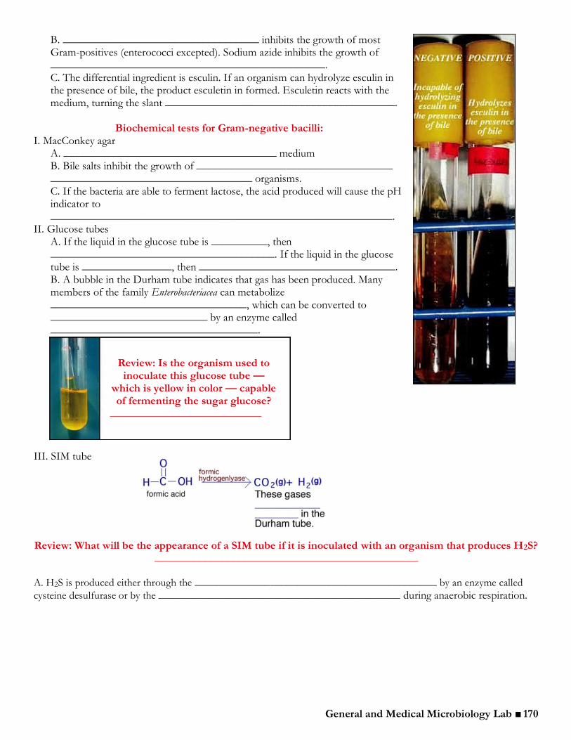
VI. Bile Esculin Agar A. Is a ______________________________________________ medium used to identify the Enterococcus spp. (Enterococcus faecium and Enterococcus faecalis)
Catalase + _________________ _________________ _________________
General and Medical Microbiology Lab ■ 170
B. ___________________________________ inhibits the growth of most Gram-positives (enterococci excepted). Sodium azide inhibits the growth of
_________________________________________________. C. The differential ingredient is esculin. If an organism can hydrolyze esculin in the presence of bile, the product esculetin in formed. Esculetin reacts with the medium, turning the slant _________________________________________.
Biochemical tests for Gram-negative bacilli:
I. MacConkey agar A. ______________________________________ medium B. Bile salts inhibit the growth of ___________________________________ ____________________________________ organisms. C. If the bacteria are able to ferment lactose, the acid produced will cause the pH indicator to _____________________________________________________________.
II. Glucose tubes A. If the liquid in the glucose tube is __________, then ________________________________________. If the liquid in the glucose tube is ________________, then ___________________________________. B. A bubble in the Durham tube indicates that gas has been produced. Many members of the family Enterobacteriacea can metabolize ___________________________________, which can be converted to ____________________________ by an enzyme called _____________________________________.
III. SIM tube
Review: What will be the appearance of a SIM tube if it is inoculated with an organism that produces H2S?
_______________________________________________
A. H2S is produced either through the _____________________________________________ by an enzyme called
cysteine desulfurase or by the _____________________________________________ during anaerobic respiration.
Review: Is the organism used to inoculate this glucose tube —
which is yellow in color — capable of fermenting the sugar glucose?
___________________________
General and Medical Microbiology Lab ■ 171
IV. Kligler’s Iron Agar (KIA) A. A differential medium that contains two sugars: ____________________________ (low concentration) and _______________________ (higher concentration).
1. Fermenters will typically utilize the
______________________ and production of acidic fermentation byproducts will cause the entire tube to turn ________________________________. However, the glucose is in short supply and, after the first few hours of growth, will all be used up. 2. After the glucose is gone, bacteria that are capable of fermenting lactose will do so. Production of acidic byproducts will continue and the medium will _________________________________________.
Review: What will a KIA tube look like if the organism can ferment both glucose and lactose?
______________________________________
3. Bacteria that are incapable of fermenting lactose have to utilize the
__________________________________ in the medium. This produces NH3 (a weak base), which
______________________________ and turns the slant red. The butt remains yellow.
Review: What will a KIA tube look like if the organism can ferment glucose but not lactose? __________________________________________
4. Non-fermenters will utilize only the amino acids and proteins in the medium. The slant will be _____________________ and the butt will remain the original red/orange color.
Review: What will a KIA tube look like if the organism can ferment neither glucose nor lactose? _______________________________________________________
B. H2S production
1. The reduction of sulfate to hydrogen sulfide, H2S, results in a ____________________________ precipitate.
V. Oxidase test A. Detects the enzyme _____________________________________, an important catalyst in the electron transport chain of some organisms. B. This test is done by smearing a colony onto filter paper and adding oxidase reagent. If the bacteria produces a cytochrome oxidase, the colony will turn ________________________________________. C. Pseudomonas and Neisseria are oxidase __________________________________.
Note: A positive and negative ______________________ should be performed every time the oxidase test is done.
VI. Urease test. A. Urea broth is used to test for the enzyme urease. ______________________________________________________________________. Since ammonia
(NH3) is alkaline, the pH indicator will produce a __________________________________________________. B. Members of the genus ________________________________ are urease positive.
Demonstrations: Two other important tests used for the identification of Streptococci spp. Are: 1. CAMP test: Diagnostic for Streptococcus agalactiae 2. Bile esculin test: Diagnostic for Enterococcus faecalis Both tests will also be discussed in lecture, and examples will be provided. Both tests are also described in the Summary of Biochemical Tests on the class website. It is important to understand the procedure and basis of these tests in the event that they need to be performed during the unknown identification process for Experiment28.

General and Medical Microbiology Lab ■ 172
Lecture 15 Tasks for the day:
Cover Lecture 15 material. Vodcast found at: https://youtu.be/RjffHJctqyc Perform Experiment 13B: Gram-positive Cocci, Lab Period Three Perform Experiment 13C: Gram-negative Rod, Lab Period Three Perform Experiment 15A: Unknown Identification Part I
I. Previous lab results
A. Staphylococcus and Streptococcus spp. 1. Coagulase
a. Enzyme that effectively ____________________________________________ b. Staphylococcus aureus: ___________________________ c. Staphylococcus epidermidis: ____________________________
2. Taxos P a. A disk that contains ______________________________ b. Streptococcus pneumoniae: __________________________ c. Streptococcus mitis: ___________________________
3. Taxos A a. A disk that contains ____________________________ b. Streptococcus pyogenes: _______________________ c. Staphylococcus aureus and other, less pathogenic Streptococcus spp (e.g. Streptococcus agalactiae): ___________________________
B. Gram-negative bacilli 1. Kigler’s Iron Agar (KIA) (see “How Does It Work?, next page)
a. Differentiates bacteria based on their ability to ferment __________________________________ _________________________. b. The KIA test is often used to identify ______ _________________________________ (e.g. Shigella), which are often more ______________ ________________________ in the GIT. c. Gas produced as a product of fermentation will form __________________________________ ____________ or completely ________________________________________ ________________________________________ of the tube.
i. ____________________________________ and _________________________________ produce gas.
d. The production of H2S results in a ____________________________________________. If the black precipitate obscures the medium color, it is ________________________________________________________________________________.

General and Medical Microbiology Lab ■ 173
2. The Nitrate Test a. Determines an organism’s ability to reduce _________________________________________________________ using the enzyme nitratase.
Note: There are ________________________________ for an organism to be positive for nitrate reduction and only __________________________________ to be negative.
How Does It Work? Deciphering KIA data
Also sometimes called nitrate reductase.
Nitratase Other e-
transfers Other nitrogenous compounds (e.g. N2(g) ______________________ _____________

General and Medical Microbiology Lab ■ 174
Expected results (circle one)
1. Escherichia coli
POSITIVE / NEGATIVE
2. Pseudomonas aeruginosa
POSITIVE / NEGATIVE
3. Staphylococcus epidermidis
POSITIVE / NEGATIVE
4. Streptococcus pneumoniae
POSITIVE / NEGATIVE
II. Unknowns A. Take one unknown Gram-negative and one Gram-positive. Both the Gram-negative and the Gram-positive should have the ______________________________________________________. B. Be certain to _________________________ all tubes/plates/slides so as not to confuse the Gram-negative and Gram-positive. C. Perform a _____________________________ of both unknowns and streak for isolation. It is very important to get an isolated colony! D. Be certain that you also receive a clue for both the Gram-negative and Gram-positive. These clues will assist you in writing your hypotheses, which are _______________________________ (along with your references) ________________________________________. Please see the description of this assignment on page xiii in the appendix.
General and Medical Microbiology Lab ■ 175
Experiment 15A: Unknown Identification Part I
Throughout Experiments 26-27, a number of biochemical tests have been introduced that can be helpful in the identification of an unknown microorganism. In this experiment, each student will be assigned two unknown bacterial isolates. One of the unknown isolates will be a Gram-positive organism and the other will be a Gram-negative organism. The first step in identifying these unknowns will be the Gram stain. In the next lab period, working and stock cultures will be prepared. Morphological and physiological criteria will be used to identify the genus and species of each isolate. During the next few lab periods, various tests will be performed on each unknown. After sifting through all the results of each test, it will be possible to deduce the true identity of each unknown organism.
Lab Period One A. Streak for Isolation Procedure Students will work individually. From the side bench, collect 1 TSA plate and 1 MacConkey plate.
1. Collect and “A” and “B” unknown culture pair from the TA or instructor. Be certain that both the “A” and “B” unknowns have the same number! Tell the TA the number of the unknown so that she or he will have an accurate record. Immediately record your unknown number in the results section of this experiment.
Note: “Unknown A” is presumed to be Gram-positive; “Unknown B” is presumed to be Gram-negative. It is very important to keep careful and detailed records. Record all
observations, test results, etc. in the table following this experiment. Be careful not to reverse the “A” and “B” unknowns! Everything from here on out should be labeled with both the
unknown number and the A or B desgination! *All results for these unknowns will be recorded in the tables on page 192-197.*
2. Observe broth growth patterns in the tubes. This will be observed again in a later labe period. Mix the two culture tubes by flicking. Be certain that both cultures are visibly turbid, indicating that they have grown and are viable. If either one of your unknowns did not grow, inform the TA or instructor and obtain a replacement culture. 3. Prepare two microscope slides by labeling the slides either “A###” or “B###.” Prepare a smear of each unknown on the appropriately labeled slide. Following are some tips for obtaining a high-quality smear preparation.
a. Clean the slide thoroughly before beginning the smear preparation. b. Do not dilute the culture. Simply use one or two loops of the concentrated broth culture. Be sure to get a loop from bacterial growth not just broth. c. Patiently let the smear air dry, and be certain to heat fix well!
4. Perform a Gram stain to verify the Gram reaction of each unknown to determine the morphology and arrangement. A focus line drawn on the top of the slide will assist in locating the smear. Save these slides and record all observations (for reference please see page 24) 5. Label both the TSA and MacConkey plates with your initials and lab section number. Label the TSA plate “A###.” Label the MacConkey plate “B###.” 6. Using these labeled plates, streak both unknowns for isolation using the T-streak method. T-streak the presumed Gram-positive onto the TSA plate. T-streak the presumed Gram-negative onto the MacConkey agar plate. Invert the plates and place them in the tray on the side bench to be incubated at 37°C. 7. Return the original unknown cultures to the rack on the west bench.
General and Medical Microbiology Lab ■ 176
Lecture 16 Tasks for the day:
■ Cover Lecture 16 material. Vodcast found at: https://youtu.be/mNVfVBsRtR4 ■ Perform Experiment 16A: Unknown Identification Part II ■ Perform Experiment 16B: Preparation of Wine, Lab Period One
I. Maintaining culture purity A. After isolating a pure culture, it is important to ___________________________________. This is difficult because the more transfers made, the greater the likelihood of contamination. B. Working and reserve stocks
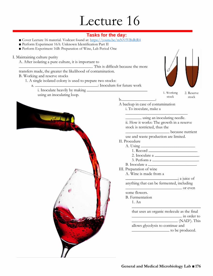
1. A single isolated colony is used to prepare two stocks: a. ______________________________: Inoculum for future work
i. Inoculate heavily by making _____________________________ using an inoculating loop.
b.________________: A backup in case of contamination
i. To inoculate, make a _______________________________ ________ using an inoculating needle.
ii. How it works: The growth in a reserve stock is restricted, thus the _____________________ because nutrient use and waste production are limited.
II. Procedure A. Using __________________________
1. Record _______________________. 2. Inoculate a _____________________. 3. Perform a _______________________.
B. Inoculate a ________________________.
III. Preparation of wine A. Wine is made from a _________________________; a juice of anything that can be fermented, including ___________________________ or even some flowers. B. Fermentation
1. An _________________________________ that uses an organic molecule as the final ________________________ in order to ______________________ (NAD’). This allows glycolysis to continue and __________________ to be produced.
stock 2. Reserve
stock
General and Medical Microbiology Lab ■ 177
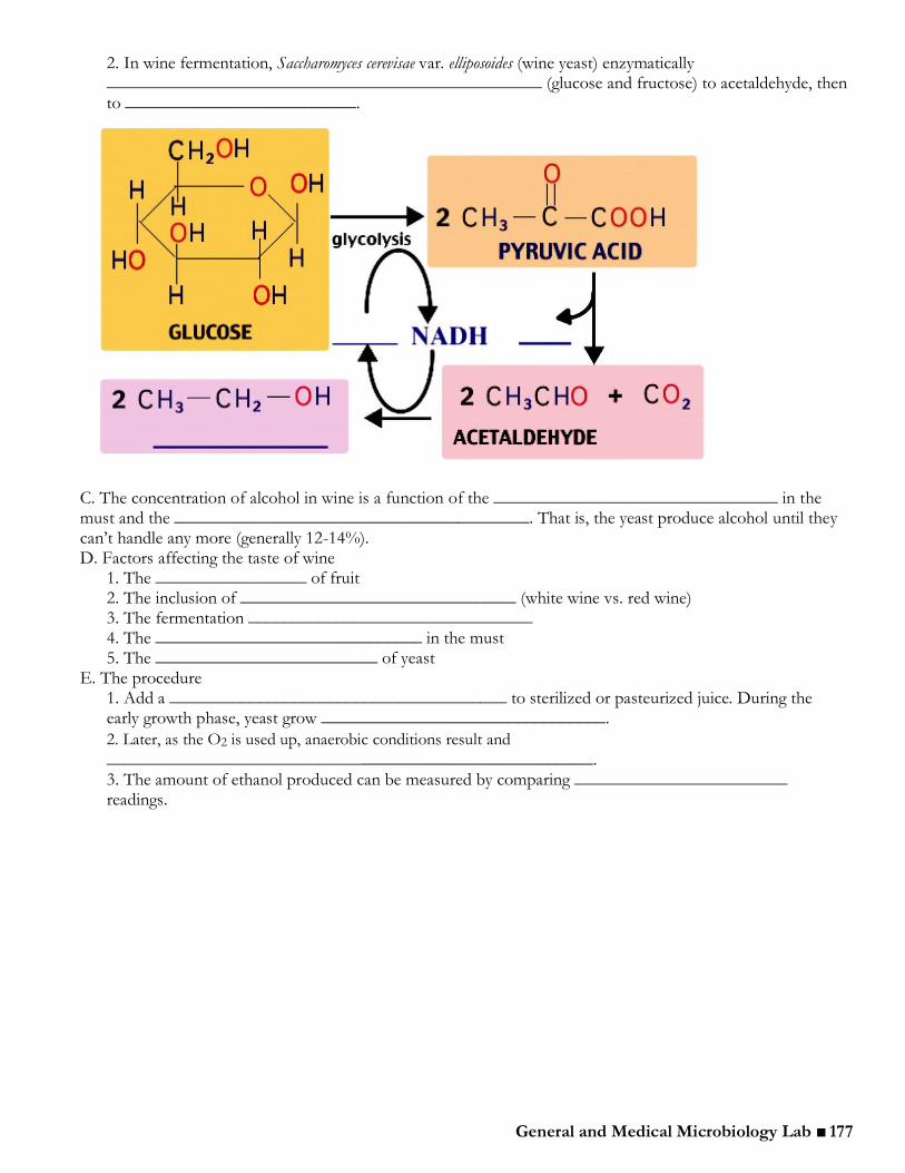
2. In wine fermentation, Saccharomyces cerevisae var. elliposoides (wine yeast) enzymatically _________________________________________________ (glucose and fructose) to acetaldehyde, then to __________________________.
C. The concentration of alcohol in wine is a function of the ________________________________ in the must and the ________________________________________. That is, the yeast produce alcohol until they can’t handle any more (generally 12-14%). D. Factors affecting the taste of wine
1. The _________________ of fruit 2. The inclusion of _______________________________ (white wine vs. red wine) 3. The fermentation ________________________________ 4. The ______________________________ in the must 5. The _________________________ of yeast
E. The procedure 1. Add a ______________________________________ to sterilized or pasteurized juice. During the early growth phase, yeast grow ________________________________.
2. Later, as the O2 is used up, anaerobic conditions result and
________________________________________________________. 3. The amount of ethanol produced can be measured by comparing ________________________ readings.

General and Medical Microbiology Lab ■ 178
Experiment 16A: Unknown Identification Part II
B. Stock cultures Once a pure bacterial isolate (single colony) has been obtained, it is important to store the culture under
appropriate conditions to maintain it in a viable state for several months. To set up a reserve stock culture, a deep stab tube is inoculated, incubated, and stored in the refrigerator. To set up
a working stock, a slant tube is inoculated, incubated, and refrigerated when not in use. The working stock is used for further tests, and the reserve stock is stored as a source of viable organisms if needed.
The goal of today’s lab is to prepare reserve stocks and working stocks of each of the two unknown bacterial isolates (A# and B#). To avoid accidentally working with a contaminant, both reserve and working stocks will be prepared using a single isolated colony. The instructions below describe how to use only one colony as the source of inoculum for both stocks and as the source of bacteria for a Gram stain.
It is critical to do this work aseptically and not to confuse the A and B cultures. Label all tubes correctly and read the labels before doing anything. Because it has been several days since the cultures were streaked, it is possible that some cells will not stain as expected due to changes in the cell walls. However, the size, shape, and arrangement of the cells should be similar to the first slide.
Procedure Students will work individually. From the side bench, collect 2 stab tubes and 2 slant tubes.
1. Collect the Unknown A and Unknown B TSA and MacConkey streak plates. Check to see that all colonies on each plate appear similar. If there are no isolated colonies, re-streak from the original culture onto a fresh TSA (Unknown A) or MacConkey (Unknown B) plate. 2. Keep the lid poised over the plate when opening it. Describe the colony morphology for Unknown A and Unknown B and record these in the results section for this experiment. Terms to describe morphology can be reviewed in Lecture 2, page 24. Describe the lactose fermentation of Unknown B on the MacConkey plate. 3. Prepare two microscope slides by labeling each slide either Unknown A or Unknown B. Place a drop of sterile water on each slide.
4. Label one stab tube and one slant tube with your initials, lab section number, date, and “A###.” Using a flamed inoculating loop, pick up approximately half of an isolated colony from the Unknown A TSA plate and heavily inoculate the slant by making a zig-zag pattern completely covering the surface of the slant. Without flaming the loop, immediately inoculate the sterile drop of water on the slide labeled “A###.” There will be plenty of bacteria remaining on the loop to prepare a smear for Gram staining. 5. Using a flamed needle, pick up the remaining half of the isolated colony from
your Unknown A TSA plate and inoculate the stab tube by making a single stab to the bottom. 6. Repeat steps 4 and 5 with Unknown B. 7. Perform a Gram stain on the prepared smears (remember to heat fix after drying). Compare these with the first Gram stain of the samples that was done during last lab period. The Gram reaction and cell shape should be the same, but cell arrangement may be slightly different because the culture came from a solid surface this time. The arrangement observed in the first Gram stain is more accurate. If the Gram reaction or cell shape is markedly different than what was observed on the first Gram stain, ask the TA or instructor for help. 8. Place the reserve stock and working stock in the tray on the side bench to be incubated at 37°C. You should have 4 tubes — 2 stabs and 2 slants. Please make sure that each tube is clearly labeled with the unknown letter and number, your name, and your section number. 9. Save your A and B streak plates by placing them in appropriately the tray on the back bench. These will be refrigerated to save in case you need them later. Make sure your initials, section number, and unknown number are written on each plate.
Tip! For easier identification, use tape to make a small label for the tubes holding your reserve and open stocks (e.g. “A###” and your initials) and put it on the top of the caps.
General and Medical Microbiology Lab ■ 179
Experiment 16B: Preparation of Wine
In the production of wine, grapes are crushed to release the juice, or “must.” Table sugar is added to increase the potential of the must to produce alcohol. A pure, aerobically growing culture of wine yeast in added as a started culture and grown in sterilized or pasteurized grape juice. During the initial stages, air is present in the liquid and rapid aerobic growth of the yeast occurs. Then, as the air is used up, anaerobic conditions develop and alcohol production begins.
Lab Period One
Procedure Students will work in groups of four.
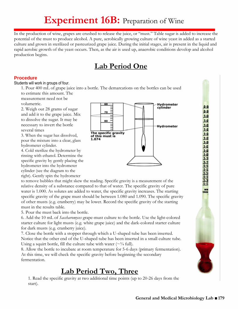
1. Pour 400 mL of grape juice into a bottle. The demarcations on the bottles can be used to estimate this amount. The measurement need not be volumetric. 2. Weigh out 28 grams of sugar and add it to the grape juice. Mix to dissolve the sugar. It may be necessary to invert the bottle several times 3. When the sugar has dissolved, pour the mixture into a clear, glass hydrometer cylinder. 4. Cold sterilize the hydrometer by rinsing with ethanol. Determine the specific gravity by gently placing the hydrometer into the hydrometer cylinder (see the diagram to the right). Gently spin the hydrometer to remove bubbles that might skew the reading. Specific gravity is a measurement of the
relative density of a substance compared to that of water. The specific gravity of pure water is 1.000. As solutes are added to water, the specific gravity increases. The starting specific gravity of the grape must should be between 1.080 and 1.090. The specific gravity of other musts (e.g. cranberry) may be lower. Record the specific gravity of the starting must in the results table. 5. Pour the must back into the bottle. 6. Add the 10 mL of Saccharomyces grape-must culture to the bottle. Use the light-colored starter culture for light musts (e.g. white grape juice) and the dark-colored starter culture for dark musts (e.g. cranberry juice). 7. Close the bottle with a stopper through which a U-shaped tube has been inserted. Notice that the other end of the U-shaped tube has been inserted in a small culture tube. Using a squirt bottle, fill the culture tube with water (~¾ full). 8. Allow the bottle to incubate at room temperature for 5-6 days (primary fermentation). At this time, we will check the specific gravity before beginning the secondary fermentation.
Lab Period Two, Three
1. Read the specific gravity at two additional time points (up to 20-26 days from the start).

General and Medical Microbiology Lab ■ 180
Experiment 16B: Results
Group Data:
Fermenting wine
Starting must __ day incubation __ day incubation __ day incubation
Aroma
Clarity
Specific Gravity
% potential
alcohol
% alcohol
produced **
**To determine the % alcohol produced, use the following formula and table below. Formula: (% alcohol produced) = (% potential alcohol in the starting must) — (% potential alcohol after incubation)
Specific Gravity % Potential Alcohol
1 0
1.01 0.9
1.02 2.3
1.03 3.7
1.04 5.1
1.05 6.5
1.06 7.8
1.08 10.6
1.09 12
1.1 13.4
1.11 14.9
1.12 16.3
1.13 17.7
Class Data:
Specific gravity
Name of wine Amount of sugar (g)
Starting Must ___ day incubation ___ day incubation ___ day incubation

General and Medical Microbiology Lab ■ 181
Lecture 17
Tasks for the day: Perform Experiment 17A: Unknown Identification Part III
You’re doing great! Remember to keep on task for your final OWL (hands-on skill tests) (page xi in the appendix).
General and Medical Microbiology Lab ■ 182
Experiment 17A: Unknown Identification Part III
B. Stock cultures 1. Collect the working and reserve cultures from the side tray. You should have an “A###” reserve and working stock and a “B###” reserve and working stock for a total of 4 tubes.
a. Place the reserve stocks into a rack labeled for the appropriate section number and put them in the refrigerator. The advantage of a stab tube is that it supports minimal growth of the organism, which restricts depletion of the nutrients in the medium; thus, the bacteria will remain viable and can be stored in this medium for long periods of time. b. The working stock should have a lot of growth on the slant face. Use this growth as a source of inoculum for the following series of tests. After using the working stock as a source of inoculating a test medium, always return it to the refrigerator with the cap tight. Make sure to flame the tube every time it is opened to avoid contamination!
2. Use the working stocks — slant tubes — to inoculate the following media on the checklist. For a review of the purpose and principles of all the tests, consult the Summary of Tests for Unknown on page xxvii in the appendix. Check off each test on the list after it is completed.
*Please use small inoculum as these working stocks will be used for several tests.
C. Checklist 1 Gram-positive Unknown A
________ Perform a Gram stain using growth from the working slant. This should be done first. If there is any sign on contamination, it will be necessary to inoculate a fresh stock before proceeding.
________ Inoculate 1 tube of TSB using an inoculating loop. ________ Inoculate one tube of nitrate broth using an inoculating loop. ________ Inoculate motility agar using a needle to make one stab down the center to the bottom of the tube. ________ Perform the catalase test (procedure can be found in Lecture 14). Use a wooden stick — not a loop! ________ If your unknown is a Gram-positive coccus, streak 1 BAP. T-streak using the entire plate; use the
inoculating loop to make several stabs in a streak-free region of the agar. ________ If your unknown is a Gram-positive rod, inoculate 1 NSM plate for a future spore stain. T-streak the plate for
isolation. ________ If your unknown is a Gram-positive rod, perform an acid-fast stain (procedure can be found in Lab 3).
Gram-negative Unknown B
________ Perform a Gram stain using growth from the working slant. This should be done first. ________ Inoculate 1 tube of TSB using an inoculating loop. ________ Inoculate one tube of nitrate broth using an inoculating loop. ________ Inoculate motility agar using a needle to make one stab down the center to the bottom of the tube. ________ Inoculate 1 mannitol, 1 glucose, and 1 lactose broth tube. Use an inoculating loop. ________ Inoculate one tube of MR-VP medium with an inoculating loop. This will need to be incubated for 2-5
days. Place rack labeled MR-VP. ________ Perform an Oxidase with controls located on the west lab bench.
After inoculation, place all the media — except the MR-VP tube — into an incubation cup or small rack. Place the cup/rack in the tray on the side bench to be incubated at 37°C. Place the MR-VP tubes into a separate, labeled rack. This rack will be incubated for 5 days. Be certain to place your working stock into the appropriate rack. This rack should be placed into the refrigerator and NOT the incubator.
Demonstrations of both positive and negative reactions for the tests that have not been previously performed will be discussed during lecture and available for comparison in the labs. It is important to know and understand all of the tests.

General and Medical Microbiology Lab ■ 183
Lecture 18 Tasks for the day:
■ Cover Lecture 18 material. Vodcast found at: https://youtu.be/3UmB753jn_s ■ Perform Experiment 18A: Unknown Identification Part IV
I. CAMP Test
A. Tests for the ability of an organism to produce the CAMP factor, as _____________________________________________________. B. Synergistic test between ______________________________________________ and ____________________________________________.
1. The two bacteria are streaked ______________________________ to one another. They do NOT touch.
2. The CAMP protein, ______________________________________________________ of S. aureus by binding to already damaged red blood cells and leading to complete lysis. As a result, ___________________________ of enhanced hemolysis is produced between the two streaks. The test is ______________________________ S. agalactiae.
II. Methyl Red Voges-Proskauer (MR-VP) A. Used to determine ______________________________________________ is used ___________________________________________________________. B. _________________________________ Fermentation Pathway
1. In this pathway, glucose is fermented to produce several _____________________________________ (lactic, acetic, succinic, and formic acids). The stable production of enough acid to overcome the phosphate buffer will result in a _______________________________________________________________________. 2. ___________________ is a pH indicator. If this indicator is added to the culture broth and the pH is below 4.4, a ____________________________________. If the MR turns _______________, pH is above 6.0 and the mixed acid fermentation pathway has ________________ been utilized.

General and Medical Microbiology Lab ■ 184
C. _______________________ Fermentation Pathway 1. In this pathway, glucose is fermented to produce a
__________________________________________________________ instead of organic acids.
2. In order to detect acetoin, alpha-napthol and KOH are added. Acetoin reacts with the alpha-napthol in the presence of KOH to produce a red color. Thus, if the culture is _________________________________, it will turn “_____________________________________.”
III. The Rapid KOH String Test1 A. This test can be used in conjunction with the Gram stain to differentiate between Gram-positive and Gram-negative cells.
Check your knowledge: What are some potential problems with using the Gram stain alone to determine cell wall structure?
______________________________________________________________________________ ______________________________________________________________________________
B. When placed in a drop of KOH base, Gram-negative cells will ______________________, releasing DNA and proteins into solution. A _________________________ of DNA and proteins is formed2. Gram-positive cells will remain clumpy in KOH.
1.This portion of the lecture was adapted from a procedure written by microbiology TA Aaron Larson in the spring of 207. 2.Sutton, Scott. “The Gram Stain.” The Microbiology Network. Feb 2006. Feb 20, 2007 <http://www.microbiol.org/WPaper.Gram. htm>
Note: A culture will usually only be
__________________________________________: either MR-positive or VP-positive. But remember, some bacteria are nonfermenters and will thus be ________________________ ______________________________.
General and Medical Microbiology Lab ■ 185
Experiment 18A: Unknown Identification Part IV
Checklist 1
Collect all test media that was inoculated during the last lab period. Record all findings in the charts at the end of this experiment. Be certain to carefully note all observations (e.g. If a lactose broth tube is neither yellow nor red but appear slightly orange, carefully write down exactly what you see). Collect the working slants for each unknown and inoculate according to Checklist 2
Checklist 2
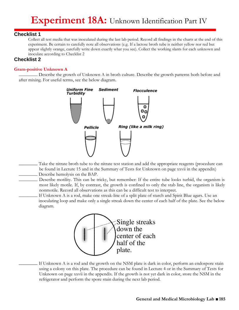
Gram-positive Unknown A ________ Describe the growth of Unknown A in broth culture. Describe the growth patterns both before and after mixing. For useful terms, see the below diagram.
________ Take the nitrate broth tube to the nitrate test station and add the appropriate reagents (procedure can
be found in Lecture 15 and in the Summary of Tests for Unknown on page xxvii in the appendix) ________ Describe hemolysis on the BAP. ________ Describe motility. This can be tricky, but remember: If the entire tube looks turbid, the organism is
most likely motile. If, by contrast, the growth is confined to only the stab line, the organism is likely nonmotile. Record all observations as this can be a difficult test to interpret.
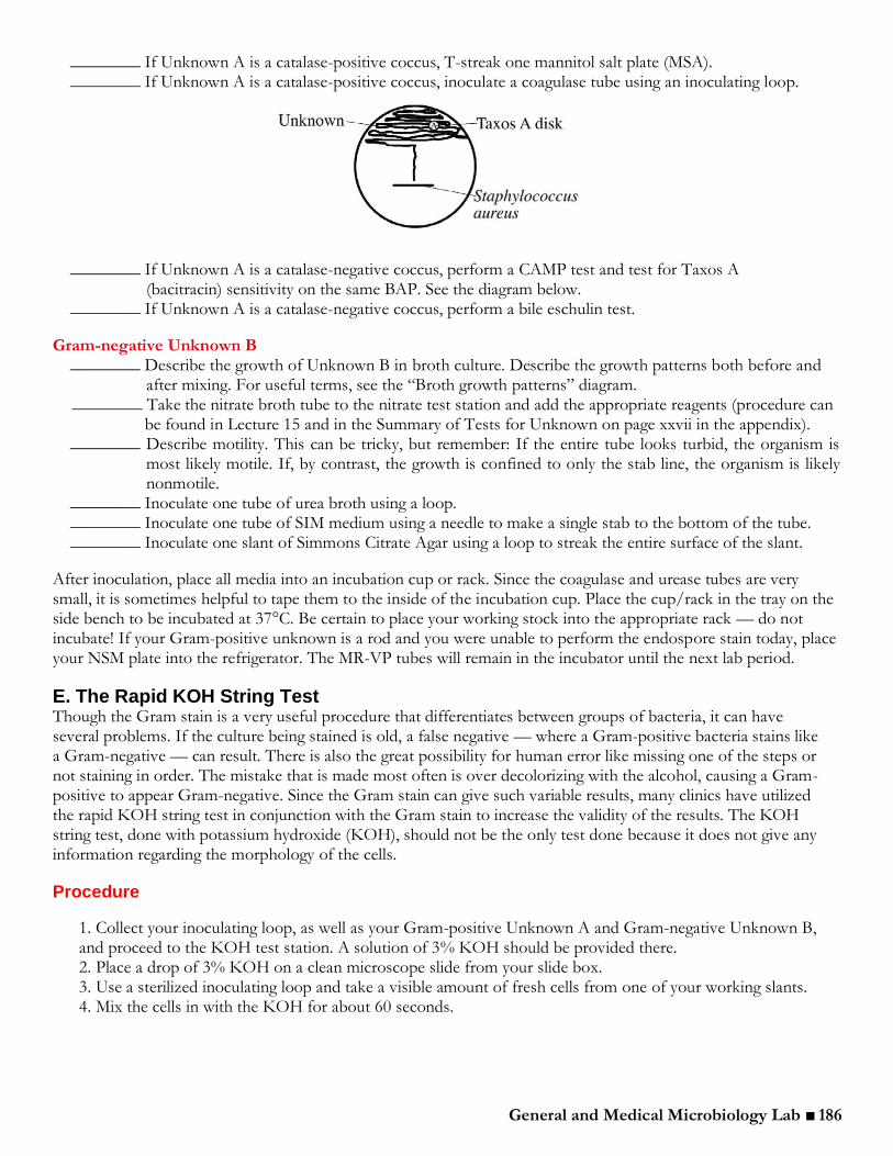
________ If Unknown A is a rod, make one streak-line of a split plate of starch and Spirit Blue agars. Use an inoculating loop and make only a single streak down the center of each half of the plate. See the below diagram.
________ If Unknown A is a rod and the growth on the NSM plate is dark in color, perform an endospore stain
using a colony on this plate. The procedure can be found in Lecture 4 or in the Summary of Tests for Unknown on page xxvii in the appendix. If the growth is not yet dark in color, store the NSM in the refrigerator and perform the spore stain during the next lab period.
General and Medical Microbiology Lab ■ 186
________ If Unknown A is a catalase-positive coccus, T-streak one mannitol salt plate (MSA). ________ If Unknown A is a catalase-positive coccus, inoculate a coagulase tube using an inoculating loop.
________ If Unknown A is a catalase-negative coccus, perform a CAMP test and test for Taxos A (bacitracin) sensitivity on the same BAP. See the diagram below.
________ If Unknown A is a catalase-negative coccus, perform a bile eschulin test.
Gram-negative Unknown B ________ Describe the growth of Unknown B in broth culture. Describe the growth patterns both before and
after mixing. For useful terms, see the “Broth growth patterns” diagram. ________ Take the nitrate broth tube to the nitrate test station and add the appropriate reagents (procedure can
be found in Lecture 15 and in the Summary of Tests for Unknown on page xxvii in the appendix). ________ Describe motility. This can be tricky, but remember: If the entire tube looks turbid, the organism is
most likely motile. If, by contrast, the growth is confined to only the stab line, the organism is likely nonmotile.
________ Inoculate one tube of urea broth using a loop. ________ Inoculate one tube of SIM medium using a needle to make a single stab to the bottom of the tube. ________ Inoculate one slant of Simmons Citrate Agar using a loop to streak the entire surface of the slant.
After inoculation, place all media into an incubation cup or rack. Since the coagulase and urease tubes are very small, it is sometimes helpful to tape them to the inside of the incubation cup. Place the cup/rack in the tray on the side bench to be incubated at 37°C. Be certain to place your working stock into the appropriate rack — do not incubate! If your Gram-positive unknown is a rod and you were unable to perform the endospore stain today, place your NSM plate into the refrigerator. The MR-VP tubes will remain in the incubator until the next lab period.
E. The Rapid KOH String Test Though the Gram stain is a very useful procedure that differentiates between groups of bacteria, it can have several problems. If the culture being stained is old, a false negative — where a Gram-positive bacteria stains like a Gram-negative — can result. There is also the great possibility for human error like missing one of the steps or not staining in order. The mistake that is made most often is over decolorizing with the alcohol, causing a Gram-positive to appear Gram-negative. Since the Gram stain can give such variable results, many clinics have utilized the rapid KOH string test in conjunction with the Gram stain to increase the validity of the results. The KOH string test, done with potassium hydroxide (KOH), should not be the only test done because it does not give any information regarding the morphology of the cells.
Procedure
1. Collect your inoculating loop, as well as your Gram-positive Unknown A and Gram-negative Unknown B, and proceed to the KOH test station. A solution of 3% KOH should be provided there. 2. Place a drop of 3% KOH on a clean microscope slide from your slide box. 3. Use a sterilized inoculating loop and take a visible amount of fresh cells from one of your working slants. 4. Mix the cells in with the KOH for about 60 seconds.

General and Medical Microbiology Lab ■ 187
5. Lift the loop off the slide and look for a slimy string connecting the loop to the slide. 6. Repeat the procedure with the second unknown.
Remember: Gram-negative cells will lyse and a string will form. Gram-positive cells will not lyse and no string will form.
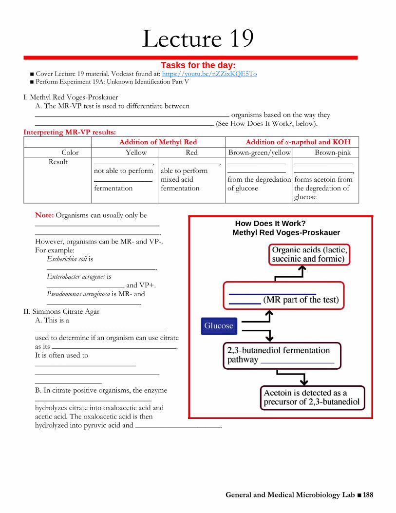
General and Medical Microbiology Lab ■ 188
Lecture 19 Tasks for the day:
■ Cover Lecture 19 material. Vodcast found at: https://youtu.be/nZZixKQE5To ■ Perform Experiment 19A: Unknown Identification Part V
I. Methyl Red Voges-Proskauer A. The MR-VP test is used to differentiate between __________________________________________________ organisms based on the way they ______________________________________________ (See How Does It Work?, below).
Interpreting MR-VP results:
Addition of Methyl Red Addition of α-napthol and KOH
Color Yellow Red Brown-green/yellow Brown-pink
Result _______________, not able to perform _______________ fermentation
_______________, able to perform mixed acid fermentation
_______________ _______________ from the degredation of glucose
_______________ _______________, forms acetoin from the degredation of glucose
Note: Organisms can usually only be ________________________________ ________________________________. However, organisms can be MR- and VP-. For example:
Escherichia coli is ____________________________. Enterobacter aerogenes is ____________________ and VP+. Pseudomonas aeruginosa is MR- and
________________________. II. Simmons Citrate Agar
A. This is a __________________________________ used to determine if an organism can use citrate as its ________________________________. It is often used to __________________________ ________________________________ _________________. B. In citrate-positive organisms, the enzyme ______________________________ hydrolyzes citrate into oxaloacetic acid and acetic acid. The oxaloacetic acid is then hydrolyzed into pyruvic acid and ______________________.
How Does It Work? Methyl Red Voges-Proskauer

General and Medical Microbiology Lab ■ 189
C. If CO2 is produced, it reacts with components of the medium to produce an
______________________________________________ (e.g. Na2CO3). The alkaline pH turns the _____________________________________ from ______________________________________. This is a ___________________ result. D. Escherichia coli and Shigella dysenteriae are _______________________ whereas Klebsiella pneumonia and Proteus mirabilis are ___________________________.
III. Spirit Blue agar A. This agar is used to identify organisms that are capable of producing the enzyme ____________________________________. This enzyme is secreted and ___________________________________ to ______________________________ and three
________________________________________. These compounds are ________________________________ to pass through the bacterial cell wall.
1. Glycerol can be converted into a __________________________________________. 2. The fatty acids can be __________________________________________________ and their fragments can eventually enter the ____________________________________________
B. Spirit Blue agar contains an emulsion of ________________________________________________________________. Bacteria that produce lipase will hydrolyze the olive oil and produce a ___________________ around the bacterial growth. C. Bacillus subtilis is lipase ___________________________.
IV. Starch hydrolysis test A. This test is used to identify bacteria that can ___________________________________________ _____________________________________ using the enzymes ________________________ and oligo-1,6-glucosidase. B. Often used to differentiate species from the genera ____________________________________.
C. Because of the _________________________________ of amylose and amylopectin molecules, these starches ______________________________________ through the bacterial cell wall.
General and Medical Microbiology Lab ■ 190
1. In order to use these starches as a carbon source, bacteria must ___________________________ α-amylase and oligo-1,6-glucosidase into the extracellular space. 2. These enzymes _______________________________________________________________ subunits which can then be utilized directly as carbon sources.
D. After incubation of the starch plates, _______________________________ is added to the top of the agar. They iodine reacts with the starch to form a _________________________________________________________. Hydrolysis of the starch will ______________________________________ around the bacterial growth. E. Bacillus subtilis is amylase _________________.
Things to remember today:
1. Perform ________________________________________ needed to help determine your unknown. 2. Determine your unknown and ______________________________________________________. 3. Consult the lab notebook for a description of the report format. If you have any questions about the report, be sure to ask today. 4. Remember the late policy: ____________________________________________ that a report is late. Late reports are not accepted after one full week. 5. Reports are due on ____________________________. 6. Once you have correctly identified your unknown, do the following: remove all tape — including that on the original plates and stocks — and dispose of it in the trash; discard all plates and slants in the biohazard containers; and wash all microscope slides.

General and Medical Microbiology Lab ■ 191
Experiment 19A: Unknown Identification Part V
Checklist 3
Procedure Gram-positive Unknown A
________ For Gram-positive rods, perform an endospore stain from the culture grown on the NSM plate if you did not do this during last lab period. The endospore stain procedure can be found in Lecture 4 and in the Summary of Staining Procedures on page xxv in the appendix.
________ For Gram-positive rods, flood the white starch side of the starch and Spirit Blue plate with iodine. Add just enough iodine to cover the surface of the starch agar and allow the plate to sit for 5-10 minutes before observing.
Gram-negative Unknown B ________ Add 6-7 drops of Kovac’s reagent to the top of the SIM tube. ________ Perform the MR-VP test using the following procedure:
1. Remove one 0.5 mL aliquot from the Unknown B MR-VP culture and place it into a test tube. 2. Label the tube containing 0.5 mL “MR” and the tube containing the remainder of the culture “VP.” 3. MR tube: Add 4 drops of methyl red pH indicator into the “MR” tube. 4. VP tube: Add 0.25 mL of alpha-napthol and 0.25 mL of 40% KOH. Mix vigorously and let it sit for about one hour.
Final unknown identification 1. Review the Unknown Microorganism Tables to be absolutely certain all tables are complete. If you need to perform additional tests or redo any of the procedures, do so now. Additional media is available on the side bench or by request. 2. Using all the information collected over the past couple weeks, in conjunction with the dichotomous keys and tables beginning on page xxxv in the appendix, make an identification of your Gram-positive and Gram-negative unknown. Verify each conclusion with the TA or instructor.
Bacterial Unknown Reports 1. Spend some time reviewing the unknown report format described on page xvii. The unknown report is worth 20 points (10% of your total lab grade). Be certain to clarify any points of confusion before attempting to write the report.
Clean up 1. All plates (including anything saved in the fridge) should be discarded in the biohazard containers. 2. All tape — including that on the original plates and stocks — must be removed from glass tubes and plates. Tubes should be placed in the discard racks on the side bench.
Nucleotide Sequence Searches The use of phenotypic characteristics to identify bacterial unknowns is an effective and straightforward way to identify organisms that can be cultured in the lab. However, there are many bacteria and archaea that cannot be cultured in the lab; thus, the biochemical methods we have investigated have little application. Fortunately, an increasing amount genotypic, or “phylogenic,” sequence data is being accumulated that allows for the identification of these unculturable prokaryotes. Because of its essential cellular function and highly conserved sequence, as well as its convenient intermediate size, the 16S rRNA is very commonly the molecule of choice for building sequence databases. Large 16S rRNA databases exist and are accessible to the public of the internet. Two such databases are the Ribosomal Database Project II (RDP II) and GenBank (NCBI).
Procedure Students will work individually.
1. Follow the procedure that is posted under additional information on the lab course web page. 16S rRNA sequence data, as gathered from RDP II will be given for the Gram-negative Unknown B organisms. Follow the steps of this tutorial to use GenBank to verify the identity of your Gram-negative unknown. This is extra credit for the Unknown Report described on page xvii.

General and Medical Microbiology Lab ■ 192
Experiment 15A-19A: Results
Table of Organism Characteristics: Gram-positive Unknown A Unknown #
Please describe the quality noted in the left-hand column for your unknown. Write a thorough observation, description, and/or draw
pictures (for example, for approximate size one might write: ‘A chain of ten bacteria took up 1/8th of the field of view. Thus, the size of
the unknown was estimated as (200 m/8 = 25 m taken up by chain. For one bacterium 25mm/10 = 2.5m)
Gram reaction
Shape
Approximate size
Arrangement
Colony morphology
Colony color
Nutrient broth growth patterns•
Smell
Endospore*
Acid-fast*
Other observations
•Uniform fine turbidity, flocculent (clumpy), sediment, ring or pellicle (surface membrane)
*Record data here only if you had a Gram-positive rod. Otherwise simply write NA.

General and Medical Microbiology Lab ■ 193
Table of Biochemical Tests: Gram-positive Unknown A
Description/Basis (see
example entries in
appendix and below)
Negative Reaction
Positive Reaction
Results of Unknown
Comments
Catalase
Hemolysis alpha (α) -----
Hemolysis beta (β) -----
Hemolysis gamma
(γ) -----
Streptolysin O Example Entry
Tests for the production of an oxygen label hemolysin called streptolysin O
No clear zone at the bottom of the stab
Very clear zone at the bottom of the stab
Negative
While there was a zone at the bottom of the stab, it was not fully clear or adequately large
Taxos A
Taxos P
This test is only used for alpha hemolytic bacteria and the unknown is not alpha
Motility
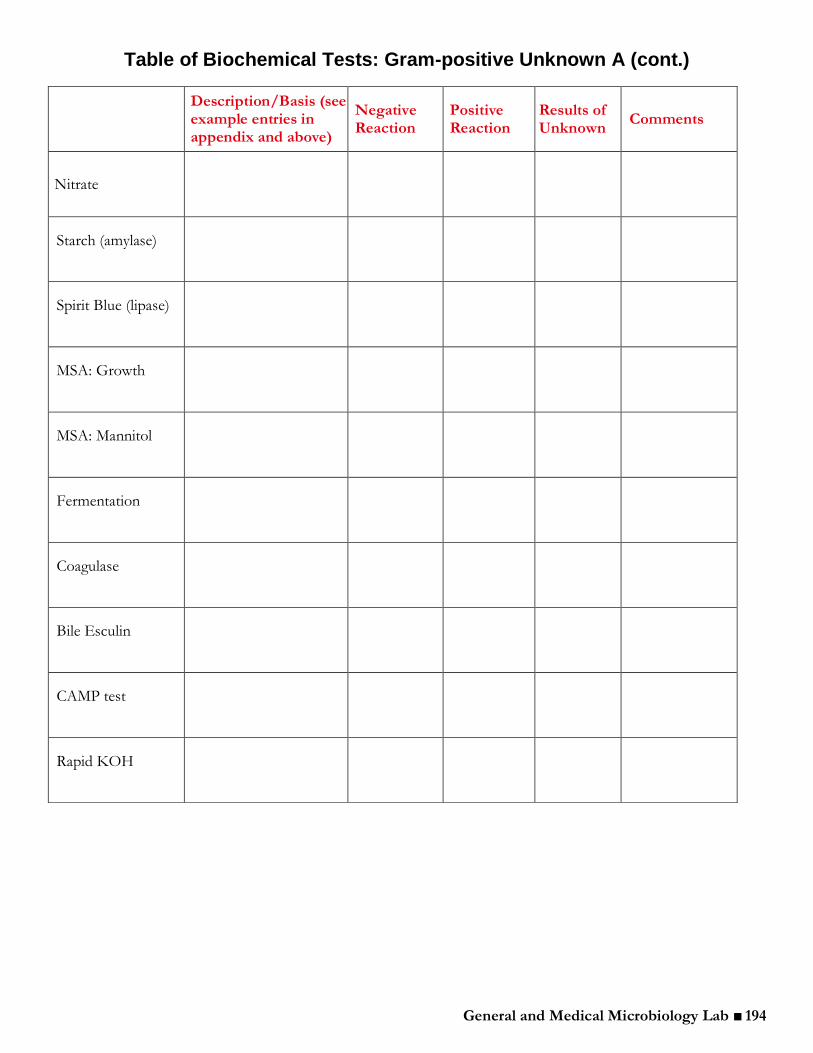
General and Medical Microbiology Lab ■ 194
Table of Biochemical Tests: Gram-positive Unknown A (cont.)
Description/Basis (see example entries in appendix and above)
Negative Reaction
Positive Reaction
Results of Unknown
Comments
Nitrate
Starch (amylase)
Spirit Blue (lipase)
MSA: Growth
MSA: Mannitol
Fermentation
Coagulase
Bile Esculin
CAMP test
Rapid KOH
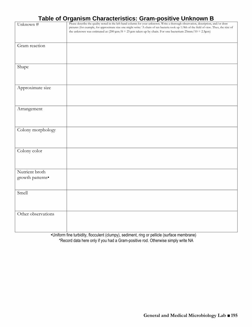
General and Medical Microbiology Lab ■ 195
Table of Organism Characteristics: Gram-positive Unknown B Unknown # Please describe the quality noted in the left-hand column for your unknown. Write a thorough observation, description, and/or draw
pictures (for example, for approximate size one might write: ‘A chain of ten bacteria took up 1/8th of the field of view. Thus, the size of
the unknown was estimated as (200 m/8 = 25 m taken up by chain. For one bacterium 25mm/10 = 2.5m)
Gram reaction
Shape
Approximate size
Arrangement
Colony morphology
Colony color
Nutrient broth growth patterns•
Smell
Other observations
•Uniform fine turbidity, flocculent (clumpy), sediment, ring or pellicle (surface membrane)
*Record data here only if you had a Gram-positive rod. Otherwise simply write NA

General and Medical Microbiology Lab ■ 196
Table of Biochemical Tests: Gram-negative Unknown B
Description/Basis (see example entries in appendix and above)
Negative Reaction
Positive Reaction
Results of Unknown
Comments
MacConkey Agar
Mannitol Fermentation / gas production
Glucose Fermentation / gas production
Lactose Fermentation / gas production
Nitrate
Oxidase
Methyl Red (MR)
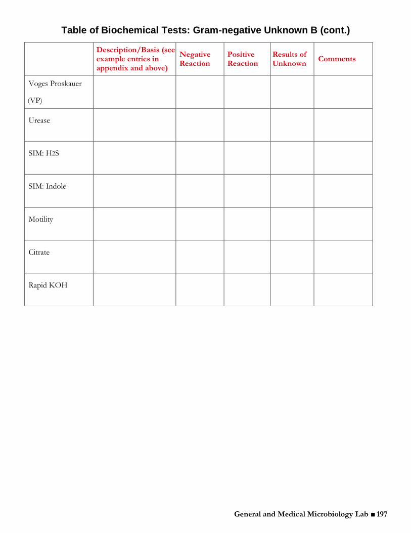
General and Medical Microbiology Lab ■ 197
Table of Biochemical Tests: Gram-negative Unknown B (cont.)
Description/Basis (see example entries in appendix and above)
Negative Reaction
Positive Reaction
Results of Unknown
Comments
Voges Proskauer
(VP)
Urease
SIM: H2S
SIM: Indole
Motility
Citrate
Rapid KOH

General and Medical Microbiology Lab ■ 198


