101
Unit III Nervous System and Electromyography
Unit IIINervous System and Electromyography
Content Introduction to Nervous System-Anatomy
The anatomy of the nervous system & The Autonomic nervous System
10-20 electrode placement system for EEG measurement
EEG machine
Evoked potentials and Types & significance of EEG signals
EEG Amplifiers& filters
Analysis of diseases using EEG Electromyography (EMG)
Muscle contraction mechanism,
Myoelectric voltages,
EMG Machine.
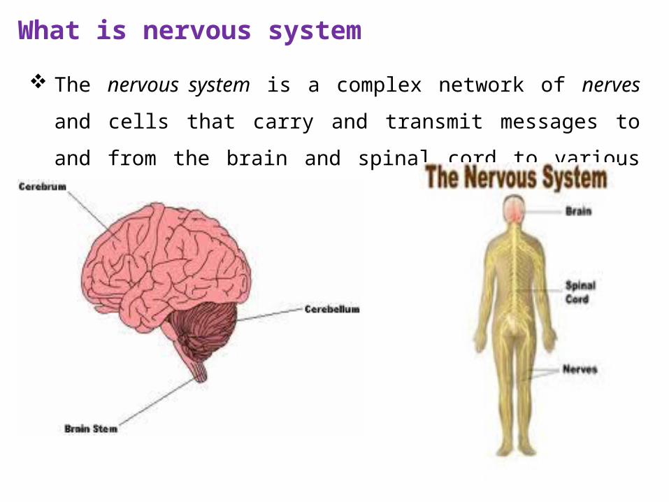
What is nervous system
The nervous system is a complex network of nerves and cells that carry
and transmit messages to and from the brain and spinal cord to
various parts of the body.
The Nervous system has three major functions:
Sensory – monitors internal & external environment through presence
of receptors (carry messages from body to brain (pain, pressure,
temperature))
Integration – interpretation of sensory information (information
processing); complex (higher order) functions
Motor – response to information processed through stimulation of
effectors(– carry messages from brain to body to respond )
Muscle contraction
Glandular secretion
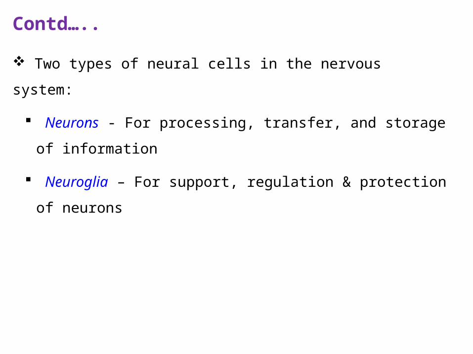
Contd…..
Two types of neural cells in the nervous system:
Neurons - For processing, transfer, and storage of information
Neuroglia – For support, regulation & protection of neurons
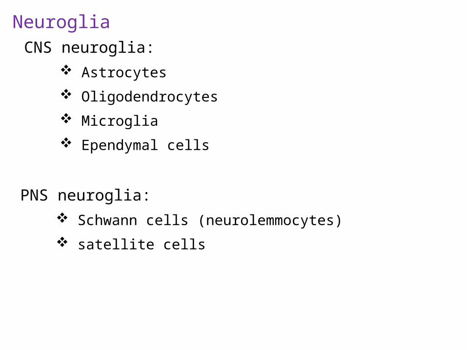
NeurogliaCNS neuroglia:
Astrocytes
Oligodendrocytes
Microglia
Ependymal cells
PNS neuroglia: Schwann cells (neurolemmocytes)
satellite cells
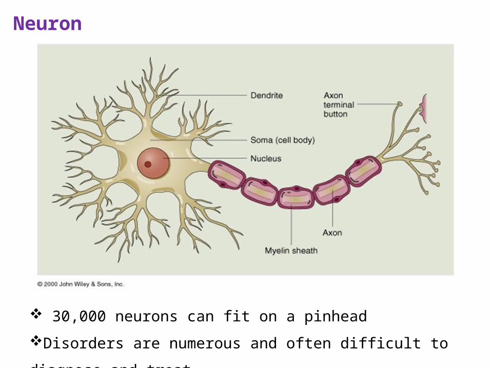
Neuron
30,000 neurons can fit on a pinhead
Disorders are numerous and often difficult to diagnose and treat
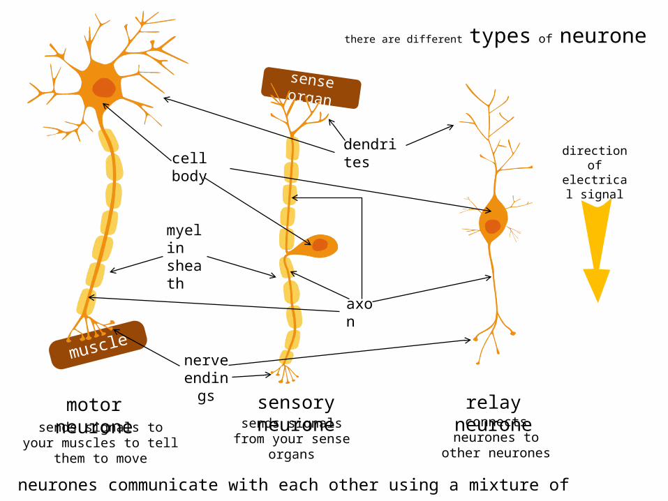
sense organ
muscle
there are different types of neurone
sensory neuronemotor neurone relay neurone
direction of electrical
signal
sends signals to your muscles to tell them to move
sends signals from your sense organs
connects neurones to other neurones
dendritescell body
axon
myelin sheath
nerve endings
neurones communicate with each other using a mixture of electrical & chemical signals
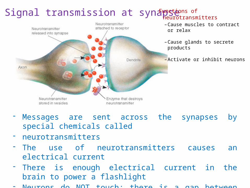
Signal transmission at synapse
- Messages are sent across the synapses by special chemicals called- neurotransmitters- The use of neurotransmitters causes an electrical current- There is enough electrical current in the brain to power a flashlight- Neurons do NOT touch; there is a gap between them called a synapse
Functions of neurotransmitters– Cause muscles to contract or
relax
– Cause glands to secrete products
– Activate or inhibit neurons
cell body
axon
myelin sheath
dendrites
nucleus
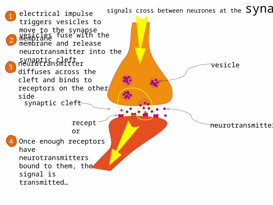
signals cross between neurones at the synapse
neurotransmitter
vesicle
synaptic cleft
receptor
electrical impulse triggers vesicles to move to the synapse membrane
1
vesicles fuse with the membrane and release neurotransmitter into the synaptic cleft
2
neurotransmitter diffuses across the cleft and binds to receptors on the other side
3
Once enough receptors have neurotransmitters bound to them, the signal is transmitted…
4
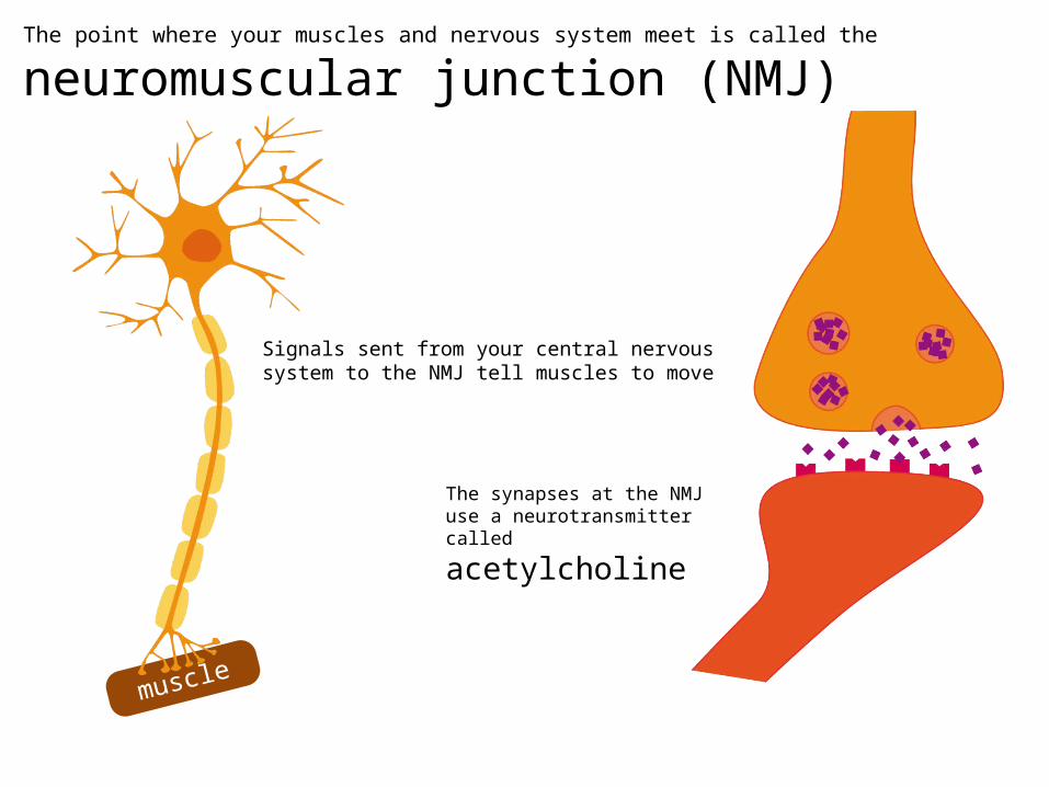
The point where your muscles and nervous system meet is called the
neuromuscular junction (NMJ)
muscle
Signals sent from your central nervous system to the NMJ tell muscles to move
The synapses at the NMJ use a neurotransmitter called
acetylcholine
Important Terms
Synapse – junction between 2 neurons that communicates the
message from the presynaptic neuron to the postsynaptic neuron
Ganglion – a cluster of neuronal cell bodies in the PNS
Preganglionic neuron – cell body lies within the CNS
Postganglionic fiber (axon) of the ganglionic neuron extends to the
visceral organs
Dendrites - fibers that receive messages from other neurons
Axons - fibers that send messages to other neurons
Apply Your Knowledge
What is the function of neurotransmitters?
ANSWER: Neurotransmitters cause muscles to contract or relax, cause glands to secret products, activate neurons to send nerve impulses, or inhibit neurons from sending them.
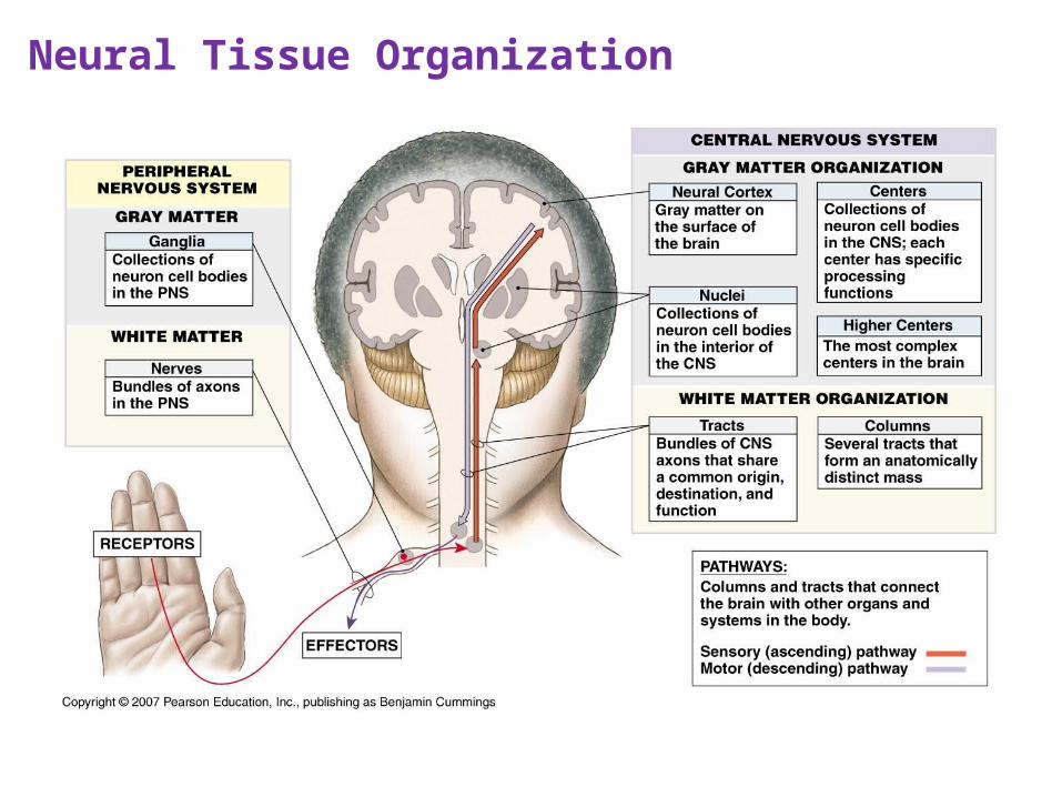
Neural Tissue Organization
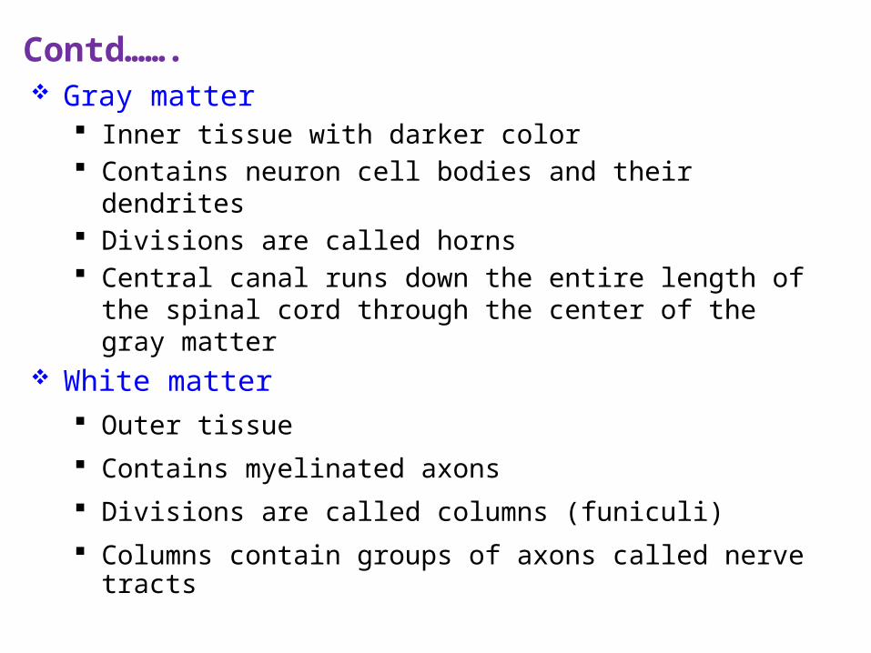
Gray matter Inner tissue with darker color Contains neuron cell bodies and their dendrites Divisions are called horns Central canal runs down the entire length of the spinal cord through
the center of the gray matter White matter
Outer tissue Contains myelinated axons Divisions are called columns (funiculi) Columns contain groups of axons called nerve tracts
Contd…….
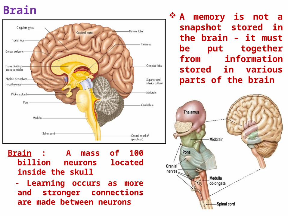
Brain
A memory is not a snapshot stored in the brain – it must be put together from information stored in various parts of the brain
Brain : A mass of 100 billion neurons located inside the skull
- Learning occurs as more and stronger connections are made between neurons
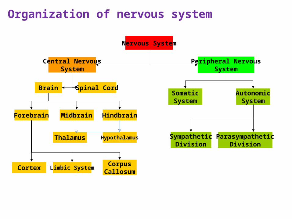
Nervous System
Central NervousSystem
Brain Spinal Cord
Forebrain Midbrain Hindbrain
Thalamus Hypothalamus
Cortex Limbic SystemCorpus
Callosum
Peripheral NervousSystem
SomaticSystem
AutonomicSystem
ParasympatheticDivision
SympatheticDivision
Organization of nervous system
Med
ical A
rt S
erv
ice, M
unic
h /
, W
ellc
om
e Im
ag
es
Cre
dit
Med
ical A
rt S
erv
ice, M
unic
h /
, W
ellc
om
e Im
ag
es
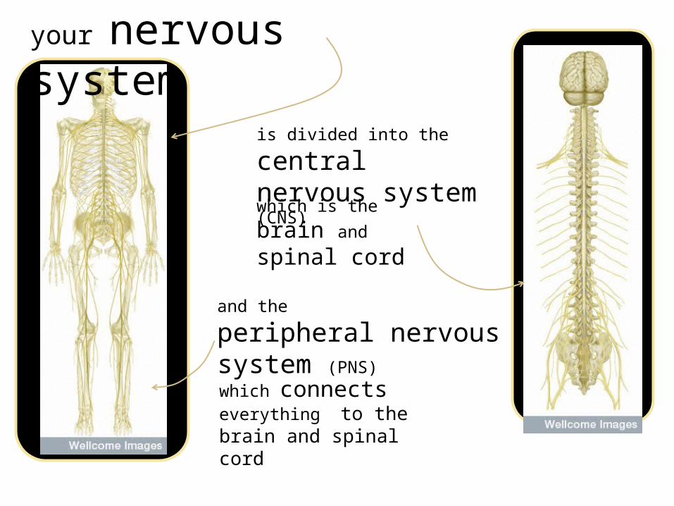
your nervous system
is divided into the central nervous system (CNS)
and the
peripheral nervous system (PNS)
which is the brain and
spinal cord
which connects everything to the brain and spinal cord
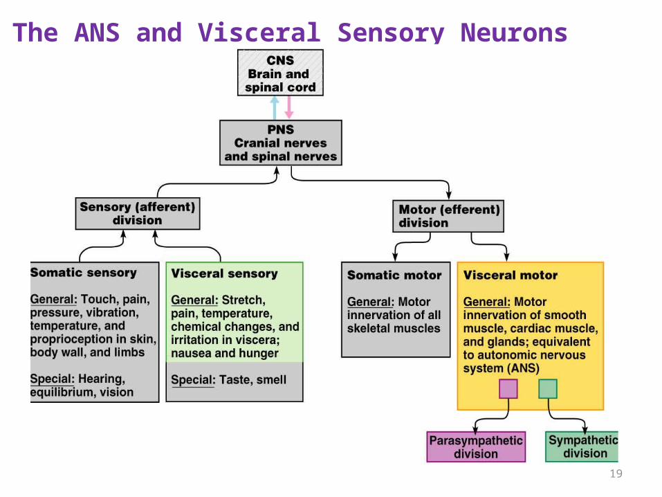
The ANS and Visceral Sensory Neurons
19
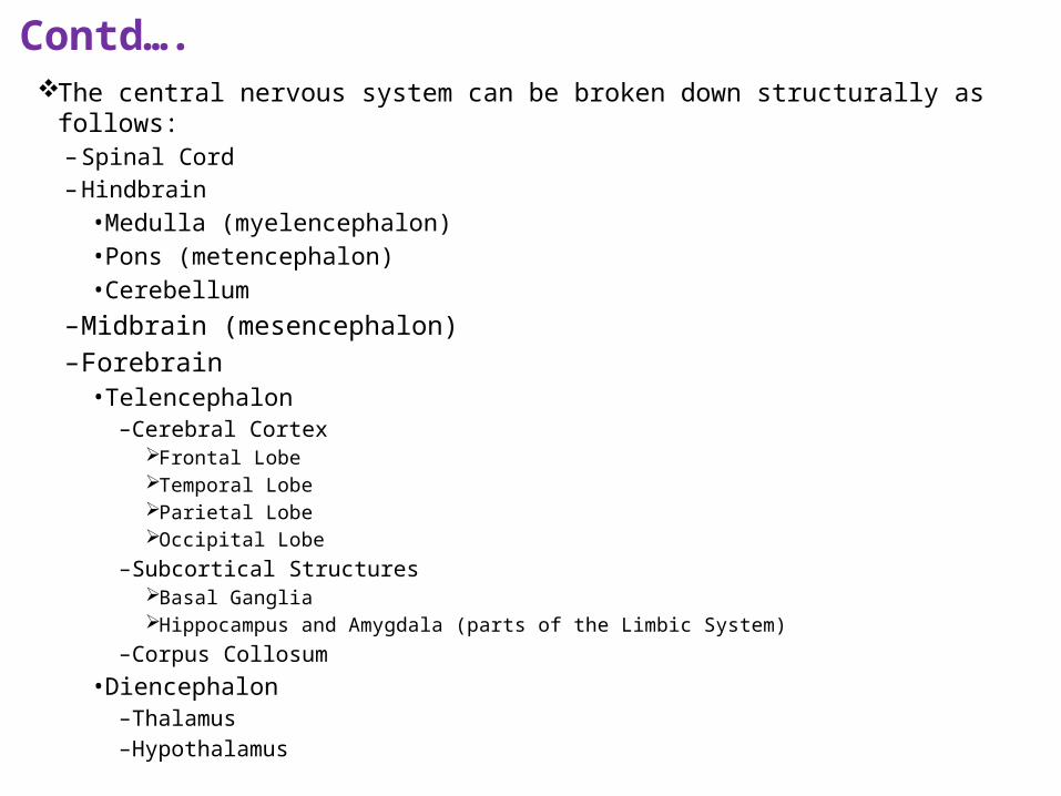
Contd…. The central nervous system can be broken down structurally as follows:
– Spinal Cord – Hindbrain
• Medulla (myelencephalon) • Pons (metencephalon) • Cerebellum
– Midbrain (mesencephalon) – Forebrain
• Telencephalon – Cerebral Cortex
Frontal Lobe Temporal Lobe Parietal Lobe Occipital Lobe
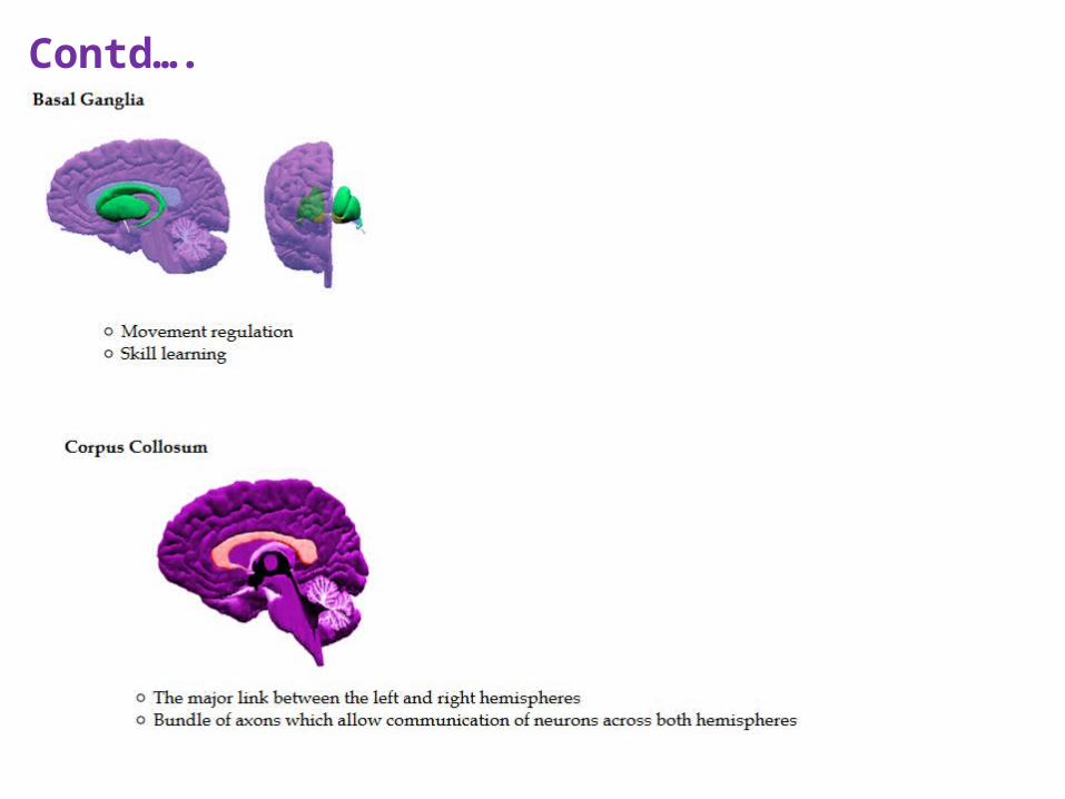
– Subcortical Structures
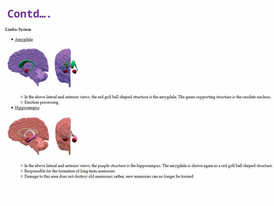
Basal Ganglia Hippocampus and Amygdala (parts of the Limbic System)
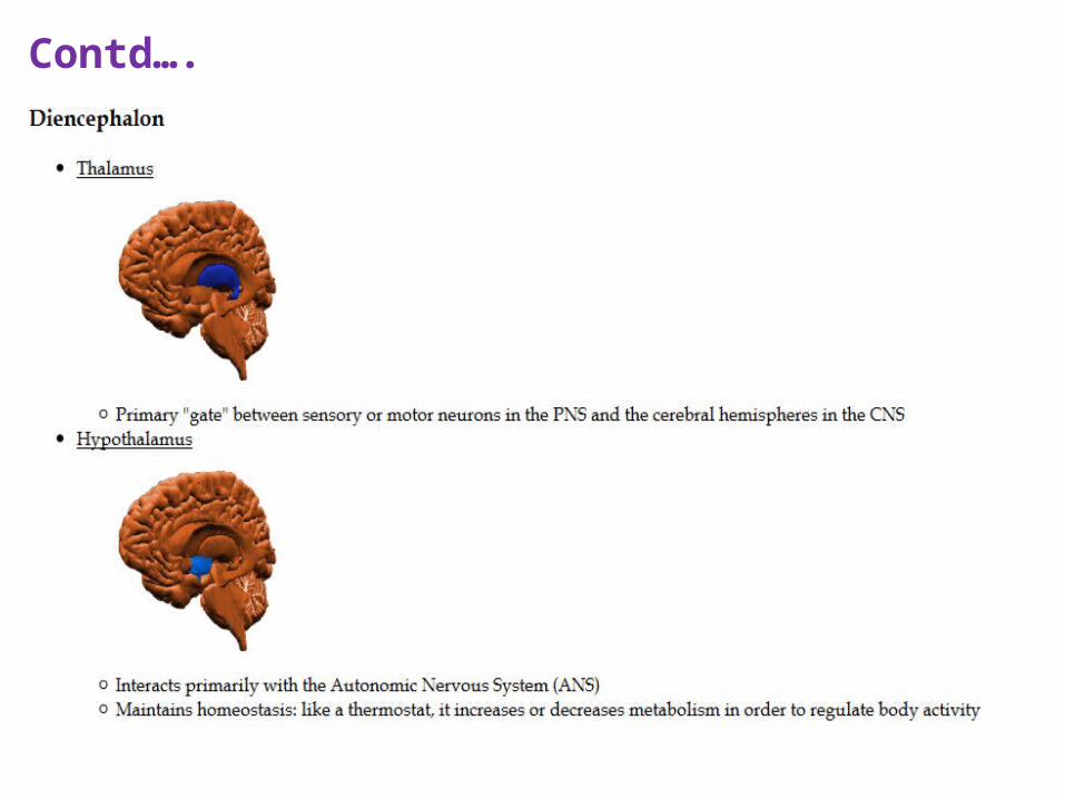
– Corpus Collosum• Diencephalon
– Thalamus – Hypothalamus
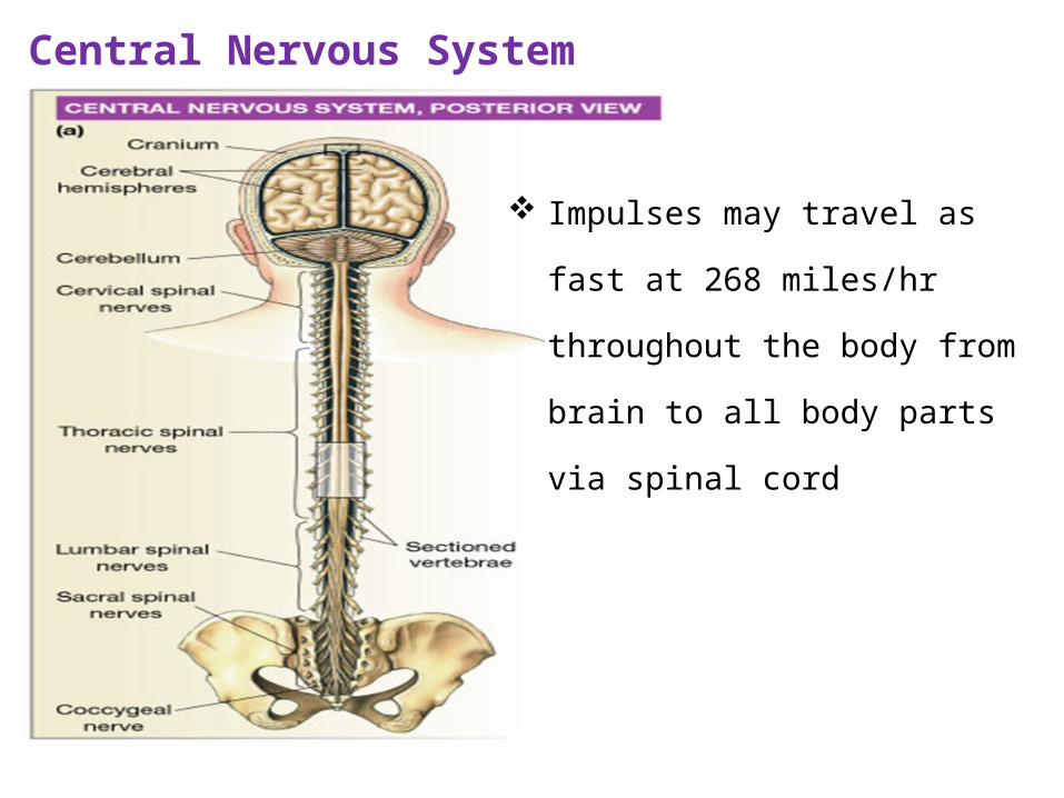
Central Nervous System
Impulses may travel as fast at 268
miles/hr throughout the body from
brain to all body parts via spinal cord
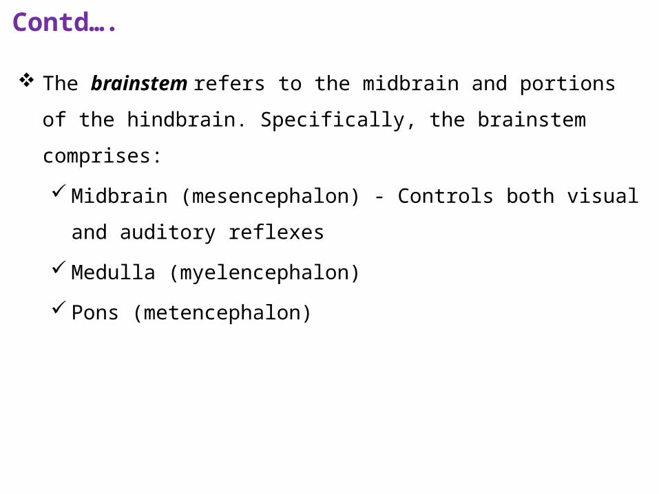
The brainstem refers to the midbrain and portions of the hindbrain.
Specifically, the brainstem comprises:
Midbrain (mesencephalon) - Controls both visual and auditory
reflexes
Medulla (myelencephalon)
Pons (metencephalon)
Contd….
Spinal cord The spinal cord is one of the two major components of the central
nervous system:
Like the brain, it is completely encased in bone. It resides within the
vertebral column
Connects directly to the medulla section of the brain
It is approximately 45 cm long in an adult
Ascending tracts Receives sensory messages and sends them to the
brain
Descending tracts carry motor information down from the brain to
muscles and glands
Also acts independently from the brain called reflexes
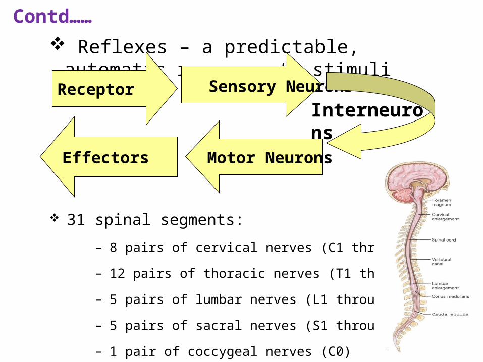
Reflexes – a predictable, automatic response to stimuli
Receptor Sensory Neurons
Effectors Motor Neurons
Interneurons
Contd……
31 spinal segments:
– 8 pairs of cervical nerves (C1 through C8)
– 12 pairs of thoracic nerves (T1 through T12)
– 5 pairs of lumbar nerves (L1 through L5)
– 5 pairs of sacral nerves (S1 through S5)
– 1 pair of coccygeal nerves (C0)
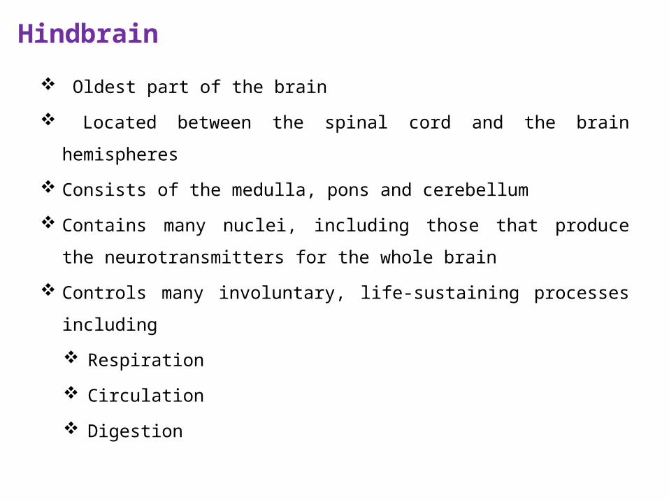
Hindbrain
Oldest part of the brain
Located between the spinal cord and the brain hemispheres
Consists of the medulla, pons and cerebellum
Contains many nuclei, including those that produce the
neurotransmitters for the whole brain
Controls many involuntary, life-sustaining processes including
Respiration
Circulation
Digestion
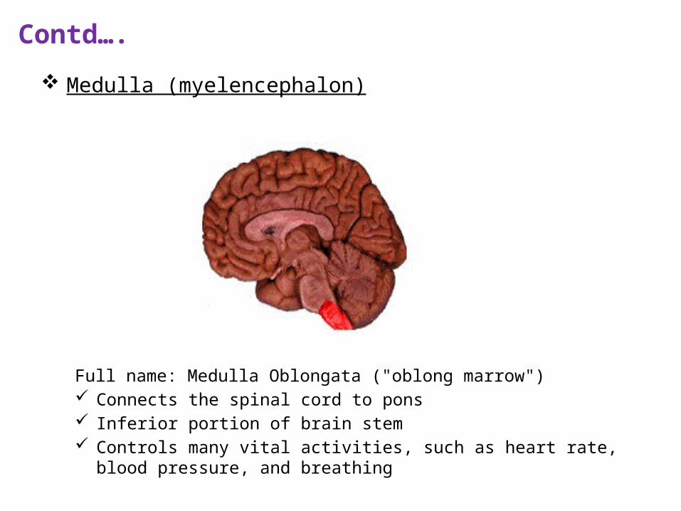
Contd….
Medulla (myelencephalon)
Full name: Medulla Oblongata ("oblong marrow") Connects the spinal cord to pons Inferior portion of brain stem Controls many vital activities, such as heart rate, blood pressure, and
breathing
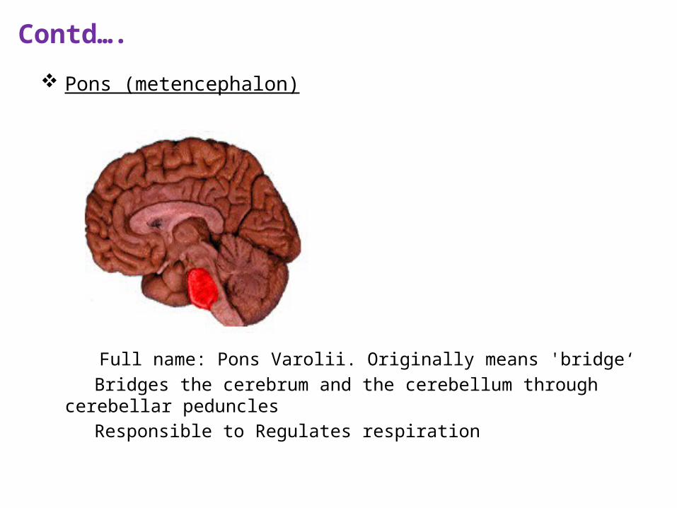
Contd….
Pons (metencephalon)
Full name: Pons Varolii. Originally means 'bridge‘ Bridges the cerebrum and the cerebellum through cerebellar peduncles Responsible to Regulates respiration

Cerebellum Location
– Inferior to the occipital lobes of the cerebrum– Posterior to the pons and medulla oblongata
Coordinates – Complex skeletal muscle contractions that are needed for
body movements– Fine movements, provide balance to the body
Contd….
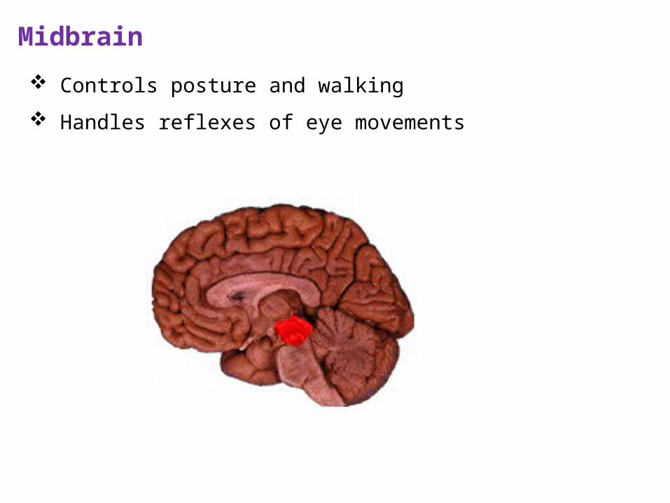
Midbrain
Controls posture and walking
Handles reflexes of eye movements
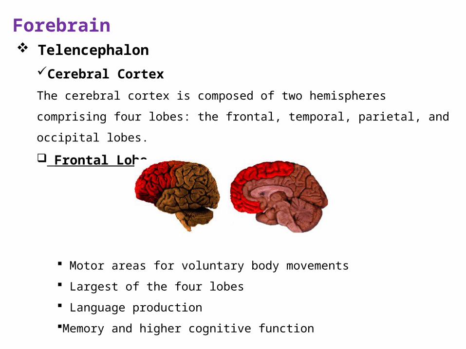
Forebrain
TelencephalonCerebral Cortex
The cerebral cortex is composed of two hemispheres comprising four lobes: the frontal,
temporal, parietal, and occipital lobes.
Frontal Lobe
Motor areas for voluntary body movements
Largest of the four lobes
Language production
Memory and higher cognitive function

Temporal Lobe Auditory processing - interpretation Memory Understanding language
Contd….
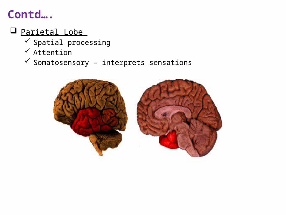
Parietal Lobe Spatial processing Attention Somatosensory – interprets sensations
Contd….
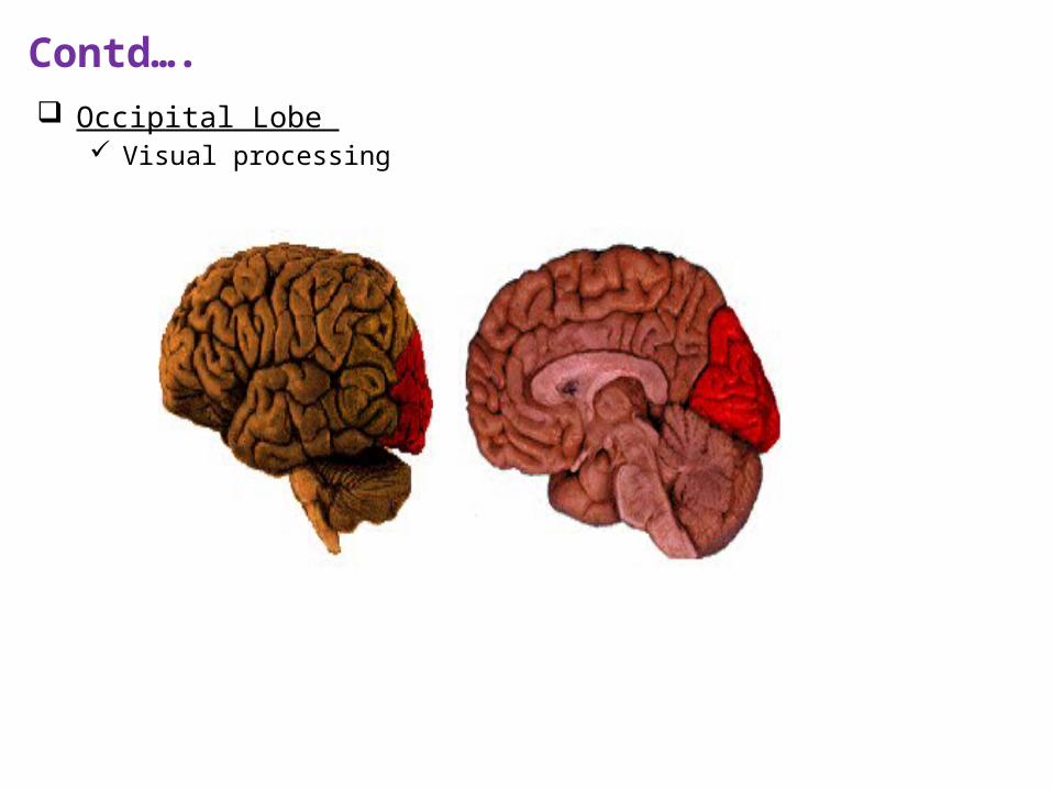
Occipital Lobe Visual processing
Contd….
Contd….
Contd….
Contd….
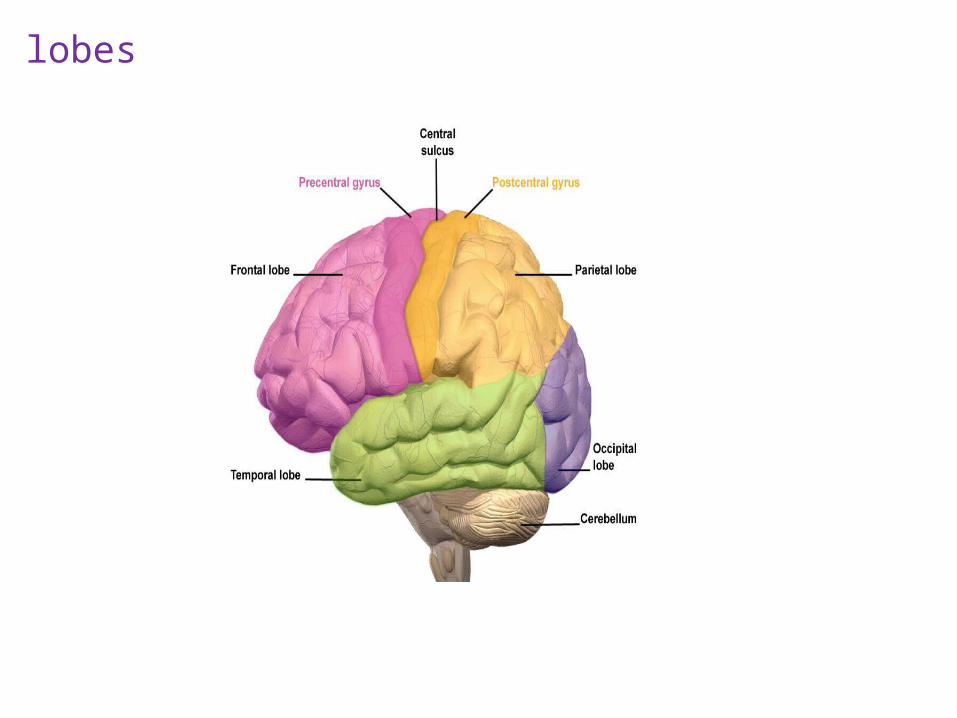
lobes
Apply Your Knowledge
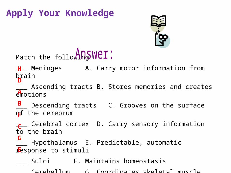
Match the following:
___ Meninges A. Carry motor information from brain
___ Ascending tracts B. Stores memories and creates emotions
___ Descending tracts C. Grooves on the surface of the cerebrum
___ Cerebral cortex D. Carry sensory information to the brain
___ Hypothalamus E. Predictable, automatic response to stimuli
___ Sulci F. Maintains homeostasis
___ Cerebellum G. Coordinates skeletal muscle contractions
___ Reflexes H. Protects the brain and spinal cord
B
F
C
G
E
D
A
H
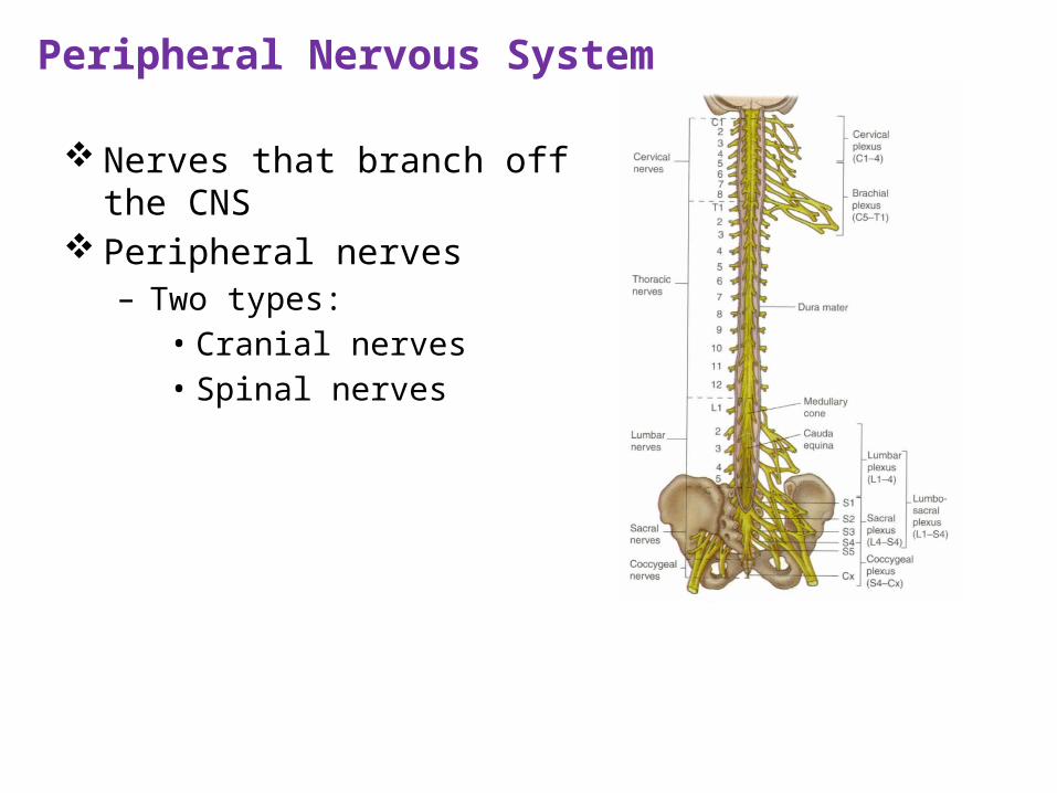
Peripheral Nervous System
Nerves that branch off the CNS Peripheral nerves
– Two types:• Cranial nerves • Spinal nerves
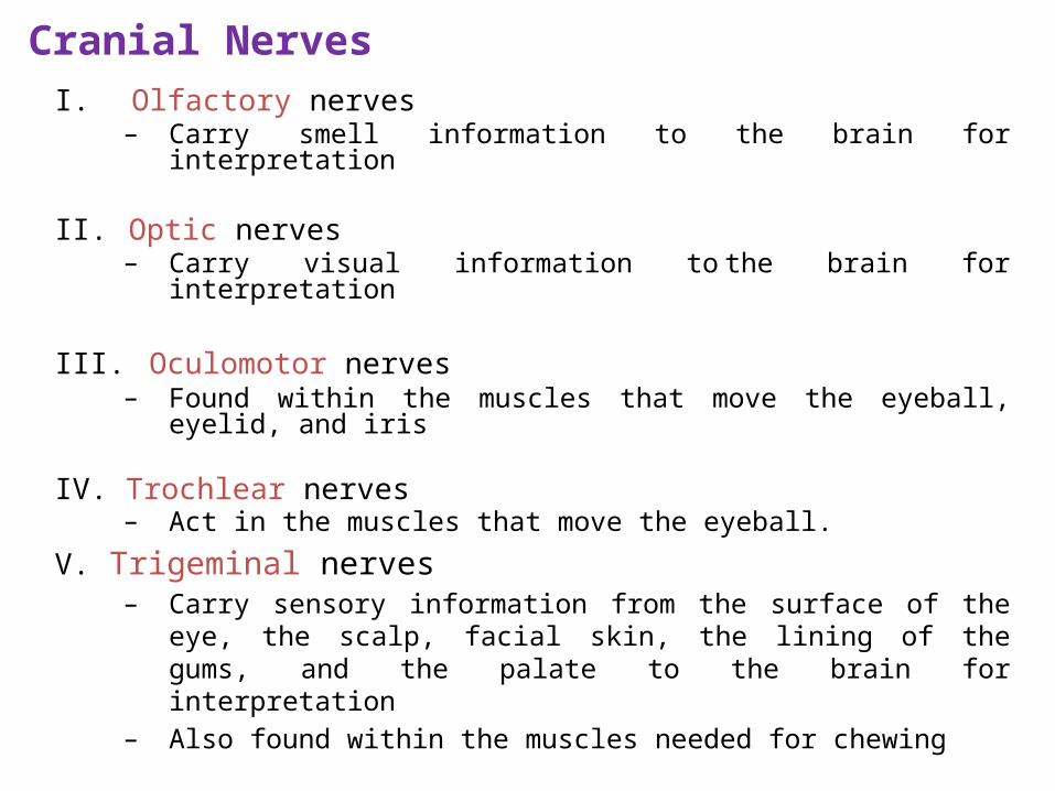
Cranial NervesI. Olfactory nerves
– Carry smell information to the brain for interpretation
II. Optic nerves – Carry visual information to the brain for interpretation
III. Oculomotor nerves – Found within the muscles that move the eyeball, eyelid, and
iris
IV. Trochlear nerves – Act in the muscles that move the eyeball.
V. Trigeminal nerves – Carry sensory information from the surface of the eye, the
scalp, facial skin, the lining of the gums, and the palate to the brain for interpretation
– Also found within the muscles needed for chewing
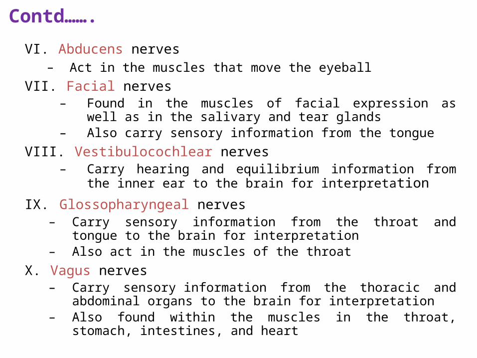
VI. Abducens nerves – Act in the muscles that move the eyeball
VII. Facial nerves – Found in the muscles of facial expression as well as in the salivary
and tear glands– Also carry sensory information from the tongue
VIII. Vestibulocochlear nerves – Carry hearing and equilibrium information from the inner ear to
the brain for interpretation
IX. Glossopharyngeal nerves – Carry sensory information from the throat and tongue to the brain
for interpretation– Also act in the muscles of the throat
X. Vagus nerves – Carry sensory information from the thoracic and abdominal organs to
the brain for interpretation– Also found within the muscles in the throat, stomach, intestines, and
heart
Contd…….
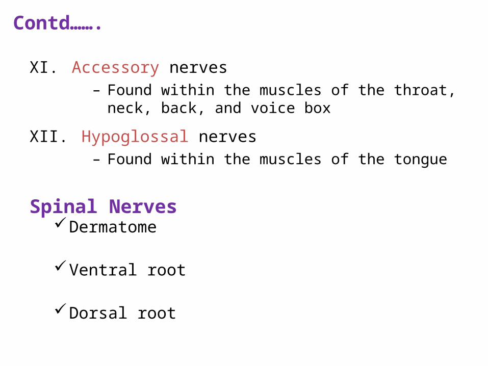
XI. Accessory nerves – Found within the muscles of the throat, neck, back, and
voice box
XII. Hypoglossal nerves – Found within the muscles of the tongue
Spinal Nerves Dermatome
Ventral root
Dorsal root
Contd…….
Introduction to ANS Regulates activity of smooth muscle, cardiac muscle and glands
Operates without conscious control
Named autonomic because was thought to be AUTONOMUS (working without CNS)
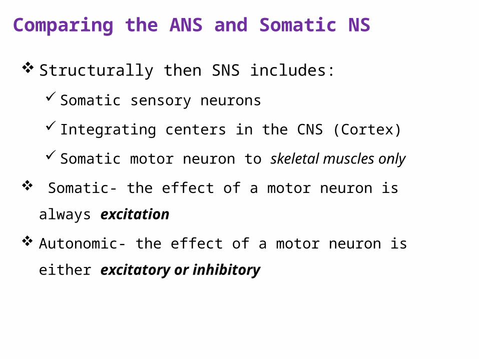
Comparing the ANS and Somatic NS
Structurally then SNS includes:
Somatic sensory neurons
Integrating centers in the CNS (Cortex)
Somatic motor neuron to skeletal muscles only
Somatic- the effect of a motor neuron is always excitation
Autonomic- the effect of a motor neuron is either excitatory or
inhibitory
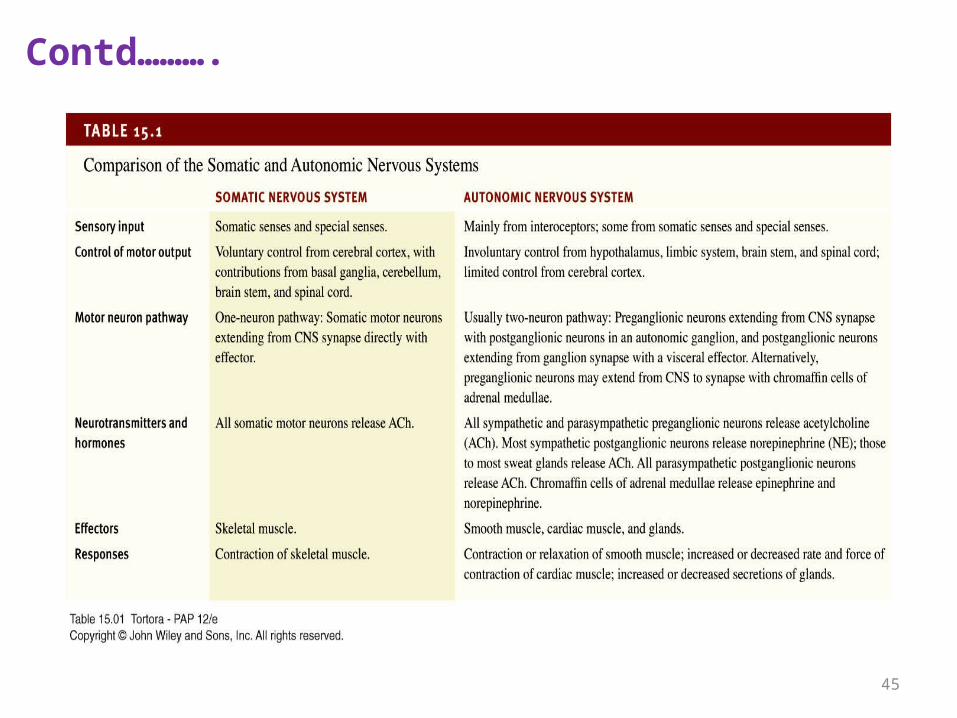
Contd……….
45
Autonomic Nervous System
Makes all routine adjustments in physiological systems.
The ANS pathway from the CNS to the effector always involves 2
neurons synapsing in an autonomic ganglion.
Preganglionic – cell body is in the CNS, axon extends to the
ganglion outside the CNS
Postganglionic – cell body is in the ganglion, axon extends to the
visceral effector
Autonomic regulation & stress
A stressful situation activates three major communication systems in the
brain that regulate bodily functions.
The first of these systems is the voluntary nervous system, which sends
messages to muscles so that we may respond to sensory information.
The second communication system is the autonomic nervous system. It
combines the sympathetic or emergency branch, which gets us going in
emergencies, and the parasympathetic or calming branch, which keeps
the body’s maintenance systems, such as digestion, in order and calms
the body’s responses to the emergency branch.
The brain’s third major communication process is the neuroendocrine
system, which also maintains the body’s internal functioning.
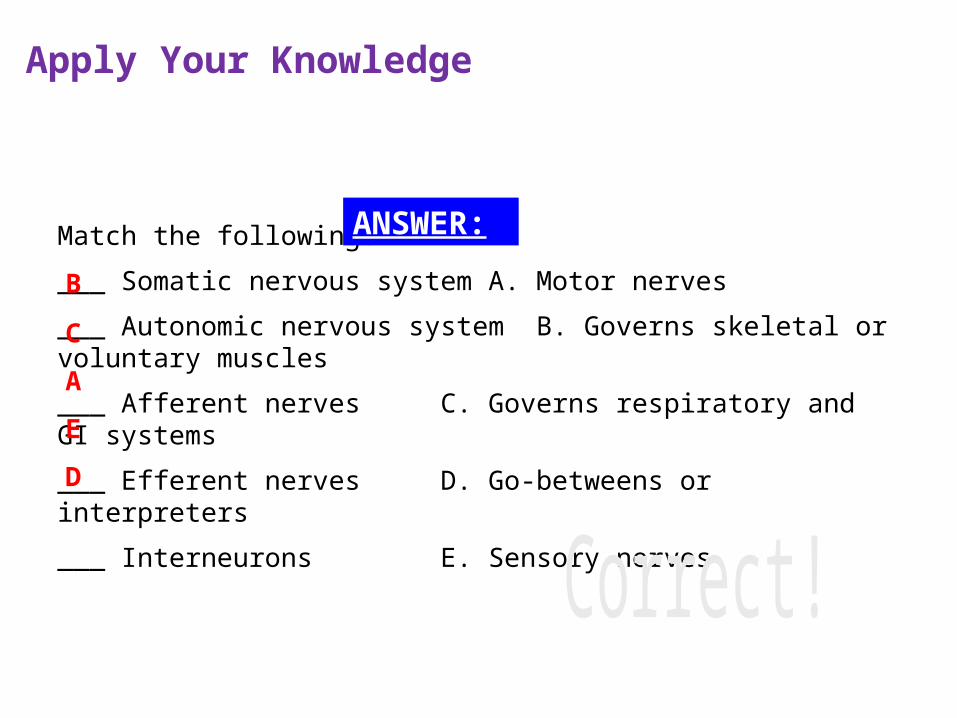
Apply Your Knowledge
Match the following:
___ Somatic nervous system A. Motor nerves
___ Autonomic nervous system B. Governs skeletal or voluntary muscles
___ Afferent nerves C. Governs respiratory and GI systems
___ Efferent nerves D. Go-betweens or interpreters
___ Interneurons E. Sensory nerves
C
A
E
D
B
ANSWER:
Subdivisions of the ANS
Sympathetic Division Fight-or-flight
Parasympathetic Division Rest-and-digest
These divisions are anatomically distinct Sympathetic Stimulates
heart beat tissue metabolism, increases alertness, prepares the body to deal with emergencies (“fight or flight” division)
Contd…..
Synapses of neurons are in a chain of ganglia that run alongside the
spinal cord
Extends on both sides of the vertebral column
Carries preganglionic fibers and cell bodies of postganglionic neurons
Rami communicantes from the spinal nerves connect to the chain
Effects of Sympathetic StimulationWidespread
• The sympathetic chain allows one preganglionic fiber to synapse with many postganglionic neurons
Enhanced & prolonged by the adrenal medulla
Neurotransmitters of Sympathetic Division
Preganglionic fibers release acetylcholine (Ach) Therefore they are
called:
Cholinergic
Postganglionic fibers (most) release norepinephrine (NE) (=
noradrenaline)
Adrenergic
Adrenal medulla releases norepinephrine and epinephrine
(adrenalin)
Functions of the Sympathetic Division
Heart: increases rate
Lung bronchioles: dilates bronchioles
Salivary glands: produce viscous fluid
Stomach: decreases motility
Pupil: dilates
Sweat glands: produce secretions
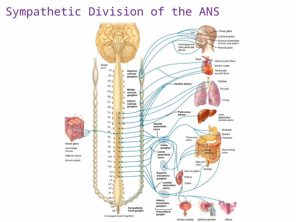
Sympathetic Division of the ANS
Parasympathetic Parasympathetic division (craniosacral)
– Cell bodies reside in the brain stem (cranial nerves) or in the sacral
portion of the spinal cord
– Slows the heart rate,
– Inhibits senses,
– Prepares the body for rest and relaxation; (“rest and digest” division).
Neurotransmitter of Parasympathetic Division
Preganglionic fibers: Acetylcholine
Postganglionic fibers: Acetylcholine
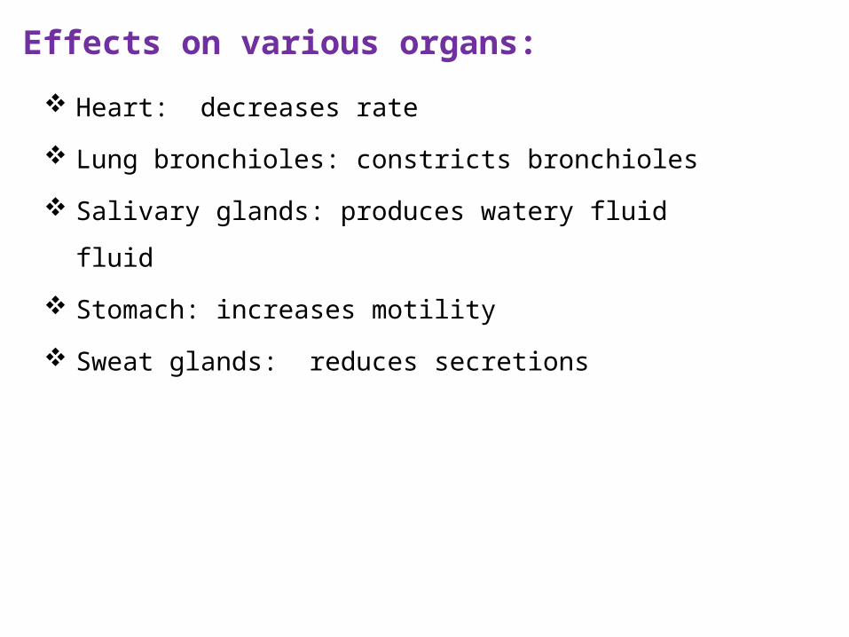
Effects on various organs:
Heart: decreases rate
Lung bronchioles: constricts bronchioles
Salivary glands: produces watery fluid fluid
Stomach: increases motility
Sweat glands: reduces secretions
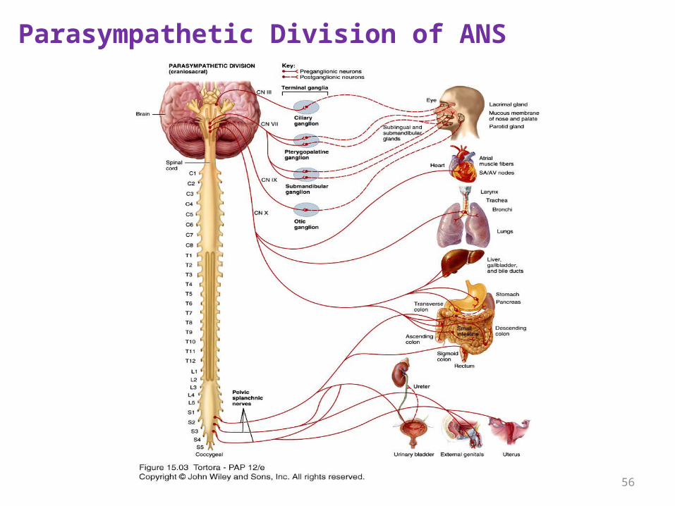
Parasympathetic Division of ANS
56
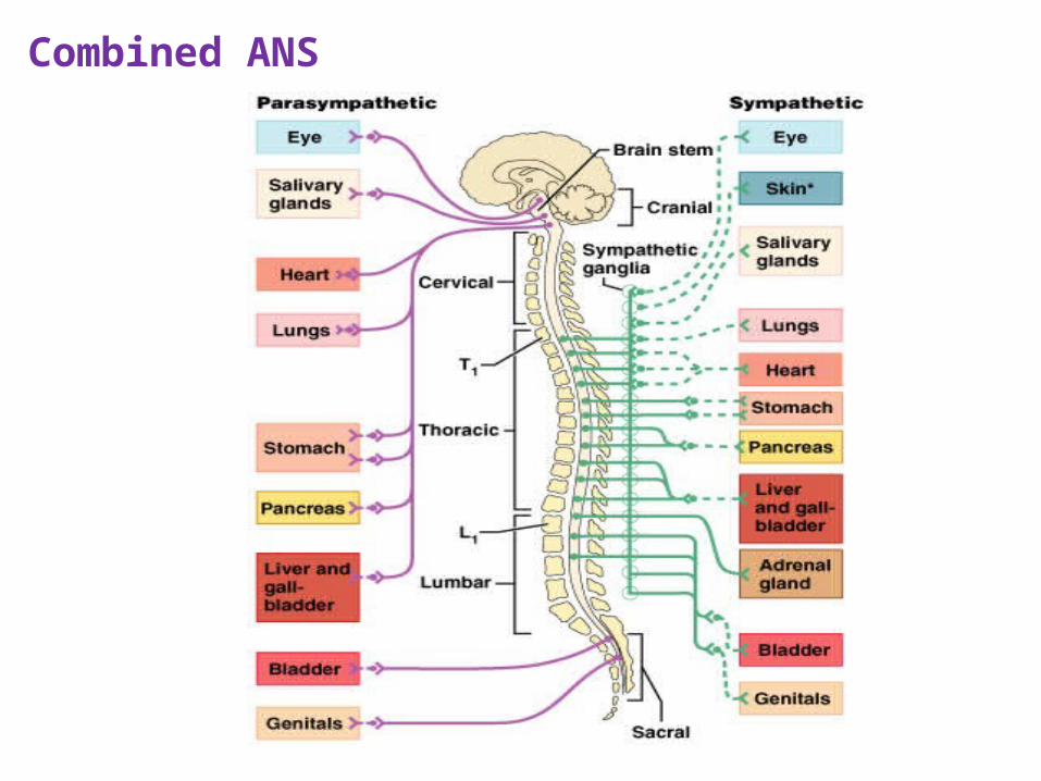
Combined ANS
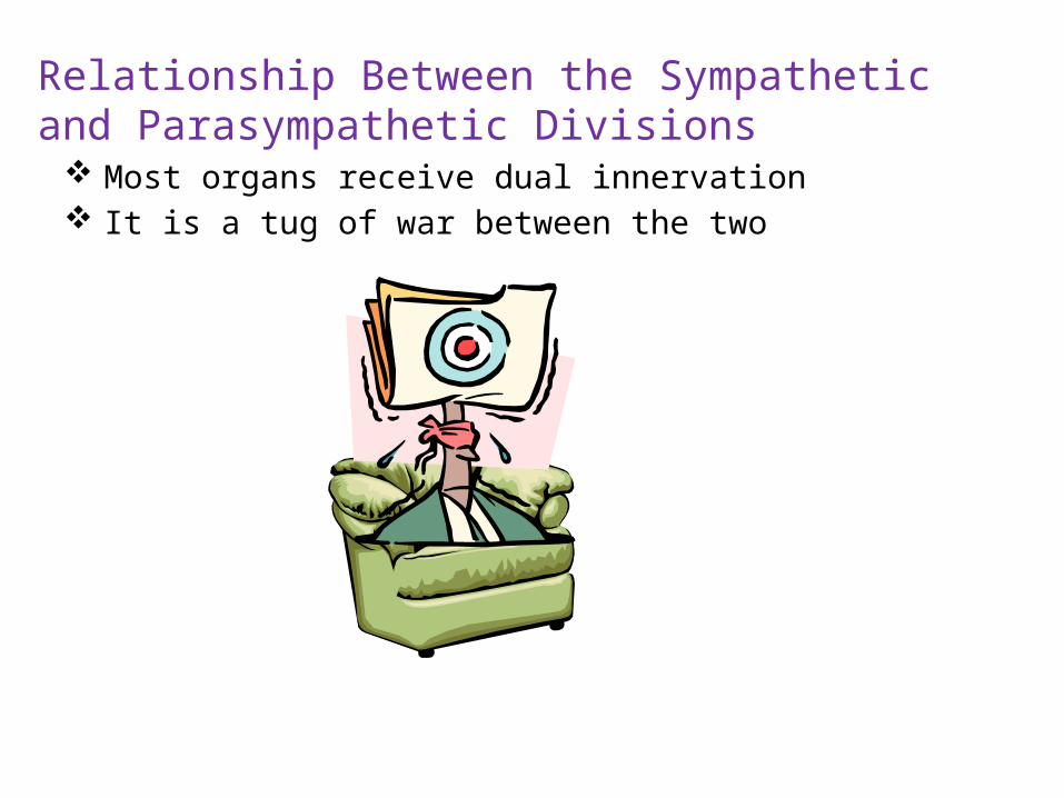
Relationship Between the Sympathetic and Parasympathetic Divisions
Most organs receive dual innervation It is a tug of war between the two
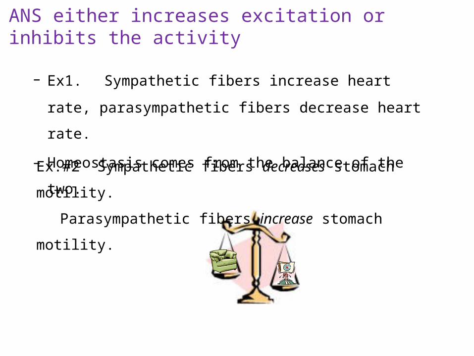
ANS either increases excitation or inhibits the activity
– Ex1. Sympathetic fibers increase heart rate, parasympathetic
fibers decrease heart rate.
– Homeostasis comes from the balance of the two.
Ex.#2 Sympathetic fibers decreases stomach motility.
Parasympathetic fibers increase stomach motility.
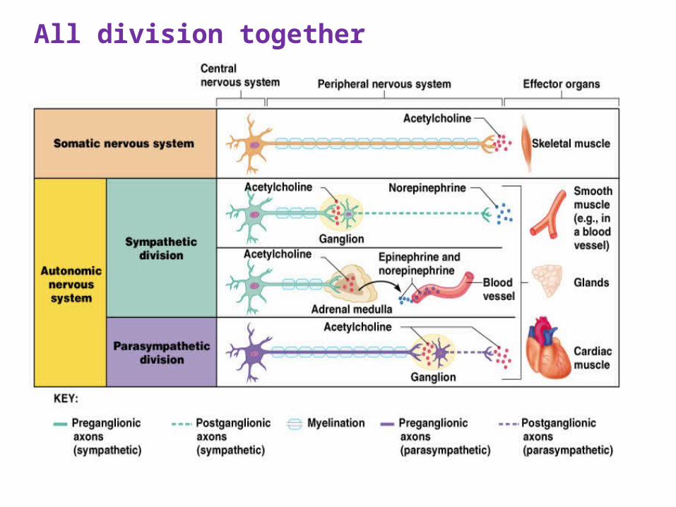
All division together
Apply Your Knowledge
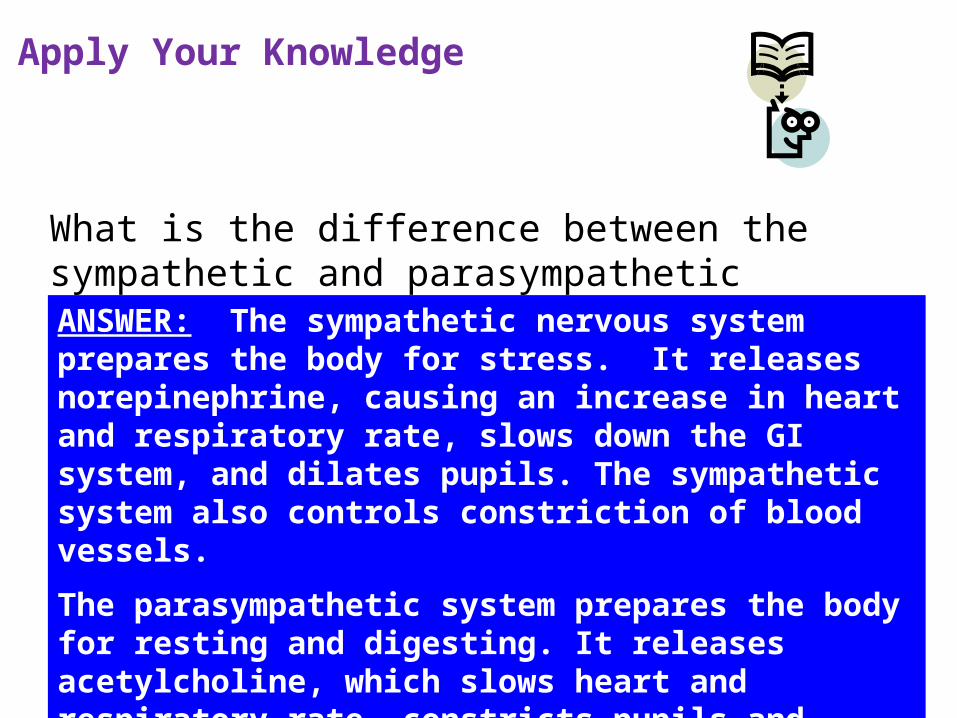
What is the difference between the sympathetic and parasympathetic nervous systems?ANSWER: The sympathetic nervous system prepares the body for stress. It releases norepinephrine, causing an increase in heart and respiratory rate, slows down the GI system, and dilates pupils. The sympathetic system also controls constriction of blood vessels.
The parasympathetic system prepares the body for resting and digesting. It releases acetylcholine, which slows heart and respiratory rate, constricts pupils and stimulates the GI system. It has no effect on most blood vessels.
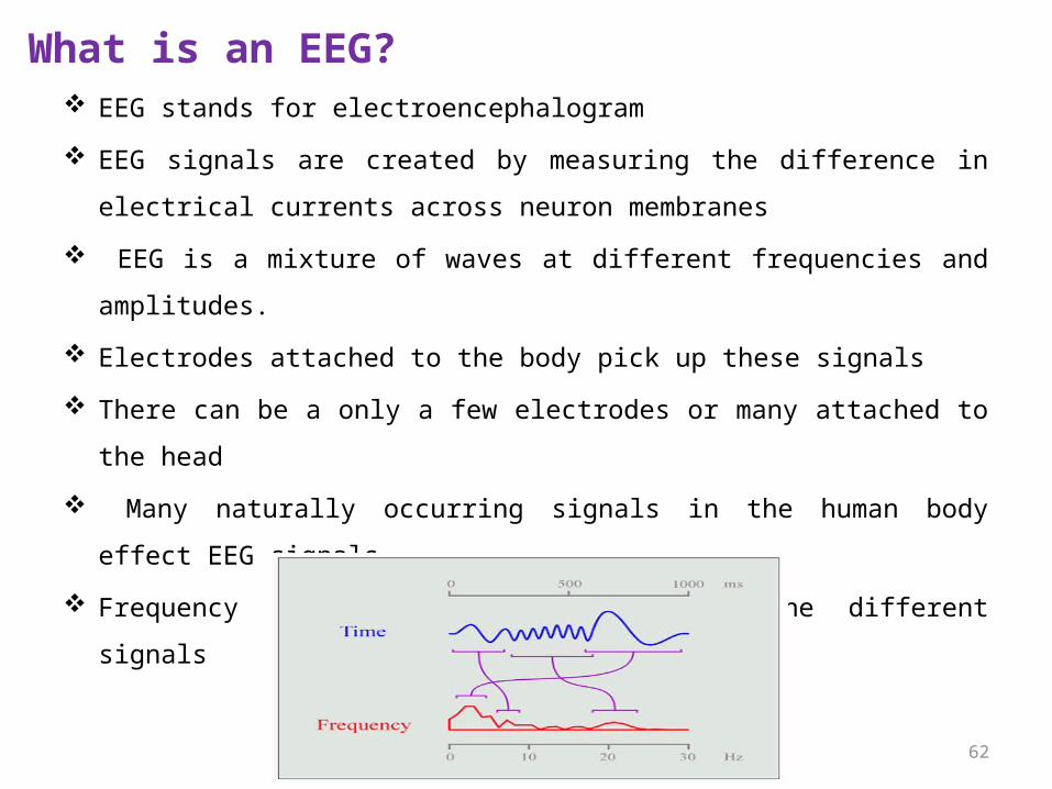
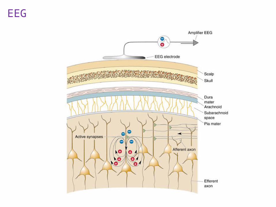
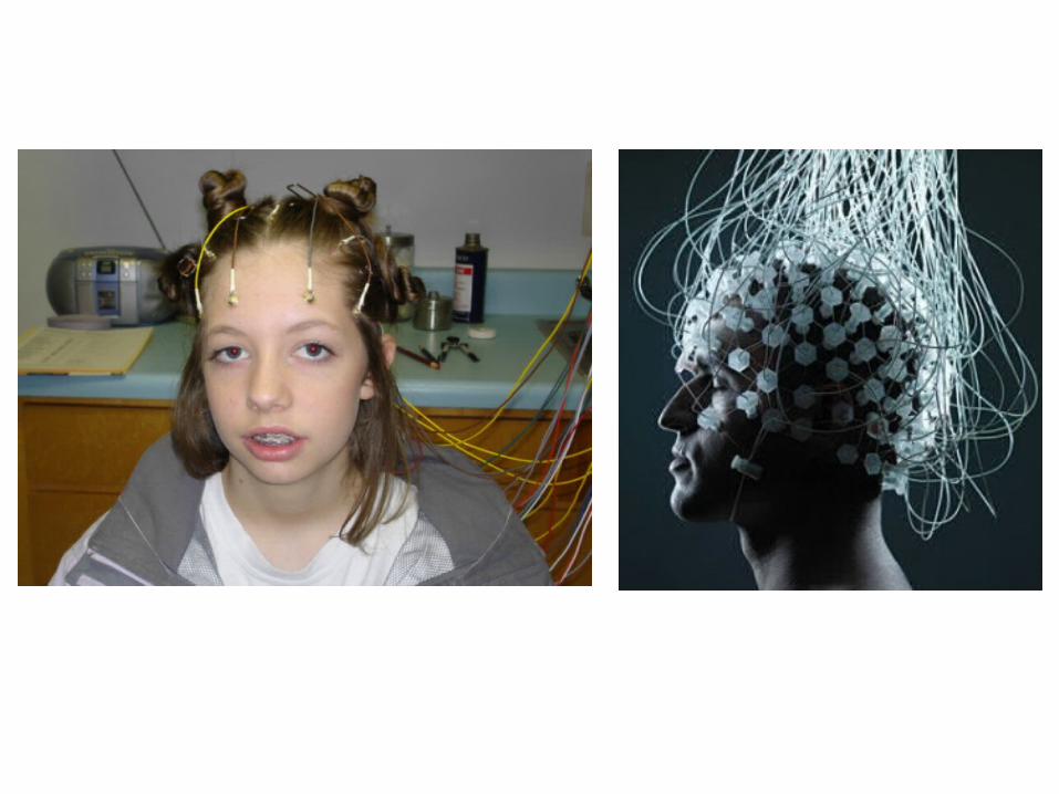
What is an EEG? EEG stands for electroencephalogram
EEG signals are created by measuring the difference in electrical currents
across neuron membranes
EEG is a mixture of waves at different frequencies and amplitudes.
Electrodes attached to the body pick up these signals
There can be a only a few electrodes or many attached to the head
Many naturally occurring signals in the human body effect EEG signals
Frequency Analysis helps to separate the different signals
62

Contd….
EEG
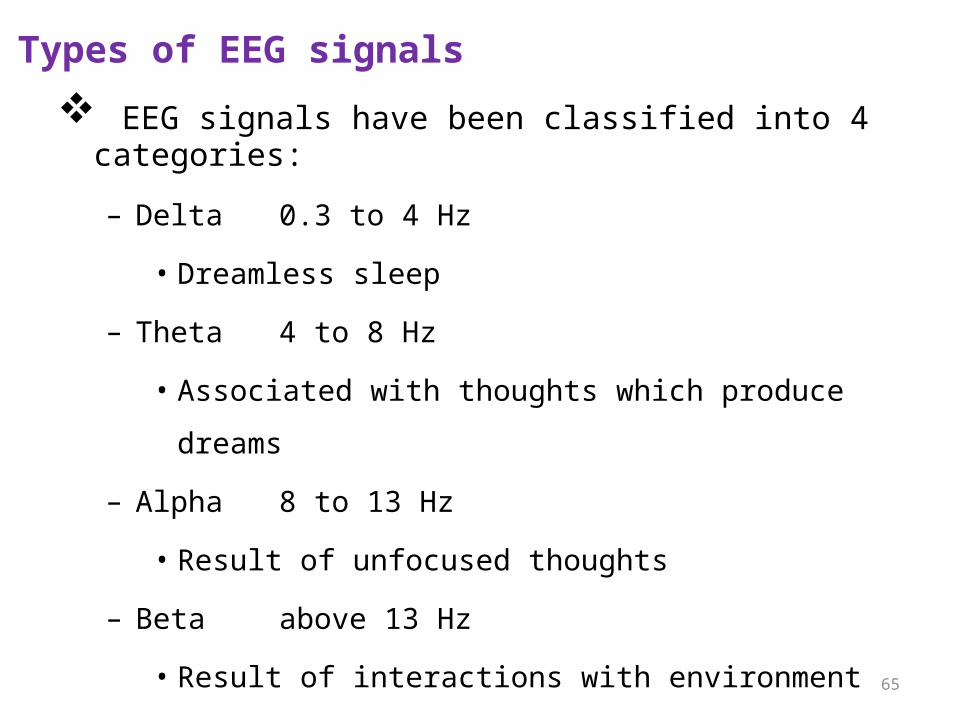
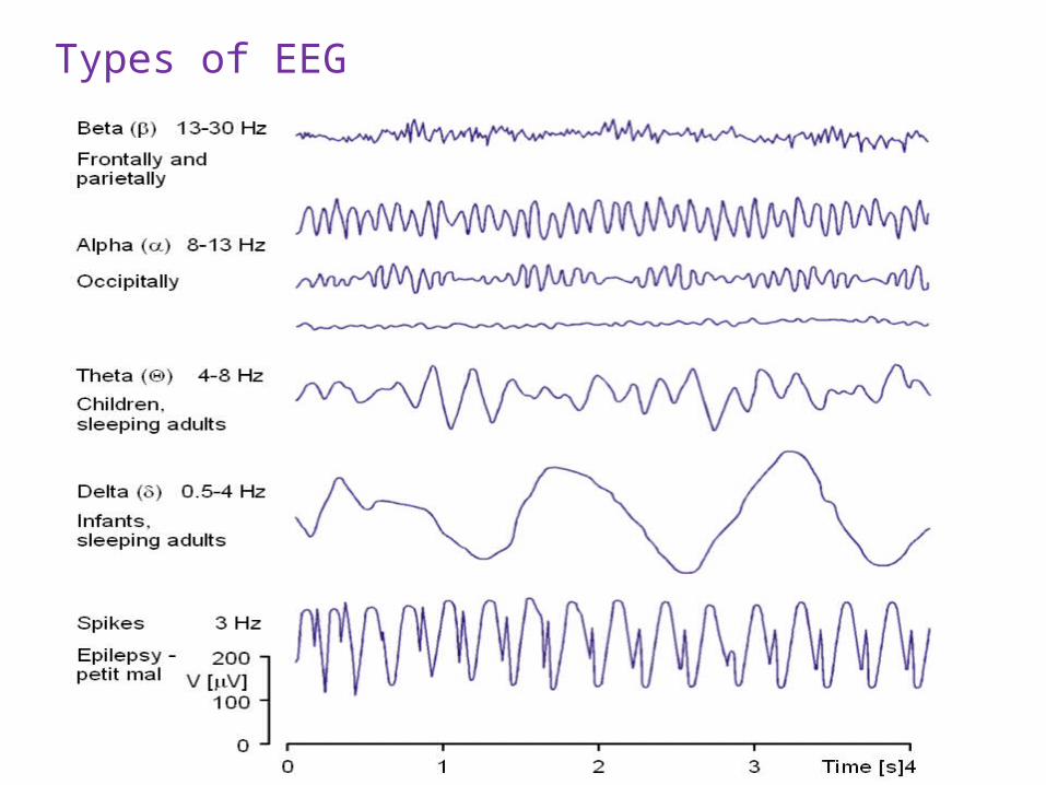
Types of EEG signals
EEG signals have been classified into 4 categories:
– Delta 0.3 to 4 Hz
• Dreamless sleep
– Theta 4 to 8 Hz
• Associated with thoughts which produce dreams
– Alpha 8 to 13 Hz
• Result of unfocused thoughts
– Beta above 13 Hz
• Result of interactions with environment
65
Types of EEG
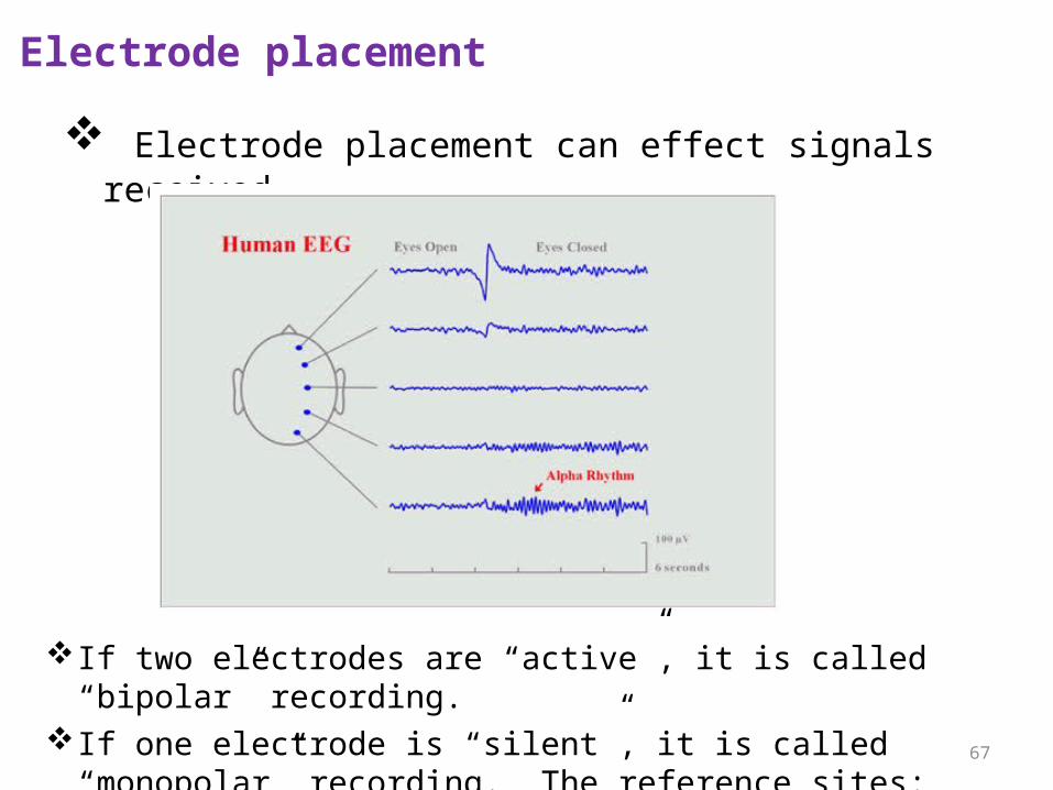
Electrode placement
Electrode placement can effect signals received
67
If two electrodes are “active”, it is called “bipolar” recording.If one electrode is “silent”, it is called “monopolar” recording. The
reference sites: ear lobe, mastoid, nose.
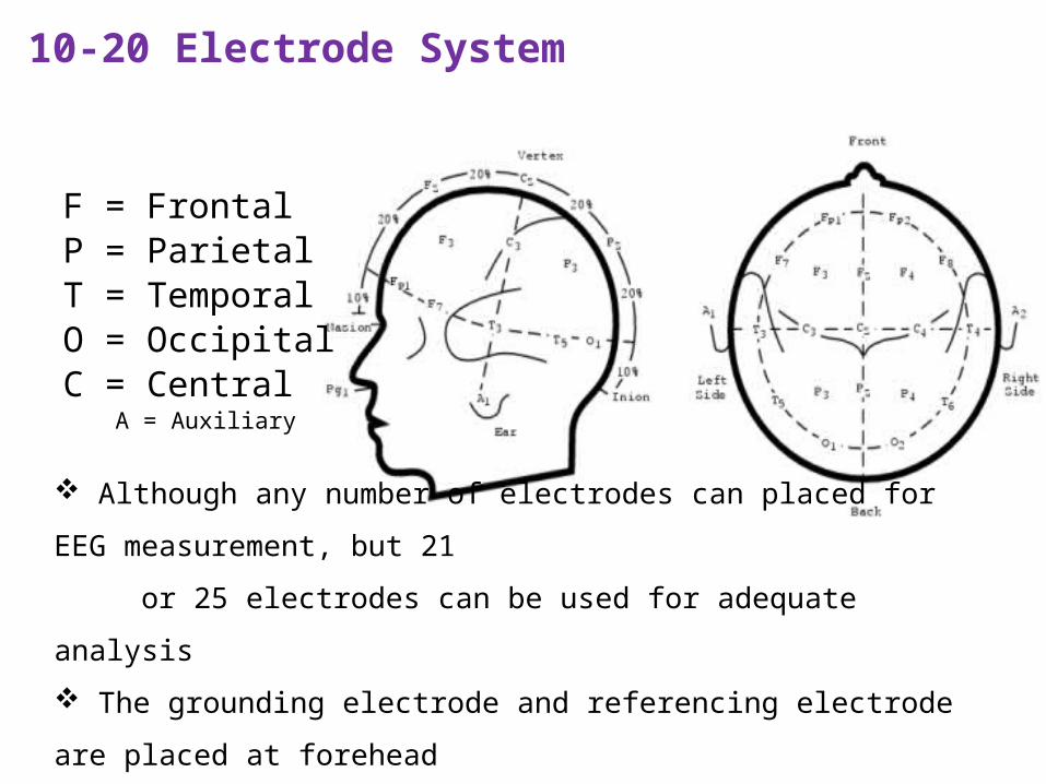
10-20 Electrode System
F = FrontalP = ParietalT = TemporalO = OccipitalC = Central
A = Auxiliary
Although any number of electrodes can placed for EEG measurement, but 21
or 25 electrodes can be used for adequate analysis
The grounding electrode and referencing electrode are placed at forehead
and right ear lobe respectively.
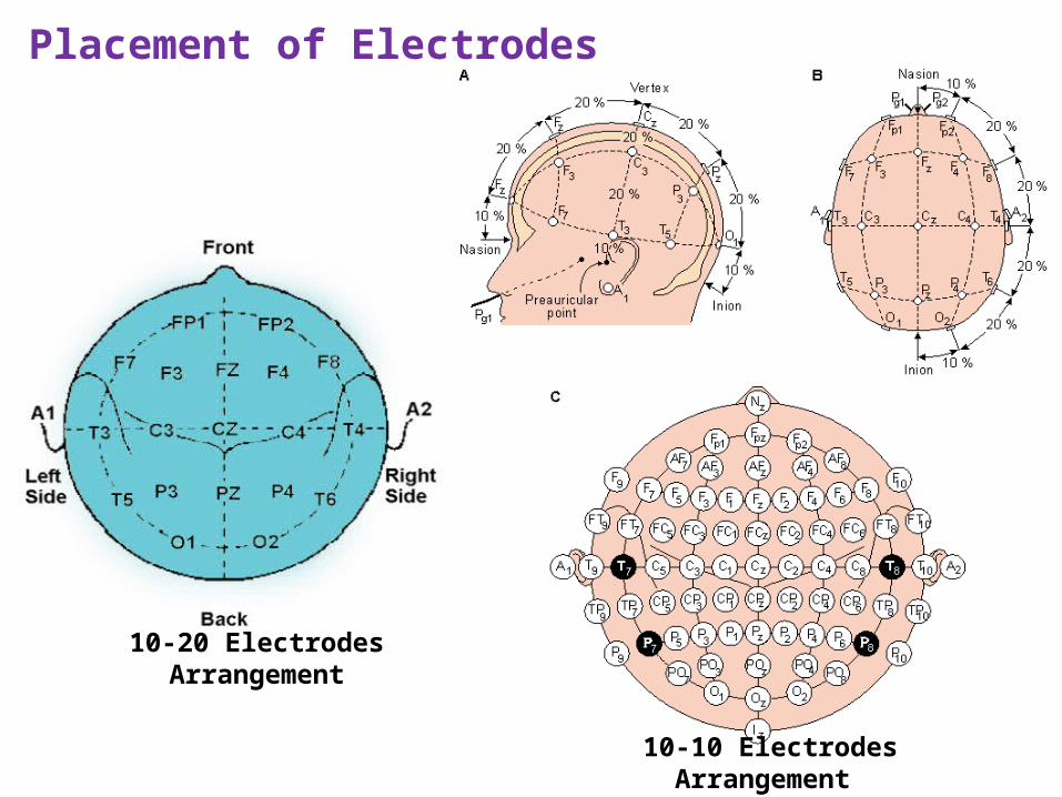
Placement of Electrodes
10-10 Electrodes Arrangement
10-20 Electrodes Arrangement
Cont….
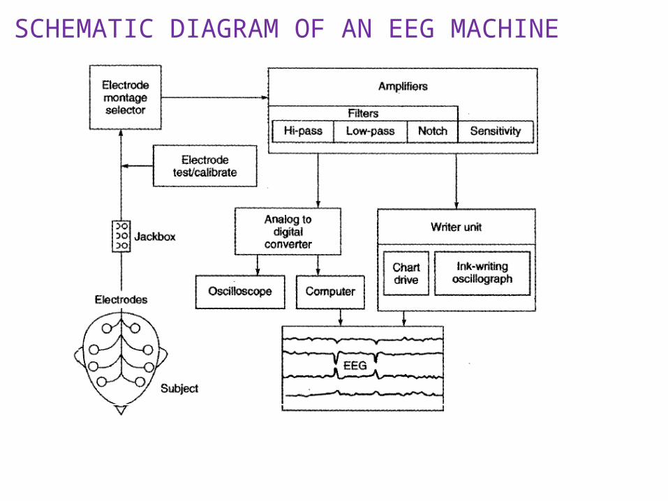
SCHEMATIC DIAGRAM OF AN EEG MACHINE
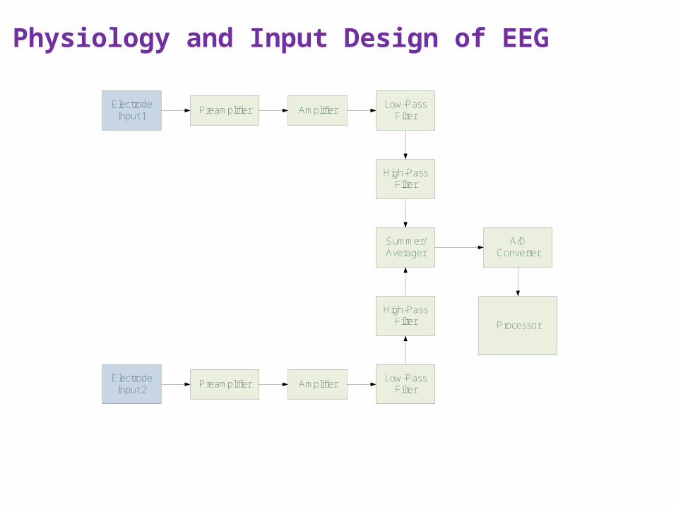
Physiology and Input Design of EEG
Electrode Input 1
Low-PassFilter
High-PassFilter
Preamplifier Amplifier
Summer/Averager
A/DConverter
Processor
Electrode Input 2
Preamplifier AmplifierLow-Pass
Filter
High-PassFilter
16
Cont…….
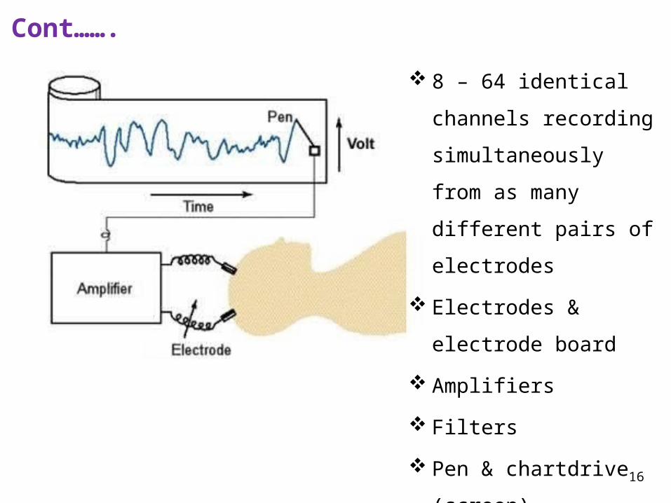
8 – 64 identical channels
recording simultaneously
from as many different
pairs of electrodes
Electrodes & electrode
board
Amplifiers
Filters
Pen & chartdrive (screen)
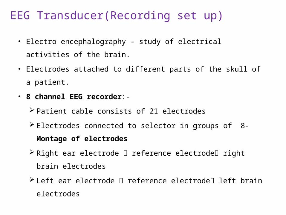
EEG Transducer(Recording set up)
• Electro encephalography - study of electrical activities of the brain.
• Electrodes attached to different parts of the skull of a patient.
• 8 channel EEG recorder:-
Patient cable consists of 21 electrodes
Electrodes connected to selector in groups of 8- Montage of
electrodes
Right ear electrode reference electrode right brain
electrodes
Left ear electrode reference electrode left brain electrodes
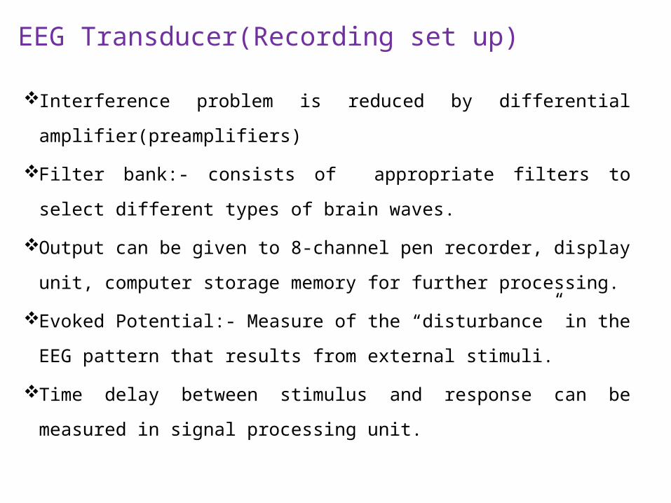
EEG Transducer(Recording set up)
Interference problem is reduced by differential
amplifier(preamplifiers)
Filter bank:- consists of appropriate filters to select different types of
brain waves.
Output can be given to 8-channel pen recorder, display unit, computer
storage memory for further processing.
Evoked Potential:- Measure of the “disturbance” in the EEG pattern
that results from external stimuli.
Time delay between stimulus and response can be measured in signal
processing unit.
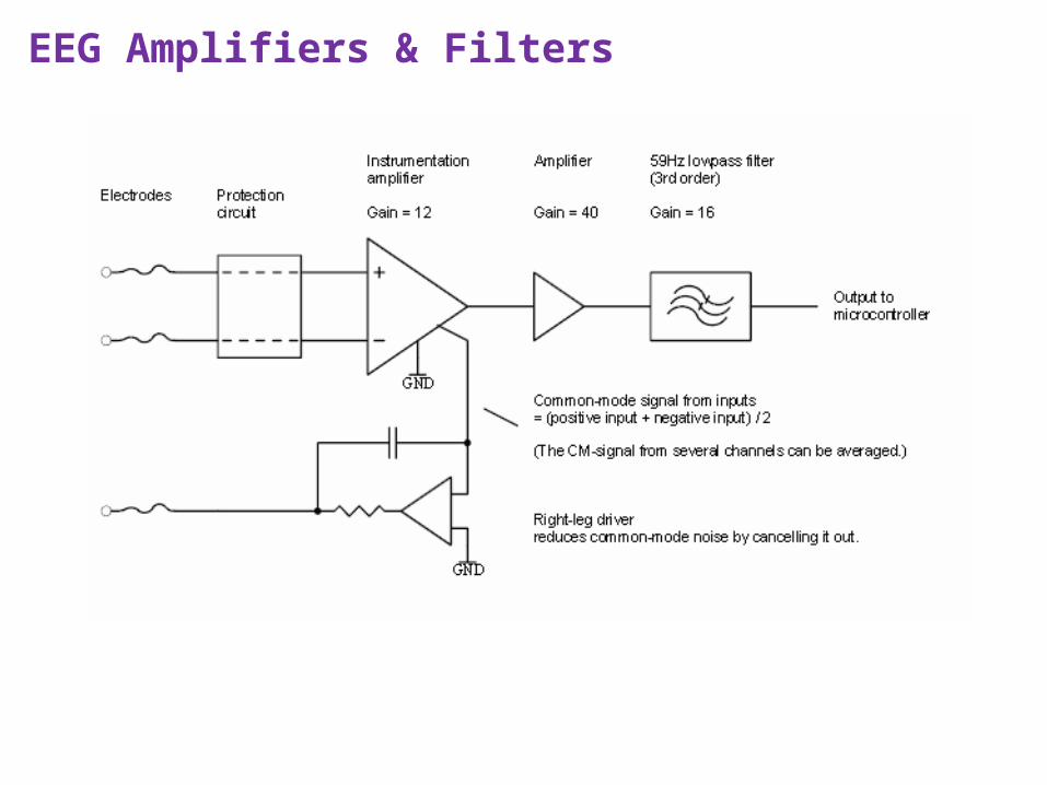
EEG Amplifiers & Filters
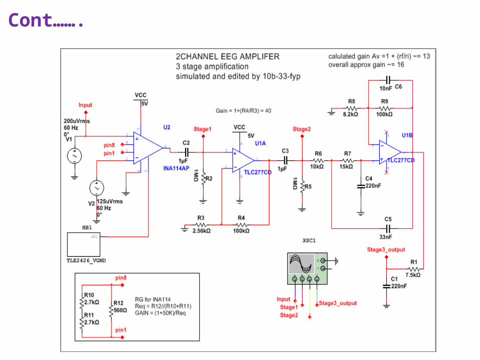
Cont…….
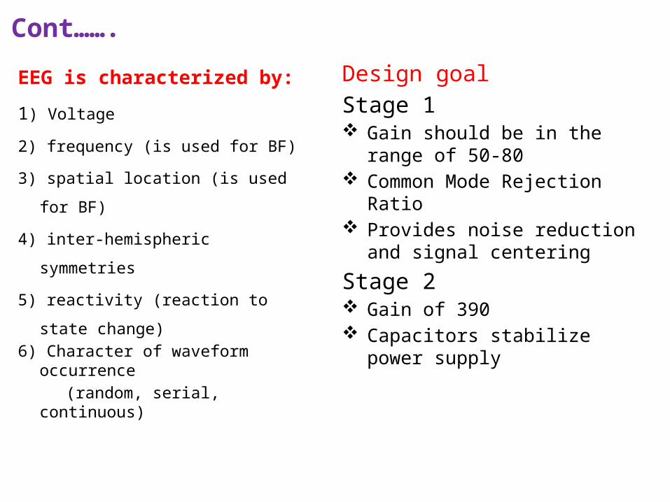
EEG is characterized by:
1) Voltage
2) frequency (is used for BF)
3) spatial location (is used for BF)
4) inter-hemispheric symmetries
5) reactivity (reaction to state change) 6) Character of waveform occurrence (random, serial, continuous)
Cont…….
Design goal Stage 1 Gain should be in the range of 50-80 Common Mode Rejection Ratio Provides noise reduction and signal
centering
Stage 2 Gain of 390 Capacitors stabilize power supply
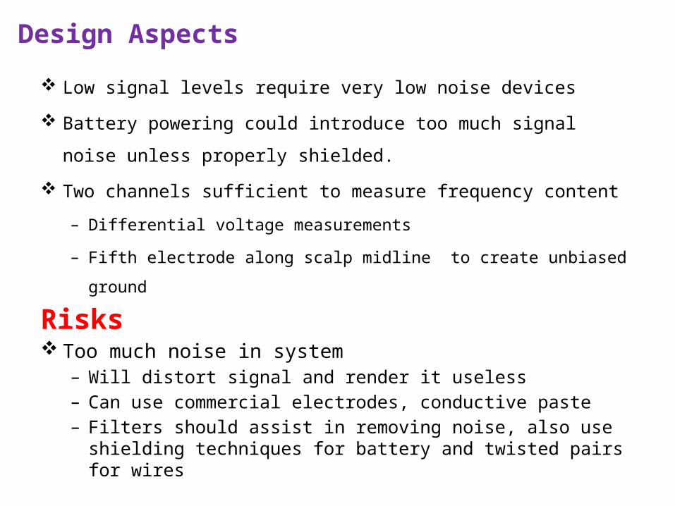
Design Aspects
Low signal levels require very low noise devices
Battery powering could introduce too much signal noise unless
properly shielded.
Two channels sufficient to measure frequency content
– Differential voltage measurements
– Fifth electrode along scalp midline to create unbiased ground
Risks Too much noise in system
– Will distort signal and render it useless– Can use commercial electrodes, conductive paste– Filters should assist in removing noise, also use shielding
techniques for battery and twisted pairs for wires
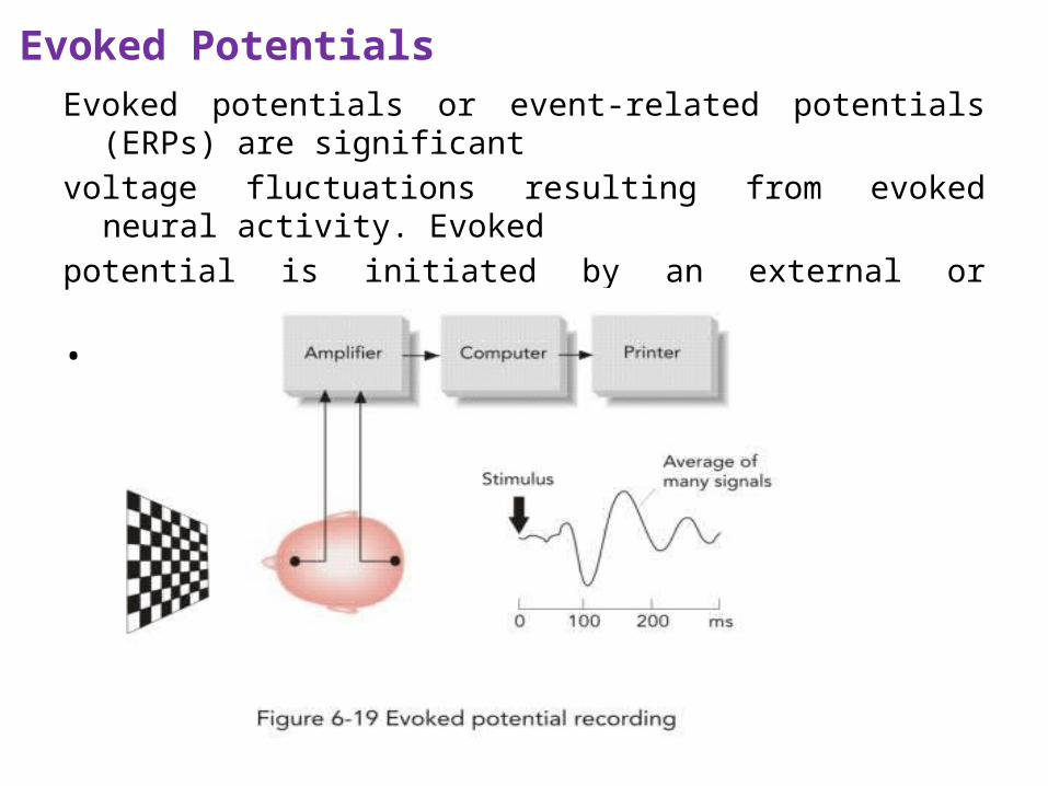
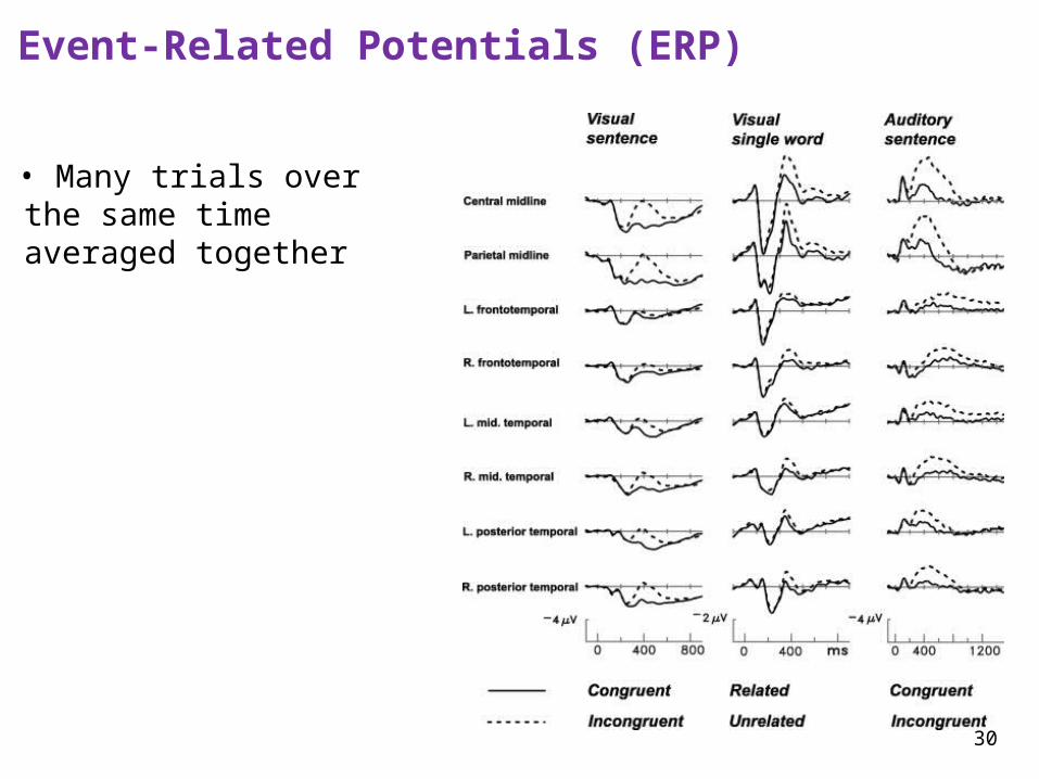
Evoked PotentialsEvoked potentials or event-related potentials (ERPs) are significantvoltage fluctuations resulting from evoked neural activity. Evoked potential is initiated by an external or internal stimulus • Visual
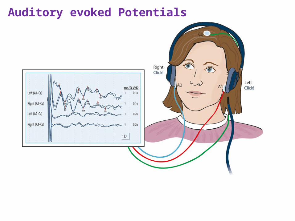
Auditory evoked Potentials
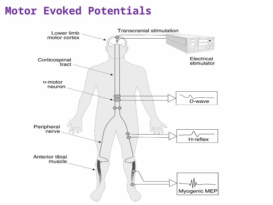
Motor Evoked Potentials
30
Event-Related Potentials (ERP)
• Many trials over the same time averaged together
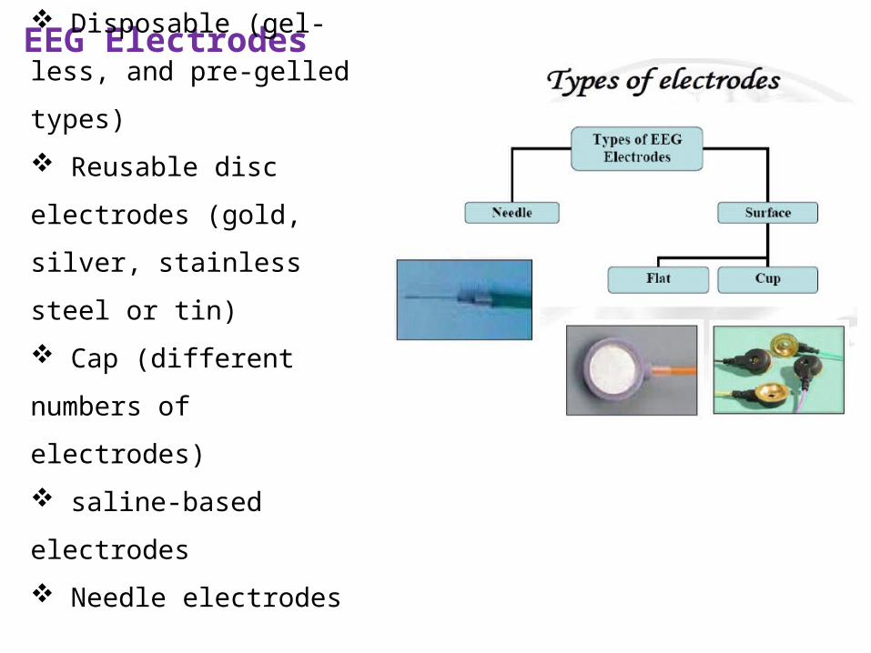
EEG Electrodes
Disposable (gel-less, and
pre-gelled types)
Reusable disc electrodes
(gold, silver, stainless steel or
tin)
Cap (different numbers of
electrodes)
saline-based electrodes
Needle electrodes
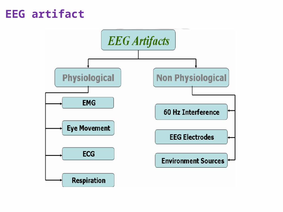
EEG artifact
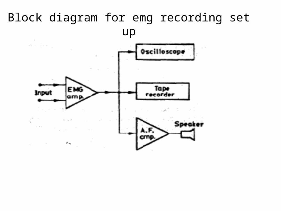
Block diagram for emg recording set up
Block diagram for emg recording set up
Electro myography :- study and interpreting of muscle action
potential.
Potentials measured by placing surface electrodes on the skin.
Individual cell potential measured by means of needle electrode
EMG appears like random noise waveform.
Contraction of muscle fibers produce action potentials
Block diagram for emg recording set up
Amplitude of EMG signals depends
• Type & placement
• Degree of muscular exertions
Normal frequency of EMG signals is 60 Hz
EMG signal amplitude ranges from 0.1 to 0.5 mV.
Amplifier with high CMRR and input impedance
Output can be given to oscilloscope, tape recorder or AF amplifier.
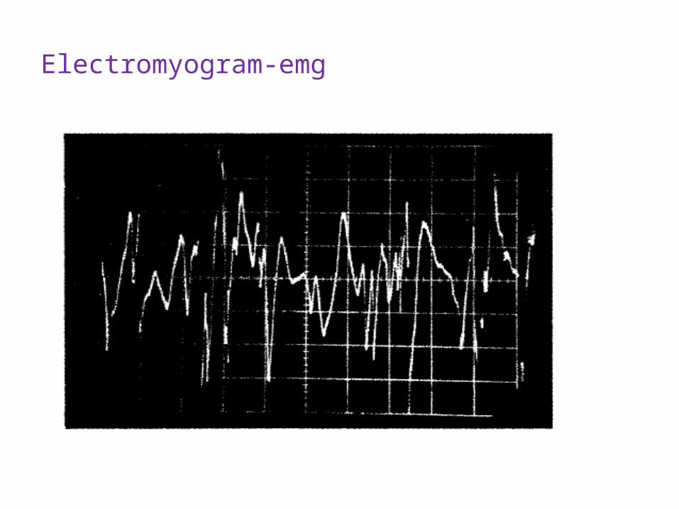
Electromyogram-emg
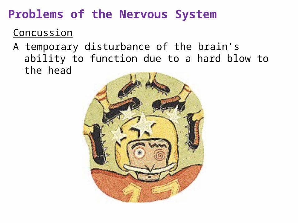
Problems of the Nervous System
ConcussionA temporary disturbance of the brain’s ability to function due to a
hard blow to the head
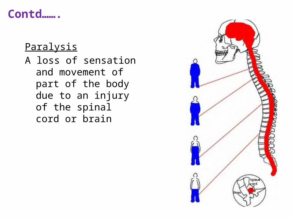
ParalysisA loss of sensation and
movement of part of the body due to an injury of the spinal cord or brain
Contd…….
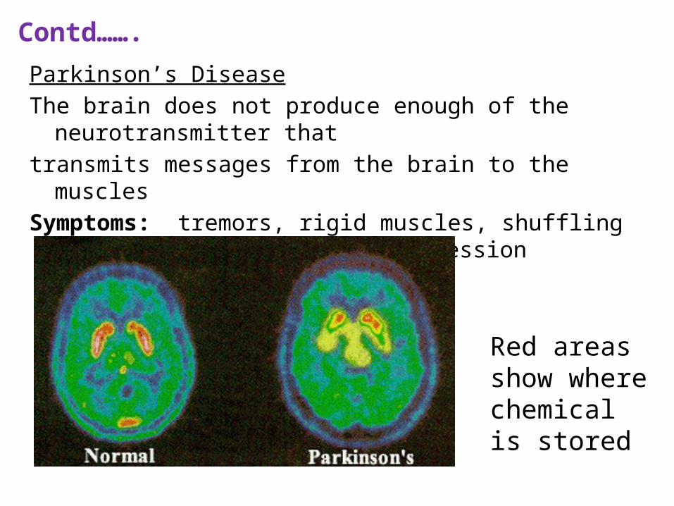
Parkinson’s DiseaseThe brain does not produce enough of the neurotransmitter that transmits messages from the brain to the musclesSymptoms: tremors, rigid muscles, shuffling walk, and loss of facial
expression
Red areas show where chemical is stored
Contd…….
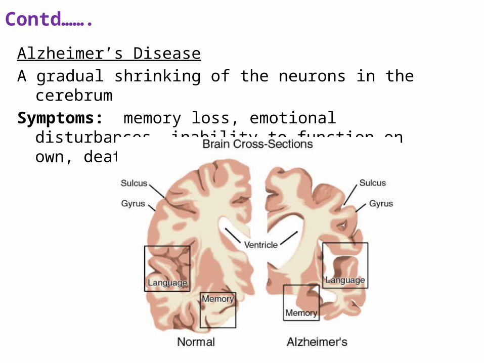
Alzheimer’s DiseaseA gradual shrinking of the neurons in the cerebrumSymptoms: memory loss, emotional disturbances, inability to
function on own, death
Contd…….
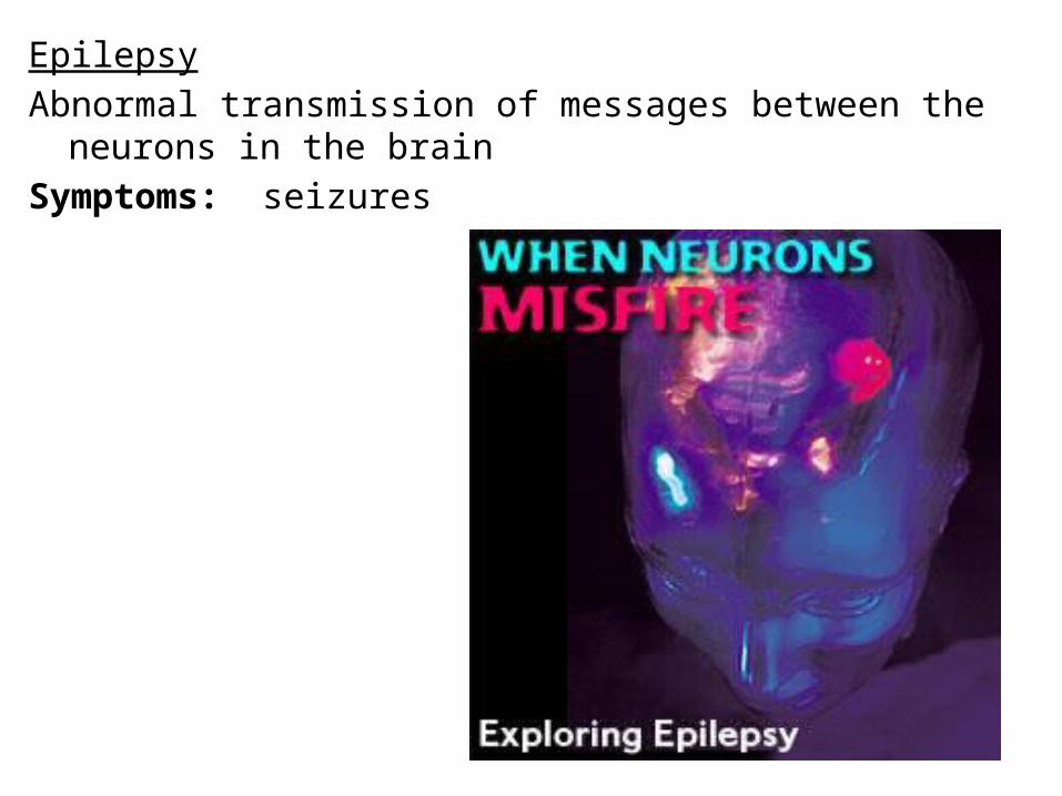
EpilepsyAbnormal transmission of messages between the neurons in the brain Symptoms: seizures
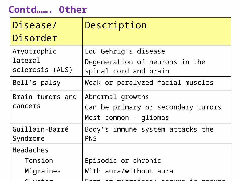
Contd……. OtherDisease/Disorder Description
Amyotrophic lateral sclerosis (ALS)
Lou Gehrig’s diseaseDegeneration of neurons in the spinal cord and brain
Bell’s palsy Weak or paralyzed facial muscles
Brain tumors and cancers
Abnormal growths Can be primary or secondary tumorsMost common – gliomas
Guillain-Barré Syndrome
Body’s immune system attacks the PNS
HeadachesTension MigrainesCluster
Episodic or chronicWith aura/without auraForm of migraines; occurs in groups
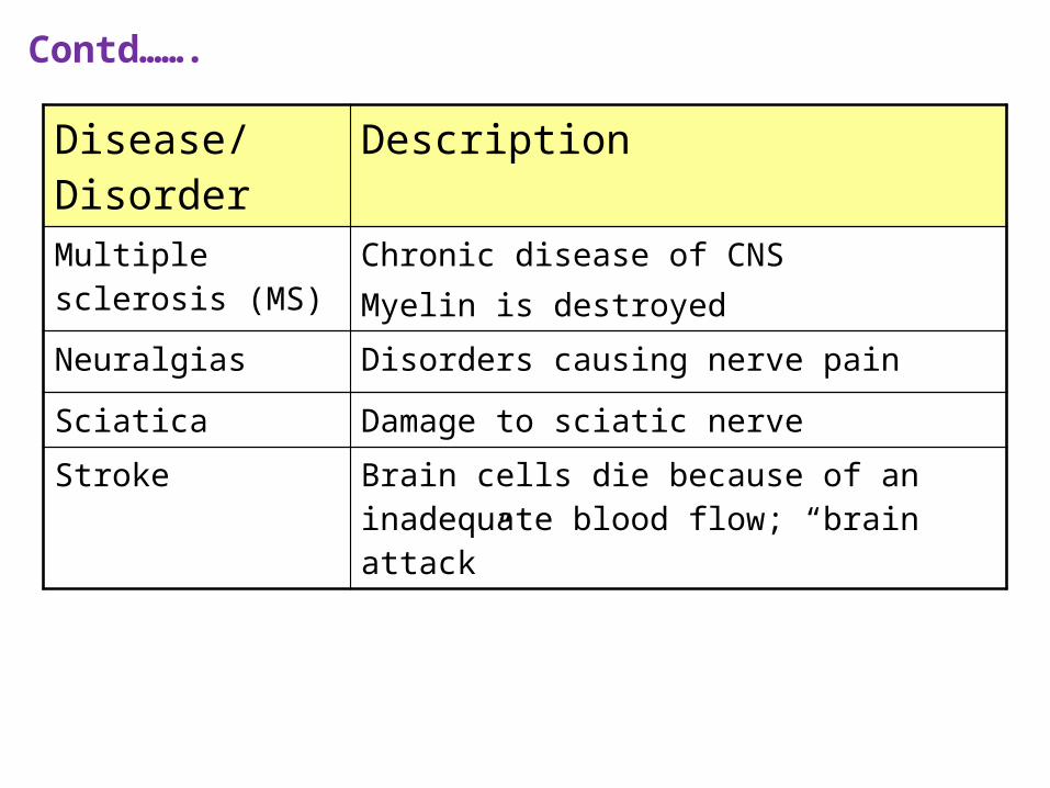
Disease/Disorder Description
Multiple sclerosis (MS)
Chronic disease of CNSMyelin is destroyed
Neuralgias Disorders causing nerve pain
Sciatica Damage to sciatic nerve
Stroke Brain cells die because of an inadequate blood flow; “brain attack”
Contd…….
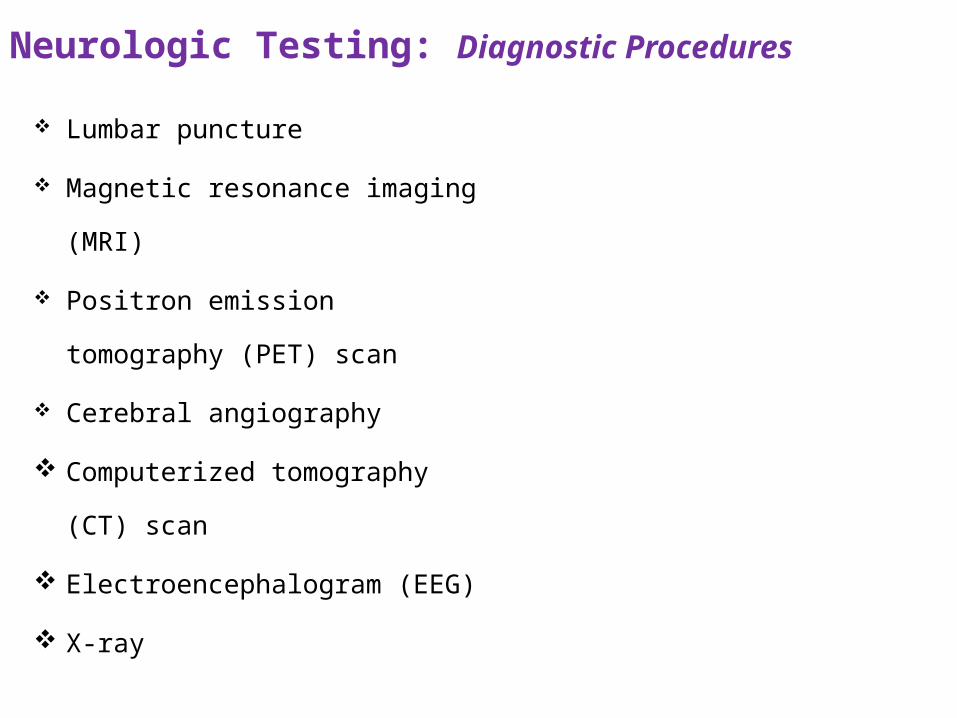
Neurologic Testing: Diagnostic Procedures
Lumbar puncture
Magnetic resonance imaging
(MRI)
Positron emission tomography
(PET) scan
Cerebral angiography
Computerized tomography (CT)
scan
Electroencephalogram (EEG)
X-ray
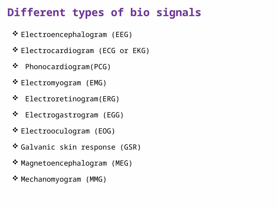
Different types of bio signals
Electroencephalogram (EEG)
Electrocardiogram (ECG or EKG)
Phonocardiogram(PCG)
Electromyogram (EMG)
Electroretinogram(ERG)
Electrogastrogram (EGG)
Electrooculogram (EOG)
Galvanic skin response (GSR)
Magnetoencephalogram (MEG)
Mechanomyogram (MMG)

University Model Questions: Q1.


