23
Unit IV: Coordination Reflex Arc Chapter 11 – pp 363-371 Chapter 12 – pp 413-421
| Date post: | 03-Jan-2016 |
| Category: |
Documents |
| Upload: | delphia-richards |
| View: | 219 times |
| Download: | 4 times |
Unit IV: CoordinationReflex Arc
Chapter 11 – pp 363-371
Chapter 12 – pp 413-421
Internal Coordination
• Endocrine and nervous system maintain internal coordination
– endocrine =
– nervous =
• Reflex Arc:
1. sense organs receive information
2. brain and spinal cord determine responses
3. brain and spinal cord issue commands to glands and muscles
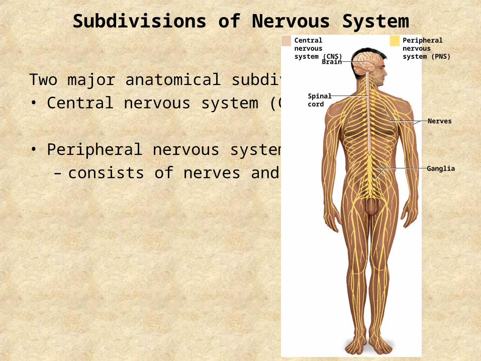
Subdivisions of Nervous System
Two major anatomical subdivisions
• Central nervous system (CNS)
• Peripheral nervous system (PNS)
– consists of nerves and ganglia
Brain
Nerves
Ganglia
Peripheral nervoussystem (PNS)
Central nervoussystem (CNS)
Spinalcord
Neural Tissue
A nerve is a bundle of nerve fibers (axons) wrapped in fibrous connective tissue.
• emerge from the CNS to carry signals between organs
• Neurons and neuroglial cells
Functions of Neurons
1. Excitability (irritability)
– stimuli
2. Conductivity
– produce traveling electrical signals
3. Secretion
– at end of nerve fiber, a neurotransmitter is secreted
Structure of a Neuron
• Soma (cell body)
• Dendrites
• Axon (nerve fiber)− trigger zone
• Myelin Sheath– some nerve fibers– insulating layer (mostly lipid) – formed by neuroglia

A Representative Neuron
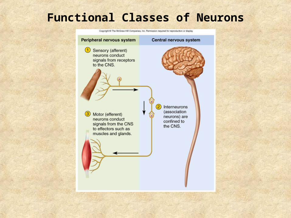
Functional Classes of Neurons

Cell body
Dendrites
Dendriticprocess
Axon
Synapticterminals
DendritesInitialsegment
Axon
Axon
Synapticterminals
Dendrites
Axon
Synapticterminals
Structural Classes of Neurons
• Multipolar neuron
• Bipolar neuron
• Unipolar neuron
– Peripheral fiber carries impulses from source of sensation
– Central fiber carriers impulses to spinal cord
• Anaxonic neuron
Neuroglial Cells
• Also known as Glial cells
• Outnumber neurons
• General functions:
− Protect
− Support, maintain physical structure of neural tissue
− Repair
− Maintain nutrient supply to neurons

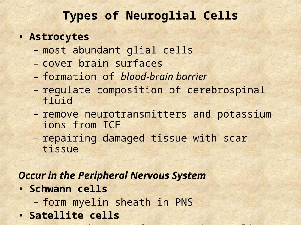
Types of Neuroglial Cells
Occur in the Central Nervous System
• Oligodendrocytes
– form myelin sheaths in CNS
• Ependymal cells
– line cavities of brain and spinal cord
– produce and circulate CSF
• Microglia
– macrophages (WBC)
– Phagocytosis
– in areas of infection, trauma or stroke
Types of Neuroglial Cells
• Astrocytes– most abundant glial cells– cover brain surfaces– formation of blood-brain barrier– regulate composition of cerebrospinal fluid – remove neurotransmitters and potassium ions from ICF– repairing damaged tissue with scar tissue
Occur in the Peripheral Nervous System• Schwann cells
– form myelin sheath in PNS• Satellite cells
– surround somas of neurons in ganglia

Neuroglial Cells of CNS
Section ofspinal cord
Ependymal cell
Microglial cell
Neurons
Gray matter White matter
Myelinatedaxons
Astrocytes
OligodendrocyteCapillary
Axon
Myelin(cut)
Nodes
Unmyelinated axon
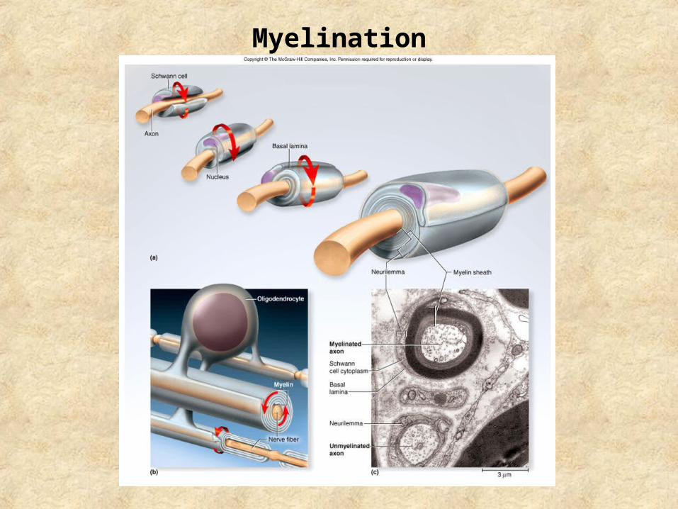
Myelination

DemyelinationHallmark of some neurodegenerative autoimmune diseases:
• Multiple Sclerosis– Demyelination in CNS– Own immune system attacks and damages myelin– Scars form in white matter of CNS– Cause unknown, no cure
• Cerebral Palsy– Damage to developing oligodendrocytes usually during infancy– Mutations, lack of oxygen, interruption of blood flow– Treatment of symptoms, no cure
• Leukodystrophies– Results from defect in the gene that controls the production of only
one component molecule of myelin– Affects growth and maintenance of white matter– Inherited, no cure
Nerve Signal
Depends on two factors:
• Presence/absence of myelin
• Diameter of fiber
- large/thicker fibers have more surface area for signals
• Functions
– fast signals employed when speedy responses are needed
– slow signals used when quick responses are not important

Axon MyelinStep 1: the axon and myelin degenerate andfragment.
Step 2: The Schwanncells proliferate and macrophages remove the debris distal to the injury site.
Macrophage
Regeneration Tube
Step 3: The axon grows along the path created by theSchwann cells.
Step 4: As the axon elongates, the Schwanncells wrap around it.
Site of injury
Regeneration of Nerve Fiber in PNS

Reflexes
Properties:
– Require stimulation
– Quick
– Involuntary
– Stereotyped
Reflex Arc:
Somatic receptors → afferent nerves → integrating center →
efferent nerves → skeletal muscles

The Stretch Reflex
• When a muscle is stretched, it contracts and maintains increased muscle tone (stretch reflex)– helps maintain equilibrium and posture– balances tension in extensors and flexors– mediated by the brain
• Reciprocal inhibition prevents muscles from working against each other
• Very sudden muscle stretch causes tendon reflex– knee-jerk (patellar) reflex is monosynaptic reflex
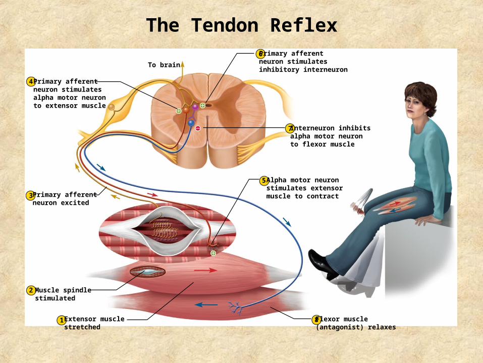
The Tendon Reflex
To brain
4
3
5
8
7
6
2
1
Primary afferentneuron stimulates alpha motor neuron to extensor muscle
Primary afferentneuron excited
Muscle spindlestimulated
Extensor musclestretched
Flexor muscle(antagonist) relaxes
Alpha motor neuronstimulates extensormuscle to contract
Interneuron inhibitsalpha motor neuron to flexor muscle
Primary afferentneuron stimulatesinhibitory interneuron
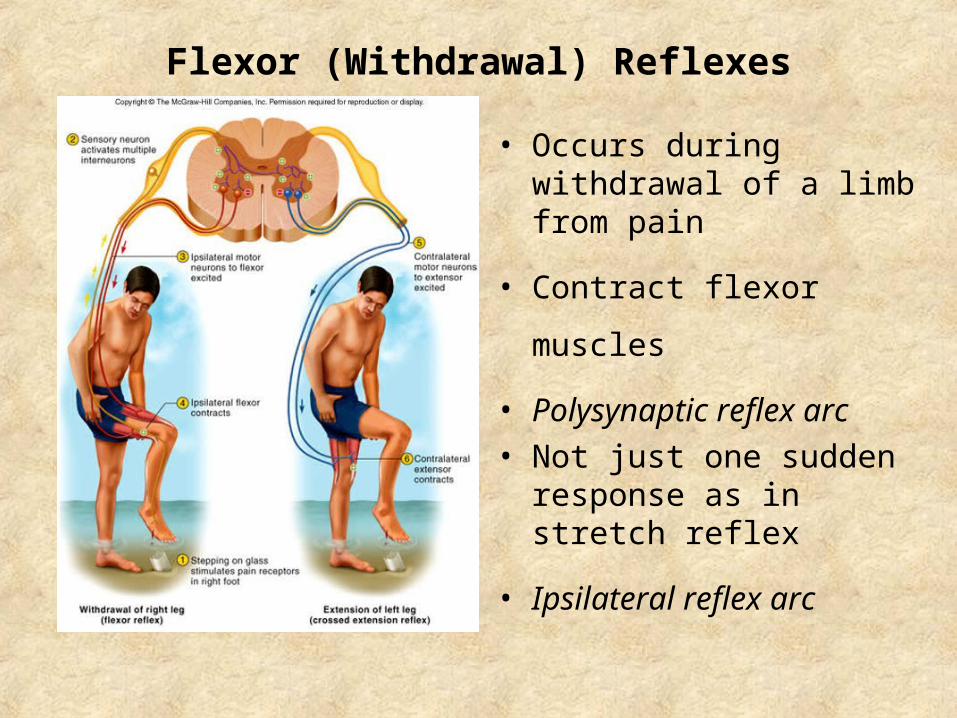
Flexor (Withdrawal) Reflexes
• Occurs during withdrawal of a limb from pain
• Contract flexor muscles
• Polysynaptic reflex arc
• Not just one sudden response as in stretch reflex
• Ipsilateral reflex arc
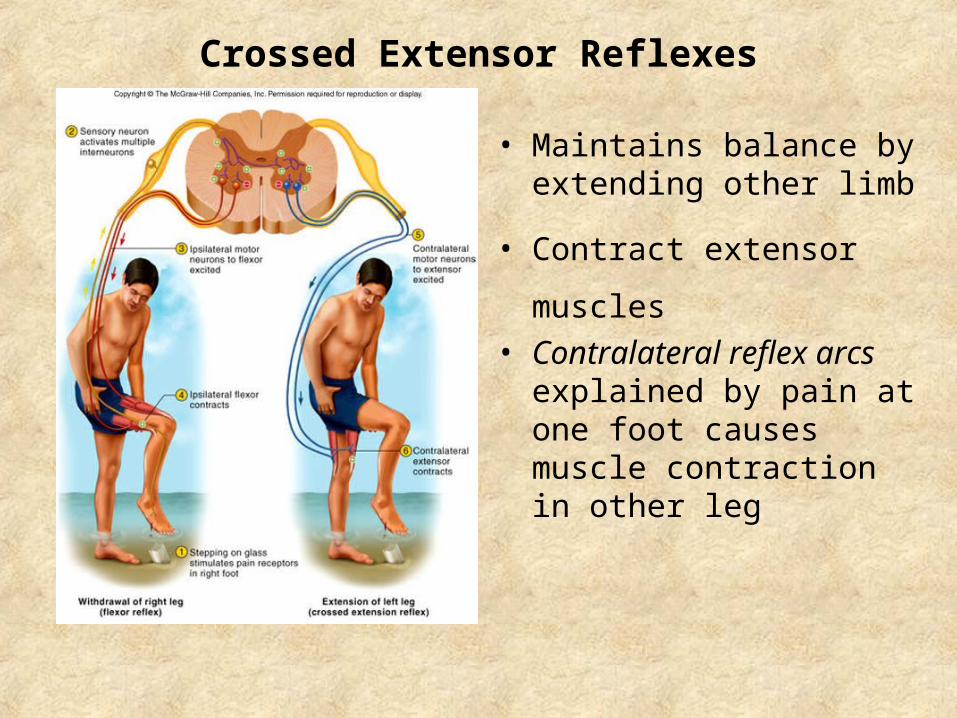
Crossed Extensor Reflexes
• Maintains balance by extending other limb
• Contract extensor muscles
• Contralateral reflex arcs explained by pain at one foot causes muscle contraction in other leg
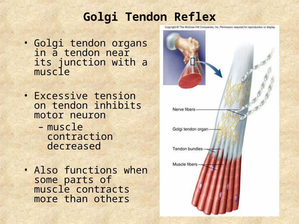
Golgi Tendon Reflex
• Golgi tendon organs in a tendon near its junction with a muscle
• Excessive tension on tendon inhibits motor neuron– muscle contraction
decreased
• Also functions when some parts of muscle contracts more than others


