UNIVERSIDADE DE LISBOA
FACULDADE DE MEDICINA DE LISBOA
Synthetic Pathogens for Integrated Biophysical and
Genetic Dissection of Antigen Cross-Presentation
RUI PEDRO DA SILVA ALBUQUERQUE E FREITAS
Doutoramento em Ciências Biomédicas
Especialidade em Ciências Morfológicas
2010
UNIVERSIDADE DE LISBOA
FACULDADE DE MEDICINA DE LISBOA
Synthetic Pathogens for Integrated Biophysical and
Genetic Dissection of Antigen Cross-Presentation
RUI PEDRO DA SILVA ALBUQUERQUE E FREITAS
Tese orientada por:
Professor Doutor Luís Filipe Ferreira Moita
Professor Doutor Darrell J. Irvine
Doutoramento em Ciências Biomédicas
Especialidade em Ciências Morfológicas
2010
Todas as afirmações efectuadas no presente documento são da exclusiva
responsabilidade do seu autor, não cabendo qualquer responsabilidade
à Faculdade de Medicina de Lisboa pelos conteúdos nele apresentados.
A impressão desta dissertação foi aprovada pela Comissão
Coordenadora do Conselho Científico da Faculdade de
Medicina de Lisboa em reunião de 27 de Janeiro de 2010
O desenvolvimento e execução gráfica da presente dissertação foram
financiados pela Fundação para a Ciência e Tecnologia
(Bolsa SFRH/BD/14316/2003)
“Para ser grande, sê inteiro: nada
Teu exagera ou exclui.
Sê todo em cada coisa. Põe quanto és
No mínimo que fazes.
Assim em cada lago a lua toda
Brilha, porque alta vive”
Fernando Pessoa
Preface
iii
One of the most important and difficult steps in a scientist short life is to decide
start writing the PhD thesis, but even before, to be brave or naive enough to start a
scientific career and to run the unknown and tricky road until the end. It is a kind
of matrimonial relation, where almost all scientists begin to love science, adore and
enjoy the idea of discover something new and interesting with the propose of
finding a solution or eliminating something that was not so bright. Therefore,
everything starts with conceptual dreamers without frontiers.
This thesis describes the work carried between January 2006 and July 2009
mainly at the Instituto de Medicina Molecular (Lisbon, Portugal). During this
period, part of the research was done at Massachusetts General Hospital (Boston,
USA), at MIT (Cambridge, USA) and at the Institute Currie (Paris, France). The
main goal was to study how the biochemical and biophysical properties of specific
particulate antigens influence the cross-presentation pathway(s) and try to dissect
and indentify the mechanism(s) behind it.
This thesis was divided in 6 Chapters:
The introduction comprises a general overview of specific key immunology
concepts, such as Innate Immunity, Dendritic Cell biology, and antigen
presentation mechanism with emphasis on antigen cross-presentation.
The second chapter focuses on particulate antigen design and the goal of
specific properties introduced in the particles; the shRNA lentiviral library
production and its application in high-throughput approaches. It includes a
summary description of my participation in the work done within this period and
the resulting publications.
The third chapter is composed by the materials and methods used throughout
my work, including the different particulate platforms design and biochemical and
cellular techniques for antigen presentation studies.
Results of my main project are described on Chapter 4, where different
platforms of particulate antigen were used to study antigen cross-presentation
mechanism(s).
Discussion is presented on Chapter 5 and concluding remarks on Chapter 6.
The results presented in this thesis, in collaboration with Darrell Irvine’s lab at
MIT, are under preparation for publication.
vii
The study of host-pathogen interactions is crucial to unveil the diversity of the
immune response outcome. Dendritic Cells (DCs) play a central role in the
initiation and regulation of T-Cell immunity, functioning as master switches that
control whether the outcome of antigen presentation results in tolerance, or
immunity. Antigen cross-presentation is a necessary mechanism to generate
immunity against tumors, bacteria and viruses. In addition, it is extremely
important to induce cytotoxic immunity by vaccination with antigens. Moreover,
particulate antigens have been used in vaccine design tools as a platform to deliver
different types of signals and in the modulation of DC-dependent immune
responses. DCs express a series of different receptors that mediate the transfer of
signals from the environment. Among them, Toll-Like Receptors (TLRs) play a
critical role in the early innate immune response to invading pathogens. These
receptors have the ability to recognize a broad range of pathogen-associated
molecular patterns (PAMPs), turning them, key receptors in distinguishing
between self/non-self antigens. The precise mechanisms underlying the crosstalk
between TLRs and antigen presentation are not entirely understood. Therefore, the
main goal of this project is to understand how TLR agonists coupled to particulate
antigens influence antigen cross-presentation.
In our studies, we have used newly synthesized particle antigens, denominated
as 'synthetic pathogens', coupled with a model antigen (Ovalbumin - OVA), and/or
a model ligand (TLR agonist). These particle platforms have distinct, well-defined
physical and biochemical properties, and function as a novel approach to elucidate
the intrinsic mechanism(s) of antigen cross-presentation. In addition, they represent
a valuable and powerful tool, which might be explored for therapy applications.
ABSTRACT
TLR4 is unique among TLRs as it can signal through both MyD88 and TRIF
adaptors upon LPS stimulation, but mainly by the TRIF pathway when LipidA is
the agonist. Our results revealed that when LPS is in the same cargo as the particle
antigen, it impairs antigen cross-presentation and dictates a shift to MHC class- II
presentation. This antigen cross-presentation abolishment is recovered on TLR4
deficient DCs and in the presence of the p38 MAPK pathway inhibitor, but not in
the absence of the MyD88 adaptor. Moreover, LipidA reproduces the same
phenotype as LPS, implicating the TLR4/TRIF-mediated signaling on particulate
antigen cross-presentation impairment. Thus, here we describe a new mechanism
of antigen selection in DCs for antigen cross-presentation that is dependent on the
antigen based-environment. We show that the efficiency of presenting antigens
from phagocytosed cargo is dependent on the presence of TLR ligands within the
cargo. The influence of the compartmentalization on the crosstalk between the
TLR-signaling and the antigen cross-presentation pathway(s), may constitute a tool
used by DCs in order to discriminate the contents of phagosomes and present an
appropriate immune response to specific stimuli. Therefore, DCs may have the
“capacity” to decide which kind of destiny an antigen should have depending on
the type and origin of the stimuli.
In order to dissect the mechanisms behind the cross-presentation phenotype, we
have addressed the role of particle LPS on several antigen presentation key steps.
Our data show that LPS-containing phagosomes enhance phagosome maturation
(higher levels of colocalization with lysosomes) characterized by higher rates of
phagosomal acidification and a decrease of phagosomal reactive oxygen species
(ROS) production. The induction of phagosome maturation mediated by LPS
signaling seems to shut down the machinery for antigen release into the cytosol,
where the epitopes for MHC class-I are predominantly generated by the
proteasome. Moreover, lower levels of antigen degradation occur when LPS is in
ix
the same cargo as antigen, mainly in a proteasome-dependent manner. This
phenotype mediated by particulate LPS stimulus seems to be related with lower
levels of particle antigen cross-presentation.
Therefore, we propose that antigen cross-presentation is enhanced during the
brief period of time when phagosomal acidification is “sustained” and an immature
phenotype is predominant, where endoplasmic reticulum machinery important for
MHC class-I presentation probably is available. In addition this phenotype allows
antigen escape into cytosol and the generation of epitopes for MHC class-I by the
proteasome. On the other hand, antigen cross-presentation is impaired when a
stimulus that induces phagosome maturation/acidification is in the same cargo as
the antigen, producing a mature phenotype, allowing the generation of epitopes on
the endocytic pathway that is compromised for MHC class-II antigen presentation.
In order to address if the abolishment on antigen cross-presentation phenotype
is transversal to others TLRs, studies were extended using different TLR specific
agonists. When particle antigen contains TLR agonists that preferentially signal
through MAPK/NF-kB pathways, antigen cross-presentation is induced. In
contrast, in the presence of TLR agonists that preferentially signals through IFN-
Type I pathway, particle antigen cross-presentation is inhibited. Therefore, a
signaling pathway correlation may exist in the outcome of antigen presentation
pathway(s) mediated by TLR agonist-containing particle antigens.
In sum, this work shows for the first time the inhibitory effect of TLR4
signaling on cross-presentation when agonists are delivered in the same cargo as
particulate antigen. This phenotype is likely to be mediated by TRIF-dependent
signaling, mainly by p38 MAPK activation. This knowledge could have a major
impact in the dissection of the antigen cross-presentation mechanism, which will
be highly valuable for novel vaccine design inducing T-Cell responses of the
desired type and specificity.
xi
O estudo das interações patogénio-hospedeiro é fundamental para a
compreensão da diversidade da resposta imunitária e para o desenvolvimento de
novas estratégias terapêuticas. As células dendríticas (DCs) desempenham um
papel central na iniciação e regulação da imunidade mediada por linfócitos T,
funcionando como “interruptores”, que podem originar uma resposta de tolerância
ou imunidade em relação a um determinado antigénio. O mecanismo de cross-
presentation de antigénios tem sido descrito como necessário para gerar imunidade
contra tumores, bactérias e vírus, e fudamental na indução de imunidade citotóxica
mediada por vacinação. Por outro lado, os antigénios particulados têm sido
utilizados como ferramentas no design de vacinas, possibilitando uma plataforma
na qual se podem integrar diferentes tipos de estímulos.
As DCs expressam uma diversidade de receptores à superficie, o que permite
uma detecção e transmissão eficazes dos vários tipos de “sinais” do meio ambiente.
Entre eles, os Toll-Like Receptors (TLRs) desempenham um papel crucial, na
resposta imune inata contra patogénios invasores. Estes receptores têm a
capacidade de reconhecer uma ampla gama de padrões moleculares associados a
patogénios (PAMPs), implicando-os como receptores-chave na distinção entre
antigénios próprios e não-próprios. O mecanismo subjacente à crosstalk entre
TLRs e a apresentação de antigénios não é totalmente conhecido. Por isso, um dos
principais objetivos do meu trabalho foi compreender como é que os agonistas dos
TLRs no mesmo contexto que antigénios particulados, influenciam a sua cross-
presentation.
Neste projecto foram utilizadas partículas sintéticas – designadas por synthetic
pathogens - na presença de um antigénio modelo (Ovalbumina) e/ou de um ligando
RESUMO
(agonista dos TLRs). Estas plataformas têm propriedades físicas e bioquímicas
distintas e bem definidas, pelo que funcionam como uma nova abordagem para
dissecar o mecanismo de cross-presentation de antigénios, bem como explorar o
seu potencial para utilizaçao nas mais diversas terapias.
Os resultados demonstram que quando o Lipopolissacarídeo (LPS - agonista do
TLR4) está presente no mesmo contexto que as partículas contendo o antigénio,
ocorre uma redução nos níveis de cross-presentation de antigénios. Este fenótipo é
acompanhado por uma mudança na via de apresentação de antigénios para MHC
classe- II, que é induzida quando comparada com as partículas só com o antigénio.
Este mecanismo foi demonstrado como sendo mediado pelo TLR4, onde a cross-
presentation de antigénios é restabelecida em DCs deficientes nesse receptor .
A origem física dos estímulos (partícula vs solúvel) parece ser crucial para a
regulação da via de cross-presentation de antigénios. Quando o LPS solúvel é co-
incubado com partículas contendo o antigénio (dois estímulos físicos diferentes),
verifica-se um aumento da activação/proliferação de células T em ambos os
contextos de apresentação de antigénios - MHC classe-I e MHC classe -II. No
entanto, quando o LPS é utilizado numa partícula diferente daquela que contém o
antigénio, não se verificam diferenças significativas na eficiência das duas vias.
A influência da compartimentação no crosstalk entre a sinalização mediada
pelos TLR e a via de cross-presentation de antigénios, pode constituir uma
ferramenta importante que as DCs utilizam para discriminar o conteúdo dos
fagossomas e iniciar uma resposta imune apropriada aos estímulos específicos.
Esta observação é de extrema importância para compreender o papel de estímulos
"patogénicos" no destino da apresentação de antigénios. Com o objectivo de
compreender o mecanismo adjacente ao fenótipo observado da via da cross-
presentation de antigénios particulados, o papel da activação do TLR4 foi estudado
em vários processos importantes na apresentação de antigénios. Os resultados
xiii
obtidos indicam que fagossomas que contêm LPS têm uma indução na maturação
(níveis mais elevados de colocalização com lisossomas), caracterizada por taxas
mais elevadas de acidificação e uma diminuição da produção de espécies reactivas
de oxigénio (ROS). A indução da maturação dos fagossomas mediada pela
sinalização por LPS parece bloquear o mecanismo de libertação do antigénio dos
fagossomas para o citosol, onde os epítopos para a apresentação em MHC classe -I
são predominantemente gerados pelo proteassoma. Para além disso, níveis mais
baixos de degradação do antigénio, mediada principalmente pelo proteassoma,
ocorrem quando o LPS está no mesmo contexto. Este fenótipo é devido à
sinalização mediada pelo LPS e parece estar relacionado com níveis baixos de
cross-presentation do antigénio particulado.
Posto isto, sugerimos que a cross-presentation de antigénios é reforçada
durante um breve período de tempo quando o pH dos fagossomas é mantido em
valores próximos do estado basal, onde um fenótipo imaturo é predominante. Este
estado imaturo é caracterizado pela existência de componentes do retículo
endoplasmático (ER) importantes para a apresentação em MHC classe-I,
permitindo o escape do antigénio para o citoplasma, onde os epítopos podem então
ser gerados pelo proteassoma e apresentados no contexto MHC classe-I à
superfície. Por outro lado, a cross-presentation de antigénios é diminuída quando
um estímulo que induz a maturação dos fagossomas que contem o antigénio está
no mesmo contexto. A formação de fagolisossomas leva à rápida acidificação e
produz um fenótipo maduro, permitindo a geração de epítopos na via endocítica
que é direccionada para a via MHC classe-II de apresentação de antigénios.
O TLR4 é singular entre os TLRs, uma vez que pode sinalizar tanto pelo
adaptador MyD88 ou pelo TRIF quando estimulado por LPS, mas
preferencialmente pelo adaptador TRIF quando LipidA é o agonista. Com o
objectivo de estudar o impacto da sinalização do TLR4 na via de cross-
presentation dos antigénios particulados, o LipidA foi utilizado no mesmo
contexto que o antigénio particulado. Observou-se uma reprodução do fenótipo de
supressão da via de cross-presentation de antigénios obtido na presença do LPS.
Além disso, a cross-presentation de antigénios particulados na presença de LPS
não foi recuperada usando DCs deficientes no adaptador MyD88, ao contrário do
que acontece quando se usa DCs deficientes no TLR4 e na presença de inibidores
de activação da via das MAPK, principalmente a p38 MAPK. Estes resultados
implicam a via TLR4/TRIF na inibição da cross-presentation de antigénios.
Com o objectivo de verificar se o efeito inibitório na via de cross-presentation
de antigénios é transversal aos outros TLRs, os estudos foram alargados usando
agonistas dos diversos TLRs no mesmo contexto que as partículas de antigénio.
Verificou-se que, os agonistas dos TLR que sinalizam preferencialmente através da
via MAPK/NF-kB induzem a cross-presentation de antigénios particulados. Em
oposição, os agonistas dos TLR que sinalizam preferencialmente através da via do
IFN Tipo-I, levam à inibição da cross-presentation destes antigénios. O TLR4
pode sinalizar através dos dois adaptadores (MyD88 e TRIF) em diferentes
localizações, sendo preferencialmente via TRIF quando o TLR4 é internalizado
nos endossomas. A inibição da via de cross-preserntation de antigénios mediada
pelo LPS quando no mesmo contexto que o antigénio particulado, poderia indicar
que o LPS em partículas sinaliza preferencialmente através da via TRIF, quando
estas são internalizadas. Esta observação corrobora os dados obtidos com os outros
agonistas de TLR que preferencialmente sinalizam através da via do IFN Tipo-I,
como é o caso do TLR3, do TLR7 e do TLR9, que estão localizados em
endossomas. Conclui-se assim que os vários TLRs estão envolvidos em
mecanismos diferentes que levam a efeitos distintos nas vias de apresentação de
antigénios. Além disso, um padrão da via de cross-presentation de antigénios
xv
parece existir mediado por partículas contendo os vários agonistas dos diferentes
TLRs.
Em colaboração com o grupo do Prof. Darrell Irvine do MIT, pretendemos
alargar estes estudos para outras plataformas de antigénios. Partículas de poli(ácido
lático-co-ácido glicólico), PLGA, e hidrogel têm sido usadas como plataforma para
administrar drogas, assim como em aplicações de biomateriais e concepção de
vacinas. De acordo com os dados obtidos para a plataforma “fixa” de antigénios
(partículas utilizadas nos ensaios anteriores), o LipidA no mesmo contexto que o
antigénio particulado inviabiliza a via de cross-presentation de antigénios,
comprovando a sua acção como um inibidor de sinalização mediada pelo TLR4,
por um mecanismo dependente do adaptador TRIF. Assim, podemos concluir que
mesmo na presença de partículas com propriedades distintas, a via mediada pelo
adaptador TRIF tem um papel importante na inibição da via de cross-presentation
de antigénios quando estimulada pelos agonistas do TLR4.
Este trabalho mostrou pela primeira vez, o efeito inibitório da crosstalk entre a
sinalização pelo TLR4 e a cross-presentation de antigénios quando os agonistas
estão no mesmo contexto do antigénio particulado. Este fenótipo é susceptível de
ser mediado pela via TLR4/TRIF, principalmente através da activação da via p38
MAPK. Estes resultados podem ter assim um impacto deveras importante na
dissecção do mecanismo de cross-presentation de antigénios, assim como na
concepção de novos protocolos de vacinação e na indução de respostas específicas
mediadas por linfócitos T.
xvii
Em primeiro lugar queria agradecer a todos que de alguma maneira ao longo
destes anos sempre tiveram disponibilidade para ensinar, apoiar, dar, partilhar,
aconselhar mas acima de tudo acreditar e fazer acreditar.
O primeiro agradecimento ao Professor Doutor Luís Ferreira Moita, pela
orientação e por me ter proporcionado as condições necessárias para desenvolver o
meu trabalho.
I would also like to thank my co-supervisor, Professor Darrell Irvine, to have
received me at MIT and to support important technology used on my work and for
helpful brainstorms. Professor Nir Hacohen (Harvard Medical School) and
Professor Sebastian Amigorena (Institute Curie) for their hospitality and
availability to help me to train and learn important techniques and for helpful
science improvement, discussion and supervision. To Anna Bershteyn for the
important collaboration, helpful work discussion and particles design.
Aos membros do comité de tese, Professor Doutor João Gonçalves, Professor
Doutor Tiago Outeiro e Professora Doutora Margarida Gama-Carvalho pela sua
paciência e conselhos durante todo este processo.
Ao Sérgio, ao “Malino”, ao “Mister”, à Ju, à Sofia, ao Daniel e ao “Zé” pela
ajuda preciosa na elaboração da tese e pela “energia” partilhada. Ao José Rino pela
ajuda na aquisição e análise de imagens por microscopia confocal.
Um agradecimento muito especial aos colegas de laboratório e dos quais
nasceram verdadeiros amigos... principalmente aos companheiros de longas
tertúlias científicas e que marcaram a diferença: “Malino”, “Mister” e Bruno. À
Catarina, à Teresa, à Helena Raquel, à Raquel, à Ana e ao Nuno um muito
obrigado.
ACKNOWLEDGEMENTS
Finalmente, gostaria de deixar um último agradecimento e abraço, aqueles que
ao longo do meu percurso académico e principalmente no IMM se interessam por
ciência e tentam fazer a diferença, aos verdadeiros amigos que nasceram entre as
pipetas, jogos de futebol e rambóia ... pela continua amizade e conselhos. Àqueles
que passaram pela minha vida e que a marcaram de alguma forma ... fazendo
sentir, sofrer, chorar, rir, pensar, reflectir, amar, parar, andar, vibrar, gostar, odiar,
transpirar, viajar, respirar, querer ... viver. Obrigado pela essência.
Aos meus Pais por serem únicos, à minha irmã e sobrinho por serem especiais,
aos meus avós onde quer que estejam ... este “momento” é vosso.
This work was funded by Human Frontier Science Program (HFSP -
RGY0058/2006-C), FCT (PTDC/SAU-MII/69280/2006) and FCT PhD fellowship
- SFRH/BD/14316/2003
xix
Ag Antigen
APCs Antigen Presenting Cells
BMDCs Bone Marrow-derived Dendritic Cells
Cat.S Cathepsin S
cDCs Concentional Dendritic Cells CFSE Carboxyfluorescein diacetate Succinimidyl Ester
CpG Cytosine-guanosine oligonucleotide
CTL Cytotoxic T Lymphocyte
Cyt c Cytochrome c
Cytcp Particles loaded with Cyt c
Cytc=LPSp Particles loaded with Cyt c and LPS
DCs Dendritic Cells
DHR123 Dihydrorhodamine 123
DQ-OVAp Particles loaded with DQ-OVA
DQ-OVA=LPSp Particles loaded with DQ-OVA and LPS
ELISA Enzyme-linked Immunosorbent Assay ER Endoplasmatic Reticulum
ERAD ER-associated Degradation
FACS Fluorescence Activated Cell Sorter
GM-CSF Granulocyte Macrophage- Colony Stimulating Factor
Hydrogel=OVAp Hydrogel particles loaded with OVA
Hydrogel=OVA=MPLAp Hydrogel particles loaded with OVA and MPLA
IFN-β Interferon beta
IFN-γ Interferon gamma
IL-… Interleukin-…
LPS Lipopolysaccharide
MPAKs Mitogen-activated protein kinases
MHC Major Histocompatibility Complex MPLA (LipidA) Monophosphoryl Lipid A
MyD88 Myeloid differentiation primary response gene 88
NF-κB Nuclear Factor kappa B
OVA Ovalbumin
OVA488 Ovalbumin–Alexa488
OVAp Particles loaded with OVA
OVA=CpGp Particles loaded with OVA and CpG
OVA=Flagellinp Particles loaded with OVA and Flagellin
OVA=LPS Particles loaded with OVA and LPS
OVA=LipidAp Particles loaded with OVA and LipidA
OVA=Pam2p Particles loaded with OVA and Pam2
LIST OF
ABREVIATIONS
OVA=Pam3p Particles loaded with OVA and Pam3
OVA=PolyI:Cp Particles loaded with OVA and PolyI:C OVA=ssRNA40p Particles loaded with OVA and ssRNAS40
OVA488p Particles loaded with OVA488
OVA488=LPSp Particles loaded with OVA488 and LPS
OVA=DHR123p Particles loaded with OVA and DHR123
OVA=DHR123=LPSp Particles loaded with OVA, DHR123 and LPS
PAMPs Pathogen-Associated Molecular Patterns
pDCs Plasmacytoid Dendritic Cells
PLGA poly(lactic-co-glycolic acid)
PLGA=OVAp poly(lactic-co-glycolic acid) particles loaded with OVA
PLGA=OVA=MPLAp poly(lactic-co-glycolic acid) particles loaded with OVA
and MPLA
PRRs Pattern Recognition Receptors RNAi RNA interference
ROS Reactive Oxygen Species
sDCs Splenic Dendritic Cells
shRNA short hairpin RNA
SIINFEKLp Particles loaded with SIINFEKL
SIINFKL=LPSp Particles loaded with SIINFEKL and LPS
TRIF TIR-domain-containing adapter-inducing interferon-β
TAP Transporter Associated with Antigen Processing
TCR T-Cell Receptor
TLR Toll-Like Receptor
u.v. Ultraviolet radiation WB Western Blot
xxi
Preface____________________________________________________________i
Abstract _________________________________________________________vii
Resumo__________________________________________________________xi
Acknowledgements________________________________________________xiv
List of Abbreviations_______________________________________________xix
Table of Contents _________________________________________________xxi
CHAPTER 1 –Introduction___________________________________________1
CHAPTER 2 - Objectives and Integrated Research Plans: Synthetic Pathogen
Platform & shRNA genetic tools ______________________________________59
Part I: Synthetic Pathogen Platform design and characterization ______66
Part II: shRNA Library: New tools for the genetic dissection of cross-
presentation pathway(s) ______________________________________ 87
CHAPTER 3 - Materials and Methods _________________________________97
CHAPTER 4 - Results_____________________________________________129
CHAPTER 5- Discussion __________________________________________179
CHAPTER 6- Concluding Remarks __________________________________203
REFERENCES__________________________________________________211
TABLE OF
CONTENTS
_______________________________________________________________Introduction
3
1. Immune System_____________________________________________
The immune system is one of nature's most fascinating creations. It is composed
of different cell types and uses almost all known proteins, with different ―jobs‖ in
fighting foreign invaders and to recognize and tolerate self. It protects against
bacteria, viruses, and other parasites, but unfortunately some pathogens can bypass
and establish diseases. In some cases, immune cells fail their function and progress
to recognize self-antigens (autoimmune response). For these reasons, immunology
is one of the most studied subjects, and its knowledge seems to be interminable and
fascinating. The immune system has been ―conceptually‖ subdivided in 2 sub-
systems, innate and adaptive, each one with a different function and role. The main
distinction relates to the mechanisms and receptors used for the immune
recognition, which will be described bellow (Medzhitov and Janeway, 2000).
1.1. Innate Immune System
The idea of a generic host defence was introduced over 100 years ago by Eli
Metchnikoff, who revealed the role of ―phagocytes‖ in destroying invading
microbes. In 1957, important components of innate immunity, named interferons,
were discovered by Alick Isaacs. The interferons were recognized as important
molecules for host defence mechanism activation with broad specificity. In 1973,
Steinman and Cohn made a big step in immunology, by identification of Dendritic
Cells (DCs) as a subtype of phagocytes, which were specialized for the capture and
presentation of antigens. Nonetheless, the ―big bang‖ in innate immunity was
introduced by Charles Janeway Jr. in his seminal 1989 commentary. There he
introduced the ―immunologist´s dirty little secret‖, i.e. that most antigens would
only elicit an adequate immune response when mixed with adjuvants containing
microbial products.
The Innate Immune system is an evolutionarily ancient part of the host defense
mechanisms and is found in all metazoans (the same molecular modules are found
Chapter 1_________________________________________________________________
4
in plants and animals, meaning that it arose before the split into these two
kingdoms) (Hoffmann et al., 1999). Innate immunity major functions are: a)
inflammatory responses through the production of chemical factors (cytokines and
chemokines); b) activation of the complement cascade that promote the clearance
of pathogens, dead cells or antibody complexes; c) identification and removal of
cellular debris, foreign particles or microorganism by phagocytosis; d) activation
of the adaptive immune system through a process known as antigen presentation.
Innate immune cells originate from pluripotent hematopoietic stem cells present in
the bone marrow and include: Natural killer cells, Mast Cells, Eosinophils,
Basophils and the phagocytic cells (Macrophages, Neutrophils and DCs) (Janeway
and Medzhitov, 2002). There are hundreds of receptors involved in innate immune
recognition and approximately 1014
and 1018
different somatically generated
immunoglobulin receptors and T-Cell receptors respectively. However, microbes
are extremely heterogenous and can mutate at a much higher rate than any of their
hosts (Medzhitov and Janeway, 2000).
Therefore, the strategy of the innate immune response may not be to recognize
every possible antigen, but rather to focus on a few, highly conserved structures
present in large groups of microorganisms, that are structurally distinct from the
host. These structures are referred to as Pathogen-Associated Molecular Patterns
(PAMPs), and the receptors of the innate immune system that evolved to recognize
them are called Pattern-Recognition Receptors (PRRs) (Janeway, 1989). These
receptors allow the first line of defence: discrimination between self and non-self
(Janeway, 1989). When Charles Janeway Jr. answered the questions: “How does
the immune system determine the origin of the antigen, and how does it decide
whether to induce and immune response or not? Does innate immune system has a
major role on that?”, the PAMPs and their recognition by PRRs, emerged as
essential components for the innate immune system to respond or not to a specific
antigen (Janeway and Medzhitov, 2002).
_______________________________________________________________Introduction
5
1.2 Adaptive Immune System
Adaptive Immunity is present only in vertebrates and it was the ―big bang‖ in
immunity. It is composed of highly specialized, systemic cells: B Lymphocytes (B
cells), T Lymphocytes (T-Cells) - CD8+ T-Cell or Cytotoxic T-Cell, CD4
+ T-Cell
or Helper T-Cells (Th1 or Th2) and gamma/delta T-Cells (Medzhitov and Janeway,
2000; Reis e Sousa, 2004a). Adaptive immunity is a relative newcomer on the
evolutionary landscape. Because the mechanism of generating receptors in the
adaptive immune system involves great variability and rearrangement of receptor
gene segments, the adaptive immune system can provide specific recognition of
foreign antigens, immunological memory of infection, and pathogen-specific
adaptor proteins. However, the adaptive immune response is also responsible for
allergy, autoimmunity, and the rejection of tissue grafts (Janeway and Medzhitov,
2002). This variability of receptors is due to Somatic hypermutation on two types
of antigen receptors: T-Cell receptors (TCRs) on T-Cells and immunoglobulin
receptors (IgR) on B cells. These antigen receptors are generated by random
somatic gene rearrangement and are expressed in a clonal fashion on lymphocytes.
Since each lymphocyte displays a single kind of structurally unique receptor, the
repertoire of antigen receptors in the entire population of lymphocyte is extremely
unique (each lymphocyte with a structurally unique receptor). Thus, these receptors
are able to recognize almost all antigens that exist in nature. However, these
receptors recognize not only pathogenic, but also environmental or self-antigens
(Hoffmann et al., 1999; Medzhitov and Janeway, 1998). Rarely, responses from
these receptors to environmental or self-antigens could lead to allergies or
autoimmune diseases. What adaptive immunity adds to the underlying innate
immune system is specific recognition of proteins, carbohydrates, lipids, nucleic
acids (Janeway, 1989). The signals induced on recognition by the innate immune
system, in turn, control the activation of adaptive immune responses that respond
to a pathogen only after it has been recognizing by the innate immune system
Chapter 1_________________________________________________________________
6
(Medzhitov and Janeway, 2000). DCs make the link between innate and adaptive
immune system by presenting antigens to naive T-Cells and expressing molecules
such as cytokines, chemokines, costimulatory molecules and proteases to initiate
an immune response (Steinman, 1991). To generate an efficient immune response
to a specific pathogen, it is critical that the recognition of a specific antigen by
lymphocyte receptors could trigger its activation and proliferation. This process
termed clonal selection is the basic property of the adaptive immune system.
Unfortunately, these receptors cannot be passed on to the next generation, even if
they give a survival advantage. Antigen Receptors have to be reinvented by every
generation (Medzhitov and Janeway, 2000). This mechanism takes 3-5 days to
produce enough number of clones and to differentiate into effector cells. For most
pathogens, this period could be enough to damage the host and establish disease
(Medzhitov and Janeway, 2000). However, the effector mechanism of innate
immune system (antimicrobial peptides, phagocytosis and alternative complement
pathway), is activated immediately after infection, which normally leads to a rapid
control of the infection pathogen. ―Retarding‖ the infection until the adaptive
immune system is ready to deal with it, is one of the main functions of innate
immunity (Janeway and Medzhitov, 2002).
_______________________________________________________________Introduction
7
2. Pathogen and Antigen Recognition________________________________
2.1. Pattern Recognition Receptors
The Pattern Recognition Receptors (PRRs) concept was introduced by Janeway
20 years ago, with the idea that immune system senses microbial infection by these
receptors that are predominantly expressed on sentinels cells. Pathogen-associated
molecular patterns (PAMPs) are the molecular signature recognized by PRRs,
which are broadly expressed in pathogens but not in host cells. Therefore, PRRs
are able to discriminate between self from non-self (Janeway, 1989). During the
past years, different families of PRRs were identified. The most important are:
Toll-like receptors (TLR), RIG-I-like receptors (RLR), NOD-like receptors (NLR)
and C-Type Lectin Receptor (CLR).
Studies have shown the existence of a cytosolic detection system for
intracellular PAMPs. These cytosolic PRRs include retinoic acid-inducible gene-I
(RIG-I)-like receptors (RLRs) and nucleotide-binding oligomerization domain
(NOD)-like receptors (NLRs). RLRs belong to the RNA helicases family that
specifically detects RNA species derived from viruses in the cytoplasm and
coordinate anti-viral programs via type I IFN induction (Yoneyama and Fujita,
2008). NLRs constitute a large family of intracellular PRRs, where the NOD1,
NOD2 and NALP3 are the most relevant (Ting et al., 2008). NOD1 and NOD2
recognize intracellular bacterial cell products, and NALP3 responds to multiple
stimuli to form a multi-protein complex termed the NALP3 inflammasome, which
promotes the release of the IL-1 family of cytokines (Fritz et al., 2006; Inohara et
al., 2005; Kanneganti et al., 2007; Meylan et al., 2006; Schweichel et al., 2006; Yu
and Finlay, 2008). CLRs, are diverse families of receptors containing one or more
C-type lectin domains, identified as a carbohydrate-binding structure. The different
groups vary widely in ligand recognition and function and only a few acts as innate
PRR mediating the recognition of PAMPs by DCs (Robinson et al., 2006). CLR
Chapter 1_________________________________________________________________
8
expressed by DCs included dectin-1, DEC-205 and DC-SIGN, which all belong to
a separate group of CLR (Zelensky and Gready, 2005). Dectin-1 has been shown to
act as PRR resulting in DC activation (Rogers et al., 2005). It recognizes
carbohydrate structures in the form of fungal β 1,3-glucans (Palma et al., 2006).
DEC-205 and DC-SIGN play a role in uptake, processing and presentation of
antigens from pathogens, but does not induce DC activation (Jiang et al., 1995;
Mahnke et al., 2000).
In addition to PAMPs, innate immunity has the potential to respond to
endogenous molecules that are released by host cells as a result of necrosis,
pathogen infection, damage, injury and certain pathological conditions, which are
directly or indirectly recognized by TLRs, NLRs, RLRs or as-yet undefined
sensors. The recognition of endogenous molecules by PRRs is tightly linked to the
pathogenesis of autoimmune and inflammatory diseases (Kawai and Akira, 2009).
TLR family detects PAMPs either on the cell surface or the lumen of
intracellular vesicles such as endosomes or lysosomes (Kawai and Akira, 2009).
This work was focused on TLRs function in antigen cross-presentation; therefore
they will be described more extensively as follows.
2.1.1. Toll-like Receptors: Overview
The first member of the Toll family, Drosophila Toll, was discovered as one of
12 maternal effect genes that function in a pathway required for dorso-ventral axis
formation in fly embryos (Hashimoto et al., 1988). Analysis of the sequence of the
toll gene revealed that it encodes a transmembrane protein with a large
extracellular domain containing leucine-rich repeats. The sequence of the
cytoplasmic domain of the toll turned out to be similar to the cytoplasmic domain
of the human interleukin-1 receptor (hIL-1R), and it became apparent that they had
possible functional similarities (Gay and Keith, 1991). Both hIL1-R and toll in
drosophila have homologous cytoplasmic TIR domains and signal through
_______________________________________________________________Introduction
9
homologous protein kinases (Pelle and IRAK). Their signal-transduction leads to
activation of transcription factors of the nuclear factor-kB (NF-kB) family pathway
(Anderson, 2000; Belvin and Anderson, 1996). Earlier studies done by Ruslan
Medzhitov in 1997, lead to the identification of a family of membrane-bound
receptors in mammalian similar to drosophila toll (Medzhitov and Janeway, 1997).
Therefore, a family of pattern-recognition receptors homologues of drosophila toll
have been identified in mammals and are referred as toll-like receptors (TLRs)
(Medzhitov and Janeway, 1997; Rock et al., 1998). Until now, there are described
13 members of mammalian TLR family (Akira et al., 2006). TLR1-9 are conserved
between humans and mice, TLR10 is not functional in mice because of a retrovirus
insertion, and TLR11, TLR12 and TLR13 are lost in human genomes.
TLRs differ from each other in: (1) ligand specificities, (2) expression patterns
and in (3) target genes they can induce (Akira et al., 2006). The TLR family
members can be conveniently divided into two subpopulations with regard to their
cellular localization. TLR1, TLR2, TLR4, TLR5, TLR6 and TLR11 are expressed
exclusively on the cell surface and recognize microbial membrane components
such as lipids, lipoproteins and proteins. TLR3, TLR7, TLR8 and TLR9 are
localized in intracellular vesicles such as the endosomes or lysosome and the
endoplasmic reticulum (ER) and predominantly recognize microbial nucleic acid
species (Akira and Takeda, 2004).
Therefore, TLR were initially implicated in the recognition of bacterial, fungal
patterns and viruses (Pasare and Medzhitov, 2005; Takeda and Akira, 2004) but the
exact mechanism of PAMPs recognition has not yet been well characterized.
Recent studies have revealed the crystal structure of TLR1, TLR2, TLR3 and
TLR4 and suggest their mechanism of ligand recognition (Jin and Lee, 2008). The
ligands for most TLRs were identified trough generation of mice deficient for each
TLR. Genetic studies revealed that TLR are able to recognize a wide range of
PAMPs including lipids, lipoproteins, proteins, glycans and nucleic acids and play
Chapter 1_________________________________________________________________
10
a central role in initiating innate immune responses (Akira et al., 2006). Bacterial
lipopeptide, a ligand for the TLR-1-TLR2 heterodimer, interacts with internal
protein pockets, and hydrophobic interactions are responsible for ligand
recognitions (Jin et al. 2007). On the other end, viral double-stranded RNA
(dsRNA), a TLR3 ligand, interacts with both the N-terminal and the C-terminal
sites on the lateral side of the convex surface of TLR3 by ionic and hydrogen
bonds with the sugar-phosphate backbones of dsRNA (Choe et al., 2005). TLR4 is
responsible for bacterial LPS recognition but there are no direct interactions. TLR4
form a complex with another LRR protein known as MD-2 (LPS-binding
component) by ionic and hydrogen bonds in two oppositely charged patches (Kim
et al., 2007; Ohto et al., 2007). All these TLR ligands induce a homodimer or
heterodimer of TLRs (TLR1-TLR2, TLR3-TLR3, TLR4-TLR4) showing an ―m‖-
shaped complexes. This dimerization is necessary for triggering downstream
signaling by recruitment the TIR domain-containing adapter protein (fig.1).
_______________________________________________________________Introduction
11
Fig.1: TLR-mediated immune responses. The TLR family can be divided into subfamilies: the TLR at the cell surface (TLR1, TLR2, TLR4, TLR5 and TLR11) primarily detected bacterial, fungal and protozoan cell components, while intracellular TLR (TLR3, TLR7/8 and TLR9) recognize nucleic acid ligands in specific endosomal compartments. TLR2 in concert with TLR1 or TLR6 discriminates between the molecular patterns of triacyl and diacyl lipopeptide, respectively. TLR3 recognizes
dsRNA. TLR4 recognizes bacterial LPS. TLR7/8 mediates recognition of imidazoquinolines and ssRNA. TLR9 recognizes CpG DNA of bacteria and viruses. TLR5 recognizes bacterial flagellin and mouse TLR11 recognizes components of uropathogenic bacteria and profilin like molecule of the protozoan parasite Toxoplasma gondii. TLR1/2 and TLR2/6 utilize MyD88 and TIRAP as essential adapters. TLR3 utilizes TRIF. TLR4 utilizes four adapters, including MyD88, TIRAP, TRIF and TRAM. TLR7/8, TLR9, TLR5 and TLR11 use only MyD88. The MyD88-dependent pathway controls inflammatory responses, while TRIF mainly mediates type I IFN responses. In addition, TLR7/8 and TLR9 induce type I IFN in a MyD88-dependent manner. Adapted from (Kawai and
Akira, 2006).
2.1.2. Toll-like Receptors at cell surface
Toll-like Receptor 4: The first human TLR (hTLR), that was identified by
Ruslan Medzhitov in 1997 is now referred to as toll-like receptor 4, and it was
shown to activate, like its drosophila homolog, NF-kB signaling pathway
(Medzhitov et al., 1997). In 1998, further four TLR were reported (Rock et al.,
1998). Through NF-kB pathway, activation of TLR4 induces the expression of a
variety of inflammatory cytokines and co-stimulatory molecules that are crucial to
adaptive immune response (Medzhitov et al., 1997). This evidence implies TLRs
as receptors of the immune system (Medzhitov and Janeway, 1997). The first link
arises when it was shown that TLR4 is the receptor for lipopolysaccharide (LPS) in
mice. Mice with either spontaneous mutation or a target disruption of the tlr4 gene,
have no response to LPS and are thus resistant to endotoxin shock (Poltorak et al.,
1998; Qureshi et al., 1999). Together, these studies demonstrated the essential role
for TLR4 in recognition of LPS, a major component of gram-negative bacteria,
which is a potent immunostimulatory molecule and cause septic shock.
TLR4 is not directly involved in LPS recognition. Soluble LPS molecules first
interact with a serum protein called lipopolysaccharide-binding protein (LBP) that
is present as a soluble protein or as a plasma membrane protein (Ulevitch and
Tobias, 1995). At the plasma membrane, LBP binds CD14, a receptor that is
Chapter 1_________________________________________________________________
12
anchored to cell surface by a glycosylphosphoinositol tail (GPI-linked cell surface
protein) and delivers LPS-LBP to the TLR4 (Miyake, 2007; Wright et al., 1990;
Wright et al., 1989).
A small protein, MD-2, is also required for TLR4-mediated recognition of LPS,
making the three components of LPS-recognition complex (TLR4, CD14 and MD-
2). MD-2 lacks a transmembrane anchor but is constitutively associated with the
extracellular region of TLR4, whereas CD14 is presumably recruited to the
complex after binding LPS (Shimazu et al., 1999). The cell-surface events that
lead to LPS recognition are not clear, but important data indicate that a complex of
TLR4/MD-2/CD14 directly binds LPS (da Silva Correia et al., 2001; Lien et al.,
2000; Poltorak et al., 2000). ‗Smooth‘ LPS is composed of a polysaccharide O-
antigen side chain and has complete core oligosaccharides, whereas ‗rough‘ LPS
lacks O-antigen and has shorter core oligosaccharides; both forms contain lipid A,
a biologically active component of LPS. Cells lacking CD14 are unresponsive to
smooth LPS; however, they still respond to rough LPS or lipid A (Dybdahl et al.,
2002; Vabulas et al., 2002). TLR4 is known to activate two signaling pathways -
the myeloid differentiation primary response gene 88 (MyD88)-dependent pathway
and the TIR-containing adapter inducing IFN-β (TRIF)-dependent pathway.
Monophosphoryl lipid A (MPLA) is a low-toxicity derivative of LPS with useful
immunostimulatory properties. MPLA is inefficient with respect to stimulation of
TLR4/MyD88-induced gene expression, it has fortuitously retained TLR4/TRIF-
associated activities, such as induction of type I interferon (Mata-Haro et al.,
2007).
These results suggest that the diversity of the structures of LPS among bacterial
species may influence selective activation of these pathways. In addition to the
detection of components of Gram-negative bacteria, TLR4 has been implicated as
well, in the recognition of liptocheic acid (LTA), the heat shock protein hsp60, and
the fusion protein of the respiratory syncytial virus (RSV) and mouse mammary
_______________________________________________________________Introduction
13
tumors virus (Kurt-Jones et al., 2000; Ohashi et al., 2000; Takeuchi et al., 1999;
Vabulas et al., 2001). The physiological relevance of these putative TLR4 ligands
remains to be demonstrated. However it is clear that mammalian TLRs do not
discriminate between classes of pathogens (Janeway and Medzhitov, 2002).
TLR4 has recently been shown to signal from two locations (Kagan et al.,
2008). At the plasma membrane, TLR4 recruits the second TIR domain adapter to
be described, termed MyD88-adapter-like protein (MAL), and MyD88, leading to
NF-kB activation. It then appears to traffic to the endosome, where it recruits two
other TIR domain adapters, translocating chain-associating membrane protein
(TRAM) and TIR-domain-containing adapter-inducing interferon-β (TRIF).
Consequently leading to activation of the protein kinase TANK-binding kinase-1
(TBK-1) and activation of interferon regulatory factor 3 (IRF3), the transcription
factor required for induction of Type I interferons and many other genes that
contain the interferon responsive response element. This capacity for signaling
from two locations appears to be unique to TLR4 among the TIR domain-
containing receptors (O'Neill, 2008).
Toll-like Receptor 2 and heterodimers: TLR2 recognizes a wide range of
PAMPs derived from various pathogens, ranging from bacteria, fungi, parasites
and viruses (Akira et al., 2006). These ligands include triacyl lipopeptides from
bacteria and mycobacteria, diacyl lipopeptides from mycoplasma, peptidoglycan
(PGN) and lipoteichoic acid (LTA) from Gram-positive bacteria, porin from
Neisseria, lipoarabinomannan from mycobacteria, zymosan (containing b-glucan,
mannans, chitin, lipid and protein) from fungi, Trypanosoma GPI-mucin (tGPI-
mucin) and hemagglutinin protein from measles virus. TLR2 generally forms a
heterodimer with TLR1, TLR6 or non-TLR molecules such as CD36, CD14 and
dectin-1 to discriminate the molecular structure of the ligands. TLR2–TLR6
Chapter 1_________________________________________________________________
14
recognizes the mycobacterial diacylated lipopeptide, LTA and zymosan, whereas
TLR2–TLR1 recognizes the bacterial triacylated lipopeptide.
The role of extracellular TLRs may be as sensors for danger signals. TLR2 and
TLR4 are also implicated in the recognition of endogenous molecules. These
include heat shock proteins (HSP60, HSP70, gp96 and HSP22), fibrinogen, the
extra domain A of fibronectins, hyaluronic acid, heparan sulfate, fatty acids, high-
mobility group box 1 (HMGB1), modified low-density lipoprotein and β-defensin
2, most of which are released during inflammation or tissue damages or by necrotic
cells (Akira and Takeda, 2004).These endogenous ligands trigger production of
TNF-α, IL-12 and nitric oxide (Miyake, 2007).
Toll-like Receptor 5: TLR5 recognizes a highly conserved central site of
flagelin, a protein that is a component of bacterial flagella, which is required for
protofilament formation and bacterial motility (Hayashi et al., 2001). TLR5 is
highly expressed on the basolateral surface of intestinal epithelial cells and
moreover preferentially in CD11c+
CD11b+ lamina propria of DCs (LPDCs) in the
small intestine (Uematsu et al., 2006), which suggests a role of TLR5 in the
detection of invasive flagellated bacteria in the gut. TLR5 on LPDCs plays a
critical role in regulating both innate and adaptive immune response in the intestine
(Uematsu et al., 2008).
Toll-like Receptor 11: Mouse TLR11, which is a relative to TLR5, is highly
expressed in the kidney and bladder. Because TLR11-deficient mice are
susceptible to uropathogenic bacteria infection, is likely to sense its products,
however a specific ligand has not been identified yet (Zhang et al., 2004). TLR11
also recognizes a parasite component from Toxoplasma gondii tachyzoites known
as soluble Toxoplasma antigen that is a potent inducer for IL-12. The active
component is a profiling-like molecule that functions as an actin-binding protein
_______________________________________________________________Introduction
15
and involved in parasite motility and invasion (Plattner et al., 2008) and is
recognized by mouse TLR11 (Yarovinsky et al., 2005).
2.1.3. Intracellular Toll-like Receptors
The intracellular TLRs, TLR3, TLR7, TLR8 and TLR9, are expressed in
intracellular compartments. They are on the ER in resting cells and trafficked to the
endosomal compartments such as endosomes and lysosomes, in response to
PAMP-mediated stimulation (Latz et al., 2004; Nishiya et al., 2005). This
intracellular localization is important for the recognition of viral nucleic acids that
are delivered to TLR expressing intracellular vesicles through the endosomal
pathway. Moreover, this is also important for discrimination of self from non-self
nucleic acids since ectopic expression of TLR9 on the macrophage cell surface
causes it to respond to DNA derived from self (Barton et al., 2006). Intracellular
TLRs appear to be sensors of foreign nucleic acids and trigger anti-viral innate
immune responses by producing type I IFN and inflammatory cytokines.
Toll-like Receptor 3: TLR3 is a receptor for dsRNA. It recognizes a synthethic
analogue of dsRNA polyinosinic-polycytidylic acid (poly:IC), genomic RNA
purified by dsRNA viruses (such as reovirus) and dsRNA produced during the
course of single-stranded RNA (ssRNA) viruses replication (such as RSV,
encephalomyocarditis virus (EMCV) and West Nile virus (WNV) (Alexopoulou et
al., 2001; Wang et al., 2004). TLR3 is also implicated in the recognition of small
interfering RNA (siRNA) in a sequence independent manner (Kleinman et al.,
2008). TLR3 mRNA is expressed in conventional Dendritic Cells (cDCs) but
mostly in CD8α+ DCs, which have high phagocytic activity for apoptotic bodies of
virus-infected or dsRNA-loaded cells. This allows dsRNA to gain access to TLR3
compartments and signaling to produce IL-12p40 and IFN-β (Schulz et al., 2005).
Chapter 1_________________________________________________________________
16
Toll-like Receptor 7: TLR7 is a natural receptor for ssRNA. It was originally
identified to recognize imidazoquinoline derivatives (imiquimod and resiquimod
(R-848)), and guanine analogues such as loxoribine. These compounds have anti-
viral and anti-tumor properties (Hemmi et al., 2002). Synthethic polyuridine
ssRNA and some siRNAs as well as natural compounds such guanosine-rich and
uridine-rich ssRNA from HIV or influenza virus were identified as ligands for
TLR7 (Diebold et al., 2004; Heil et al., 2004). TLR7 is mostly expressed in a
subset of DCs, plasmacytoid Dendritic Cells (pDCs), which are unique in their
capacity to rapidly secrete vast amounts of type I IFN in response to viral
infections. These response to influenza or VSV was impaired in TLR7-deficient
pDCs and it is independent of envelope virus replication such influenza or herpes
viruses. It seems that these virus are endocyted to intracellular compartments, were
viral particles are degraded, allowing the viral RNA to engage TLR7 (Gilliet et al.,
2008).
Toll-like Receptor 8: TLR8 is related phylogenetically with TLR7. TLR8 is
mostly expressed in monocytes, and is up-regulated upon bacterial infection.
Human TLR8 recognizes R-848, ssRNA from virus (HIV, VSV and influenza A)
and bacterial RNA, however, TLR8-deficient mice responds normally to these
molecules suggesting a species-specific function of TLR8 (Heil et al., 2004).
Toll-like Receptor 9: TLR9 was identified to recognize DNA motifs that are
frequently present in bacteria, but are rare in mammalians - unmethylated 2´-
deoxyribo cytidine-phosphate-guanosine (CpG) (Hemmi et al., 2000). Synthethic
CpG oligodeoxynuvleotides (ODNs) are TLR9 ligands. This recognition is
independent of the base sequence, and the sugar backbone 2´-deoxyribose of DNA
is sufficient for signaling (Haas et al., 2008). TLR9 is highly expressed in pDCs as
TLR3 and TLR7, which serve as sensor for virus infection. pDCs respond to DNA
_______________________________________________________________Introduction
17
virus infection (such as MCMV,HSV-1 and HSV-2) and to CpG ODNs by
production high amounts of type I IFN and this response was totally dependent on
TLR9 (Krug et al., 2004a; Krug et al., 2004b; Lund et al., 2003). Recently, a
compound derived from Plasmodium falciparum - hemozoin (Hz)– potentially
activates, trough TLR9, macrophages and DCs to produce inflammatory cytokines
and chemokines (Coban et al., 2005; Parroche et al., 2007; Pichyangkul et al.,
2004).
2.1.4. Toll-like Receptors signaling pathway(s)
TLRs are type I transmembrane proteins (N-terminal is outside de membrane)
composed of three major domains. The ectodomain is responsible for PAMPs
recognition and is characterized by Leucin-Rich Repeats (LRRs). There is a
transmembrane domain and an intracellular domain homologous to that of the IL1-
R known as Toll/IL1R (TIR) domain which is required for initiation of the
downstream signaling pathways. The LRR domain is composed of 19-25 tandem
copies of LRR motifs, 20-30 amino acids in length, that contain the ‗xLxxLxLxx‘
motif as well as ‗xUxxUxxxxUxxLx (U: hydrophobic)‘ sequences. LRR domain
contains a beta-strand and an alpha-helix linked by loops, which leads to the
prediction that the LRR has a horseshoe-like structure.
Differences in the TIR-domain-containing adaptors used for downstream
signaling by TLRs have a crucial influence on the cytokine patterns induced in
response to ligand recognition (fig.2). Except for TLR3 which exclusively signals
via the TIR domain-containing adaptor inducing IFN-β (TRIF), all TLR share the
adaptor molecule MyD88 (O'Neill and Bowie, 2007). While the TIR domain of
MyD88 is recruited directly to the cytoplasmic TIR domain of most TLR, TLR2
and TLR4 require the MyD88-adaptor-like adaptor (MAL), also called TIR-
domain-containing adaptor protein (TIRAP), as a bridge for MyD88 recruitment
(Fitzgerald et al., 2001; Horng et al., 2002; Kagan and Medzhitov, 2006;
Chapter 1_________________________________________________________________
18
Yamamoto et al., 2002). In addition to these two TIR-containing adaptor
molecules, TLR4 also signals via TRIF and TRIF-related adaptor molecule
(TRAM) (Fitzgerald et al., 2003; Yamamoto et al., 2003). These differences in
adaptor molecule usage between the TLR leads to activation of various
transcription factors in the downstream signaling cascade. Triggering of TLR2
homo and heterodimers and TLR5 lead exclusively to NF-kB activation, whereas
TLR4 additionally induces interferon regulatory factor 3 (IRF3) in a TRIF/TRAM-
dependent manner (Fitzgerald et al., 2003). While IRF3 is crucial for the induction
of interferon-β (IFN-β) in response to TLR4, NF-kB-mediated immune activation
leads to the rapid induction of pro-inflammatory cytokines such IL-6, IL-12 and
TNF-α. IRF5 was shown to play a crucial role in the induction of pro-inflammatory
cytokines and IFN type-I in response to TLR activation in general (Takaoka et al.,
2005).
As a summary, the general consequence of TLR activation results in induction
of mitogen activated protein kinases (MAPK) p38, ERK and JNK and transcription
of nuclear factor kB (NF-kB) and interferon regulatory factor (IRF)-responsive
genes pivotal to immunity (fig.2).
_______________________________________________________________Introduction
19
Fig.2: TLR signaling pathway(s). Lipopolysaccharide recognition by TLR4 initiates both MyD88-dependent and TRIF-dependent pathways. The TLR4–MD-2 complex engages with LPS on the cell surface via LBP and CD14 and then recruits a TIR domain-containing adapter complex including
TIRAP and MyD88. The TLR4–MD-2–LPS complex is subsequently trafficked to the endosome, where it recruits TRAM and TRIF adapters (not shown (Kagan et al., 2008)). TIRAP–MyD88 recruits IRAK family members and TRAF6 to activate TAK1. The TAK1 complex activates the IKK complex composed of IKKa, IKKb and NEMO (IKKc), which catalyze phosphorylation of IkB proteins. Phosphorylated IkB proteins are degraded, allowing NF-kB to translocate to the nucleus. TAK1 simultaneously activates the MAPK pathway that induces the activator protein-1 (AP-1). The activation of NF-kB and AP-1 results in induction of inflammatory cytokine genes (MyD88- dependent pathway). TRAM–TRIF recruits TRAF6 and RIP-1 for activation of TAK1 as well as TRAF3 for activation of TBK1– IKKi that phosphorylates and activates IRF3, in addition to NF-κB
and AP-1. Whereas NF-kB and MAPK regulate expression of inflammatory cytokine genes in both pathways, IRF3 regulates expression of type I IFN in the TRIF-dependent pathway only. TLR7 and
TLR9 reside in the ER and interact with UNC93B and traffic to the endosome to recognize viral ssRNA and DNA, respectively. These TLRs recruit MyD88, IRAK4 and TRAF6, which in turn activates TAK1, IRF5 and TRAF3. TAK1 mediates activation of NF-kB and MAPK, which leads to the induction of inflammatory cytokine genes. TRAF3 activates IRAK1 and IKKα, which catalyze
Chapter 1_________________________________________________________________
20
the phosphorylation of IRF7 and induce type I IFN genes. TLR3 signaling basically goes through the same pathway as the TLR4-TRIF-dependent pathway, without TRAM. TLR7 and -9 initiate only the MyD88-dependent pathway, but they induce IFNα expression by activating IRF7 via TNF receptor-associated factor 3 (TRAF3). Adapted from (Kawai and Akira, 2009; Lee and Kim, 2007).
Upon PAMPs recognition, TLRs induce inflammatory responses and a variety
of antimicrobial effector responses. In particular, TLR ligation on specialized
antigen-presenting cells called Dendritic Cells (DCs) directly induces a
differentiation program, called DC maturation, which is characterized by the
induction of co-stimulatory molecules on the cell surface. The co-stimulatory
signal "flags" the antigenic peptides as foreign and is required (along with the TCR
ligand–MHC/peptide complex) for the activation of T lymphocytes. Thus, by
recognizing microbial molecular patterns, TLRs couple recognition of infection
with the induction of pathogen-specific adaptive immune responses. TLR have also
been implicated in autoimmunity and their manipulation has been seen to be
extremely important in immunotherapies (Janeway and Medzhitov, 2002).
The activation of DCs determines the ability to deliver three signals to naive T
lymphocytes (fig.3). The three signal model is composed by: Signal 1: delivered to
the T-Cell receptor by the engagement of peptide-MHC complex of DCs. Antigens
internalized that are delivered to late endosomes compartments, could be processed
and loaded onto MHC class-II molecules (Turley et al., 2000). However, Blander
and Medzhitov have shown that the response to peptide loading after phagocytosis
by MHC class-II only occurs if TLR signals are triggered (Blander and Medzhitov,
2006b), however little is known about the implication of TLR signaling on MHC
class-I antigen cross-presentation. Signal 2: delivered to T-Cells through co-
stimulatory molecules such, CD40, CD80, CD83 and CD86. T-Cell activation is
determined by the expression of these co-stimulatory molecules on DCs
(Banchereau and Steinman, 1998; Iwasaki and Medzhitov, 2004; Reis e Sousa,
2006) that is induced by triggering TLR signaling pathways (Iwasaki and
Medzhitov, 2004). In the absence of co-stimulatory signal naive T-Cells are
_______________________________________________________________Introduction
21
tolerized (when receive signal 1 alone) and primed when signal 1 and signal 2 are
both present. Signal 3: refers to DC-derived signals, such as cytokines, and
determine the T-Cell differentiation fate and for consequence the outcome of
immune response (Reis e Sousa, 2006). As initially postulated, many of these
signals are controlled by TLR (Akira et al., 2006; Amsen et al., 2004; Reis e
Sousa, 2006).
Fig.3: Interactions between Dendritic Cell and naive T-Cell. Signal 1 is the antigen-specific signal that is mediated through T-Cell receptor (TCR) triggering by MHC class molecules peptides processed from pathogens after internalization through specialized pattern recognition receptors (PRRs). Signal 2 is the co-stimulatory signal, mainly mediated by triggering of CD28 by CD80 and CD86 that are expressed by DCs after ligation of PRRs, such as Toll-like receptors (TLRs) that are specialized to sense infection through recognition of pathogen-associated molecular patterns (PAMPs) or inflammatory tissue factors. Signal 3 is the polarizing signal that is mediated by various soluble or membrane-bound factors, such as interleukins and chemokines. The nature of signal 3
depends on the activation of particular PRRs, such as TLRs by PAMPs. Optimal activation of DCs requires feedback stimulation by CD40 ligand (CD40L) expressed by T-Cells after activation by signals 1 and 2. Adapted from (Kapsenberg, 2003).
Chapter 1_________________________________________________________________
22
3. Antigen Presenting Cells (APCs)_______________________________
3.1. The importance of being professional
Virtually any cell type expressing cell surface MHC class-I and II α/β
heterodimers was able to engage T-Cells in an antigen-specific manner (Malissen
et al., 1984). However, different cell types can process and present antigens with
different efficiencies (Mellman et al., 1998; Robadey et al., 1996; Schneider and
Sercarz, 1997). This finding has led investigators to consider certain cells as
antigen presenting cells (APCs), a group that typically includes B Lymphocytes (B
Cells), Macrophages (MØ), and especially Dendritic Cells (DCs).
3.2. Dendritic Cells (DCs): The key players in antigen presentation
The historical, functional, and morphological definition of a DC is a veiled cell
characterized by the presence of numerous membrane processes, that extended for
up to hundreds micrometers in the form of dendrites, pseudopods or veils.
Additional, morphological features of DCs included high concentrations of
intracellular structures related to antigen processing such as endosomes and
lysosomes. The fundamental characteristic is the unique ability to stimulate a naive
T-Cell into cycle (Steinman, 1991). Phenotypically, DCs expressed in their surface
large amounts of MHC class-II molecules, but are excluded of lineage markers
including CD14 (monocyte), CD3 (T-Cell) CD19, 20, 24 (B cell), DCD56 (natural
killer cell) and CD66b (Granulocyte) (Hart, 1997). Concerning their antigen
presenting function, DCs also express various adhesion molecules: CD11a (LFA-
1), CD11c, CD50 (ICAM-2), CD54 (ICAM-1), CD58 (LFA-3) and CD102
(ICAM-3), as well expressed in monocytes and macrophages (Hart and Prickett,
1993); and co-stimulatory molecules: such as CD80 (B7.1), CD86 (B7.2) and
CD40. The adhesion molecules, co-stimulatory molecules and MHC class-II
_______________________________________________________________Introduction
23
molecules are up-regulated upon activation (Banchereau et al., 1994; Fagnoni et
al., 1995).
DCs are the sentinels of the immune system, which play a critical role in the
regulation of the adaptive immune response, making the link to adaptive immune
system. These ―professional‖ APCs function as key players as their primary
function is to present antigens being unique among APCs for their critical role in
stimulating naive T-Cells. DCs are rare cells present in blood, skin and all
lymphoid organs, however, they are crucial for normal immune responses
(Banchereau et al., 2000; Banchereau and Steinman, 1998). Mice depleted of DCs
display defective immune responses to viral (Ciavarra et al., 2006), parasitic (Jung
et al., 2002; Liu et al., 2006) and bacterial infections (Jung et al., 2002).
DCs function as ―master switches‖, being able to control whether the antigen
gives a tolerance or a cellular immune response (Steinman et al., 2003). They
reside in immature state in peripheral tissues to ―sample‖ the environment. When
encounter bacteria, viral particles or apoptotic cells, DCs could enter in a mature
stage. This maturation program leads to migration to secondary lymphoid organs,
and consequently antigens could be presented directly in MHC class-II molecules,
triggering CD4+ T-Cells or cross-presented on MHC class-I molecules to CD8
+ T-
Cells (Basta and Alatery, 2007; Bevan, 1976b; Rock and Shen, 2005). In this way,
all systemically and peripherally expressed antigens can be presented to T-Cells.
The existence of distinct DC subsets is due to their inherent plasticity and to the
changing microenvironment modulating their immunological properties. Because
different DC types may play distinct roles in induction of immunity and tolerance
(Belz et al., 2002a; Belz et al., 2002b; Chilton et al., 2004; Heath et al., 2004;
O'Keeffe et al., 2005) the outcome of the vaccination strategy in each scenario
might vary (Corbett et al., 2005). DCs form a heterogeneous cell population, which
could be classified as plasmacytoid (pDCs) or conventional DCs (cDCs) (Shortman
and Naik, 2007).
Chapter 1_________________________________________________________________
24
3.2.1. DCs sub-populations: Conventional Dendritic Cells (cDCs)
Within mouse lymphoid organs the cDCs can be recognized by the expression
of high levels of CD11c and MHC class-II (Shortman and Liu, 2002). Further
separation of the cDCs into subsets depends upon the organ in question and can be
subdivided into migratory DCs and resident DCs (Villadangos and Schnorrer,
2007). Migratory DCs develop in peripheral tissues and migrate constitutively into
lymph nodes, even in the absence of inflammatory stimuli (Walton et al., 2006;
Wilson et al., 2008). These include Epidermal Langerhans cells (LCs) and different
subsets of epidermal DCs: pulmonary CD103+CD11b
- and CD103
-CD11b
+ DC
(Hintzen et al., 2006) and their recently described dermal counterparts CD103+
CD11b-langerin
+ and CD103
-CD11b
+langerin
- dermal DCs (Bursch et al., 2007;
Ginhoux et al., 2007; Poulin et al., 2007). Resident DCs differentiate in the
lymphoid organs from blood-borne precursors (Liu et al., 2007; Naik et al., 2006)
and can be divided by the CD8αα homodimer marker into three ‗‗conventional‘‘
(CD11chigh
) DCs subsets: CD8+CD4
- DC, CD8
- CD4
- DC and DN DC. These
subsets are discussed as follow.
3.2.1.1 Splenic Dendritic Cells (sDCs)
sDCs compose 1% of total splenocytes. Of these, approximately 80% are cDC
and the remaining 20% are pDC (Asselin-Paturel et al., 2003). The separation of
spleen cDC into functionally distinct subsets is possible by the separation of
CD11chiMHC II
+ cells based on the expression of CD4 and the CD8αα homodimer
(Vremec et al., 2000).
CD8+ Spleen DC: The CD8
+CD4
− subset compose about 25% of total spleen
cDC and resides in the T-Cell areas of spleen being the shortest lived cDCs
subtype, with a turnover rate about three days (Kamath et al., 2000). The CD8+
DCs are important in vivo due to their ability to secrete extremely high levels of the
pro-inflammatory cytokine IL-12p70 upon activation, lending these cells with a
_______________________________________________________________Introduction
25
Th-1-inducing profile (Maldonado-Lopez et al., 1999). The CD8+ cDCs also
express other cytokines upon activation including IL-6, TNF-α, and low levels of
chemokines including Mip-1α and β and RANTES (Proietto et al., 2004). Under
some circumstances the CD8α+ DCs also produce type I IFN (Hochrein et al.,
2001). The CD8+ cDCs in all organs are unique amongst the cDCs as they have the
exquisite ability to constitutively present exogenous cell-associated or soluble
proteins very efficiently in the context of MHC class-I (den Haan et al., 2000;
Pooley et al., 2001; Schnorrer et al., 2006).
CD4+ Spleen DC: The CD4
+CD8
− cDCs of mouse spleen comprise about 50%
of spleen cDCs and are located in non-T-Cell zones of the spleen. They require
IRF4 and IRF2 for development and normal function (Ichikawa et al., 2004). The
CD4+ cDCs stand out as the cDCs population that produces the highest levels of
inflammatory-type chemokines: Mip-3α, Mip-3β, RANTES (Proietto et al., 2004).
However, the CD4+ cDCs display a high capacity to stimulate CD4
+ and CD8
+ T -
Cells in ―direct‖ presentation assays. In fact the CD4+ cDCs, together with the
CD4−CD8
− cDCs are the most potent presenters of MHC class-II antigen
complexes to CD4+ T-Cells (den Haan and Bevan, 2002; Pooley et al., 2001).
Contrary to CD8+ cDC, the CD4
+ cDCs and CD4
−CD8
− cDCs have been shown to
induce TH2 responses by responder T-Cells (Hammad et al., 2004; Maldonado-
Lopez et al., 1999). When compared to the CD8+ cDCs, the antigen cross-
presentation capacity of CD4+ cDCs in the steady state is poor.
CD8-CD4
- (DN) Spleen DC: The CD4
−CD8
− DCs in mouse spleen comprise
about 20–25% of spleen cDCs and closely resemble the CD4+ DCs in function.
Similarly, they also produce high levels of Mip-3α, Mip-3β and RANTES,
although the levels produced in the steady state are considerably lower than those
produced by the CD4+ cDCs. The CD4
−CD8
− cDCs are also poor at cross-
presenting exogenous antigen, although they are as efficient as CD4+
cDCs in
direct MHC class-I and MHC class-II presentation (Schnorrer et al., 2006).
Chapter 1_________________________________________________________________
26
Microarray data does suggest that at least the majority of cells that are identified as
CD4−CD8
− DCs are extremely closely related to the CD4
+ cDCs (Edwards et al.,
2003; Lahoud et al., 2006). Mice treated with fms-like tyrosine kinase 3 ligand
(Flt3-L) have a large increase in CD4−CD8
− and CD8
+ cDCs, and, in contrast, the
CD4+
cDC are preferentially increased in a mouse treated with GM-CSF (O'Keeffe
et al., 2002). There are other cDCs: Thymic cDCs, Lymph Node cDCs and
Immature cDCs of Blood and Bone Marrow (Hochrein and O'Keeffe, 2008).
Plasmacytoid DC (pDCs): The phenotype of pDCs is always CD11cint
MHC
IIloCD11b
− CD205
− and they defy the classical definition of a DC since in the
steady state they completely lack any cytoplasmic protrusions or veils (plasma cell
morphology) and lack the ability to stimulate naive T-Cells into cycle. However,
upon activation by viruses, TLR7, 8, or 9 ligands they rapidly acquire the
morphological and phenotypical characteristics of a cDC together with their trade
mark high type I IFN production. There is no doubt that these cells have a major
function in innate immune responses with their exceptional ability to produce
rapid, high levels of type I IFN upon activation (Barchet et al., 2005a; Barchet et
al., 2005b; Fuchsberger et al., 2005; Liu, 2005; Naik et al., 2005a; Soumelis and
Liu, 2006). It remains unclear the real ability of pDC to stimulate naive T-Cells
into cycle when compared to the potent stimulatory activity of cDCs. Several
recent reports cite pDCs as capable and necessary of maintaining tolerance (Abe et
al., 2005; de Heer et al., 2004; Ochando et al., 2006). cDCs phenotype is
summarized on following table and compared to their in vitro equivalent.
_______________________________________________________________Introduction
27
Table I: Mouse cDCs subsets, in vitro equivalent and respective phenotype.
CD8
+ DC CD8
- DC DN DC pDCs Monocyte Derived
CD11c +++ +++ +++ + +++
CD8 ++ - - +/- -
CD4 - + - - -
CD205 ++ - +/- - +/-
CD11b - ++ ++ ++
Langerin + - - - -
Ly6C - - - ++ ++
In vitro
equivalent
Bone-Marrow precursors plus FLT3-L BM, spleen or blood
percursosr plus GM-CSF Adapted from (Villadangos and Schnorrer, 2007)
3.2.2. DC generation in vitro and their in vivo counterpart
At least two distinct pathways of DC development from pluripotent bone
marrow stem cells have been identified in mice, myeloid and lymphoid, which
differ in phenotype, localization, and function (Cella et al., 1997; Steinman, 1991).
Both subsets express high levels of CD11c, MHC class-II complex, and the co-
stimulatory molecules CD86 and CD40. To date, the most reliable marker to
distinguishing these two subsets is CD8α, which is expressed as a homodimer on
the lymphoid DC, but is absent from the myeloid subset. Other markers such as
DEC-205 and CD1d are expressed at higher levels on lymphoid DCs, but they can
be up-regulated on myeloid DCs by in vitro culture or by LPS treatment
(Maraskovsky et al., 1996; Pulendran et al., 1997; Vremec and Shortman, 1997;
Wu et al., 1996). The establishment of defined cell-culture systems to generate
DCs in vitro has been instrumental in assessing the functional properties of these
Chapter 1_________________________________________________________________
28
cells. Granulocyte/macrophage colony stimulating factor (GM-CSF) preferentially
expands the myeloid DC subset in vivo (Pulendran et al., 1999). Several methods
to generate mouse DCs in vitro have been described (Shortman and Naik, 2007).
The most common involves culturing bone-marrow (primitive hematopoietic
progenitors) or spleen precursors in medium that is supplemented with GM-CSF,
which are able to fully function as a DC capable of priming antigen specific T-Cell
responses, with or without interleukin-4 (IL-4) (Inaba et al., 1992; Scheicher et al.,
1992). The DCs generated by this method resemble monocyte-derived DCs, which
almost certainly do not correspond with any of the lymphoid-organ-resident DC
subsets found in vivo (Shortman and Naik, 2007). DCs can also arise from
lymphoid-committed precursors (Ardavin et al., 1993; Saunders et al., 1996)
(Steinman et al., 1997; Vremec et al., 1992; Wu et al., 1996). To generate these,
bone-marrow precursors must be cultured with FMS-like tyrosine kinase 3 ligand
(FLT3L) (Lyman and Jacobsen, 1998), and both lymphoid and myeloid DC
numbers increase dramatically upon Flt3-L injection (Pulendran et al., 1997;
Shurin et al., 1997). It is expected that this culture system will allow comparative
studies among resident DC subtypes, and between these DCs and monocyte-
derived DCs (Shortman and Naik, 2007).
_______________________________________________________________Introduction
29
4. The mechanism of Antigen Presentation_________________________
As described, DCs are the ―professional‖ APCs, because they are able to
degrade antigens in a ―fine-tune‖ way, presenting the resulting fragments at cell
surface and activate both naive CD4+ T-Cells and CD8
+ T-Cells (Banchereau and
Steinman, 1998). DCs process intracellular and extracellular antigens differently,
using different pathways, which could outcome in a different immune response.
4.1. Antigen presentation pathways
4.1.1. Classical pathways
4.1.1.1 Extracellular antigens - MHC Class-II pathway
Extracellular antigens are internalized by endocytosis (exogenous pathway).
As the phagosome matures it acquires the machinery for antigen processing and
loading. MHC class-II molecules (MHC-II) are assembled as dimmers in the
endoplasmic reticulum (ER) with help of the specialized chaperone invariant chain
(li), which occupied the peptide-binding groove. These MHC-II/li complexes are
transported to the MHC class-II containing compartments (MIIC). Here the
invariant chain is degraded by cathepsins and proteases until only the part that
occupies the peptide-binding groove is left, which is called CLIP. In these
compartments, MHC-II encounters antigenic peptides fragments, in size between
9-25 mer, derived from proteins degraded in the endocytic track. CLIP is then
exchanged for one of these fragments with the help of the chaperone HLA-DM.
The resulting peptide fragments associate with MHC class-II molecules presents
within phagosomes and migrate to cell membrane to be presented to CD4+ T helper
cells that stimulate mainly the production of antibodies (Ramachandra et al., 1999;
Rocha and Neefjes, 2007).
Chapter 1_________________________________________________________________
30
4.1.1.2. Intracellular antigens - MHC Class-I pathway
Intracellular antigens from self or from pathogens, such as virus, are degraded
in the cytoplasm or nucleus by the ubiquitin-proteasome pathway (endogenous
pathway), where the proteins are conjugated with a chain of ubiquitin molecules, a
marker for rapid degradation (Goldberg and Rock, 1992; Hershko and
Ciechanover, 1998; Rock et al., 1994). The proteasome degrades proteins into
oligopeptides ranging in size from 2-3 residues to >20 amino acids, and the
majority of these residues are further hydrolyzed by cytosolic peptidases ultimately
into amino acids that are re-utilized for protein synthesis or energy (Rock and
Goldberg, 1999). However, a small fraction of resulting peptides >7 residues that
survived to complete destruction are shuttled into the endoplasmatic reticulum
(ER) through the transporter associated with antigen processing (TAP protein). In
the ER, long peptides are trimmed by ER aminopeptidase-1 (ERAP1) to peptides
of 8-9 residues (York et al., 2002). 8-mer or 9-mer peptides with appropriate
sequences, are loaded into newly synthesized MHC class-I molecules by loading
complex (ER chaperones clanexin, calreticulin and tapasin). MHC class-I/peptide
complexes are then transported to the cell surface, where it stimulates cytotoxic T
lymphocytes (CTLs) that kill directly infected cells. The MHC class-I presentation
of endogenous antigen by professional APCs to elicit an immune response is also
called direct presentation.
CD8+ cytotoxic lymphocytes are crucial for clearance of infected cells and
provide the major defense against cancer. However, this pathway does not explain
how can CTL responses be elicited against tumor cells of non-hematopoietic origin
or against viruses that no not infect professional APCs? The first evidence was
shown by Bevan in 1976, were CTL responses could be elicited against antigens
derived from an exogenous source. He showed that minor histocompatibility
antigens could be transferred from donor cells to host APCs which result in T-Cell
priming, ―cross-priming‖ (Bevan, 1976a, 1976b). Consequently, extracellular
_______________________________________________________________Introduction
31
antigens can have access into endogenous pathway by a mechanism not well
known, and antigens could be present into MHC class-I complex. This mechanism
is denominated antigen cross-presentation and it is almost restricted to DCs,
which should have specialized machinery. The term ―cross priming‖ is used when
presentation results in activation and ―cross tolerance‖ is used when there is
tolerization of CD8+ T-Cells (Heath and Carbone, 2001b; Touret et al., 2005;
Yewdell et al., 1999). Antigen cross-presentation is teleologically attractive in that
it serves three evolutionary purposes (Chen et al., 2004b): (1) as a fallback
mechanism for viruses that evade CTL activity by failing to replicate within APCs;
(2) as a tumor surveillance mechanism and (3) as a means for inducing peripheral
tolerance to self-antigens not synthesized within APCs (Lin et al., 2008b).
4.1.2. Antigen cross-presentation
4.1.2.1. Internalization pathways for antigen cross-presentation
Immunity to microbial infections or tissue remodeling (eliminating self)
depends on engulfment into intracellular vesicles by specialized phagocytic cells
(Macrophages, Neutrophils and DCs). However, these two different processes
usually give rises to a different immune response, where phagocytosis of microbes
triggers inflammatory response but not engulfment and degradation of apoptotic
cells. These inflammatory vs anti-inflammatory outcome depends on the type of
receptors and signaling pathways that are engaged during recognition (Greenberg
and Grinstein, 2002; Henson et al., 2001; Underhill and Ozinsky, 2002).
The uptake of particles >0.5 μm in size is termed phagocytosis, whereas
particles < 0.5 μm are taken up by receptor-mediated endocytosis or pinocytosis
(Rejman et al., 2004). Distinct types of phagocytosis tend to be ligand specific:
bacteria (~0.5–3 μm) or yeast (~3–4 μm) are internalized by macrophages through
scavenger receptors. Microorganisms can also be coated with serum components
(for example, complement) or antibodies and then taken up through complement
Chapter 1_________________________________________________________________
32
(Underhill and Ozinsky, 2002; van Lookeren Campagne et al., 2007) or Fc-
receptors (Nimmerjahn and Ravetch, 2006; Swanson and Hoppe, 2004),
respectively. Cells that undergo apoptosis, which can range in size from 5 to 50
μm, must also be removed (Kinchen and Ravichandran, 2008).
DCs acquire exogenous antigens through four major pathways. The nature of
the antigen determines which internalization route is used. (a) Large particulate
antigens (such as bacteria, cell debris, apoptotic cells and biologically inert
particles) are internalized by phagocytosis, a clathrin-independent process trough
phagosomes (a membrane-bound organelle formed when a phagocytic cell engulfs
particulate material) (Brown, 1986). (b) Small particulate antigens enter the cell by
endocytosis, the formation of vesicles of between 150-200nm formed at sites of
membrane invaginations, termed coated pits, mediated by cells surface receptors
(Mellman, 1996). (c) Pinocytosis describes the uptake of soluble antigens as part of
the extracellular fluid present in the vicinity of the budding endosome. Uptake of
endocytic vesicles can be both clathrin dependent and independent. (d) Large fluid
volumes are internalized by macropinocytosis, by the formation of nonspecifically
large vacuoles around 200-500nm at sites of membrane ruffling (Brode and
Macary, 2004).
Antigen from endocytic vesicles is either retro-translocated into the cytosol for
presentation via the classical pathway or loaded on MHC class-I molecules within
the endocytic compartment for antigen cross-presentation, or in MHC class-II
molecules in vacuolar pathway (Brode and Macary, 2004). This balance is not well
understood so far as well the type of stimulus that favors the different pathways.
However, physiologically, phagocytosis is probably a major route for antigen
uptake and cross-presentation (Savina and Amigorena, 2007). Phagocytosis is the
center of immune response by providing a route for destruction of pathogens and
generation of antigenic ligands. This process could be divided in four stages: (1)
particle attachment to cell surface and target recognition by surface receptors that
_______________________________________________________________Introduction
33
initiate cytoskeletal rearrangements and membrane trafficking; (2) particle
internalization, characterized by the flow redistribution of the plasma membrane to
surround the particle; (3) phagosome formation / maturation by exchange material
initially with early endosomes and subsequently with late endosomes and
lysosomes to form phagolysosomes; (4) fusion with plasma membrane leading to
antigen presentation in MHC class molecules to T-Cells (Greenberg and Grinstein,
2002).
Work done in DCs, showed that particle antigens (i.e. Ovalbumin adsorbed to
latex beads), that force internalization by phagocytosis, strongly increased the
efficiency of antigen cross-presentation (Kovacsovics-Bankowski et al., 1993;
Shen et al., 1997). Initially it had been suggested that the efficiency of antigen
cross-presentation may be due to differences in phagocytic capacity (Albert et al.,
1998). Indeed, all DCs subsets have equivalent capacities of both soluble and
particulate antigens. However they have a different ability to cross-present (den
Haan et al., 2000; Pooley et al., 2001; Schulz et al., 2002). It is important to note
that uptake of particulate antigens may trigger a different pathway(s) than uptake
of soluble antigens, which could lead to different efficiencies in antigen cross-
presentation (Graham et al., 2007). Therefore, whether receptors only selectively
drive antigens to specific endocytic cross-presentation compartments or just
accumulate antigens within DCs to favor antigen cross-presentation is still
unknown (Guermonprez et al., 2002). Once antigen is in the phagosome, how are
presented peptides generated and how do they get to MHC class-I molecules?
Chapter 1_________________________________________________________________
34
4.1.2.2. Antigen loading into MHC class I pathway - Mechanism(s)
There are two main pathways for presentation of exogenous antigens via MHC
class–I: (1) one requires escape of exogenous antigen via active transfer from
phagosomes into the cytosol for proteasomal degradation – Cytosolic Pathway
(Arnold et al., 1995; Kovacsovics-Bankowski and Rock, 1995; Norbury et al.,
1995; Reis e Sousa and Germain, 1995); (2) the other involves the activity of
lysosomal proteases for peptide generation within the phagosome itself – Vacuolar
Pathway (Shen et al., 2004). There is another interesting model proposed by J.
Neefjes‘ lab, which implicates GAP junctions in the swapping of intracellular
peptides to adjacent APCs (Neijssen et al., 2005), but its relevance has not been
addressed so far. The cytosolic pathway is considered the most important pathway
under physiological conditions for antigen cross-presentation, while the
―alternative‖ vacuolar pathway contribution in vivo is relatively insignificant (Rock
and Shen, 2005).
4.1.2.2.1. The Vacuolar Pathway – TAP independent pathway
The vacuolar pathway model (fig.4) propose that MHC class-I molecules
encounter and bind exogenously derived peptides in post-Golgi or endolysosomal
compartments, in the same way as MHC class-II molecules, before being
transported to the cell surface. Indeed, MHC class I-β2-microglobulin dimmers are
present in the endosomes of immature DCs and can traffic rapidly to the cell
surface upon encounter with a maturation signal (Ackerman and Cresswell, 2003;
Kleijmeer et al., 2001; MacAry et al., 2001). How peptide-receptive MHC class-I
molecules traffic to the vacuole is unknown, although there are several possible
routes of entry. Specific sorting of MHC class-I molecules from the plasma
membrane into endosomes that could fuse with phagosomal compartments (Chiu et
al., 1999; Reid and Watts, 1990), is mediated by a highly conserved tyrosine motif
_______________________________________________________________Introduction
35
within the cytoplasmic tail region (Lizee et al., 2003). Intriguingly, deletion or
mutation of this motif abrogates acquisition and antigen cross-presentation of
exogenously derived peptides in vitro and attenuate T lymphocyte responses to
immunodominant viral epitopes in vivo (Lizee et al., 2003). Consistent with these
possibilities, vacuolar antigen cross-presentation was reported to be insensitive to
brefeldin A (BFA) which blocks exocytosis of proteins from the ER (Pfeifer et al.,
1993). This mechanism does not require TAP and was insensitive to proteasome
inhibitors and therefore was clearly different from the phagosome-to-cytosol
pathway (Song and Harding, 1996). Different types of antigens have been shown to
be cross-presented, at least in part, by this vacuolar pathway: Proteins associated
with E.coli (Campbell et al., 2000; Pfeifer et al., 1993; Song and Harding, 1996;
Wick and Pfeifer, 1996), poly(lactic-coglycolic acid) (PLGA) particles (Shen et al.,
2004), viral proteins/virus-like particles (Bachmann et al., 1995; Ruedl et al., 2002;
Stober et al., 2002) or even soluble antigens (Chen and Jondal, 2004).
For vacuolar pathway presentation, antigens are not generated in the cytosol but
instead within endocytic vacuoles. The proteases resident in these compartments
may play an important role in peptide generation. Protease inhibitor leupeptin,
several cathepsins resident in the endocytic compartments shown to be sensitive to
leupeptin (Chapman et al., 1997; Villadangos et al., 1999), and DCs deficient on
cathepsin S were unable to present Ovalbumin antigen by the vacuolar pathway but
does not affect the phagosome-to-cytosol Ovalbumin antigen presentation pathway
(Shen et al., 2004). These indicate that the proteases involved in the two pathways
were clearly distinct. However, an absence of cathepsin B, L or D had no effect on
antigen cross-presentation by both pathways (Shen et al., 2004). Cysteine
Proteases, including cathepsin S, are able to generate peptides for MHC class-II
presentation pathway. Shen and colleagues shown that chatepsin S could play a
key and non-redundant role in the vacuolar pathway of antigen cross-presentation,
Chapter 1_________________________________________________________________
36
at least for several antigens, and was not substitute by others cathepsins (Shen et
al., 2004).
Fig.4: Vacuolar pathway of antigen cross-presentation: In the vacuolar pathway, antigen is internalized into phagosomes where it is degraded into oligopeptides by cathepsin S and possibly other endosomal proteases. The resulting peptides are probably loaded onto major histocompatibility complex class-I molecules (MHC-I) that have trafficked into the vesicle from the plasma membrane or from the endoplasmic reticulum (ER), either by internalization, transport or ER–phagosome fusion,
and presented at cell surface. See text above for details. Adapted from (Rock and Shen, 2005).
_______________________________________________________________Introduction
37
4.1.2.2.2. Phagosome-to-cytosol pathway - TAP-dependent pathway
The Phagosome-to-cytosol pathway model (fig.5) proposes that the antigen
internalized into endosomes is followed by release into the cytosol (translocation)
of the APCs (Delamarre et al., 2003; Rodriguez et al., 1999). In the cytosol,
proteins need to be degraded (Cresswell et al., 2005) by proteasome before being
transported into the lumen of the endoplasmatic reticulum (ER) via TAP. In the ER
the antigens are loaded onto newly synthesized MHC class-I molecules, that are
transported to the cell surface to be presented to CD8+ T-Cells. At least, three
observations support this model: Antigen cross-presentation is abrogated by the
inhibition of proteasomal degradation using lactacystin (Rodriguez et al., 1999); by
inhibition of TAP or in TAP-deficient professional APCs (den Haan et al., 2000);
and by inhibition of the secretory pathway and the trans-golgi network using
brefeldin A (Kovacsovics-Bankowski and Rock, 1995).
4.1.2.2.2.1. The ER-Phagosome Model
Recent revelations regarding the phagocytic process support an alternative
molecular model for antigen cross-presentation. This may occur autonomously
through an involvement of the ER in the generation of phagosome compartments
but in a TAP - dependent manner, the ER-phagosome fusion model. This model
proposes that during phagosome formation there is an ER recruitment, which
allows the phagosomes to contain all MHC class-I loading machinery (Pierre,
2005) and other required ER-components important for antigen cross-presentation
(Gagnon et al., 2002; Houde et al., 2003). This model is similar to the phagosome-
to-cytosol model in that antigen retro-translocated to the cytosol for proteasome-
mediated processing. However, it differs in that peptides are transported back into
the phagosomes, instead of the ER, for the MHC class-I/peptide complexes
formation. It has been a paradigm for more than three decades that the plasma
Chapter 1_________________________________________________________________
38
membrane, through invaginations, provides all of the membrane required to form
complete phagosomes. Active phagocytes are capable of engulfing large numbers
of particles without any apparent loss from their plasma membrane (Werb and
Cohn, 1972). Membrane regeneration process alone is difficult to explain this
observation. The ER-phagosome fusion model was originally suggested by
proteomic analysis (mass spectrometry and two-dimensional gel electrophoresis) of
latex bead phagosomes from mouse macrophages that shown presence of many
ER-derived components, including the MHC-I loading machinery in phagosomes
(Garin et al., 2001). Further analysis showed that phagosomes fuse with the ER
during particle engulfment, and that ER membranes constitute a large part of
phagosomal membranes (Gagnon et al., 2002). Three independent studies came out
at same time with the evidence that phagosomes in both macrophages (Houde et
al., 2003) and DCs (Ackerman et al., 2003; Guermonprez et al., 2003) were
competent organelles for antigen cross-presentation. Additionally, antigen cross-
presentation studies via this route was only partially inhibited by brefeldin A
(blocks exocytosis of proteins from the ER), suggesting that the ER-fused
phagosomes are able to transport peptide-loaded MHC class-I complexes directly
to the cell surface independent of Golgi-mediated transport (Yewdell and Haeryfar,
2005). Briefly, ER membranes fuse with the plasma membrane to form the
complete phagosome with a large portion of ER-derived membranes and proteins
including all the major elements of the MHC class-I loading complex such as
TAP, tapasin, calnexin, MHC class-I heavy chain, ER chaperones, disulphide
isomerases (Grp78, ERp57), ER-aminopeptidases associated with peptide
trimming (ERAP) and the peptide translocation channel (sec61) (Gagnon et al.,
2002; Houde et al., 2003). Despite much controversy regarding the purity of the
phagosomal preparations in such studies (Touret et al., 2005), subsequent studies
confirmed the existence of endoplasmic reticulum–based proteins in phagosomes
(Ackerman et al., 2003; Guermonprez et al., 2003; Houde et al., 2003).
_______________________________________________________________Introduction
39
Phagosomes gradually progress through the endocytic pathway and acquire
hydrolases such as cathepsins. Partial proteolysis by cathepsins generates
polypeptides suitable for export into cytosol by Sec61. Cytosolic peptides are poly-
ubiquitinated by the ubiquitinating enzyme complex (UBC) become substrates for
proteasomal degradation. After proteasomal degradation, processed peptides are re-
imported (translocated) by the TAP into the lumen of phagosomes compartments
and trimmed by ER aminopeptidase (ERAP) into 8-9 residues. Indeed, the import
of peptides is dependent on TAP, as demonstrated by: antibodies against TAP, the
inhibitory peptide ICP47 derived from herpes simplex virus, and US6 a low-
molecular weight cytomegalovirus protein (Ackerman and Cresswell, 2003). This
8-9 length ideal peptides are loaded into MHC class-I molecules by the MHC
class-I loading complex. Phagosomes containing loaded MHC class-I molecules
may recycle back to the plasma membrane by exocytosis to be present to CD8+ T-
Cell. The role of the cytosolic pathway in antigen cross-presentation was recently
confirmed in vivo, by the selective suicide of DCs that are able to translocate
exogenous proteins, using cytocrome c intravenously (Lin et al., 2008a).
Chapter 1_________________________________________________________________
40
Fig.5: Antigen cross-presentation: Phagosome-to-cytosol pathway. In the phagosome-to-cytosol pathway, antigen is internalized into phagosomes or macropinosomes and then transferred into the cytosol. Recently, it was found that a subset of phagosome acquires transporter associated with
antigen processing (TAP), MHC class-I, Tapasin, and Sec61 from the ER, and it is not presently clear to what extent these vesicles versus standard phagosomes participate in this pathway. The mechanism by which proteins are transferred from phagosomes into the cytosol is not understood, although it has been hypothesized that this export may occur through Sec61. Once in the cytosol, the antigen is hydrolyzed by proteasome into oligopeptides that are then transported by TAP and loaded onto MHC class-I molecules in the endoplasmic reticulum (ER) or the ‗ER – phagosome‘ vesicles, and presented at cell surface. See text above for details. Adapted from (Rock and Shen, 2005).
_______________________________________________________________Introduction
41
4.1.2.3. Key steps in MHC class-I antigen cross-presentation
These exogenous antigens to be presented to CD8+ T-Cells need to utilize the
conventional MHC class-I presentation pathway, where exists three crucial steps:
the release into cytosol (retro-translocation machinery) (Gagnon et al., 2002;
Houde et al., 2003), the antigen processing mediated by proteasome complex and
the transport associated (Brossart and Bevan, 1997; Kovacsovics-Bankowski and
Rock, 1995) and the control of phagosomal maturation (Blander and Medzhitov,
2006a).
4.1.2.3.1. Antigen transport into the cytosol (retro-translocation)
For the antigens to be degraded by the proteasome the antigens should have
access to the cytosol (Pierre, 2005). Therefore, the logical question to ask is, how
are antigens internalized into endosomes/phagosomes transferred across
membranes into cytosol? There is a simple explanation that antigens could
passively egress through ―leaky‖ phagosomal membranes (Reis e Sousa and
Germain, 1995). However this observation is questioned by Amigorena´s lab, were
they shown that immune complexes tracked microscopically by fluorescent dye
retained their immunoglobulin portion within endosomal compartments after
cytosolic translocation (Rodriguez et al., 1999). In this work, they also proposed
the existence of a selective size-specific process that permits retro-translocation of
the phagosome into the cytosol. The nature of the channel or complex is not well
defined (Lilley and Ploegh, 2004; Ye et al., 2004), however there is some evidence
from the interaction of Ovalbumin antigen with the ER-associated degradation
(ERAD) pathway translocon, sec61 (Imai et al., 2005). Another channel protein,
Derlin1, another component of the ERAD pathway, was also implicated (Lilley
and Ploegh, 2004; Ye et al., 2004).
Chapter 1_________________________________________________________________
42
Sec61 is normally involved in the import of newly synthesized proteins from
ribosomes into the endoplasmic reticulum. However, in ways that are incompletely
understood, the direction of transport through Sec61 can be reversed (Tsai et al.,
2002). Sec61 is known to be involved in the retro-translocation of misfolded
proteins from the endoplasmic reticulum to the cytosol for degradation, so it has
been proposed that Sec61 might be involved in the translocation of proteins in
phagosomes to the cytosol (Ackerman et al., 2003). Sec61 has been isolated from
purified phagosomes from DCs, but there is no crucial experiments showing
mediated export of internalized antigens (apart from dextrans) (Rodriguez et al.,
1999) from phagosomes into cytosol. Recently, Cresswell‘s lab made use of
exotoxin A, inhibitor of sec61 transporter, which results in inhibition of antigen
cross-presentation of soluble Ovalbumin (Ackerman et al., 2006). The exogenous
antigens were rapidly found in cytosol within 1-2hr of internalization and sec61
appears to be involved (Guermonprez et al., 2003; Houde et al., 2003; Roy, 2002).
This translocation was also observed with the colera toxin subunit 1 (CTA1), a
known substrate for Sec61, suggesting the role of this complex in the translocation
events observed (Schmitz et al., 2000).
However, a major problem exists: the size of sec61 channel. A crystal structure
for secY (homolog of sec61), showed that the diameter of this pore-like channel is
around 5-8Å in diameter (Van den Berg et al., 2004), which appear to be too small
to accommodate large proteins as 30-60 KDa, dextrans, luciferase or horse radish
peroxidase, with an expected radii of >30Å (Van den Berg et al., 2004), that have
been previously shown to egress from phagosomes into the cytosol (Norbury et al.,
1997; Norbury et al., 1995; Rodriguez et al., 1999). In a recent review, Lin and
colleagues suggest that this could be due to: 1) the sec61 channel has the capacity
to widen, 2) proteins are transferred out of phagosomes in an unfolded state and
subsequently refolded in the cytosol or, 3) additional pathways for cytosolic
transfer are available (Lin et al., 2008b; Rock, 2006).
_______________________________________________________________Introduction
43
4.1.2.3.2. Antigen processing /degradation
To be presented in MHC class-I molecules, antigenic peptides of defined length
(usually 8–10 amino-acid residues long) and sequence have to be generated. Where
and how are generated these peptides? Houde and colleagues have found that some
of the antigens internalized into phagosomes are polyubiquitinated (Houde et al.
2003). Proteasome - an ATP dependent, multisubunit protease - is the central
proteolytic machinery in the cell, involved in the turnover of proteins and plays a
critical role in initiation protein breakdown to generate most antigenic peptide
ligands for MHC class-I molecules (Rock et al., 1994; York et al., 1999).
Proteasome are required to make the C-terminal cleavages that generate the
presented peptide (Goldberg et al., 2002). Proteasome tends to cleave after
hydrophobic or basic residues — residues that are favored as carboxy-terminal
anchor residues – this final N-terminal peptide generation is crucial for correct
loading into MHC class-I molecules (Beninga et al., 1998; Mo et al., 1999). Other
proteases and peptidases could contribute to the MHC class-I peptide pool, but in a
minor extension (Schwarz et al., 2000).
Because large percentage of antigenic peptides are not generated as 8 or 10-
residue products that bind MHC class-I molecules, but precursors peptides of 10-
12 residues, they required to be cleaved at N-terminal by cytosolic and ER amino
peptidases (ERAAP or ERAP1) (Lauvau et al., 1999; Mo et al., 1999). This
trimming should in some way controlled, a role develop by cytosolic chaperons
who protect MHC class-I epitopes for total degradation in cytosol.
Polyubiquitylated proteins are marked for rapid degradation by 26S proteasome,
composed of the 20S proteasome, representing the catalytic core, and two 19S
regulator complexes that are responsible for the binding and unfolding of
substrates (Kloetzel, 2001). The proteasome composition and expression of
modulatory cofactors can vary accordingly the type of inflammatory environment,
which will affect the repertoire of peptides produced (Naujokat et al., 2007;
Chapter 1_________________________________________________________________
44
Visekruna et al., 2006). However, the evidence that proteasomes are involved in
cross-presenting of exogenous antigens remains indirect and only based on in vitro
data using proteasome inhibitors (Ackerman and Cresswell, 2004; Kovacsovics-
Bankowski and Rock, 1995; Norbury et al., 1997; Norbury et al., 1995; Shen et al.,
2004). Proteasomes are localized in the cytosol and are not found in the ER and
endolysosomes, demonstrating that cross-presented proteins are processed by
either form of proteasomes should intersect the cytosol during antigen cross-
presentation. Typical proteasome are not always well suited for generating
antigenic peptides. To process antigens more efficiently, the cell replaces some of
its proteasomal subunits with more appropriate subunits. Exposure of cells to IFN-
γ induces the synthesis of three proteolytic proteasome subunits (LMP2 (βli),
LMP7 (β5i), and MECL-1(β2i)), which are incorporated into an alternative form of
proteasome, called immunoproteasome, displacing the constitutive subunits β1, β2,
and β5, respectively (Aki et al., 1994; Kelly et al., 1991; Martinez and Monaco,
1991; Ortiz-Navarrete et al., 1991). Other stimuli like TNF-α and IFN-β as well as
stimulation of DCs can influence the expression levels of the three immuno-
subunits (Jamaluddin et al., 2001; Kuckelkorn et al., 2002; Loukissa et al., 2000).
It had been previously demonstrated that defined T-Cell epitopes are exclusively
generated by immunoproteasomes and fail to be generated by the constitutive
proteasomes (Cerundolo et al., 1995; Chen et al., 2001; Gileadi et al., 1999; Toes
et al., 2001; Van Kaer et al., 1994). Immunoproteasomes should generate more
peptides with C-terminal hydrophobic or basic residues, the kinds of peptides that
preferentially bind to TAP and MHC class-I molecules (Goldberg et al., 2002).
Internalized antigens into phagosomes, follow normal maturation process, enter
the endocytic pathway and are processing for presentation in MHC class-II
molecules. This pathway is able to generated suitable peptides for MHC class-I
molecules (Shen et al., 2004). During phagosome maturation process (vesicular
interactions with endosomes and lysosomes) phagosomes acquired molecules
_______________________________________________________________Introduction
45
involved in generation of toxic reactive oxygen species (ROS), lysosomal
proteases (e.g., cathepsins S, L, B, D, and AEP) and proton pumps (e.g., V-
ATPase) (Delamarre et al., 2005; Lennon-Dumenil et al., 2002). The acidic
environment (drop in pH) that is coupled to phagosome maturation, creates an
optimal environment for catabolitic protease activity, that contributes to both
killing of microbes and proteolytic processing of antigens (Ramachandra et al.,
2009). Chatepsin S could play a key and non-redundant role in the vacuolar
pathway of antigen cross-presentation, at least for several antigens, and is not
substitute by others cathepsins. An explanation could be the differences in their
cleavage site or be the predominat protease on this vacuolar compartments
(Lennon-Dumenil et al., 2002), but most important the unique ability to cathepsin
S to be catalytically active at neutral pH (Chapman et al., 1997; Shi et al., 1992).
In contrast all others cathepsins require acid environment for catalytic activity,
which may be crucial, because at low pH, class-I molecules may not be able to
stably bind peptides (Chapman et al., 1997; Villadangos et al., 1999). Another
possibility is that peptides generated in endocytic compartments could be loaded
onto class-I molecules in another location in the cell for antigen cross-presentation.
It is reported that peptides from phagosomes could traffic to MHC class-I
molecules in the ER through a retrograde transport mechanism (Day et al., 1997).
The precise site(s) of processing and peptide loading was not well identified;
however lysosomes (or late endosomes) are obvious candidates, by their capacity
of digestion of internalized antigens were loading machinery accumulates
(Trombetta and Mellman, 2005).
However, a fine balance should be achieved between proteolytic function to
process antigens and complete destruction, for the antigens to be present by MHC
molecules. This are regulated at phagosomal acidification level as well as at
protease activity during different stages of APCs activation or maturation
(Delamarre et al., 2005; Trombetta et al., 2003; Trombetta and Mellman, 2005).
Chapter 1_________________________________________________________________
46
DCs have adapted their intracellular machinery to focus on antigen presentation
rather than microorganism killing (antigen degradation) that is best played by
macrophages and neutrophils (Savina et al., 2006). This type of mechanism by
DCs can generate long-lived peptide-MHC complexes leading to ―antigenic
memory‖ which result in high efficiency in antigen presentation (Trombetta and
Mellman, 2005; Villadangos et al., 2005).
4.1.2.3.3. Regulation of phagosomal maturation
Regulated activation of lysosomal acidification appears to be an important
element controlling antigen presentation during DC maturation. The control of
lysosomal pH depends on many factors and is primarily due to the influx of H+ into
the lysosome (Mellman et al., 1986) mediated by the vacuolar ATPase (V-ATPase)
(Nishi and Forgac, 2002; Stevens and Forgac, 1997). After internalization, a
gradual remodeling of the phagosomal membrane and contents occurs through a
finely coordinated sequence of fusion events with vesicular components of the
endocytic and possibly also secretory pathways (Beron et al., 1995; Desjardins et
al., 1997; Mayorga et al., 1991; Pitt et al., 1992). This process was described as
―phagosome maturation‖ (fig.6) (Vieira et al., 2002). Despite multiple rounds of
fusion, the surface area of the phagosome remains approximately constant (Bajno
et al., 2000; Hackam et al., 1998; Holevinsky and Nelson, 1998) by virtue of
concomitant fission events that contribute to remodeling. Maturing phagosomes
ultimately fuse with lysosomes for terminal degradation of the cargo and killing of
internalized microorganisms. This process is accompanied by a progressive
decrease in phagosomal pH, and could reach lower values such pH 4.5 in
lysosomes (Hayashi et al., 1973). Hydrolytic enzymes resident in late endosomes
and lysosomes have low pH optimum, which ensures that their activities are
confined to a particular stage in the endocytic pathway (Blander and Medzhitov,
2006a). Acidification of the phagosomal lumen is generated by the vacuolar
_______________________________________________________________Introduction
47
ATPase (V-ATPase) (composed by V1 domain that bind and hydrolyze ATP and
V0 domain that serves as the pore for protons transport) delivered via fusion with
membranes of the endocytic pathway (Forgac, 1998; Nishi and Forgac, 2002).
Fig.6: The sequential incorporation of early endosomes, late endosomes and lysosomes to phagosomes drives phagosome maturation. These vesicles (identities indicated by different colors) are recruited to the phagosomal surfaces and then fuse with phagosomes, providing the phagosome with a variety of protein and lipid materials composition. Phagosome luminal pH starts to decrease after the completion of engulfment and reaches the lowest level when a phagosome evolves into a phagolysosome. A phagolysosome gradually decreases in size and eventually disappears. Adapted
from (Zhou and Yu, 2008).
In addition, as found for the endocytic and secretory systems, there is mounting
evidence suggesting that acidification is not only a consequence but also a
determinant of phagosomal maturation (Gordon et al., 1980) and phagosome-
endosome fusion (Gordon et al., 1980; Hart and Young, 1991). The control and
regulation of phagosome maturation is not yet fully understood. Phagosome
acidification seems to be tailored to the functions of the particular cell type. The
outcome of phagosome maturation in macrophage is the killing of pathogens and
complete degradation and clearance of phagosomal cargo. Instead, in DCs, it
serves to prevent complete degradation of cargo antigens such MHC molecules can
present the right epitope to T-Cells. DCs seems to actively maintain a more
alkaline pH within their phagosomes (Savina et al., 2006). A progressive decrease
in phagosomal pH occurs over time in macrophages whereas no significant
acidification seems to occur in DCs (Janssen et al., 2006). The steps in phagosomal
Chapter 1_________________________________________________________________
48
maturation seems to be conserved among different cell types, however phagosomes
are not created equal and there is a significant degree of phagosome heterogeneity
and individuality that is dictated primarily by the cargo contained in phagosomes
as well as by external signals and the activation/or differentiation state of
phagocytic cells (Griffiths, 2004) .
Recent proteomic studies, revealed phagosomal complexity and association
with hundreds of proteins that differ upon stimulation (Desjardins et al., 1994;
Garin et al., 2001; Griffiths and Mayorga, 2007). The rate of phagosome
maturation is: i) dependent on its contents, illustrated by the ability of some
intracellular pathogens to arrest phagosome development to aid their survival
(Vergne et al., 2004) and ii) mediated by signaling pathways activated during
engulfment as suggested by studies inhibiting Toll-like receptor signaling (Blander
and Medzhitov, 2004; Shiratsuchi et al., 2004). At the phagosomal level, receptor-
ligand interactions during phagocytosis could have an important role.
4.1.2.4. Antigen cross-presentation: State-of-the-art
4.1.2.4.1 Antigen cross-presentation in vivo - Differential contribution of
different mechanisms
The different mechanisms for antigen cross-presentation have been
characterized in vitro. Are similar mechanisms operative in vivo? Ken Rock´s lab
has shown that the same particulate antigens that are cross-presented by APCs in
vitro are similarly acquired by these cells when injected in vivo and stimulate
strong CTL immunity (Falo et al., 1995; Kovacsovics-Bankowski et al., 1993). In
contrast, injection of soluble antigen fails to prime CD8+ T-Cell response.
Therefore in vivo, as in vitro phagocytosis and phagosomes plays an important role
in antigen cross-presentation (Rock and Shen, 2005). Some studies using wild type
irradiated mice reconstituted with TAP-deficient bone marrow macrophages and
DCs, have shown that TAP-dependent antigen cross-presentation is active in vivo
_______________________________________________________________Introduction
49
and plays a major role in cross-priming of CD8+ T-Cells to viruses, tumors (Huang
et al., 1996; Sigal et al., 1999) and transplant cells (Shen et al., 2004). In contrast,
in TAP-deficient bone marrow chimeras, CD8+ T-Cell response was primed to
PLGA-OVA, high-dose influenza virus, and also weakly to transplant cells (Shen
et al., 2004). Therefore, the cathepsin S-dependent pathway is operative in vivo,
although its contribution is less than the TAP-dependent pathway. TAP-
independent pathway can be up-regulated during DC maturation, either by LPS or
CpG, suggesting that under certain conditions it can play a more significant role
(Chen and Jondal, 2004; Robson et al., 2003). It potentially plays a major role in
situations where the TAP-dependent pathway is inhibited (due to infection with
virus encoding immune evasion molecules or with antigens that are unable to
access the cytosolic pathway).
4.1.2.4.2 Are all DC sub-types able to cross-present antigens?
Not all DCs are created equal, as some subsets may have a greater capacity to
cross-present antigens to CD8+ T-Cells (Lin et al., 2008b; Villadangos and Heath,
2005). Bevan´s group was the first to show differences in antigen cross-
presentation ability to cell-associated antigens between the various DC subsets,
reporting that all subsets were capable of antigen uptake, but only the CD8+ DC
subset are able to cross-present in vivo (den Haan et al., 2000). Further studies
confirmed the same observation, shown that cell-associated antigen was cross-
presented by CD8+ but not CD8
- DC (Iyoda et al., 2002; Naik et al., 2005b; Schulz
and Reis e Sousa, 2002). The capacity of the CD8+ DC in uptake of dead cells was
in the origin of the unique ability to cross-presenting this form of antigen (den
Haan et al., 2000; Iyoda et al., 2002; Kerksiek et al., 2005; Schulz and Reis e
Sousa, 2002; Valdez et al., 2002). Moreover, antigens in immunocomplexed form
(OVA/Ig complexes), or associated to bacteria, were reportedly cross-presented by
both CD8+ and CD8
- DC (den Haan and Bevan, 2002; Iyoda et al., 2002; Schulz
Chapter 1_________________________________________________________________
50
and Reis e Sousa, 2002). Thus, the context of antigen with additional activator
ligands, if in an immune complex (via the FcR-γ ), or additionally if seen together
with Toll-like receptor ligands (associated with bacteria), may endow cDC, at least
in spleen, with the ability to cross-present antigens (Maurer et al., 2002).
Therefore, both spleen CD8−
cDC subsets, under certain circumstances, are able to
cross-present antigen at least as well as CD8+ cDC.
These findings led to the hypothesis that all DCs can cross-present and that the
role of each DC subset in cross-presentation of a given antigen is dictated simply
by their ability to capture that antigen. However, work done with soluble antigen
by Pooley et al., shown that both CD8+
and CD8- DC captured this form of antigen
in vivo, but only the CD8+
DC cross-presented it efficiently (Pooley et al., 2001).
Recent work done by Schnorrer et al., support the notion that CD8+ DC possess
specialized machinery to deliver different forms of antigen to the cross-
presentation pathway and that this machinery is largely absent in the other splenic
DC subsets (Schnorrer et al., 2006). Indeed, the CD8+
DC capacity for antigen
cross-presentation was extended to soluble antigen (Pooley et al., 2001), viral
(such as herpes simplex virus, vaccinia and influenza) (Belz et al., 2004a; Smith et
al., 2003), bacterial (such as Listeria monocytogenes) (Belz et al., 2005) and
apoptotic cells (Iyoda et al., 2002). In contrast, CD8- DCs generally appear to be
required for CD4+ T-Cell immunity to soluble antigens (Pooley et al., 2001), HSV-
2 infections (Zhao et al., 2003) and cutaneous Leishmania (Filippi et al., 2003).
pDCs have been implicated in antigen cross-presentation at least in vitro (Hoeffel
et al., 2007). However, in most studies, the efficacy of antigen cross-presentation
by pDCs is modest compared with cDCs (Salio et al., 2004; Schnurr et al., 2005).
However the route of administration (such as intravenous, lung or intragastric
delivery) of cross-presented antigen may also determine the DC subset involved.
The general conclusion of different in vivo studies using different routes of
administration is that resident CD8+
DC in spleen and peripheral lymph nodes are
_______________________________________________________________Introduction
51
potent at cross-presenting, whereas migratory CD8- DCs have a small capacity to
cross-present (Belz et al., 2004b; Chung et al., 2005; Iwasaki and Kelsall, 2001;
O'Connell et al., 2003; Pillarisetty et al., 2004; Stoitzner et al., 2006; Vremec et
al., 2000; Waithman et al., 2007). Therefore, in a per cell basis the CD8+ DCs have
higher antigen cross-presentation capacity. A summary of CD8+ and CD8
- DCs
antigen presentation capacities are represented in the following table.
Table II: Summary of CD8+ and CD8- DCs antigen presentation and uptake abilities mediated by the source of the antigen.
Antigen CD8+ DCs CD8
- DCs
Uptake MHC-I MHC-II Uptake MHC-I MHC-II
Endogenous ø ++ ++ ø ++ ++
Phagocytosed
Cells-antigen ++ ++ + +/- - +/-
Beads-antigen ++ ++ +/- ++ - ++
Pinocytosed ++ ++ ++ ++ +/- ++
Adapted from (Villadangos and Schnorrer, 2007)
4.1.2.4.3 Antigen cross-presentation in tolerance and immunity
During these past years has been clear that immune system uses antigen cross-
presentation to monitor tissues for the presence of foreign antigens in cells.
However it is not clear exactly how these proteins are acquired by the APCs, by
released of cellular proteins by secretion or cell death or even by material
―sequestration‖ of living cells by APCs (Rock and Shen, 2005). The cross-
presented antigens can be acquired in several different forms including DNA or
RNA, peptides, peptide-HSP complexes (Freigang et al., 2003; Suto and
Srivastava, 1995; Udono and Srivastava, 1993). However, they play a minor role
(Wolkers et al., 2004) where cellular proteins appear to be the major source of
cross-presented antigens in vivo (Shen and Rock, 2004). The aggregation or
Chapter 1_________________________________________________________________
52
association of antigen with cellular debris may promote the internalization of the
exogenous antigens into the antigen cross-presentation pathways of the APCs
(Carbone and Bevan, 1990; Li et al., 2001).
Cross-tolerance requires the constitutive presentation of self-antigens to cause
deletion of self-reactive CTLs (Davey et al., 2002; Kurts et al., 1997). Several
studies have reported that pancreatic β-cell expression of model self-antigens leads
to cross-tolerance by host bone marrow-derived DCs (Kurts et al., 1997; Kurts et
al., 1999; Morgan et al., 1999), and later identified as CD8+
DC (Belz et al.,
2002a). A model to study antigen cross-presentation of apoptotic cells in vivo was
developed by Steinman and co-workers (Liu et al., 2002). In this study deletion
tolerance of OVA-transgenic CD8+ T-Cell was induced following antigen cross-
presentation of apoptotic cells carrying osmotically loaded OVA by the CD8+ DC
subset (Liu et al., 2002). Thus, together these data indicate that CD8+ DCs plays an
important role in constitutively antigen cross-presentation in the steady state,
resulting in deletion of naïve peripheral T-Cells and antigen-specific tolerance.
CD8- DCs were recently implicated in cross-tolerance to intestinal soluble OVA
(Chung et al., 2005) and to OVA expressed in keratinocytes (Waithman et al.,
2007) as well as CD4+ T-Cell tolerance to gastric (Scheinecker et al., 2002) and
pancreatic autoantigens (Hugues et al., 2002).
Adjuvants, most of them from microbial origins, play an essential role in the
generation of immunity, and have been described to enhance immune responses. In
the absence of these stimuli, antigen can lead to tolerance instead of immunity
(Dresser, 1961; Heath et al., 1998; Hunter, 2002). Adjuvants are thought to exert
their effects at least in part by stimulating DCs to fully mature and express
costimulatory molecules. Therefore, tissue antigens that are internalized by DCs, in
absence of these stimuli will be predicted to induce cross-tolerance (Heath et al.,
1998; Kurts et al., 1997). However, the release of adjuvants from cells provides a
_______________________________________________________________Introduction
53
mechanism by which a dying cell‘s antigen can stimulate immunity instead of
tolerance (Kurts et al., 1998; Shi et al., 2003; Shi et al., 2000).
4.1.2.4.4 Antigen cross-presentation in therapy
Antigen cross-presentation can be the dominant pathway for vaccinia-induced
CTL responses. There are a number of infectious diseases for which vaccines are
unavailable or only stimulate suboptimal immunity (Gasteiger et al., 2007). The
evidence for the role of antigen cross-presentation of certain cellular antigens in
vivo (such as minor histocompatibility antigens, protein-coated cells, intracellular
bacteria, intracellular protozoa, certain virus like HSV, influenza and vaccinia)
appears convincing (Heath et al., 2004). Moreover, there are non-infectious
indications, such as cancer, that could be potentially treated with vaccines (Rock
and Shen, 2005; Thomas et al., 2004; Valmori et al., 2007). Antigen cross-
presentation of dying target cells may be important not only in the pathogenesis of
a CTL-mediated autoimmune disease like type 1 diabetes (Liadis et al., 2005) but
also in epitope spreading in this disease (Krishnamurthy et al., 2006; Yamanouchi
et al., 2003). Most vaccines consist of non-living components of pathogens (killed
or subunit vaccines). However, they normally fail to elicit CD8+
T-Cell immunity,
which is extremely important in most of viral infections and cancer, because
antigens in these preparations do not get presented on the MHC class-I molecules
of APCs. The major problem could be the way that antigen is delivered. Particulate
form of the antigen are taken up efficiently by APCs, and presented on both MHC
class-I and class -II molecules (Falo et al., 1995; Raychaudhuri and Rock, 1998;
Rock and Clark, 1996). Immature DCs avidly take up particulate antigens, which
are precisely the cells that can most efficiently stimulate immune responses. Many
studies have been done with particulate preparations of biocompatible and
biodegradable materials such polylactide-co-glycolide (Desai et al., 1997; Fu et al.,
2000; Kaiser-Schulz et al., 2007; Newman et al., 2002; Newman et al., 1998;
Chapter 1_________________________________________________________________
54
Raychaudhuri and Rock, 1998; Waeckerle-Men et al., 2006; Waeckerle-Men et al.,
2004; Walter et al., 1999; Wang et al., 1999). One advance is the possibility to
target particulate antigens in ways to elicit CD4+, CD8
+ T-Cell immunity or both
responses to better mount a specific immune response to a specific disease. This
could be done in vivo, by directing this particles to DCs or introduce antigens into
the antigen cross-presentation pathways of DCs ex vivo and then inject these APCs
back in vivo as a cellular vaccine (Celluzzi and Falo, 1998; Moron et al., 2004;
Nestle et al., 2005).
4.1.2.4.5 TLRs in DCs subsets and antigen cross-presentation
The separation of DCs into multiple subsets based on phenotype normally
correlates with a difference in function. These functional differences could lead to
understand and manipulate the immune response to pathogens, tumors and self
(autoimmune diseases). The outcome of immune responses depends on the state of
DC differentiation or maturation. During the steady state, DCs reside in an
immature form, and can promote immune tolerance (Davis et al., 1999). Exposure
to stimuli such as pathogens activates or matures DCs and initiates immunity.
Therefore, the type of immunity depends the upon the particular maturation
stimulus that the DC encounters (Dhodapkar et al., 2008; Granucci et al., 2003).
TLRs, the ancient and highly conserved family of receptors, have been
implicated in the immune responses to pathogens and many of these could lead to
many autoimmune pathology. Activation of TLRs by agonist, leads to DC to enter
into a maturation process, undergoing a number of phenotypical and functional
changes (Janeway and Medzhitov, 2002). The maturation process, in general,
involves a redistribution of MHC molecules from intracellular endocytic
compartments to the DC surface, down-regulation of antigen internalization, an
increase in expression of co-stimulatory molecules (CD80, CD86, CD40) and
MHC class-II molecules at cell surface. It is normally characterized by
_______________________________________________________________Introduction
55
morphological changes (e.g. dendrites formation), cytoskeleton re-organization,
surface expression of adhesion molecules and chemokine receptors and secretion
of chemokines, proteases and cytokines (Akira, 2006). A wide variety of cytokines
may be expressed (not necessarily simultaneously) by mature DCs: IL-12, IL-1α,
IL1-β, IL-15, IL-18, IFN-α, IFN-β, IL-4, IL-10, IL-6, IL-17, TNF-α and MIF
(Morelli et al., 2001). The qualitative and quantitative composition of the cytokine
pattern induced in response TLR stimulation depends on the receptor that are
triggered, the ligand that are recognized, and the cell type that are activated and
their maturation stage (Reis e Sousa, 2004a, 2004b).
Several populations of DC have been described (Shortman and Liu, 2002;
Villadangos and Heath, 2005) with different capacities of antigen cross-
presentation (den Haan et al., 2000; Heath et al., 2004; Iyoda et al., 2002; Pooley
et al., 2001; Schnorrer et al., 2006; Schulz and Reis e Sousa, 2002) and different
expression of TLR family members (Boonstra et al., 2003; Iwasaki and Medzhitov,
2004). However, how this differential expression is related with different capacities
of DCs substes to cross-present antigens is not well understood. TLR expression on
different sDCs and in vitro generated DCs are summarized in the following table.
Table III: TLR expression by mouse splenic DCs subsets and in vitro generated DCs.
Mouse DC TLR1 TLR2 TLR3 TLR4 TLR5 TLR6 TLR7 TLR8 TLR9
Spleen CD4+ ++ ++ + + ++ +++ ++ ++ ++
Spleen CD8+ ++ ++ +++ + low ++ - ++ ++
Spleen CD4-CD8- ++ ++ ++ + + ++ + ++ ++
Spleen pDC ++ ++ low + + ++ +++ ++ +++
Adapted from (Boonstra et al., 2003; Iwasaki and Medzhitov, 2004)
in vitro DCs (IL-4 + GM-CSF)
++ ++
Chapter 1_________________________________________________________________
56
4.1.2.4.5.1 How do TLRs influence antigen cross-presentation?
TLR signaling has been shown to have influence in the efficiency of antigen
processing and presentation (Apetoh et al., 2007; Blander and Medzhitov, 2006b;
West et al., 2004; Yarovinsky et al., 2006; Zaru et al., 2007). TLRs have been
implicated in antigen cross-presentation efficiency of different ―forms‖ of antigen
(Bevaart et al., 2004; Chen et al., 2005; Datta and Raz, 2005; Datta et al., 2003;
Heit et al., 2003; Lin et al., 2008a; Schulz et al., 2005; Weck et al., 2007).
TLR ligands were shown to acutely stimulate antigen macropinocytosis, leading
to enhanced presentation by MHC class-II and MHC class-I (West et al., 2004).
Soluble antigens are poor substrates for antigen cross-presentation and their uptake
and traffic differs from particulate antigens (Shen et al., 1997). It has been shown
that cross-priming in mice and even in primates is more efficient if an adjuvant like
CpG oligonucleotide (ODNs) or other immunostimulatory DNA sequences were
chemically linked to antigen in a TLR9-dependent manner (Cho et al., 2000; Heit
et al., 2005; Schirmbeck et al., 2003; Wille-Reece et al., 2005). More recently,
Christian Kurts and colleagues demonstrated that efficient antigen cross-
presentation of soluble antigen required TLR-MyD88 signaling and appeared
biased towards antigens containing microbial molecular patterns (Burgdorf et al.,
2008). TLR3 stimulation resulted in cross-priming in two vaccine models:
vaccination with either virally infected cells or isolated Ovalbumin proteins
(Schroder and Bowie, 2005; Schulz et al., 2005). Reis e Sousa and colleagues
showed that cross-priming of cell-associated antigens is more potent when the cells
were either virus infected or loaded with poly I:C (Schulz et al., 2005).
Studies from Laurence Zitvogel‘s laboratory demonstrated that activation of
tumor antigen-specific T-Cell immunity requires secretion of the high-mobility-
group box 1 (HMGB1) protein by necrotic tumor cells and the action of HMGB1
on TLR4 expressed by DCs. DCs required signaling through TLR4 and MyD88 for
efficient processing and cross-presentation of antigen from dying tumor cells
_______________________________________________________________Introduction
57
(Apetoh et al., 2007). In humans, there is evidence that addition of CpG ODNs to a
melanoma vaccine resulted in a CTL response (Speiser et al., 2005).
Using biodegradable microspheres, Marcus Groettrup and colleagues found that
the co-encapsulation of TLR ligands and antigen onto this microspheres, compared
with co-injection of antigen with TLR ligands, enhance cytotoxic T-Cell activation
in vaccinated mice (Schlosser et al., 2008).
The "communication‖ between TLR signaling pathways and the phagosome/
lysosome pathway leads to the control of phagosome maturation and MHC class-II
presentation (Blander and Medzhitov, 2006b). Whether cross-presentation of
phagocytosed antigens is also dependent on TLR signaling, as shown for MHC
class-II presentation, is not clear. In the absence of TLR signaling, antigen cross-
presentation may occur constitutively to ensure that CD8+
T-Cells, with auto-
reactivities to endogenous cellular proteins, encounter autoantigens in the absence
of inflammation and become tolerant. Alternatively, concerning the physiological
role of antigen cross-presentation in antiviral immunity (Heath and Carbone,
2001a; Rock and Shen, 2005), only infected apoptotic cells may successfully be
cross-presented, where viral nucleic acids within these cells could trigger TLRs.
Conflicting studies exist showing both outcomes (Chen et al., 2004a; Datta and
Raz, 2005; Datta et al., 2003; Delamarre et al., 2003; Hamilton-Williams et al.,
2005; Heit et al., 2003; Palliser et al., 2004; Salio and Cerundolo, 2005; Schulz et
al., 2005; Tabeta et al., 2006; Wagner et al., 2004; Wilson et al., 2006; Winau et
al., 2006). The general consensus is that constitutive antigen cross-presentation
does occur at steady state (Heath et al., 2004). A direct comparison, however, in
the presence or absence of TLR signals has not been enlightened.
Therefore, we propose to identify whether TLRs also control antigen cross-
presentation of phagocytosed antigens and the intrinsic mechanism(s).
______________________________Synthetic pathogen platform and shRNA Library tool
61
1. Integrated research plans_________________________________
Particles carrying antigen have been extensively applied to study antigen cross-
presentation (Kovacsovics-Bankowski et al., 1993; Kovacsovics-Bankowski and
Rock, 1994; Reis e Sousa and Germain, 1995) and for the design of improved
vaccines (Jain et al., 2005; Langer et al., 1997; Marx et al., 1993; Singh and
O'Hagan, 1999). In a few studies, these particle carriers have also been modified
with ligands to improve uptake by target cells (Keegan et al., 2003; Kempf et al.,
2003) or to modulate APCs function. Thus, we proposed to study the
internalization, traffic and processing of „synthetic pathogens‟- model particles
with distinct, well-defined physical and biochemical properties - by using a novel
approach. It may allow us to understand how the biophysical nature of particulate
antigens influences their uptake and fate in APCs. Signals from pathogen
structure/composition itself, which modulate phagocytosis, can be compared and
subsequent immunity in the context of a single well-defined particle platform.
Previous results from our lab using particle antigen co-delivered with a phagocytic
ligand (circulating opsonins: complement and IgG) led to higher levels of CD8+
T-
Cell responses comparable to those induced by Ovalbumin coated particles
(unpublished data). These results might be explained by enhanced phagocytic
levels or/and re-routing the particles to specific compartments for efficient antigen
cross-presentation. In order to understand how the signals from pathogens integrate
and influence antigen presentation outcome, and how parasites can subvert the
endocytic traffic/antigen presentation pathways for evasion and disease
establishment, particle antigen coupled with a modulation signal (TLR agonists)
were used. Making use of the RNAi technology to generate loss of function
phenotypes, we proposed to determine the signaling events that regulate this
process and to probe the role of both known and newly identified genes on antigen
cross-presentation of model particle antigens, complementing with biochemical
Chapter 2 _________________________________________________________________
62
approach to characterize each step in detail. This work could have an important
role on elucidating how pathogen structure and chemistry dictates signaling,
intracellular traffic, antigen processing, immune responses and pathogen survival
or elimination.
2. Synthetic Pathogen Platform & shRNA genetic tools____________
2.1 Why to use particle antigens?
DCs are able to cross-present exogenous proteins in soluble form only when
they are incubated with very high concentration (Kovacsovics-Bankowski et al.,
1993; Norbury et al., 1995; Rock et al., 1990). This finding suggested that antigens
are taken up by fluid-phase endocytosis and access the antigen cross-presentation
pathway inefficiently, which may explain why immunization with soluble protein
antigens generally fails to stimulate CTL immunity. Ken Rock and co-workers
found that when a soluble protein was “made” particulate (adsorption to particles
of 1-5µm) antigen cross-presentation occurs at 103-10
4 fold lower concentrations of
antigen (Harding and Song, 1994; Kovacsovics-Bankowski et al., 1993;
Kovacsovics-Bankowski and Rock, 1994). As, immature DCs are highly
phagocytic, they avidly ingest particulate antigens. The strong CTL response was
also observed in vivo when Ovalbumin coated particles are inject into animals
(Falo et al., 1995; Harding and Song, 1994; Kovacsovics-Bankowski et al., 1993)
and when another particulate antigens, bacterial antigens, were used (Pfeifer et al.,
1993). This phenomenon generally explains why cellular antigens are cross-present
in vivo efficiently, because cell-associated antigens are essentially particulate in
nature (Carbone and Bevan, 1990; Li et al., 2001). Thus, why is particulate antigen
cross-presented much more efficiently than soluble antigens? It seems that the
amount of antigen internalized by phagocytosis is much greater comparing when
______________________________Synthetic pathogen platform and shRNA Library tool
63
soluble proteins are internalized by fluid-phase pinocytosis. However, when the
amount of antigen internalized is kept constant, phagocystosis is more efficient
than endocytosis in antigen cross-presentation (Reis e Sousa and Germain, 1995).
Therefore, it seems that the pathway by which particulate antigens are internalized
gives access to a type of compartment with an “easy access” to the antigen cross-
presentation pathway.
2.2 Applications in vaccine design
The ultimate goal of the vaccine field is to develop effective immunity after a
single vaccine injection. Therefore, strategies for preventive and therapeutic
vaccines have focused on the ability to deliver antigen to DCs in a target and
prolonged manner. DCs have important properties for vaccination as they
controlling adaptive immune response by internalization and processing antigen
through MHC class-I and class-II pathways, presenting antigenic peptides to CD8+
and CD4+ T Lymphocytes respectively (Banchereau and Palucka, 2005;
Banchereau and Steinman, 1998; Nestle et al., 2001). This highlights the need to
develop technologies that effect the robust and simple targeting of DCs, using
biomaterial vectors. The most promising biomaterials for drug vehicles are
biodegradable polymer microparticles and nanoparticles (Lutolf and Hubbell,
2005; Peppas and Langer, 1994). These polymer particles are suitable for
conjugation or loading with antigens and adjutants, protecting the antigen from
complete degradation in vivo during particular steps of the cell-internalization
pathways. From these particles, antigen could be released intracellularly, in a
manner that can activate both antigen presentation pathways and consequently
CD4+
and CD8+ T-Cell immunity. Moreover, the surface of these biomaterial
vehicles can be conjugated with DC-specific antibodies or ligands (danger signals)
to increase targeting specificity or activation, enhancing the adjuvant effects on
Chapter 2 _________________________________________________________________
64
DCs (Reddy et al., 2006). These strategies demonstrate how biomaterials can be
designed to respond specifically to the intracellular environment for efficient
antigen release and specific processing and presentation and elicit an ideal immune
response to a specific antigen.
2.3 What is the effect of TLR agonists on these particles?
Activation of the immune system by a vaccine requires: (i) the delivery of a
sufficient amount of antigen to antigen presenting cells (Macrophages and DCs),
(ii) the controlled presentation of antigen molecules to target immune cells (CD4+
and/or CD8+
T-Cells), (iii) the proliferation of effector cells such as cytotoxic T
lymphocytes and plasma B cells, and (iv) the maintenance of an activated immune
system for the desired period of time. Adjuvants could play an important role on
those requirements (Reddy et al., 2006).
Successful immunization results in activation of adaptive immunity, which
could be performed for example by TLR agonists, at least by up-regulation the
expression of MHC molecules and co-stimulatory molecules (Iwasaki and
Medzhitov, 2004). TLRs have been implicated in particle antigen uptake and
antigen presentation. A recent work showed that TLR ligands differentially affect
uptake and presentation of cellular antigens (Weck et al., 2007). Blander has
shown that TLRs are also implicated in antigen uptake of particulate antigens and
MHC class-II presentation (Blander, 2007; Blander and Medzhitov, 2004).
PLGA microspheres have been used as a vaccine platform (Acharya et al.,
2009; Cleland et al., 1994; Singh et al., 2006; Sun et al., 2003). It has been shown
that surface coating or micro-encapsulation of an adjuvant and an antigen yields
better antibody titers in vaccine animals (Hunter et al., 2001; Kazzaz et al., 2006;
Singh et al., 2004) or better T helper cell proliferation and cytokine secretion in
vitro (Westwood et al., 2006). More recently, was reported that strong CTL
______________________________Synthetic pathogen platform and shRNA Library tool
65
response to Ovalbumin could be elicited with PLGA microspheres containing both
Ovalbumin and CpG oligonucleotide (ODNs) than when antigen and adjuvant are
loaded into separately microspheres (Heit et al., 2007; Schlosser et al., 2008) or co-
injected (Storni et al., 2004). The same phenotype was observed for Poly I:C but
less stronger comparing to CpG oligonucleotide (Schlosser et al., 2008). Another
study has shown that immunization of mice with PLGA microspheres containing
recombinant prion protein and CpG oligonucleotide was able to induce antibodies,
T helper- and CTL responses to the prion protein, showing that the paradigm of co-
encapsulation is not valid only for vaccination with one model antigen (Kaiser-
Schulz et al., 2007).
As such, antigen cross-presentation is an important mechanism leading to
effective vaccine response against intracellular pathogens that required CTL-
mediated immunity (Heath and Carbone, 2001; Touret et al., 2005; Yewdell et al.,
1999). All these observations have important consequences for the design of
microparticulate vaccines, specifically if they can be extended to cross-priming
CTLs. Some studies described above, implicated TLR agonists in antigen cross-
presentation but the question whether an adjuvant and antigen must be co-localized
within one and the same particle in order to optimal elicit cross-priming is not
completely solved, being the principal aim of my PhD work.
Chapter 2 _________________________________________________________________
66
Part I: Synthetic Pathogen Platform design and characterization
TLR agonists have been implicated as important molecules to elicit and induce
efficient immune response and have been focus of innumerous studies in the
vaccine field for anti-tumor treatment and infectious diseases (Erard and Ryffel,
2008; Ishii and Akira, 2007; Pulendran, 2007; Wolska et al., 2009). To better study
the interaction of particle antigens with DCs (host-pathogen interaction) we
focused on TLRs to design our synthetic pathogen platform. Understanding
whether the capacity to cross-present is dictated by antigen capture or handling is
important for the design of vaccination strategies based on antigen targeting in vivo
(Bonifaz et al., 2002; Corbett et al., 2005; Wille-Reece et al., 2005a; Wille-Reece
et al., 2005b). To address de question of whether an adjuvant and antigen must be
co-localized in the same particle in order to optimally elicit cross-priming, or to
evaluate the role of a single ligand in a particulate antigen, or even to study de
dynamic of antigen and ligand exposure, we have proposed to create 3 types of
model particles that would deliver antigen and cell-modulating ligands (TLR
agonists) to DCs in a different way:
(1) Fixed-ligand particles (Polystyrene), which have covalently
immobilized antigen and ligand on their surface;
(2) Mobile-ligand particles (PLGA), where antigen and ligands are tethered
to a lipid bilayer coated on the surface of the poly (lactic-co-glycolic acid)
particle;
(3) Sequestered dynamic-ligand particles (Hydrogel), which have antigen
and ligands „masked‟ by a thin polymer shell which is designed to
dissolve at phagolysosomal pH (lower pH), allowing staged delivery of
signals to DCs.
______________________________Synthetic pathogen platform and shRNA Library tool
67
1. Polystyrene particles (fixed-ligand particles)
Commercially available monodisperse carboxylated polystyrene microspheres
from Polysciences were used as the basis of our fixed-„Synthetic Pathogens‟.
Particles with sizes range from 50 nm to 6.0 µm were applied, in order to cover a
relevant range to intact microbes. Ovalbumin protein was used as a model antigen
to be delivered by these particles. In order to provide a „universal‟ platform, TLR
agonists were co-attached to the particle antigen. These particles allow the creation
of a simple model, where the influence of a specific TLR agonist in antigen cross-
presentation can be addressed.
1.1 Two-step coupling of Protein and TLR ligands to Carboxylated
Microspheres
Carbodiimide chemistry was the basis for these model particles syntheses.
EDC/NHS coupling chemistry was used to attach antigen and TLR agonist to the
surface of carboxylated polystyrene particles. The principle of this chemistry is
based on: N-hydroxysulfosuccinimide Sulfo-NHS (Pierce - Sulfo-NHS is water-
soluble, but not membrane-permeable. NHS is membrane-permeable but not water-
soluble) was used to modify a carboxyl group to an amine-reactive ester. This is
accomplished by mixing Sulfo-NHS with a carboxyl-containing molecule and a
dehydrating reagent such as EDC (Pierce). The addition of EDC will cause a
dehydration reaction between the carboxyl and the NHS hydroxyl group, giving
rise to a NHS-ester-activated molecule. The NHS-ester-containing molecule can
then react spontaneously with a primary amine-containing molecule (fig.7). While
the carboxyl-containing molecule can be made to react directly with the amine-
containing molecule by the addition of EDC, the reaction is much more efficient
with Sulfo-NHS present because a stable intermediate is created (Grabarek and
Gergely, 1990; Staros et al., 1986).
Chapter 2 _________________________________________________________________
68
Fig.7: Activation and cross-linking mechanism of NHS/EDC. EDC reacts with a carboxyl group on carboxylated particle, forming an amine-reactive O-acylisourea intermediate. This intermediate may react with an amine on amine group ligand yielding a conjugate of the two molecules joined by a stable amide bond. However, the intermediate is also susceptible to hydrolysis, making it unstable and short-lived in aqueous solution. The addition of Sulfo-NHS stabilizes the amine-reactive intermediate by converting it to an amine-reactive Sulfo-NHS ester, thus increasing the efficiency of EDC-mediated coupling reactions. The amine-reactive Sulfo-NHS ester intermediate has sufficient stability to permit two-step cross-linking procedures, which allows the carboxyl groups on amine group ligand
to remain unaltered. Adapted from http://www.piercenet.com
The reaction should be quenched by adding a solution with BSA and sodium
azide to remove any un-reacted NHS present in solution. This method of
quenching causes hydrolysis to occur with any un-reacted NHS present, very
quickly. This is recommended if the proteins are not susceptible to high pH
extremes. Carboxylate microspheres will be covalently linked to a mixture of the
protein and ligand-modified, via carbodiimide coupling of the core particle‟s
carboxy groups and exposed amines of ligands. As model ligands LPS (TLR4
agonist) and unmethylated CpG (TLR9 agonist) were used as extracellular and
intracellular agonists respectively.
The LPS has phosphatidylethanolamine functional groups, so the most efficient
and direct route is to couple LPS to carboxylic acid functionalized microspheres by
carbodiimide crosslinker as used for Ovalbumin stimulation. In any event, using
these available amines is by far the easiest way of achieving covalent conjugation.
A ninhydrin test for amines could be used, so that the moles of primary amine per
milligram LPS could be determine. This will be helpful in situations
where we may want more control over the amount of LPS that are loaded onto the
______________________________Synthetic pathogen platform and shRNA Library tool
69
particles. Through knowing the moles of amine, we are able to titrate in a
competing amine, such as glycine or ethanolamine so that we saturate the particles'
functional groups while controlling LPS content. In some cases, a non-chemical
approach that would leave LPS structurally intact was used. This reaction would be
to simply mix a solution of LPS with Ovalbumin-coated microspheres. Ovalbumin
has a very high affinity for fatty acids and binds LPS very efficiently. We will use
this approach in other different type of approaches when conformational alteration
of protein is not appropriate.
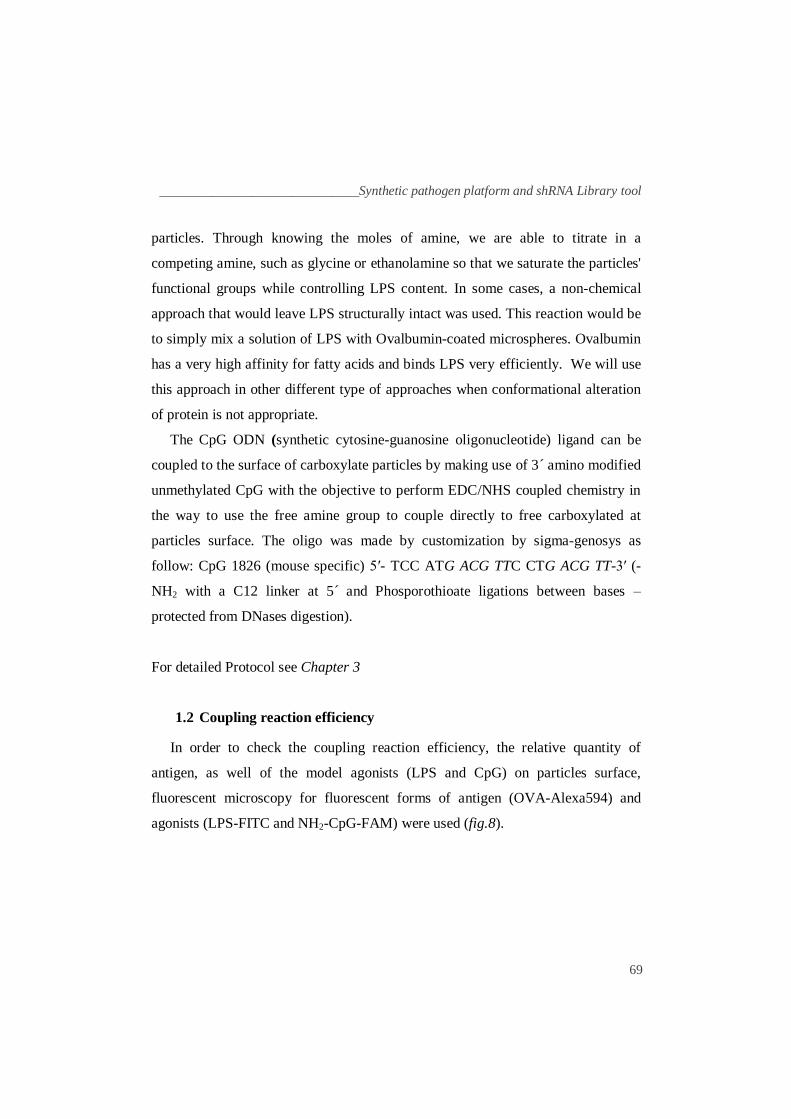
The CpG ODN (synthetic cytosine-guanosine oligonucleotide) ligand can be
coupled to the surface of carboxylate particles by making use of 3´ amino modified
unmethylated CpG with the objective to perform EDC/NHS coupled chemistry in
the way to use the free amine group to couple directly to free carboxylated at
particles surface. The oligo was made by customization by sigma-genosys as
follow: CpG 1826 (mouse specific) 5′- TCC ATG ACG TTC CTG ACG TT-3′ (-
NH2 with a C12 linker at 5 ́ and Phosporothioate ligations between bases –
protected from DNases digestion).
For detailed Protocol see Chapter 3
1.2 Coupling reaction efficiency
In order to check the coupling reaction efficiency, the relative quantity of
antigen, as well of the model agonists (LPS and CpG) on particles surface,
fluorescent microscopy for fluorescent forms of antigen (OVA-Alexa594) and
agonists (LPS-FITC and NH2-CpG-FAM) were used (fig.8).
Chapter 2 _________________________________________________________________
70
A) B) C)
Fig.8: Model particles antigen loading efficiency by CLSM images. Images of 1.0 µm polystyrene particles loaded with (A) 200µg of OVA-Alexa594 (red) or (B) 20µg of NH2-CpG-FAM (green) or (C) 20µg of LPS-FITC (green). Bright-field images are shown in upper panels and fluorescence
images in lower panels. Objective of magnification 63x was used (Scale bar 1.0µm).
These images show that either the protein or the model agonists form a well-
defined layer at the surface of the polystyrene particles, which confirms the
efficiency of the coupling reaction. However, a question arises: Does the presence
of agonists on coupling reaction affect the binding efficiency of the model antigen
between the different type of particles used (OVAp, OVA=LPSp and
OVA=CpGp)? To address this, antibodies were used against Ovalbumin to
measure the amount of antigen at particle surface by FACS, as follows:
______________________________Synthetic pathogen platform and shRNA Library tool
71
Fig.9: Ovalbumin-loaded particles efficiency by FACS based assay. Ovalbumin measurement at 1.0 µm polystyrene particles surface (OVAp, OVA=LPSp and OVA=CpGp) using Rabbit polyclonal antibody for Ovalbumin (Abcam) and 2nd anti-Rabbit conjugated to Alexa488 (Abcam). The white filled plot represents correspondent particles loaded only with 2nd antibody and the blue filled plot represents Ovalbumin staining. Quantitative coupling of Ovalbumin was examined by FACS by analyzing the Mean of fluorescence intensity (MFI) in the FL1-H channel. Numbers represent the MFI in the FLH-1 channel of stained particles. The graph is representative of at least three independent experiments.
These plots show that the loading efficiency (values of MFI) was similar in
model particles either in the presence or absence of agonists (LPS and CpG) during
the coupling reaction.
1.3 Particles Quantification
To better study particulate antigen presentation, the number of particles should
be measured. Particles were titrated by absorbance at 600nm. An example of
particle quantification is shown in figure 10 using 1.0µm particles as a model.
Using a calibration curve, the estimate number of particles in each condition could
be measured indirectly by replacing the y value with the absolute value of
absorbance at 600 nm (fig.10). The remaining particle sizes (50 nm to 6µm) were
measured in a concentration range where absorbance is linear.
Chapter 2 _________________________________________________________________
72
Table IV: Calibration curve: Values of absorbance at 600nm for the different particles concentration.
Particles/100µl 9,1x10⁷ 4,55x10
7 2,28x10
7 1,52x10
7 1,14x10
7 9,10x10
6 0
Abs 600nm 0,340 0,210 0,157 0,133 0,120 0,111 0,080
Fig.10: Particles Titration: Calibration curve of model particles with 1.0µm in size: particles concentration vs Abs at 600nm.
1.4 Coupling TLR ligands using a sonication protocol
To study the influence of other TLR agonists, we made use of an adapted
protocol from Yates et al., to couple different TLR mouse agonist for TLR (TLR1-
9 Agonist Kit from InvivoGen). This kit contains: TLR1/2 agonist - Pam3CSK4,
TLR2 agonist – HKLM, TLR3 agonist - Poly(I:C), TLR4 agonist - LPS-EK,
TLR5 agonist - ST-FLA, TLR6/2 agonist - FSL1, TLR7 agonist - ssRNA40,
TLR9 agonist - ODN1826 were adsorbed onto the surface of OVA-coated particles
by sonication with 100ng /µl of ligand in PBS for 20 minutes at 40°C. The particles
were washed extensively in PBS prior to use (Yates and Russell, 2005). The
conjugation efficiency can be assessed by measuring inflammatory cytokine
production after challenging DCs. In order to confirm the efficiency of this
y = 3E-09x + 0,087R² = 0,9986
0
0.1
0.2
0.3
0.4
0.5
0.0E+00 5.0E+07 1.0E+08 1.5E+08
Ab
s 6
00
nm
(particles/ml)
Calibration curve (1.0µm )
______________________________Synthetic pathogen platform and shRNA Library tool
73
method, we made use of available fluorescent ligands (LPS-FITC, NH2-CpG-
FAM) and compared with data from EDC/NHS coupling reaction with the same
fluorescent ligands (fig.11).
Fig.11: Adsorption of model agonists to Ovalbumin particles by sonication. Fluorescent LPS and CpG were adsorbed onto the surface of the 1.0 µm polystyrene Ovalbumin-coated particles by sonication of the particles in 100ng/ml of ligand in PBS for 20 minutes at 40°C. OVAp alone (grey line) and adsorbed with LPS-FITC (blue line) and NH2-CpG-FAM (red line). Quantitative coupling of LPS and CpG were examined by FACS by analyzing the Mean of Fluorescence Intensity (MFI) in the FL1-H channel. Numbers represent the MFI in the FLH-1 channel. The graph is representative of at least three independent experiments.
The loading efficiency of LPS and CpG by sonication method was similar as for
EDC/NHS coupling reaction. This approach can be very useful, and allow the use
of another conjugation strategy to study model particles antigen presentation.
1.5 Relative antigen coupling estimation for different particles size
Particles size from 50nm to 6µm was used to cover a broad range of pathogens
ranging from virus to bacteria to address the question if size influences antigen
cross-presentation, using the same particle ratio per DCs. In order to estimate the
amount of antigen loaded into different particles size the following equation was
used to predict the maximum protein loaded on particles surface (summarized in
table V).
Chapter 2 _________________________________________________________________
74
Table V: Expected maximum protein at close packing on microspheres surface for the different particles size. The equation was adapted from Prof. Darrell Irvine.
Size Group Quantity Surface Area Nº Protein Protein (x/1.0µm)
0.05µm -COOH 3,64x10¹⁴ p./ml 7,8x10⁻³ µm2 307 3,0x10⁻³
0.10µm -COOH 4,55x10¹³ p./ml 0,031 µm2 1097 1,1x10⁻²
0.20µm -COOH 5,68x10¹² p./ml 0,126 µm2 4141 4,2x10⁻²
0.50µm -COOH 3,64x10¹¹ p./ml 0,785 µm2 24971 2,53x10⁻¹
0.75µm -COOH 1,08x10¹¹ p./ml 1,766 µm2 55734 5,65x10⁻¹
1.0µm -COOH 4,55x10¹⁰ p./ml 3,14 µm2 98683 1,0
2.0µm -COOH 5,68x10⁹ p./ml 12,56µm2 392342 3,97
3.0µm -COOH 1.68 x 10⁹p./ml 28,26µm2 880981 8,93
6.0µm -COOH 2,10x10⁸ p./ml 113,04µm2 3516776 35,64
Using Alexa Fluor 488 Ovalbumin (OVA488) the loading efficiency at the
surface of different particles size can be measured by FACS (fig.12).
Fig.12: Antigen loading efficiency to particles with different sizes. Fluorescent Ovalbumin (Alexa Fluor 488 Ovalbumin - OVA488) was covalently linked to model particles of 500nm, 1.0µm and 3.0µm in size. Quantitative coupling of fluorescent particles antigen was examined by FACS and the mean of fluorescence intensity (MFI) in the FLH-1 channel were analyzed. Numbers represent the MFI in the FLH-1 channel of fluorescent particles. The graph is representative of at least three
independent experiments.
______________________________Synthetic pathogen platform and shRNA Library tool
75
2. PLGA particles (Biodegradable particles)
Biodegradable particles made of the polymer Poly D,L-lactic-co-glycolic acid
(PLGA), can be used as antigen delivery devices for macrophages and DCs
(Gander, et al., 2005). PLGA particles of about 0,5-5µm in diameter are actively
phagocytosed by human and murine DCs, and can be used to bind to or encapsulate
proteins and peptides, in addition to adjuvants such as DNA or RNA (Newman et
al., 2002; Newman et al., 1998; Wang et al., 1999). The PLGA particles by
themselves do not trigger DC maturation (Waeckerle-Men et al., 2004). PLGA
polymer hydrolyzes slowly in aqueous environments, and releases encapsulated
peptides and proteins into the processing pathways for presentation on either MHC
class-I and class-II pathways (Otten et al., 2003; Partidos et al., 1997; Waeckerle-
Men et al., 2006). PLGA has been successful as a biodegradable polymer because
it undergoes hydrolysis in the body to produce the original monomers, lactic acid
and glycolic acid. These two monomers under normal physiological conditions are
by-products of various metabolic pathways in the body. Since the body effectively
deals with the two monomers, there is very minimal systemic toxicity associated
with using PLGA for drug delivery or biomaterial applications (Waeckerle-Men et
al., 2004). As an example, a commercially available drug delivery device using
PLGA is Lupron DepotⓇ used in the treatment of advanced prostate cancer. State-
of-the-art of PLGA particles: i) clinically proven biocompatibility for poly(D,L-
lactide-co-glycolide); ii) promising candidate technique for vaccinations (delivery
of PLGA complexes by various routes; including oral, nasal and subcutaneous); iii)
protected protein antigen and increasing delivery efficiency (Acharya et al., 2009).
The illustrated protocol for PLGA micro-particles synthesis with and without a
lipid layer will be described as follows. For detailed protocol see chapter 3. This
work has been done in collaboration with Prof. Darrell Irvine´s group at MIT.
Chapter 2 _________________________________________________________________
76
2.1. PLGA microspheres loaded with protein mixed in the core
Illustrative representation of PLGA microspheres loaded with protein mixed in
the core (fig.13). For detailed protocol see Chapter 3.
Fig.13: Schematic illustrating the process of PLGA particles synthesis. Particles were formed by homogenization of polymer-containing organic phase into water, followed by evaporation of the organic solvent overnight and centrifugation for 5 minutes at 2.000xg. Scanning electron images of the pellets and supernatants (not shown) indicate that micron scale particles, mimicking bacteria, were separated from 100 nm-scale particles, mimicking viruses. (Adapted from Irvine´s Group – unpublished data).
In order to address the oligonucleotide (oligo) and protein conjugation to PLGA
particles, fluorescent equivalents (oligo-Texas red and OVA-Alexa594) were used
(fig.14).
Fig.14: Fluorescence
microscopy of
PLGA particles. Bright-field images (left panels) and fluorescent (right
panels) of PLGA particles loaded with 5nmol oligo-Texas Red and 10µg Ova-Alexa594. Objective of magnification 63x was used (Scale bar 5µm).
______________________________Synthetic pathogen platform and shRNA Library tool
77
This image shows that both antigen and oligos were widely distributed on
PLGA particles due to surfactant effect. Confocal microscopy was employed using
OVA-FITC loading PLGA particles (fig.15), confirming the widely pattern
distribution of Ovalbumin on PLGA particles as follows in next figure.
A B C
Fig.15: CLSM images of PLGA particles loaded with fluorescent antigen. (A) bright-field images, (B) fluorescence images of PLGA particles loaded with OVA-FITC and (C) Merged images.
Objective of magnification 63x was used with FITC filters (Scale bar 1.0µm).
2.2 PLGA microspheres with lipid layer to mimic pathogens and allow
protein and ligand conjugation
Illustrative representation of PLGA microspheres synthesis with lipid layer and
loaded with protein and MPLA (fig.16). For detailed protocol see Chapter 3.
Fig.16: Schematic illustrating the process of PLGA microspheres synthesis with lipid layer. Particles were formed by homogenization of a lipid- and polymer-containing organic phase into water, followed by evaporation of the organic solvent overnight and centrifugation for 5 minutes at 2.000xg. Scanning electron images of the pellets and supernatants (not shown) indicate that micron
scale particles, mimicking bacteria, were separated from 100 nm-scale particles, mimicking viruses. (Adapted from Irvine´s Group – unpublished data).
Chapter 2 _________________________________________________________________
78
To analyze PLGA particles synthesis and morphology by this method, scanning
electron microscopy (SEM) were performed. The antigen conjugation efficiency
was addressed by conjugating Alexa Fluor 488 Ovalbumin with maleimide-
modified PLGA particles via this method and analyzing by CLSM (fig.17).
Fig.17: Schematic illustrating the process of particle synthesis and morphology of particles observed by scanning electron microscopy (SEM). Particles were formed by homogenization of a lipid- and polymer-containing organic phase into water, followed by evaporation of the organic solvent overnight and centrifugation for 5 minutes at 2.000xg. Left: Scanning electron images of the
particles indicate that micronscale particles, mimicking bacteria, were separated from 100 nm-scale particles, mimicking viruses. Scanning electron micrograph showing 1-5 µm particle diameter (scale bar 10 µm) Right: Confocal micrograph of Alexa Fluor 488 Ovalbumin conjugated to maleimide-modified PLGA particles via this method (scale bar 5 µm). (This image was kindly provided by Anna Bershteyn from Irvine´s lab at MIT).
In order to address the Lipid bilayer formation and Ovalbumin distribution/
conjugation onto PLGA particles, a fluorescent dye DiI (1,1´-dioctadecyl 3,3,3 ,́3´-
tetramethylindo- carboxycyanate perchlorate from invitrogen) Red labeling (which
fluorescence was readily detected after binding to phospholipid bilayer
membranes), and Alexa Fluor 488 Ovalbumin were used (fig.18).
______________________________Synthetic pathogen platform and shRNA Library tool
79
A B C D
Fig.18: CLSM images of PLGA particles loaded with fluorescent antigen and lipids. (A) bright-field images and (B) fluorescence images of maleimide-modified PLGA particles conjugated to Alexa Fluor 488 OVA (green) and (C) DiI Lipophilic tracer (red) by method previously described. (D) Merged image of the three previous ones. Colocalization appears in yellow. Images were obtained with 63x objective amplification with respective filters (Scale bar 0.5µm).
These images showed that both Ovalbumin and lipids are widely distributed on
particles surface and in some extend in the core of particles. By this process, the
lipid bilayer keeps most of the antigen at the cell surface. To visualize the
morphology of the lipid layer at particles surface and address if it mimics a cellular
bilayer, Cryo-TEM were used (fig.19).
Fig.19: Cryo-TEM micrographs of lipid-coated particles. Particles synthesized with a 1:25 weight ratio of DMPC to PLGA were enveloped by single shells of lipid resembling previous Cryo-TEM studies of lipid-coated silica nanoparticles. PLGA particles made of the same materials but smaller, to
allow us to visualize the lipid surface. (This image was kindly provided by Anna Bershteyn from Irvine´s lab at MIT).
Chapter 2 _________________________________________________________________
80
Cryo-TEM images show a well defined lipid bilayer formed at PLGA particles
surface, which support this method of PLGA particles synthesis as an ideal system
to use it to mimic pathogens and to better study host-pathogen interactions.
3. Hydrogel particles (Sequestered dynamic-ligand particles)
During the past few years, many potentially powerful therapeutic strategies for
the treatment of diseases require improvement of delivery of drugs into the cytosol
or nuclear compartments of cells. Examples include therapeutic protein delivery
(Determan et al., 2006; Schweichel et al., 2006), anti-tumor toxin delivery
(Borghouts et al., 2005; Devalapally et al., 2007; Son et al., 2003), gene therapy
mediated by plasmid DNA (Medina-Kauwe et al., 2005; Putnam, 2006),
RNA/DNA that trigger potent anti-viral immune responses (Diebold et al., 2003)
and gene silencing via RNA interference (Tagami et al., 2007). Internalized
compounds/macromolecules by cells are confined to closed vesicles (endosomes or
phagosomes), where the pH is progressively lowered by fusion with lysosomes.
The pH could reach lower values such pH 4.5 and where the degradation
machinery of the cell are activated (Akinc and Langer, 2002; Asokan and Cho,
2002). This process could lead to rapid destruction of therapeutic molecules with
little or no release to cytosol. PLGA particles have been used as polymer-based
delivery systems to encapsulate DNA and provide sustained release as the polymer
degrades. However the major issue is to avoid the DNA degradation by low pH and
enzymes from lysosomes before it can be successfully releases to the cytosol (Fu
et al., 2000; Walter et al., 1999). In addition, delivery system using PLGA possess
a very poor ability to escape from endolysosomes (Cui and Mumper, 2002).
Therefore, endosomal escape is a major and critical point of current intracellular
delivery systems and synthetic carrier materials that respond to changes in pH
could be useful for drug or antigen release from these compartments before
______________________________Synthetic pathogen platform and shRNA Library tool
81
degradation. The idea of using this type of particles platform that allows the
endosomal particles escape contents could have different applications as follows:
3.1. Genesis of Hydrogel pH-responsive particles: Mechanism of exogenous
antigens delivery into MHC class-I pathway
Antigen presentation to cytotoxic T-Cells is greatly induced (up to 1000-fold)
by delivery antigens to the cytosolic of DCs, to allow the intracellular machinery to
load the right peptide onto MHC class-I molecules (Zarei et al., 2003). The
biomaterial vehicle must release the antigen intracellularly in a manner that will
enable processing by MHC class-I, class-II or both pathways. To deliver
exogenous antigens to MHC class-I pathway, they have to bypass the rapidly
traffic of phagosomes to lysosomes where antigens are then degraded
enzimatically, preventing the antigen to be processed and presented intracellularly
(Banchereau, et al. 2000). To avoid lysosomal trafficking, Murphy and colleagues
have designed smart polymers that use acid-degradable acetal bonds to disrupt
endosomes in a pH-dependent manner (Murthy et al., 2003). From an extracellular
pH of 7.4, particles that were internalized will eventually fuse with lysosomes and
achieve a pH ~5-5.5 (Underhill et al., 1999). These particles allow antigen and
adjuvants to release into cytosol as the endosomes are acidified before lysosomal
fusion, which enhance the processing by the MHC class-I pathway instead of MHC
class-II (Murthy et al., 2003).
Our approach is based on de-constructible hydrogen-bonded multilayers, a
concept originally demonstrated by Sukhishvili and colleagues (Sukhishvili et al.,
2000, 2002). Hydrogen-bonded multilayers of neutral and polyacid chains
assembled on the surface of colloidal particles have been reported (Kozlovskaya et
al., 2005). Here we extended this concept to the assembly of neutral and polybase
chains assembled on our functionalized particles, to create an acid-responsive
Chapter 2 _________________________________________________________________
82
protective shell (illustrated in Fig.21). Polymers bearing hydrophilic groups such
as –OH, CONH, -COOH, -SO3H and NH2 can be crosslinked to form hydrogels.
The swelling properties of ionic hydrogels are unique due to the ionization of their
pendant functional groups, and the equilibrium degree of swelling can be changed
suddenly by several orders of magnitude near the pKa or pKb of the hydrogels.
Taking advantage of the swelling ability of cationic gels, they can be applied for
endosomal disruption at low pH (Khare and Peppas, 1993). It has been shown that
cationic hydrogels made from diethyl aminoethyl methacrylate (DEAEMA) and
poly (ethylene glycol) monomethacrylate (PEGDMA) have a pKb ~7 which is the
ideal pKb to respond to endosomal pH (Podual et al., 2000). At pH below 7 a
fraction of the tertiary amine groups of core on the poly (DEAEMA-co-PEGDMA)
were protonated and thus positively charged, while the net surface (shell) charge is
negative due to primary amines that remain charged at all moderate pH, allowing
electrostatically-driven adsorption (fig.21). Thus, siRNA, oligos or antigen could
be electrostatically bound to the surface of the particles. At pH higher than 7.0,
poly (DEAEMA-co-PEGDMA) are largely uncharged and capable of strong
hydrogen bonding. In near-neutral extracellular conditions, the polymer multilayer
coating will remain hydrogen-bonded and prevent access of the „masked‟ ligands
to cells. On internalization, the drop in pH within the phagocytic pathway will
induce ionization of the poly (DEAEMA-co-PEGDMA) chains (due to the pKa of
the tertiary amino groups in the polymer, which is near neutral pH (Schwarte et al.
1998), leading to loss of hydrogen bonding, dissolution of the coating, and
exposure of the masked ligand (fig.20). Antigen and/or selected ligands could be
adsorbed to poly (DEAEMA-co-PEGDMA) core-shell nanoparticles. Our research
was thus to investigate the use of synthetic pH-sensitive hydrogel nanoparticles as
a novel intracellular antigen delivery system to cytosol, bypassing the requirement
of retro-translocation machinery in antigen cross-presentation.
______________________________Synthetic pathogen platform and shRNA Library tool
83
Because the surface membrane or envelope of some pathogens is not
compromised until fusion of the phagosome with lysosomes, internal components
of these pathogens may not be exposed to antigen processing machinery until late
stages of the phagolysosomal processing pathway. With this approach, TLR
agonists and antigen could be selectively exposure to endosome environment as the
pH drops during phagosome maturation. This hypothesis is consistent with the
localization of certain TLRs that recognize internal components of pathogens to
phagolysosomal compartments (Latz et al., 2004; Oshiumi et al., 2003), rather than
at surface of phagocytes. To determine whether sequential encounter of antigen or
activating signals impacts the response of phagocytes to pathogens, hydrogel
particles with „masked‟ antigen or TLR agonist layers could be synthesized, which
can be selectively exposed based on the pH of the particle microenvironment as
illustrated in the next figure.
Fig.20: Hydrogel pH-
responsive particles cellular mechanism. Particles are internalized along with an
agonist and a model antigen into endosome. During internalization, endosomes matures and become acidic (pH<7.0) to break down internalized molecules. The particles begin to be protonated at this pH, absorbing protons
that are pumped into the endosome. As protons are absorbed, anions are also pumped into the endosome to maintain charge neutrality.This causes an osmotic pressure
buildup that will drive water into the endosome, eventually disrupting or rupturing the membrane and causing release of the ligand, antigen and particle into the cytosol. Adapted from (Hu et al., 2007).
Chapter 2 _________________________________________________________________
84
3.2. New application system: RNAi delivery on same context as antigen
Up regulation of MHC class-II and CD86 were adopted as a surrogate marker of
DC maturation with the assumption that this always correlates with
immunogenicity (Finkelman et al., 1996). DCs were later found to also induce
tolerance, and it was suggested that tolerance and immunity were mediated by
immature and mature DCs, respectively (Finkelman et al., 1996; Steinman and
Nussenzweig, 2002). Most researchers interpreted „immature tolerogenic DCs‟ to
refer to MHC class IIlow
CD86low
DCs. Besides this, many studies have used this
terminology while relying on naive T-Cell proliferation as a correlate of
immunogenicity, which is not correct because T-Cell proliferation can lead to
tolerance as well as shown by „phenotypically mature‟ DCs were found to induce
tolerance (Albert et al., 2001) or at least not to induce immunity (Sporri and Reis e
Sousa, 2005). Therefore, an interesting approach could be explored for the efficient
delivery of RNAi for regulatory molecules of antigen presentation in same context
as a particular antigen in order to amplify or suppress adaptive immune response
for vaccines or immunotherapy (Greenland et al., 2007). An ideal delivery system:
(1) be able to bind RNAi in a reversible manner as to ensure the subsequent release
of the RNAi; (2) escape from endosomal compartment; and (3) be biocompatible.
Therefore, pH-sensitive core-shell nanoparticles have recently been proved to be a
good delivery system for RNAi (Blackburn et al., 2009; Hu et al., 2009). The
promise of RNAi will only be a clinical reality when safe and efficient delivery
systems become well established.
______________________________Synthetic pathogen platform and shRNA Library tool
85
3.3 Synthesis and characterization of pH-sensitive core-shell nanoparticles
Illustrative representation of Hydrogel pH-responsive core-shell nanoparticles
synthesis and chemical composition (fig.21). For detailed protocol see Chapter 3.
Fig.21: Schematic structure and chemical composition of pH-responsive core-shell nanoparticles. At extracellular/cytosolic pH, tertiary amines of DEAEMA repeat units in the particle
cores are largely uncharged, and the particles are collapsed; at endolysosomal pH, the core tertiary amines ionize, and the particles swell. Surfactant-free polymerization of DEAEMA formed the core structure of hydrogel nanoparticles, crosslinked by PEGDMA. AEMA was polymerized in a second stage to form a thin shell structure rich in primary amines. Particles can swell ~2-fold (8- fold volume change) in response to pH drop below ~7. Adapted from (Hu et al., 2007).
In order to show physiologic properties of Hydrogel particles comparing to
PLGA particles, calcein, a membrane-impermeant fluorophore, was used as a
model drug molecule and tracer to monitor the stability of endo/phagosomes
following particle uptake (fig.22). This work has been done in collaboration with
Prof. Darrell Irvine´s group at MIT.
Chapter 2 _________________________________________________________________
86
Fig.22: Endosomal escape of the
membrane-impermeable dye molecule
calcein into the cytosol of DCs in the
presence of pH-responsive core-shell
nanoparticles comparing to PLGA nanoparticles and microparticles. CLSM images at 40x - Fluorescence overlays (red, nanoparticles; green, calcein). sDCs were co-incubated with
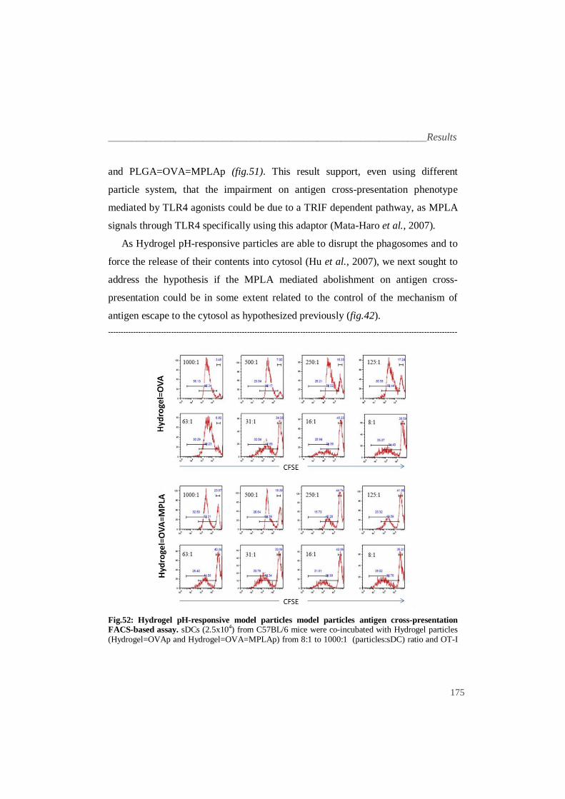
1µM of LysoTracker Red DND-99 (to label endolysosomes), 0.24 mM of calcein, and 1:20 (DCs:particles) ratio. (A) Cells were treated with calcein alone. Cells were co-incubated with (B) calcein and Hydrogel=OVA nanoparticles (C) with calcein and PLGA=OVA microparticles (D) with calcein and PLGA=OVA nanoparticles
(Scale bar 20µm). (This image was kindly provided by Anna Bershteyn from Irvine´s lab at MIT).
Hydrogel nanoparticles exhibited calcein fluorescence throughout the cytosol
and nucleus (fig. 22-B). Calcein entry into the cytosol is triggered by the presence
of nanoparticles required at the pH-sensitive core (fig. 22-B), as calcein remained
in an endosomal distribution in cells co-incubated with calcein and PLGA nano
and microparticles (fig.22-C and D). Therefore, hydrogel particles are able to
delivery components into cytosol upon internalization by DCs, but not PLGA
nanoparticles. These results implicate hydrogel pH-responsive particles as a good
delivery vehicle into cytosol for antigen cross-presentation studies.
______________________________Synthetic pathogen platform and shRNA Library tool
87
Part II: shRNA Library: New tools for the genetic dissection of antigen
cross-presentation pathway(s)
1. Technology overview and design
The information resulting from genome-sequence increased the need for tools
that allow genome-scale functional studies. In model organisms such
Caenorhabditis elegans and Drosophila melanogaster, the recognition that RNA
interference (RNAi) can be used to suppress gene expression (Fire et al., 1998;
Kennerdell and Carthew, 1998), has lead to identification of the genes underlying
many biological processes through loss-of-functions screens (Bettencourt-Dias et
al., 2004; Boutros et al., 2004; Fraser et al., 2000; Kamath et al., 2003; Kiger et
al., 2003; Lum et al., 2003). Chemically synthesized RNAi also suppresses gene
expression in mammalian cells and become essential tool for biological studies
(Elbashir et al., 2001). RNAi screen have been done with commercially available
libraries (Aza-Blanc et al., 2003; MacKeigan et al., 2005; Pelkmans et al., 2005).
As many mammalian cell types are resistant to transfection methods, an alternative
approach has to be used to introduce synthetic siRNA into cells. In 2002 emerged a
new “transfection” technology based in transduction mammalian cells with viruses
carrying expression cassettes that encode short hairpin RNAs (shRNAs) to
generate gene-specific siRNAs in cells. This approach produces stable and highly
effective gene suppression in a variety of mammalian cell types (Abbas-Terki et
al., 2002; Brummelkamp et al., 2002; Paddison et al., 2002; Stewart et al., 2003).
2. The RNAi consortium (TRC)
The RNAi Consortium (TRC) is a collaborative group of 11 world-renowned
academic and corporate life science research groups whose mission is to create
comprehensive tools for functional genomics research. The RNAi Consortium
Chapter 2 _________________________________________________________________
88
emerged with the objective of generating genome-scale shRNA libraries in viral
vectors to target almost all human and mouse gene, that allows gene silencing in
most dividing and non-dividing cell types.
2.1 Lentiviral shRNA library production
The TRC lentiviral shRNA library contains now constructs targeting all human
and mouse genome, with ~5 distinct shRNA constructs per gene. The inclusion of
five different shRNA constructs targeting each gene increases the opportunity to
achieve strong knockdown, often offers a gradation of knockdown and provides the
means to rapidly evaluate the gene specificity of a phenotypic hit. Constructs were
designed using a siRNA rules based algorithm consisting of sequence, specificity
and position scoring for optimal hairpins, that attempt for maximize knock-down
and minimize off-target effects, as well as to ensure that most genes in the library
contain shRNAs that target both the 3´ unstranlated region (UTR) and coding
sequence (CDS) of their transcripts (Khvorova et al., 2003; Schwarz et al., 2003).
For each shRNA, they designed a 21 base stem for the target transcript and an
intervening 6 base loop consisting of a XhoI site. The hairpins were cloned into the
pLKO.1 vector, which carries the puromycin-resistance gene and drives shRNA
expression from a human U6 promoter (fig.24). The pLKO1 lentiviral vector
enables efficient transduction of primary and non-dividing cells making it easy to
perform RNAi studies in these hard to transfect cell lines (Federico, 2003).
Typically, 3-5 shRNA constructs are created for each target gene to provide
varying levels of knockdown and to target different regions of mRNA transcript.
One in five clones will typically provide at least 70% knockdown of the gene
target. Sense and anti-sense hairpin oligonucleotide pairs for 90 hairpin sequence
were annealed separately and ligated into pLKO.1 at AgeI and EcoRI restriction
sites, and the ligations were transformed into competent bacteria in a 96-well plate.
______________________________Synthetic pathogen platform and shRNA Library tool
89
The 90 transformations in each plate were then pooled and plated onto a large agar
plate. A total of 672 colonies were selected robotically for growth, plasmid
purification, and sequencing. This process yields 94% of the designed clones, each
gene has an average of 4.7 unique shRNA constructs, and 96% of the genes have
four or more different constructs (Moffat et al., 2006). The TRC creates a
production pipeline to generate a library of sequence verified shRNAs in pLKO.1.
In summary, the production of the library comprises several steps: 1) Hairpin
Design; 2) Vector Preparation; 3) Oligo Pair Annealing, Ligation and
Transformation; 4) Colony Picking and Sequencing Validation; 5) Quality
Assessment of Library Glycerol Plates; 6) Tests of Recombination; 7) High-
Throughput DNA Production; 8) Lentiviral Production; 9) Lentiviral infections;
10) Quantitative RT-PCR and 11) Titering Assay (Moffat et al., 2006).
2.2 High-Throughput lentiviral particle production
A high-throughput (HT) method to generate high-titer lentiviruses was
generated by TRC shRNA library consortium. Although preparation of
transfection-quality plasmid DNA and subsequent packaging of the viral plasmids
into viruses is quite straightforward for individual samples, performing this process
efficiently in a high-throughput 96-well format is more challenging; furthermore,
high-throughput screening demands high and uniform viral titers. TRC consortium
has developed protocols for DNA and viral production for the TRC library. These
protocols are frequently updated with improvements and the latest versions can be
found online (http://www.broad.mit.edu/genome_bio/trc/protocols.html).
A semi-automated procedure was optimized in 96-well plates in which
HEK293T cells were transfected with library and packaging plasmids (pCMV-
dR8.91) and envelope plasmid (pMD2.G). pCMV-dR8.91 and the envelope
plasmid pMD2.G are available from Addgene. Transfection-quality DNA was
Chapter 2 _________________________________________________________________
90
prepped using 96-well PureLink kits (Invitrogen) with average yields of 4 μg
DNA/well, quantified using a PicoGreen assay (Molecular Probes), and normalized
robotically in each plate. Lentiviruses were made in 96-well format by transfecting
packaging cells (HEK 293T) with a three-plasmid lentivirus packaging system
(Naldini et al., 1996; Zufferey et al., 1997). 300 µl of transfected cell supernatants
containing VSV-G pseudotyped lentiviruses are collected over 36–60 hrs and
aliquoted and stored these lentivirus containing supernatants at 80ºC. With current
HT methods of viral production in 96-well plates, viral titters with average of
3x10⁷ infectious units (I.U)/ml in A549 cells infected were obtained. Typically,
50% of wells fall within a twofold range of viral titer. Thus, the procedure above
yields sufficient volumes of lentiviral supernatants (300µl) from a single 96-well
plate, is sufficient to provide virus for several hundreds shRNA infectious,
depending as well on the transducibility of the target cells (Root et al., 2006). One
consequence of using lentiviral vectors with high titer and broad tropism is the
requirement for adequate biosafety procedures for manipulation. The three
plasmid–based lentivirus productions of self-inactivating viruses nearly eliminates
the possibility of recombination to create replication-competent viruses, and the
rate of recombination is several orders of magnitude lower than that of comparable
amphotropic retroviral systems. Many institutions apply biosafety level 2 practices
to work with third-generation lentiviruses. TRC consortium used biosafety level 2+
precautions for all lentiviral shRNA library work with appropriate containment and
decontamination procedures. An example of institutional guidelines for lentiviral
work is available online (http://www.ohsu.edu/research/rda/ibc/protocols.shtml)
(Root et al., 2006).
______________________________Synthetic pathogen platform and shRNA Library tool
91
2.3 Subsets compilation
During the first year of my PhD I have been in Boston at Nir Hacohens‟ lab
(Harvard/MGH/Broad institute), one of the main collaborative groups that
compose the TRC consortium, where the UBCSI lab was originated. The objective
was to learn about the state-of-art shRNA library its compilation and how to
transfer specific subsets of the shRNA library to perform loss-of-function screens,
in order to perform the rapid identification of the genes underlying many biological
processes such as antigen presentation and inflammation that are the two main
areas of interest for the UBCSI lab. With this powerful tool we are able to generate
and study loss-of-function phenotypes of genes that compose specific protein
functional families. As a summary, we organized different subsets of shRNA for
mice and human genes, in order to produce bacteria, DNA and lentivirus (fig.23):
Fig.23: Relative representation of different shRNA bacterial glycerol stock library collections for Human and Mouse genome. Numbers represent the sets of shRNA target genes in each
collection: Splicing factors, Kinases/Phosphatases, vesicle traffic, antigen presentation and others small collections.
2.4 Library production and use
The next figure shows a schematic represenation of shRNA lentiviral library
production, lentiviral infection and phenotype assay (fig.24).
Chapter 2 _________________________________________________________________
92
Fig.24: Scheme for library production and use. Bacterial glycerol production, pLKO.1shRNA
constructs DNA prep, viral production and transduction method. Vector map for the pLKO.1
lentiviral vector: The self-inactivating lentiviral vector backbone contains elements for efficient viral packaging and shRNA expression. Expression of the shRNA is driven by the human U6 promoter (hU6). The lentiviral vector also contains the mammalian selection marker puromycin resistance gene (PAC) and the bacterial ampicillin resistance gene (AmpR). Part I: Inoculation, growth, and
duplication of glycerol stocks in 96-well plates: To create the different shRNA library families, different colonies were re-organized from the master TRC library collection into new 96-well plates.
Colonies were inoculated one by one, and grown for 17hrs at 37ºC in Terrific Broth supplemented with 100ng/µl of Carbenicillin with constant shaking at 300 rpm in an appropriated shaker for 96-well plates. After, glycerol stocks were prepared using 40µl autoclaved 50% glycerol and 80µl of culture from deep well growth plate into each destination plate to make replicate copies. These plates were freeze immediately and store at -80ºC. Part II: Preparation of Transfection-Quality Plasmid DNA
in 96-well Plates: The rest of bacterial culture was used to prepare of transfection-quality plasmid DNA using TRC library protocols for glycerol and plasmid preparation. See detailed protocol: https://www.broadinstitute.org/genome_bio/trc/protocols/trcGlycerolStockPlasmidPrep.pdf. Part III:
lentiviral Production: Packaging Cells (HEK 293T) were transfected with the 3 lentivirus plasmids
(hairpin-pLKO.1 vector, packaging plasmid and envelope plasmid). At 18 hours post-transfection: medium were removed and replaced with fresh high-serum media (30% FBS). At 24 hours viruses were harvest by replacing the medium with C10. At 48hr afterwards, viruses were harvested again and packaging cells were discarded according to TRC library protocols for lentiviral production. See
______________________________Synthetic pathogen platform and shRNA Library tool
93
detailed protocol: http://www.broadinstitute.org/genome_bio/trc/protocols/trcLentiVirusProd.pdf.Part IV:
Lentiviral infection: High titer of lentiviruses was used (10µl) to transduce target cells by spinoculation (2200rpms at 37ºC during 90 min). Polybrene were used as 8µg/ml as final concentration. After 2 days at 37ºC, cells were selected with an optimal concentration of puromycin (concentration should be optimized for each cell line; typical concentrations range from 2-5 μg/ml). Puromycin selection requires at least 48hrs. Incubated periods are highly dependent on the post-infection assay. Part V: Phenotypic assay: could be performed 3+ days after puromycin incubation.
Validation by mRNA Knockdown (qPCR) or protein Knockdown (Western/FACS) could be performed 2+ days or 3+ days respectively. Adapted from http://www.broadinstitute.org/genome_bio/trc/publicProtocols.html and (Moffat et al., 2006).
2.5. Publications
The ultimate objective with this tool was to create a specific collection of
shRNA, which we called as “antigen presentation collection”, to generate loss-of-
function of specific genes involved in different key steps of antigen presentation
pathways. Using this powerful tool, we initially proposed to dissect and clarify the
antigen cross-presentation mechanism(s) mediated by our platform of synthetic
model particles. In addition to the knowledge of the technology behind shRNA
platform, another proposed was to generate important subsets of families of genes
crucial for the development of different projects that were occurring in the lab
(UBCSI at Instituto de Medicina Molecular) and with collaboration of different
groups abroad:
i) One of the collaborations was done with Anjana Rao´s lab in Cambridge at
Harvard Medical School, with the aim to identify splicing factors required for the
activation-induced switch from CD45RA+ isoforms to the short isoform CD45RO
(exclusion of exons 4-6 (A-C) of CD45 transcripts). As the transition from naïve to
activated T-Cells is marked by alternative splicing of pre-mRNA encoding the
transmembrane phosphatase CD45, it is of great importance to understand how this
regulation occurs. From this work using the Splicing Factors shRNA library, we
identified a single factor, heterogeneous ribonucleoprotein L-like (HNRPLL),
which is up-regulated in response to PMA stimulation and whose depletion
Chapter 2 _________________________________________________________________
94
eliminated stimulation-dependent CD45RO expression. HNRPLL is necessary and
sufficient to induce CD45RO expression in B and T-Cell lines and primary T-
Cells. We conclude that HNRPLL is a crucial regulator of CD45 alternative
splicing in activated T-Cells. This work was published in Science. 2008 Aug 1;
321 (5889):686-91).
ii) Another project developed in collaboration with Sebastian Amigorena´s lab
in Paris at Curie Institute, proposed to identify vesicle traffic proteins involved on
the control of different steps of the Exosome secretion pathway. Exosomes are
secreted by several cell types and can be involved in intercellular communication
and in the pathogenesis of infectious and degenerative diseases. The molecular
mechanisms of their biogenesis and secretion are, however, poorly understood.
Using a shRNA interference screen for vesicle traffic proteins, we identified 5
small GTPases of the Rab family involved in exosome secretion in HeLa cells,
were the two Rab27, Rab27a and Rab27b, play a major role but different and
complementary in the exosomal pathway. By showing that major inhibition of
exosome secretion is associated with alterations of late endocytic compartments,
we demonstrate that exosomes originate mainly from MVEs. This work was
published in Nature Cell Biol. 2010 Jan;12(1):19-30.
iii) Caetano Reis e Sousa´s lab in London at Cancer Research Institute was
interested in fungal response and the mechanism behind that. Caetanos´s lab has
shown that with Dectin-1, a PRR for fungi, a novel innate signaling pathway
involving Syk kinase and the adaptor CARD9 is defined, which is critical for
inducing Th17 responses to fungal infection. We demonstrated that another C-type
lectin, Dectin-2, also signals via Syk indirectly trough association with FcRγ, and
CARD9, and contributes to Dendritic Cell activation by fungal particles. We
concluded from this work that Dectin-2 constitutes a major fungal PRR that can
couple to the Syk–CARD9 innate signaling pathway to activate DCs and regulate
______________________________Synthetic pathogen platform and shRNA Library tool
95
adaptive immune responses to fungal infection. This work was published in
Journal of Experimental Medicine 2009 Aug 206: 2037-2051.
iv) In collaboration with Prof. João Gonçalves´ lab in Lisbon at Faculdade de
Farmácia, we proposed to identify novel HIV-1 replication of druggable targets
identified by a shRNA-based screen enriched in kinases/Phosphatases in T
lymphocytes. HIV-1 is a complex retrovirus that uses host machinery to promote
its replication. Understanding cellular proteins involved in the multistep process of
HIV-1 infection may result in the discovery of more adapted and effective
therapeutic targets. Kinases and phosphatases are a druggable class of proteins
critically involved in regulation of signal pathways of eukaryotic cells. Here, we
have focused in the discovery of kinases and phosphatases that are essential for
HIV-1 replication but dispensable for cell viability. We have performed an iterative
screen in Jurkat T-Cells with a short-hairpin-RNA (shRNA) library highly enriched
for human kinases and phosphatases. We identified new 14 proteins essential for
HIV-1 replication that do not affect cell viability. These proteins were described to
be involved in MAPK, JNK and ERK pathways, vesicular traffic and DNA repair.
Moreover, we have shown that most proteins do not affect viral integration but
rather affect viral transcription/translation. This study brings new insights for the
complex interplay of HIV-1/host cell and opens new possibilities for antiviral
strategies. This work was published in PLoS ONE 2010 Feb 17;5(2):e9276.
The results presented in this thesis, in collaboration with Darrell Irvine‟s lab at
MIT, are under preparation for publication:
Freitas RP, Bershteyn A, Moita C, Irvine DJ, Moita LF. The particle antigen
cross-presentation is impaired by a crosstalk effect of TLR4 signaling in a phagosome dependent manner. In preparation.
____________________________________________________ Materials and Methods
99
Mice
We have used 6- to 12-week-old C57BL/6 Wild Type, MyD88KO, TLR4KO,
OT-I and OT-II mice. All mice were bred and maintained under specific pathogen-
free conditions at Instituto de Medicina Molecular animal breeding facility
according to institutional guidelines.
Cells Isolation/Preparation
Bone Marrow-derived Dendritic Cells (BMDCs)
DCs differ in developmental origin and most DCs are considered to be of
myeloid origin; the main evidence for this comes from studies of DC development
in cultures stimulated by Granulocyte-macrophage colony-stimulating factor (GM-
CSF) (Inaba et al., 1993). The principle method for generating BMDCs with GM-
CSF was adapted from previous publications (Inaba et al., 1992a; Inaba et al.,
1992b; Inaba et al., 2001; Scheicher et al., 1992). Culture of DCs in vitro was
carried out in RPMI medium 1640 (GIBCO) supplemented with 10% heat-
inactivated and filtered (0.22 µm, Milli-Pore) Fetal Bovine Serum (endotoxin-free,
BIOWEST), β-mercaptoethanol (50µM Sigma- GIBCO) L-glutamine 100 units/ml
(2mM, GIBCO), penicillin (100 U/ml, GIBCO) streptomycin (100µg/ml GIBCO)
and GM-CSF (15-30% J558 supernatant, depending on GM-CSF concentration,
tested previously in culture) - Complete Medium (C10). The BMDCs were
differentiated in 96 round-well plates (corning) or large Petri dish (non treated
dish) depending the number of DCs and the type of experiment. Bone-Marrow
derived Dendritic Cells (BMDCs) isolation from mice bone-marrow was done as
follow:
Day 0: Femurs and tibiae of female, 6–12 weeks old female C57BL/6 or
TLR4KO, were removed and purified from the surrounding muscle tissue. Then
Chapter 3_________________________________________________________________
100
both ends were cut with scissors and the marrow flushed with BMDCs Medium
using a Syringe with a 25G needle. Clusters within the marrow suspension were
disintegrated by vigorous pipetting and filtered. After spin down cells were
resuspended in 2 ml of TAC buffer to lysis Red blood cells (8.32 g NH4Cl; 0.82 g
NaHCO3; 0.043 g EDTA in 1L of miliQ water) for 2 minutes. This reaction was
stopped with 8 ml of complete medium. The cells were pelleted and counted. 15-
30% J558 supernatant (depending on GM-CSF concentration) was added. BMDCs
were plated in 96 well plates at 5x10⁴/well in 200µl or alternatively in 150 mm
petri dishes (non treated dish) at 10-12x106/petri dish in 20ml of medium and
incubated at 37ºC. For 96 well plates, BMDCs care is performed every two days
until BMDCs are ready to harvest on day 6 or 7. Remove old medium - Carefully
aspirate in circular fashion ¾ of medium. This sucks up nutrient depleted medium
and non-adherent-non DCs. Prepare new medium - add J5 (1:30 dilution) to C10
and replace with ¾ of volume.
Day 3: For the petri dishes cultured BMDCs, take the supernatant with floating
cells and transfer it into a 50 ml tube. Add 10 ml of phosphate-buffered saline (BS)
to the dish and swirl gently trying to detach some cell clusters (avoiding bubbles).
Mix this PBS containing cells with the floating cells supernatant. Add 4ml RT -
trypsin to the dish and let it for 2 minutes; stop by adding 4 ml BMDC medium and
swirl gently. Transfer this volume and mix with the rest of the cells. After this
short trypsin treatment many cells remain attached to the bottom of the dish. Do
not try to take them; most of them have a macrophage-like phenotype. Centrifuge
the cells and resuspend them in some volume of BMDC medium; count them and
plate again into 150 mm dishes (10-12 .106 cells in 20ml/Petri dish).
Day 7: Repeat as described for day3.
____________________________________________________ Materials and Methods
101
Day 9-11: Repeat as described for day3. Perform CD11c staining for
Fluorescent Activated Cell Sorter (FACS) analysis. The level of CD11c+
population should be more than 70% from day 10-11 and increase over time. It is
important to verify if CD11c+ cells are immature and how is their capacity of
maturation. Treat some cells during at least 20 hours with 10 µg/ml LPS. Perform
staining of MHC-II, CD40, CD86 in treated and not treated cells and analyze by
FACs. The levels of these 3 markers should be low in non-treated cells and
importantly increased in LPS treated DCs.
Once the population is 80-90% CD11c+, cells can be platted at higher
concentration (around 15-20x106 cells in 20ml/Petri dish). They can be used for 7-
10 days depending on maturation markers. CD11c staining for FACs analysis was
performed (fig.25). The level of CD11c+ population should be more than 70% from
day 6 and increase over time. To verify if CD11c+ cells are immature and how is
their capacity of maturation, BMDCs were stained with anti-CD86, anti-CD40 and
anti-MHC-II treated and not treated with LPS stimulation (10ng/ml) for at least 20
hours and analyzed by FACs for the surface expression of maturation markers. The
levels of these 3 markers should be low in non-treated cells and increased in LPS
treated DCs (fig.26).
Fig.25: BMDCs staining at day 9 with anti- CD11c
+-PE antibody. Upper left panel
unstained BMDCs population vs upper right panel stained BMDCs with anti-CD11c+-PE antibody (Abcam). Lower graph shows histogram of BMDCs unstained (dot curve) and stained with CD11c+-PE (filled curve). Number represents the percentages of the
positive CD11c+ cells, analyzed on PE channel. These data are representative from one experiment repeated at least three times with similar results.
Chapter 3_________________________________________________________________
102
Fig.26: BMDCs maturation with GM-CSF, staining at day 3, 6 and 9 in culture. BMDCS were
stained with anti-CD86, anti-CD40, anti-MHC-II and anti-CD11c antibodies (Abcam), with and without LPS stimulation (10ng/ml) for at least 20 hours. BMDCs were analyzed by FACS and plots represent surface expression of CD86, CD40 and MHC class-II vs DC marker (CD11c+). Numbers show the percentage of cells on CD11c+ quadrants. Antibodies were used with 1:200 dilution. These data are representative from one experiment repeated at least three times with similar results.
____________________________________________________ Materials and Methods
103
Splenic Dendritic Cells (sDC)
DCs form lymphoid origin were isolated form mouse spleen as previously
described (Vremec et al., 2000), making use of Immunomagnetic bead purification
kit (CD11c MicroBeads mouse - AutoMACS; Miltenyi Biotec) (Ing et al., 2006;
Williamson et al., 2002). The spleen was cut into small fragments and left for 30
minutes for enzymatic disaggregation in medium with Collagenase D (1mg/ml)
and DNaseI (0,02mg/ml) at 37ºC to generate single cell suspension. After, the
fragments were resuspended and filtered through 30µm nylon mesh, to remove cell
clumps that may clog the column. To prevent DC maturation during the isolation
protocol, the procedure was performed on ice or at 4ºC. Cell number was
determined and cells were recovered from the digestion by centrifugation for
200xg for 5 minutes. Cell pellet was resuspended in 400µl of MACS buffer
(solution containing PBS pH 7.2, 0.5% bovine serum albumin (BSA), and 2 mM
EDTA by diluting MACS BSA Stock Solution 1:20 with autoMACS™ Rinsing
Solution per 108 total cells (keep buffer cold,4−8 °C). 100µl of CD11c MicroBeads
were added per 108 of total cells. The cells were mixed with the beads and incubate
for 15 minutes in the refrigerator (2-8ºC). Cells were washed by adding 1 ml of
buffer per 107 cells and centrifuge at 200xg for 5 minutes. Cells were resuspended
up to 108 cells in 500µl of MACS Buffer. Next magnetic separation was
performed: Place MS column in the magnetic field separator and equilibrate with
500µl of MACS Buffer. Apply cell suspension onto the column and collect flow-
through containing unlabeled cells. Always wait until the column reservoir is
empty before proceeding to next step. Wash column 3x with 500µl of Buffer and
collect again the flow-trough. Remove column from the separator and placed in a
15ml falcon tube. Elute the CD11c+ cells by pippeting 500µl of MACs buffer onto
the column and immediately flush out the magnetically labeled cells by firmly
Chapter 3_________________________________________________________________
104
pushing the plunger into the column. This protocol excluded B220+ „„plasmacytoid
DC‟‟ from the DC preparation.
T-Cells (CD8+ Cytotoxic T-Cells and CD4
+ T Helper Cells)
CD8+ Cytotoxic T-Cells and CD4
+ T helper cells were isolated from spleen of
OT-I and OT-II mice respectively, using two different T-Cell isolation Kits from
MACS Miltenyi Biotec. T-Cell Isolation Kit is an indirect magnetic labeling
system for the isolation of untouched T-Cells from single-cell suspensions of
lymphoid organs. Consequently, CD8α+ T-Cells were isolated by depletion of non-
CD8α+
T-Cells and CD4+ T-Cells were isolated by depletion of non-CD4
+ T-Cells
(negative selection), as described elsewhere (Gruber and Brocker, 2005). Highly
pure untouched CD8α+ T-Cells and CD4
+ T-Cells are isolated respectively using
this approach. Non-CD8α+ T-Cells, i.e. T helper cells, B cells, NK cells, DCs,
macrophages, granulocytes, and erythroid cells, are indirectly magnetically labeled
with a cocktail of biotin-conjugated antibodies against CD4 (L3T4), CD45R
(B220), CD49b (DX5), CD11b (Mac-1), and Ter-119, as well as anti-Biotin
MicroBeads. Non-CD4+ T-Cells i.e. cytotoxic T-Cells, B cells, NK cells, DCs,
macrophages, granulocytes, and erythroid cells, are indirectly magnetically labeled
with a cocktail of biotin-conjugated antibodies against CD8a (Ly-2), CD45R
(B220), CD49b (DX5), CD11b (Mac-1), and Ter-119, as well as Anti-Biotin
MicroBeads. Isolation of T-Cells was achieved by depletion of the magnetically
labeled cells. The protocol is the same for CD8+ T- Cell or CD4
+ T-Cell isolation,
using however different magnetic beads cocktail described previously.
Sample preparation: The spleen was dissociated mechanically, between 2
slides sterilized with ethanol 70%. The resulting tissues were resuspended with a
pipette and the cell suspension was filtered. Cells were recovered by centrifugation
at 300xg during 5 minutes. 2 ml of TAC were added for 5 minutes at RT to remove
____________________________________________________ Materials and Methods
105
erythrocytes. Complete medium were added to stop the reaction and the pellet was
recovered by centrifugation at 300xg during 5 minutes. Pellet was resuspended in 1
ml MACS buffer and cell number was determined.
Magnetic labeling: Cell suspension was centrifuge at 300xg during 5 minutes
and pellet was resuspended in 40µl of MACS buffer per 107
of total cells. 10 µl of
Biotin-Antibody Cocktail (Cocktail of biotin-conjugated monoclonal antibodies),
were added per 107 cells and incubated at 4ºC during 15 minutes (mixture were
mixed every 5 minutes). 30µl of MACs Buffer and 20 µl of Anti-Biotin
Microbeads (monoclonal antibodies – secondary labeling reagent) were added per
107 of total cells. Resuspension was mixed and refrigerated for 15 minutes at 4ºC.
Cells were washed by adding 1 ml per 107 cells and centrifuged at 300xg for 5
minutes. Pellet was resuspended up to 108 cells in 500µl (for higher cell number
buffer volume was scale up).
Magnetic Separation: MS columns were placed in the magnetic field of MACS
separator. Cell column were equilibrated by adding 500µl of MACS buffer. Cell
suspension was applied onto the column (Cells were allowed to pass through and
collect effluent as fraction with unlabeled cells, representing the enriched T-Cell
fraction). Column was washed 3x with 500µl of MACS Buffer and the effluent
was collected in the same tube. The purity of the enriched CD8α+ T-Cells or CD4
+
T-Cells were evaluated by FACS with a fluorochrome-conjugated antibody against
CD8α (CD8α-FITC) or CD4 (CD4-FITC) respectively.
Chapter 3_________________________________________________________________
106
Synthetic Pathogen Platform
Fixed-ligand particles – Polystyrene particles
Two-step coupling to Carboxylated Microspheres
Method of preparation:
A two-step coupling reaction of protein and TLR agonists was performed using
carboxylated microspheres. 100µl of 1.0 µm microspheres (4.55 x 10¹⁰
particles/ml
from Polysciences) from stock suspension were pelleted at 13.000 rpm, 5 minutes
and washed 2x with 1ml of PBS. Particles were resuspended in 1,5ml of Activation
Buffer (0.1 M MES 0.5 M NaCl, pH 6.0). Pre-solution of 0,1g/ml of EDC (Pierce)
and Sulfo-NHS (Pierce) were prepared in activation buffer. 2mg of EDC (~6,7mM)
and Sulfo-NHS (6mM) were added to beads suspension, vortexed, and incubated
covered on shaker for 15 minutes at RT. 2µl of 2-Mercaptoethanol (Pierce), at final
concentration of ~20mM, were added to quench EDC. Particles were pelleted and
washed 1x with activation buffer. Particles were resuspended in 100µl of Coupling
Buffer 2x (0.2 M MES 1M NaCl, pH 7.0) and divided into 3 batches (50 µl each):
1- Antigen only and 2- Antigen and LPS and 3- Antigen and CpG, as shown in
following table.
Table VI: Covalent conjugation of OVA and TLR agonists (LPS and CpG) to polystyrene particles.
OVAp OVA=LPSp OVA=CpGp
Coupling Buffer 2x 50µl 50µl 50µl
Ovalbumin endograde (10mg/ml) 20µl 20µl 20µl
LPS endo-toxin free (1mg/ml) - 20µl -
CpG-NH2 (100ng/µl) - - 20µl
MQ water (endotoxin free) 30µl 10µl 10µl
Total volume 100µl 100µl 100µl
____________________________________________________ Materials and Methods
107
This reaction was mixed by vortex and incubated on a rotator covered for 3
hours at RT. Beads were pelleted and 1ml of Quenching Buffer (1%BSA, 50mM
Tris, 0,1%NaN3, pH7,4 in PBS ) was added to stop the coupling reaction and
incubated on a rotator covered for 1 hour. Beads were pelleted and washed 1x with
1ml of quenching buffer and 3x more with 1ml of PBS 1x (if they form “clusters”
beads were washed 1x PBS 0.05%Tween). Resuspended in 100µl of PBS 1x and
stored at 4ºC. For particles with a size range inferior to 0.5µm, micro vectaspin
columns were used in centrifugation steps (Whatman).
The optimal Ovalbumin and agonists‟ concentrations were titrated and used in
saturation conditions. Different concentration ranges have been used with no
significance differences in loading efficiency as measured by FACS (data not
shown). Quantitative methods could be applied to measure the conjugation
efficiency (amount of Ovalbumin and agonists at particle surface), based either on
the amount of the compound that remains in supernatant (Jain et al., 2005) or a
more specific method based on a FACS assay using calibrated fluorescent particles
labeled with the same dye as the compounds. However, the aim of this work was
not to quantify the precise amount of antigen or agonists on model particles, but
instead a more qualitative approach.
Mobile-ligand particles – PLGA particles
PLGA microspheres loaded with protein mixed in the core
Method of preparation:
Depending on the ratio of lactide to glycolide used for the polymerization,
different forms of PLGA can be obtained. These are usually identified in regard to
the monomers' ratio used (PLGA 50:50 - identifies a copolymer whose
composition is 50% lactic acid and 50% glycolic acid - from Lakeshore
Chapter 3_________________________________________________________________
108
Biomaterials). PLGA particles loaded with protein mixed in the core were prepared
as described: Weigh 100mg of PLGA in Kimble glass tube (from Laboratory
Disposable Products, Inc) with cap, and then add 1ml of dichloromethane, cover
with cap and vortex at highest speed (or sonicate less than 30 seconds) until it
dissolves- [solution 3]. Weigh 20mg of BSA in a 1.5ml eppendorf tube and add
100µl 2% PVA (use pipette and gently pipette up and down so we can prevent
bubbles- [solution 4]. If incorporating protein, then add this BSA solution (BSA is
used to protect the protein encapsulated in the PLGA particle) as the carrier protein
to the lyophilized Ovalbumin protein vial (20µg), gently pipette up and down, and
this will be our solution 4 in this case. Add solution 4 to solution 3 and emulsify by
homogenization originally at speed 1, then immediately turn up to speed 3 (10.000
rpm) for 1 minute. Add 5 nmol (estimated quantity) of oligo and or 20µg of LPS
and repeat last step. Then place homogenizer tip in the solution 1 beaker (2%
PVA/50ml Water MQ) and start at speed 1, then add (use blue pipette) the above
emulsion (from (3), ~1.1ml) to solution 1 and turn up to speed 3 to homogenize,
since this is in a beaker, so move the homogenizer tip around once while
homogenizing so can make it more homogenously homogenized, then place in the
middle of the solution for 1 minute. Place solution 2 (1%PVA/ 100ml Water MQ)
on stir plate and start stirring at speed 4, then add above emulsion (from (3), total
~51,1ml) to solution 2 and stir for 3-4 hours at speed 4 without homogenization
(1.000rpm at RT to evaporate DCM) on stir plate (VT= 150ml). At the end of
stirring, dispense it in three 50ml conical tubes and centrifuge at 10.000rpm for 20
minutes. Pellet the particles and collect in one 50ml conical tube. Wash 2x more.
After the last centrifugation, decant supernatant; add 2-3ml of water. Particles
could be separated by size, using vectaspin micro 0.02µm and 0.2µm (from
whatman) and stored at 4ºC for a couple of days. For a long storage, they could be
____________________________________________________ Materials and Methods
109
place in liquid nitrogen and then cover the tube with Kimwipe and place in
lyophilizer.
PLGA microspheres with lipid layer to mimic pathogens -allow
protein and ligand conjugation
With this technology, it is possible to create an improved dynamic system,
where particles with surface lipid layers mimicking the composition of lipid-
enveloped pathogens. Our collaborators described the self-assembly of different
components of biological membranes or lipid-like tracer molecules at the surface
of PLGA particles (Bershteyn et al. 2008). We employed the emulsion approach to
fabricate lipid-enveloped polymer microparticles and nanoparticles.
Method of preparation:
a) Stock solutions: 80 mg of PLGA were pre-aliquot into eppendorfs and stored
at -20oC. This is to avoid moisture entering the polymer stock due to numerous
freeze-thaw cycles. Lipid stocks were resuspended in chloroform and can be pre-
mixed and stored in aliquots.
DOPC, 1,2-Dioleoyl-sn-Glycero-3-Phosphocholine,18:1PC(cis) (Avanti; 11.7
mg/ml stock in chloroform). DOPG,1,2-Dioleoyl-sn-Glycero-3-[Phospho-rac-(1-
glycerol)](SodiumSalt)18:1 PG (Avanti; 3.00 mg/ml stock in chloroform). DSPE-
PEG-maleimide,1,2-Distearoyl-sn-Glycero-3-Phosphoethanolamine-N [Maleimide
(Polyethylene Glycol)2000] (Ammonium Salt) (Avanti; 5.53 mg/ml stock in
chloroform). MPLA Lipid A, monophosphoryl from Salmonella enterica serotype
minnesota Re 595 (Sigma; 3.32 mg/ml stock in chloroform).
These stocks were stored at -20ºC or -80ºC ideally with Teflon tape wrapped
around the seal to keep solvent in, or at least with parafilm to keep moisture out.
When possible, the containers were purged with nitrogen gas to remove oxygen
before storage.
Chapter 3_________________________________________________________________
110
b) Preparation of organic phase: The stocks of lipid and polymer should be
leave at room temperature for a few minutes, until the vials are no longer cold to
touch. This will keep condensation from entering the stocks. Add 250 µl of DOPC,
DOPG, and PEG-maleimide stock. For MPLA-containing beads, also add 250 µl
of MPLA stock. Use a steady stream of nitrogen to dry the lipid in the vial,
removing the chloroform solvent or a vacuum chamber with a bump trap to remove
residual chloroform. Add 80 mg PLGA to each vial and add 1 ml dichloromethane.
Let stand, swirling or pipeting occasionally, until polymer has dissolved. Add 4 ml
more dichloromethane.
c) Synthesis of microparticles: Clean the homogenizer (Ika T25 Homogenizer)
with acetone, ethanol, and water, in this sequence. Pour 40 ml pure water into an
Erlenmeyer flask and begin homogenizing at speed 3. Use a 5 ml pipet to slowly
pour the organic solution along the homogenizer tip. Polymer and lipid co-
dissolved in dichloromethane are emulsified into ultrapure water with a
homogenizer; slow evaporation of the organic solvent led to formation of solid
particles. After dispensing all organic solution, homogenize for an additional 2
minutes. Add stir bar and stir at ~400 rpm at room temperature overnight. Clean
the homogenizer by blotting away excess solution, squirting with acetone, and
cleaning with acetone, ethanol, and water. Repeat for each sample.
After 12-24 hours, strain particles through a 40 µm cell strainer. The
polydisperse products of this synthesis can be separated by centrifugation (5
minutes at 2.000xg) into bacterial-sized and virus-sized populations. Place particles
into a capped container for storage at 4ºC.
d) Conjugation of model antigen – Ovalbumin:
i) Modification of OVA and quenching: Use Frozen aliquots of
SAT(PEO)4 at 250 mM in DMSO. Ovalbumin dissolved in sterile PBS at 4.5
mg/mL (0.1 mM) should be added a 10-fold molar excess of a freshly thawed
____________________________________________________ Materials and Methods
111
aliquot of SAT(PEO)4 (1:250 for 1 mM final). Incubate Ovalbumin on rotator for
30 minutes. (When 1 batch of plain and 1 batch of MPLA beads, started with 130
µl of OVA and add 0.52 µl SAT(PEO)4.). Add 1:40 quenching buffer (75 mg/ml
or 1 M glycine in PBS stock for final 25 mM) and rotate for another 15 minutes.
ii) Deprotection of -SH groups on ova: Prepare a Zeba desalt column
according to the instructions, spinning 4 additional times for buffer exchange with
PBS. After Ovalbumin reaction is complete, desalt OVA into PBS. (Any excess
modified OVA can be stored at 4ºC at this point.) Measure the volume of eluted
OVA and add 1 part deacetylation solution (0.5 M hydroxylamine, 25 mM EDTA
in PBS pH 7.2-7.5) per 10 parts of OVA (~20 µl deacetylation solution to ~200 µl
OVA). Incubate on rotator for at least 2 hours to deprotect reactive groups.
iii) Reaction with microparticles: Coupling buffer (10 mM EDTA,
0.01mM TCEP in PBS pH 7.2-7.5) must use a fresh addition of 0.01-0.02 mM
TCEP due to the short half-life of this reagent in PBS. Weigh a minimal but
accurate amount of TCEP and dissolve at 1 mg/ml in pure water. Incubate on
rotator to dissolve. Meanwhile, spin down 1 ml beads at 1.100xg for 1 minute to
remove small particles. Resuspend in 50 µl PBS, add 950 µl PBS, and spin again at
13.000 xg for 5 minutes. Resuspend beads in 50 µl coupling buffer (no TCEP).
At this point, mix coupling buffer with 2x TCEP by adding 14.3 µl of 1 mg/ml
TCEP per 2.5 ml coupling buffer precursor (Remember that the TCEP will have a
short half-life.). Solvent exchange a Zeba desalt column into this solution. Desalt
the OVA from deacetylation solution into coupling buffer with TCEP. Finally, add
50% of the OVA/coupling buffer/TCEP solution to a batch of plain beads, and
50% to a batch of MPLA-containing beads. Let the reaction for at least 3 hours,
and then wash beads 3x with PBS at 13.000xg for 5 minutes before quantifying
using hemocytometer and OD600.
Chapter 3_________________________________________________________________
112
Sequestered dynamic-ligand particles – Hydrogel particles
Synthesis and characterization of pH-sensitive core-shell
nanoparticles
Reagents:
2-diethylamino ethylmethacrylate (DEAEMA), 2-aminoethyl methacrylate
hydrochloride (AEMA 90%) and ammonium peroxodisulfate (APS) were
purchased from Sigma-Aldrich Co. Poly (ethylene glycol) dimethacrylate
(PEGDMA, MWPEO = 200g/mol - cross-linker) were purchased from
Polysciences Inc.
Method of preparation:
Disperse DEAEMA (1ml, 4.97mmol) premixed with PEGDMA 200 (10μl,
0.03mmol) in water (9ml) with stirring and equilibrated at 70°C for 15 minutes.
DAEMA is hydrophobic, thus surfactants are not necessary. Ovalbumin (100µg)
and TLR agonists (LipidA – 20µg) could be pre-mixed with this solution, before
adding APS (50μl of 200mg/ml freshly made solution) as the initiator - the solution
becomes white in ~ 1 minute. The emulsion polymerization occurs at 70°C for 3
hours to grow the particle core, followed by injection of AEMA (50μl of 800mg/ml
freshly made solution, 0.24mmol) to grow the particle shells for an additional 1.5
hours.
The nanoparticles were purified by dialysis (10,000 MWCO Slide-A-Lyzer®
Dialysis Cassettes, Pierce Chemical Co.) in deionized water pH~ 5-6 (membrane of
8-12kDa) for three days followed by ultrafiltration 3x (10.000 MWCO PLGC
Ultrafiltration Membrane, Millipore Co.) and centrifugation 3x with PBS (pH 7.4)
at 15.000xg. Purified particles can be stored in PBS at 4°C. Another approach
used to couple Ovalbumin and TLR agonists is to pre-mix with core-shell
nanoparticles for 5 minutes to allow electrostatic adsorption of the protein and
agonists to the cationic surfaces of the core-shell particles. The majority of free
____________________________________________________ Materials and Methods
113
Ovalbumin and agonists could be removed by centrifugation. Ovalbumin and
agonists are tightly absorbed on the surface of the core-shell nanoparticles due to
the positively charged amine groups in the PAEMA shell.
Antigen Presentation to Naive T Cells in vitro
o Antigen Presentation Model
To study antigen presentation we made use of an ex vivo model using primary
APCs (either BMDCs or sDCs), model particles covalently loaded with model
antigen (Ovalbumin) and/or TLR agonist. Primary T-Cells isolated either from OT-
I mice (CD8+ T-Cells with a transgenic T-Cell receptor (TCR) K
b/SIINFEKL-
specific) and OT-II mice (H-2b-restricted OVA class-II epitope OVA4 (OVA323-
339) were co-incubated with DCs to measure MHC class-I and MCH class-II
antigen presentation respectively. T-Cell proliferation/activation can be measured
by:
1) Fluorescence-activated cell sorting (FACS) using the FACSCaliber (BD
instruments), following T-Cell division.
2) ELISPOT assay (ELISA) for detection of IFN-γ or IL-2 released by
activated antigen-specific T-Cells.
The antigen presentation model is schematically represented in the following
figure:
Chapter 3_________________________________________________________________
114
Antigen Presentation Model
Fig.27: Antigen Presentation Model: C57BL/6 mice (in some cases TLR4KO and MyD88KO mice were used) DCs are isolated either from Spleen (sDCs) or from Bone Marrow (BMDCs) and
incubated with antigens during 5 hours. Primary T-Cells from OT-I (CD8+ T-Cells with a transgenic T-Cell receptor (TCR) Kb/SIINFEKL-specific – OVA257-264:K
b) or OT-II (transgenic CD4 TCR specific for the MHC class II–restricted OVA peptide aa323–339 - OVA323-339:I-A
b) mice were isolated from spleen and co-culture with DCs. T-Cell activation are follow at day 3, by measuring T-Cell proliferation by FACS or by Cytokine production (IL-2/IFN-γ) by ELISA.
1) FACS Proliferation assay - CFSE
T-Cell activation can be follow by FACS, using a Fluorescent T-Cell staining
dye (carboxy-succinimidyl-fluorescein-ester - CFSE; Molecular Probes, Eugene,
OR). During each round of cell division, relative fluorescence intensity of the dye
is decreased by half, thus cell division can be follow by looking to the clearly
defined peaks following division in FACS plot.
Individual DCs populations (BMDCs or sDCs) were plated in 96-well round
bottom plates (Costar-Corning) at 2.5x10⁴ cells per well and challenged with the
indicated number/concentration depending on the type of experiment:
- Soluble antigen OVA (endotoxin free Ovalbumin from PROFOS AG
endotoxin concentration < 1 EU/mg; and BSA fraction V from Sigma as a
negative control.
- Soluble peptides: Kb
epitope pOVA257-264 – SIINFEKL from NeoMPS; H-
2b epiope pOVA 323-339 – OVA4 from NeoMPS
____________________________________________________ Materials and Methods
115
- Synthetic Particles (Fixed-ligand particles: Particles loaded with OVA-
OVAp; OVA particles coupled with TLR ligands- OVA=LPSp,
OVA=CpGp, OVA=LipidA, OVA=Pam2p, OVA=Pam2p, OVA=PolyI:C,
OVA=ssRNA40 and OVA=Flagellinp. Mobile-ligand particles: PLGA
particles loaded with OVA- PLGA=OVA; and PLGA=OVA particles
loaded with MPLA. Sequestered dynamic-ligand particles: Hydrogel
particles loaded with OVA- Hydrogel=OVA; and Hydrogel=OVA
particles loaded with MPLA).
- Soluble TLR agonists (LPS, CpG, PolyI:C and LipidA) .
- Fixed OVA-expressing E. coli (a kind gift from Darren Higgins‟ lab),
- OVA-loaded dying cells were prepared by osmotic shock as described
previously (Liu et al., 2002).
DCs were challenged for at least 2 hours at 37°C in complete medium followed
by extensive washes in PBS. DCs were allowed to internalize soluble OVA and
OVA particles for the indicated times. Alternately, DCs were loaded with the
specific OVA peptide as a control of surface exposure of MHC class-I molecules
and BSA as a negative control. For TLR4 inhibition pathway assay, DCs were pre-
incubated for 2 hours before antigen pulse with 10µM of drugs for the tree main
MAPK pathway. Drugs were kept during the entire pulse period. The drugs used
were: P38 inhibitor – SB203580 from Promega; MEK inhibitor- PD98059 from
Cell Signaling and JNK inhibitor- SP600125 from Promega. DCs were washed 3x
and resuspended in 200 µl of complete medium containing 5x10⁴ CFSE-labeled
OT-I or OT-II cells. T-Cells were labeled as follow: T-Cells were resuspended in
medium without serum at 106 cell/ml. 2µl of CFSE (invitrogen) (5mM) were added
to each 106
cell/ml, and incubated 10 minutes at 37°C. Cells were pellet at 200xg
for 5 minutes and resuspend in complete medium in manner to add the same
number of T-Cells as initial number of DCs. T-Cell proliferation was analyzed
after 60–65 hours of culture by FACS as described elsewhere (Wilson et al., 2003).
Chapter 3_________________________________________________________________
116
Supernatants form 60–65 hours of culture was stored at -20ºC to further cytokines
analysis by ELISA. Each determination was performed in triplicate.
2) ELISA assay (Enzyme Linked Immuno Sorbent Assay)
o IFN-γ and IL-2
Elisa 96-well plates (Nunc) were coated with 50µl/well of the respective
capture antibody diluted in PBS and incubate at 4ºC overnight. Plate were washed
3x with 0.01% Tween20 in PBS. 100μl/well of Blocking buffer were added and
plate were incubated 1 hour at RT. Plate were washed 3x with 0.01% Tween20 in
PBS. Samples and standards (50 µl/well) were added and incubated 1 hour at RT
or at 4ºC O/N. Plates were washed 3x with 0.01% Tween 20 in PBS. 50 µl/well of
2nd
antibody diluted in 5 ml of blocking buffer (each antibody as an appropriate
dilution) were added and incubated 1 hour at RT. Plates were washed 3x with
0.01% Tween20 in PBS. 50µl/well of streptavidin diluted in PBS (1:200) were
added and incubated for 30 minutes at RT. Plates were washed 3x with 0.01%
Tween20 in PBS. 50 µl/well of TMB (5 ml) were added and disclosed in the dark.
When a difference between all the standards and sample were observed, reaction
was stopped by the addition of 50 µl/well of H2SO4. The absorbance at 450nm was
measured in TECAN infinite®
200 plate reader.
3) B3Z assay:
After challenge for 6-12 hours with antigen and ligands, sDCs were fixed with
0.08% glutaraldehyde during 5 minutes, and stopped in glycine 0.2M in 96 flat-
well plates. Cells were washed and co-cultured (105 per well in 96 flat well plates)
for 18 hours with the B3Z CD8+ T-Cells, a T-Cell hybridoma specific for theH-
2Kb/OVA257–264 complex (10
5 per well) (Karttunen et al., 1992). B3Z activation was
____________________________________________________ Materials and Methods
117
monitored by measuring the induction of lacZ reporter under NF-AT elements
using 100 µl of 0.15mM the CPRG substrate (Roche) in PBS/0.5% NP-40. The
absorbance of wells was read after 4 hours incubation at 37ºC, at 595nm
(Karttunen et al., 1992).
4) Antibody staining - H-2Kb/OVA
For labeling H-2Kb/OVA complexes on DCs surface we made use of
Phycoerythrin (PE) anti-Kb/OVA 25-D1.16 (eBioscience). The 25-D1.16
monoclonal antibody reacts with the Ovalbumin-derived peptide SIINFEKL bound
to H-2Kb of MHC class-I, but not with unbound H-2Kb, or H-2Kb bound with an
irrelevant peptide. DCs were pulsed for 30 minutes and chased for 2 to 16 hours
with model particles. Cells were washed extensively with PBS. Staining with 25-
D1.16 antibody, or Mouse IgG1 isotype matched control, were performed during
30 minutes on ice and extensively washed in PBS. Staining was analyzed by FACS
in the FLH-2 channel. Data were analyzed against control without antigen
stimulation.
ELISA assay for pro-inflammatory and anti-inflammatory cytokines__
o IL-6, IL-12 and TNF-α
Protocol was the same as for IFN- γ and IL-2 as described previously.
o IFN-β
To measure IFN- β, we made use of Mouse Interferon Beta Single Plate (96 Well) ELISA Kit from R&D. The protocol was done as described in Product Data sheets
for Mouse Interferon Beta ELISA Kit v.1.4.
Chapter 3_________________________________________________________________
118
Phagocytosis Assay___________________________________________
Phagocytosis was determined using particles covalently coupled with OVA-
Alexa488 by EDC/NHS chemistry. sDCs (2×10⁵ cells/250 µl) were incubated for 1
hour at 37°C in the presence of fluorescent particles. The cells were then
extensively washed with cold PBS and immediately analyzed by FACS. The
percentage of phagocyting cells (phagocytic index) was calculated by FACS
measuring the MFI in the FLH-1 channel. A control at 4°C and other with a 2
hours pre-incubation with 10 M of cytochalasin D, potent inhibitor of actin
polymerization, (Sigma), was done to calculate the proportion of particles that are
associated with but not phagocytosed by DCs. As an additional experiment control,
cells were washed 3x in PBS, distributed on to poly-l-lysine coated slides, and
fixed in 4% paraformaldehyde before imaging by confocal microscopy. Cells were
visualized on a confocal microscope equipped with LSM image analysis software
(Carl Zeiss, Inc.). Images were acquired using a 60X objective lens with a 10X
ocular lens. Image processing was performed in Image J and LSM Image Browser
confocal software.
Antigen Degradation Assay____________________________________
To measure antigen degradation we used different approaches: antigen
processing of DQ-Ovalbumin visualized by FACS (Daro et al., 2000;
Santambrogio et al., 1999) and levels of Ovalbumin by Western Blot (Savina et al.,
2009).
____________________________________________________ Materials and Methods
119
o Self-quenched fluorescent Ovalbumin (DQ-Ovalbumin)
DQ-Ovalbumin (molecular probes) is a self-quenched conjugate of Ovalbumin
that exhibits bright green fluorescence upon proteolytic degradation. This
substrate, which is labeled by BODIPY®
FL a pH insensitive dye in a range of 3-9,
is designed especially for the study of antigen processing and presentation.
The coupling of DQ-Ovalbumin to polystyrene carboxylated particles was
performed by passive adsorption, instead of coupling conjugation, to avoid protein
unfolding or desnaturation. Passive adsorption was performed as described by
Amigorena´s lab (Savina et al., 2006). Briefly, 100µl of 1.0µm carboxylate
polystyrene particles from stock solution were pellet (4 minutes at 13.000 rpm),
washed 2x in PBS and resuspended in a solution of DQ-Ovalbumin in PBS at
10mg/ml. The reaction was performed on a rotator overnight at 4°C. Particles were
centrifuged during 4 minutes at 13.000 rpm to remove soluble DQ-Ovalbumin and
4 washes were performed in PBS. The last resuspension was done in PBS in the
same volume as the stock solution (100µl or more) and stored at 4ºC. Polystyrene
particles are ready to use. LPS was adsorbed to DQ-Ovalbumin particles as
describe previously (20µg during 2 hours shaking at RT followed by extensive
wash in PBS). DCs were pulsed and chased at the indicated times with the coupled
polystyrene particles. DCs (5×10⁵ /200 µl DME) were pulsed with DQ-Ovalbumin
coupled particles(1:20) for 15 minutes at 37°C, washed extensively with cold PBS
and resuspended in 500µl of complete medium. They were cultured for the
indicated time points. When indicated, sDCs were incubated with inhibitors for
proteases, Z-FL-COCHO (calbiochem), highly specific inhibitor of Cathepsin S,
and MG132- Z-Leu-Leu-Leu-CHO (calbiochem), a highly specific, fully reversible
inhibitor of proteasomal proteolytic activity. OVA-processing was followed by
measuring the MFI in the FLH-1 channel by FACS.
Chapter 3_________________________________________________________________
120
o Ovalbumin antigen manipulation for Western Blot assay
To measure antigen processing we made use of Ovalbumin loaded particles or
biotinylated Ovalbumin loaded particles. The biotinylation was performed as
follows: Primary amines of Ovalbumin were biotinylated using the EZ-Link NHS-
PEO4-Biotin Biotinylation Kit a polyethylene glycol (PEG)-containing reagent.
The proteins at a concentration of 5 mg/ml in PBS were incubated at room
temperature for 30 minutes with 50x molar excess of NHS-PEO4-Biotin. The
reaction was stopped by adding excess amount of ethanolamine–HCl (pH 8.5). The
reaction mixture was loaded onto Zeba Desalt Spin Columns to remove un-reacted
biotin and recover the final product. The number of biotin molecules incorporated
into protein molecules could be determined using a standard HABA assay
according to the manufacturer‟s protocol. Molar ratios of the biotin
moieties/proteins could range from 0.8 to 13. The biotinylated Ovalbumin were
incubated with streptavidin-coated particles for 1 hour at RT and washed
extensively. LPS was adsorbed to biotinylated Ovalbumin particles as describe
previously (20µg during 2 hours shaking at RT followed by extensive wash in
PBS). DCs were pulsed and chased at the indicated times with the coupled
polystyrene particles. Dendritic cells (5×10⁵/200 µl DME) were pulsed with
particles at 1:20 (DC:particles) ratio for 15 minutes at 37°C. DCs were washed
extensively with cold PBS and resuspended in 500µl complete medium, and
cultured for the indicated time points. When indicated, sDCs were incubated 2
hours previously with inhibitors for proteases (Z-FL-COCHO (calbiochem), highly
specific inhibitor of cathepsin S, and MG132 - Z-Leu-Leu-Leu-CHO (calbiochem),
a highly specific and fully reversible inhibitor of proteasomal proteolytic activity)
and kept during the incubation time points.
____________________________________________________ Materials and Methods
121
Western Blot:
sDCs from the different experiments were collected and washed 2x with cold
PBS solution. Cells were lysed for 15 minutes at 4ºC using RIPA buffer (50 mM
Tris-HCl at pH=7.4, 1% NP-40, 0.25% Sodium Deoxicholate, 150 mM NaCl, 1
mM EDTA, 1 mM Na3VO4, 1 mM NaF in the presence of proteases inhibitor
cocktail from Roche). Lysed cells were centrifuged at 14.000 rpm for 15 minutes at
4ºC, and supernatants kept at -20ºC.
Protein concentration in whole cell lysates was determined with RC/DC Protein
Assay (Bio-Rad, Hercules, CA). Cell lysates were denatured at 95ºC for 10
minutes in the presence of Laemli buffer (Biorad) and 30 μg separated by 10%
Sodium Dodecyl Sulfate (SDS)-PAGE gels. Electrophoresis was performed at 110
V for 2 hours, and gels transferred to nitrocellulose membranes (Whatman) at 220
mA for 1 hour. Membranes were blocked in a solution containing 5% Milk 0.05%
Tween20 in PBS for 1 hour. To detect Ovalbumin antigen, the membrane was
incubated with anti-Ovalbumin rabbit IgG from Abcam (1:1000). Membranes were
washed 3x with 0.1% PBS-Tween20 (Sigma-Aldrich) and incubated with the
respective HRP conjugated secondary antibodies (Molecular Probes, Eugene, OR)
for 1 hour at room temperature, followed by 3x 10 minutes washes with 0.1% PBS-
Tween20, before developing with ECL plus reagent (GE Healthcare). Membranes
were developed in anAGFA Curix 60® equipment. To detect biotinilated
Ovalbumin antigen, the membrane was incubated with HRP-Sav (1:500) (elisa kit
from R&D), washed 3x with 0.1% PBS-Tween20 before developing with ECL
plus reagent (GE Healthcare). Membranes were developed in an AGFA Curix 60®
equipment. To control loadings, the membrane was stripped using Restore WB
Stripping Buffer (Pierce, Rockford, IL) and incubated with anti-β-Actin.
Chapter 3_________________________________________________________________
122
o Antigen available in cytosol (apoptosis mediated by Cytocrome c)
Cyt c from horse heart (Sigma) was adsorbed to carboxylated polystyrene
particles in water at 4ºC O/N at 10mg/ml. Particles were extensively washed in
PBS. LPS was adsorbed to cyt c particles as describe previously (20µg during 2
hours shaking at RT followed by extensive wash in PBS). BMDCs were pulsed and
chased at the indicated times with the coupled polystyrene particles. BMDCs
(5×10⁵/200 µl DME) were pulsed with particles at 1:20 (BMDCs:particles) ratio
for 15 minutes at 37°C, washed extensively with cold PBS and cultured for the
indicated time points in 500µl of complete medium (C10). BMDCS were analyzed
for apoptosis by FACS using an Annexin-V-FITC Kit (Pierce). Briefly, After 18
hours of chase, BMDCs were washed 2x in cold PBS and resupended in Binding
Buffer. 1x10⁶ BMDCs were incubated with 1:200 of Annexin-V-FITC for 15
minutes at RT in dark and analyzed immediately by FACS in the FLH-1 channel.
PI can be used as the viability marker (5 µl of a 50 µg/ml stock solution).
Western Blot for p38-P and IKb-α (TLR pathway signaling)
sDCs were pulsed and chased at the indicated time points with the Ovalbumin
particles (OVAp) and Ovalbumin particles containing LPS (OVA=LPSp).sDCs
(5×10⁵/200 µl DME) were pulsed with particles at 1:20 (sDCs:particles) ratio for
15 minutes at 37°C, washed extensively with cold PBS and cultured for the
indicated time points in 500µl of complete medium. sDCs from the different
experiments were collected and washed twice with cold PBS. Cells were lysed for
15 minutes at 4ºC using RIPA buffer (50 mM Tris-HCl at pH=7.4, 1% NP-40,
0.25% Sodium Deoxicholate, 150 mM NaCl, 1 mM EDTA, 1 mM Na3VO4, 1 mM
NaF in the presence of proteases inhibitor cocktail - Roche). Lysed cells were
centrifuged at 14.000 rpm for 15 minutes at 4ºC, and supernatants kept at -20ºC.
____________________________________________________ Materials and Methods
123
Protein concentration in whole cell lysates was determined with RC/DC Protein
Assay (Bio-Rad, Hercules, CA).Cell lysates were denatured at 95ºC for 10 minutes
in the presence of Laemli buffer (Biorad) and 30 μg separated by 10% Sodium
Dodecyl Sulfate (SDS)-PAGE gels. Electrophoresis was performed at 110 V for 2
hours, and gels transferred to nitrocellulose membranes (Whatman) at 220 mA for
1 hour. Membranes were blocked in a solution containing 5% Milk 0.05%
Tween20 in PBS for 1 hour. Primary antibodies incubation for p38-P (38KDa)
(cell signaling) and IkB-α (39KDa) (cell signaling) were performed for 1 hour at
room temperature. Membranes were washed 3x with 0.1% PBS-Tween20 (Sigma-
Aldrich) and incubated with the respective HRP conjugated secondary antibodies
(Molecular Probes, Eugene, OR) for 1 hour at room temperature followed by 3x 10
minutes washes with 0.1% PBS-Tween20, before developing with ECL plus
reagent (GE Healthcare). Membranes were developed in an AGFA Curix 60®
equipment. To control loadings, the membrane was stripped using Restore WB
Stripping Buffer (Pierce, Rockford, IL) and incubated with anti-β-Actin.
Phagosome maturation assay___________________________________
o Measurement of Phagosomal pH
The phagosomal measurements were done as previously described (Savina et
al., 2006). Three micrometers of amino polybeads previously loaded with
Ovalbumin, were covalently coupled with FITC (pH sensitive) and FluoProbes 647
(pH insensitive) in the presence of sodium hydrogen carbonate buffer at pH 8 for 2
hours at room temperature. After extensively washing with glycine 100 mM, the
particles were suspended in PBS. BMDCs were pulsed with the coupled particles
for 10 minutes and then extensively washed in cold PBS. The cells were then
incubated at 37°C (chase) for the indicated times and immediately analyzed by
Chapter 3_________________________________________________________________
124
FACS, using a FL1 (FITC)/FL4 (FluoProbes 647) gate selective for cells that have
phagocytosed one latex particles. The ratio of the mean fluorescence intensity
(MFI) emission between the two dyes was determined. Values were compared with
a standard curve obtained by resuspending the cells that had phagocytosed beads
for 2 hours at a fixed pH (ranging from pH 5.5 to 8) and containing 0.1% Triton
X100. The cells were immediately analyzed by FACS to determine the emission
ratio of the two fluorescent probes at each pH. To certify that the decrease in FITC
fluorescence observed at low pH was not due to loss of FITC from the beads, the
pH 5.5 buffer was neutralized after measurement using NaOH. In some cases, to
evaluate the effect on pH of blocking V-ATPase activity, 200nM of Bafilomycin A
was added to WT DCs 30 minutes before pulse, and kept during chase.
o ROS measuring using DHR123
Ovalbumin loaded particles with 1µm (prepared as described previously) were
covalently coupled with dihydrorhodamine 123 (DHR123) from Invitrogen, in the
presence of sodium hydrogen carbonate buffer at pH 8 for 2 hours at room
temperature. After extensively washing with glycine 100 mM, particles were
resuspended in PBS. BMDCs were pulsed with the DHR123-coupled particles
during 20 minutes in CO2-independent medium and then extensively washed in
cold PBS. After incubation with the particles, the cells were chased for the
indicated times and immediately analyzed by FACS using a FCS/SSC gating
selective for cells that have phagocytosed DHR123 particles. The variation of the
MFI emission in the FL-1channel of DHR123 was determined.
o Confocal Microscopy
BMDCs were seeded on poly-L-lysine-coated glass coverslips (12 mm) for 15
minutes at room temperature (RT). The coverslips were washed, and complete
____________________________________________________ Materials and Methods
125
medium was added to further incubate the attached cells at 37°C and 5% CO2 for 1
hour. Attached cells were pulsed with OVA-Alexa488 particles at 1:20
(BMDCs:particles) ratio during 15 minutes at 37ºC in RPMI and extensively
washed with cold PBS. Completed medium was added subsequently and cells were
chased for 30 minutes, 1 hour and 2 hours at 37ºC an atmosphere of 5% CO2.
When indicated, as a control for phagocytosis, 10nM of Cytochalasin D was used
30 minutes before incubation and kept during chasing. 10 minutes before each time
point, 15mM of lysotracker was added in RPMI. The cells were then washed and
fixed in 2% paraformaldehyde (PFA) for 10 minutes at RT followed by 3x washes
with 1 mM glycine PBS. When indicated, Actin was labeled using 1U/ml of
Phalloidin Red (Invitrogen), which binds specifically to F-Actin skeleton, for 30
minutes, and washed 3x with 1 mM glycine PBS. Coverslips were mounted on
glass slides using vectashield.
All immunofluorescence images were acquired on a Zeiss LSM 510 META
inverted confocal laser scanning microscope (Carl Zeiss, Jena, Germany) using a
PlanApochromat 63×1.4 oil immersion objective. DAPI fluorescence was detected
with a violet 405 nm diode laser (30 mW nominal output) and a BP 420-480 filter.
Alexa Fluor 488 fluorescence was detected using the 488 nm laser line of an Ar
laser (45 mW nominal output) and a BP 505-550 filter. Phalloidin Red
fluorescence was detected using a 561 nm DPSS laser (15 mW nominal output)
and a LP 575 nm filter. Sequential multi-track/frame imaging sequences were used
to avoid any potential bleed-through from the different fluorophores. All confocal
images were acquired with a frame size of 1024x1024 pixels and with the pinhole
aperture set to 1 Airy unit. Fluorescence intensity around phagosomes and
colocalization of Alexa488 and Lysotracker Red stainings were assessed using
ImageJ (http://rsbweb.nih.gov.ij) to perform image processing and quantification.
Briefly, each image was thresholded in the green and red channels before
Chapter 3_________________________________________________________________
126
colocalization was determined for each pixel, using the ImageJ plugin RG2B
colocalization (http://rsbweb.nih.gov/ij/plugins/rg2bcolocalization.html) which
creates a binary mask for colocalizing pixels. This image mask was further
processed with a median filter (radius = 1 pixel) to remove noise. For each image,
the number of beads with positive colocalization events where then counted.
shRNA Lentivirus Production_________________________________
Plate cells: 2.2 x 10⁴ HEK 293T cells/well were splited on a 96 Flat well plate
in 100 µl DMEM + 10%FBS, without P/S, at 37ºC and incubated for 24h.
Transfecion: 100ng of viral vector DNA (10ng/µl) were transferred to 96 well
plates. 100ng/well (10ng/µl) of viral FuGW vector DNA and 100ng/well (10ng/µl)
viral siSCRAM vector DNA are used as controls. 24.26µl/well of Optimem and
0,6ul/well of transfection reagent (transit-LT1) are incubated for 5 minutes at RT.
The packaging and envelope plasmids were mixed, 0.133µl/well of Δ8.9 (0.75
µg/ul) and 0.01µl of VSV-g (1µg/µl) at a total volume of 25µl/well. The total
volume (25µl) of the mix were added immediately on DNA plate and incubated for
30 minutes at RT. The transfection mixture (35µl/well) was pipetted very gently
onto cells (do not mix). Cells were incubated at 37ºC overnight. On the next 24
hours, all media were removed and replaced with 180 µl medium with 30% FBS,
1% L-glut, 1% P/S.
Lentivirus harvest: 150µl of virus were collected after another 24 hours and
medium were replaced with C10. After another 24 hours, all medium were
collected and plate was trashed with bleach. Supernatants with Lentiviral particles
were aliquoted and stored at -80ºC.
____________________________________________________ Materials and Methods
127
Lentiviral Infection__________________________________________
Day 0: plate 1x10⁵ BMDCs/well in 200 µl of BMDCs medium (C10) + GM-
CSF (or J5) in round bottom 96 well plates.
Day 2: All media were removed carefully remove without disturbing the cells in
the bottom; 10 µl/well of virus were added to transduced BMDCs and resuspended
~5x; 40 µl/well of C10 + polyB (1:1000 final concentration) were added.
Spinoculation protocol for infection was used: Centrifugation at 2200 rpm, 37ºC,
90 minutes. Plates were wrapped in Saran wrap or equivalent to avoid evaporation.
The media were removed (50 µl/well) and 200ul/well C10 + GM-CSF (or J5) were
added. BMDCs were incubated during 2 days @ 37ºC.
Day 4: 100µl of culture medium were removed and 150 µl of puromycin added
to each well to a final concentration of 5 g/ml using C10 + GM-CSF (or J5).
Day 6: Harvest and proceed with experiments (Puromycin was kept in medium
when they are used for long time points).
Note: The tips were discarded into a recipient with bleach for 24 hours (trashed
normally afterwards).
Knock Down phenotype validation ______________________________
o FACS staining for TLR4 and TLR9
At day 6 (4 days after puromycin selection) of BMDCs transduction with
shRNAs lentivirus for TLR4 and TLR9 and siSCRAM, cells were collected and
washed 3x in PBS. Primary Antibodies for mouse TLR4 and TLR9, anti-TLR4 and
anti-TLR9 from Abcam, were used with a 1:200 dilution and incubated with cells
in PBS at 4ºC for 30 minutes. Cells were washed 3x with PBS and incubated with
secondary antibody at 4ºC for 30 minutes on dark. BMDCs were washed and
analyzed by FACS.
Chapter 3_________________________________________________________________
128
o Real-time PCR (qPCR) for TLR4 and TLR9
Total RNA was extracted with Trizol reagent (Invitrogen, Carlsbad, CA, USA),
according to the manufacturer's instructions. Following treatment with 2U/sample
of RQ1 DNase, in the presence of 50 U/sample of RNase inhibitor (Invitrogen), for
30 minutes at 37°C, 1 μg of RNA was reverse transcribed, using Superscript
reverse transcriptase (Invitrogen), following normalized against the expression of
the housekeeping gene glyceraldehyde-3-phosphate dehydrogenase (GAPDH) and
the relative expression of each gene was calculated using Pfaffl‟s method (for
details see ABI PRISM 7700 – User Bulletin #2) (Pfaffl, 2001). The primers were
designed using different online software, MGH/Harvard Primer bank
(http://pga.mgh.harvard.edu/primerbank/). The oligonucleotides used were for
GAPDH, TLR4 and TLR9. Primers reverse and forward sequences are shown.
Expression levels were evaluated by quantitative real-time PCR (qRT-PCR) with
the ABI PRISM 7700 instrument (Applied Biosystems, Forster City, CA, USA)
using 1x SYBR Green PCR Master Mix (Applied Biosystems).
GAPDH_F 5’ GAGTCAACGGATTTGGTCGT 3’
GAPDH_R 5’ TTGATTTTGGAGGGATCTCG 3’
TLR4_F 5’ ATGGCATGGCTTACACCACC 3’
TLR4_R 5’ GAGGCCAATTTTGTCTCCACA 3’
TLR9_F 5’ ATGGTTCTCCGTCGAAGGACT 3’
TLR9_R 5’ GAGGCTTCAGCTCACAGGG 3’
Statistical analysis ________________________________________________
To test the significance of the differences observed the Student‟s T-test was
used. In all tests the statistical significance was 2-sided and considered at * P <
0.05, ** P < 0.01 and ***P < 0.001. Data are displayed as mean +/± 1SD.
___________________________________________________________________Results
131
The main objectives of this work were to understand and to reveal intrinsic
mechanism(s) of antigen cross-presentation to specific physical and biochemical
forms of antigen. In order to achieve those goals we have made use of particle
antigens with different biophysical properties.
Antigen cross-presentation model characterization _
To study antigen cross-presentation we made use of an ex vivo model, where
DCs and T-Cells were isolated from mice and cultured in vitro. We started by
characterizing this model using particulate antigens (OVA particles- OVAp),
soluble antigens (OVA protein and SIINFEKL peptide) and as a negative control
BSA protein. Antigen presentation by DCs (either sDCs or BMDCs) has been
measured in the context of MHC class-I and II. In order to measure MHC class-I
antigen presentation, CD8+ T-Cells were isolated from OT-I mice (CD8
+ T-Cells
with a transgenic T-Cell receptor (TCR) -Kb/SIINFEKL, restricted class I epitope -
OVA257-264) and assays for T-Cell proliferation/activation were performed. In the
case of MHC class-II antigen presentation, a similar approach was used, although
CD4+ T-Cells were isolated from OT-II mice (CD4
+ T-Cells with a transgenic T-
Cell receptor - H-2b/ISQAVHAAHAEINEAGR-OVA4, restricted MHC class-II
epitope - OVA323-339). T-Cell proliferation could be followed by Fluorescent
Activated Cell Sorter (FACS), using a Fluorescent T-Cell staining dye (Carboxy-
Succinimidyl-Fluorescein-Ester - CFSE; Molecular Probes, Eugene, OR) – where
during each round of cell division, relative fluorescence intensity of the dye is
decreased by half. Consequently cell division could be followed by looking to the
well defined peaks, representing each division, observed in the FACS plot (fig.28).
Therefore, proliferation was quantified by determining the geometric mean of
fluorescence of CFSE using Flowjo (Treestar, Inc.) or by analyzing the percentage
of proliferation relative to the steady state (i.e. cells that do not divide). In addition,
Chapter 4________________________________________________________________
132
T-Cell activation could be followed by ELISPOT assay (ELISA), detecting the
IFN-γ released into the supernatants of activated antigen-specific T-Cells.
-------------------------------------------------------------------------------------------------------------
(A) (B)
Fig.28: Pilot antigen cross-presentation FACS-based assay: OT-I T-Cell proliferation. sDCs (2.5x10⁴) from C57BL/6 mice were incubated with OVAp at 1:10 (sDC:particles) ratio. OT-I T-Cells (1x10⁵) stained with CFSE were co-incubated with sDCs. T-Cell proliferation was measured by FACS at day 3. Graphs represent T-Cell population gated on SSC vs CFSE plots. (A) FACS plot gated on OT-I T-Cells labeled with CFSE. (B) Histogram representing the same population of OT-I
T-Cells. Open grey line plot represents control OT-I T-Cells that do not divide and blue filled plot represents OT-I T-Cells proliferation under OVAp stimulus. The numbers correspond to cell cycles, thus each peak corresponds to one cell division.
----------------------------------------------------------------------------------------------------
In order to validate the ex vivo antigen presentation model, both soluble and
particulate antigens were used. As described before, Ken Rock and co-workers
have shown that when a soluble protein was “made” particulate (adsorption to
particles of 1-5µm) antigen cross-presentation occurs at 103-10
4 fold lower antigen
concentrations (Kovacsovics-Bankowski et al., 1993; Kovacsovics-Bankowski and
Rock, 1994). This observation is one of the starting points for the use of particulate
antigens to dissect and study antigen cross-presentation mechanism with a potential
therapeutic target as the main goal. Antigen presentation by sDCs was measured by
T-Cell proliferation FACS-based assay using soluble antigens at different
concentrations (soluble OVA, soluble BSA and soluble OVA peptide -SIINFEKL)
___________________________________________________________________Results
133
and particulate antigens at different ratios (i.e. 1:10– ten particle antigens per one
DC), as shown in fig.29.
------------------------------------------------------------------------------------------------------------------------------------------
Fig.29: Antigen cross-presentation
FACS-based assay: Soluble and
particulate antigens. (A) sDCs (2.5x10⁴) from C57BL/6 mice were incubated with a broad range of soluble model antigens
concentration: OVA (10ng/µl -1µg/µl), SIINFEKL peptide (0,1ng/µl - 10ng/µl) and BSA (10ng/µl - 1µg/µl) as a control. (B) sDCs (2.5x10⁴) from C57BL/6 mice were incubated with model particles. Model antigens, OVA, BSA and SIINFEKL were covalently conjugated to 1.0µm polystyrene
particles by a coupling reaction (see
protocol for details). Model particles
were incubated with sDCs at different ratios (sDC:particles - 1:2, 1:5, 1:10, 1:20, 1:50). OT-I T-Cells (1x10⁵) stained with CFSE were co-incubated with sDCs. T-Cell proliferation was measured by FACS at day 3. Histograms represent T-Cell population gated on SSC vs CFSE
plots. Open grey line plots represent control OT-I T-Cells that do not divide and blue filled plots represent OT-I T-Cells proliferation under specific stimulus. Numbers represent the percentages of the proliferating cells of total OT-I T-Cells. These data are representative from one experiment repeated at least three times with
similar results.
------------------------------------------------------------------------------------------------------------------------------------------
Chapter 4________________________________________________________________
134
As expected, the OT-I T-Cell proliferation is antigen dose-dependent. Soluble
OVA and SIINFEKL peptide induced high levels of T-Cell proliferation even at
low concentrations (0.01µg/µl and 0.1ng/µl respectively - fig.29). However, lower
amounts of soluble antigen should be used if one wants to characterize antigen-
dose concentration on T-Cell responses. BSA protein was used as a negative
control, as BSA processing by DCs does not generate the correct peptide for OT-I
T-Cell receptor (TCR) transgenic mice recognition. Clonal expansion of OT-I
CD8+ T-Cells is specific for K
b-SIINFEKL generated by OVA processing.
To compare the “immunogenicity” of soluble and particulate antigen, the
amount of proteins that are covalently attached to particles surface were estimated.
Therefore, we made use of the following function that relates the surface area of
microspheres with the size of the loading antigen.
Equation: Estimated maximum number of proteins molecules at particle surface:
Adapted from Prof. Darrell Irvine
Comparing soluble OVA and particulate OVA, similar levels of OT-I T-Cell
proliferation were achieved when particulate antigen has used at 1:50 (sDC:OVAp)
ratio or 0.01µg/µl of OVA soluble (2µg on total) were incubated with DCs (94.3%
and 93.8% respectively). In total, 2.5x10⁹ OVA molecules exist when a ratio of
1:50 particles is used to stimulate 2.5x10⁴
sDCs. In a soluble condition, 2.68x10¹³
OVA molecules exist in 200µl for a soluble concentration of 0.01µg/µl. The
relative ratio: 2.68x10¹³
/ 2.5x10⁹
1x10⁴.
Moreover, in our model, when comparing the proliferation index (percentage of
cells that divide at least once), a particle antigen is presented at least ~104 times
more efficiently than soluble antigen. These results are in line with the previous
___________________________________________________________________Results
135
studies performed by Ken Rock’ lab (Falo et al., 1995; Kovacsovics-Bankowski et
al., 1993). However, for more detailed conclusion, the relative amount of OVA
molecules at the particle surface can be quantified, using fluorescent OVA (OVA-
FITC) and calibrated particles with FITC dye. Our data revealed that the OVA
measured at particles surface is majorly lower than expected theoretically,
highlighting previous observation (data not shown).
Size-dependent antigen cross-presentation _
It was reported that particles size itself (devoid of ligand) can determine the
uptake pathway (Rejman et al., 2004). Given the strict dependency of the immune
response on the size of the microorganisms, from virus to bacteria, the possible
role of particle size in antigen presentation has emerged. Thus, we set to determine
the “best” particle size for antigen cross-presentation studies. To observe the effect
of particle size on antigen cross-presentation efficiency, sDCs were exposed to
OVA-loaded synthetic particles with sizes ranging from 0.05µm to 6.0µm,
covering sizes from virus to bacteria. EDC/NHS coupling chemistry was employed
to covalently attach OVA to polystyrene particles. For all particle sizes, different
ratios (DC:particles) were used as described in fig.30.
Chapter 4________________________________________________________________
136
------------------------------------------------------------------------------------------------------------------------------------------
Fig.30: OVA particles size dependent antigen cross-presentation FACS-based assay. sDCs (2.5x10⁴) from C57BL/6 mice were incubated at different ratios (sDC:particles -1:10, 1:20, 1:50) with a broad range of polystyrene particle size (0.05µm – 6.0µm), covalently coupled with OVA. OT-I T-Cells (1x10⁵) stained with CFSE were co-incubated with sDCs. T-Cell proliferation was measured by
FACS at day 3. Histograms represent T-Cell population gated on SSC vs CFSE plots. Open grey line plots represents control OT-I T-Cells that do not divide and blue filled plots represent OT-I T-Cells proliferation under OVAp stimulus. Numbers represent the percentages of the proliferating cells in total number of OT-I T-Cells. These data are representative from one experiment repeated at least three times with similar results. ------------------------------------------------------------------------------------------------------------------------------------------
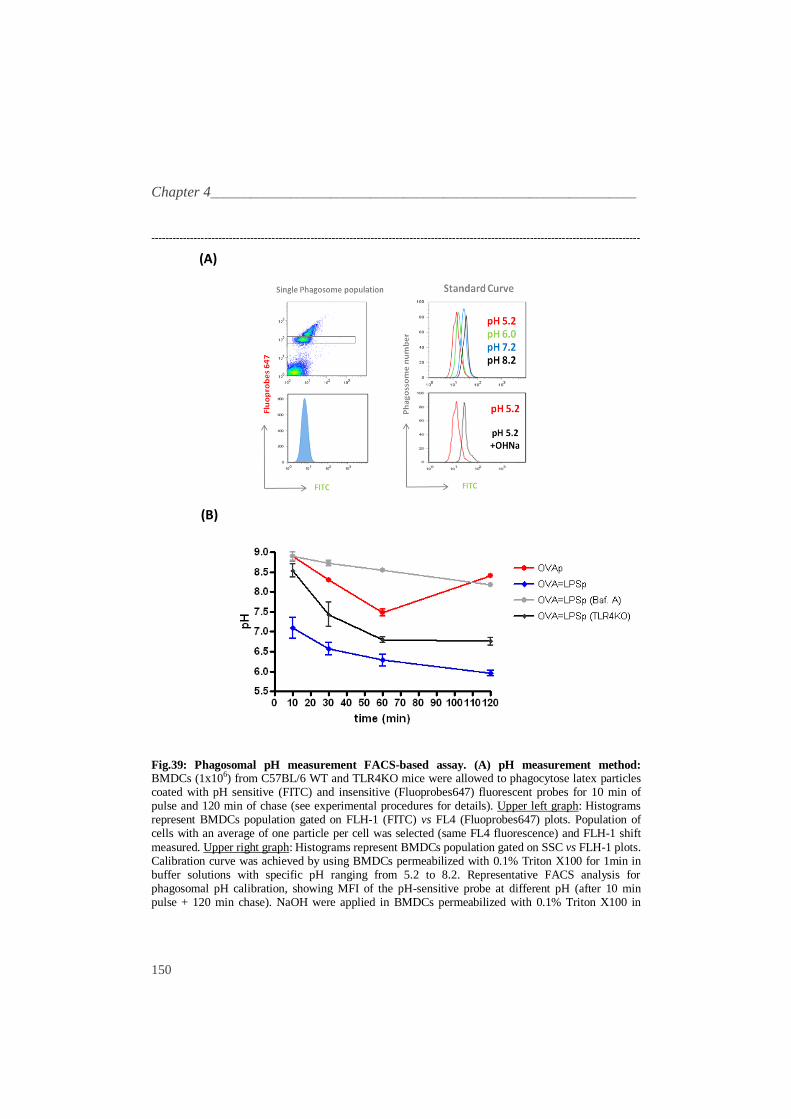
Preliminary data revealed changes in the magnitude of OVA antigen cross-
presentation by sDCs according to the size of the synthetic particles used. These
data showed that 1.0µm particles are cross-presented more efficiently by DCs,
being evident at lower ratios (Fig.30). Since particles size also affects the amount
of antigen taken up by cells (Desai et al., 1997), the differences in antigen cross-
presentation could simply be attributed to the total amount of antigen internalized
by DCs. Thus, to be able to address other type of conclusions, the amount of OVA
covalently attached to the surface of the particles should be normalized, as well as
the amount of internalized OVA using fluorescent-labeled OVA. The uptake of
particles > 0.5 μm in size is termed phagocytosis, whereas particles < 0.5 μm are
___________________________________________________________________Results
137
taken up by receptor-mediated endocytosis or pinocytosis (Rejman et al., 2004). To
address if there were differences in the uptake by phagocytosis of the different
particles size used, the amount of antigen internalized by phagocytosis was
measured. Polystyrene particles of various diameters (0.5, 1.0 and 3.0µm)
covalently coupled with fluorescent OVA-Alexa488 (Molecular Probes) were
used. Phagocytosis was determined by a FACS based assay. Quantification was
performed by determining the geometric mean of fluorescence in the FLH-1
channel, using Flowjo software (Treestar, Inc.).
------------------------------------------------------------------------------------------------------------------------------------------
0
250
500
750
1000
4ºC
37ºC/Cyto.D
37ºC
0.5=OVA488 1.0=OVA488 3.0=OVA488
MF
I (F
L1-H
)
Fig.31: Size dependent quantitative uptake of fluorescent particles antigen. Fluorescent OVA (OVA-Alexa Fluor 488 - OVA488) was covalently coupled to model particles of 0.5 µm (0.5=OVA488), 1µm (1.0=OVA488) and 3.0µm (3.0=OVA488) in size. Particles were incubated with sDCs (5x10⁴) from C57BL/6 at 1:10 (sDC:particles) ratio for initial 10 minutes of pulse and 2hrs of chase (phagocytosis). Cyto. D (10nM), a phagocytosis inhibitor, was incubated 1hr previously to particle addition. Quantitative uptake (Mean fluorescence intensity–MFI) of green fluorescent particles antigen by sDCs was examined by FACS in the FLH-1 channel. The graph represents the
average + 1SD of three independent experiments. No statistically significant differences (P>0.05) were observed between amounts of fluorescent OVA of different size particles internalized by sDCs. ------------------------------------------------------------------------------------------------------------------------------------------
There was no significant difference in the amount of antigen of the different
size particles that were phagocyted by DCs (fig.31). This result supports the
Chapter 4________________________________________________________________
138
evidence that the difference in antigen cross-presentation efficiency observed
mediated by LPS is not due to the difference on the amount of antigen internalized.
To determine whether the difference in the efficiency on particles size antigen
cross-presentation is related to antigen processing/loading or transport to cytosol,
we made use as antigen, SIINFEKL peptide (MHC class I–restricted OVA peptide)
instead of OVA protein. A similar approach was performed for SIINFEKL peptide
antigen presentation as used for OVA protein.
------------------------------------------------------------------------------------------------------------------------------------------
Fig.32: SIINFEKL particles size dependent antigen cross-presentation FACS-based assay. sDCs (2.5x10⁴) from C57BL/6 mice were incubated with a broad range size of polystyrene particles (0.05µm – 6.0µm) covalently coupled with SIINFEKL, at a low ratio (sDC:particles – 1:2). OT-I T-Cells (1x10⁵) stained with CFSE were co-incubated with sDCs. T-Cell proliferation was measured by
FACS at day 3. Equimolar molecules of SIINFEKL peptide (comparing to OVA protein used on previous assays), were coupled to particles by covalent chemistry. Histograms represent T-Cell population gated on SSC vs CFSE plots. Open grey line plots represent control OT-I T-Cells that do not divide and blue filled plots represent OT-I T-Cells proliferation under SIINFEKLp stimulus. Numbers represent the percentages of the proliferating cells of total OT-I T-Cells. These data are representative from one experiment repeated at least three times with similar results. ------------------------------------------------------------------------------------------------------------------------------------------
SIINFEKL particles (SIINFEKLp) induced higher and similar levels of T-Cell
proliferation / activation, even at low ratios of sDC:particles (1:2) and among the
different range of particle sizes used (0.05µm-6.0µm) (fig.32). These data support
the assumption that the different efficiency on particle antigen cross-presentation,
due to different forms of uptake, may not be related to the amount of antigen
internalized but instead on intracellular processing/loading mechanism of different
___________________________________________________________________Results
139
antigen particles size. Data obtained with different OVA particles size showed that
the 1.0 µm particle-containing phagosomes are the most efficient on antigen cross-
presentation (fig.30). We can hypothesize that the size of antigen carriers plays a
critical role in directing antigen to the MHC class-I antigen presentation pathway.
From now on, 1.0µm particles were used as a model size for further studies to
better dissect the antigen cross-presentation mechanism.
TLR signaling on antigen cross-presentation _ _
As the cellular mechanism of antigen cross-presentation is not well understood,
as well the possible specificity of a specific stimulus, we have proposed to
understand how TLR agonists in particulate antigens may influence their cross-
presentation. To address the role of pathogen-like particles in antigen cross-
presentation, model agonists for TLR4 and TLR9 were used, representing these
receptors exogenous and endogenous pathogen “sensors” respectively. CpG oligo
(a TLR9 agonist) or LPS (a TLR4 agonist) were covalently attached to Ova-
conjugated particles using the EDC/HNS coupling reaction. Antigen model
particles and a FACS-proliferation based antigen cross-presentation assay were
used.
Chapter 4________________________________________________________________
140
------------------------------------------------------------------------------------------------------------------------------------------
Fig.33: TLR model particles antigen cross-presentation FACS-based assay. sDCs (2.5x10⁴) from C57BL/6 mice were co-incubated with model particles (OVAp, OVA=CpGp and OVA=LPSp) at 1:20 and 1:10 (sDC:particles) ratios, and with OT-I T-Cells (1x10⁵) for 3 days. T-Cell proliferation was measured using CFSE staining by FACS. Histograms represent T-Cell population gated on SSC
vs CFSE plots. Open grey line plots represent control OT-I T-Cells that do not divide and blue filled plots represent OT-I T-Cells proliferation under specific stimulus. Numbers represent the percentages of the proliferating cells of total OT-I T-Cells. These data are representative from one experiment repeated at least three times with similar results. ------------------------------------------------------------------------------------------------------------------------------------------
Surprisingly, these preliminary data revealed that antigen cross-presentation of
OVA particles is almost abolished when TLR agonists (CpG and LPS) are in the
same cargo (fig.33). However, this phenotype was not as evident at higher ratios as
1:50 (data not shown), which could be due to “saturation” of the DCs antigen
presentation machinery. As a result, proliferation of CD8+ T-Cells was significantly
hampered in cells incubated with OVA=CpGp or OVA=LPSp conjugated particles
at different ratios (1:10 and 1:20), when compared to cells incubated with “naked”
OVAp (fig.33). Similar results were obtained, in a minor level, when BMDCs were
used as model DCs instead of sDCs (data not shown).
___________________________________________________________________Results
141
Antigen cross-presentation phenotype using TLR KD models _______
To address if this phenotype is TLR dependent or due to other different
mechanism or even an “artifact”, sDCs from TLR4KO mice and BMDCs
Knocked-Down (KD) for TLR4 or TLR9 by shRNA lentivirus were used. sDCs
were isolated from WT and TLR4KO C57BL/6 mice as described previously, and
model particles (OVAp and OVA=LPSp) were used to measure OT-I T-Cell
proliferation, as shown in fig.34.
------------------------------------------------------------------------------------------------------------------------------------------
Fig.34: TLR model particles antigen cross-presentation FACS-based assay: WT vs TLR4KO DCs. sDCs (2.5x10⁴) from C57BL/6 WT and TLR4KO mice were co-incubated with model particles (OVAp and OVA=LPSp) at 1:10 (sDC:particles) ratio and with OT-I T-Cells (1x10⁵) for 3 days. T-
Cell proliferation was measured using CFSE staining by FACS. Histograms represent T-Cell population gated on SSC vs CFSE plots. Open grey line plots represent control OT-I T-Cells that do not divide and blue filled plots represent OT-I T-Cells proliferation under specific stimulus. Numbers represent the percentages of the proliferating cells of total OT-I T-Cells. These are representative data from one experiment repeated at least three times with similar results. ------------------------------------------------------------------------------------------------------------------------------------------
As expected by previous data, antigen cross-presentation of WT sDCs was
abolished when LPS is in the same cargo as OVA particles. However, antigen
cross-presentation of OVA=LPSp by TLR4KO sDCs was not impaired, instead,
Chapter 4________________________________________________________________
142
relative higher proliferation rates of OT-I T-Cells were observed (fig.34).
Moreover, OVAp antigen cross-presentation phenotype (OT-I T-Cell proliferation)
was recovered for OVA=LPSp in sDCs deficient on TLR4. These data showed that
the effect of particulate LPS in antigen cross-presentation phenotype is TLR4
dependent, suggesting the specificity of the LPS agonist through TLR4 signaling
pathway. The TLR4KO proliferation data, where OVA=LPSp induce higher levels
of T-Cell proliferation comparing to OVAp, suggest that particulate LPS in the
absence of TLR4 could signal through other type of receptor, thus increasing (even
in a low magnitude) antigen cross-presentation (fig.34- lower panels). In order to
extend these studies to other type of DCs, BMDCs and shRNA lentivirus were
used to Knock-Down TLR4 and TLR9.
------------------------------------------------------------------------------------------------------------------------------------------
___________________________________________________________________Results
143
Fig.35: TLR model particles antigen cross-presentation FACS-based assay: TLR4 and TLR9 KD in DCs using shRNA. BMDCs (5x10⁴) from C57BL/6 mice were transduced with 5 different lentivirus encoding shRNAs targeting TLR4 (upper graph) and TLR9 (lower graph). A random sequence (siSCRAM) was used as control. After selection at day 2 with puromycin, BMDCs were co-incubated at day 6 with model particles (OVAp, OVA=CpGp and OVA=LPSp) at 1:10 (BMDCs: particles) ratio and OT-I T-Cells (1x10⁵) for 3 days. At day 6, BMDCs were analyzed by FACS
staining for TLR4 and TLR9 to evaluate the KD of TLR4 and TLR9. T-Cell activation was measured by ELISA for IFN-γ, using supernatants at 60-65 hrs. The graphs represent the average + 1SD of three independent experiments. The asterisks represent statistically significant differences between OVAp and OVA=LPSp and OVAp and OVACpGp for the same shRNA (*P<0.05;**P< 0.01). ------------------------------------------------------------------------------------------------------------------------------------------
As expected, the levels of T-Cell activation (IFN-γ production) by BMDCs
were lower comparing to sDCs (see next figure- fig.36). These data in BMDCs
shown that T-Cell activation was decreased when OVA=LPSp and OVA=CpGp
were used as particle antigen comparing to “naked” OVAp (fig.35), reproducing
the same results obtained with sDCs (fig.33). The results may suggest that this
phenotype is “transversal” to DCs populations, therefore proving BMDCS to be a
good model for the use of shRNA interference tool in antigen cross-presentation
phenotype characterization. Using two or more shRNAs constructs to target TLR4
and TLR9 on BMDCs (where KD efficiency was previously confirmed), in the
presence of OVA=LPSp or OVA=CpGp respectively, the antigen cross-
presentation phenotype was recovered (levels of OT-I T-Cell activation were
similar as observed for “naked” OVAp), suggesting the specificity effect of the
TLR agonists (LPS and CpG) through their TLR signaling pathway (fig.35).
Chapter 4________________________________________________________________
144
MHC class-II antigen presentation pathway _ _
LPS was used as the “model” TLR agonist aiming to better characterize the
antigen cross-presentation phenotype and to try to dissect the underlying
mechanism. We decided to use LPS for different reasons, mainly because: 1) it is
one of the most used TLR agonists and TLR4 is the usual target of immunologists
and vaccinologists, 2) it is easy to conjugate to different type of model particles, 3)
the nature of its receptor, TLR4 is unique among TLRs, because signals through
two different adaptors, MyD88 and TRIF.
As MHC class-II is the classical pathway for extracellular antigen presentation
and in order to have a mechanistic control comparing to antigen cross-presentation,
we next addressed the role of LPS on MHC class-II presentation. Therefore,
activation of OT-II Th1 Cells (CD4+) and OT-I cytotoxic T-Cells (CD8
+) by DCs
was addressed by measuring their IFN-γ production.
------------------------------------------------------------------------------------------------------------------------------------------
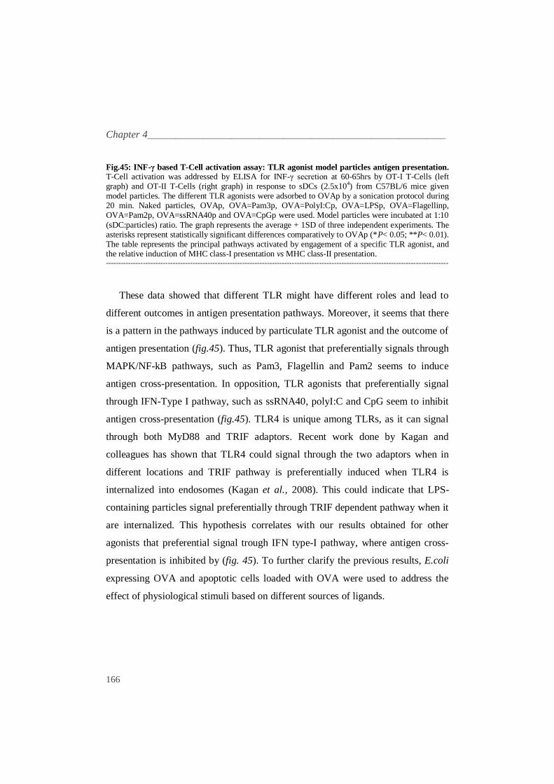
Fig.36: Antigen presentation: INF-γ based T-Cell activation assay. T-Cell activation was addressed by ELISA for INF-γ secretion at 60-65hrs by OT-I T-Cells (left graph) and OT-II T-Cells (right graph) in response to sDCs (2.5x10⁴) from C57BL/6 mice given diverse stimuli. Particulate antigens: Naked particles, OVAp and OVA=LPSp at 1:10 (sDC:particles) ratio; soluble antigen:
OVA endograde (100ng/µl), SIINFEKL peptide (1ng/µl) and OVA4 peptide (1ng/µl) were used.
___________________________________________________________________Results
145
Soluble LPS was used at 10ng/µl. The graph represents the average + 1SD of three independent experiments. The asterisks represent statistically significant differences comparatively to OVAp (*P< 0.05; **P< 0.01 *** P< 0.001). ------------------------------------------------------------------------------------------------------------------------------------------
The ELISA data for IFN-γ produced by OT-I T-Cells, reproduced the previous
results obtained for OT-I T-Cell proliferation (fig.33). Thus, in the presence of LPS
in the same cargo as particle antigen, OT-I T-Cells activation (IFN-γ) was
decreased approximately 3 times comparing to OVAp. Moreover, OVA=LPSp
induced OT-II T-Cell proliferation, approximately 3 times, comparing to OVAp
(fig.36). The induction observed on MHC class-II antigen presentation pathway
mediated by LPS on same cargo as particle antigen corroborates previous data
obtained by Blander et.al (Blander and Medzhitov, 2006b). It is interesting to
notice that the relative antigen presentation for OVAp in absence of LPS stimulus
is higher in MHC-class I when comparing to MHC class-II. When soluble LPS was
co-incubated with OVAp (two different stimuli) there was an increase in T-Cell
activation (IFN-γ) and proliferation (FACS analysis - data not shown) in both
MHC class-I and MHC class-II contexts. Furthermore the magnitude was much
higher for MHC-class-II (~ 4.5x) when compared to MHC class-I (~1.25x) (fig.33).
These data suggest that LPS when in same cargo as antigen impairs antigen cross-
presentation and dictates a shift to MHC class-II antigen presentation.
TLR signaling in a different physical form _
Concerning the physical nature of stimulus (particulate vs soluble), we next
addressed if the efficiency of presenting antigens from phagocytosed particles is
dependent on the presence of TLR4 agonist within the antigen cargo. T-Cell
activation was measured using IFN-γ ELISA assay.
Chapter 4________________________________________________________________
146
------------------------------------------------------------------------------------------------------------------------------------------
Fig.37: INF-γ based T-Cell activation assay: Particle antigen presentation using different physical LPS stimuli. T-Cell activation was addressed by ELISA for INF-γ secretion at 60-65hrs by OT-I T-Cells (left graph) and OT-II T-Cells (right graph) in response to sDCs from C57BL/6 mice given particulate antigens: BSAp, BSA=LPSp, OVAp and OVA=LPSp. Particles were used at 1:10
(sDC:particle) ratio. Soluble LPS was used at 10ng/µl. The graph represents the average + 1SD of three independent experiments. The asterisks represent statistically significant differences comparatively to OVAp (*P< 0.05; **P< 0.01 *** P< 0.001). ------------------------------------------------------------------------------------------------------------------------------------------
These data suggest that when LPS is in a particulate form, but in a different
cargo as particle antigen, there is no significant difference between these two
stimuli (OVAp and OVAp + BSA=LPSp), either in MHC class-I and MHC class-II
antigen presentation (fig.37). In opposition, when LPS is present in a soluble form,
(fig.36 and 37), antigen presentation is induced in both MHC class-I and MHC
class-II context, with higher magnitude for MHC class-II presentation. As a
control, model particles with OVA labelled with a fluorescent dye were used, to
ensure that the amount of antigen internalized was the same when sDCs where co-
cultured with OVAp alone or with OVAp and BSAp. However, no significant
differences were observed (data not shown). Our results suggest that soluble LPS
is able to activate all the antigen presentation machinery (up-regulation of
___________________________________________________________________Results
147
molecules involved in antigen presentation – MHC class-I and II and co-stimulatoy
molecules). However, when LPS is in particulate state, relies the concept of
“phagosome autonomous maturation” in antigen presentation (Blander and
Medzhitov, 2006a), where distinct phagosomes are processed individually on the
same cell. Thus, the TLR agonist must be in same cargo as antigen to signal a
specific pathogen-mediated antigen fate.
To better characterize the antigen cross-presentation phenotype, some “key”
steps in antigen presentation should be dissected. We have proposed to characterize
important mechanisms, such as uptake ratio, antigen processing and phagosome
maturation mediated by antigen model particles. To control if the efficiency on
antigen cross-presentation is due to different capacities on particle antigen
internalization, we addressed the role of phagocytic ability on DCs mediated by
LPS stimulus.
Phenotype characterization I - Uptake__________________________________
It has been suggested that the efficiency of antigen cross-presentation is due to
differences at phagocytic capacity level (Albert et al., 1998). As the amount of
antigen that was internalized by DCs could influence the antigen presentation, the
question if TLR agonists (more specifically LPS) influenced this process should be
addressed. The implication of TLR signaling in phagocytosis and antigen uptake
has been described but is quite controversial (Diwan et al., 2003; Khan et al., 2007;
Weck et al., 2007). In order to address the possible involvement of TLR signaling
in antigen uptake, phagocytosis of antigen model particles using OVA labeled with
alexa 488 (OVA488) as model antigen was measured using a FACS-based assay.
Chapter 4________________________________________________________________
148
------------------------------------------------------------------------------------------------------------------------------------------
Fig.38: Uptake assay of model particles: FACS: Measurement of antigen uptake by sDCs from C57BL/6 WT and TLR4KO mice using green fluorescent OVA (OVA488) loaded particles (OVA488p and OVA488=LPSp). Particles were co-incubated with sDCs at 1:10 (sDC:particles) ratio for initial 10 min of pulse and 2hrs of chase (phagocytosis). Soluble LPS was used at 10ng/µl. Cyto. D (10nM) and LPS (10ng/ml) were incubated 1hr previously to particle addition. MFI in the FLH-1 channel was calculated for phagocytic cells. The graph represents the average + 1SD of three
independent experiments. No statistically significant differences (P>0.05) were observed between amounts of fluorescent OVA488 model particles internalized by sDC from C57BL/6 WT and TLR4KO mice. Confocal images: Particles were co-incubated with BMDCS (plated in cover slips 12hrs before to allow adhering) from C57BL/6 mice at 1:10 (sDC:particles) ratio during 10 min (pulse). After 2 hrs of incubation (chase), PFA1% was added during 5 min and cells were washed with PBS. Cyto. D (10nM) was incubated 1hr previously to OVA=488p addition. Phalloidin red was added at 1:40 dilution during 20 min, and BMDCs mounted in coverslips with vectashield medium for confocal analysis with a 63x objective (Scale bar, 2 µm). ------------------------------------------------------------------------------------------------------------------------------------------
The FACS assay showed that the uptake capacity of sDCs was similar for OVA
model particles (OVAp, OVA=LPSp and for OVAp + soluble LPS) and between
WT and TLR4KO sDCs populations (fig.38). Therefore, TLR4 signaling does not
affect in a significant way particle antigen internalization, when LPS is present
either in same cargo as antigen or in a soluble form. As such, we may assume that
the amount of antigen that reached the phagosomes by model particles is
___________________________________________________________________Results
149
approximately the same. Therefore, the differences observed in antigen
presentation studies should not be due to differences in phagocytosis uptake of
model particles used.
Phenotype characterization II – Phagosome maturation (pH) _
Efficient antigen processing in phagosomes requires limited and controlled
antigen degradation, generating the correct array of peptides to be loaded on MHC
molecules and to avoid total destruction by unspecific lysosomal proteases (Savina
et al., 2006). Endosomal pH regulation is one of the most direct ways to control the
lysosomal proteases activity (Delamarre et al., 2005) and it has been described to
be involved in antigen presentation (Blander and Medzhitov, 2006a).
In order to address the role of LPS on pH of phagosome containing particle
antigen, we proposed to measure phagosomal pH in DCs accurately by a FACS
assay, using pH-sensitive particles, developed and already published by
Amigorena´s lab (Savina et al., 2006). Briefly, latex 3.0µm amino particles
adsorbed with OVA were coated with pH-sensitive (Fluorescein isothiocyanate –
FITC) and pH-insensitive (FluoProbe647) fluorescent dyes. After different time
points of particles phagocytosis, the fluorescence intensity of the two dyes was
quantified using a FACS-based assay. Population of DCs with an average of one
particle internalized was selected on FluoProbe647 channel (FL4). The differences
in pH was measured by deviations on FITC channel (FLH-1), reflecting the pH in
the phagosomal environment. The absolute value of phagosomal pH was calibrated
with a standard curve established in cells permeabilized with 0.1% Triton X100
and immersed in buffers of fixed calibrated pH (Fig. 39-A).
Chapter 4________________________________________________________________
150
------------------------------------------------------------------------------------------------------------------------------------------
(A)
(B)
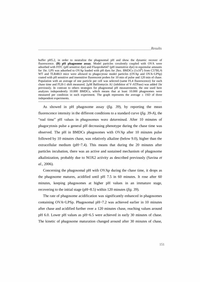
Fig.39: Phagosomal pH measurement FACS-based assay. (A) pH measurement method: BMDCs (1x10⁶) from C57BL/6 WT and TLR4KO mice were allowed to phagocytose latex particles coated with pH sensitive (FITC) and insensitive (Fluoprobes647) fluorescent probes for 10 min of pulse and 120 min of chase (see experimental procedures for details). Upper left graph: Histograms represent BMDCs population gated on FLH-1 (FITC) vs FL4 (Fluoprobes647) plots. Population of cells with an average of one particle per cell was selected (same FL4 fluorescence) and FLH-1 shift
measured. Upper right graph: Histograms represent BMDCs population gated on SSC vs FLH-1 plots. Calibration curve was achieved by using BMDCs permeabilized with 0.1% Triton X100 for 1min in buffer solutions with specific pH ranging from 5.2 to 8.2. Representative FACS analysis for phagosomal pH calibration, showing MFI of the pH-sensitive probe at different pH (after 10 min pulse + 120 min chase). NaOH were applied in BMDCs permeabilized with 0.1% Triton X100 in
___________________________________________________________________Results
151
buffer pH5.2, in order to neutralize the phagosomal pH and show the dynamic recover of fluorescence. (B) pH phagosome assay. Model particles covalently coupled with OVA were adsorbed with FITC (pH sensitive dye) and Fluoprobe647 (pH insensitive dye) in equimolar amounts for 1hr. LPS was adsorbed to OVAp loaded with pH dyes for 2hrs. BMDCs (1x10⁶) from C57BL/6 WT and TLR4KO mice were allowed to phagocytose model particles (OVAp and OVA=LPSp) coated with pH sensitive and insensitive fluorescent probes for 10 min of pulse and 120 min of chase.
Population with an average of one particle per cell was selected (same FL4 fluorescence) for each chase time and FLH-1 shift measured. 2µM Bafilomycin A1 (inhibitor of V-ATPase) was added 1hr previously. In contrast to others strategies for phagosomal pH measurements, the one used here analyzes independently 10.000 BMDCs, which means that at least 10.000 phagosomes were measured per condition in each experiment. The graph represents the average ± 1SD of three independent experiments. ------------------------------------------------------------------------------------------------------------------------------------------
As showed in pH phagosome assay (fig. 39), by reporting the mean
fluorescence intensity in the different conditions to a standard curve (fig. 39-A), the
“real time” pH values in phagosomes were determined. After 10 minutes of
phagocytosis pulse a general pH decreasing phenotype during the chase time was
observed. The pH in BMDCs phagosomes with OVAp after 10 minutes pulse
followed by 10 minutes chase, was relatively alkaline (below 9.0), higher than the
extracellular medium (pH~7.4). This means that during the 20 minutes after
particles incubation, there was an active and sustained mechanism of phagosome
alkalinization, probably due to NOX2 activity as described previously (Savina et
al., 2006).
Concerning the phagosomal pH with OVAp during the chase time, it drops as
the phagosome matures, acidified until pH 7.5 in 60 minutes. It rose after 60
minutes, keeping phagosomes at higher pH values in an immature stage,
recovering to the initial stage (pH~8.5) within 120 minutes (fig. 39).
The rate of phagosome acidification was significantly enhanced in phagosomes
containing OVA=LPSp. Phagosomal pH~7.2 was achieved earlier in 10 minutes
after chase and acidified further over a 120 minutes chase, reaching values around
pH 6.0. Lower pH values as pH~6.5 were achieved in early 30 minutes of chase.
The kinetic of phagosome maturation changed around after 30 minutes of chase,
Chapter 4________________________________________________________________
152
and was kept at slower rate until longer time points of incubation (120 minutes),
decreased only 0.5 units from pH 6.5 to pH 6.0 (fig. 39).
In the presence of Bafilomycin A, a V-ATPase inhibitor, the higher phagosomal
acidification kinetics induced by OVA=LPSp was blocked. The pH decreased only
0.5 units from pH~8.9 – pH~8.4, during the entire chase time (120 minutes). This
result could imply V-ATPase in phagosomal pH acidification mediated by LPS
signaling. In the absence of TLR4 (BMDCs from TLR4 KO), the rate of
phagosome maturation induced by OVA=LPSp particles was decreased, with a 0.8
pH units of difference at the end of chase time. A slower rate of phagosome
maturation mediated by OVA=LPSp was observed, from pH 8.5 at 10 minutes to
pH 7.0 in 120 minutes of chase. After 30 minutes, the pH achieved a “plateau”
(lowest value - pH~6.8) and remained quite stable during the rest of chase.
However, the phagosomal pH did not rise again to the initial stage as observed in
absence of LPS stimulus on WT BMDCs, within 120 minutes (fig. 39). Therefore,
the phagosome acidification related to OVA=LPSp may not totally dependent on
TLR4-mediated signaling. Moreover, this observation implies TLR4 signaling on
phagosomal acidification mediated by LPS
The control and regulation of phagosomal pH is mediated in majority by the
function of two complexes, V-ATPase and NADPH Oxidase NOX2 (Savina et al.,
2006). Amigorena´s lab described NOX2 as a specific adaptation of DCs endocytic
pathway to the antigen presentation function by causing an active and sustained
phagosome alkalinization, keeping pH at relative high values (Savina et al., 2006).
The NOX2 generates ROS, causing transient phagosome alkalinization, in part
through the consumption of protons in the phagocytic lumen (Lee et al., 2003;
Segal, 2005). To better address the role of LPS signal within the cargo on
phagosome maturation, we proposed to measure the phagosomal ROS production
in order to correlate with phagosomal pH data.
___________________________________________________________________Results
153
Phenotype characterization II – Phagosome maturation (ROS) _
To address ROS production, specifically in phagosomes containing OVAp and
OVA=LPSp, dihydrorhodamine 123 (DHR123), a dye that only emits fluorescence
under oxidative conditions (Vowells et al., 1995), was covalently linked to OVAp
and OVA=LPSp. DHR123 is a pH stable molecule, thus the possibility of DHR123
degradation in phagosomal lumen could be excluded (Savina et al., 2006).
------------------------------------------------------------------------------------------------------------------------------------------
Fig.40: Phagosomal ROS
measurement based assay. Measurement of phagosomal ROS production using DHR123 coupled to model particles (OVA=DHR123p and OVA=DHR123=LPSp).
MFI of FLH-1 channel was calculated for phagocytic cells. 2x10⁵ BMDCs per time point were pulsed for 10 min in CO2 independent medium using 1:10 (BMDCs:particles) ratio and chased for 5 min, 15 min, 30 min, 60 min and 120 min. The graph represents the
average ± 1SD of three independent experiments.
------------------------------------------------------------------------------------------------------------------------------------------ As shown by FACS analysis, DHR-coated particles became fluorescent after
phagocytosis, showing the production of ROS in DCs phagosomes. Moreover,
ROS production in phagosomes containing model particles decreased during the
chase time (fig.40). This means that phagosomal environment in DCs is oxidative
due to ROS generation. ROS production in phagosomes containing OVAp was
higher comparing to phagosomes containing OVA=LPSp, even at earlier chase
time points (5 minutes) and during the entire chase time (120 minutes) (fig.40). It
seems that the production of ROS decreased during the phagosome maturation
mediated by model particles and it was more evident in phagosomes containing
Chapter 4________________________________________________________________
154
OVA=LPSp. After 60 minutes of chase, the ROS production on phagosomes
containing OVAp seemed to stabilize, with a tendency to recover even with low
rate. However, ROS production in OVA=LPSp phagosomes appeared to decrease
continually until the end of chase, with a higher rate (fig.40). As a result, these data
showed that when LPS is in same cargo as OVA particles mediates the reduction of
ROS production in phagosomes, which could be related to the higher rate of
phagosome maturation/acidification induced by OVA=LPSp during the
experimental time points (fig. 39). These data support the evidence showed by the
pH results, which suggested that “naked” OVAp kept the phagosomes in a
immature stage comparing to OVA=LPSp which enhance phagosome maturation
(fig.39 and 40).
Phenotype characterization III – Phagosome maturation (Confocal) _
DCs have been shown to have lower contents of lysosomal proteases and
acidification mechanism (Delamarre et al., 2005). In order to link phagosomal
maturation with the pH regulation and ROS production profiles obtained above
with model particles (fig.39 and 40), the intracellular localization of model
particles was examined by confocal microscopy. Phagosome maturation ratios with
different cargos was assessed by fluorescent microscopy, by qualitatively merging
the phagosomes containing OVA488 fluorescent particles (OVA-Alexa488 - pH
stable fluorescence) and lysosomes labeled with Lysotracker Red (Invitrogen).
This colocalization could be quantified by determining the total colocalization area
(pixel quantification using the ImageJ plugin RG2B colocalization which creates a
binary mask for colocalizing pixels). Lysotracker probes have high selectivity for
labeling and tracking acidic organelles in live cells, as the result of protonation,
results in a pH dependent increase of fluorescent intensity (Shiratsuchi et al.,
2004).
___________________________________________________________________Results
155
------------------------------------------------------------------------------------------------------------------------------------------
Fig.41: Confocal imaging based phagosomal maturation assay. Representative fluorescent micrographs showing phagocytosis of OVA488 fluorescent particles (green) and lysosomes (red). BMDCs from C57BL/6 mice were incubated with 1µm OVA488 fluorescent polystyrene model particles (OVA488p and OVA488=LPSp) at a ratio (BMDCs:particles) of 1:10 for 15 min, washed, and further incubated for either 30 min or 120 min. BMDCs were stained with Lysotracker Red to label lysosomes and DAPI (blue) to label nuclei and fixed with 4% of paraformaldehyde. BMDCs
Chapter 4________________________________________________________________
156
were washed and mounted in coverslips with vectashield medium for confocal analysis with a 63x objective. Merged images are shown in yellow and colocalization in white with values of total pixel colocalization. Slides were examined by confocal microscopy to determine the intracellular localization. The color images represent particles (green), lysosomes (red), lysosomes fused with phagosomes containing particles (yellow). NH4Cl (50mM) were used as a weak base that raises the pH of acidic compartments to disrupt the low pH at lysosomes and worked as a loading control.
Images are representative at least from three independent experiments (Scale bar, 1µm). ------------------------------------------------------------------------------------------------------------------------------------------
The confocal data showed that phagosomes containing OVA488=LPSp mature
with higher ratio than those containing OVA488p (fig.41). This observation is
independent of the phagocytic index, which is not different for OVAp and
OVA=LPSp and not markedly agonist specific (fig.38). After 30 minutes of
incubation, OVAp did not colocalize with lysosomes and phagosomes exhibited an
alkaline environment where the pH is around 8.5 (fig.39). Whereas the majority of
OVA=LPSp colocalize with a higher extend (Fig.41) showing an average pH of
6.5 (fig.39). Even after 120 minutes incubation, phagosomes containing OVAp had
reduced values of colocalization with lysosomes (Fig.41), with higher pH values
around 8.5 (fig.39). In contrast, phagosomes containing OVA=LPSp completely
merged with lysosomes, indicating that internalized particles were routed into
lysosomes (Fig.41), where phagosomal pH reaches lower values as pH 6.0 (fig.39).
These data indicate that LPS-containing phagosomes completely mature into
phagolysosomes. As a phagosomal maturation control, NH4Cl was used as a weak
base that raises the pH of acidic compartments to disrupt the low pH. Therefore,
the colocalization between OVA488=LPSp and lysosomes was abolished in the
presence of NH4Cl (fig.41), suggesting that the fusion was mediated by lower pH
values. In addition, OVA488=LPSp and lysosomes appears to be concentrated on
the periphery of the cell nucleus (labeled blue by DAPI), a feature of late
endosomal and lysosomal compartments (Tran and Shen, 2009). Moreover, these
results are reliable with the phagosomal pH profiles obtained before (fig.39).
___________________________________________________________________Results
157
Phenotype characterization IV – Antigen Processing / Degradation _
Phagosomal pH regulation (either by V-ATPase or NOX2) is the main
mechanism used by DCs to “protect” peptides from complete degradation, with the
ultimate goal being to generate T-Cell epitopes (Savina et al., 2006; Trombetta et
al., 2003). Therefore, we next sought to evaluate the role and functional
consequences of such a drop in phagosomal pH and ROS production mediated by
LPS in antigen processing/degradation.
------------------------------------------------------------------------------------------------------------------------------------------
Chapter 4________________________________________________________________
158
Fig.42: Antigen processing/degradation based assays. A) Quantitative antigen processing/ degradation was examined by FACS by measuring the MFI of DQ-OVA in the FLH-1 channel. sDCs (1x10⁵) from C57BL/6 mice were challenged with particles containing DQ-OVA (DQ-OVAp) and DQ-OVA + LPS (DQ-OVA=LPSp) at 1:10 (sDC:particles) ratio during 2hr. DQ-OVA (100ng/ml) and LPS (10n/ml) were adsorbed to 1.0 µm polystyrene particles during 2hr. Proteasome inhibiting drug, MG-132 (Z-leu-Leu-Leu-CHO) and the inhibitor of Cathepsin S (Z-FL-COCHO), were added
1hr previously. The asterisks represent statistically significant differences (*P< 0.05; **P< 0.01). B) sDCs (1x10⁵) from C57BL/6 mice were incubated with model particles (OVAp and OVA=LPSp) at 1:10 (sDC:particles) ratio during 30 and 120 min. OVA were detected using anti-OVA monoclonal antibody (Abcam). Bradford normalization was performed. The chase with particles was performed without washing the remaining particles after pulse (long course kinetic). 30 μg of total protein extracts from sDCs were separated by 10% SDS-PAGE. C) sDCs (1x10⁵) from C57BL/6 mice were pulsed with streptavidin particles coupled with OVA-biotin (OVAp) and OVA-biotin + LPS (OVA=LPSp) at 1:10 (sDC:particles) ratio for 10 min, washed extensively and chased during 30 min
and 120 min. OVA-biotin (100ng/ml) and LPS (10ng/ml) were adsorbed to 1.0 µm polystyrene particles during 2hr. OVA-biotin from whole cell lysates after particles phagocytosis was detected by Western blot using Sav-HRP from ELISA kit. As a loading control β-actin staining was performed. Lanes: 1) Control: only sDCs 2) OVAp 30 min + MG132 3) OVA=LPSp 30 min + MG132 4) OVAp 30 min 5) OVA=LPSp 30 min 6) OVAp 2hrs + MG132 7) OVA=LPSp 2hrs + MG132 8) OVAp 2hrs 9) OVA=LPSp 2hrs. 30 μg of total protein extracts from sDCs were separated by 10% SDS-PAGE. D) Antigen retro-translocation assay (availability in cytosol) using cyt c particles (apoptosis assay). Horse Heart cyt c and LPS was adsorbed to 1.0µm polystyrene particles during 2hr. BMDCs (1x10⁵)
from C57BL/6 mice were challenged with particles loaded with cyt c (Cyt.cp) and particles loaded with cyt c and LPS (Cyt.c=LPSp) at 1:10 (sDC:particles) ratio during 30 min and washed extensively. The amount of free cyt c in cytosol was measured by apoptosis assay using Annexin V-FITC kit at 18hrs. Apoptosis was examined by FACS measuring the MFI in the FLH-1 channel. The asterisk represents statistically significant differences (*P< 0.05). E) sDCs (2.5x10⁴) from C57BL/6 mice were co-incubated with model particles (SIINFEKLp, SIINFEKL=LPSp) at 1:2 (sDC:particles) ratio, and OT-I T-Cells (1x10⁵) for 3 days. T-Cell proliferation was measured using CFSE staining by FACS. Histograms represent T-Cell population gated on SSC vs CFSE plots. Open grey line plots represent control OT-I T-Cells that do not divide and blue filled plots represent OT-I T-Cells
proliferation under specific stimulus. Numbers represent the percentages of the proliferating cells of total OT-I T-Cells. These data are representative from one experiment repeated at least three times with similar results. ------------------------------------------------------------------------------------------------------------------------------------------
Using the performed assays, we are not able to distinguish between antigen
processing (peptide epitope generation) and degradation (random cleavage). DCs
have reduced expression levels and low recruitment of proteolytic enzymes to
phagosomes, which leads to low lysosomal proteolysis (Delamarre et al., 2005;
Jancic et al., 2007) and a limited phagosomal acidification by V-ATPase (Savina et
al., 2006; Trombetta et al., 2003). Therefore, we next sought to address the role of
proteasome as an important protease catalytic complex in particle antigen
___________________________________________________________________Results
159
processing/degradation and cross-presentation in DCs. Thus, we have addressed
the role of LPS on particle antigen processing/degradation using different
molecular approaches as follows:
i) As starting point, antigen degradation was followed by FACS analysis using
DQ-OVA (a self-quenched conjugate of OVA that exhibits bright green
fluorescence upon proteolytic degradation) as a model antigen. The data showed
that after 2 hrs of chase, the overall particle antigen processing/degradation was
reduced when LPS (similar results were obtained in the presence of CpG – data not
shown) was in same cargo as antigen (fig.42-A). To address the role of proteasome,
as a key step in antigen processing for antigen cross-presentation (Guermonprez
and Amigorena, 2005; Houde et al., 2003; Vyas et al., 2008), we used a
proteasome inhibitor, MG-132 (Z-leu-Leu-Leu-CHO), at lower concentrations as
possible to avoid unspecific inhibition of other proteases. MG-132 is highly
specific, fully reversible inhibitor of proteasomal proteolytic activity (bind
specifically 20S subunit). When proteasome activity was inhibited, at 120 minutes
of incubation, there was a significant reduction of antigen processing/degradation,
either on OVAp and OVA=LPSp for similar levels (fig.42-A). This suggests that
proteasome plays an important and crucial role on particle antigen
processing/degradation. In the presence of Z-FL-COCHO (Cathepsin S - Cat.S -
inhibitor that do not block the activity of non-proteasomal serine proteases) antigen
processing/degradation was reduced but not as evident as observed upon
proteasome inhibition (fig. 42-A). These results could indicate a minor role of
Cat.S in particle antigen degradation, either when an LPS stimulus occurs.
Moreover, “blocking” an important endoprotease of vacuolar pathway degradation
(Cat.S), the role of proteasome degradation on particulate antigen degradation was
highlighted, existing at higher level when LPS stimulus was not present (fig.42-A).
Chapter 4________________________________________________________________
160
ii) As a simple approach, particle antigen degradation (OVAp and OVA=LPSp)
was followed by immunobloting detection, using 30 minutes and 120 minutes for
the time course experiment without pulse (antigen was continuously internalized
by DCs) (fig.42-B). In agreement with results obtained previously by DQ-OVA
measurement (fig.42-A), western blot for OVA antigen showed a higher antigen
processing/degradation for OVAp comparing to OVA=LPSp already at 30 minutes,
being clearer at 120 minutes of incubation (fig.42-B). These differences observed
on the antigen levels were not due to differences in antigen uptake, as it was shown
previously (fig.38).
iii) To support previous data and to better address the role of proteasome
complex in particulate antigen processing/degradation, biotinylated OVA was
conjugated to model particles. Consequently products of antigen degradation could
be followed by immunobloting. Proteasome inhibitor was used to reveal the role of
this protease complex in particle antigen processing/degradation. Using OVAp and
OVA=LPSp, in combination with MG-132, during the first 30 minutes of chase
there was no significant differences on antigen processing/degradation (fig.42-C).
However, they were observed using a different immunobloting approach described
previously (fig.42–B). Therefore, this could be due to OVA biotinylation that could
influence their cleavage, or simply due to experimental differences. When a
continuously challenge with particle antigen was used (fig.42-B), the amount of
antigen internalized was higher and the differences on antigen processing/
degradation between OVAp and OVA=LPSp could be better addressed. At 120
minutes, almost total antigen degradation was observed for OVAp but in a lesser
extended for OVA=LPSp (fig.42–B). Analyzing the immunobloting data on the
end of chase (120 minutes), it was evident that OVA protein and intermediate
processing products occurred at higher levels compared to those observed when
LPS was in same cargo as antigen (fig.42-C), confirming our previous results (fig.
___________________________________________________________________Results
161
42-B). However, in the presence of MG-132, OVA processing/degradation was
inhibited significantly, but in a lower level when LPS was in same cargo. The
degradation of OVAp was apparently totally reverted but in a minor extend for
OVA=LPSp (fig. 42-C).
This could highlight the role of proteasome on particle antigen processing/
degradation when a “bacterial” signal such as LPS is absent. Moreover, particle
LPS may have a role on the control of proteasome-independent antigen
processing/degradation, such as vacuolar endoproteases.
iv) Reduced antigen processing/degradation in the presence of LPS may result
from a decrease in proteasome activity. Alternatively, it may result by a reduction
of antigen escape from the phagosome into the cytosol (retro-translocation), either
by channel recruitment or kinetic function. The nature of this phagosomal “pore”
remains controversial, although it appears to have a size limited. Internalized
antigens with molecular masses of <40 kDa have been reported to gain access to
the cytosol rapidly (Rodriguez et al., 1999), making small proteins ideal to study
the amount of antigen that was available in cytosol. To relatively quantify the
amount of internalized antigen available in cytosol, which results from the egress
from phagosomes, cytochrome c (cyt c) a soluble 13-kDa mitochondrial protein
was used. As proof of principle, when cyt c is available in the cytosol apoptosis
was induced (Cai et al., 1998). As this assay was not suitable for sDCs (due to the
short half-life in culture) BMDCs were employed as a model. Given the strict
dependence of efficient antigen cross-presentation mechanism on the capacity to
transfer exogenous antigens to the cytosol, exogenous cyt c would preferentially
induce apoptosis when the mechanism of retro-translocation is more efficient (Hao
et al., 2005; Schafer and Kornbluth, 2006). Our data showed that when LPS is in
same cargo as cyt c particles, lower amounts of cyt c were available in the cytosol
(less apoptosis), comparing to cyt c particles (fig. 42-D). Lower levels of apoptosis
Chapter 4________________________________________________________________
162
could be due either higher degradation or to lower rates of cyt c escape from
phagosomes into cytosol. Supporting previous data, where antigen degradation
occurs at higher efficiency in the absence of LPS (fig.42- A, B and C) and the
inhibition of proteasome almost completely abolished antigen degradation (fig.42-
A and C), we have observed less cyt c in cytosol (apoptosis) in LPS-containing
particles (fig. 42-D). In addition, our previous data showed that phagosome
maturation occurs at a higher rate in the presence of LPS containing particles
(fig.39 and 41). Therefore we hypothesized that fewer antigen amounts could
escape from phagosomes to cytosol, due to maturation kinetics, when LPS is
present. These results are then in accordance with the low apoptosis level observed
in this assay, when LPS is in same cargo as cyt c (fig. 42-D). Thus, we propose that
reduced antigen processing/degradation mediated by LPS could be due not only to
low proteasomal activity but also to low retro-translocation efficiency.
v) Using SIINFEKL peptide as a model antigen, the retro-translocation step to
the cytosol could be bypassed, as well as the requirement of the proteasome
processing for antigen cross-presentation. Our data showed that the particle antigen
cross-presentation is similar either in the presence or absence of LPS (fig. 42- E).
LPS in same cargo as antigen did not impair SIINFEKL peptide cross-presentation,
whereas in same cargo as OVA did (fig. 33). The influence of LPS in antigen retro-
translocation and/or proteasome activity was thus supported by this assay.
Phenotype characterization V – The role of p38 in cross-presentation _ _
Blander and Medzhitov have found that activation of the p38 mitogen-activated
protein (MAP) kinase downstream of TLRs was needed for accelerated phagosome
maturation and MHC class-II antigen presentation (Blander and Medzhitov, 2004,
2006a). As a downstream signal molecule of TLRs, and specifically TLR4, it
makes sense to further address their role on the antigen cross-presentation
___________________________________________________________________Results
163
phenotype observed, using a p38 MAP kinase inhibitor, as other inhibitor drugs for
the other MAPKs, ERK and JNK.
------------------------------------------------------------------------------------------------------------------------------------------
Fig.43: Particle antigen cross-presentation based assay using MAPKs inhibiting drugs. sDCs (2.5x10⁴) from C57BL/6 mice were incubated previously with inhibiting drugs for major MAPK pathway (p38, ERK and JNK) and DMSO as negative control for 2hr. Model particles (OVA and OVA=LPS) were incubated at 1:10 (sDC:particles) ratio. Left graph: OT-I T-Cells (1x10⁵) were co-cultured with sDCs for 3 days. T-Cell proliferation was measured using CFSE staining by FACS. Histograms represent T-Cell population gated on SSC vs CFSE plots. Open grey line plots represent
control OT-I T-Cells that do not divide and blue filled plots represent OT-I T-Cells proliferation under specific stimulus. Numbers represent the percentages of the proliferating cells of total OT-I T-Cells. These data are representative from one experiment repeated at least three times with similar results. Right graph: T-Cell activation was addressed by ELISA for IFN-γ secretion at 60-65hrs by OT-I T-Cells. The graph represents the average + 1SD of three independent experiments. The asterisks represent statistically significant differences comparatively to OVA=LPSp in absence of inhibiting drugs (*P< 0.05; **P< 0.01; *** P< 0.001). ------------------------------------------------------------------------------------------------------------------------------------------
These data showed that in the presence of an inhibitor of p38 phosphorilation
(SB203580), similar levels of OT-I T-cell proliferation / activation were achieved
for both OVAp and OVA=LPSp (fig. 43), reversing the inhibitory effect on antigen
cross-presentation mediated by LPS on same cargo as OVA particles. Therefore,
this result indicates that p38 activation might have a major role on antigen cross-
presentation impairment mediated by LPS signaling. Additionally, inhibiting drugs
Chapter 4________________________________________________________________
164
for the others MAPK, JNK (SP600125) and ERK (PD98059), have also been used,
but the effect on recovering of the phenotype was not as evident as shown with the
p38 inhibiting drug (fig.43). Upon activation of TLR4, p38 becomes
phosphorylated and IkB-α is degraded, players of two different main pathways
(Fitzgerald et al., 2003). Therefore we decided to evaluate p38 phosphorylation
state and IkB-α expression by protein immunoblot, as a manner to measure
downstream TLR4 signaling activation mediated by particle LPS stimulus.
------------------------------------------------------------------------------------------------------------------------------------------
Fig. 44: Western blot for p-p38 and IKB-α upon model particles stimulation. sDCs (1x10⁵) from C57BL/6 were pulsed with 1:10 (sDC:particles) ratio with OVAp and OVA=LPSp for 15 min, and chased for indicated time points (5 min, 15 min, 30 min, 1hr and 2hr). Whole cell lysates were analyzed for p38 phosphorylation and NF-kB activation (degradation of IkB-α) by WB using 1:200 dilution of anti-p-38 and IKB-α antibodies (Abcam). 30 μg of total protein extracts from sDCs were
separated by 10% SDS-PAGE. β-actin staining was used as loading control (bottom). ------------------------------------------------------------------------------------------------------------------------------------------
These data showed that in the presence of OVA=LPSp, p38 phosphorylation
and IKB-α degradation occurs with higher kinetics comparing to OVAp (fig. 44).
Indeed, LPS on particulate state, effectively induce TLR4-mediated signaling, as
showed by p38 and NFk-B signaling transduction pathways activation.
___________________________________________________________________Results
165
Phenotype characterization VI – The role of other TLRs agonists _
To address if the effect was “transversal” to other TLRs, agonists for different
TLRs (Pam3, PolyI:C, LPS, Flagellin, Pam2, ssRNA40 and Cpg) were used
coupled to OVA particles, using a sonication protocol as described by Yates et.al,
(Yates and Russell, 2005).
------------------------------------------------------------------------------------------------------------------------------------------
Chapter 4________________________________________________________________
166
Fig.45: INF-γ based T-Cell activation assay: TLR agonist model particles antigen presentation. T-Cell activation was addressed by ELISA for INF-γ secretion at 60-65hrs by OT-I T-Cells (left graph) and OT-II T-Cells (right graph) in response to sDCs (2.5x10⁴) from C57BL/6 mice given model particles. The different TLR agonists were adsorbed to OVAp by a sonication protocol during 20 min. Naked particles, OVAp, OVA=Pam3p, OVA=PolyI:Cp, OVA=LPSp, OVA=Flagellinp, OVA=Pam2p, OVA=ssRNA40p and OVA=CpGp were used. Model particles were incubated at 1:10
(sDC:particles) ratio. The graph represents the average + 1SD of three independent experiments. The asterisks represent statistically significant differences comparatively to OVAp (*P< 0.05; **P< 0.01). The table represents the principal pathways activated by engagement of a specific TLR agonist, and the relative induction of MHC class-I presentation vs MHC class-II presentation. ------------------------------------------------------------------------------------------------------------------------------------------
These data showed that different TLR might have different roles and lead to
different outcomes in antigen presentation pathways. Moreover, it seems that there
is a pattern in the pathways induced by particulate TLR agonist and the outcome of
antigen presentation (fig.45). Thus, TLR agonist that preferentially signals through
MAPK/NF-kB pathways, such as Pam3, Flagellin and Pam2 seems to induce
antigen cross-presentation. In opposition, TLR agonists that preferentially signal
through IFN-Type I pathway, such as ssRNA40, polyI:C and CpG seem to inhibit
antigen cross-presentation (fig.45). TLR4 is unique among TLRs, as it can signal
through both MyD88 and TRIF adaptors. Recent work done by Kagan and
colleagues has shown that TLR4 could signal through the two adaptors when in
different locations and TRIF pathway is preferentially induced when TLR4 is
internalized into endosomes (Kagan et al., 2008). This could indicate that LPS-
containing particles signal preferentially through TRIF dependent pathway when it
are internalized. This hypothesis correlates with our results obtained for other
agonists that preferential signal trough IFN type-I pathway, where antigen cross-
presentation is inhibited by (fig. 45). To further clarify the previous results, E.coli
expressing OVA and apoptotic cells loaded with OVA were used to address the
effect of physiological stimuli based on different sources of ligands.
___________________________________________________________________Results
167
-----------------------------------------------------------------------------------------------------------------------------------------
Fig.46: INF-γ based T-Cell activation assay using: Antigen presentation of model particles, E.coli expressing OVA and apoptotic cells OVA-loaded by WT and TLR4 KO DCs. T-Cell activation was addressed by ELISA for INF-γ secretion at 60-65hrs by OT-I T-Cells (upper graph) and OT-II T-Cells (lower graph) in response to sDCs (2.5x10⁴) from C57BL/6 WT and TLR4KO mice stimulated with OVAp, OVA=LPSp, E.coli expressing OVA (E.coli=OVA) and apoptotic cells OVA-loaded (Apoptotic=OVA). Model particles, E.coli and apoptotic cells were incubated at 1:10 (sDC:particles) ratio. The graph represents the average + 1SD of three independent experiments. The
asterisks represent statistically significant differences (*P< 0.05; **P< 0.01). ------------------------------------------------------------------------------------------------------------------------------------------
Chapter 4________________________________________________________________
168
As shown previously by the proliferation data (fig. 34), similar levels of OT-I
T-Cell activation were achieved for both OVAp and OVA=LPSp using sDCs from
TLR4KO mice (fig.46). Thus the inhibitory effect of LPS on same cargo as OVA
particles was abolished. In the case of MHC class-II presentation, in the absence of
TLR4, the OVA=LPSp induced similar levels of antigen presentation as for OVAp
(fig.46). Therefore, the inducible effect of LPS observed on MHC class-II particle
antigen presentation is abolished in absence of TLR4. Using E. coli expressing
OVA, antigen presentation was induced at higher levels in both MHC class-I and
MHC class-II comparing to OVA particles. Moreover, there are not significantly
changes on antigen presentation mediated by E. coli when sDCs deficient on TLR4
were used (fig.46). It is possible that E.coli could induce a different pattern of
receptors, resulting in a general inductor effect of antigen presentation.
When u.v. irradiated cells loaded with OVA were used, antigen presentation
was induced in both MHC class-I and MHC class-II comparing to OVA particles.
Surprisingly, in the absence of TLR4, antigen cross-presentation of dead cells
loaded with OVA was inhibited but not the MHC class-II presentation (fig.46).
This could be due to specific danger signals that are produced during u.v.
irradiation, which may stimulate TLR4 in a way, inducing antigen cross-
presentation.
As mentioned before, TLR4 could signal through two adapters, therefore the
effect of LPS in antigen cross-presentation could be MyD88 dependent, TRIF
dependent or both. Our previous data indicate that the inhibitory mechanism could
be mediated mainly by a TRIF dependent mechanism (fig.45). To address this
hypothesis, sDCs from MyD88KO mice were used to confirm the role of this
adaptor in antigen cross-presentation phenotype, mediated by particulate LPS in
same cargo as antigen.
___________________________________________________________________Results
169
------------------------------------------------------------------------------------------------------------------------------------------
Fig.47: INF-γ based T-Cell activation assay: Antigen presentation of model particles by MyD88 KO DCs. T-Cell activation was addressed by ELISA for INF-γ secretion at 60-65hrs by OT-I T-Cells (left graph) and OT-II T-Cells (right graph) in response to sDCs (2.5x10⁴) from MyD88KO mice in the presence of different particulate antigens: Naked particles, OVAp, OVA=LPSp and OVA=CpGp. Model particles were incubated at 1:10 (sDC:particles) ratio. The graph represents the average + 1SD of three independent experiments. The asterisks represent statistically significant differences
comparing to OVAp (*P< 0.05). ------------------------------------------------------------------------------------------------------------------------------------------
The basal antigen cross-presentation capacity of OVA particles by sDCs from
MyD88KO mice is lower when compared with WT sDCs (~3 times less), not
verified for MHC class-II antigen presentation (fig.46 and 47). These data showed
that the enhanced effect of particulate LPS and CpG in MHC class-II presentation
(fig.45) is abolished in MyD88 deficient sDCs (fig.47). The antigen cross-
presentation phenotype of model particles mediated by MyD88 deficient sDCs is
comparable with WT sDCs. Indeed, similar inhibitory phenotype of antigen cross-
presentation were observed for OVA=LPSp and OVA=CpGp (fig.45), even in
absence of MyD88 adapter (fig.47). Therefore, these results suggest that signaling
through MyD88 adapter by TLR4 is not responsible for the abolishment effect on
antigen cross-presentation mediated by LPS-containing particles. Unexpectedly, in
absence of MyD88 adaptor, CpG impairs particle antigen cross-presentation. As
Chapter 4________________________________________________________________
170
MyD88 is the only adaptor known for TLR9, this effect on antigen cross-
presentation could be due to signaling through another adaptor not described so far.
The impairment on antigen cross-presentation mediated by CpG-containing
particles be through other receptor is excluded by previous data, where KD of
TLR9 revert the abolishment of antigen cross-presentation (fig.35).
Phenotype characterization VII – Cytokines _
Cytokines has been described as an important signal on the outcome of immune
response, mainly by controlling of T-Cell activation (Macagno et al., 2007). In
order to address the effect of our model particles on cytokine production,
Inflammatory cytokines such as IL-6, IL-12, TNF-α, and IFN type I cytokines such
as IFN-β, were measured upon stimulation with model particles, by ELISA assay.
------------------------------------------------------------------------------------------------------------------------------------------
Fig.48: Inflammatory and anti-inflammatory cytokine secretion by DCs in response to model particles. ELISA measurement of cytokine secretion by sDCs (2.5x10⁴) from C57BL/6 mice at 12 hrs after challenged with model particles. Left graph: Inflammatory cytokines (IL-6, IL-12 and TNF-
α); Right graph: Anti-Inflammatory cytokines (IFN-β). Model particles: Naked particles, OVAp, OVA=CpGp, OVA=LPSp, OVA=PolyI:Cp and OVA=LipidAp were co-incubated at 1:10 (sDC:particles) ratio. The graph represents the average + 1SD of three independent experiments. ------------------------------------------------------------------------------------------------------------------------------------------
___________________________________________________________________Results
171
As expected, OVA particles loaded with TLR agonists induced higher levels of
co-stimulatory cytokines (IL-6, IL-12 and TNF-α) when compared with “naked”
OVAp. However, TNF-α levels were not induced by OVA=PolyI:Cp (fig.43). IFN-
β, one of the major cytokines produced upon IFN-type I pathway activation, was
also induced by particulate TLR agonists, but with a higher extent for
OVA=LipidAp (fig.48). As described, monophosphoryl LipidA (MPLA) is an
agonist of TLR4 that induces particularly TLR4/TRIF-dependent pathway (Mata-
Haro et al., 2007). To address the possible effect of IFN-β on particle antigen
cross-presentation impairment mediated by TLR4 agonists (more specifically
LipidA), we made use of soluble rIFN-β in combination with OVAp.
------------------------------------------------------------------------------------------------------------------------------------------
Fig.49: INF-γ based T-Cell activation assay: Antigen presentation of model particles using soluble rIFN-β. T-Cell activation was addressed by ELISA for INF-γ secretion at 60-65hrs by OT-I T-Cells (left graph) and OT-II T-Cells (right graph) in response to sDCs (2.5x10⁴) from C57BL/6
mice given particulate antigens: Naked particles, OVAp and OVA=LipidAp. In some conditions, OVAp were co-delivered with a range (0.1–25U) of soluble rIFN-β concentration. 1U of rIFN-β corresponds to ~50 pg/ml. Model particles were incubated at 1:10 (sDC:particles) ratio. The graph represents the average + 1SD of three independent experiments. The asterisks represent statistically significant differences comparing to OVAp (*P< 0.05). ------------------------------------------------------------------------------------------------------------------------------------------
Chapter 4________________________________________________________________
172
These data showed that OVA=LipidAp have a similar antigen cross-
presentation phenotype as verified for OVA=LPSp (fig.36), leading to an inhibition
on OT-I T-Cell activation when compared to OVAp. However, in the case of MHC
class-II presentation no significant differences were observed between OVAp and
OVA=LipidAp (fig.49). Instead, an increase in MHC class-II presentation occurred
when LPS was in same cargo as OVA particles (fig.36). This might suggest, in
accordance with MyD88KO data (fig.47), that the inhibitory effect of antigen
cross-presentation could be mainly due to a TRIF-dependent pathway.
To address if this phenotype was related with IFN-β secretion by DCs upon
model particles stimulation, soluble rIFN-β was used. Using lower concentrations
of soluble rIFN-β, in a range observed by ELISA measurement of IFN-β produced
upon stimulation with model particles (0.1-25 U), there was no effect in MHC
class-II antigen presentation (fig.49). Concentrations as high as 25U, appear to be
toxic to the cells as antigen presentation on both MHC class-I and MHC class-II
were complete abolished (fig.49). Concerning to antigen cross-presentation, there
was not a general inhibitory effect due to soluble rIFN-β, but slight tendency on
antigen cross-presentation inhibition, only when IFN-β was present around 1U of
concentration (fig.49). On contrary, and increase on antigen cross-presentation
occurs, when IFN-β was present around 5U of concentration. Nevertheless, the
rINF-β used could be not in an “ideal” conformation/processing state to signal
properly through the IFN-R, as it was known to be critical to IFN-β signaling.
However, we cannot exclude the hypothesis that soluble IFN-β could have a
role on antigen cross-presentation inhibitory effect due to particle LPS/LipidA
stimulation, although, further characterization should be done as other mechanism
may also occur.
___________________________________________________________________Results
173
Phenotype characterization VIII – PLGA and Hydrogel particle platform _
PLGA biodegradable particles have been used as in vivo carriers, and could be
used as a dynamic system for antigen delivery (Acharya et al., 2009). Thus, a
different platform of particulate antigen was used to address the role of TRIF-
dependent pathway mediated by TLR4. As described, MPLA (Monophosphoryl
LipidA) is a low-toxicity derivative of LPS with useful immunostimulatory
properties that trigger exclusively the TRIF-dependent pathway and has been used
as in vivo modulation ligand (Mata-Haro et al., 2007). In fact, MPLA is in
regulatory approval for use as a human vaccine adjuvant. Therefore, we next
sought to evaluate the role of PLGA particles loaded with model antigen in the
presence of MPLA on both MHC class-I and MHC class-II antigen presentation
pathways.
------------------------------------------------------------------------------------------------------------------------------------------
Fig.50: PLGA model particles antigen cross-presentation FACS-based assay. sDCs (2.5x10⁴) from C57BL/6 mice were incubated with model particles (PLGAp, PLGA=OVAp and
PLGA=OVA=MPLAp) at 1:40 (sDC:particles) ratio. Similar results were obtained with lower ratios 1:20 and 1:10. 1x10⁵ OT-I T-Cells (upper panel) and OT-II T-cells (lower panel) were co-incubated
Chapter 4________________________________________________________________
174
for 3 days. Histograms represent T-Cell population gated on SSC vs CFSE plots. T-Cell proliferation was measured using CFSE staining by FACS. Numbers represent the percentages of the proliferating cells of total OT-I T-Cells. These are representative data from one experiment repeated at least three times with similar results. ------------------------------------------------------------------------------------------------------------------------------------------
The results obtained with the PLGA model particles were in total accordance
with previous ones obtained using fixed ligand (polystyrene) particles (fig.49),
supporting the effect of TLR4 agonists as an inhibitory signal for antigen cross-
presentation when present in same context as particle antigen (fig. 50). Therefore,
when MPLA is on same cargo as PLGA=OVAp, there was an inhibitory effect on
antigen cross-presentation and a slightly increase MHC class-II presentation
(fig.50). As TRIFKO mice were not available, TRIF/MyD88 double KO mice were
used to address the direct role of TLR4/TRIF dependent pathway on antigen cross-
presentation phenotype inhibition mediated by MPLA-containing particles.
------------------------------------------------------------------------------------------------------------------------------------------
Fig.51: PLGA model particles antigen cross-presentation FACS-based assay by TRIF/MyD88 double KO DCs. sDCs (2.5x10⁴) from TRIF/MyD88 double KO mice were co-incubated with model particles (PLGAp, PLGA=OVAp and PLGA=OVA=MPLAp) at 1:40 ratio (similar results with lower ratios 1:20 / 1:10) and OT-I T-Cells (1x10⁵) for 3 days. T-Cell proliferation was measured using CFSE staining by FACS. Histogram represents T-Cell population gated on SSC vs CFSE plots. Numbers represent the percentages of the proliferating cells of total OT-I T-Cells These are representative data from one experiment repeated at least three times with similar results. ------------------------------------------------------------------------------------------------------------------------------------------
As expected, in the absence of TRIF-dependent pathway, the inhibitory effect
of MPLA-containing PLGA particles on antigen cross-presentation was abolished
and similar levels of OT-I T-Cell proliferation were obtained for PLGA=OVAp
___________________________________________________________________Results
175
and PLGA=OVA=MPLAp (fig.51). This result support, even using different
particle system, that the impairment on antigen cross-presentation phenotype
mediated by TLR4 agonists could be due to a TRIF dependent pathway, as MPLA
signals through TLR4 specifically using this adaptor (Mata-Haro et al., 2007).
As Hydrogel pH-responsive particles are able to disrupt the phagosomes and to
force the release of their contents into cytosol (Hu et al., 2007), we next sought to
address the hypothesis if the MPLA mediated abolishment on antigen cross-
presentation could be in some extent related to the control of the mechanism of
antigen escape to the cytosol as hypothesized previously (fig.42).
------------------------------------------------------------------------------------------------------------------------------------------
Fig.52: Hydrogel pH-responsive model particles model particles antigen cross-presentation FACS-based assay. sDCs (2.5x10⁴) from C57BL/6 mice were co-incubated with Hydrogel particles (Hydrogel=OVAp and Hydrogel=OVA=MPLAp) from 8:1 to 1000:1 (particles:sDC) ratio and OT-I
Chapter 4________________________________________________________________
176
T-Cells (1x10⁵) for 3 days. T-Cell proliferation was measured using CFSE staining by FACS. Histogram represents T-Cell population gated on SSC vs CFSE plots. Numbers represent the percentages of total OT-I T-Cells. These are representative data from one experiment repeated at least three times with similar results. ------------------------------------------------------------------------------------------------------------------------------------------
These data showed that even using a particle system that forces the escape of
antigen to cytosol, the inhibitory effect of MPLA on particles was evident even
when antigen was present at higher ratios of concentration (fig.52). These data
support the role of TLR4 agonists-containing particles on antigen cross-
presentation impairment. From these data we could hypothesize that antigen cross-
presentation inhibitory effect mediated by TLR4 agonists is not only due to
decreased antigen availability on cytosol and processing by the proteasome (fig.42)
or phagosome maturation (fig.39 and 41), but also to another intrinsic mechanism.
However, hydrogel particles were at a range size of ~0.5µm (smaller than
polystyrene and PLGA) and could reach different niches of internalized
phagosomes that potentially have different antigen cross-presentation abilities
(Kutomi et al., 2009; Reinicke et al., 2009; Tran and Shen, 2009). Further studies
should be done in order to characterize this assumption.
In sum, all these results showed that the impairment of particle antigen cross-
presentation of phagocytosed cargo is dependent on the presence of TLR4 agonists
within the cargo (summarized in Table VII). All these data will be discussed in the
following chapter.
___________________________________________________________________Results
177
Table VII: Representative illustration of principal model particles antigen presentation on both MHC class-I and MHC class-II pathways. LPS-mediated phenotype and influence on antigen presentation major key steps.
______________________________________________________________Discussion
181
Antigen presentation is at the basis of the CD8+ T-Cell immune response
against tumours or viral infections but also in self-tolerance mechanisms. Cross-
presentation of antigens in DCs, is a key pathway to elicit effective CD8+
T-Cell
responses of exogenously-delivered antigens (Heath and Carbone, 2001; Touret et
al., 2005; Yewdell et al., 1999). However, little is known about its mechanistic
basis and how it responds to different types of foreign and self-antigens. Classical
antigen presentation studies have focused on soluble extracellular antigens, but
much evidence suggests that particulate antigens, such as bacteria, fungi and
microparticles, are processed much more efficiently to stimulate CD8+ T-Cells
(Houde et al., 2003; Kovacsovics-Bankowski et al., 1993; Stuart and Ezekowitz,
2005). DCs express a series of different receptors that mediate the transfer of such
signals from the environment. Toll-Like Receptors play a critical role in the
immune response to invading pathogens by sensing microorganisms (Akira, 2006).
As the role of these receptors in the outcome of the immune response and antigen
presentation is not well understood, here we discuss how TLR agonists in same
cargo as particulate antigens influence cross-presentation and its possible intrinsic
mechanism.
Particles size influences OVA antigen cross-presentation _
As a starting point and given the strict dependency of the immune response on
the size of the microorganisms, we first looked at the role of particle size on
antigen presentation. It was described that the size of ligand-devoid particles can
determine their pathway(s) of entry (Rejman et al., 2004). Our data revealed a size-
dependent efficiency of particulate antigen cross-presentation by DCs (Chapter 4).
Since particle size also affects the amount of antigen uptake (Desai et al., 1997) the
differences in antigen cross-presentation could be attributed to the total amount of
antigen taken up by DCs. However, there were no significant differences in the
Chapter 5 ________________________________________________________________
182
amount of antigens of particles with different sizes that were internalized by DCs.
In addition, particulate MHC class-I OVA epitope (SIINFEKL) was efficiently
cross-presented and not size related (Chapter 4). This evidence supports the idea
that antigen cross-presentation size dependency is not due to different amounts of
OVA antigen internalized, but instead, it can be related to different intracellular
processing/loading mechanisms of antigens resulting from different uptake
mechanisms. One possible explanation for these differences is suggested by the
observation that after internalization, different kinds of particles localize to
different niches of phagosomes (Brewer et al., 2004; Cervi et al., 2004; Oh and
Swanson, 1996). Consequently, it is possible that the different OVA particles end
up in different phagosomes with different antigen cross-presentation capabilities
(Belizaire and Unanue, 2009; Tran and Shen, 2009). Recently, it was shown that
particle size influences the phagosomal pH which is directly related with antigen
cross-presentation efficiency. It was proposed that antigen bounded to 50 nm
particles is shuttled rapidly to an acidic environment leading to its rapid and
unregulated degradation and inefficient cross-presentation. In contrast, antigen
bounded to 500 nm and 3 µm beads remained in a more neutral environment,
which preserved the majority of antigens, leaving it available for the generation of
peptides to be loaded onto MHC class-I molecules (Tran and Shen, 2009).
Altogether, these observations suggest that the size of antigen carriers plays a
critical role in directing antigens to the MHC class-I pathway. Therefore,
enhancing phagocytic levels and/or re-route the particles to specific compartments
could improve the efficiency of antigen cross-presentation.
______________________________________________________________Discussion
183
“Pathogen-like” stimulus impairs particulate antigen cross-presentation _
Previous studies showed that particulate antigens do not elicit a significant T-
Cell response in the context of MHC class-II. Moreover, these same studies
revealed that co-stimulation of APCs with pathogen “sensors”, like TLR agonists,
boost MHC class-II antigen presentation pathway (Blander, 2007a; Blander and
Medzhitov, 2006). In addition, it was also described that antigen cross-presentation
can be induced by TLR agonists (Bevaart et al., 2004; Chen et al., 2005; Datta and
Raz, 2005; Datta et al., 2003; Heit et al., 2003; Schulz et al., 2005; Weck et al.,
2007). Unexpectedly, our data revealed that cross-presentation of OVA particles is
almost abolished when the TLR4 agonist LPS (same observation for the TLR9
agonist CpG) is present in same context as the antigen. On contrary, we observed
an induction of the MHC class-II antigen presentation. The decrease in antigen
cross-presentation mediated by LPS in same cargo as OVA particles was recovered
in DCs deficient on TLR4 signaling, using both TLR4KO DCs and shRNA
lentivirus mediated TLR4 gene knockdown (and TLR9 gene knockdown in case of
OVA particles containing CpG).Targeting an important downstream pathway,
MAPK pathway of TLR signaling, using inhibitors for JNK, ERK and p38, the
levels of antigen cross-presentation of OVA particles containing LPS were
recovered (higher extent for p38 MAPK inhibition), suggesting the LPS-mediated
effect is through TLR4 signaling pathway (Chapter 4). This important discovery
reveals that, when LPS is in same cargo as particle antigen, antigen cross-
presentation is impaired and MHC class-II presentation is increased, mediated by a
TLR4-dependent signaling crosstalk. This impairment effect on antigen cross-
presentation mediated by TLR agonists on same cargo as particle antigen was
never described before. However, when soluble LPS is co-incubated with OVA
particles (two different physic stimuli) an increase of T-Cell activation/
proliferation occurs in both MHC class-I and MHC class-II (Chapter 4). It seems
Chapter 5 ________________________________________________________________
184
that a strong soluble stimulus, such as LPS, is able to activate and reprogram all the
antigen presentation machinery (up-regulation of molecules involved in antigen
presentation – MHC class-I and class-II, co-stimulatory molecules, as well as other
important cellular processes) leading to improved capacity to present an antigen in
both classical pathways. The potential impact of this assumption, could justify its
application on vaccine design projects, as TLR4 agonists were never described or
used as a negative regulators of antigen cross-presentation response to a specific
protein vaccine in vivo.
The origin of pathogen-like stimuli (particulate vs soluble) seems to be critical
for cross-presentation pathway(s) regulation (Chapter 4). When LPS is used in a
different particle of that containing the antigen, no significant differences on the
efficiency of MHC class-I and MHC class-II antigen presentation are observed
(Chapter 4). Altogether, these data suggest that when in a soluble form, LPS
stimulus could be able to activate all the machinery for antigen presentation, on
contrary, when LPS is present in a particulate state, one can apply the concept of
“phagosome autonomous maturation” in antigen presentation described by
Blander et. al (Blander and Medzhitov, 2006a). According to this assumption, DCs
distinguish between self and non-self antigens by selectively maturing phagosomes
that contain the TLR agonist, as the TLR4 agonist LPS. This may constitute a
regulatory mechanism to avoid the recognition of self antigens by the immune
system (Blander and Medzhitov, 2006a). Our results with the TLR4 agonist and the
antigen located on different particles, seem to support the finding that two
phagosomes in the same DCs are “processed” individually (Blander 2008; Blander
and Medzhitov, 2004, 2006b).This assumption is of great relevance to understand
the role of “pathogen” stimuli on antigen fate and the capacity of an antigen-
presenting cell to decide the antigen presentation outcome of a specific antigen
based-environment. The influence of the compartmentalization on the crosstalk
______________________________________________________________Discussion
185
between the TLR-signaling and the antigen cross-presentation pathways may
constitute a tool that the DCs use to discriminate between the contents of
phagosomes and to better mount an appropriate immune response to a specific
stimulus. In sum, here we describe a new mechanism of antigen selection in DCs
for cross-presentation that is based on the origin of the antigen. We show that the
efficiency of presenting antigens from phagocytosed cargo is dependent on the
presence of TLR agonists within the cargo.
Antigen uptake is not regulated by particulate TLR stimulus _
As the amount of antigen that is internalized by DCs may influence the antigen
presentation, and moreover, TLRs have been implicated on phagocytosis (Blander,
2007a), we addressed whether a TLR4 agonist influences the particulate antigen
uptake. Our results revealed that the uptake capacity of DCs is similar for particle
antigen, when LPS is present in a soluble form or in the same context as antigen
(Chapter 4). As such, the amount of antigen that reaches the phagosomes is almost
the same, thus, the differences observed in antigen presentation for particle antigen
and particle antigen containing LPS do not appear to be due to differences in
phagocytic uptake.
Phagosome maturation mediated by LPS impairs antigen cross-presentation___
The maturation of phagosomes containing antigens has been described as an
important process related to antigen presentation (Blander, 2007a; Blander and
Medzhitov, 2006a; Russell and Yates, 2007). It was previously reported that
bacterial LPS enhances phagosome maturation/acidification (Blander and
Medzhitov, 2004, , 2006a; Trombetta et al., 2003) and improves DC performance
in antigen presentation (Blander and Medzhitov, 2006a; Trombetta and Mellman,
2005). In agreement with the published studies, our data showed that, if present in
Chapter 5 ________________________________________________________________
186
the same context as particulate antigens, LPS stimulus induces higher levels of
phagosome maturation (phagosome colocalization with lysosomes). Moreover, in
the absence of signals from TLR4, phagosomes failed to mature efficiently
(Chapter 4).
Our data link phagosome maturation enhancement with a phagosomal
acidification (decreased pH) mediated by LPS-containing phagosomes. In the
absence of LPS stimulus, phagosomal pH is maintained at higher pH values,
indicative of an immature stage (Chapter 4), which enhances the fusion between
phagosomes and early endosomes (Hart and Young, 1991; Kjeken et al., 2004;
Pais and Appelberg, 2004). This blockade on phagosome maturation
(phagolysosome formation) leads to phagosomal enrichment on ER machinery
important for antigen cross-presentation (Ackerman et al., 2003; Claus et al., 1998;
Houde et al., 2003; Howland and Wittrup, 2008) and supported the observation
that only early phagosomes are able to contribute to antigen cross-presentation in
DCs (Ackerman et al., 2003; Houde et al., 2003).This is supported by the evidence
that antigen cross-presentation may occur in a time-dependent manner,
preferentially early after phagocytosis (Howland and Wittrup, 2008). Additionally,
antigen cross-presentation is described to be enhanced in the presence of a
lysosomotropic agent (cause an increase in lysosomal pH) (Accapezzato et al.,
2005; Howland and Wittrup, 2008) as low pH values perturb the loading of
peptides onto MHC class-I molecules (Ackerman and Cresswell, 2004; Ackerman
et al., 2003) and favor antigen degradation mediated by vacuolar endoproteases
(Claus et al., 1998; Delamarre et al., 2005; Manoury et al., 1998).
Taken together our results with others, we hypothesize that the effect of LPS
stimulus on phagosome maturation and pH regulation is related with particle
antigen cross-presentation impairment.
______________________________________________________________Discussion
187
The pH variation induced by LPS is blocked in the presence of Bafilomycin A
(V-ATPase inhibitor), which implicates the V-ATPase complex on phagosomal pH
regulation mediated by LPS. However, the TLR4KO pH phenotype mediated by
LPS does not resemble completely the immature stage, suggesting that phagosomal
pH kinetics mediated by LPS is not totally dependent on TLR4 mediated signaling,
but seems to plays a major role (Chapter 4). V-ATPase is the principal
responsible for phagosomal pH acidification and its function could be regulated by
the control of phagosomal efflux of protons (Nishi and Forgac, 2002; Stevens and
Forgac, 1997). This process is important to maintain phagosomal electrochemical
balance, allowing its acidification and maturation (Nishi and Forgac, 2002). P2x4
receptor can play a role on this balance, as it is induced on phagosomes upon
stimulation (Raouf et al., 2007). Knockdown of P2x4 receptor enhanced particulate
antigen cross-presentation. This effect was not observed following P2x7 receptor
knockdown as a control (data not shown). As its function was described to be
enhanced by LPS (Raouf et al., 2007), P2x4 receptor could have an important role
on phagosome maturation and consequently on particle antigen cross-presentation.
A direct link between LPS signaling and V-ATPase control/regulation was not
shown, however we can hypothesize the existence of such a link and that P2x4
receptor could have an important role on that. Further studies should be done in
order to support this assumption based on preliminary data.
A recent study reported the DCs phagosomal pH to range between 7.0-7.5 upon
challenge with pH sensitive particles (Savina et al., 2006). However, our results
show higher pH values at the starting point (pH~8.8) and also different kinetics of
pH variation following the addition of particles. These differences might be due to
the fact that, unlike the previous study that use plain amino particles as backbone
to coupled the fluorescent dyes, we have used particles loaded with OVA, similar
to the ones used during the antigen presentation studies. It was previously shown
Chapter 5 ________________________________________________________________
188
that OVA is able to target specific receptors on DCs for efficient uptake and
antigen presentation (Burgdorf et al., 2007; Hao et al., 2007; Idoyaga et al., 2008).
Studies performed with soluble OVA, implicated Scavanger Receptores, DEC-205
and most importantly Mannose Receptors (MR) in re-routing endocytosis
pathways to introduce soluble antigens into separate intracellular compartments
(Burgdorf et al., 2007). The MR was also described to supply an early endosomal
compartment distinct from lysosomes, which was committed to cross-presentation
(Burgdorf et al., 2007). A similar mechanism may also explain the different
phagosomal pH values obtained in DCs in the presence of particles loaded with
OVA. In this case, phagocytosis might end up on more alkaline compartments
giving rise to higher pH values when using particles loaded with OVA. This would
then explain the different observations made previously regarding the controversial
effect (negative or neutral effect) of LPS on phagosomal maturation (Shiratsuchi et
al., 2004; Yates and Russell, 2005) and its relation with antigen cross-presentation.
LPS plays a negative role on ROS production – a step required for efficient
cross-presentation
The control of phagosomal pH in DCs relies on the activity of the V-ATPase
complex but also on the activity of NOX2 (Savina et al., 2006), and its regulation
results from a delicate balance between the recruitment and activity of these two
complexes (DeCoursey et al., 2001; DeCoursey et al., 2003; Nanda et al., 1994;
Savina et al., 2006). NOX2 has been described as a major player, limiting “blunt”
phagosomal acidification in DCs, providing a sustained alkaline phagosomal
environment (pH 7-7.5) (Savina et al., 2006). Moreover, ROS production by
NOX2 activity is required for efficient antigen cross-presentation (Mantegazza et
al., 2008; Savina et al., 2006).
______________________________________________________________Discussion
189
We showed that LPS-containing particles stimulus may reduce ROS production
into phagosomes (Chapter 4). Thus, the lower rate of phagosomal maturation
observed in the absence of LPS stimulus, maybe due in part to higher levels of
ROS production. Increased ROS levels could consume the protons generated by V-
ATPase, therefore able to block phagosome acidification. This regulation could
occur due to the control of the activity and/or recruitment of NOX2 to LPS
particles-containing phagosomes. Moreover, V-ATPase and NOX2 activities could
be regulated by signals from TLRs, either by controlling the density and assembly
of these complexes on the phagosomal membrane or by regulating the activity of
associated proteins (Nishi and Forgac, 2002). Although, the effect of TLR
activation on NOX2 recruitment to phagosomes has not been studied directly, it
was shown that soluble LPS enhances NOX2 activity and expression in DCs
(Vulcano et al., 2004). This finding is in opposition to our results; however a
simple explanation could be based on a different experimental set up, the physical
nature of the LPS stimulus. Where others have used soluble LPS during a long
time (~24h), and subsequently challenged with PMA, we used a LPS-particulate
stimulus that could signal in a different time window or activation state of NOX2
regulation/recruitment. Besides the pH regulation, ROS production in phagosomes
may also modify molecular antigen conformation by oxidation, which could affect
the antigen sensitivity to different proteases or influence the peptide export to the
cytosol (Amigorena and Savina, 2010), and consequently antigen cross-
presentation. We suggest that higher levels of phagosome maturation of LPS-
containing phagosomes should be due to a regulatory mechanism mediated by LPS
stimulus that leads to a higher rate of phagosomal acidification but also to a
decrease of phagosomal ROS production (Chapter 4). Taken together these data,
we hypothesize that antigen cross-presentation is enhanced during the period of
time when pH is maintained at near alkaline values, where phagosomal
Chapter 5 ________________________________________________________________
190
acidification is “sustained” and occurs and active ROS production which resembles
an immature phenotype. On the other hand, antigen cross-presentation is impaired
when a stimulus induces phagosome maturation, acidification and decreased ROS
production, producing a mature phenotype that is compromised for MHC class-II
antigen presentation. In sum, we propose that phagosomal maturation mediated by
LPS stimulus impairs antigen cross-presentation, due to enhanced acidification and
decreased ROS production, two compensatory mechanisms.
LPS plays a role on particle antigen processing/degradation which is important
for efficient cross-presentation
DCs have adapted their intracellular machinery to focus on antigen presentation
rather than on antigen degradation (Savina et al., 2006). As described, phagosomal
pH regulation (either by V-ATPase or NOX2) is the main mechanism used by DCs
to “protect” peptides from complete degradation, with the ultimate goal being to
generate T-Cell epitopes (Savina et al., 2006; Trombetta et al., 2003).
Antigen processing/degradation during antigen presentation by DCs is mediated
either by the vacuolar pathway, compromised with the MHC class-II epitope
generation or by the proteasome (cytosolic) pathway, that plays a major role on
MHC class-I epitope generation (Belizaire and Unanue, 2009; Trombetta and
Mellman, 2005). Our results show a major role of the proteasome on particulate
antigen processing/degradation, as this process is almost abolished when a
proteasomal inhibitor is used. In the presence of LPS, lower levels of particulate
antigen processing/degradation were observed, which could imply LPS as a
negative regulator of the proteasome activity. In contrast, inhibition of Cathepsin S
(a major endoprotease of the vacuolar degradation pathway) has a small effect on
particulate antigen processing/degradation (Chapter 4).
______________________________________________________________Discussion
191
Exogenous antigen processing by the proteasome is described as a key step and
required for their cross-presentation (Houde et al., 2003). As we have previously
shown, LPS-containing particles impairs antigen cross-presentation which could be
in part explained by their negative regulation in proteasome-dependent degradation
(Chapter 4), leading to less MHC-I epitopes generation. Further experiments
should be then performed to analyse MHC-I/SIINFEKL complexes at cell surface,
to show if LPS influences the amount of the first signal on antigen cross-
presentation. By using the methods available at the moment, either an anti-mouse
H-2Kb OVA agonist peptide antibody or a B3Z T-Cell hybridoma based assay
(Guermonprez et al., 2003; Kwon et al., 2005), we observed no differences in the
MHC-I/SIINFEKL complexes at the cell surface, when LPS was in same cargo as
particle antigen (data not shown). Nevertheless, other authors considered these
methods with no sensitivity enough to evaluate particulate antigen presentation
(Amigorena and Savina, 2010). Therefore, new and more efficient methods are
required to accurately ascertain this question.
In addition to proteasome, proteolytic activity of endosomal proteases has also
been described to be important for an efficient antigen cross-presentation
(Chapman, 2006). Antigen cross-presentation is favoured by a neutral pH in DC
phagosomes (Savina et al., 2006). The Cathepsin S (Cat.S) endoprotease has been
shown to have an optimal pH around 7.0 (Claus et al., 1998) and to be able to
generate the correct OVA peptide (SIINFEKL). This fulfils the requirements for
the OVA antigen to be loaded in MHC class-I molecules (Shen et al., 2004),
favoring its cross-presentation. Our data don´t address a direct role for Cat.S
activity on particle antigen cross-presentation. However, particulate LPS stimulus
seems to have an important role on the control of proteasome-dependent antigen
processing/degradation, but not on endosomal proteases activity (Chapter 4). On
the other hand, it has been shown that certain antigens such as different bacterial
Chapter 5 ________________________________________________________________
192
antigens do not require a cytosolic step for their cross-presentation (Belizaire and
Unanue, 2009). Therefore, the proteasome and endoproteases may play important
but different roles on particulate antigen processing for presentation on different
pathways, depending on the type of signal in same context as antigen. We believe
that depending on the context of antigen or the nature of the stimuli, endoproteases,
may also generate MHC class-I restricted peptides that can be loaded in
phagosomes or endosomes and presented to CD8+
T-Cells.
A possible role of LPS on the retro-translocation of antigen to the cytosol during
cross-presentation
The decreased antigen processing observed for particulate antigens in the
presence of LPS may result exclusively from reduced proteasomal activity but may
also result from a decreased antigen escape from the phagosome to the cytosol
(retro-translocation) (Guermonprez and Amigorena, 2005; Houde et al., 2003;
Vyas et al., 2008).
In order to address this assumption, an indirect measurement of antigen
availability on the cytosol was performed. We showed that LPS on same cargo as
particulate antigen reduces the antigen availability on the cytosol (as demonstrated
by lower levels of apoptosis) (Chapter 4). This could be due to: 1) lower levels of
retro-translocation; 2) higher levels of phagosomal degradation; 3) rapid shuttling
to the proteasome degradation machinery. The 3rd
hypothesis could be excluded by
data showing that degradation of OVA occurs at higher efficiency in the absence of
LPS and was almost abolished in the presence of proteasome inhibitor (Chapter
4). Concerning the other two hypotheses, and assuming that phagosome maturation
is enhanced in particles with LPS, we hypothesize that particle antigen degradation
takes place in acidified compartments at later stages of maturation rather than in
the cytosol mediated by the proteasome activity. This would ultimately result in
______________________________________________________________Discussion
193
reduced levels of antigen in the cytosol and is in accordance with our results
showing that LPS in the same cargo as particulate antigen reduces the antigen
availability on cytosol (Chapter 4). However, inhibition of Cathepsin S (a major
endoprotease of the vacuolar degradation pathway) has a small effect on particulate
antigen processing/degradation even in the presence of LPS (Chapter 4). Recent
studies have shown that the transport of antigens to cytosol is a key step for antigen
cross-presentation and could also require high pH in the lumen of the endocytic
pathway (Mantegazza et al., 2008). Our results show that in the presence of LPS,
phagosomes loose the immature stage (pH>7.0), therefore unfavorable for retro-
translocation (Chapter 4). This observation suggests that retro-translocation could
be directly impaired by LPS stimulus. In the future, the ubiquitination state of
cytoplasmic OVA should be addressed, in order to dissect the real value of LPS
signaling on retro-translocation step on particulate antigen cross-presentation
mediated by proteasome.
In order to bypass the retro-translocation step as well the requirement of the
proteasome or/and endoproteases processing for cross-presentation, SIINFEKL
particles were used as a model antigen. On contrary to what was observed when
using OVA particles, our results showed that LPS does not influence antigen cross-
presentation of particle antigen peptide (Chapter 4). Thus, the influence of LPS
signaling in decreasing antigen retro-translocation and proteasome processing is
once more supported by this assay.
We propose that the higher rate of phagosome maturation mediated by
particulate LPS, could give rise to less antigen escape to cytosol and consequently
lower levels of antigen loading into MHC class-I presentation pathway. Taken
together our results and other published studies (Howland and Wittrup, 2008;
Kovacsovics-Bankowski et al., 1993; Palliser et al., 2005), allow us to speculate
that phagolysosome formation mediated by LPS signaling may shut down the
Chapter 5 ________________________________________________________________
194
machinery necessary for antigen egress to the cytosol. Alternatively, phagosome
maturation mediated by LPS stimulus may avoid the recruitment of vesicles with
specific cargos important for retro-translocation mechanism. These processes may
impose another limit on the time window for an efficient antigen release and cross-
presentation. It seems that there is a LPS-dependent “check point” on phagosome
maturation that blocks protein release to cytosol –“the point of no return”. Several
groups have reported that the time of antigen persistence and degradative
environment of exogenously-delivered antigen in phagosomes is critical to
preserve peptides available for loading onto MHC class-I molecules (Howland and
Wittrup, 2008; Savina et al., 2006), that was shown to occur efficiently in early
endosomes (Burgdorf et al., 2007). In sum, we showed that the release of antigens
from phagosomes to the cytosol was higher during early stages of phagosomal
maturation, which is a crucial step for antigen cross-presentation. Taking all these
observations together, we hypothesize that the epitopes for MHC class-II
presentation are generated in phagolysosomes and should be retained on the
endocytic pathway. In opposition, the MHC class-I epitopes should be generated
predominantly in a proteasome dependent manner upon egress, during early stages
of phagosome maturation.
It is generally accepted that the Sec61 complex plays a role in ER-associated
degradation (ERAD) in phagosome-cytosol export and in cross-presentation
mechanism, however little is still yet known about this process (Guermonprez et
al., 2003; Houde et al., 2003; Roy, 2002). Preliminary data, using shRNA
lentivirus for Sec61g in BMDCs showed that Sec61 translocon is not the most
important transporter when we made use of particulate antigen, as no significant
differences on antigen cross-presentation were observed when sec61g was KD
(data not shown). In fact, this was challenged by others that have implicated
Derlin-1 in ER retro-translocation (Lilley and Ploegh, 2004; Ye et al., 2004) which
______________________________________________________________Discussion
195
could have a role on particulate antigen cross-presentation. Nevertheless, we
cannot exclude the importance of Sec61complex for other type and physical forms
of antigens and further validations should be done in our system, as a
compensatory effect, such as unspecific transport, could also occur. Even though
all data obtained so far suggest a role of ERAD in antigen cross-presentation, the
mechanisms and origins of antigen export involved in this antigen presentation
pathway are not completely understood.
TLR4 signaling mediated by p38 MAPK plays a major role on particle antigen
cross-presentation impairment
The role of TLR signaling on antigen cross-presentation is not clear and
remains quite controversial. The TLR-p38 signaling pathway was implicated on
the inducible mode of phagosome maturation and MHC class-II presentation
(Blander, 2007b; Blander and Medzhitov, 2006a). Moreover, a simple event such
as phosphorylation of p38 MAPK molecules by signals from TLRs can induce
dynamic changes in the composition of the phagosome proteome (Cavalli et al.,
2001). Our data showed that the abolishment in antigen cross-presentation
mediated by LPS in same cargo as particulate antigen was recovered when p38
phosphorylation was inhibited, and in a lesser extend for others MAPKs (Chapter
4). Integrating our data with others (Blander and Medzhitov, 2006a), it may
suggest that a link between phagosome maturation and the impairment of antigen
cross-presentation mediated by particulate LPS, could be played in major part by
p38 MAPK activation. However, further studies should be done in order to address
the precise role of p38 MAPK in particle antigen cross-presentation by analyzing
the regulatory proteins upstream and downstream on the signaling pathway. This
result implicates for the first time p38 MAPK activation mediated by TLR4 as a
major negative regulator on antigen cross-presentation. However the precise
Chapter 5 ________________________________________________________________
196
mechanism is not known. A recent work reports that TLR4 coordinates recruitment
and signaling through TIRAP-MyD88 and TRAM-TRIF sequentially rather than
simultaneously (Kagan et al., 2008). In addition, these signals seem to be separated
compartmentally but also temporally (Kagan et al., 2008). The p38 activation
could be mediated either by MyD88 and TRIF-dependent signaling pathways
(Cekic et al., 2009; Oda and Kitano, 2006). Indeed, it was reported that
endocytosis of TLR4 terminates an initial phase of MyD88-dependent signaling at
cell surface leading to an early activation of NF-kB and p38 MAPK. Sequentially,
endocytosis leads the start of a second phase of TRIF-dependent signal
transduction from TLR4 located in endosomes inducing a late activation of NF-kB,
p38 MAPK and IFN type I cytokine production such as IFN-β (Kagan et al., 2008).
We have demonstrated that phospho-activation of p38 and degradation of IkB-α,
occurs with higher kinetics in the presence of particulate LPS stimulus. The earlier
activation of p38 at 5 minutes of chase, should be then due to signaling from cell
surface mediated by MyD88 or from phagosomes mediated by TRIF, as DCs were
allowed to uptake particles before chase. There is no evidence of a second wave of
signaling within 2 hours of chase probably mediated by TRIF, which could occur
later on (Chapter 4). However, we cannot exclude that the MyD88 and TRIF
pathways could crosstalk in a time and spatial dependent way. As shown recently,
synthetic derivate of LipidA, an agonist of TLR4 that tigers exclusively the TRIF-
dependent pathway (Mata-Haro et al., 2007), induces strong p38 MAPK but weak
JNK activation (Cekic et al., 2009). Moreover, p38 phosphorylation and NF-kB
activation is delayed in cells lacking MyD88 (Hoebe et al., 2003; Yamamoto et al.,
2003), which could be due to TRIF-dependent signaling. Our data using
MyD88KO DCs, could exclude the MyD88 adapter as having a role in particle
antigen cross-presentation impairment. In fact, in the absence of MyD88, the
inducible effect on MHC class-II and abolishment of cross-presentation mediated
______________________________________________________________Discussion
197
by particle antigen containing LPS does not occur (Chapter 4). These observations
support the effect of TRIF-dependent pathway later on time, after particles
internalization, on antigen cross-presentation impairment mediated mainly by p38
MAPK. Therefore, these data suggest that MyD88 adaptor is not responsible for
the abolishment of particle antigen cross-presentation, instead, it´s responsible for
MHC class-II induction when LPS is in the same context as particle antigen.
Interestingly, particulate CpG could indeed mediate the same phenotype either in
MHC class-I and MHC class-II antigen presentation, by signaling through TLR9 as
shown by using shRNA for TLR9 (Chapter 4). Another interesting observation
was observed in the absence of MyD88, as an inhibitory effect on particle antigen
cross-presentation containing CpG occurs. This effect implies that the impairment
on particle antigen cross-presentation mediated by TLR9 occurs by another adaptor
beside MyD88. Our previous results exclude in part the impairment of particle
antigen cross-presentation mediated by CpG through another receptor, as in
absence of TLR9 the phenotype is reverted (Chapter 4). This assumption has not
been addressed so far, and further studies should be done to clearly show this new
observation. A further step should be done, using shRNA lentiviral to perform KD
for all four main adapters known (MyD88, TIRAP, TRIF and TRAM- which
mediate TLR signaling and share significant amino-acid sequence similarity within
their TIR domains) (O'Neill et al., 2003), in order to dissect a possible role of
different adapters beside MyD88 in TLR9 signaling upon CpG-containing particle
antigen stimulation.
TRIF/IFN-β pathway may have a role on particle antigen cross-presentation
abolishment mediated by TLR4 agonists
It is well accepted that cytokine production by DCs sensing pathogens can have
a role on T-Cell response (Macagno et al., 2007). Moreover, a link between
Chapter 5 ________________________________________________________________
198
antigen presentation pathway(s) and the expression cytokines could occur. As we
made use of particles, internalization could lead to a second wave of signaling
either amplifying or be suppress the first one originated from plasma membrane.
The levels of inflammatory cytokines produced such as, IL-6, IL-12 and TNF-α,
were induced by antigen particles containing TLR agonists. This induction was
similar for particles containing LPS and LipidA but in a less extend for CpG
containing particles (Chapter 4). These cytokines did not show a significant
contribution in the antigen cross-presentation phenotype mediated by particulate
antigen. In the presence of cytokines, in a physiological range induced by
particulate stimulus, no differences on antigen cross-presentation were observed
(data not shown). Thus we propose that particulate LPS and mostly LipidA could
signaling through phagosomes and are able to activate IFN type-I pathway leading
to IFN-β production, probably by a TRIF-dependent/MyD88-independent pathway
(Chapter 4).
We have described that the TRIF-dependent pathway may be responsible for
the particle antigen cross-presentation abolishment. The main evidence emerges by
the observation that LipidA-containing particle impairs antigen cross-presentation
in same extend as observed for LPS, which implies the TRIF-dependent pathway
in the antigen cross-presentation impairment mediated by TLR4 signaling
(Chapter 4). However, the TRIF/IFN-β pathway has been implicated in response
to viral infection (Yamamoto et al., 2002). The direct influence of IFN-β on
antigen presentation was not described. However, the impairment on particulate
antigen cross-presentation mediated by LipidA and LPS could be related in some
extend to the stimulation mediated by soluble IFN-β. Our data shown that, there
was not a general inhibitory effect due to soluble IFN-β, but slight tendency on
antigen cross-presentation inhibition, only when IFN-β was present on a specific
range of concentration (~1U- maximum produced by particle antigens under
______________________________________________________________Discussion
199
physiological conditions). Nevertheless, the soluble INF-β used could not be in an
“ideal” conformation/processing state to signal properly through the IFN-R, as it
was none to be critical to IFN-β signaling. Moreover, soluble IFN-β at the same
concentrations has no influence on MHC class-II (Chapter 4). Indeed, this subtle
effect should not be the major responsible for the mechanism of particulate antigen
cross-presentation abolishment mediated by TLR4 pathway. However, this
evidence implies again the TLR4/TRIF-dependent pathway on the inhibitory effect
of particle antigen cross-presentation. Concerning the cytokine pattern mediated by
particulate TLR agonists, signaling from MyD88 adaptor could cooperate with
TRIF adaptor for synergistic induction of a set of target genes and activation
events. A crosstalk between the MyD88 and TRIF pathways for a robust TLR-
mediated activation of the immune system could occur (Ouyang et al., 2007) and
be important on particulate antigen presentation mediated by LPS/LipidA. A time-
dependent activation of these two signaling pathways mediated by particulate LPS
and LipidA should be further dissected.
Antigen cross-presentation abolishment is “transversal” to other TLR/TRIF
mediated signaling
Different TLR signaling pathways may have different roles on particle antigen
cross-presentation (Datta et al., 2003; Weck et al., 2007). Therefore, our data
indicates that when in same cargo as particulate antigen, TLR agonist that
preferentially signals through MAPK/NF-kB pathways seems to induced cross-
presentation, instead, TLR agonists that preferentially signals through IFN Type-I
pathway seems to inhibit cross-presentation (Chapter 4). However, TLR4 is
singular as it could signal through the two adaptors at different locations, and TRIF
pathway is preferentially induced when TLR4 is internalized into endosomes
(Kagan et al., 2008). This is one more evidence showing that the abolishment on
Chapter 5 ________________________________________________________________
200
cross-presentation is due to the signaling of particulate LPS preferentially through
TRIF-dependent pathway when it is internalized, correlating with other TLR
agonists that signals preferentially through IFN type-I pathway, such as - TLR3,
TLR7 and TLR9, localized in endosomes. Thus, these data support the
TLR4/TRIF-dependent inhibitory mechanism on particle antigen cross-
presentation mediated by TLR4 agonists. Therefore, different TLR could have
different roles and lead to different outcomes in antigen presentation pathways.
Moreover, a pathway pattern seems to exist in the outcome of antigen presentation
mediated by a specific particulate TLR agonist.
“Integrated” stimuli affect differently antigen cross-presentation when compared
to specific stimulus by “synthetic pathogens”
Our results clearly suggest that TLR cooperation seems to have a role on the
outcome of antigen presentation. E.coli expressing OVA and OVA loaded
Apoptotic cells increases antigen presentation in both MHC class-I and class-II
context when compared to particulate OVA antigen (Chapter 4). In absence of
TLR4, no major effect was observed on antigen presentation mediated by E.coli;
surprisingly, there was a significant decrease on cross-presentation but not on
MHC class-II antigen presentation mediated by apoptotic cells (Chapter 4).
Therefore, E.coli could induce a different pattern of receptors beside TLR4, which
results in a general enhancing effect on antigen presentation. Furthermore, there is
a hypothesis that apoptotic cells, during u.v irradiation could generate specific
“danger” components that signals through TLR4 in a way that results in antigen
cross-presentation induction when compared in the absence of this receptor
(Chapter 4). It has been described that OVA from dead opsonised Toxoplama
gondii is not able to be cross-presented, while OVA expressed in other pathogens
such as E.coli and Salmonella is cross-presented (Pfeifer et al., 1993; Svensson and
______________________________________________________________Discussion
201
Wick, 1999). The explanation claimed by the authors is that the amount of OVA in
dead parasites is limiting (even if it is sufficient for presentation on MHC class-II
molecules) and that the recruitment of ER components by the live Toxoplama
gondii to the resident vacuole makes the cross-presentation mechanism more
efficient (Goldszmid et al., 2009). Further studies should be done to address the
role of cross-presentation mechanism against intracellular pathogens, by using
model particles, with different molecules from several pathogens. Co-deliver
antigen with a phagocytic ligand and a modulation signal allow the possibility to
determine how uptake and inflammatory signals integrate to be cross-presented.
LipidA has the same inhibitory effect on particulate antigen cross-presentation
even in other particles platforms: PLGA and Hydrogel
In collaboration with Darrel Irvine´s group of Bioengineering at MIT, we
proposed to extend our studies to other platforms of antigen delivering with the
aim to use in in vivo studies for a vaccine application. Properties such as in vivo
biodegradability, an adjustable and dynamic system for delivery and the very high
encapsulation capacity are strong arguments to explore PLGA microspheres as
antigen delivery systems for vaccination for more than 20 years (Acharya et al.,
2009; Cleland et al., 1994; Shi et al., 2002; Singh et al., 2006; Sun et al., 2003;
Waeckerle-Men et al., 2005). As expected, when LipidA was in same cargo as
PLGA particles containing antigen, an inhibitory effect on cross-presentation and a
slightly increase on MHC class-II presentation occurs. This effect was abolished in
the absence of TRIF-dependent pathway when MyD88/TRIF double KO was used
(Chapter 4). Thus, these data highlight previous results obtained using fixed
ligand (polystyrene) particles, supporting the inhibitory outcome of TRIF-
dependent pathway on particulate antigen cross-presentation and an induction on
MHC class-II presentation pathway mediated by TLR4 agonists.We suggest that
Chapter 5 ________________________________________________________________
202
TRIF-dependent pathway mediated by LipidA has an inhibitory role on antigen
cross-presentation even when antigen is forced to “run away” from the specific
phagosomal signaling cargo, achieved when using Hydrogel particles as antigen
carriers (Chapter 4). Based only on these data, we can hypothesize that, antigen
cross-presentation inhibitory effect is not only due to decreased antigen availability
on cytosol and processing by the proteasome or by enhanced phagosomal
maturation (Chapter 4), but also to another mechanism mediated by TLR4 agonist
signaling . However, these results could be related to a size-dependent phenotype.
Hydrogel particles were used at a range of size < 0,5µm (smaller than polystyrene
and PLGA) and could reach different “niches” of endocytic compartments,
comparing to other different size/nature OVA particles,(Brewer et al., 2004; Cervi
et al., 2004; Harding et al., 1991; Oh and Swanson, 1996) that potentially have
different abilities or mechanisms for cross-presentation (Kutomi et al., 2009;
Reinicke et al., 2009; Tran and Shen, 2009). Moreover, we can speculate that this
type of niches could be “chosen” by signals present on same context as antigen
such as TLR agonists.
In sum, our data suggest that cross-presentation inhibitory effect mediated by
TLR4/TRIF-dependent pathway upon LPS/LipidA-containing particle antigen
stimulation is reproducible by using different particle systems. In order to achieve
a good vaccine response against pathogens/tumor antigens, cross-presentation is
required (Heath and Carbone, 2001; Touret et al., 2005; Yewdell et al., 1999).
LipidA have been used in in vivo studies and vaccine design because of its lower
toxicity (Persing et al., 2002). In vivo studies should be performed with our model
particles in order to address the direct role of TLR4 agonist on antigen cross-
presentation impairment. Its knowledge could be of outstanding interest on vaccine
research and have tremendous impact unveiling a key for an efficient immune
response upon vaccination.
______________________________________________________Concluding Remarks
205
Using different particle platforms carrying antigen (Polystyrene, PLGA and
Hydrogel) with different properties, we have shown that particulate antigen cross-
presentation is impaired when co-signaling through the TLR4 pathway. However,
besides antigen cross-presentation impairment, MHC class-II antigen presentation
is induced. This negative regulation is triggered by the TLR4 agonists, LPS or
LipidA, in same cargo as antigen. This is supposed to be mediated by TLR4/TRIF-
dependent pathway, instead of TLR4/MyD88-dependent pathway. This result
implicates for the first time TLR4 signaling as a negative regulator on antigen
cross-presentation. As TLR4 signals preferentially through TRIF-dependent
pathway when located in the endosomes, the LPS on same context as particle
antigen is suggested to target TLR4/TRIF signaling after particles internalization,
in a spatial and temporal manner. These findings support the effect of
compartmentalization on TLR4 negative signaling crosstalk with antigen cross-
presentation pathway, mediated by agonists when present in same cargo as the
antigen. Moreover, this phenotype is reproducible with the different particle
platforms studied, highlighting the role of the TLR4/TRIF-dependent pathway on
the impairment of particle antigen cross-presentation. The crosstalk effect of TLR4
signaling and particle antigen cross-presentation impairment is proposed to be
mediated by p38 MAPK-dependent activation. This kinase is known to play a
major role on phagosome maturation, and consequently on MHC class-II
presentation. For the first time p38 MAPK activation mediated by TLR4 signaling
was described as a negative regulator on antigen cross-presentation. However, we
could not show how this negative regulation occurs, and further studies should be
done to better address this assumption, and associate this phenotype with
phagosomal maturation. To address a direct link between the effect of phagosomal
maturation/pH and particle antigen cross-presentation mediated by LPS, drugs for
phagosome maturation/pH inhibition should be used in further antigen presentation
Chapter 6________________________________________________________________
206
assays. Additionally we have shown that TLR agonists that preferentially signal
through the MAPK/NF-kB pathway have an inducible effect on particle antigen
cross-presentation. In opposition TLR agonists that preferentially signal through
IFN-Type I pathway seem to inhibit cross-presentation. Therefore, this observation
supports that the inhibitory mechanism of cross-presentation mediated by TLR4
agonist-containing particle antigen is mostly TRIF-dependent. Different TLRs
could play different roles and lead to different outcomes in antigen presentation
pathways. Moreover, a relation seems to exist in the outcome of antigen
presentation mediated by particulate TLR agonist. This knowledge could be of
extreme importance to address future questions in different fields, such as the
antigen presentation response against tumours and pathogens. To dissect the
specific pathway mediated by TLR4 and other TLRs, shRNA for the main adaptors
(TIRAP, MyD88, TRAM and TRIF) and downstream signaling proteins should be
then used.
Concerning the mechanism, we propose that the impairment of cross-
presentation mediated by particulate antigen containing TLR4 agonist is related
with phagosome maturation, retro-translocation and antigen processing, which are
major steps in antigen presentation. Therefore, LPS in particulate state induces
phagosome maturation, either by decreasing pH and ROS production or enhancing
the inducible rate of fusion with lysosomes; decreasing the amount of antigen
available in cytosol; or/and decreasing antigen processing mediated by proteasome.
In order to integrate these data and to evaluate the importance of TLR4/TRIF
signaling pathway on these key processes in antigen cross-presentation phenotype,
TRIF and p38 KO DCs should be used to analyse each step in detail, mediated by
particulate antigens in presence of TLR4 agonists: phagosome maturation,
phagosomal pH and ROS regulation, antigen processing and retro-translocation
should be addressed.
______________________________________________________Concluding Remarks
207
Experimental observations imply an endocytic selective sorting of antigen
cargo, where particles antigens are targeted to a specific pathway depending of the
origin of stimuli present and the type of receptor engaged during internalization.
Concerning antigen presentation, there are two different compartments that mature
with different kinetics acquiring different phenotypes. One is compromised to
antigen cross-presentation and the other one to MHC class-II presentation.
Endosomes that would be “arrested” in an early stage, in an alkaline environment
avoiding fusion with lysosomes favour cross-presentation. Early compartments are
enriched in ER components, with high pH and low proteolytic environment that
may allow antigen escape to cytosol and MHC-I peptide loading. Engagement of
TLR4 would drive the cargo to MHC class-II compartments, which in contrary to
immature ones are competent to fuse with lysosomes. As phagosome matures, the
presence of ER components decreases as well the pH and proteolysis levels are
enhanced. Therefore, these comportments become incompetent for cross-
presentation but specialized for MHC class-II restricted presentation (fig.54).
Phagosome purification approach should be performed to address the biochemical
alterations in phagosome proteomics during maturation steps in the presence of
LPS stimulus. Important traffic proteins such as RABs, SNAREs and Syntaxins,
pH regulatory complexes and transporter channels should be the first targets of
study. A FACS analysis for antigen degradation of purified phagosomes should be
done to better dissect the role of LPS stimulus on proteasome-dependent antigen
particulate degradation and its direct role on particle antigen cross-presentation. A
next logical and crucial step should be done in order to confirm antigen cross-
presentation phenotype in vivo, by analyzing CD8+ cytotoxic T-Cell response to
particles antigen as well memory long-term response establishment. PLGA model
particles should be on the first line, as they have been used as in vivo carriers due
to their biocompatibility properties and potential for vaccine design.
Chapter 6________________________________________________________________
208
As a conclusion, the compartmentalized nature on the crosstalk between the
TLR-signaling and the antigen cross-presentation phenotype, could suggest the
existence of a mechanism of selective maturation by which DCs may use to
discriminate the contents of phagosomes and better mount an appropriate immune
response. Therefore, DCs may have the “capacity” to decide which kind of destiny
an antigen should have depending on the type and origin of the stimuli. This
assumption was of great relevance to understand the role of “pathogen” stimuli on
antigen fate and the capacity of an antigen-presenting cell to decide the outcome of
a specific antigen based-environment. According to our data, we hypothesised that
depending the nature of antigen, tumour/viral vs bacterial, DCs may use different
cross-presentation pathways with different strength, depending on the antigen
context and environment. In the absence of a pathogen-like stimulus, a cross-
presentation mechanism appears to be preferred, in detriment of MHC class-II
which is the “classical” antigen presentation pathway against pathogens. Together
our data suggest that cross-presentation might not be the preferred pathway against
pathogens but can, notwithstanding, have a dramatic impact in challenges deprived
of TLR agonists, namely against tumor cells or in self/altered-self recognition.
The dissection and knowledge of the mechanism behind TLR signaling
mediated by agonists in same context as antigen could be of extreme importance to
the design of novel therapies. Our hypothesis could be highly valuable to design
novel vaccination methodologies to induce T-Cell responses of the desired type
and specificity, unveiling a key for an efficient immune response upon vaccination.
Therefore, synthetic well-defined TLR agonist-antigen particles conjugates
could be designed for optimal DCs activation and specific T-Cell induction to
better study and elucidate how pathogen structure and chemistry dictates signaling,
intracellular traffic, antigen processing, immune responses and pathogen survival
or elimination.
______________________________________________________Concluding Remarks
209
No TLR4 signaling Phagosomal-mediated TLR4 signaling
Fig.54: Proposed model of crosstalk between TLR4 signaling and particle antigen presentation pathway(s). Early phagosomes are enriched in certain ER compounds and display high pH and a low
Chapter 6________________________________________________________________
210
degradation environment due to sustained ROS production by NOX2. These are most probably the compartments that allow antigen escape to the cytosol probably through Sec61. After proteasome processing in the cytosol, the resulting peptides can either be translocated into the ER and follow the endogenous route for MHC class-I molecules or be re-imported to the endocytic compartments for loading on MHC class-I molecules. As phagosomes mature through a TLR4-mediated signal from LPS-containing phagosomes, the presence of ER-derived proteins decreases, and the pH drops. The
compartments become incompetent for antigen cross-presentation, but acquire the environment required for MHC class-II antigen presentation. This phenotype is supposed to be mediated by a TLR4/TRIF/p38-dependent mechanism. See Chapter 5 and text above for details.
_______________________________________________________________ References
213
Abbas-Terki, T., Blanco-Bose, W., Deglon, N., Pralong, W. and Aebischer, P. (2002). "Lentiviral-mediated RNA
interference." Hum Gene Ther 13(18): 2197-201
Abe, M., Wang, Z., de Creus, A. and Thomson, A. W. (2005). "Plasmacytoid dendritic cell precursors induce allogeneic T-
cell hyporesponsiveness and prolong heart graft survival." Am J Transplant 5(8): 1808-19
Acharya, A. P., Clare-Salzler, M. J. and Keselowsky, B. G. (2009). "A high-throughput microparticle microarray platform
for dendritic cell-targeting vaccines." Biomaterials 30(25): 4168-77
Ackerman, A. L. and Cresswell, P. (2003). "Regulation of MHC class I transport in human dendritic cells and the dendritic-
like cell line KG-1." J Immunol 170(8): 4178-88
Ackerman, A. L. and Cresswell, P. (2004). "Cellular mechanisms governing cross-presentation of exogenous antigens." Nat
Immunol 5(7): 678-84
Ackerman, A. L., Giodini, A. and Cresswell, P. (2006). "A role for the endoplasmic reticulum protein retrotranslocation
machinery during crosspresentation by dendritic cells." Immunity 25(4): 607-17
Ackerman, A. L., Kyritsis, C., Tampe, R. and Cresswell, P. (2003). "Early phagosomes in dendritic cells form a cellular
compartment sufficient for cross presentation of exogenous antigens." Proc Natl Acad Sci U S A 100(22): 12889-94
Aki, M., Shimbara, N., Takashina, M., Akiyama, K., Kagawa, S., Tamura, T., Tanahashi, N., Yoshimura, T., Tanaka, K.
and Ichihara, A. (1994). "Interferon-gamma induces different subunit organizations and functional diversity of proteasomes." J
Biochem 115(2): 257-69
Akinc, A. and Langer, R. (2002). "Measuring the pH environment of DNA delivered using nonviral vectors: implications for
lysosomal trafficking." Biotechnol Bioeng 78(5): 503-8
Akira, S. (2006). "TLR signaling." Curr Top Microbiol Immunol 311: 1-16
Akira, S. and Takeda, K. (2004). "Toll-like receptor signalling." Nat Rev Immunol 4(7): 499-511
Akira, S., Uematsu, S. and Takeuchi, O. (2006). "Pathogen recognition and innate immunity." Cell 124(4): 783-801
Albert, M. L., Jegathesan, M. and Darnell, R. B. (2001). "Dendritic cell maturation is required for the cross-tolerization of
CD8+ T cells." Nat Immunol 2(11): 1010-7
Albert, M. L., Pearce, S. F., Francisco, L. M., Sauter, B., Roy, P., Silverstein, R. L. and Bhardwaj, N. (1998). "Immature
dendritic cells phagocytose apoptotic cells via alphavbeta5 and CD36, and cross-present antigens to cytotoxic T lymphocytes." J
Exp Med 188(7): 1359-68
Alexopoulou, L., Holt, A. C., Medzhitov, R. and Flavell, R. A. (2001). "Recognition of double-stranded RNA and activation
of NF-kappaB by Toll-like receptor 3." Nature 413(6857): 732-8
Amigorena, S. and A. Savina. (2010). "Intracellular mechanisms of antigen cross presentation in dendritic cells." Curr Opin
Immunol 22(1): 109-17.
Amsen, D., Blander, J. M., Lee, G. R., Tanigaki, K., Honjo, T. and Flavell, R. A. (2004). "Instruction of distinct CD4 T
helper cell fates by different notch ligands on antigen-presenting cells." Cell 117(4): 515-26 Anderson, K. V. (2000). "Toll signaling pathways in the innate immune response." Curr Opin Immunol 12(1): 13-9
Apetoh, L., Ghiringhelli, F., Tesniere, A., Obeid, M., Ortiz, C., Criollo, A., Mignot, G., Maiuri, M. C., Ullrich, E.,
Saulnier, P., Yang, H., Amigorena, S., Ryffel, B., Barrat, F. J., Saftig, P., Levi, F., Lidereau, R., Nogues, C., Mira, J. P. ,
Chompret, A., Joulin, V., Clavel-Chapelon, F., Bourhis, J., Andre, F., Delaloge, S., Tursz, T., Kroemer, G. and Zitvogel, L.
(2007). "Toll-like receptor 4-dependent contribution of the immune system to anticancer chemotherapy and radiotherapy ." Nat
Med 13(9): 1050-9
Ardavin, C., Wu, L., Li, C. L. and Shortman, K. (1993). "Thymic dendritic cells and T cells develop simultaneously in the
thymus from a common precursor population." Nature 362(6422): 761-3
Arnold, D., Faath, S., Rammensee, H. and Schild, H. (1995). "Cross-priming of minor histocompatibility antigen-specific
cytotoxic T cells upon immunization with the heat shock protein gp96." J Exp Med 182(3): 885-9
Asokan, A. and Cho, M. J. (2002). "Exploitation of intracellular pH gradients in the cellular delivery of macromolecules." J
Pharm Sci 91(4): 903-13
Asselin-Paturel, C., Brizard, G., Pin, J. J., Briere, F. and Trinchieri, G. (2003). "Mouse strain differences in plasmacytoid
dendritic cell frequency and function revealed by a novel monoclonal antibody." J Immunol 171(12): 6466-77
Aza-Blanc, P., Cooper, C. L., Wagner, K., Batalov, S., Deveraux, Q. L. and Cooke, M. P. (2003). "Identification of
modulators of TRAIL-induced apoptosis via RNAi-based phenotypic screening." Mol Cell 12(3): 627-37
Bachmann, M. F., Oxenius, A., Pircher, H., Hengartner, H., Ashton-Richardt, P. A., Tonegawa, S. and Zinkernagel, R.
M. (1995). "TAP1-independent loading of class I molecules by exogenous viral proteins." Eur J Immunol 25(6): 1739-43
Bajno, L., Peng, X. R., Schreiber, A. D., Moore, H. P., Trimble, W. S. and Grinstein, S. (2000). "Focal exocytosis of
VAMP3-containing vesicles at sites of phagosome formation." J Cell Biol 149(3): 697-706
Banchereau, J., Bazan, F., Blanchard, D., Briere, F., Galizzi, J. P., van Kooten, C., Liu, Y. J., Rousset, F. and Saeland, S.
(1994). "The CD40 antigen and its ligand." Annu Rev Immunol 12: 881-922
Banchereau, J., Briere, F., Caux, C., Davoust, J., Lebecque, S., Liu, Y. J., Pulendran, B. and Palucka, K. (2000).
"Immunobiology of dendritic cells." Annu Rev Immunol 18: 767-811
Banchereau, J. and Palucka, A. K. (2005). "Dendritic cells as therapeutic vaccines against cancer." Nat Rev Immunol 5(4):
296-306
Banchereau, J. and Steinman, R. M. (1998). "Dendritic cells and the control of immunity." Nature 392(6673): 245-52
Barchet, W., Blasius, A., Cella, M. and Colonna, M. (2005a). "Plasmacytoid dendritic cells: in search of their niche in
immune responses." Immunol Res 32(1-3): 75-83
_________________________________________________________________________
214
Barchet, W., Cella, M. and Colonna, M. (2005b). "Plasmacytoid dendritic cells--virus experts of innate immunity." Semin
Immunol 17(4): 253-61
Barton, G. M., Kagan, J. C. and Medzhitov, R. (2006). "Intracellular localization of Toll-like receptor 9 prevents
recognition of self DNA but facilitates access to viral DNA." Nat Immunol 7(1): 49-56
Basta, S. and Alatery, A. (2007). "The cross-priming pathway: a portrait of an intricate immune system." Scand J Immunol
65(4): 311-9
Belizaire, R. and Unanue, E. R. (2009). "Targeting proteins to distinct subcellular compartments reveals unique requirements
for MHC class I and II presentation." Proc Natl Acad Sci U S A 106(41): 17463-8
Belvin, M. P. and Anderson, K. V. (1996). "A conserved signaling pathway: the Drosophila toll-dorsal pathway." Annu Rev
Cell Dev Biol 12: 393-416
Belz, G. T., Behrens, G. M., Smith, C. M., Miller, J. F., Jones, C., Lejon, K., Fathman, C. G., Mueller, S. N., Shortman,
K., Carbone, F. R. and Heath, W. R. (2002a). "The CD8alpha(+) dendritic cell is responsible for inducing peripheral self-
tolerance to tissue-associated antigens." J Exp Med 196(8): 1099-104
Belz, G. T., Heath, W. R. and Carbone, F. R. (2002b). "The role of dendritic cell subsets in selection between tolerance and
immunity." Immunol Cell Biol 80(5): 463-8
Belz, G. T., Shortman, K., Bevan, M. J. and Heath, W. R. (2005). "CD8alpha+ dendritic cells selectively present MHC class
I-restricted noncytolytic viral and intracellular bacterial antigens in vivo." J Immunol 175(1): 196-200
Belz, G. T., Smith, C. M., Eichner, D., Shortman, K., Karupiah, G., Carbone, F. R. and Heath, W. R. (2004a). "Cutting
edge: conventional CD8 alpha+ dendritic cells are generally involved in priming CTL immunity to viruses." J Immunol 172(4):
1996-2000
Belz, G. T., Smith, C. M., Kleinert, L., Reading, P., Brooks, A., Shortman, K., Carbone, F. R. and Heath, W. R. (2004b).
"Distinct migrating and nonmigrating dendritic cell populations are involved in MHC class I-restricted antigen presentation after
lung infection with virus." Proc Natl Acad Sci U S A 101(23): 8670-5
Beninga, J., Rock, K. L. and Goldberg, A. L. (1998). "Interferon-gamma can stimulate post-proteasomal trimming of the N
terminus of an antigenic peptide by inducing leucine aminopeptidase." J Biol Chem 273(30): 18734-42
Beron, W., Alvarez-Dominguez, C., Mayorga, L. and Stahl, P. D. (1995). "Membrane trafficking along the phagocytic
pathway." Trends Cell Biol 5(3): 100-4
Bettencourt-Dias, M., Giet, R., Sinka, R., Mazumdar, A., Lock, W. G., Balloux, F., Zafiropoulos, P. J., Yamaguchi, S.,
Winter, S., Carthew, R. W., Cooper, M., Jones, D., Frenz, L. and Glover, D. M. (2004). "Genome-wide survey of protein
kinases required for cell cycle progression." Nature 432(7020): 980-7
Bevaart, L., Van Ojik, H. H., Sun, A. W., Sulahian, T. H., Leusen, J. H., Weiner, G. J., Van De Winkel, J. G. and Van Vugt, M. J. (2004). "CpG oligodeoxynucleotides enhance FcgammaRI-mediated cross presentation by dendritic cells." Int
Immunol 16(8): 1091-8
Bevan, M. J. (1976a). "Cross-priming for a secondary cytotoxic response to minor H antigens with H-2 congenic cells which do
not cross-react in the cytotoxic assay." J Exp Med 143(5): 1283-8
Bevan, M. J. (1976b). "Minor H antigens introduced on H-2 different stimulating cells cross-react at the cytotoxic T cell level
during in vivo priming." J Immunol 117(6): 2233-8
Blackburn, W. H., Dickerson, E. B., Smith, M. H., McDonald, J. F. and Lyon, L. A. (2009). "Peptide-Functionalized
Nanogels for Targeted siRNA Delivery." Bioconjug Chem 20(5): 960-8
Blander, J. M. (2007). "Coupling Toll-like receptor signaling with phagocytosis: potentiation of antigen presentation ." Trends
Immunol 28(1): 19-25
Blander, J. M. (2007). "Signalling and phagocytosis in the orchestration of host defence." Cell Microbiol 9(2): 290-9
Blander, J. M. (2008). "Phagocytosis and antigen presentation: a partnership initiated by Toll-like receptors." Ann Rheum Dis
67 Suppl 3: iii44-9
Blander, J. M. and Medzhitov, R. (2004). "Regulation of phagosome maturation by signals from toll-like receptors." Science
304(5673): 1014-8
Blander, J. M. and Medzhitov, R. (2006a). "On regulation of phagosome maturation and antigen presentation." Nat Immunol
7(10): 1029-35
Blander, J. M. and Medzhitov, R. (2006b). "Toll-dependent selection of microbial antigens for presentation by dendritic
cells." Nature 440(7085): 808-12
Bonifaz, L., Bonnyay, D., Mahnke, K., Rivera, M., Nussenzweig, M. C. and Steinman, R. M. (2002). "Efficient targeting of
protein antigen to the dendritic cell receptor DEC-205 in the steady state leads to antigen presentation on major
histocompatibility complex class I products and peripheral CD8+ T cell tolerance." J Exp Med 196(12): 1627-38
Boonstra, A., Asselin-Paturel, C., Gilliet, M., Crain, C., Trinchieri, G., Liu, Y. J. and O'Garra, A. (2003). "Flexibility of
mouse classical and plasmacytoid-derived dendritic cells in directing T helper type 1 and 2 cell development: dependency on
antigen dose and differential toll-like receptor ligation." J Exp Med 197(1): 101-9
Borghouts, C., Kunz, C. and Groner, B. (2005). "Current strategies for the development of peptide-based anti-cancer
therapeutics." J Pept Sci 11(11): 713-26
Boutros, M., Kiger, A. A., Armknecht, S., Kerr, K., Hild, M., Koch, B., Haas, S. A., Paro, R. and Perrimon, N. (2004).
"Genome-wide RNAi analysis of growth and viability in Drosophila cells." Science 303(5659): 832-5
Brewer, J. M., Pollock, K. G., Tetley, L. and Russell, D. G. (2004). "Vesicle size influences the trafficking, processing, and
presentation of antigens in lipid vesicles." J Immunol 173(10): 6143-50
Brode, S. and Macary, P. A. (2004). "Cross-presentation: dendritic cells and macrophages bite off more than they can chew!"
Immunology 112(3): 345-51
_______________________________________________________________ References
215
Brossart, P. and Bevan, M. J. (1997). "Presentation of exogenous protein antigens on major histocompatibility complex class I
molecules by dendritic cells: pathway of presentation and regulation by cytokines." Blood 90(4): 1594-9
Brown, E. J. (1986). "The role of extracellular matrix proteins in the control of phagocytosis." J Leukoc Biol 39(5): 579-91
Brummelkamp, T. R., Bernards, R. and Agami, R. (2002). "A system for stable expression of short interfering RNAs in
mammalian cells." Science 296(5567): 550-3
Burgdorf, S., Kautz, A., Bohnert, V., Knolle, P. A. and Kurts, C. (2007). "Distinct pathways of antigen uptake and
intracellular routing in CD4 and CD8 T cell activation." Science 316(5824): 612-6
Burgdorf, S., Scholz, C., Kautz, A., Tampe, R. and Kurts, C. (2008). "Spatial and mechanistic separation of cross-
presentation and endogenous antigen presentation." Nat Immunol 9(5): 558-66
Bursch, L. S., Wang, L., Igyarto, B., Kissenpfennig, A., Malissen, B., Kaplan, D. H. and Hogquist, K. A. (2007).
"Identification of a novel population of Langerin+ dendritic cells." J Exp Med 204(13): 3147-56
Cai, J., Yang, J. and Jones, D. P. (1998). "Mitochondrial control of apoptosis: the role of cytochrome c." Biochim Biophys
Acta 1366(1-2): 139-49
Campbell, D. J., Serwold, T. and Shastri, N. (2000). "Bacterial proteins can be processed by macrophages in a transporter
associated with antigen processing-independent, cysteine protease-dependent manner for presentation by MHC class I molecules."
J Immunol 164(1): 168-75
Carbone, F. R. and Bevan, M. J. (1990). "Class I-restricted processing and presentation of exogenous cell-associated antigen
in vivo." J Exp Med 171(2): 377-87
Cavalli, V., Corti, M. and Gruenberg, J. (2001). "Endocytosis and signaling cascades: a close encounter." FEBS Lett 498(2-
3): 190-6
Cavalli, V., Vilbois, F., Corti, M., Marcote, M. J., Tamura, K., Karin, M., Arkinstall, S. and Gruenberg, J. (2001). "The
stress-induced MAP kinase p38 regulates endocytic trafficking via the GDI:Rab5 complex." Mol Cell 7(2): 421-32
Cekic, C., Casella, C. R., Eaves, C. A., Matsuzawa, A., Ichijo, H. and Mitchell, T. C. (2009). "Selective activation of the p38
MAPK pathway by synthetic monophosphoryl lipid A." J Biol Chem
Cella, M., Sallusto, F. and Lanzavecchia, A. (1997). "Origin, maturation and antigen presenting function of dendritic cells."
Curr Opin Immunol 9(1): 10-6
Celluzzi, C. M. and Falo, L. D., Jr. (1998). "Physical interaction between dendritic cells and tumor cells results in an
immunogen that induces protective and therapeutic tumor rejection." J Immunol 160(7): 3081-5
Cerundolo, V., Kelly, A., Elliott, T., Trowsdale, J. and Townsend, A. (1995). "Genes encoded in the major
histocompatibility complex affecting the generation of peptides for TAP transport." Eur J Immunol 25(2): 554-62
Cervi, L., MacDonald, A. S., Kane, C., Dzierszinski, F. and Pearce, E. J. (2004). "Cutting edge: dendritic cells copulsed
with microbial and helminth antigens undergo modified maturation, segregate the antigens to distinct intracellular compartments,
and concurrently induce microbe-specific Th1 and helminth-specific Th2 responses." J Immunol 172(4): 2016-20
Chapman, H. A. (2006). "Endosomal proteases in antigen presentation." Curr Opin Immunol 18(1): 78-84
Chapman, H. A., Riese, R. J. and Shi, G. P. (1997). "Emerging roles for cysteine proteases in human biology." Annu Rev
Physiol 59: 63-88
Chen, K., Lu, J., Wang, L. and Gan, Y. H. (2004a). "Mycobacterial heat shock protein 65 enhances antigen cross-
presentation in dendritic cells independent of Toll-like receptor 4 signaling." J Leukoc Biol 75(2): 260-6
Chen, L. and Jondal, M. (2004). "Alternative processing for MHC class I presentation by immature and CpG-activated
dendritic cells." Eur J Immunol 34(4): 952-60
Chen, M., Barnfield, C., Naslund, T. I., Fleeton, M. N. and Liljestrom, P. (2005). "MyD88 expression is required for
efficient cross-presentation of viral antigens from infected cells." J Virol 79(5): 2964-72
Chen, W., Masterman, K. A., Basta, S., Haeryfar, S. M., Dimopoulos, N., Knowles, B., Bennink, J. R. and Yewdell, J. W.
(2004b). "Cross-priming of CD8+ T cells by viral and tumor antigens is a robust phenomenon." Eur J Immunol 34(1): 194-9
Chen, W., Norbury, C. C., Cho, Y., Yewdell, J. W. and Bennink, J. R. (2001). "Immunoproteasomes shape
immunodominance hierarchies of antiviral CD8(+) T cells at the levels of T cell repertoire and presentation of viral antigens ." J
Exp Med 193(11): 1319-26
Chilton, P. M., Rezzoug, F., Fugier-Vivier, I., Weeter, L. A., Xu, H., Huang, Y., Ray, M. B. and Ildstad, S. T. (2004).
"Flt3-ligand treatment prevents diabetes in NOD mice." Diabetes 53(8): 1995-2002
Chiu, I., Davis, D. M. and Strominger, J. L. (1999). "Trafficking of spontaneously endocytosed MHC proteins." Proc Natl
Acad Sci U S A 96(24): 13944-9
Cho, H. J., Takabayashi, K., Cheng, P. M., Nguyen, M. D., Corr, M., Tuck, S. and Raz, E. (2000). "Immunostimulatory
DNA-based vaccines induce cytotoxic lymphocyte activity by a T-helper cell-independent mechanism."Nat Biotechnol 18(5):509
14
Choe, J., Kelker, M. S. and Wilson, I. A. (2005). "Crystal structure of human toll-like receptor 3 (TLR3) ectodomain."
Science 309(5734): 581-5
Chung, Y., Chang, J. H., Kweon, M. N., Rennert, P. D. and Kang, C. Y. (2005). "CD8alpha-11b+ dendritic cells but not
CD8alpha+ dendritic cells mediate cross-tolerance toward intestinal antigens." Blood 106(1): 201-6
Ciavarra, R. P., Stephens, A., Nagy, S., Sekellick, M. and Steel, C. (2006). "Evaluation of immunological paradigms in a
virus model: are dendritic cells critical for antiviral immunity and viral clearance?" J Immunol 177(1): 492-500
_________________________________________________________________________
216
Claus, V., Jahraus, A., Tjelle, T., Berg, T., Kirschke, H., Faulstich, H. and Griffiths, G. (1998). "Lysosomal enzyme
trafficking between phagosomes, endosomes, and lysosomes in J774 macrophages. Enrichment of cathepsin H in early
endosomes." J Biol Chem 273(16): 9842-51
Cleland, J. L., Powell, M. F., Lim, A., Barron, L., Berman, P. W., Eastman, D. J., Nunberg, J. H., Wrin, T. and Vennari,
J. C. (1994). "Development of a single-shot subunit vaccine for HIV-1." AIDS Res Hum Retroviruses 10 Suppl 2: S21-6
Coban, C., Ishii, K. J., Kawai, T., Hemmi, H., Sato, S., Uematsu, S., Yamamoto, M., Takeuchi, O., Itagaki, S., Kumar, N.,
Horii, T. and Akira, S. (2005). "Toll-like receptor 9 mediates innate immune activation by the malaria pigment hemozoin ." J Exp
Med 201(1): 19-25
Corbett, A. J., Caminschi, I., McKenzie, B. S., Brady, J. L., Wright, M. D., Mottram, P. L., Hogarth, P. M., Hodder, A.
N., Zhan, Y., Tarlinton, D. M., Shortman, K. and Lew, A. M. (2005). "Antigen delivery via two molecules on the CD8-
dendritic cell subset induces humoral immunity in the absence of conventional "danger" ." Eur J Immunol 35(10): 2815-25
Cresswell, P., Ackerman, A. L., Giodini, A., Peaper, D. R. and Wearsch, P. A. (2005). "Mechanisms of MHC class I-
restricted antigen processing and cross-presentation." Immunol Rev 207: 145-57
Cui, Z. and Mumper, R. J. (2002). "Genetic immunization using nanoparticles engineered from microemulsion precursors."
Pharm Res 19(7): 939-46
da Silva Correia, J., Soldau, K., Christen, U., Tobias, P. S. and Ulevitch, R. J. (2001). "Lipopolysaccharide is in close
proximity to each of the proteins in its membrane receptor complex. transfer from CD14 to TLR4 and MD-2." J Biol Chem
276(24): 21129-35
Daro, E., Pulendran, B., Brasel, K., Teepe, M., Pettit, D., Lynch, D. H., Vremec, D., Robb, L., Shortman, K., McKenna,
H. J., Maliszewski, C. R. and Maraskovsky, E. (2000). "Polyethylene glycol-modified GM-CSF expands
CD11b(high)CD11c(high) but notCD11b(low)CD11c(high) murine dendritic cells in vivo: a comparative analysis with Flt3
ligand." J Immunol 165(1): 49-58
Datta, S. K. and Raz, E. (2005). "Induction of antigen cross-presentation by Toll-like receptors." Springer Semin
Immunopathol 26(3): 247-55
Datta, S. K., Redecke, V., Prilliman, K. R., Takabayashi, K., Corr, M., Tallant, T., DiDonato, J., Dziarski, R., Akira, S., Schoenberger, S. P. and Raz, E. (2003). "A subset of Toll-like receptor ligands induces cross-presentation by bone marrow-
derived dendritic cells." J Immunol 170(8): 4102-10
Davey, G. M., Kurts, C., Miller, J. F., Bouillet, P., Strasser, A., Brooks, A. G., Carbone, F. R. and Heath, W. R. (2002).
"Peripheral deletion of autoreactive CD8 T cells by cross presentation of self-antigen occurs by a Bcl-2-inhibitable pathway
mediated by Bim." J Exp Med 196(7): 947-55
Davis, T. A., Czerwinski, D. K. and Levy, R. (1999). "Therapy of B-cell lymphoma with anti-CD20 antibodies can result in
the loss of CD20 antigen expression." Clin Cancer Res 5(3): 611-5
Day, P. M., Yewdell, J. W., Porgador, A., Germain, R. N. and Bennink, J. R. (1997). "Direct delivery of exogenous MHC
class I molecule-binding oligopeptides to the endoplasmic reticulum of viable cells." Proc Natl Acad Sci U S A 94(15): 8064-9
de Heer, H. J., Hammad, H., Soullie, T., Hijdra, D., Vos, N., Willart, M. A., Hoogsteden, H. C. and Lambrecht, B. N.
(2004). "Essential role of lung plasmacytoid dendritic cells in preventing asthmatic reactions to harmless inhaled antigen." J Exp
Med 200(1): 89-98
DeCoursey, T. E., Cherny, V. V., Morgan, D., Katz, B. Z. and Dinauer, M. C. (2001). "The gp91phox component of
NADPH oxidase is not the voltage-gated proton channel in phagocytes, but it helps." J Biol Chem 276(39): 36063-6
DeCoursey, T. E., Morgan, D. and Cherny, V. V. (2003). "The voltage dependence of NADPH oxidase reveals why
phagocytes need proton channels." Nature 422(6931): 531-4
Delamarre, L., Holcombe, H. and Mellman, I. (2003). "Presentation of exogenous antigens on major histocompatibility
complex (MHC) class I and MHC class II molecules is differentially regulated during dendritic cell maturation ." J Exp Med
198(1): 111-22
Delamarre, L., Pack, M., Chang, H., Mellman, I. and Trombetta, E. S. (2005). "Differential lysosomal proteolysis in
antigen-presenting cells determines antigen fate." Science 307(5715): 1630-4
den Haan, J. M. and Bevan, M. J. (2002). "Constitutive versus activation-dependent cross-presentation of immune complexes
by CD8(+) and CD8(-) dendritic cells in vivo." J Exp Med 196(6): 817-27
den Haan, J. M., Lehar, S. M. and Bevan, M. J. (2000). "CD8(+) but not CD8(-) dendritic cells cross-prime cytotoxic T cells
in vivo." J Exp Med 192(12): 1685-96
Desai, M. P., Labhasetwar, V., Walter, E., Levy, R. J. and Amidon, G. L. (1997). "The mechanism of uptake of
biodegradable microparticles in Caco-2 cells is size dependent." Pharm Res 14(11): 1568-73
Desjardins, M., Celis, J. E., van Meer, G., Dieplinger, H., Jahraus, A., Griffiths, G. and Huber, L. A. (1994). "Molecular
characterization of phagosomes." J Biol Chem 269(51): 32194-200
Desjardins, M., Nzala, N. N., Corsini, R. and Rondeau, C. (1997). "Maturation of phagosomes is accompanied by changes in
their fusion properties and size-selective acquisition of solute materials from endosomes." J Cell Sci 110 ( Pt 18): 2303-14
Determan, A. S., Wilson, J. H., Kipper, M. J., Wannemuehler, M. J. and Narasimhan, B. (2006). "Protein stability in the
presence of polymer degradation products: consequences for controlled release formulations." Biomaterials 27(17): 3312-20
Devalapally, H., Shenoy, D., Little, S., Langer, R. and Amiji, M. (2007). "Poly(ethylene oxide)-modified poly(beta-amino
ester) nanoparticles as a pH-sensitive system for tumor-targeted delivery of hydrophobic drugs: part 3. Therapeutic efficacy and
safety studies in ovarian cancer xenograft model." Cancer Chemother Pharmacol 59(4): 477-84
Dhodapkar, M. V., Dhodapkar, K. M. and Palucka, A. K. (2008). "Interactions of tumor cells with dendritic cells: balancing
immunity and tolerance." Cell Death Differ 15(1): 39-50
_______________________________________________________________ References
217
Diebold, S. S., Kaisho, T., Hemmi, H., Akira, S. and Reis e Sousa, C. (2004). "Innate antiviral responses by means of TLR7-
mediated recognition of single-stranded RNA." Science 303(5663): 1529-31
Diebold, S. S., Montoya, M., Unger, H., Alexopoulou, L., Roy, P., Haswell, L. E., Al-Shamkhani, A., Flavell, R., Borrow, P. and Reis e Sousa, C. (2003). "Viral infection switches non-plasmacytoid dendritic cells into high interferon producers." Nature
424(6946): 324-8
Diwan, M., Elamanchili, P., Lane, H., Gainer, A. and Samuel, J. (2003). "Biodegradable nanoparticle mediated antigen
delivery to human cord blood derived dendritic cells for induction of primary T cell responses." J Drug Target 11(8-10): 495-507
Dresser, D. W. (1961). "Effectiveness of lipid and lipidophilic substances as adjuvants." Nature 191: 1169-71
Dybdahl, B., Wahba, A., Lien, E., Flo, T. H., Waage, A., Qureshi, N., Sellevold, O. F., Espevik, T. and Sundan, A. (2002).
"Inflammatory response after open heart surgery: release of heat-shock protein 70 and signaling through toll-like receptor-4."
Circulation 105(6): 685-90
Edwards, A. D., Chaussabel, D., Tomlinson, S., Schulz, O., Sher, A. and Reis e Sousa, C. (2003). "Relationships among
murine CD11c(high) dendritic cell subsets as revealed by baseline gene expression patterns." J Immunol 171(1): 47-60
Elbashir, S. M., Harborth, J., Lendeckel, W., Yalcin, A., Weber, K. and Tuschl, T. (2001). "Duplexes of 21-nucleotide
RNAs mediate RNA interference in cultured mammalian cells." Nature 411(6836): 494-8
Erard, F. and Ryffel, B. (2008). "Toll like receptor - potential drug targets in infectious disease." Infect Disord Drug Targets
8(4): 221-31
Fagnoni, F. F., Takamizawa, M., Godfrey, W. R., Rivas, A., Azuma, M., Okumura, K. and Engleman, E. G. (1995). "Role
of B70/B7-2 in CD4+ T-cell immune responses induced by dendritic cells." Immunology 85(3): 467-74
Falo, L. D., Jr., Kovacsovics-Bankowski, M., Thompson, K. and Rock, K. L. (1995). "Targeting antigen into the phagocytic
pathway in vivo induces protective tumour immunity." Nat Med 1(7): 649-53
Federico, M. (2003). "From lentiviruses to lentivirus vectors." Methods Mol Biol 229: 3-15
Filippi, C., Hugues, S., Cazareth, J., Julia, V., Glaichenhaus, N. and Ugolini, S. (2003). "CD4+ T cell polarization in mice is
modulated by strain-specific major histocompatibility complex-independent differences within dendritic cells." J Exp Med 198(2):
201-9
Finkelman, F. D., Lees, A., Birnbaum, R., Gause, W. C. and Morris, S. C. (1996). "Dendritic cells can present antigen in
vivo in a tolerogenic or immunogenic fashion." J Immunol 157(4): 1406-14
Fire, A., Xu, S., Montgomery, M. K., Kostas, S. A., Driver, S. E. and Mello, C. C. (1998). "Potent and specific genetic
interference by double-stranded RNA in Caenorhabditis elegans." Nature 391(6669): 806-11
Fitzgerald, K. A., Palsson-McDermott, E. M., Bowie, A. G., Jefferies, C. A., Mansell, A. S., Brady, G., Brint, E., Dunne,
A., Gray, P., Harte, M. T., McMurray, D., Smith, D. E., Sims, J. E., Bird, T. A. and O'Neill, L. A. (2001). "Mal (MyD88-
adapter-like) is required for Toll-like receptor-4 signal transduction." Nature 413(6851): 78-83
Fitzgerald, K. A., Rowe, D. C., Barnes, B. J., Caffrey, D. R., Visintin, A., Latz, E., Monks, B., Pitha, P. M. and
Golenbock, D. T. (2003). "LPS-TLR4 signaling to IRF-3/7 and NF-kappaB involves the toll adapters TRAM and TRIF." J Exp
Med 198(7): 1043-55
Forgac, M. (1998). "Structure, function and regulation of the vacuolar (H+)-ATPases." FEBS Lett 440(3): 258-63
Fraser, A. G., Kamath, R. S., Zipperlen, P., Martinez-Campos, M., Sohrmann, M. and Ahringer, J. (2000). "Functional
genomic analysis of C. elegans chromosome I by systematic RNA interference." Nature 408(6810): 325-30
Freigang, S., Egger, D., Bienz, K., Hengartner, H. and Zinkernagel, R. M. (2003). "Endogenous neosynthesis vs. cross-
presentation of viral antigens for cytotoxic T cell priming." Proc Natl Acad Sci U S A 100(23): 13477-82
Fritz, J. H., Ferrero, R. L., Philpott, D. J. and Girardin, S. E. (2006). "Nod-like proteins in immunity, inflammation and
disease." Nat Immunol 7(12): 1250-7
Fu, K., Pack, D. W., Klibanov, A. M. and Langer, R. (2000). "Visual evidence of acidic environment within degrading
poly(lactic-co-glycolic acid) (PLGA) microspheres." Pharm Res 17(1): 100-6
Fuchsberger, M., Hochrein, H. and O'Keeffe, M. (2005). "Activation of plasmacytoid dendritic cells." Immunol Cell Biol
83(5): 571-7
Gagnon, E., Duclos, S., Rondeau, C., Chevet, E., Cameron, P. H., Steele-Mortimer, O., Paiement, J., Bergeron, J. J. and Desjardins, M. (2002). "Endoplasmic reticulum-mediated phagocytosis is a mechanism of entry into macrophages." Cell 110(1):
119-31
Garin, J., Diez, R., Kieffer, S., Dermine, J. F., Duclos, S., Gagnon, E., Sadoul, R., Rondeau, C. and Desjardins, M. (2001).
"The phagosome proteome: insight into phagosome functions." J Cell Biol 152(1): 165-80
Gasteiger, G., Kastenmuller, W., Ljapoci, R., Sutter, G. and Drexler, I. (2007). "Cross-priming of cytotoxic T cells dictates
antigen requisites for modified vaccinia virus Ankara vector vaccines." J Virol 81(21): 11925-36
Gay, N. J. and Keith, F. J. (1991). "Drosophila Toll and IL-1 receptor." Nature 351(6325): 355-6
Gileadi, U., Moins-Teisserenc, H. T., Correa, I., Booth, B. L., Jr., Dunbar, P. R., Sewell, A. K., Trowsdale, J., Phillips, R. E. and Cerundolo, V. (1999). "Generation of an immunodominant CTL epitope is affected by proteasome subunit composition
and stability of the antigenic protein." J Immunol 163(11): 6045-52
Gilliet, M., Cao, W. and Liu, Y. J. (2008). "Plasmacytoid dendritic cells: sensing nucleic acids in viral infection and
autoimmune diseases." Nat Rev Immunol 8(8): 594-606
Ginhoux, F., Collin, M. P., Bogunovic, M., Abel, M., Leboeuf, M., Helft, J., Ochando, J., Kissenpfennig, A., Malissen, B., Grisotto, M., Snoeck, H., Randolph, G. and Merad, M. (2007). "Blood-derived dermal langerin+ dendritic cells survey the skin
in the steady state." J Exp Med 204(13): 3133-46
_________________________________________________________________________
218
Goldberg, A. L., Cascio, P., Saric, T. and Rock, K. L. (2002). "The importance of the proteasome and subsequent proteolytic
steps in the generation of antigenic peptides." Mol Immunol 39(3-4): 147-64
Goldberg, A. L. and Rock, K. L. (1992). "Proteolysis, proteasomes and antigen presentation." Nature 357(6377): 375-9
Goldszmid, R. S., Coppens, I., Lev, A., Caspar, P., Mellman, I. and Sher, A. (2009). "Host ER-parasitophorous vacuole
interaction provides a route of entry for antigen cross-presentation in Toxoplasma gondii-infected dendritic cells." J Exp Med
206(2): 399-410.
Gordon, A. H., Hart, P. D. and Young, M. R. (1980). "Ammonia inhibits phagosome-lysosome fusion in macrophages."
Nature 286(5768): 79-80
Grabarek, Z. and Gergely, J. (1990). "Zero-length crosslinking procedure with the use of active esters." Anal Biochem
185(1): 131-5
Graham, D. B., Stephenson, L. M., Lam, S. K., Brim, K., Lee, H. M., Bautista, J., Gilfillan, S., Akilesh, S., Fujikawa, K.
and Swat, W. (2007). "An ITAM-signaling pathway controls cross-presentation of particulate but not soluble antigens in dendritic
cells." J Exp Med 204(12): 2889-97
Granucci, F., Zanoni, I., Feau, S. and Ricciardi-Castagnoli, P. (2003). "Dendritic cell regulation of immune responses: a
new role for interleukin 2 at the intersection of innate and adaptive immunity." Embo J 22(11): 2546-51
Greenberg, S. and Grinstein, S. (2002). "Phagocytosis and innate immunity." Curr Opin Immunol 14(1): 136-45
Greenland, J. R., Geiben, R., Ghosh, S., Pastor, W. A. and Letvin, N. L. (2007). "Plasmid DNA vaccine-elicited cellular
immune responses limit in vivo vaccine antigen expression through Fas-mediated apoptosis." J Immunol 178(9): 5652-8
Griffiths, G. (2004). "On phagosome individuality and membrane signalling networks." Trends Cell Biol 14(7): 343-51
Griffiths, G. and Mayorga, L. (2007). "Phagosome proteomes open the way to a better understanding of phagosome function ."
Genome Biol 8(3): 207
Gruber, A. and Brocker, T. (2005). "MHC class I-positive dendritic cells (DC) control CD8 T cell homeostasis in vivo: T cell
lymphopenia as a prerequisite for DC-mediated homeostatic proliferation of naive CD8 T cells." J Immunol 175(1): 201-6
Guermonprez, P. and Amigorena, S. (2005). "Pathways for antigen cross presentation." Springer Semin Immunopathol 26(3):
257-71
Guermonprez, P., Saveanu, L., Kleijmeer, M., Davoust, J., Van Endert, P. and Amigorena, S. (2003). "ER-phagosome
fusion defines an MHC class I cross-presentation compartment in dendritic cells." Nature 425(6956): 397-402
Guermonprez, P., Valladeau, J., Zitvogel, L., Thery, C. and Amigorena, S. (2002). "Antigen presentation and T cell
stimulation by dendritic cells." Annu Rev Immunol 20: 621-67
Haas, T., Metzger, J., Schmitz, F., Heit, A., Muller, T., Latz, E. and Wagner, H. (2008). "The DNA sugar backbone 2'
deoxyribose determines toll-like receptor 9 activation." Immunity 28(3): 315-23
Hackam, D. J., Rotstein, O. D., Sjolin, C., Schreiber, A. D., Trimble, W. S. and Grinstein, S. (1998). "v-SNARE-dependent
secretion is required for phagocytosis." Proc Natl Acad Sci U S A 95(20): 11691-6
Hamilton-Williams, E. E., Lang, A., Benke, D., Davey, G. M., Wiesmuller, K. H. and Kurts, C. (2005). "Cutting edge: TLR
ligands are not sufficient to break cross-tolerance to self-antigens." J Immunol 174(3): 1159-63
Hammad, H., de Vries, V. C., Maldonado-Lopez, R., Moser, M., Maliszewski, C., Hoogsteden, H. C. and Lambrecht, B. N. (2004). "Differential capacity of CD8+ alpha or CD8- alpha dendritic cell subsets to prime for eosinophilic airway
inflammation in the T-helper type 2-prone milieu of the lung." Clin Exp Allergy 34(12): 1834-40
Hao, S., Bai, O., Li, F., Yuan, J., Laferte, S. and Xiang, J. (2007). "Mature dendritic cells pulsed with exosomes stimulate
efficient cytotoxic T-lymphocyte responses and antitumour immunity." Immunology 120(1): 90-102
Hao, Z., Duncan, G. S., Chang, C. C., Elia, A., Fang, M., Wakeham, A., Okada, H., Calzascia, T., Jang, Y., You-Ten, A.,
Yeh, W. C., Ohashi, P., Wang, X. and Mak, T. W. (2005). "Specific ablation of the apoptotic functions of cytochrome C reveals
a differential requirement for cytochrome C and Apaf-1 in apoptosis." Cell 121(4): 579-91
Harding, C. V., Collins, D. S., Slot, J. W., Geuze, H. J. and Unanue, E. R. (1991). "Liposome-encapsulated antigens are
processed in lysosomes, recycled, and presented to T cells." Cell 64(2): 393-401
Harding, C. V. and Song, R. (1994). "Phagocytic processing of exogenous particulate antigens by macrophages for
presentation by class I MHC molecules." J Immunol 153(11): 4925-33
Hart, D. N. (1997). "Dendritic cells: unique leukocyte populations which control the primary immune response." Blood 90(9):
3245-87
Hart, D. N. and Prickett, T. C. (1993). "Adhesion molecules in tonsil DC-T cell interactions." Adv Exp Med Biol 329: 65-9
Hart, P. D. and Young, M. R. (1991). "Ammonium chloride, an inhibitor of phagosome-lysosome fusion in macrophages,
concurrently induces phagosome-endosome fusion, and opens a novel pathway: studies of a pathogenic mycobacterium and a
nonpathogenic yeast." J Exp Med 174(4): 881-9
Hashimoto, C., Hudson, K. L. and Anderson, K. V. (1988). "The Toll gene of Drosophila, required for dorsal-ventral
embryonic polarity, appears to encode a transmembrane protein." Cell 52(2): 269-79
Hayashi, F., Smith, K. D., Ozinsky, A., Hawn, T. R., Yi, E. C., Goodlett, D. R., Eng, J. K., Akira, S., Underhill, D. M. and
Aderem, A. (2001). "The innate immune response to bacterial flagellin is mediated by Toll-like receptor 5." Nature 410(6832):
1099-103
Hayashi, M., Hiroi, Y. and Natori, Y. (1973). "Effect of ATP on protein degradation in rat liver lysosomes." Nat New Biol
242(119): 163-6
Heath, W. R., Belz, G. T., Behrens, G. M., Smith, C. M., Forehan, S. P., Parish, I. A., Davey, G. M., Wilson, N. S.,
Carbone, F. R. and Villadangos, J. A. (2004). "Cross-presentation, dendritic cell subsets, and the generation of immunity to
cellular antigens." Immunol Rev 199: 9-26
_______________________________________________________________ References
219
Heath, W. R. and Carbone, F. R. (2001a). "Cross-presentation in viral immunity and self-tolerance." Nat Rev Immunol 1(2):
126-34
Heath, W. R. and Carbone, F. R. (2001b). "Cross-presentation, dendritic cells, tolerance and immunity." Annu Rev Immunol
19: 47-64
Heath, W. R., Kurts, C., Miller, J. F. and Carbone, F. R. (1998). "Cross-tolerance: a pathway for inducing tolerance to
peripheral tissue antigens." J Exp Med 187(10): 1549-53
Heckelsmiller, K., Beck, S., Rall, K., Sipos, B., Schlamp, A., Tuma, E., Rothenfusser, S., Endres, S. and Hartmann, G.
(2002). "Combined dendritic cell- and CpG oligonucleotide-based immune therapy cures large murine tumors that resist
chemotherapy." Eur J Immunol 32(11): 3235-45
Heil, F., Hemmi, H., Hochrein, H., Ampenberger, F., Kirschning, C., Akira, S., Lipford, G., Wagner, H. and Bauer, S. (2004). "Species-specific recognition of single-stranded RNA via toll-like receptor 7 and 8." Science 303(5663): 1526-9
Heit, A., Huster, K. M., Schmitz, F., Schiemann, M., Busch, D. H. and Wagner, H. (2004). "CpG-DNA aided cross-priming
by cross-presenting B cells." J Immunol 172(3): 1501-7
Heit, A., Maurer, T., Hochrein, H., Bauer, S., Huster, K. M., Busch, D. H. and Wagner, H. (2003). "Cutting edge: Toll-like
receptor 9 expression is not required for CpG DNA-aided cross-presentation of DNA-conjugated antigens but essential for cross-
priming of CD8 T cells." J Immunol 170(6): 2802-5
Heit, A., Schmitz, F., Haas, T., Busch, D. H. and Wagner, H. (2007). "Antigen co-encapsulated with adjuvants efficiently
drive protective T cell immunity." Eur J Immunol 37(8): 2063-74
Heit, A., Schmitz, F., O'Keeffe, M., Staib, C., Busch, D. H., Wagner, H. and Huster, K. M. (2005). "Protective CD8 T cell
immunity triggered by CpG-protein conjugates competes with the efficacy of live vaccines." J Immunol 174(7): 4373-80
Hemmi, H., Kaisho, T., Takeuchi, O., Sato, S., Sanjo, H., Hoshino, K., Horiuchi, T., Tomizawa, H., Takeda, K. and Akira, S. (2002). "Small anti-viral compounds activate immune cells via the TLR7 MyD88-dependent signaling pathway." Nat
Immunol 3(2): 196-200
Hemmi, H., Takeuchi, O., Kawai, T., Kaisho, T., Sato, S., Sanjo, H., Matsumoto, M., Hoshino, K., Wagner, H., Takeda,
K. and Akira, S. (2000). "A Toll-like receptor recognizes bacterial DNA." Nature 408(6813): 740-5
Henson, P. M., Bratton, D. L. and Fadok, V. A. (2001). "The phosphatidylserine receptor: a crucial molecular switch?" Nat
Rev Mol Cell Biol 2(8): 627-33
Hershko, A. and Ciechanover, A. (1998). "The ubiquitin system." Annu Rev Biochem 67: 425-79
Hintzen, G., Ohl, L., del Rio, M. L., Rodriguez-Barbosa, J. I., Pabst, O., Kocks, J. R., Krege, J., Hardtke, S. and Forster, R. (2006). "Induction of tolerance to innocuous inhaled antigen relies on a CCR7-dependent dendritic cell-mediated antigen
transport to the bronchial lymph node." J Immunol 177(10): 7346-54
Hochrein, H. and O'Keeffe, M. (2008). "Dendritic cell subsets and toll-like receptors." Handb Exp Pharmacol(183): 153-79
Hochrein, H., Shortman, K., Vremec, D., Scott, B., Hertzog, P. and O'Keeffe, M. (2001). "Differential production of IL-12,
IFN-alpha, and IFN-gamma by mouse dendritic cell subsets." J Immunol 166(9): 5448-55
Hoebe, K., Du, X., Georgel, P., Janssen, E., Tabeta, K., Kim, S. O., Goode, J., Lin, P., Mann, N., Mudd, S., Crozat, K.,
Sovath, S., Han, J. and Beutler, B. (2003). "Identification of Lps2 as a key transducer of MyD88-independent TIR signalling."
Nature 424(6950): 743-8
Hoeffel, G., Ripoche, A. C., Matheoud, D., Nascimbeni, M., Escriou, N., Lebon, P., Heshmati, F., Guillet, J. G., Gannage, M., Caillat-Zucman, S., Casartelli, N., Schwartz, O., De la Salle, H., Hanau, D., Hosmalin, A. and Maranon, C. (2007).
"Antigen crosspresentation by human plasmacytoid dendritic cells." Immunity 27(3): 481-92
Hoffmann, J. A., Kafatos, F. C., Janeway, C. A. and Ezekowitz, R. A. (1999). "Phylogenetic perspectives in innate
immunity." Science 284(5418): 1313-8
Holevinsky, K. O. and Nelson, D. J. (1998). "Membrane capacitance changes associated with particle uptake during
phagocytosis in macrophages." Biophys J 75(5): 2577-86
Horng, T., Barton, G. M., Flavell, R. A. and Medzhitov, R. (2002). "The adaptor molecule TIRAP provides signalling
specificity for Toll-like receptors." Nature 420(6913): 329-33
Houde, M., Bertholet, S., Gagnon, E., Brunet, S., Goyette, G., Laplante, A., Princiotta, M. F., Thibault, P., Sacks, D. and Desjardins, M. (2003). "Phagosomes are competent organelles for antigen cross-presentation." Nature 425(6956): 402-6
Howland, S. W. and Wittrup, K. D. (2008). "Antigen release kinetics in the phagosome are critical to cross-presentation
efficiency." J Immunol 180(3): 1576-83
Hu, Y., Atukorale, P. U., Lu, J. J., Moon, J. J., Um, S. H., Cho, E. C., Wang, Y., Chen, J. and Irvine, D. J. (2009).
"Cytosolic delivery mediated via electrostatic surface binding of protein, virus, or siRNA cargos to pH-responsive core-shell gel
particles." Biomacromolecules 10(4): 756-65
Hu, Y., Litwin, T., Nagaraja, A. R., Kwong, B., Katz, J., Watson, N. and Irvine, D. J. (2007). "Cytosolic delivery of
membrane-impermeable molecules in dendritic cells using pH-responsive core-shell nanoparticles." Nano Lett 7(10): 3056-64
Huang, A. Y., Bruce, A. T., Pardoll, D. M. and Levitsky, H. I. (1996). "In vivo cross-priming of MHC class I-restricted
antigens requires the TAP transporter." Immunity 4(4): 349-55
Hugues, S., Mougneau, E., Ferlin, W., Jeske, D., Hofman, P., Homann, D., Beaudoin, L., Schrike, C., Von Herrath, M., Lehuen, A. and Glaichenhaus, N. (2002). "Tolerance to islet antigens and prevention from diabetes induced by limited apoptosis
of pancreatic beta cells." Immunity 16(2): 169-81
Hunter, R. L. (2002). "Overview of vaccine adjuvants: present and future." Vaccine 20 Suppl 3: S7-12
Hunter, S. K., Andracki, M. E. and Krieg, A. M. (2001). "Biodegradable microspheres containing group B Streptococcus
vaccine: immune response in mice." Am J Obstet Gynecol 185(5): 1174-9
_________________________________________________________________________
220
Ichikawa, E., Hida, S., Omatsu, Y., Shimoyama, S., Takahara, K., Miyagawa, S., Inaba, K. and Taki, S. (2004).
"Defective development of splenic and epidermal CD4+ dendritic cells in mice deficient for IFN regulatory factor-2." Proc Natl
Acad Sci U S A 101(11): 3909-14
Idoyaga, J., Cheong, C., Suda, K., Suda, N., Kim, J. Y., Lee, H., Park, C. G. and Steinman, R. M. (2008). "Cutting edge:
langerin/CD207 receptor on dendritic cells mediates efficient antigen presentation on MHC I and II products in vivo." J Immunol
180(6): 3647-50
Imai, J., Hasegawa, H., Maruya, M., Koyasu, S. and Yahara, I. (2005). "Exogenous antigens are processed through the
endoplasmic reticulum-associated degradation (ERAD) in cross-presentation by dendritic cells." Int Immunol 17(1): 45-53
Inaba, K., Inaba, M., Deguchi, M., Hagi, K., Yasumizu, R., Ikehara, S., Muramatsu, S. and Steinman, R. M. (1993).
"Granulocytes, macrophages, and dendritic cells arise from a common major histocompatibility complex class II-negative
progenitor in mouse bone marrow." Proc Natl Acad Sci U S A 90(7): 3038-42
Inaba, K., Inaba, M., Romani, N., Aya, H., Deguchi, M., Ikehara, S., Muramatsu, S. and Steinman, R. M. (1992).
"Generation of large numbers of dendritic cells from mouse bone marrow cultures supplemented with granulocyte/macrophage
colony-stimulating factor." J Exp Med 176(6): 1693-702
Inaba, K., Steinman, R. M., Pack, M. W., Aya, H., Inaba, M., Sudo, T., Wolpe, S. and Schuler, G. (1992). "Identification
of proliferating dendritic cell precursors in mouse blood." J Exp Med 175(5): 1157-67
Inaba, K., Swiggard, W. J., Steinman, R. M., Romani, N. and Schuler, G. (2001). "Isolation of dendritic cells." Curr Protoc
Immunol Chapter 3: Unit 3 7
Ing, R., Segura, M., Thawani, N., Tam, M. and Stevenson, M. M. (2006). "Interaction of mouse dendritic cells and malaria-
infected erythrocytes: uptake, maturation, and antigen presentation." J Immunol 176(1): 441-50
Inohara, Chamaillard, McDonald, C. and Nunez, G. (2005). "NOD-LRR proteins: role in host-microbial interactions and
inflammatory disease." Annu Rev Biochem 74: 355-83
Ishii, K. J. and Akira, S. (2007). "Toll or toll-free adjuvant path toward the optimal vaccine development." J Clin Immunol
27(4): 363-71
Iwasaki, A. and Kelsall, B. L. (2001). "Unique functions of CD11b+, CD8 alpha+, and double-negative Peyer's patch
dendritic cells." J Immunol 166(8): 4884-90
Iwasaki, A. and Medzhitov, R. (2004). "Toll-like receptor control of the adaptive immune responses." Nat Immunol 5(10):
987-95
Iyoda, T., Shimoyama, S., Liu, K., Omatsu, Y., Akiyama, Y., Maeda, Y., Takahara, K., Steinman, R. M. and Inaba, K. (2002). "The CD8+ dendritic cell subset selectively endocytoses dying cells in culture and in vivo." J Exp Med 195(10): 1289-302
Jain, S., Yap, W. T. and Irvine, D. J. (2005). "Synthesis of protein-loaded hydrogel particles in an aqueous two-phase system
for coincident antigen and CpG oligonucleotide delivery to antigen-presenting cells." Biomacromolecules 6(5): 2590-600
Jamaluddin, M., Wang, S., Garofalo, R. P., Elliott, T., Casola, A., Baron, S. and Brasier, A. R. (2001). "IFN-beta mediates
coordinate expression of antigen-processing genes in RSV-infected pulmonary epithelial cells." Am J Physiol Lung Cell Mol
Physiol 280(2): L248-57
Jancic, C., Savina, A., Wasmeier, C., Tolmachova, T., El-Benna, J., Dang, P. M., Pascolo, S., Gougerot-Pocidalo, M. A., Raposo, G., Seabra, M. C. and Amigorena, S. (2007). "Rab27a regulates phagosomal pH and NADPH oxidase recruitment to
dendritic cell phagosomes." Nat Cell Biol 9(4): 367-78
Janeway, C. A., Jr. (1989). "Approaching the asymptote? Evolution and revolution in immunology." Cold Spring Harb Symp
Quant Biol 54 Pt 1: 1-13
Janeway, C. A., Jr. and Medzhitov, R. (2002). "Innate immune recognition." Annu Rev Immunol 20: 197-216
Janssen, E., Tabeta, K., Barnes, M. J., Rutschmann, S., McBride, S., Bahjat, K. S., Schoenberger, S. P., Theofilopoulos, A. N., Beutler, B. and Hoebe, K. (2006). "Efficient T cell activation via a Toll-Interleukin 1 Receptor-independent pathway."
Immunity 24(6): 787-99
Jiang, Q., Griffin, D. A., Barofsky, D. F. and Hurst, J. K. (1997). "Intraphagosomal chlorination dynamics and yields
determined using unique fluorescent bacterial mimics." Chem Res Toxicol 10(10): 1080-9
Jiang, W., Swiggard, W. J., Heufler, C., Peng, M., Mirza, A., Steinman, R. M. and Nussenzweig, M. C. (1995). "The
receptor DEC-205 expressed by dendritic cells and thymic epithelial cells is involved in antigen processing." Nature 375(6527):
151-5
Jiang, Z., Georgel, P., Du, X., Shamel, L., Sovath, S., Mudd, S., Huber, M., Kalis, C., Keck, S., Galanos, C., Freudenberg,
M. and Beutler, B. (2005). "CD14 is required for MyD88-independent LPS signaling." Nat Immunol 6(6): 565-70
Jin, M. S. and Lee, J. O. (2008). "Structures of the toll-like receptor family and its ligand complexes." Immunity 29(2): 182-91
Jung, S., Unutmaz, D., Wong, P., Sano, G., De los Santos, K., Sparwasser, T., Wu, S., Vuthoori, S., Ko, K., Zavala, F., Pamer, E. G., Littman, D. R. and Lang, R. A. (2002). "In vivo depletion of CD11c(+) dendritic cells abrogates priming of
CD8(+) T cells by exogenous cell-associated antigens." Immunity 17(2): 211-20
Kagan, J. C. and Medzhitov, R. (2006). "Phosphoinositide-mediated adaptor recruitment controls Toll-like receptor
signaling." Cell 125(5): 943-55
Kagan, J. C., Su, T., Horng, T., Chow, A., Akira, S. and Medzhitov, R. (2008). "TRAM couples endocytosis of Toll-like
receptor 4 to the induction of interferon-beta." Nat Immunol 9(4): 361-8
Kaiser-Schulz, G., Heit, A., Quintanilla-Martinez, L., Hammerschmidt, F., Hess, S., Jennen, L., Rezaei, H., Wagner, H.
and Schatzl, H. M. (2007). "Polylactide-coglycolide microspheres co-encapsulating recombinant tandem prion protein with CpG-
oligonucleotide break self-tolerance to prion protein in wild-type mice and induce CD4 and CD8 T cell responses." J Immunol
179(5): 2797-807
_______________________________________________________________ References
221
Kamath, A. T., Pooley, J., O'Keeffe, M. A., Vremec, D., Zhan, Y., Lew, A. M., D'Amico, A., Wu, L., Tough, D. F. and Shortman, K. (2000). "The development, maturation, and turnover rate of mouse spleen dendritic cell populations." J Immunol
165(12): 6762-70
Kamath, R. S., Fraser, A. G., Dong, Y., Poulin, G., Durbin, R., Gotta, M., Kanapin, A., Le Bot, N., Moreno, S.,
Sohrmann, M., Welchman, D. P., Zipperlen, P. and Ahringer, J. (2003). "Systematic functional analysis of the Caenorhabditis
elegans genome using RNAi." Nature 421(6920): 231-7
Kanneganti, T. D., Lamkanfi, M. and Nunez, G. (2007). "Intracellular NOD-like receptors in host defense and disease."
Immunity 27(4): 549-59
Kapsenberg, M. L. (2003). "Dendritic-cell control of pathogen-driven T-cell polarization." Nat Rev Immunol 3(12): 984-93
Karttunen, J., Sanderson, S. and Shastri, N. (1992). "Detection of rare antigen-presenting cells by the lacZ T-cell activation
assay suggests an expression cloning strategy for T-cell antigens." Proc Natl Acad Sci U S A 89(13): 6020-4
Kawai, T. and Akira, S. (2006). "TLR signaling." Cell Death Differ 13(5): 816-25
Kawai, T. and Akira, S. (2009). "The roles of TLRs, RLRs and NLRs in pathogen recognition." Int Immunol 21(4): 317-37
Kazzaz, J., Singh, M., Ugozzoli, M., Chesko, J., Soenawan, E. and O'Hagan, D. T. (2006). "Encapsulation of the immune
potentiators MPL and RC529 in PLG microparticles enhances their potency." J Control Release 110(3): 566-73
Keegan, M. E., Whittum-Hudson, J. A. and Mark Saltzman, W. (2003). "Biomimetic design in microparticulate vaccines."
Biomaterials 24(24): 4435-43
Kelly, A., Powis, S. H., Glynne, R., Radley, E., Beck, S. and Trowsdale, J. (1991). "Second proteasome-related gene in the
human MHC class II region." Nature 353(6345): 667-8
Kempf, M., Mandal, B., Jilek, S., Thiele, L., Voros, J., Textor, M., Merkle, H. P. and Walter, E. (2003). "Improved
stimulation of human dendritic cells by receptor engagement with surface-modified microparticles." J Drug Target 11(1): 11-8
Kennerdell, J. R. and Carthew, R. W. (1998). "Use of dsRNA-mediated genetic interference to demonstrate that frizzled and
frizzled 2 act in the wingless pathway." Cell 95(7): 1017-26
Kerksiek, K. M., Niedergang, F., Chavrier, P., Busch, D. H. and Brocker, T. (2005). "Selective Rac1 inhibition in dendritic
cells diminishes apoptotic cell uptake and cross-presentation in vivo." Blood 105(2): 742-9
Khan, S., Bijker, M. S., Weterings, J. J., Tanke, H. J., Adema, G. J., van Hall, T., Drijfhout, J. W., Melief, C. J.,
Overkleeft, H. S., van der Marel, G. A., Filippov, D. V., van der Burg, S. H. and Ossendorp, F. (2007). "Distinct uptake
mechanisms but similar intracellular processing of two different toll-like receptor ligand-peptide conjugates in dendritic cells." J
Biol Chem 282(29): 21145-59
Khare, A. R. and Peppas, N. A. (1993). "Release behavior of bioactive agents from pH-sensitive hydrogels." J Biomater Sci
Polym Ed 4(3): 275-89
Khvorova, A., Reynolds, A. and Jayasena, S. D. (2003). "Functional siRNAs and miRNAs exhibit strand bias." Cell 115(2):
209-16
Kiger, A. A., Baum, B., Jones, S., Jones, M. R., Coulson, A., Echeverri, C. and Perrimon, N. (2003). "A functional genomic
analysis of cell morphology using RNA interference." J Biol 2(4): 27
Kim, H. M., Park, B. S., Kim, J. I., Kim, S. E., Lee, J., Oh, S. C., Enkhbayar, P., Matsushima, N., Lee, H., Yoo, O. J. and Lee, J. O. (2007). "Crystal structure of the TLR4-MD-2 complex with bound endotoxin antagonist Eritoran." Cell 130(5): 906-17
Kinchen, J. M. and Ravichandran, K. S. (2008). "Phagosome maturation: going through the acid test." Nat Rev Mol Cell
Biol 9(10): 781-95
Kjeken, R., Egeberg, M., Habermann, A., Kuehnel, M., Peyron, P., Floetenmeyer, M., Walther, P., Jahraus, A., Defacque, H., Kuznetsov, S. A. and Griffiths, G. (2004). "Fusion between phagosomes, early and late endosomes: a role for
actin in fusion between late, but not early endocytic organelles." Mol Biol Cell 15(1): 345-58
Kleijmeer, M. J., Escola, J. M., UytdeHaag, F. G., Jakobson, E., Griffith, J. M., Osterhaus, A. D., Stoorvogel, W., Melief,
C. J., Rabouille, C. and Geuze, H. J. (2001). "Antigen loading of MHC class I molecules in the endocytic tract." Traffic 2(2):
124-37
Kleinman, M. E., Yamada, K., Takeda, A., Chandrasekaran, V., Nozaki, M., Baffi, J. Z., Albuquerque, R. J., Yamasaki,
S., Itaya, M., Pan, Y., Appukuttan, B., Gibbs, D., Yang, Z., Kariko, K., Ambati, B. K., Wilgus, T. A., DiPietro, L. A., Sakurai, E., Zhang, K., Smith, J. R., Taylor, E. W. and Ambati, J. (2008). "Sequence- and target-independent angiogenesis
suppression by siRNA via TLR3." Nature 452(7187): 591-7
Kloetzel, P. M. (2001). "Antigen processing by the proteasome." Nat Rev Mol Cell Biol 2(3): 179-87
Kovacsovics-Bankowski, M., Clark, K., Benacerraf, B. and Rock, K. L. (1993). "Efficient major histocompatibility complex
class I presentation of exogenous antigen upon phagocytosis by macrophages." Proc Natl Acad Sci U S A 90(11): 4942-6
Kovacsovics-Bankowski, M. and Rock, K. L. (1994). "Presentation of exogenous antigens by macrophages: analysis of major
histocompatibility complex class I and II presentation and regulation by cytokines." Eur J Immunol 24(10): 2421-8
Kovacsovics-Bankowski, M. and Rock, K. L. (1995). "A phagosome-to-cytosol pathway for exogenous antigens presented on
MHC class I molecules." Science 267(5195): 243-6
Krishnamurthy, B., Dudek, N. L., McKenzie, M. D., Purcell, A. W., Brooks, A. G., Gellert, S., Colman, P. G., Harrison,
L. C., Lew, A. M., Thomas, H. E. and Kay, T. W. (2006). "Responses against islet antigens in NOD mice are prevented by
tolerance to proinsulin but not IGRP." J Clin Invest 116(12): 3258-65
Krug, A., French, A. R., Barchet, W., Fischer, J. A., Dzionek, A., Pingel, J. T., Orihuela, M. M., Akira, S., Yokoyama, W. M. and Colonna, M. (2004). "TLR9-dependent recognition of MCMV by IPC and DC generates coordinated cytokine responses
that activate antiviral NK cell function." Immunity 21(1): 107-19
_________________________________________________________________________
222
Krug, A., Luker, G. D., Barchet, W., Leib, D. A., Akira, S. and Colonna, M. (2004). "Herpes simplex virus type 1 activates
murine natural interferon-producing cells through toll-like receptor 9." Blood 103(4): 1433-7
Kuckelkorn, U., Ruppert, T., Strehl, B., Jungblut, P. R., Zimny-Arndt, U., Lamer, S., Prinz, I., Drung, I., Kloetzel, P. M., Kaufmann, S. H. and Steinhoff, U. (2002). "Link between organ-specific antigen processing by 20S proteasomes and CD8(+) T
cell-mediated autoimmunity." J Exp Med 195(8): 983-90
Kurt-Jones, E. A., Popova, L., Kwinn, L., Haynes, L. M., Jones, L. P., Tripp, R. A., Walsh, E. E., Freeman, M. W.,
Golenbock, D. T., Anderson, L. J. and Finberg, R. W. (2000). "Pattern recognition receptors TLR4 and CD14 mediate
response to respiratory syncytial virus." Nat Immunol 1(5): 398-401
Kurts, C., Kosaka, H., Carbone, F. R., Miller, J. F. and Heath, W. R. (1997). "Class I-restricted cross-presentation of
exogenous self-antigens leads to deletion of autoreactive CD8(+) T cells." J Exp Med 186(2): 239-45
Kurts, C., Miller, J. F., Subramaniam, R. M., Carbone, F. R. and Heath, W. R. (1998). "Major histocompatibility complex
class I-restricted cross-presentation is biased towards high dose antigens and those released during cellular destruction." J Exp
Med 188(2): 409-14
Kurts, C., Sutherland, R. M., Davey, G., Li, M., Lew, A. M., Blanas, E., Carbone, F. R., Miller, J. F. and Heath, W. R. (1999). "CD8 T cell ignorance or tolerance to islet antigens depends on antigen dose." Proc Natl Acad Sci U S A 96(22): 12703-7
Kutomi, G., Tamura, Y., Okuya, K., Yamamoto, T., Hirohashi, Y., Kamiguchi, K., Oura, J., Saito, K., Torigoe, T., Ogawa, S., Hirata, K. and Sato, N. (2009). "Targeting to static endosome is required for efficient cross-presentation of
endoplasmic reticulum-resident oxygen-regulated protein 150-peptide complexes." J Immunol 183(9): 5861-9
Lahoud, M. H., Proietto, A. I., Gartlan, K. H., Kitsoulis, S., Curtis, J., Wettenhall, J., Sofi, M., Daunt, C., O'Keeffe, M.,
Caminschi, I., Satterley, K., Rizzitelli, A., Schnorrer, P., Hinohara, A., Yamaguchi, Y., Wu, L., Smyth, G., Handman, E., Shortman, K. and Wright, M. D. (2006). "Signal regulatory protein molecules are differentially expressed by CD8- dendritic
cells." J Immunol 177(1): 372-82
Langer, R., Cleland, J. L. and Hanes, J. (1997). "New advances in microsphere-based single-dose vaccines." Adv Drug Deliv
Rev 28(1): 97-119
Latz, E., Schoenemeyer, A., Visintin, A., Fitzgerald, K. A., Monks, B. G., Knetter, C. F., Lien, E., Nilsen, N. J., Espevik, T. and Golenbock, D. T. (2004). "TLR9 signals after translocating from the ER to CpG DNA in the lysosome." Nat Immunol 5(2):
190-8
Lauvau, G., Kakimi, K., Niedermann, G., Ostankovitch, M., Yotnda, P., Firat, H., Chisari, F. V. and van Endert, P. M.
(1999). "Human transporters associated with antigen processing (TAPs) select epitope precursor peptides for processing in the
endoplasmic reticulum and presentation to T cells." J Exp Med 190(9): 1227-40
Lee, M. S. and Kim, Y. J. (2007). "Signaling pathways downstream of pattern-recognition receptors and their cross talk."
Annu Rev Biochem 76: 447-80
Lee, W. L., Harrison, R. E. and Grinstein, S. (2003). "Phagocytosis by neutrophils." Microbes Infect 5(14): 1299-306
Lennon-Dumenil, A. M., Bakker, A. H., Maehr, R., Fiebiger, E., Overkleeft, H. S., Rosemblatt, M., Ploegh, H. L. and
Lagaudriere-Gesbert, C. (2002). "Analysis of protease activity in live antigen-presenting cells shows regulation of the
phagosomal proteolytic contents during dendritic cell activation." J Exp Med 196(4): 529-40
Li, M., Davey, G. M., Sutherland, R. M., Kurts, C., Lew, A. M., Hirst, C., Carbone, F. R. and Heath, W. R. (2001). "Cell-
associated ovalbumin is cross-presented much more efficiently than soluble ovalbumin in vivo." J Immunol 166(10): 6099-103
Liadis, N., Murakami, K., Eweida, M., Elford, A. R., Sheu, L., Gaisano, H. Y., Hakem, R., Ohashi, P. S. and Woo, M.
(2005). "Caspase-3-dependent beta-cell apoptosis in the initiation of autoimmune diabetes mellitus." Mol Cell Biol 25(9): 3620-9
Lien, E., Means, T. K., Heine, H., Yoshimura, A., Kusumoto, S., Fukase, K., Fenton, M. J., Oikawa, M., Qureshi, N.,
Monks, B., Finberg, R. W., Ingalls, R. R. and Golenbock, D. T. (2000). "Toll-like receptor 4 imparts ligand-specific
recognition of bacterial lipopolysaccharide." J Clin Invest 105(4): 497-504
Lilley, B. N. and Ploegh, H. L. (2004). "A membrane protein required for dislocation of misfolded proteins from the ER."
Nature 429(6994): 834-40
Lin, M. L., Zhan, Y., Proietto, A. I., Prato, S., Wu, L., Heath, W. R., Villadangos, J. A. and Lew, A. M. (2008a). "Selective
suicide of cross-presenting CD8+ dendritic cells by cytochrome c injection shows functional heterogeneity within this subset ."
Proc Natl Acad Sci U S A 105(8): 3029-34
Lin, M. L., Zhan, Y., Villadangos, J. A. and Lew, A. M. (2008b). "The cell biology of cross-presentation and the role of
dendritic cell subsets." Immunol Cell Biol 86(4): 353-62
Liu, C. H., Fan, Y. T., Dias, A., Esper, L., Corn, R. A., Bafica, A., Machado, F. S. and Aliberti, J. (2006). "Cutting edge:
dendritic cells are essential for in vivo IL-12 production and development of resistance against Toxoplasma gondii infection in
mice." J Immunol 177(1): 31-5
Liu, K., Iyoda, T., Saternus, M., Kimura, Y., Inaba, K. and Steinman, R. M. (2002). "Immune tolerance after delivery of
dying cells to dendritic cells in situ." J Exp Med 196(8): 1091-7
Liu, K., Waskow, C., Liu, X., Yao, K., Hoh, J. and Nussenzweig, M. (2007). "Origin of dendritic cells in peripheral
lymphoid organs of mice." Nat Immunol 8(6): 578-83
Liu, Y. J. (2005). "IPC: professional type 1 interferon-producing cells and plasmacytoid dendritic cell precursors." Annu Rev
Immunol 23: 275-306
Lizee, G., Basha, G., Tiong, J., Julien, J. P., Tian, M., Biron, K. E. and Jefferies, W. A. (2003). "Control of dendritic cell
cross-presentation by the major histocompatibility complex class I cytoplasmic domain." Nat Immunol 4(11): 1065-73
Loukissa, A., Cardozo, C., Altschuller-Felberg, C. and Nelson, J. E. (2000). "Control of LMP7 expression in human
endothelial cells by cytokines regulating cellular and humoral immunity." Cytokine 12(9): 1326-30
_______________________________________________________________ References
223
Lukacs, G. L., Rotstein, O. D. and Grinstein, S. (1990). "Phagosomal acidification is mediated by a vacuolar-type H(+)-
ATPase in murine macrophages." J Biol Chem 265(34): 21099-107
Lum, L., Yao, S., Mozer, B., Rovescalli, A., Von Kessler, D., Nirenberg, M. and Beachy, P. A. (2003). "Identification of
Hedgehog pathway components by RNAi in Drosophila cultured cells." Science 299(5615): 2039-45
Lund, J., Sato, A., Akira, S., Medzhitov, R. and Iwasaki, A. (2003). "Toll-like receptor 9-mediated recognition of Herpes
simplex virus-2 by plasmacytoid dendritic cells." J Exp Med 198(3): 513-20
Lutolf, M. P. and Hubbell, J. A. (2005). "Synthetic biomaterials as instructive extracellular microenvironments for
morphogenesis in tissue engineering." Nat Biotechnol 23(1): 47-55
Lyman, S. D. and Jacobsen, S. E. (1998). "c-kit ligand and Flt3 ligand: stem/progenitor cell factors with overlapping yet
distinct activities." Blood 91(4): 1101-34
Macagno, A., Napolitani, G., Lanzavecchia, A. and Sallusto, F. (2007). "Duration, combination and timing: the signal
integration model of dendritic cell activation." Trends Immunol 28(5): 227-33
MacAry, P. A., Lindsay, M., Scott, M. A., Craig, J. I., Luzio, J. P. and Lehner, P. J. (2001). "Mobilization of MHC class I
molecules from late endosomes to the cell surface following activation of CD34-derived human Langerhans cells." Proc Natl Acad
Sci U S A 98(7): 3982-7
MacKeigan, J. P., Murphy, L. O. and Blenis, J. (2005). "Sensitized RNAi screen of human kinases and phosphatases
identifies new regulators of apoptosis and chemoresistance." Nat Cell Biol 7(6): 591-600
Mahnke, K., Guo, M., Lee, S., Sepulveda, H., Swain, S. L., Nussenzweig, M. and Steinman, R. M. (2000). "The dendritic
cell receptor for endocytosis, DEC-205, can recycle and enhance antigen presentation via major histocompatibility complex class
II-positive lysosomal compartments." J Cell Biol 151(3): 673-84
Maldonado-Lopez, R., De Smedt, T., Michel, P., Godfroid, J., Pajak, B., Heirman, C., Thielemans, K., Leo, O., Urbain, J. and Moser, M. (1999). "CD8alpha+ and CD8alpha- subclasses of dendritic cells direct the development of distinct T helper cells
in vivo." J Exp Med 189(3): 587-92
Malissen, B., Price, M. P., Goverman, J. M., McMillan, M., White, J., Kappler, J., Marrack, P., Pierres, A., Pierres, M.
and Hood, L. (1984). "Gene transfer of H-2 class II genes: antigen presentation by mouse fibroblast and hamster B-cell lines."
Cell 36(2): 319-27
Manoury, B., Hewitt, E. W., Morrice, N., Dando, P. M., Barrett, A. J. and Watts, C. (1998). "An asparaginyl
endopeptidase processes a microbial antigen for class II MHC presentation." Nature 396(6712): 695-9
Mantegazza, A. R., Savina, A., Vermeulen, M., Perez, L., Geffner, J., Hermine, O., Rosenzweig, S. D., Faure, F. and Amigorena, S. (2008). "NADPH oxidase controls phagosomal pH and antigen cross-presentation in human dendritic cells."
Blood 112(12): 4712-22
Maraskovsky, E., Brasel, K., Teepe, M., Roux, E. R., Lyman, S. D., Shortman, K. and McKenna, H. J. (1996). "Dramatic
increase in the numbers of functionally mature dendritic cells in Flt3 ligand-treated mice: multiple dendritic cell subpopulations
identified." J Exp Med 184(5): 1953-62
Martinez, C. K. and Monaco, J. J. (1991). "Homology of proteasome subunits to a major histocompatibility complex-linked
LMP gene." Nature 353(6345): 664-7
Marx, P. A., Compans, R. W., Gettie, A., Staas, J. K., Gilley, R. M., Mulligan, M. J., Yamshchikov, G. V., Chen, D. and
Eldridge, J. H. (1993). "Protection against vaginal SIV transmission with microencapsulated vaccine." Science 260(5112): 1323-
7
Mata-Haro, V., Cekic, C., Martin, M., Chilton, P. M., Casella, C. R. and Mitchell, T. C. (2007). "The vaccine adjuvant
monophosphoryl lipid A as a TRIF-biased agonist of TLR4." Science 316(5831): 1628-32
Maurer, T., Heit, A., Hochrein, H., Ampenberger, F., O'Keeffe, M., Bauer, S., Lipford, G. B., Vabulas, R. M. and Wagner, H. (2002). "CpG-DNA aided cross-presentation of soluble antigens by dendritic cells." Eur J Immunol 32(8): 2356-64
Mayorga, L. S., Bertini, F. and Stahl, P. D. (1991). "Fusion of newly formed phagosomes with endosomes in intact cells and
in a cell-free system." J Biol Chem 266(10): 6511-7
Medina-Kauwe, L. K., Xie, J. and Hamm-Alvarez, S. (2005). "Intracellular trafficking of nonviral vectors." Gene Ther
12(24): 1734-51
Medzhitov, R. and Janeway, C., Jr. (2000). "Innate immunity." N Engl J Med 343(5): 338-44
Medzhitov, R. and Janeway, C. A., Jr. (1997). "Innate immunity: the virtues of a nonclonal system of recognition." Cell 91(3):
295-8
Medzhitov, R., Preston-Hurlburt, P. and Janeway, C. A., Jr. (1997). "A human homologue of the Drosophila Toll protein
signals activation of adaptive immunity." Nature 388(6640): 394-7
Mellman, I. (1996). "Endocytosis and molecular sorting." Annu Rev Cell Dev Biol 12: 575-625
Mellman, I., Fuchs, R. and Helenius, A. (1986). "Acidification of the endocytic and exocytic pathways." Annu Rev Biochem
55: 663-700
Mellman, I., Turley, S. J. and Steinman, R. M. (1998). "Antigen processing for amateurs and professionals." Trends Cell
Biol 8(6): 231-7
Meylan, E., Tschopp, J. and Karin, M. (2006). "Intracellular pattern recognition receptors in the host response." Nature
442(7098): 39-44
Miyake, K. (2007). "Innate immune sensing of pathogens and danger signals by cell surface Toll-like receptors." Semin
Immunol 19(1): 3-10
Mo, X. Y., Cascio, P., Lemerise, K., Goldberg, A. L. and Rock, K. (1999). "Distinct proteolytic processes generate the C and
N termini of MHC class I-binding peptides." J Immunol 163(11): 5851-9
_________________________________________________________________________
224
Moffat, J., Grueneberg, D. A., Yang, X., Kim, S. Y., Kloepfer, A. M., Hinkle, G., Piqani, B., Eisenhaure, T. M., Luo, B.,
Grenier, J. K., Carpenter, A. E., Foo, S. Y., Stewart, S. A., Stockwell, B. R., Hacohen, N., Hahn, W. C., Lander, E. S.,
Sabatini, D. M. and Root, D. E. (2006). "A lentiviral RNAi library for human and mouse genes applied to an arrayed viral high-
content screen." Cell 124(6): 1283-98
Morelli, A. E., Zahorchak, A. F., Larregina, A. T., Colvin, B. L., Logar, A. J., Takayama, T., Falo, L. D. and Thomson, A. W. (2001). "Cytokine production by mouse myeloid dendritic cells in relation to differentiation and terminal maturation
induced by lipopolysaccharide or CD40 ligation." Blood 98(5): 1512-23
Morgan, D. J., Kurts, C., Kreuwel, H. T., Holst, K. L., Heath, W. R. and Sherman, L. A. (1999). "Ontogeny of T cell
tolerance to peripherally expressed antigens." Proc Natl Acad Sci U S A 96(7): 3854-8
Moron, G., Dadaglio, G. and Leclerc, C. (2004). "New tools for antigen delivery to the MHC class I pathway." Trends
Immunol 25(2): 92-7
Murthy, N., Campbell, J., Fausto, N., Hoffman, A. S. and Stayton, P. S. (2003). "Bioinspired pH-responsive polymers for
the intracellular delivery of biomolecular drugs." Bioconjug Chem 14(2): 412-9
Naik, S. H., Corcoran, L. M. and Wu, L. (2005a). "Development of murine plasmacytoid dendritic cell subsets." Immunol
Cell Biol 83(5): 563-70
Naik, S. H., Metcalf, D., van Nieuwenhuijze, A., Wicks, I., Wu, L., O'Keeffe, M. and Shortman, K. (2006). "Intrasplenic
steady-state dendritic cell precursors that are distinct from monocytes." Nat Immunol 7(6): 663-71
Naik, S. H., Proietto, A. I., Wilson, N. S., Dakic, A., Schnorrer, P., Fuchsberger, M., Lahoud, M. H., O'Keeffe, M., Shao, Q. X., Chen, W. F., Villadangos, J. A., Shortman, K. and Wu, L. (2005b). "Cutting edge: generation of splenic CD8+ and
CD8- dendritic cell equivalents in Fms-like tyrosine kinase 3 ligand bone marrow cultures." J Immunol 174(11): 6592-7
Naldini, L., Blomer, U., Gallay, P., Ory, D., Mulligan, R., Gage, F. H., Verma, I. M. and Trono, D. (1996). "In vivo gene
delivery and stable transduction of nondividing cells by a lentiviral vector." Science 272(5259): 263-7
Nanda, A., Curnutte, J. T. and Grinstein, S. (1994). "Activation of H+ conductance in neutrophils requires assembly of
components of the respiratory burst oxidase but not its redox function." J Clin Invest 93(4): 1770-5
Naujokat, C., Fuchs, D. and Berges, C. (2007). "Adaptive modification and flexibility of the proteasome system in response to
proteasome inhibition." Biochim Biophys Acta 1773(9): 1389-97
Neijssen, J., Herberts, C., Drijfhout, J. W., Reits, E., Janssen, L. and Neefjes, J. (2005). "Cross-presentation by
intercellular peptide transfer through gap junctions." Nature 434(7029): 83-8
Nestle, F. O., Banchereau, J. and Hart, D. (2001). "Dendritic cells: On the move from bench to bedside." Nat Med 7(7): 761-5
Nestle, F. O., Farkas, A. and Conrad, C. (2005). "Dendritic-cell-based therapeutic vaccination against cancer." Curr Opin
Immunol 17(2): 163-9
Newman, K. D., Elamanchili, P., Kwon, G. S. and Samuel, J. (2002). "Uptake of poly(D,L-lactic-co-glycolic acid)
microspheres by antigen-presenting cells in vivo." J Biomed Mater Res 60(3): 480-6
Newman, K. D., Samuel, J. and Kwon, G. (1998). "Ovalbumin peptide encapsulated in poly(d,l lactic-co-glycolic acid)
microspheres is capable of inducing a T helper type 1 immune response." J Control Release 54(1): 49-59
Nimmerjahn, F. and Ravetch, J. V. (2006). "Fcgamma receptors: old friends and new family members." Immunity 24(1): 19-
28
Nishi, T. and Forgac, M. (2002). "The vacuolar (H+)-ATPases--nature's most versatile proton pumps." Nat Rev Mol Cell Biol
3(2): 94-103
Nishiya, T., Kajita, E., Miwa, S. and Defranco, A. L. (2005). "TLR3 and TLR7 are targeted to the same intracellular
compartments by distinct regulatory elements." J Biol Chem 280(44): 37107-17
Norbury, C. C., Chambers, B. J., Prescott, A. R., Ljunggren, H. G. and Watts, C. (1997). "Constitutive macropinocytosis
allows TAP-dependent major histocompatibility complex class I presentation of exogenous soluble antigen by bone marrow-
derived dendritic cells." Eur J Immunol 27(1): 280-8
Norbury, C. C., Hewlett, L. J., Prescott, A. R., Shastri, N. and Watts, C. (1995). "Class I MHC presentation of exogenous
soluble antigen via macropinocytosis in bone marrow macrophages." Immunity 3(6): 783-91
O'Connell, P. J., Son, Y. I., Giermasz, A., Wang, Z., Logar, A. J., Thomson, A. W. and Kalinski, P. (2003). "Type-1
polarized nature of mouse liver CD8alpha- and CD8alpha+ dendritic cells: tissue-dependent differences offset CD8alpha-related
dendritic cell heterogeneity." Eur J Immunol 33(7): 2007-13
O'Keeffe, M., Brodnicki, T. C., Fancke, B., Vremec, D., Morahan, G., Maraskovsky, E., Steptoe, R., Harrison, L. C. and
Shortman, K. (2005). "Fms-like tyrosine kinase 3 ligand administration overcomes a genetically determined dendritic cell
deficiency in NOD mice and protects against diabetes development." Int Immunol 17(3): 307-14
O'Keeffe, M., Hochrein, H., Vremec, D., Pooley, J., Evans, R., Woulfe, S. and Shortman, K. (2002). "Effects of
administration of progenipoietin 1, Flt-3 ligand, granulocyte colony-stimulating factor, and pegylated granulocyte-macrophage
colony-stimulating factor on dendritic cell subsets in mice." Blood 99(6): 2122-30
O'Neill, L. A. (2008). "The interleukin-1 receptor/Toll-like receptor superfamily: 10 years of progress." Immunol Rev 226: 10-
8
O'Neill, L. A. and Bowie, A. G. (2007). "The family of five: TIR-domain-containing adaptors in Toll-like receptor signalling."
Nat Rev Immunol 7(5): 353-64
O'Neill, L. A., Fitzgerald, K. A. and Bowie, A. G. (2003). "The Toll-IL-1 receptor adaptor family grows to five members."
Trends Immunol 24(6): 286-90
Ochando, J. C., Homma, C., Yang, Y., Hidalgo, A., Garin, A., Tacke, F., Angeli, V., Li, Y., Boros, P., Ding, Y., Jessberger, R., Trinchieri, G., Lira, S. A., Randolph, G. J. and Bromberg, J. S. (2006). "Alloantigen-presenting plasmacytoid
dendritic cells mediate tolerance to vascularized grafts." Nat Immunol 7(6): 652-62
_______________________________________________________________ References
225
Oh, Y. K. and Swanson, J. A. (1996). "Different fates of phagocytosed particles after delivery into macrophage lysosomes." J
Cell Biol 132(4): 585-93
Ohashi, K., Burkart, V., Flohe, S. and Kolb, H. (2000). "Cutting edge: heat shock protein 60 is a putative endogenous ligand
of the toll-like receptor-4 complex." J Immunol 164(2): 558-61
Ohto, U., Fukase, K., Miyake, K. and Satow, Y. (2007). "Crystal structures of human MD-2 and its complex with
antiendotoxic lipid IVa." Science 316(5831): 1632-4
Ortiz-Navarrete, V., Seelig, A., Gernold, M., Frentzel, S., Kloetzel, P. M. and Hammerling, G. J. (1991). "Subunit of the
'20S' proteasome (multicatalytic proteinase) encoded by the major histocompatibility complex." Nature 353(6345): 662-4
Oshiumi, H., Matsumoto, M., Funami, K., Akazawa, T. and Seya, T. (2003). "TICAM-1, an adaptor molecule that
participates in Toll-like receptor 3-mediated interferon-beta induction." Nat Immunol 4(2): 161-7
Otten, G., Schaefer, M., Greer, C., Calderon-Cacia, M., Coit, D., Kazzaz, J., Medina-Selby, A., Selby, M., Singh, M.,
Ugozzoli, M., zur Megede, J., Barnett, S. W., O'Hagan, D., Donnelly, J. and Ulmer, J. (2003). "Induction of broad and potent
anti-human immunodeficiency virus immune responses in rhesus macaques by priming with a DNA vaccine and boosting with
protein-adsorbed polylactide coglycolide microparticles." J Virol 77(10): 6087-92
Ouyang, X., Negishi, H., Takeda, R., Fujita, Y., Taniguchi, T. and Honda, K. (2007). "Cooperation between MyD88 and
TRIF pathways in TLR synergy via IRF5 activation." Biochem Biophys Res Commun 354(4): 1045-51
Paddison, P. J., Caudy, A. A., Bernstein, E., Hannon, G. J. and Conklin, D. S. (2002). "Short hairpin RNAs (shRNAs)
induce sequence-specific silencing in mammalian cells." Genes Dev 16(8): 948-58
Pais, T. F. and Appelberg, R. (2004). "Induction of Mycobacterium avium growth restriction and inhibition of phagosome-
endosome interactions during macrophage activation and apoptosis induction by picolinic acid plus IFNgamma." Microbiology
150(Pt 5): 1507-18
Palliser, D., Guillen, E., Ju, M. and Eisen, H. N. (2005). "Multiple intracellular routes in the cross-presentation of a soluble
protein by murine dendritic cells." J Immunol 174(4): 1879-87
Palliser, D., Huang, Q., Hacohen, N., Lamontagne, S. P., Guillen, E., Young, R. A. and Eisen, H. N. (2004). "A role for
Toll-like receptor 4 in dendritic cell activation and cytolytic CD8+ T cell differentiation in response to a recombinant heat shock
fusion protein." J Immunol 172(5): 2885-93
Palma, A. S., Feizi, T., Zhang, Y., Stoll, M. S., Lawson, A. M., Diaz-Rodriguez, E., Campanero-Rhodes, M. A., Costa, J., Gordon, S., Brown, G. D. and Chai, W. (2006). "Ligands for the beta-glucan receptor, Dectin-1, assigned using "designer"
microarrays of oligosaccharide probes (neoglycolipids) generated from glucan polysaccharides." J Biol Chem 281(9): 5771-9
Parroche, P., Lauw, F. N., Goutagny, N., Latz, E., Monks, B. G., Visintin, A., Halmen, K. A., Lamphier, M., Olivier, M.,
Bartholomeu, D. C., Gazzinelli, R. T. and Golenbock, D. T. (2007). "Malaria hemozoin is immunologically inert but radically
enhances innate responses by presenting malaria DNA to Toll-like receptor 9." Proc Natl Acad Sci U S A 104(6): 1919-24
Partidos, C. D., Vohra, P., Jones, D., Farrar, G. and Steward, M. W. (1997). "CTL responses induced by a single
immunization with peptide encapsulated in biodegradable microparticles." J Immunol Methods 206(1-2): 143-51
Pasare, C. and Medzhitov, R. (2005). "Toll-like receptors: linking innate and adaptive immunity." Adv Exp Med Biol 560: 11-
8
Pelkmans, L., Fava, E., Grabner, H., Hannus, M., Habermann, B., Krausz, E. and Zerial, M. (2005). "Genome-wide
analysis of human kinases in clathrin- and caveolae/raft-mediated endocytosis." Nature 436(7047): 78-86
Peppas, N. A. and Langer, R. (1994). "New challenges in biomaterials." Science 263(5154): 1715-20
Persing, D. H., Coler, R. N., Lacy, M. J., Johnson, D. A., Baldridge, J. R., Hershberg, R. M. and Reed, S. G. (2002).
"Taking toll: lipid A mimetics as adjuvants and immunomodulators." Trends Microbiol 10(10 Suppl): S32-7
Pfeifer, J. D., Wick, M. J., Roberts, R. L., Findlay, K., Normark, S. J. and Harding, C. V. (1993). "Phagocytic processing
of bacterial antigens for class I MHC presentation to T cells." Nature 361(6410): 359-62
Pichyangkul, S., Yongvanitchit, K., Kum-arb, U., Hemmi, H., Akira, S., Krieg, A. M., Heppner, D. G., Stewart, V. A.,
Hasegawa, H., Looareesuwan, S., Shanks, G. D. and Miller, R. S. (2004). "Malaria blood stage parasites activate human
plasmacytoid dendritic cells and murine dendritic cells through a Toll-like receptor 9-dependent pathway." J Immunol 172(8):
4926-33
Pierre, P. (2005). "Dendritic cells, DRiPs, and DALIS in the control of antigen processing." Immunol Rev 207: 184-90
Pillarisetty, V. G., Shah, A. B., Miller, G., Bleier, J. I. and DeMatteo, R. P. (2004). "Liver dendritic cells are less
immunogenic than spleen dendritic cells because of differences in subtype composition ." J Immunol 172(2): 1009-17
Pitt, A., Mayorga, L. S., Schwartz, A. L. and Stahl, P. D. (1992). "Transport of phagosomal components to an endosomal
compartment." J Biol Chem 267(1): 126-32
Plattner, F., Yarovinsky, F., Romero, S., Didry, D., Carlier, M. F., Sher, A. and Soldati-Favre, D. (2008). "Toxoplasma
profilin is essential for host cell invasion and TLR11-dependent induction of an interleukin-12 response." Cell Host Microbe 3(2):
77-87
Podual, K., Doyle, F. J., 3rd and Peppas, N. A. (2000). "Dynamic behavior of glucose oxidase-containing microparticles of
poly(ethylene glycol)-grafted cationic hydrogels in an environment of changing pH." Biomaterials 21(14): 1439-50
Poltorak, A., He, X., Smirnova, I., Liu, M. Y., Van Huffel, C., Du, X., Birdwell, D., Alejos, E., Silva, M., Galanos, C., Freudenberg, M., Ricciardi-Castagnoli, P., Layton, B. and Beutler, B. (1998). "Defective LPS signaling in C3H/HeJ and
C57BL/10ScCr mice: mutations in Tlr4 gene." Science 282(5396): 2085-8
Poltorak, A., Ricciardi-Castagnoli, P., Citterio, S. and Beutler, B. (2000). "Physical contact between lipopolysaccharide and
toll-like receptor 4 revealed by genetic complementation." Proc Natl Acad Sci U S A 97(5): 2163-7
_________________________________________________________________________
226
Pooley, J. L., Heath, W. R. and Shortman, K. (2001). "Cutting edge: intravenous soluble antigen is presented to CD4 T cells
by CD8- dendritic cells, but cross-presented to CD8 T cells by CD8+ dendritic cells." J Immunol 166(9): 5327-30
Poulin, L. F., Henri, S., de Bovis, B., Devilard, E., Kissenpfennig, A. and Malissen, B. (2007). "The dermis contains
langerin+ dendritic cells that develop and function independently of epidermal Langerhans cells." J Exp Med 204(13): 3119-31
Proietto, A. I., O'Keeffe, M., Gartlan, K., Wright, M. D., Shortman, K., Wu, L. and Lahoud, M. H. (2004). "Differential
production of inflammatory chemokines by murine dendritic cell subsets." Immunobiology 209(1-2): 163-72
Pulendran, B. (2007). "Tolls and beyond--many roads to vaccine immunity." N Engl J Med 356(17): 1776-8
Pulendran, B., Lingappa, J., Kennedy, M. K., Smith, J., Teepe, M., Rudensky, A., Maliszewski, C. R. and Maraskovsky,
E. (1997). "Developmental pathways of dendritic cells in vivo: distinct function, phenotype, and localization of dendritic cell
subsets in FLT3 ligand-treated mice." J Immunol 159(5): 2222-31
Pulendran, B., Smith, J. L., Caspary, G., Brasel, K., Pettit, D., Maraskovsky, E. and Maliszewski, C. R. (1999). "Distinct
dendritic cell subsets differentially regulate the class of immune response in vivo." Proc Natl Acad Sci U S A 96(3): 1036-41
Putnam, D. (2006). "Polymers for gene delivery across length scales." Nat Mater 5(6): 439-51
Qureshi, S. T., Lariviere, L., Leveque, G., Clermont, S., Moore, K. J., Gros, P. and Malo, D. (1999). "Endotoxin-tolerant
mice have mutations in Toll-like receptor 4 (Tlr4)." J Exp Med 189(4): 615-25
Ramachandra, L., Simmons, D. and Harding, C. V. (2009). "MHC molecules and microbial antigen processing in
phagosomes." Curr Opin Immunol 21(1): 98-104
Ramachandra, L., Song, R. and Harding, C. V. (1999). "Phagosomes are fully competent antigen-processing organelles that
mediate the formation of peptide:class II MHC complexes." J Immunol 162(6): 3263-72
Raouf, R., Chabot-Dore, A. J., Ase, A. R., Blais, D. and Seguela, P. (2007). "Differential regulation of microglial P2X4 and
P2X7 ATP receptors following LPS-induced activation." Neuropharmacology 53(4): 496-504.
Raychaudhuri, S. and Rock, K. L. (1998). "Fully mobilizing host defense: building better vaccines." Nat Biotechnol 16(11):
1025-31
Reddy, S. T., Swartz, M. A. and Hubbell, J. A. (2006). "Targeting dendritic cells with biomaterials: developing the next
generation of vaccines." Trends Immunol 27(12): 573-9
Reeves, E. P., Lu, H., Jacobs, H. L., Messina, C. G., Bolsover, S., Gabella, G., Potma, E. O., Warley, A., Roes, J. and
Segal, A. W. (2002). "Killing activity of neutrophils is mediated through activation of proteases by K+ flux." Nature 416(6878):
291-7
Reid, P. A. and Watts, C. (1990). "Cycling of cell-surface MHC glycoproteins through primaquine-sensitive intracellular
compartments." Nature 346(6285): 655-7
Reinicke, A. T., Omilusik, K. D., Basha, G. and Jefferies, W. A. (2009). "Dendritic cell cross-priming is essential for
immune responses to Listeria monocytogenes." PLoS One 4(10): e7210
Reis e Sousa, C. (2004a). "Activation of dendritic cells: translating innate into adaptive immunity." Curr Opin Immunol 16(1):
21-5
Reis e Sousa, C. (2004b). "Toll-like receptors and dendritic cells: for whom the bug tolls." Semin Immunol 16(1): 27-34
Reis e Sousa, C. (2006). "Dendritic cells in a mature age." Nat Rev Immunol 6(6): 476-83
Reis e Sousa, C. and Germain, R. N. (1995). "Major histocompatibility complex class I presentation of peptides derived from
soluble exogenous antigen by a subset of cells engaged in phagocytosis." J Exp Med 182(3): 841-51
Rejman, J., Oberle, V., Zuhorn, I. S. and Hoekstra, D. (2004). "Size-dependent internalization of particles via the pathways
of clathrin- and caveolae-mediated endocytosis." Biochem J 377(Pt 1): 159-69
Robadey, C., Wallny, H. J. and Demotz, S. (1996). "Cell type-specific processing of the I-Ed-restricted hen egg lysozyme
determinant 107-116." Eur J Immunol 26(7): 1656-9
Robinson, M. J., Sancho, D., Slack, E. C., LeibundGut-Landmann, S. and Reis e Sousa, C. (2006). "Myeloid C-type lectins
in innate immunity." Nat Immunol 7(12): 1258-65
Robson, N. C., Beacock-Sharp, H., Donachie, A. M. and Mowat, A. M. (2003). "Dendritic cell maturation enhances CD8+
T-cell responses to exogenous antigen via a proteasome-independent mechanism of major histocompatibility complex class I
loading." Immunology 109(3): 374-83
Rocha, N. and Neefjes, J. (2007). "MHC class II molecules on the move for successful antigen presentation." Embo J
Rock, F. L., Hardiman, G., Timans, J. C., Kastelein, R. A. and Bazan, J. F. (1998). "A family of human receptors structurally
related to Drosophila Toll." Proc Natl Acad Sci U S A 95(2): 588-93
Rock, K. L. (2006). "Exiting the outside world for cross-presentation." Immunity 25(4): 523-5
Rock, K. L. and Clark, K. (1996). "Analysis of the role of MHC class II presentation in the stimulation of cytotoxic T
lymphocytes by antigens targeted into the exogenous antigen-MHC class I presentation pathway." J Immunol 156(10): 3721-6
Rock, K. L., Gamble, S. and Rothstein, L. (1990). "Presentation of exogenous antigen with class I major histocompatibility
complex molecules." Science 249(4971): 918-21
Rock, K. L. and Goldberg, A. L. (1999). "Degradation of cell proteins and the generation of MHC class I-presented peptides."
Annu Rev Immunol 17: 739-79
Rock, K. L., Gramm, C., Rothstein, L., Clark, K., Stein, R., Dick, L., Hwang, D. and Goldberg, A. L. (1994). "Inhibitors of
the proteasome block the degradation of most cell proteins and the generation of peptides presented on MHC class I molecules."
Cell 78(5): 761-71
Rock, K. L. and Shen, L. (2005). "Cross-presentation: underlying mechanisms and role in immune surveillance." Immunol
Rev 207: 166-83
Rodriguez, A., Regnault, A., Kleijmeer, M., Ricciardi-Castagnoli, P. and Amigorena, S. (1999). "Selective transport of
internalized antigens to the cytosol for MHC class I presentation in dendritic cells." Nat Cell Biol 1(6): 362-8
_______________________________________________________________ References
227
Rogers, N. C., Slack, E. C., Edwards, A. D., Nolte, M. A., Schulz, O., Schweighoffer, E., Williams, D. L., Gordon, S., Tybulewicz, V. L., Brown, G. D. and Reis e Sousa, C. (2005). "Syk-dependent cytokine induction by Dectin-1 reveals a novel
pattern recognition pathway for C type lectins." Immunity 22(4): 507-17
Root, D. E., Hacohen, N., Hahn, W. C., Lander, E. S. and Sabatini, D. M. (2006). "Genome-scale loss-of-function screening
with a lentiviral RNAi library." Nat Methods 3(9): 715-9
Roy, C. R. (2002). "Exploitation of the endoplasmic reticulum by bacterial pathogens." Trends Microbiol 10(9): 418-24
Ruedl, C., Storni, T., Lechner, F., Bachi, T. and Bachmann, M. F. (2002). "Cross-presentation of virus-like particles by
skin-derived CD8(-) dendritic cells: a dispensable role for TAP." Eur J Immunol 32(3): 818-25
Russell, D. G. and Yates, R. M. (2007). "Toll-like receptors and phagosome maturation." Nat Immunol 8(3): 217; author reply
217-8
Salio, M. and Cerundolo, V. (2005). "Viral immunity: cross-priming with the help of TLR3." Curr Biol 15(9): R336-9
Salio, M., Palmowski, M. J., Atzberger, A., Hermans, I. F. and Cerundolo, V. (2004). "CpG-matured murine plasmacytoid
dendritic cells are capable of in vivo priming of functional CD8 T cell responses to endogenous but not exogenous antigens." J
Exp Med 199(4): 567-79
Santambrogio, L., Sato, A. K., Carven, G. J., Belyanskaya, S. L., Strominger, J. L. and Stern, L. J. (1999). "Extracellular
antigen processing and presentation by immature dendritic cells." Proc Natl Acad Sci U S A 96(26): 15056-61
Saunders, D., Lucas, K., Ismaili, J., Wu, L., Maraskovsky, E., Dunn, A. and Shortman, K. (1996). "Dendritic cell
development in culture from thymic precursor cells in the absence of granulocyte/macrophage colony-stimulating factor." J Exp
Med 184(6): 2185-96
Savina, A., Jancic, C., Hugues, S., Guermonprez, P., Vargas, P., Moura, I. C., Lennon-Dumenil, A. M., Seabra, M. C.,
Raposo, G. and Amigorena, S. (2006). "NOX2 controls phagosomal pH to regulate antigen processing during crosspresentation
by dendritic cells." Cell 126(1): 205-18
Savina, A., Peres, A., Cebrian, I., Carmo, N., Moita, C., Hacohen, N., Moita, L. F. and Amigorena, S. (2009). "The small
GTPase Rac2 controls phagosomal alkalinization and antigen crosspresentation selectively in CD8(+) dendritic cells." Immunity
30(4): 544-55
Schafer, Z. T. and Kornbluth, S. (2006). "The apoptosome: physiological, developmental, and pathological modes of
regulation." Dev Cell 10(5): 549-61
Scheicher, C., Mehlig, M., Zecher, R. and Reske, K. (1992). "Dendritic cells from mouse bone marrow: in vitro
differentiation using low doses of recombinant granulocyte-macrophage colony-stimulating factor." J Immunol Methods 154(2):
253-64
Scheinecker, C., McHugh, R., Shevach, E. M. and Germain, R. N. (2002). "Constitutive presentation of a natural tissue
autoantigen exclusively by dendritic cells in the draining lymph node." J Exp Med 196(8): 1079-90
Schirmbeck, R., Riedl, P., Zurbriggen, R., Akira, S. and Reimann, J. (2003). "Antigenic epitopes fused to cationic peptide
bound to oligonucleotides facilitate Toll-like receptor 9-dependent, but CD4+ T cell help-independent, priming of CD8+ T cells."
J Immunol 171(10): 5198-207
Schlosser, E., Mueller, M., Fischer, S., Basta, S., Busch, D. H., Gander, B. and Groettrup, M. (2008). "TLR ligands and
antigen need to be coencapsulated into the same biodegradable microsphere for the generation of potent cytotoxic T lymphocyte
responses." Vaccine 26(13): 1626-37
Schmitz, A., Herrgen, H., Winkeler, A. and Herzog, V. (2000). "Cholera toxin is exported from microsomes by the Sec61p
complex." J Cell Biol 148(6): 1203-12
Schneider, S. C. and Sercarz, E. E. (1997). "Antigen processing differences among APC." Hum Immunol 54(2): 148-58
Schnorrer, P., Behrens, G. M., Wilson, N. S., Pooley, J. L., Smith, C. M., El-Sukkari, D., Davey, G., Kupresanin, F., Li, M., Maraskovsky, E., Belz, G. T., Carbone, F. R., Shortman, K., Heath, W. R. and Villadangos, J. A. (2006). "The dominant
role of CD8+ dendritic cells in cross-presentation is not dictated by antigen capture." Proc Natl Acad Sci U S A 103(28): 10729-
34
Schnurr, M., Chen, Q., Shin, A., Chen, W., Toy, T., Jenderek, C., Green, S., Miloradovic, L., Drane, D., Davis, I. D.,
Villadangos, J., Shortman, K., Maraskovsky, E. and Cebon, J. (2005). "Tumor antigen processing and presentation depend
critically on dendritic cell type and the mode of antigen delivery." Blood 105(6): 2465-72
Schroder, M. and Bowie, A. G. (2005). "TLR3 in antiviral immunity: key player or bystander?" Trends Immunol 26(9): 462-8
Schulz, O., Diebold, S. S., Chen, M., Naslund, T. I., Nolte, M. A., Alexopoulou, L., Azuma, Y. T., Flavell, R. A.,
Liljestrom, P. and Reis e Sousa, C. (2005). "Toll-like receptor 3 promotes cross-priming to virus-infected cells." Nature
433(7028): 887-92
Schulz, O., Pennington, D. J., Hodivala-Dilke, K., Febbraio, M. and Reis e Sousa, C. (2002). "CD36 or alphavbeta3 and
alphavbeta5 integrins are not essential for MHC class I cross-presentation of cell-associated antigen by CD8 alpha+ murine
dendritic cells." J Immunol 168(12): 6057-65
Schulz, O. and Reis e Sousa, C. (2002). "Cross-presentation of cell-associated antigens by CD8alpha+ dendritic cells is
attributable to their ability to internalize dead cells." Immunology 107(2): 183-9
Schwarz, D. S., Hutvagner, G., Du, T., Xu, Z., Aronin, N. and Zamore, P. D. (2003). "Asymmetry in the assembly of the
RNAi enzyme complex." Cell 115(2): 199-208
Schwarz, K., de Giuli, R., Schmidtke, G., Kostka, S., van den Broek, M., Kim, K. B., Crews, C. M., Kraft, R. and Groettrup, M. (2000). "The selective proteasome inhibitors lactacystin and epoxomicin can be used to either up- or down-
regulate antigen presentation at nontoxic doses." J Immunol 164(12): 6147-57
_________________________________________________________________________
228
Schweichel, D., Steitz, J., Tormo, D., Gaffal, E., Ferrer, A., Buchs, S., Speuser, P., Limmer, A. and Tuting, T. (2006).
"Evaluation of DNA vaccination with recombinant adenoviruses using bioluminescence imaging of antigen expression: impact of
application routes and delivery with dendritic cells." J Gene Med 8(10): 1243-50
Segal, A. W. (2005). "How neutrophils kill microbes." Annu Rev Immunol 23: 197-223
Shen, L. and Rock, K. L. (2004). "Cellular protein is the source of cross-priming antigen in vivo." Proc Natl Acad Sci U S A
101(9): 3035-40
Shen, L., Sigal, L. J., Boes, M. and Rock, K. L. (2004). "Important role of cathepsin S in generating peptides for TAP-
independent MHC class I crosspresentation in vivo." Immunity 21(2): 155-65
Shen, Z., Reznikoff, G., Dranoff, G. and Rock, K. L. (1997). "Cloned dendritic cells can present exogenous antigens on both
MHC class I and class II molecules." J Immunol 158(6): 2723-30
Shi, G. P., Munger, J. S., Meara, J. P., Rich, D. H. and Chapman, H. A. (1992). "Molecular cloning and expression of
human alveolar macrophage cathepsin S, an elastinolytic cysteine protease." J Biol Chem 267(11): 7258-62
Shi, L., Caulfield, M. J., Chern, R. T., Wilson, R. A., Sanyal, G. and Volkin, D. B. (2002). "Pharmaceutical and
immunological evaluation of a single-shot hepatitis B vaccine formulated with PLGA microspheres." J Pharm Sci 91(4): 1019-35
Shi, Y., Evans, J. E. and Rock, K. L. (2003). "Molecular identification of a danger signal that alerts the immune system to
dying cells." Nature 425(6957): 516-21
Shi, Y., Zheng, W. and Rock, K. L. (2000). "Cell injury releases endogenous adjuvants that stimulate cytotoxic T cell
responses." Proc Natl Acad Sci U S A 97(26): 14590-5
Shimazu, R., Akashi, S., Ogata, H., Nagai, Y., Fukudome, K., Miyake, K. and Kimoto, M. (1999). "MD-2, a molecule that
confers lipopolysaccharide responsiveness on Toll-like receptor 4." J Exp Med 189(11): 1777-82
Shiratsuchi, A., Watanabe, I., Takeuchi, O., Akira, S. and Nakanishi, Y. (2004). "Inhibitory effect of Toll-like receptor 4 on
fusion between phagosomes and endosomes/lysosomes in macrophages." J Immunol 172(4): 2039-47
Shortman, K. and Liu, Y. J. (2002). "Mouse and human dendritic cell subtypes." Nat Rev Immunol 2(3): 151-61
Shortman, K. and Naik, S. H. (2007). "Steady-state and inflammatory dendritic-cell development." Nat Rev Immunol 7(1): 19-
30
Shurin, M. R., Pandharipande, P. P., Zorina, T. D., Haluszczak, C., Subbotin, V. M., Hunter, O., Brumfield, A., Storkus,
W. J., Maraskovsky, E. and Lotze, M. T. (1997). "FLT3 ligand induces the generation of functionally active dendritic cells in
mice." Cell Immunol 179(2): 174-84
Sigal, L. J., Crotty, S., Andino, R. and Rock, K. L. (1999). "Cytotoxic T-cell immunity to virus-infected non-haematopoietic
cells requires presentation of exogenous antigen." Nature 398(6722): 77-80
Singh, J., Pandit, S., Bramwell, V. W. and Alpar, H. O. (2006). "Diphtheria toxoid loaded poly-(epsilon-caprolactone)
nanoparticles as mucosal vaccine delivery systems." Methods 38(2): 96-105
Singh, M., Kazzaz, J., Chesko, J., Soenawan, E., Ugozzoli, M., Giuliani, M., Pizza, M., Rappouli, R. and O'Hagan, D. T. (2004). "Anionic microparticles are a potent delivery system for recombinant antigens from Neisseria meningitidis serotype B." J
Pharm Sci 93(2): 273-82
Singh, M. and O'Hagan, D. (1999). "Advances in vaccine adjuvants." Nat Biotechnol 17(11): 1075-81
Smith, C. M., Belz, G. T., Wilson, N. S., Villadangos, J. A., Shortman, K., Carbone, F. R. and Heath, W. R. (2003).
"Cutting edge: conventional CD8 alpha+ dendritic cells are preferentially involved in CTL priming after footpad infection with
herpes simplex virus-1." J Immunol 170(9): 4437-40
Son, Y. J., Jang, J. S., Cho, Y. W., Chung, H., Park, R. W., Kwon, I. C., Kim, I. S., Park, J. Y., Seo, S. B., Park, C. R. and Jeong, S. Y. (2003). "Biodistribution and anti-tumor efficacy of doxorubicin loaded glycol-chitosan nanoaggregates by EPR
effect." J Control Release 91(1-2): 135-45
Song, R. and Harding, C. V. (1996). "Roles of proteasomes, transporter for antigen presentation (TAP), and beta 2-
microglobulin in the processing of bacterial or particulate antigens via an alternate class I MHC processing pathway." J Immunol
156(11): 4182-90
Soumelis, V. and Liu, Y. J. (2006). "From plasmacytoid to dendritic cell: morphological and functional switches during
plasmacytoid pre-dendritic cell differentiation." Eur J Immunol 36(9): 2286-92
Speiser, D. E., Lienard, D., Rufer, N., Rubio-Godoy, V., Rimoldi, D., Lejeune, F., Krieg, A. M., Cerottini, J. C. and
Romero, P. (2005). "Rapid and strong human CD8+ T cell responses to vaccination with peptide, IFA, and CpG
oligodeoxynucleotide 7909." J Clin Invest 115(3): 739-46
Sporri, R. and Reis e Sousa, C. (2005). "Inflammatory mediators are insufficient for full dendritic cell activation and promote
expansion of CD4+ T cell populations lacking helper function." Nat Immunol 6(2): 163-70
Staros, J. V., Wright, R. W. and Swingle, D. M. (1986). "Enhancement by N-hydroxysulfosuccinimide of water-soluble
carbodiimide-mediated coupling reactions." Anal Biochem 156(1): 220-2
Steinman, R. M. (1991). "The dendritic cell system and its role in immunogenicity." Annu Rev Immunol 9: 271-96
Steinman, R. M., Hawiger, D. and Nussenzweig, M. C. (2003). "Tolerogenic dendritic cells." Annu Rev Immunol 21: 685-
711
Steinman, R. M. and Nussenzweig, M. C. (2002). "Avoiding horror autotoxicus: the importance of dendritic cells in
peripheral T cell tolerance." Proc Natl Acad Sci U S A 99(1): 351-8
Steinman, R. M., Pack, M. and Inaba, K. (1997). "Dendritic cells in the T-cell areas of lymphoid organs." Immunol Rev 156:
25-37
Stevens, T. H. and Forgac, M. (1997). "Structure, function and regulation of the vacuolar (H+)-ATPase." Annu Rev Cell Dev
Biol 13: 779-808
_______________________________________________________________ References
229
Stewart, S. A., Dykxhoorn, D. M., Palliser, D., Mizuno, H., Yu, E. Y., An, D. S., Sabatini, D. M., Chen, I. S., Hahn, W. C., Sharp, P. A., Weinberg, R. A. and Novina, C. D. (2003). "Lentivirus-delivered stable gene silencing by RNAi in primary cells."
Rna 9(4): 493-501
Stober, D., Trobonjaca, Z., Reimann, J. and Schirmbeck, R. (2002). "Dendritic cells pulsed with exogenous hepatitis B
surface antigen particles efficiently present epitopes to MHC class I-restricted cytotoxic T cells." Eur J Immunol 32(4): 1099-108
Stoitzner, P., Tripp, C. H., Eberhart, A., Price, K. M., Jung, J. Y., Bursch, L., Ronchese, F. and Romani, N. (2006).
"Langerhans cells cross-present antigen derived from skin." Proc Natl Acad Sci U S A 103(20): 7783-8
Storni, T., Ruedl, C., Schwarz, K., Schwendener, R. A., Renner, W. A. and Bachmann, M. F. (2004). "Nonmethylated CG
motifs packaged into virus-like particles induce protective cytotoxic T cell responses in the absence of systemic side effects ." J
Immunol 172(3): 1777-85
Stuart, L. M. and Ezekowitz, R. A. (2005). "Phagocytosis: elegant complexity." Immunity 22(5): 539-50
Sun, H., Pollock, K. G. and Brewer, J. M. (2003). "Analysis of the role of vaccine adjuvants in modulating dendritic cell
activation and antigen presentation in vitro." Vaccine 21(9-10): 849-55
Suto, R. and Srivastava, P. K. (1995). "A mechanism for the specific immunogenicity of heat shock protein-chaperoned
peptides." Science 269(5230): 1585-8
Svensson, M. and Wick, M. J. (1999). "Classical MHC class I peptide presentation of a bacterial fusion protein by bone
marrow-derived dendritic cells." Eur J Immunol 29(1): 180-8
Swanson, J. A. and Hoppe, A. D. (2004). "The coordination of signaling during Fc receptor-mediated phagocytosis." J Leukoc
Biol 76(6): 1093-103
Tabeta, K., Hoebe, K., Janssen, E. M., Du, X., Georgel, P., Crozat, K., Mudd, S., Mann, N., Sovath, S., Goode, J.,
Shamel, L., Herskovits, A. A., Portnoy, D. A., Cooke, M., Tarantino, L. M., Wiltshire, T., Steinberg, B. E., Grinstein, S. and Beutler, B. (2006). "The Unc93b1 mutation 3d disrupts exogenous antigen presentation and signaling via Toll-like receptors 3, 7
and 9." Nat Immunol 7(2): 156-64
Tagami, T., Barichello, J. M., Kikuchi, H., Ishida, T. and Kiwada, H. (2007). "The gene-silencing effect of siRNA in cationic
lipoplexes is enhanced by incorporating pDNA in the complex." Int J Pharm 333(1-2): 62-9
Takaoka, A., Yanai, H., Kondo, S., Duncan, G., Negishi, H., Mizutani, T., Kano, S., Honda, K., Ohba, Y., Mak, T. W.
and Taniguchi, T. (2005). "Integral role of IRF-5 in the gene induction programme activated by Toll-like receptors." Nature
434(7030): 243-9
Takeda, K. and Akira, S. (2004). "TLR signaling pathways." Semin Immunol 16(1): 3-9
Takeuchi, O., Hoshino, K., Kawai, T., Sanjo, H., Takada, H., Ogawa, T., Takeda, K. and Akira, S. (1999). "Differential
roles of TLR2 and TLR4 in recognition of gram-negative and gram-positive bacterial cell wall components." Immunity 11(4): 443-
51
Thomas, A. M., Santarsiero, L. M., Lutz, E. R., Armstrong, T. D., Chen, Y. C., Huang, L. Q., Laheru, D. A., Goggins, M.,
Hruban, R. H. and Jaffee, E. M. (2004). "Mesothelin-specific CD8(+) T cell responses provide evidence of in vivo cross-priming
by antigen-presenting cells in vaccinated pancreatic cancer patients." J Exp Med 200(3): 297-306
Ting, J. P., Lovering, R. C., Alnemri, E. S., Bertin, J., Boss, J. M., Davis, B. K., Flavell, R. A., Girardin, S. E., Godzik, A.,
Harton, J. A., Hoffman, H. M., Hugot, J. P., Inohara, N., Mackenzie, A., Maltais, L. J., Nunez, G., Ogura, Y., Otten, L. A.,
Philpott, D., Reed, J. C., Reith, W., Schreiber, S., Steimle, V. and Ward, P. A. (2008). "The NLR gene family: a standard
nomenclature." Immunity 28(3): 285-7
Toes, R. E., Nussbaum, A. K., Degermann, S., Schirle, M., Emmerich, N. P., Kraft, M., Laplace, C., Zwinderman, A., Dick, T. P., Muller, J., Schonfisch, B., Schmid, C., Fehling, H. J., Stevanovic, S., Rammensee, H. G. and Schild, H. (2001).
"Discrete cleavage motifs of constitutive and immunoproteasomes revealed by quantitative analysis of cleavage products." J Exp
Med 194(1): 1-12
Touret, N., Paroutis, P., Terebiznik, M., Harrison, R. E., Trombetta, S., Pypaert, M., Chow, A., Jiang, A., Shaw, J., Yip,
C., Moore, H. P., van der Wel, N., Houben, D., Peters, P. J., de Chastellier, C., Mellman, I. and Grinstein, S. (2005).
"Quantitative and dynamic assessment of the contribution of the ER to phagosome formation ." Cell 123(1): 157-70
Tran, K. K. and Shen, H. (2009). "The role of phagosomal pH on the size-dependent efficiency of cross-presentation by
dendritic cells." Biomaterials 30(7): 1356-62
Trombetta, E. S., Ebersold, M., Garrett, W., Pypaert, M. and Mellman, I. (2003). "Activation of lysosomal function during
dendritic cell maturation." Science 299(5611): 1400-3
Trombetta, E. S. and Mellman, I. (2005). "Cell biology of antigen processing in vitro and in vivo." Annu Rev Immunol 23:
975-1028
Tsai, B., Ye, Y. and Rapoport, T. A. (2002). "Retro-translocation of proteins from the endoplasmic reticulum into the cytosol."
Nat Rev Mol Cell Biol 3(4): 246-55
Turley, S. J., Inaba, K., Garrett, W. S., Ebersold, M., Unternaehrer, J., Steinman, R. M. and Mellman, I. (2000).
"Transport of peptide-MHC class II complexes in developing dendritic cells." Science 288(5465): 522-7
Udono, H. and Srivastava, P. K. (1993). "Heat shock protein 70-associated peptides elicit specific cancer immunity." J Exp
Med 178(4): 1391-6
Uematsu, S., Fujimoto, K., Jang, M. H., Yang, B. G., Jung, Y. J., Nishiyama, M., Sato, S., Tsujimura, T., Yamamoto, M.,
Yokota, Y., Kiyono, H., Miyasaka, M., Ishii, K. J. and Akira, S. (2008). "Regulation of humoral and cellular gut immunity by
lamina propria dendritic cells expressing Toll-like receptor 5." Nat Immunol 9(7): 769-76
Uematsu, S., Jang, M. H., Chevrier, N., Guo, Z., Kumagai, Y., Yamamoto, M., Kato, H., Sougawa, N., Matsui, H., Kuwata, H., Hemmi, H., Coban, C., Kawai, T., Ishii, K. J., Takeuchi, O., Miyasaka, M., Takeda, K. and Akira, S. (2006).
_________________________________________________________________________
230
"Detection of pathogenic intestinal bacteria by Toll-like receptor 5 on intestinal CD11c+ lamina propria cells." Nat Immunol
7(8): 868-74
Ulevitch, R. J. and Tobias, P. S. (1995). "Receptor-dependent mechanisms of cell stimulation by bacterial endotoxin." Annu
Rev Immunol 13: 437-57
Underhill, D. M. and Ozinsky, A. (2002). "Phagocytosis of microbes: complexity in action." Annu Rev Immunol 20: 825-52
Underhill, D. M., Ozinsky, A., Hajjar, A. M., Stevens, A., Wilson, C. B., Bassetti, M. and Aderem, A. (1999). "The Toll-
like receptor 2 is recruited to macrophage phagosomes and discriminates between pathogens." Nature 401(6755): 811-5
Vabulas, R. M., Ahmad-Nejad, P., da Costa, C., Miethke, T., Kirschning, C. J., Hacker, H. and Wagner, H. (2001).
"Endocytosed HSP60s use toll-like receptor 2 (TLR2) and TLR4 to activate the toll/interleukin-1 receptor signaling pathway in
innate immune cells." J Biol Chem 276(33): 31332-9
Vabulas, R. M., Ahmad-Nejad, P., Ghose, S., Kirschning, C. J., Issels, R. D. and Wagner, H. (2002). "HSP70 as
endogenous stimulus of the Toll/interleukin-1 receptor signal pathway." J Biol Chem 277(17): 15107-12
Valdez, Y., Mah, W., Winslow, M. M., Xu, L., Ling, P. and Townsend, S. E. (2002). "Major histocompatibility complex
class II presentation of cell-associated antigen is mediated by CD8alpha+ dendritic cells in vivo." J Exp Med 195(6): 683-94
Valmori, D., Souleimanian, N. E., Tosello, V., Bhardwaj, N., Adams, S., O'Neill, D., Pavlick, A., Escalon, J. B., Cruz, C.
M., Angiulli, A., Angiulli, F., Mears, G., Vogel, S. M., Pan, L., Jungbluth, A. A., Hoffmann, E. W., Venhaus, R., Ritter, G., Old, L. J. and Ayyoub, M. (2007). "Vaccination with NY-ESO-1 protein and CpG in Montanide induces integrated antibody/Th1
responses and CD8 T cells through cross-priming." Proc Natl Acad Sci U S A 104(21): 8947-52
Van den Berg, B., Clemons, W. M., Jr., Collinson, I., Modis, Y., Hartmann, E., Harrison, S. C. and Rapoport, T. A.
(2004). "X-ray structure of a protein-conducting channel." Nature 427(6969): 36-44
Van Kaer, L., Ashton-Rickardt, P. G., Eichelberger, M., Gaczynska, M., Nagashima, K., Rock, K. L., Goldberg, A. L.,
Doherty, P. C. and Tonegawa, S. (1994). "Altered peptidase and viral-specific T cell response in LMP2 mutant mice." Immunity
1(7): 533-41
van Lookeren Campagne, M., Wiesmann, C. and Brown, E. J. (2007). "Macrophage complement receptors and pathogen
clearance." Cell Microbiol 9(9): 2095-102
Vergne, I., Chua, J., Singh, S. B. and Deretic, V. (2004). "Cell biology of mycobacterium tuberculosis phagosome." Annu Rev
Cell Dev Biol 20: 367-94
Villadangos, J. A., Bryant, R. A., Deussing, J., Driessen, C., Lennon-Dumenil, A. M., Riese, R. J., Roth, W., Saftig, P.,
Shi, G. P., Chapman, H. A., Peters, C. and Ploegh, H. L. (1999). "Proteases involved in MHC class II antigen presentation."
Immunol Rev 172: 109-20
Villadangos, J. A. and Heath, W. R. (2005). "Life cycle, migration and antigen presenting functions of spleen and lymph node
dendritic cells: limitations of the Langerhans cells paradigm." Semin Immunol 17(4): 262-72
Villadangos, J. A. and Schnorrer, P. (2007). "Intrinsic and cooperative antigen-presenting functions of dendritic-cell subsets
in vivo." Nat Rev Immunol 7(7): 543-55
Villadangos, J. A., Schnorrer, P. and Wilson, N. S. (2005). "Control of MHC class II antigen presentation in dendritic cells:
a balance between creative and destructive forces." Immunol Rev 207: 191-205
Visekruna, A., Joeris, T., Seidel, D., Kroesen, A., Loddenkemper, C., Zeitz, M., Kaufmann, S. H., Schmidt-Ullrich, R.
and Steinhoff, U. (2006). "Proteasome-mediated degradation of IkappaBalpha and processing of p105 in Crohn disease and
ulcerative colitis." J Clin Invest 116(12): 3195-203
Vowells, S. J., Sekhsaria, S., Malech, H. L., Shalit, M. and Fleisher, T. A. (1995). "Flow cytometric analysis of the
granulocyte respiratory burst: a comparison study of fluorescent probes." J Immunol Methods 178(1): 89-97
Vremec, D., Pooley, J., Hochrein, H., Wu, L. and Shortman, K. (2000). "CD4 and CD8 expression by dendritic cell subtypes
in mouse thymus and spleen." J Immunol 164(6): 2978-86
Vremec, D. and Shortman, K. (1997). "Dendritic cell subtypes in mouse lymphoid organs: cross-correlation of surface
markers, changes with incubation, and differences among thymus, spleen, and lymph nodes." J Immunol 159(2): 565-73
Vremec, D., Zorbas, M., Scollay, R., Saunders, D. J., Ardavin, C. F., Wu, L. and Shortman, K. (1992). "The surface
phenotype of dendritic cells purified from mouse thymus and spleen: investigation of the CD8 expression by a subpopulation of
dendritic cells." J Exp Med 176(1): 47-58
Vulcano, M., Dusi, S., Lissandrini, D., Badolato, R., Mazzi, P., Riboldi, E., Borroni, E., Calleri, A., Donini, M., Plebani, A., Notarangelo, L., Musso, T. and Sozzani, S. (2004). "Toll receptor-mediated regulation of NADPH oxidase in human
dendritic cells." J Immunol 173(9): 5749-56
Vyas, J. M., Van der Veen, A. G. and Ploegh, H. L. (2008). "The known unknowns of antigen processing and presentation."
Nat Rev Immunol 8(8): 607-18
Waeckerle-Men, Y., Allmen, E. U., Gander, B., Scandella, E., Schlosser, E., Schmidtke, G., Merkle, H. P. and Groettrup,
M. (2006). "Encapsulation of proteins and peptides into biodegradable poly(D,L-lactide-co-glycolide) microspheres prolongs and
enhances antigen presentation by human dendritic cells." Vaccine 24(11): 1847-57
Waeckerle-Men, Y., Gander, B. and Groettrup, M. (2005). "Delivery of tumor antigens to dendritic cells using
biodegradable microspheres." Methods Mol Med 109: 35-46
Waeckerle-Men, Y., Scandella, E., Uetz-Von Allmen, E., Ludewig, B., Gillessen, S., Merkle, H. P., Gander, B. and Groettrup, M. (2004). "Phenotype and functional analysis of human monocyte-derived dendritic cells loaded with biodegradable
poly(lactide-co-glycolide) microspheres for immunotherapy." J Immunol Methods 287(1-2): 109-24
Wagner, H., Heit, A., Schmitz, F. and Bauer, S. (2004). "Targeting split vaccines to the endosome improves vaccination."
Curr Opin Biotechnol 15(6): 538-42
_______________________________________________________________ References
231
Waithman, J., Allan, R. S., Kosaka, H., Azukizawa, H., Shortman, K., Lutz, M. B., Heath, W. R., Carbone, F. R. and Belz, G. T. (2007). "Skin-derived dendritic cells can mediate deletional tolerance of class I-restricted self-reactive T cells." J
Immunol 179(7): 4535-41
Walter, E., Moelling, K., Pavlovic, J. and Merkle, H. P. (1999). "Microencapsulation of DNA using poly(DL-lactide-co-
glycolide): stability issues and release characteristics." J Control Release 61(3): 361-74
Walton, K. L., He, J., Kelsall, B. L., Sartor, R. B. and Fisher, N. C. (2006). "Dendritic cells in germ-free and specific
pathogen-free mice have similar phenotypes and in vitro antigen presenting function ." Immunol Lett 102(1): 16-24
Wang, D., Robinson, D. R., Kwon, G. S. and Samuel, J. (1999). "Encapsulation of plasmid DNA in biodegradable poly(D, L-
lactic-co-glycolic acid) microspheres as a novel approach for immunogene delivery." J Control Release 57(1): 9-18
Wang, T., Town, T., Alexopoulou, L., Anderson, J. F., Fikrig, E. and Flavell, R. A. (2004). "Toll-like receptor 3 mediates
West Nile virus entry into the brain causing lethal encephalitis." Nat Med 10(12): 1366-73
Weck, M. M., Grunebach, F., Werth, D., Sinzger, C., Bringmann, A. and Brossart, P. (2007). "TLR ligands differentially
affect uptake and presentation of cellular antigens." Blood 109(9): 3890-4
Werb, Z. and Cohn, Z. A. (1972). "Plasma membrane synthesis in the macrophage following phagocytosis of polystyrene latex
particles." J Biol Chem 247(8): 2439-46
West, M. A., Wallin, R. P., Matthews, S. P., Svensson, H. G., Zaru, R., Ljunggren, H. G., Prescott, A. R. and Watts, C. (2004). "Enhanced dendritic cell antigen capture via toll-like receptor-induced actin remodeling." Science 305(5687): 1153-7
Westwood, A., Elvin, S. J., Healey, G. D., Williamson, E. D. and Eyles, J. E. (2006). "Immunological responses after
immunisation of mice with microparticles containing antigen and single stranded RNA (polyuridylic acid) ." Vaccine 24(11): 1736-
43
Wick, M. J. and Pfeifer, J. D. (1996). "Major histocompatibility complex class I presentation of ovalbumin peptide 257-264
from exogenous sources: protein context influences the degree of TAP-independent presentation." Eur J Immunol 26(11): 2790-9
Wille-Reece, U., Flynn, B. J., Lore, K., Koup, R. A., Kedl, R. M., Mattapallil, J. J., Weiss, W. R., Roederer, M. and Seder, R. A. (2005). "HIV Gag protein conjugated to a Toll-like receptor 7/8 agonist improves the magnitude and quality of Th1
and CD8+ T cell responses in nonhuman primates." Proc Natl Acad Sci U S A 102(42): 15190-4
Wille-Reece, U., Wu, C. Y., Flynn, B. J., Kedl, R. M. and Seder, R. A. (2005). "Immunization with HIV-1 Gag protein
conjugated to a TLR7/8 agonist results in the generation of HIV-1 Gag-specific Th1 and CD8+ T cell responses." J Immunol
174(12): 7676-83
Williamson, E., Bilsborough, J. M. and Viney, J. L. (2002). "Regulation of mucosal dendritic cell function by receptor
activator of NF-kappa B (RANK)/RANK ligand interactions: impact on tolerance induction." J Immunol 169(7): 3606-12
Wilson, N. S., Behrens, G. M., Lundie, R. J., Smith, C. M., Waithman, J., Young, L., Forehan, S. P., Mount, A., Steptoe,
R. J., Shortman, K. D., de Koning-Ward, T. F., Belz, G. T., Carbone, F. R., Crabb, B. S., Heath, W. R. and Villadangos, J. A. (2006). "Systemic activation of dendritic cells by Toll-like receptor ligands or malaria infection impairs cross-presentation and
antiviral immunity." Nat Immunol 7(2): 165-72
Wilson, N. S., El-Sukkari, D., Belz, G. T., Smith, C. M., Steptoe, R. J., Heath, W. R., Shortman, K. and Villadangos, J. A.
(2003). "Most lymphoid organ dendritic cell types are phenotypically and functionally immature." Blood 102(6): 2187-94
Wilson, N. S., Young, L. J., Kupresanin, F., Naik, S. H., Vremec, D., Heath, W. R., Akira, S., Shortman, K., Boyle, J.,
Maraskovsky, E., Belz, G. T. and Villadangos, J. A. (2008). "Normal proportion and expression of maturation markers in
migratory dendritic cells in the absence of germs or Toll-like receptor signaling." Immunol Cell Biol 86(2): 200-5
Winau, F., Weber, S., Sad, S., de Diego, J., Hoops, S. L., Breiden, B., Sandhoff, K., Brinkmann, V., Kaufmann, S. H. and Schaible, U. E. (2006). "Apoptotic vesicles crossprime CD8 T cells and protect against tuberculosis." Immunity 24(1): 105-17
Wolkers, M. C., Brouwenstijn, N., Bakker, A. H., Toebes, M. and Schumacher, T. N. (2004). "Antigen bias in T cell cross-
priming." Science 304(5675): 1314-7
Wolska, A., Lech-Maranda, E. and Robak, T. (2009). "Toll-like receptors and their role in carcinogenesis and anti-tumor
treatment." Cell Mol Biol Lett 14(2): 248-72
Wright, S. D., Ramos, R. A., Tobias, P. S., Ulevitch, R. J. and Mathison, J. C. (1990). "CD14, a receptor for complexes of
lipopolysaccharide (LPS) and LPS binding protein." Science 249(4975): 1431-3
Wright, S. D., Tobias, P. S., Ulevitch, R. J. and Ramos, R. A. (1989). "Lipopolysaccharide (LPS) binding protein opsonizes
LPS-bearing particles for recognition by a novel receptor on macrophages." J Exp Med 170(4): 1231-41
Wu, L., Li, C. L. and Shortman, K. (1996). "Thymic dendritic cell precursors: relationship to the T lymphocyte lineage and
phenotype of the dendritic cell progeny." J Exp Med 184(3): 903-11
Yamamoto, M., Sato, S., Hemmi, H., Hoshino, K., Kaisho, T., Sanjo, H., Takeuchi, O., Sugiyama, M., Okabe, M.,
Takeda, K. and Akira, S. (2003). "Role of adaptor TRIF in the MyD88-independent toll-like receptor signaling pathway."
Science 301(5633): 640-3
Yamamoto, M., Sato, S., Hemmi, H., Uematsu, S., Hoshino, K., Kaisho, T., Takeuchi, O., Takeda, K. and Akira, S. (2003). "TRAM is specifically involved in the Toll-like receptor 4-mediated MyD88-independent signaling pathway." Nat Immunol
4(11): 1144-50
Yamamoto, M., Sato, S., Mori, K., Hoshino, K., Takeuchi, O., Takeda, K. and Akira, S. (2002). "Cutting edge: a novel
Toll/IL-1 receptor domain-containing adapter that preferentially activates the IFN-beta promoter in the Toll-like receptor
signaling." J Immunol 169(12): 6668-72
Yamanouchi, J., Verdaguer, J., Han, B., Amrani, A., Serra, P. and Santamaria, P. (2003). "Cross-priming of diabetogenic
T cells dissociated from CTL-induced shedding of beta cell autoantigens." J Immunol 171(12): 6900-9
_________________________________________________________________________
232
Yarovinsky, F., Kanzler, H., Hieny, S., Coffman, R. L. and Sher, A. (2006). "Toll-like receptor recognition regulates
immunodominance in an antimicrobial CD4+ T cell response." Immunity 25(4): 655-64
Yarovinsky, F., Zhang, D., Andersen, J. F., Bannenberg, G. L., Serhan, C. N., Hayden, M. S., Hieny, S., Sutterwala, F. S., Flavell, R. A., Ghosh, S. and Sher, A. (2005). "TLR11 activation of dendritic cells by a protozoan profilin-like protein." Science
308(5728): 1626-9
Yates, R. M., Hermetter, A., Taylor, G. A. and Russell, D. G. (2007). "Macrophage activation downregulates the
degradative capacity of the phagosome." Traffic 8(3): 241-50
Yates, R. M. and Russell, D. G. (2005). "Phagosome maturation proceeds independently of stimulation of toll-like receptors 2
and 4." Immunity 23(4): 409-17
Ye, Y., Shibata, Y., Yun, C., Ron, D. and Rapoport, T. A. (2004). "A membrane protein complex mediates retro-
translocation from the ER lumen into the cytosol." Nature 429(6994): 841-7
Yewdell, J. W. and Haeryfar, S. M. (2005). "Understanding presentation of viral antigens to CD8+ T cells in vivo: the key to
rational vaccine design." Annu Rev Immunol 23: 651-82
Yewdell, J. W., Norbury, C. C. and Bennink, J. R. (1999). "Mechanisms of exogenous antigen presentation by MHC class I
molecules in vitro and in vivo: implications for generating CD8+ T cell responses to infectious agents, tumors, transplants, and
vaccines." Adv Immunol 73: 1-77
Yoneyama, M. and Fujita, T. (2008). "Structural mechanism of RNA recognition by the RIG-I-like receptors." Immunity
29(2): 178-81
York, I. A., Chang, S. C., Saric, T., Keys, J. A., Favreau, J. M., Goldberg, A. L. and Rock, K. L. (2002). "The ER
aminopeptidase ERAP1 enhances or limits antigen presentation by trimming epitopes to 8-9 residues." Nat Immunol 3(12): 1177-
84
York, I. A., Goldberg, A. L., Mo, X. Y. and Rock, K. L. (1999). "Proteolysis and class I major histocompatibility complex
antigen presentation." Immunol Rev 172: 49-66
Yu, H. B. and Finlay, B. B. (2008). "The caspase-1 inflammasome: a pilot of innate immune responses." Cell Host Microbe
4(3): 198-208
Zarei, S., Arrighi, J. F., Ongaro, G., Calzascia, T., Haller, O., Frossard, C., Piguet, V., Walker, P. R. and Hauser, C.
(2003). "Efficient induction of CD8 T-associated immune protection by vaccination with mRNA transfected dendritic cells." J
Invest Dermatol 121(4): 745-50
Zaru, R., Ronkina, N., Gaestel, M., Arthur, J. S. and Watts, C. (2007). "The MAPK-activated kinase Rsk controls an acute
Toll-like receptor signaling response in dendritic cells and is activated through two distinct pathways." Nat Immunol 8(11): 1227-
35
Zelensky, A. N. and Gready, J. E. (2005). "The C-type lectin-like domain superfamily." Febs J 272(24): 6179-217
Zhang, D., Zhang, G., Hayden, M. S., Greenblatt, M. B., Bussey, C., Flavell, R. A. and Ghosh, S. (2004). "A toll-like
receptor that prevents infection by uropathogenic bacteria." Science 303(5663): 1522-6
Zhao, X., Deak, E., Soderberg, K., Linehan, M., Spezzano, D., Zhu, J., Knipe, D. M. and Iwasaki, A. (2003). "Vaginal
submucosal dendritic cells, but not Langerhans cells, induce protective Th1 responses to herpes simplex virus-2." J Exp Med
197(2): 153-62
Zhou, Z. and Yu, X. (2008). "Phagosome maturation during the removal of apoptotic cells: receptors lead the way." Trends
Cell Biol 18(10): 474-85
Zufferey, R., Nagy, D., Mandel, R. J., Naldini, L. and Trono, D. (1997). "Multiply attenuated lentiviral vector achieves
efficient gene delivery in vivo." Nat Biotechnol 15(9): 871-5