UNIVERSIDADE DE LISBOA FACULDADE DE CIÊNCIAS DEPARTAMENTO DE BIOLOGIA VEGETAL Analysis of signalling pathways required for hemocyte navigation in Drosophila melanogaster Ana Sofia Da Silva Pereira Brandão Mestrado em Biologia Molecular e Genética 2011
Transcript
UNIVERSIDADE DE LISBOA
FACULDADE DE CIÊNCIAS
DEPARTAMENTO DE BIOLOGIA VEGETAL
Analysis of signalling pathways required for
hemocyte navigation in Drosophila
melanogaster
Ana Sofia Da Silva Pereira Brandão
Mestrado em Biologia Molecular e Genética
2011
UNIVERSIDADE DE LISBOA
FACULDADE DE CIÊNCIAS
DEPARTAMENTO DE BIOLOGIA VEGETAL
Analysis of signalling pathways required for
hemocyte navigation in Drosophila
melanogaster
Ana Sofia Da Silva Pereira Brandão
Dissertation supervisors
External supervisor: Dr. Anna Rachel Zaidman-Rémy, Faculdade de Medicina da
Universidade de Lisboa
Internal supervisor: Prof. Dr. Margarida Telhada, Faculdade de Ciências da
Universidade de Lisboa
Mestrado em Biologia Molecular e Genética
2011
This project was exclusively performed in Instituto de Medicina Molecular, Faculdade de
Medicina da Universidade de Lisboa.
II
Acknowledgements
I would like to thank Dr. António Jacinto, for giving me the opportunity to work in his
laboratory. I couldn‟t have asked for a better place to do my master project.
To my wonderful supervisors, Anna Zaidman-Rémy and Jennifer Regan, for teaching
me how to do science, for the help and advice they gave me throughout this year, and most
importantly for believing in my work. You´re the best (Hemo team rules)!
To Professor Margarida Telhada for being my internal supervisor.
To all members of the Tissue Morphogenesis and Repair Unit for making the lab a
cheerful place to work. A special thanks to: Lara Carvalho and Telmo Pereira for all the
advice and support and for being such nice friends; Gonçalo Brito, for making me smile;
Marco Antunes, Rita Mateus and Sara Sousa for the friendship and the relaxing evenings;
and Soren Prag, for advice.
To all my friends, that have always supported me, for making life easy and happy and
for making me laugh during all these years, in particular to my amazing and beautiful friends:
Daniela Brito, Inês Marques, Ana Moreno and Inês Lagoas. Also to Miguel Quitério,
Sebastião Martins, Diogo Costa and Tiago Zeferino.
And lastly, to the most important people in my life: my parents, grandparents and
João Costa for putting up with me during my times of stress, for their patience and for
believing in my capabilities and hearing me talk about flies all the time. And most importantly
for all their love. You make me feel special!
III
Abbreviations
AMP: Antimicrobial Peptides
Btl: Breathless
CA: Constitutively Active
DAPI: 4'-6-Diamidino-2-phenylindole
DECadh: Drosophila E-cadherin
D.m.: Drosophila melanogaster
DN: Dominant Negative
dsRNA: Double stranded Ribonucleic Acid
ECM: Extracellular Matrix
Efgr: Epidermal growth factor receptor
EGTA: Ethylene Glycol Tetraacetic Acid
EH: Embryonic Hemocytes
Ena: Enabled
GDP: Guanosine Diphosphate
GFP: Green Fluorescent Protein
GPCR: G-Protein-Coupled Receptor
GRHR: Gonadotropin-Releasing Hormone Receptor
GRHRII: Gonadotropin-Releasing Hormone Receptor II or Corazonin Receptor
GTP: Guanosine Triphosphate
L3W: L3 Wandering larva (end of 3rd instar larval stage)
Alexa568, goat anti-rabbit Alexa488, goat anti-rabbit Alexa568 and goat anti-mouse
Alexa488 were all used at 1:200 (Invitrogen). Immunolabelling was performed as follows:
dissected guts and bleeds, from the same gender, were fixed for 15 minutes in 4%
formaldehyde in 1x PBS and then washed in PBST (0.1% Triton X-100 in PBS1x; Sigma-
Aldrich). Guts were serially dehydrated in methanol and left in 100% methanol for 1 hour at -
20˚C. Guts were rehydrated by following the reverse procedure and washed in PBST. Guts
and bleeds were incubated 1 hour at room temperature (RT) in blocking solution (PBST + 1%
Bovine Serum Albumin), before an overnight incubation at 4˚C with primary antibodies
diluted in blocking solution. Samples were rinsed and washed 6x 25 minutes in PBST,
incubated 30 minutes in blocking solution, then 1.5 hour in secondary antibodies at RT. After
3x 10 minutes washing (PBST), the samples were incubated in DAPI solution (4', 6-
diamidino-2-phenylindole; 1µm/ml, 5 minutes at RT) and washed 3x 10 minutes before
mounting.
For TUNEL (Terminal deoxynucleotidyl transferase dUTP nick end labelling) analysis,
guts and bleeds were fixed and washed as described above, before permeabilisation for 20
minutes on ice (0.1% sodium citrate 0.1% Triton X-100 in PBS). A positive control was
incubated at 37˚C for 30 minutes in a solution of 2.5% DNAse1 in 10x buffer (50 mM Tris-
HCl, pH7.5, 1 mg/mL BSA), in a humidity chamber. The TUNEL reaction was carried out
using the kit as recommended by the manufacturer (Roche, In Situ Cell Death Kit, AP), with
90 minutes or 1 hour incubation at 37˚C for guts and bleeds respectively. The samples were
washed in PBS before DAPI staining, as described above. Remaining Cherry fluorescence
was sufficient to visualize hemocytes in these experiments.
All immunolabelling experiments were repeated at least three times, with three or
more guts per condition in each experiment. For bleed samples (TUNEL, PH3-staining), we
aimed for 1000 free-hemocytes imaged per condition in each experiment.
Phalloidin staining was performed on hemocyte bleeds. The bleeds were fixed as usual
and incubated 30 minutes at RT in a blocking solution (PBST + 1% Bovine Serum Albumin).
The bleeds were incubated with phalloidin (1:200) and DAPI (1 µm/ml) for 30 minutes. Then
washed 1 hour in PBS and mounted in mounting medium.
2.7 Cell clustering assay
Hml∆Gal4>UAS-GFP, PI3K CA larvae, Hml∆Gal4>UAS-GFP, PI3K DN larvae or
control Hml∆Gal4>UAS-GFP larvae were bled into 40 µl of Schneider medium with or without
30 mM EGTA. The cells were submitted to a gentle agitation for 20 min, and then left to
13
settle for 20 min before fixation and imaging. Fluorescence from the GFP remained sufficient
after fixation to visualize the hemocytes.
2.8 Imaging
Imaging was performed using a confocal laser line-scanning microscope (LSM 5 Live;
Carl Zeiss), a confocal point-scanning microscope (LSM710; Carl Zeiss), using a 40x oil
objective (except for the clustering analysis, 10x air), or a Widefield Fluorescence
Microscope (DM5000B; Leica Microsystems; 5x, 10x, 20x and 40x air objectives). Images
were analyzed using Fiji (ImageJ NIH) and produced using Adobe Photoshop and Illustrator.
2.9 Scoring and statistical analysis
Gut and sessile hemocytes were scored manually from original images on Fiji software.
For cell morphology analysis, we categorized cells in three morphological groups:
round, with few or no lamellae; intermediate, with some filopodia and partial lamellae; and
serrate, with a large, well spread and serrated lamella around the cell periphery. We blind
scored each category from colour-free images, assigning genotype after scoring.
For hemocyte-clustering assay, area measurements were done automatically with Fiji
software. The areas were grouped in categories according to the corresponding hemocyte
number, this correspondence was based on area measurements performed manually on
single and grouped hemocytes, where, for example, a single cell can have an area between
20 and 90 µm2 (mean of 60 µm2). The categories chosen were: 20-90 µm2 (one cell); 90-150
µm2 (clusters of two cells); 150-600 µm2 (clusters of three to ten cells); 600-3000 µm2
(clusters of eleven to fifty cells); 3000-15000 µm2 (clusters of fifty-one to two hundred cells).
This assay was performed three times giving consistently the same profile.
For all analyses, we compared only guts, SP or bleeds from the same gender, because
previous analysis pointed out that hemocyte numbers differ between genders. Statistical
significance was defined by pair-wise comparison to controls using the Mann-Whitney U test.
Indicated p-values are two-tailed. Calculations and graphs were produced using Excel
(Microsoft) and Prism (Graphpad).
14
Chapter 3. Results
3.1 PI3K phenotype characterization
PI3K is known to be important for directional migration of single motile cells in a variety
of organisms, including homing of immune cells [11, 42] in mammals, and hemocyte
recruitment to wounds in Drosophila embryos [57]. For this reason, we decided to analyze
whether PI3K could have a role in homing of tissue-associated hemocytes.
3.1.1 A balance in PI3K signalling is required for regulation of tissue-associated
hemocytes
To investigate whether the localization of tissue-associated hemocytes is regulated by
the PI3K signalling pathway, we analyzed the behaviour and number of two hemocyte
populations: PV-enclosed hemocytes and SP, with either inactivated (DN) or constitutively-
activated (CA) PI3K signalling. We manipulated Dp110 (PI3K92E), the only catalytic subunit
of Class I PI3K in D. melanogaster [83]. In comparison to control larvae (Fig. 4I, B, F), we
observed significantly more hemocytes at the PV (Fig. 4I, C) and less in both SP (Fig. 4J, G)
when a PI3K DN transgene was specifically expressed in hemocytes. Conversely,
significantly less PV hemocytes where found when expressing a myc-tagged PI3K CA
construct (CA-myc; Fig. 4I, D) and a high number of hemocytes were observed in the sessile
population (Fig. 4J, H). In addition, when we tested a second CA construct with a different
insertion site (CA), we also observed fewer hemocytes in the PV than in the control (Fig. 4I)
and more in both sessile patches (Fig. 4J). However, the myc-tagged PI3K CA construct had
a stronger effect then the non-tagged PI3K CA construct suggesting that the myc-tag, by
recruiting the PI3K CA protein to the cytoplasmic membrane, makes it even more efficient.
These data suggest that active PI3K signalling in hemocytes is required for regulating
the size of these tissue associated hemocytes. PI3K signaling in hemocytes is not required
for the initial recruitment to the PV, this may rely on another signal, but appears to be
necessary for recruitment to the SP
3.1.2 Changes in population size are not explained by alterations in cell survival or
proliferation
PI3K regulates many cellular processes including proliferation and survival [84].
Therefore, we tested whether alterations in hemocyte survival or proliferation rates could
account for the changes in the number of PV hemocytes or in the sessile population in larvae
with modified PI3K signalling. Proliferation was analyzed by an anti-phosphohistone H3
(PH3) immunostaining that labels mitotic nuclei specifically at the point of the cell cycle when
Histone H3 is phosphorylated. We also analyzed apoptosis using the TUNEL technique,
which labels apoptotic cells by detecting DNA fragmentation through an enzymatic reaction.
15
These analyses were performed on PV-enclosed hemocytes and in bleeds, but we couldn‟t
perform them in the SP due to technical limitations; however, circulating hemocytes
retrieved from bleeds should behave similarly to the sessile population since SP are derived
from the circulation. Both percentages of apoptosis and proliferation were very low in normal
conditions, as previously reported [85, 86], and were not significantly affected in Hml>PI3K
CAmyc and Hml>PI3K DN larvae PV-enclosed hemocytes or in the circulating population
(Table 1). Although we did not find PH3-positive hemocytes within the PV in any of the
genetic backgrounds analyzed (Table 1), we consistently observed dividing cells among the
neighbouring gut cells in the PV, in particular within a known stem cell niche [87] (Suppl. Fig.
1D-F).
Figure 4: Fine-tuned regulation of PI3K signaling regulates hemocyte number at the PV and at SP. A:
Schematic of upper midgut showing location of PV-enclosed hemocytes (green). B-D, F-H: Representative examples of PV and SP hemocytes from control Hml∆>GFP larva (B,F); Hml∆>GFP, PI3K DN larva (C,G); Hml∆>GFP, PI3K CAmyc larva (D,H). E: Scheme of a prepupa. I: Statistical analysis of PV-hemocyte number reveals a lower number in PI3K CA larvae and a higher number in PI3K DN larvae when compared to the control. J: quantification of the number of hemocytes present in the posterior SP reveals a lower number in PI3K CA prepupa and a higher number in PI3K DN prepupa when compared to the control. Dashed boxes in A and E
correspond to the regions imaged in the next panels. Bars in I and J represent means and standard error of the mean. Scale bars: 20 µm. *: p<0.05; **: p<0.01; ***: p<0.001.
16
Table 1: PI3K signalling does not affect cell death or proliferation of hemocytes in L3W larvae.
Quantifications of the percentage of cell proliferation and apoptosis upon modulating PI3K activity. Percentages are displayed as the mean of three independent experiments, except when indicated. (*): this experiment was only performed twice but yielded similar results; sd: standard deviation
PV - hemocytes Circulating hemocytes
Control PI3K DN PI3K Camyc Control PI3K DN PI3K Camyc
Proliferation (%)
0 sd:0
0 sd:0
0 sd:0
0,6 sd:0,14
0,6 sd:0,26
0,5 sd:0,31
Apoptosis (%)
0 sd:0
2,09 sd:1,93
0,71 sd:1,08
0,14* 0,24* 0,1*
3.1.3 Alteration in population size may be partially explained by differences in the
adhesive properties of the cells
During our quantification of PV-hemocytes, we noticed a difference in appearance
and size of hemocytes in the different PI3K contexts (Fig. 4C, D). Hml>PI3K DN hemocytes
were smaller and rounder than controls while Hml>PI3K CAmyc hemocytes were more
spread (Fig. 4B-D). One hypothesis is that this reflects the space available in the PV for the
cells to spread in each condition, due to the respective lack or excess of hemocytes.
However, when we imaged live cells from circulation ex vivo, which are free of space
constraints, we noticed a similar effect of PI3K modulation on cell morphology. We observed
different cell morphologies in control bleeds, which is in agreement with the literature [88,
89]. These different morphologies were categorized in three groups: round, with few or no
projections; intermediate, with some filopodia and partial lamellae; and serrate, with large,
well-spread and serrated lamellae (Fig. 5). Interestingly, when compared to controls, the
Hml>PI3K DN hemocyte population had a slightly greater proportion of round cells whereas
the Hml>PI3K CAmyc population had a significantly greater proportion of serrate cells (Fig.
5). Therefore, the change in cell morphology observed in the PV in different PI3K contexts
correlates with a change in the global hemocyte population, suggesting a modification of the
adhesive properties of the cells to the substrate.
Figure 5: Cell morphology distribution in live bleeds is altered by PI3K signalling. Representative images of
cell morphology categories observed in live bleeds, and graph showing the distribution of these classes in Hml∆>GFP, Hml∆>GFP, PI3K CAmyc (CA) and Hml∆>GFP, PI3K DN (DN) larvae, as compared to the control.
White dashed line added to better illustrate cell edges. *: p<0.05; **: p<0.01. Bar scales: 25 µm
17
We also noticed, in our ex vivo preparations, that circulating hemocytes from
Hml>PI3K CAmyc larvae tended to form groups more often than control or Hml>PI3K DN
hemocytes, further suggesting a change in cell-cell adhesion in cells with over-activated
PI3K. To quantify this phenotype, we performed a hemocyte-grouping assay by which we
examined the number and size of cell groups in each context, using an automated analysis
of fluorescence area. In control larval bleeds (Hml∆>GFP), hemocytes appeared mostly, as
single circulating cells; small groups of cells are relatively common (aprox. 25% of the
population) and bigger groups with more than 10 cells are rare (Fig. 6A, D). The Hml>PI3K
DN circulating population had a higher percentage of single hemocytes and very few
hemocyte clusters of any size when compared to the control (Fig. 6B, D). In contrast, the
Hml>PI3K CAmyc hemocyte population had a lower percentage of single circulating cells
and a higher percentage of small and big groups (Fig. 6C, D) when compared to the control.
This suggests that PI3K signalling modifies cell-cell adhesive properties of the hemocytes.
Altogether, these results reveal the importance of finely-tuned regulation of PI3K
signalling for the proper maintenance of hemocytes at the gut and SP, and demonstrate that
these changes observed upon modulation of this pathway could be explained in part by a
role for PI3K in cell adhesion (see Discussion).
Figure 6: PI3K signalling modulates the formation of cell-to-cell adhesion between hemocytes. A-C:
Representative images of hemolymph smears from control and PI3K-modulated hemocytes (10x). A: Hml∆>GFP. B: Hml∆>GFP, PI3K DN (DN). C: Hml∆>GFP, PI3K CAmyc (CA). Scale bar represents 100 µm. D: Graph showing the distribution of cluster size (µm
2) in PI3K-modulated hemocyte smears.
18
3.2 Upstream of PI3K: Genetic screen for receptors required for hemocyte location
After characterizing the phenotype obtained upon modulation of PI3K signalling, we
aimed to find receptors that could act upstream of PI3K in hemocyte navigation. We decided
to performed an RNAi candidate screen on a selective list of RTKs and GPCRs and their
associated G-proteins, to identify those that show defects in cell migration; particularly
interesting would be those giving a similar phenotype to PI3K downregulation. In addition, if
one of these genes is involved in the sensing of the recruitment signal of the hemocytes to
the gut (which is, as we have previously seen, independent of the PI3K signalling), we would
expect to observe less PV-hemocytes in this genetic context.
Table 2: Results from genetic screen targeting genes involved in hemocyte migration. Resumé of the
phenotypes obtained when using an RNAi or other type of construct against the indicated genes. Each line was analyzed according to: the number of hemocytes present in the proventriculus (PV), in the anterior and posterior sessile patches (SP), Lymph Gland phenotype and other observed abnormalities. The number (n) of PV and SP analyzed are displayed for each line. p values are shown when the number of hemocytes is significantly different between the line and the control: *, p<0.05; **, p<0.01; ***, p<0.001. Hp: Hemocyte phenotype; - : decrease in hemocyte number; +: increase in hemocyte number; ns: non-significant.
Gene
Observed phenotype
PV Anterior SP Posterior SP Other
Hp n Hp n Hp n
Gα 47A ns
n=19 ns
n=5 ns
n=5
Gα 60A ns
n=11 + p=0,0054
(**) n=5 ns
n=5
Gα 49B ns
n=12 + p=0,0044
(**) n=5 ns
n=5
Gα 73B - p=0,0068
(**) n=13 ns
n=5 -
p=0,0173 (*)
n=5
Gβ 76C ns
n=22 ns
n=5 ns
n=5
Gβ 13F ns
n=7 + p=0,0022
(**) n=5 ns
n=4
Mthl5 ns
n=12 ns
n=5 ns
n=5
Mthl6 ns
n=11 ns
n=5 ns
n=5
Mthl8 ns
n=10 ns
n=4 ns
n=4
CG7497 ns
n=10 ns
n=5 ns
n=5
CG4313 ns
n=11 ns
n=5 ns
n=5
GRHR ns
n=10 ns
n=5 ns
n=5 Abnormal
Lymph Gland
GRHRII + p=0,0056
(**) n=32 -
p=0,0266 (*)
n=12 ns
n=12
Tre1 - p=0,0055
(**) n=15 ns
n=5 ns
n=5
Tre1 ∆EP5
ns
n=12 ns
n=5 ns
n=5
Moody ns
n=13 ns
n=5 ns
n=5
PVR - p=0,0002
(***) n=25 -
p=0,0157 (*)
n=10 - p=0,0107
(*) n=10
Lamellocytes; increase in
Hml negative population
EgfR ns
n=12 ns
n=6 ns
n=6
Btl ns
n=10 + p=0,0026
(**) n=5 ns
n=5
19
3.2.1 G-proteins
In Drosophila there are six α, three β and two γ subunits [70]; we tested the available
RNAi for four α, and two β subunits (Table 2). Phenotypic classes observed are discussed
below.
Absence of significant phenotype: Hemocyte-specific expression of RNAi against Gα
47A and Gβ 76C subunits gave no significant difference in hemocyte number compared to
control at either location (Table 2).
Increased number of hemocytes in the anterior SP: Hemocytes expressing RNAi
against the subunits Gα 60A, Gα 49B and Gβ 13F, gave a mild phenotype: in all three cases,
there was a significant higher number of hemocytes in the anterior sessile patch, but no
change for the posterior sessile patch or the PV as compared to the control (Table 2). This
could suggest a GPCR is required to limit the number of cells at the patch, for example.
Reduced number of hemocytes in the PV and in one SP: The strongest phenotype
was obtained with Gα 73B RNAi, which lead to a decreased number of hemocytes in the PV
and in the posterior sessile patch in comparison to the control (Table 2). One possibility is
that a GPCR is required for the recruitment of hemocytes to these locations.
These changes in hemocyte number could implicate a role for G-proteins (and
therefore GPCRs) in hemocyte location. Functional redundancy of G protein subunits, in
particular for the α-subunits, and/or the hypomorphic effect of the RNAi could explain why we
do not observe stronger phenotypes.
3.2.2 GPCRs
The fruitfly genome contains approximately 270 genes coding for GPCRs (Table 2)
[26], which is a high number of genes to screen. For this reason, we decided to restrict our
screen to GPCRs known to be expressed in hemocytes. After a thorough bibliographical
search, largely of microarrays and expression databases, we selected 9 GPCRs that were
found to be expressed in hemocytes, either at embryonic or larval stages [68, 71, 90].(Table
2): Mthl5, Mthl6 and Mthl8 from the Methuselah-like Receptor family, some members of
which have been involved in life span and stress response [70, 91]; GRHR and GRHRII (also
known as Corazonin Receptor), from the family of the Gonadotropin-releasing hormone
receptors, of which, GRHR, is known to be required for regulation of fat and carbohydrate
accumulation and mobilization [92-94]; Trapped in endoderm 1 (Tre1), involved in germ-cell
migration [71, 72]; Moody, which has a role in maintaining the integrity of the blood-brain
barrier in the adult fly and regulates drug related behaviours [95]; and lastly 2 GPCRs with
unknown function: CG7497 and CG4313. Phenotypic classes observed are discussed below.
Absence of significant phenotype: Mthl5, mthl6, mthl8, CG7497, CG4313 and moody
RNAi-expressing lines did not show significantly different numbers compared to the control,
20
suggesting that these genes are not involved in the recruitment or maintenance of the PV
and SP hemocytes (Table 2).
Abnormal Lymph gland: When expressing GRHR RNAi in hemocytes, we observed
an abnormal size and position of the lymph gland (the larval hematopoietic organ) as
compared to the control (data not shown). Although out of the scope of the screen, this is an
interesting phenotype, which suggests a role for this receptor in the regulation of hemocyte
proliferation and differentiation in this organ.
Reduced number of PV-hemocytes but normal number of hemocytes in the SP: Tre1
RNAi phenotype belongs to this class that suggests a role in hemocyte navigation to the gut,
but not to the epidermis (Table 2). We attempted to confirm the Tre1 phenotype using a
mutant allele of the gene, Tre1∆EP5. However, we did not observe a significant difference in
hemocyte number at the gut or SP in this mutant as compared to the control (Table 2),
suggesting that our result with Tre1 RNAi is a “false positive” phenotype.
Reduced number of PV-hemocytes and higher number of SP hemocytes:
Interestingly, this class is comprised of a GPCR of the same family as GRHR, GRHR II (or
Corazonin Receptor). We observed a phenotype in Hml∆>GFP, GRHR II RNAi progeny that
resembles the PI3K DN phenotype: more hemocytes in the PV (Fig. 7A-C) and less in the
anterior SP (Fig. 7D-F). GRHRII could represent an important gene for our studies: a
receptor acting upstream of PI3K in the regulation of hemocyte localization. In order to
confirm this particularly interesting candidate, we repeated the experiment, yielding the same
result (note the large number of animals tested for this gene, Table 2). We will develop in the
discussion how we are now aiming to further characterize this promising hit.
Figure 7: GRHRII RNAi presents a similar phenotype to PI3K DN. Representative images of PV from: control Hml∆>GFP larva (A) and Hml∆>GFP, GRHR II RNAi larva (B). C: Statistical analysis of PV-hemocyte number revealing a higher number in Hml∆>GFP, GRHR II RNAi larvae as compared to control. D, E: Representative image of the dorsal middle region of a prepupa, showing the two middle SP (empty arrow heads) from control Hml∆>GFP prepupa (D) and Hml∆>GFP, GRHR II RNAi prepupa (E). F: Statistical analysis which reveals a lower number of hemocytes in the anterior SP in Hml∆>GFP, GRHR II RNAi prepupae, when compared to control. Anterior is up in all images. *: p<0.05; **: p<0.01. Scale bars: 50 µm for PV and 500 µm for prepupa images.
21
3.2.3 RTKs
We analyzed the following set of RTKs, already described in the Introduction chapter,
that are known to be expressed in hemocytes (Table 2) [68, 90]: Pvr; Egfr; and btl.
Phenotypic classes observed are discussed below.
Absence of significant phenotype: We did not observe significant changes in
hemocyte number at the PV or SP after expression of Egfr RNAi (Table 2). This suggests
that this RTK is not involved in the regulation of the hemocyte location at these two sites.
Increased number of hemocytes in a sessile patch: We observed that hemocytes
expressing the btl RNAi accumulated in higher numbers at the level of the anterior SP, as
compared to the control, while their number at the PV was not modified (Table 2). This could
imply that Btl signalling is required to limit the number of cells in the patch or direct
hemocytes to other sites in the larva, for example.
Reduced number of hemocytes in both locations: When we analyzed the Hml∆>GFP,
Pvr RNAi larvae, we observed a reduced number of hemocytes in the PV (Fig. 8A-C) but
also in the two SP (Fig. 8D-F). This could reflect a general defect in cell migration of the
hemocytes to these two different sites, reminiscent of the role of Pvr in hemocyte migration
and dispersal in embryos [57]. However, it is also known that Pvr is implicated in hemocyte
survival in embryos [79] therefore a general decrease in hemocyte survival in Hml∆>GFP,
Pvr RNAi larvae could also be the cause of the decrease in hemocytes in both locations.
Last, Pvr could be required for both cell survival and migration at larval stages.
Figure 8: Pvr could play a role in hemocyte migration and/or survival in third instar larvae. A,B: Representative images of PV from: control Hml∆>GFP (A) and Hml∆>GFP, PVR RNAi larvae (B). C: Graph showing a statistical analysis of PV-hemocyte number reveals a significantly lower number in Hml∆>GFP, PVR RNAi larvae, compared to control. D,E: Representative images of dorsal middle region of control Hml∆>GFP (D) and Hml∆>GFP, Pvr RNAi prepupae (E) showing two middle SP(empty arrow heads). F: Statistical analysis of posterior SP revealing a significantly lower number of hemocytes in Hml∆>GFP, PVR RNAi prepupae, compared to control. Anterior is up. *: p<0.05; ***: p<0.001. Scale bars: 50 µm for PV and 500 µm for prepupa images.
22
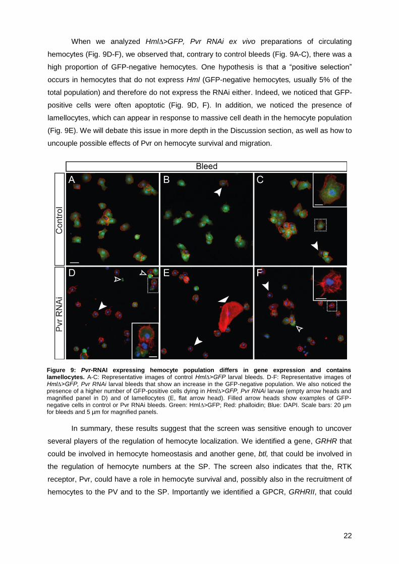
When we analyzed Hml∆>GFP, Pvr RNAi ex vivo preparations of circulating
hemocytes (Fig. 9D-F), we observed that, contrary to control bleeds (Fig. 9A-C), there was a
high proportion of GFP-negative hemocytes. One hypothesis is that a “positive selection”
occurs in hemocytes that do not express Hml (GFP-negative hemocytes, usually 5% of the
total population) and therefore do not express the RNAi either. Indeed, we noticed that GFP-
positive cells were often apoptotic (Fig. 9D, F). In addition, we noticed the presence of
lamellocytes, which can appear in response to massive cell death in the hemocyte population
(Fig. 9E). We will debate this issue in more depth in the Discussion section, as well as how to
uncouple possible effects of Pvr on hemocyte survival and migration.
In summary, these results suggest that the screen was sensitive enough to uncover
several players of the regulation of hemocyte localization. We identified a gene, GRHR that
could be involved in hemocyte homeostasis and another gene, btl, that could be involved in
the regulation of hemocyte numbers at the SP. The screen also indicates that the, RTK
receptor, Pvr, could have a role in hemocyte survival and, possibly also in the recruitment of
hemocytes to the PV and to the SP. Importantly we identified a GPCR, GRHRII, that could
Figure 9: Pvr-RNAI expressing hemocyte population differs in gene expression and contains lamellocytes. A-C: Representative images of control Hml∆>GFP larval bleeds. D-F: Representative images of Hml∆>GFP, Pvr RNAi larval bleeds that show an increase in the GFP-negative population. We also noticed the presence of a higher number of GFP-positive cells dying in Hml∆>GFP, Pvr RNAi larvae (empty arrow heads and magnified panel in D) and of lamellocytes (E, flat arrow head). Filled arrow heads show examples of GFP-negative cells in control or Pvr RNAi bleeds. Green: Hml∆>GFP; Red: phalloidin; Blue: DAPI. Scale bars: 20 µm for bleeds and 5 µm for magnified panels.
23
be functioning upstream of PI3K in hemocyte localization and could represent the receptor
we were initially aiming to identify.
3.3 Downstream of PI3K: what potential downstream effectors can modify hemocyte
location in the larva?
PI3K is known to have a wide range of downstream effectors that could potentially
modify hemocyte behaviour, including migration and adhesion [40]. Our next aim was to
address which downstream effectors of PI3K regulate hemocyte location in the larva.
In our characterization of the PI3K phenotype, we noticed a change in the cell
adhesive properties (3.1.3). Obvious candidates to mediate cell-cell adhesion are the
Cadherin family of transmembrane binding proteins, which play a key role in the dynamics of
cell–cell contact formation, cell signalling and remodelling of junctions and tissues [96]. There
are approximately 16 different Cadherins in the Drosophila genome [97]. In order to know
whether any of these Cadherins play a role in hemocyte adhesion as a possible effector of
PI3K we took advantage of the fact that Cadherin-mediated adhesion is strictly Ca2+
dependent. We blocked the availability of extracellular Ca2+ by using a Ca2+ chelator,
ethylene glycol tetraacetic acid (EGTA), and tested whether this treatment had an effect on
cell clustering in ex vivo preparations of hemocytes, especially in PI3K over-activated (CA)
context, in which hemocytes tend to aggregate more. We found that hemocyte group sizes
were reduced in Hml>PI3K CA, control and even Hml>PI3K DN bleeds after treatment
with the Ca2+ chelator, with single cells becoming more numerous in all contexts (Fig. 10).
This suggests that the clustering phenotype observed upon PI3K modulation relies on a
change in Cadherin-mediated hemocyte-hemocyte adhesion.
We next aimed to determine whether hemocytes express Cadherins. Although
prohemocytes in the medullary zone of the lymph gland have been reported to express
Cadherins [98], it has not yet been shown that mature hemocytes can express Cadherins.
Figure 10: Regulation of hemocyte adhesive properties appears to be Ca 2+
dependent. Cell clustering is
reduced in EGTA-treated hemocyte bleeds.
24
Using immunofluorescence, we tested whether the hemocytes express a well-studied
Drosophila cadherin, the Drosophila E-cadherin (DEcadh, also known as Shotgun) [99]. We
detected a low (as compared to the epithelial expression) but distinct expression of DECadh
in PV-hemocytes, often enriched at cell boundaries (Fig. 11) but we failed to detect any
expression of this particular Cadherin in circulating hemocytes.
Taken together, this data opens the possibility that Cadherins play a role downstream
of PI3K signalling in the regulation of hemocyte-hemocyte and hemocyte-epithelium
adhesion, particularly in the PV. The changes in cell adhesion properties could in turn be, at
least partially, responsible for the change in hemocyte number observed after modulation of
PI3K signalling in these two locations.
Figure 11: Hemocyte adhesion in the PV may require Cadherins. DE-Cadh is expressed in PV hemocytes. A:
Scheme of upper midgut showing the region of the PV imaged in the next panels; PV-enclosed hemocytes are illustrated in green. B: anti-DE-Cadh staining highlights PV epithelium (green, first panel). C: anti-GFP expression in PV-hemocytes (hml>GFP larva, pseudocoloured red). C: merge, including DAPI staining (blue).
Arrow heads show DE-Cadh expression in hemocytes which is stronger at cell boundaries. Scale bar represents 20 µm.
25
Chapter 4. Discussion
To date, most of our knowledge about directed cell migration comes from cell culture
studies using mammalian cells. It is a major challenge to study single cell migration in the
complexity of a living organism and observe the behaviour of cells in contact with a variety of
different stimuli. Therefore, a simple but reliable in vivo system is needed to uncover the
mechanisms underlying single cell migration and chemotaxis. Drosophila hemocytes have
recently emerged as a model for cell migration. Indeed, it was shown that they share many
similarities concerning the basic mechanisms of cell migration with mammalian leukocytes.
Additionally, the available techniques and genetics of the fruitfly present many advantages
for identifying genes involved in cell migration in vivo. In this project, we aimed to better
establish this model system, by uncovering the molecular mechanisms that allow the
recruitment and maintenance of tissue-associated hemocyte populations at the gut and
dorsal epithelium of the larva.
4.1 PI3K phenotype characterization
Here we showed that PI3K regulates the number of PV-enclosed hemocytes at the gut
(PV) and dorsal epithelium (SP) in L3W larvae. Modulation of PI3K signalling using three
different constructs was sufficient to alter the size of these populations. Expressing a PI3K
DN construct specifically in hemocytes, we observed more hemocytes at the PV and less at
the SP. Conversely, upon expressing PI3K CA constructs we observed less hemocytes at
the PV and more at the SP (Fig. 4). We show that these differences in number are
independent of alterations in cell proliferation and survival of hemocytes (Table 1).
Importantly, PI3K could regulate these two populations in different ways. PI3K is not required
for recruitment of hemocytes to the PV, as they are present in high numbers when PI3K
signalling is blocked (PI3K DN). This initial recruitment may involve other signals, possibly
relying on a GPCR or an RTK. However, our results indicate that PI3K is involved in
hemocyte maintenance at the PV. Our working model is that hemocytes at the PV are still
able to sense and respond to signals from the rest of the organism, and that this
responsiveness is PI3K-dependent. When PI3K is over-activated in the hemocytes they tend
to leave the PV because of over-responsiveness to attractive signals from other tissues, for
example the dorsal epithelium. Indeed, our data indicate that PI3K signalling functions in
hemocyte recruitment and/or adhesion to the dorsal epithelium, since PI3K signalling
inhibition by PI3K DN expression leads to a decrease in hemocytes at this location.
Interestingly, the results we obtain in the D. melanogaster resemble recent findings in mouse
models of colitis, in which the authors observed a higher number of macrophages and more
inflammation in the gut of mice mutant for PI3Kγ or PI3Kδ [43, 44].
26
In addition, PI3K may have a role in regulating hemocyte morphology and adhesion.
Hemocytes expressing PI3K CA tend to be more spread, both in the PV and in ex vivo
preparations (Fig. 5), and also tend to form larger clusters in circulation (Fig. 6). It is possible
that PI3K modifies hemocyte adhesion to other tissues, one possible way of regulating
tissue-associated hemocyte populations.
4.2 Genetic screen to find receptors working upstream of PI3K
We performed an RNAi-based genetic screen to uncover RTKs and GPCRs and their
associated G-proteins involved in hemocyte recruitment to the PV and SP. We were
especially interested in finding a phenotype similar to the PI3K DN phenotype, which could
indicate an involvement of the target receptor upstream of PI3K in this process. We chose a
candidate approach, testing in priority genes that were described to be expressed in
hemocytes. This type of screen has advantages and disadvantages when compared to an
unbiased screen. It is faster, since the number of candidates found to be specifically
expressed in hemocytes is much smaller than the total number of GPCRs and RTKs coded
by the D. melanogaster genome; but consequently, there is also a higher probability of
missing an important candidate. We used RNAi for the screen, because of its many
advantages: it enables a fast and efficient silencing of endogenous gene expression in a
tissue-specific manner (here, in hemocytes), whereby flies are often viable; large libraries of
RNAi lines are now available, allowing one to target any gene of interest; and newer libraries
have targeted insertions, thus eliminating one cause of off-target effects. Disadvantages
include inefficiency in silencing target gene expression (hypomorphism), which can
potentially give rise to false negatives; and off-target effects, due for example to sequence
similarities with another gene, which can lead to false positives. It is essential to take into
account all these issues when analyzing the data and use alternative techniques to
overcome these problems, for example by using mutants to confirm RNAi phenotypes.
This genetic screen allowed us to uncover several genes that could be involved in
regulating hemocyte homeostasis and recruitment. However, we decided to focus the
discussion on the two most interesting results, obtained with RNAi against GRHRII and Pvr.
4.2.1 GRHRII
The first indications that a GPCR could be acting upstream of PI3K were the
phenotypes observed in larvae with RNAi against different G-protein subunits (Table 2).
However, these results were difficult to interpret due to potential redundancy between G
protein subunits, added to a likely incomplete loss-of-function due to RNAi hypomorphism.
GRHRII RNAi larvae presented a phenotype that we were most interested in further
analyzing, since it was similar to that obtained in the PI3K DN context: more hemocytes at
the PV and less in the SP (Fig. 7). This suggests that this GPCR might function upstream of
27
PI3K in regulating the number of hemocytes at these two locations. Importantly, to date no
GPCR has been found in Drosophila that is required for hemocyte recruitment, which makes
this finding very interesting. However, GRHRII RNAi possesses a less striking phenotype
than the one obtained with PI3K DN: only the anterior SP had significantly less hemocytes
when compared to control (Table 2). This could be explained by the inefficiency of the RNAi
in completely abolishing expression of the receptor; a low level of remaining expression
could be enough to trigger a signal that could then be amplified inside the cell, leading to a
subtler phenotype. Another possible explanation is redundancy between receptors of the
same family, such as between GRHRII and GRHR. This hypothesis seems unlikely in this
case, as GRHR and GRHRII RNAi gave different phenotypes in our screen.
To further confirm the GRHRII phenotype and make sure that this result is not a false
positive, due to an off-target effect for example, we will now test a mutant line for GRHRII.
The importance of this confirmation is reinforced by the example of the Tre1 RNAi
phenotype, which we identified as a false positive result when it was not confirmed by the
Tre1 mutant. Another way of testing whether this receptor is important for hemocyte
recruitment will be to misexpress its ligand, corazonin (Crz), in a specific location, and see
whether this is sufficient to recruit hemocytes. Lastly, one of the main questions remaining to
be addressed is whether this receptor is indeed working upstream of PI3K, which will be
tested by epistatic analysis of GRHRII and PI3K.
4.2.2 Pvr
Pvr was previously described to be required for hemocyte migration and survival in
the embryo [57, 79]. During our analysis of Pvr RNAi, we observed fewer hemocytes in both
the PV and SP (Fig.8). These effects could be explained by either a general defect in cell
survival (hemocytes start to die when Pvr expression is down-regulated), or an impairment in
cell migration (hemocytes require Pvr to migrate to these locations), or both. When we
analyzed bleeds of HmlΔ>Pvr RNAi larvae, we found that most circulating hemocytes were
GFP-negative (Fig.9), meaning that they were not expressing the HmlΔ-Gal4 driver, which is
normally expressed in 95% of all hemocytes in the L3W larvae. Consequently, it is possible
that in our experiments on Pvr we were unable to visualize and therefore quantify a
proportion of the hemocytes, making the results difficult to interpret. This problem highlights
one disadvantage of using the GAL4/UAS system; the labelling of cells is not direct, which
means that if expression of the driver gene is down-regulated, the cells will lose GFP
expression and will no longer be visualized.
Several explanations could account for the decrease in the GFP-positive hemocyte
population. It could be due to a down-regulation of Hml expression in the Pvr RNAi context,
which would imply that Pvr is required for Hml expression, as yet unreported. Another
possibility is that the two UAS constructs present in the HmlΔ -GAL4, UAS-GFP; UAS-Pvr
28
RNAi larvae are competing for the GAL4 transcription factor, decreasing the amount of GAL4
protein available to activate both UAS promoters. However, this situation is not observed for
other RNAi constructs, which makes this hypothesis less likely. Finally, the observed effect
could be the result of a positive selection: as Pvr is important for hemocyte survival, cells
expressing HmlΔ-Gal4 and thus UAS-Pvr RNAi (and UAS-GFP) would be disadvantaged in
comparison to cells not expressing HmlΔ-Gal4. As a consequence, HmlΔ-Gal4>Pvr RNAi,
GFP cells would tend to undergo apoptosis while the 5% of cells not expressing HmlΔ-Gal4
would continue proliferating, eventually representing a much larger proportion of the
population than normal. One way to corroborate this hypothesis is to perform TUNEL
analysis and PH3 immunostaining to see whether GFP-positive hemocytes have a higher
rate of apoptosis and/or GFP-negative hemocytes a higher rate of proliferation.
In order to determine whether Pvr is involved in hemocyte recruitment to the PV or
solely modifies global survival, we will need to uncouple the potential survival and migration
effects of Pvr in the L3W larvae. In order to do so, we can attempt to modulate the
expression of the ligands, instead of the receptor itself. Misexpression or down-regulation (by
RNAi) of Pvr ligands Pvf-1,-2 and -3, in the PV should indicate whether Pvf/Pvr signalling is
needed for hemocyte recruitment to this location. Interestingly, at late embryogenesis the PV
starts to express Pvf-1 and 2, which further suggests that Pvr is required for hemocyte
recruitment to the PV in response to this signal (Berkeley Drosophila Genome Project).
4.3 Possible downstream effectors of PI3K
We observed, upon modulation of PI3K signalling, changes in cell shape and in the
rate of adhesion between hemocytes in circulation in the PI3K CA context. Obvious
candidates that could function downstream of PI3K and mediate cell-cell adhesion are
Cadherins. This hypothesis was reinforced by the decrease in hemocyte clustering after
EGTA treatment (Fig. 10). We also observed that PV-enclosed hemocytes expressed DE-
cadherin, suggesting a role for DE-cadherin in adhesion of hemocytes to the PV epithelium
(Fig. 11). However, we were not able to observe the expression of this Cadherin in circulating
hemocytes, which could indicate that other types of Cadherin are involved in hemocyte-
hemocyte interactions. To determinate which Cadherin is responsible for adhesion between
hemocytes, we could perform a new screen in which we silence the expression of each
Cadherin, either alone or in a PI3K CA context, to see which one abolishes the clustering
phenotype (“modifier” screen). To limit the number of Cadherins to test (there are
approximately 16 Cadherin genes in the fly genome), we could first perform expression
analysis in hemocytes using available antibodies against other Cadherins. Together, our
results suggest that Cadherins could act as downstream effectors of PI3K signalling in the
regulation of hemocyte-hemocyte and hemocyte-gut epithelium adhesion. This change of
adhesion could in turn account, at least in part, for the PI3K-dependent regulation of
29
hemocyte localisation. It must be noted that other types of adhesion molecules, such as
integrins, could also be involved in hemocyte adhesion. More generally, it is likely that PI3K
regulates different aspects of hemocyte biology through diverse effectors that together
contribute to the hemocyte localization phenotypes we observe.
4.4 Final remarks
Populations of cells associated with tissues are maintained through a succession of
key events, including i) cell migration towards the tissue in response to a signalling cue, and
ii) retention, possibly through adhesion, of the cells in that particular location. Here we
analyzed potential molecules and receptors that could be important for recruitment and
maintenance of two tissue-associated hemocyte populations in D. melanogaster larvae.
We demonstrate that PI3K is involved in the regulation of hemocyte localization,
possibly, in part, through Cadherin-mediated modulation of hemocyte adhesive properties.
Importantly, we also provide insights into potential receptors involved in hemocyte
recruitment and population maintenance in the PV and in the SP. Although much work
remains to establish their functions, we can propose a preliminary working model, in which
the RTK Pvr, independently of PI3K, recruits hemocytes to the PV, where its ligands Pvf-1
and -2 appear to be expressed. Concurrently, hemocytes receive recruitment signals from
other locations in the organism, such as the dorsal epithelium. These signals are received
through the GPCR GRHRII, which in turn activates PI3K.
If we can confirm the involvement of this GPCR in the regulation of hemocyte
navigation it will be very interesting in terms of evolution. While mammalian leukocytes
undergo chemotaxis mainly through chemokine receptors, the Drosophila genome does not
code for GPCRs from the chemokine receptor subfamily and remarkably, no other GPCRs
have yet been implicated in hemocyte navigation. Therefore GRHRII could be the first GPCR
regulating immune cell migration in the fruitfly. One interesting question for the future is
whether this GPCR or others are involved in the recruitment of hemocytes in „crisis‟
situations, such as the response to damaged or infected tissues.
Taken together, our results indicate that hemocyte navigation in the D. melanogaster
larva is tightly regulated, and strongly suggest that hemocytes have to integrate competing
signals from different tissues in their passage through the organism. Importantly, our data
provide new insights about the signalling pathways that regulate hemocyte migration in the
larva, and recruitment and maintenance at target tissues. We hope this work will contribute to
better establish Drosophila hemocytes as a model for single cell migration in vivo.
30
Chapter 5. Bibliography
1. Lauffenburger, D.A. and A.F. Horwitz, Cell migration: a physically integrated molecular process. Cell, 1996. 84: 359-69.
2. Dormann, D. and C.J. Weijer, Imaging of cell migration. EMBO J, 2006. 25: 3480-93. 3. Rorth, P., Collective guidance of collective cell migration. Trends Cell Biol, 2007. 17: 575-9. 4. Affolter, M. and C.J. Weijer, Signaling to cytoskeletal dynamics during chemotaxis. Dev Cell, 2005. 9: 19-
34. 5. Horwitz, R. and D. Webb, Cell migration. Curr Biol, 2003. 13: R756-9. 6. Raftopoulou, M. and A. Hall, Cell migration: Rho GTPases lead the way. Dev Biol, 2004. 265: 23-32. 7. Janetopoulos, C. and R.A. Firtel, Directional sensing during chemotaxis. FEBS Lett, 2008. 582: 2075-85.
8. Charest, P.G. and R.A. Firtel, Feedback signaling controls leading-edge formation during chemotaxis. Curr Opin Genet Dev, 2006. 16: 339-47.
9. Nourshargh, S., P.L. Hordijk, and M. Sixt, Breaching multiple barriers: leukocyte motility through venular walls and the interstitium. Nat Rev Mol Cell Biol, 2010. 11: 366-78.
10. Sanchez-Madrid, F. and M.A. del Pozo, Leukocyte polarization in cell migration and immune interactions. EMBO J, 1999. 18: 501-11.
11. Ward, S.G. and F.M. Marelli-Berg, Mechanisms of chemokine and antigen-dependent T-lymphocyte navigation. Biochem J, 2009. 418: 13-27.
13. Petrie, R.J., A.D. Doyle, and K.M. Yamada, Random versus directionally persistent cell migration. Nat Rev Mol Cell Biol, 2009. 10: 538-49.
14. Chodniewicz, D. and R.L. Klemke, Guiding cell migration through directed extension and stabilization of pseudopodia. Exp Cell Res, 2004. 301: 31-7.
15. Ridley, A.J., et al., Cell migration: integrating signals from front to back. Science, 2003. 302: 1704-9. 16. Springer, T.A., Traffic signals for lymphocyte recirculation and leukocyte emigration: the multistep
602. 19. Frondorf, K., et al., Phosphatidic acid is a leukocyte chemoattractant that acts through S6 kinase
signaling. J Biol Chem, 2010. 285: 15837-47.
20. Ley, K., et al., Getting to the site of inflammation: the leukocyte adhesion cascade updated. Nat Rev Immunol, 2007. 7: 678-89.
21. Thelen, M., Dancing to the tune of chemokines. Nat Immunol, 2001. 2: 129-34. 22. Luster, A.D., R. Alon, and U.H. von Andrian, Immune cell migration in inflammation: present and future
75-84. 24. Johnston, B. and E.C. Butcher, Chemokines in rapid leukocyte adhesion triggering and migration. Semin
Immunol, 2002. 14: 83-92. 25. Metpally, R.P. and R. Sowdhamini, Cross genome phylogenetic analysis of human and Drosophila G
protein-coupled receptors: application to functional annotation of orphan receptors. BMC Genomics, 2005. 6: 106.
26. Fredriksson, R. and H.B. Schioth, The repertoire of G-protein-coupled receptors in fully sequenced genomes. Mol Pharmacol, 2005. 67: 1414-25.
27. Lattin, J., et al., G-protein-coupled receptor expression, function, and signaling in macrophages. J Leukoc Biol, 2007. 82: 16-32.
28. Tuteja, N., Signaling through G protein coupled receptors. Plant Signal Behav, 2009. 4: 942-7. 29. Daaka, Y., S-nitrosylation-regulated GPCR signaling. Biochim Biophys Acta, 2011. 30. Robinson, D.R., Y.M. Wu, and S.F. Lin, The protein tyrosine kinase family of the human genome.
Oncogene, 2000. 19: 5548-57. 31. Schlessinger, J., Cell signaling by receptor tyrosine kinases. Cell, 2000. 103: 211-25. 32. McKay, M.M. and D.K. Morrison, Integrating signals from RTKs to ERK/MAPK. Oncogene, 2007. 26:
3113-21. 33. Dossenbach, C., S. Rock, and M. Affolter, Specificity of FGF signaling in cell migration in Drosophila.
Development, 2001. 128: 4563-72.
34. Ledda, F. and G. Paratcha, Negative Regulation of Receptor Tyrosine Kinase (RTK) Signaling: A Developing Field. Biomark Insights, 2007. 2: 45-58.
35. Robertson, S.C., J. Tynan, and D.J. Donoghue, RTK mutations and human syndromes: when good receptors turn bad. Trends Genet, 2000. 16: 368.
36. Barber, M.A. and H.C. Welch, PI3K and RAC signalling in leukocyte and cancer cell migration. Bull Cancer, 2006. 93: E44-52.
37. Ward, S.G., Do phosphoinositide 3-kinases direct lymphocyte navigation? Trends Immunol, 2004. 25:
67-74. 38. Cain, R.J. and A.J. Ridley, Phosphoinositide 3-kinases in cell migration. Biol Cell, 2009. 101: 13-29. 39. Koyasu, S., The role of PI3K in immune cells. Nat Immunol, 2003. 4: 313-9.
31
40. Abrams, C.S., A little grease helps the cell to stick. blood, 2005. 106: 4-5: 3.
41. Liu, L., et al., Leukocyte PI3Kgamma and PI3Kdelta have temporally distinct roles for leukocyte recruitment in vivo. Blood, 2007. 110: 1191-8.
42. Oak, J.S., et al., Lymphocyte cell motility: the twisting, turning tale of phosphoinositide 3-kinase. Biochem Soc Trans, 2007. 35: 1109-13.
43. Uno, J.K., et al., Altered macrophage function contributes to colitis in mice defective in the phosphoinositide-3 kinase subunit p110delta. Gastroenterology, 2010. 139: 1642-53, 1653 e1-6.
44. van Dop, W.A., et al., The absence of functional PI3Kgamma prevents leukocyte recruitment and ameliorates DSS-induced colitis in mice. Immunol Lett, 2010. 131: 33-9.
45. Parsons, J.T., A.R. Horwitz, and M.A. Schwartz, Cell adhesion: integrating cytoskeletal dynamics and cellular tension. Nat Rev Mol Cell Biol, 2010. 11: 633-43.
46. Ridley, A.J., Life at the leading edge. Cell, 2011. 145: 1012-22. 47. Crozatier, M. and A. Vincent, Drosophila: a model for studying genetic and molecular aspects of
haematopoiesis and associated leukaemias. Dis Model Mech, 2011. 4: 439-45. 48. Santos, A.C. and R. Lehmann, Germ cell specification and migration in Drosophila and beyond. Curr
Biol, 2004. 14: R578-89. 49. Tekotte, H., D. Tollervey, and I. Davis, Imaging the migrating border cell cluster in living Drosophila egg
chambers. Dev Dyn, 2007. 236: 2818-24. 50. Williams, M.J., Drosophila hemopoiesis and cellular immunity. J Immunol, 2007. 178: 4711-6. 51. Lemaitre, B. and J. Hoffmann, The host defense of Drosophila melanogaster. Annu Rev Immunol, 2007.
25: 697-743.
52. Wood, W. and A. Jacinto, Drosophila melanogaster embryonic haemocytes: masters of multitasking. Nat Rev Mol Cell Biol, 2007. 8: 542-51.
53. Babcock, D.T., et al., Circulating blood cells function as a surveillance system for damaged tissue in Drosophila larvae. Proc Natl Acad Sci U S A, 2008. 105: 10017-22.
54. Tirouvanziam, R., et al., Fluorescence-activated cell sorting (FACS) of Drosophila hemocytes reveals important functional similarities to mammalian leukocytes. Proc Natl Acad Sci U S A, 2004. 101: 2912-7.
55. Holz, A., et al., The two origins of hemocytes in Drosophila. Development, 2003. 130: 4955-62. 56. Crozatier, M. and M. Meister, Drosophila haematopoiesis. Cell Microbiol, 2007. 9: 1117-26.
57. Wood, W., C. Faria, and A. Jacinto, Distinct mechanisms regulate hemocyte chemotaxis during development and wound healing in Drosophila melanogaster. J Cell Biol, 2006. 173: 405-16.
58. Fauvarque, M.O. and M.J. Williams, Drosophila cellular immunity: a story of migration and adhesion. J Cell Sci, 2011. 124: 1373-82.
59. Moreira, S., et al., Prioritization of competing damage and developmental signals by migrating macrophages in the Drosophila embryo. Curr Biol, 2010. 20: 464-70.
60. Stramer, B., et al., Live imaging of wound inflammation in Drosophila embryos reveals key roles for small GTPases during in vivo cell migration. J Cell Biol, 2005. 168: 567-73.
61. Paladi, M. and U. Tepass, Function of Rho GTPases in embryonic blood cell migration in Drosophila. J Cell Sci, 2004. 117: 6313-26.
62. Zanet, J., et al., Fascin is required for blood cell migration during Drosophila embryogenesis. Development, 2009. 136: 2557-65.
63. Tucker, P.K., I.R. Evans, and W. Wood, Ena drives invasive macrophage migration in Drosophila embryos. Dis Model Mech, 2011. 4: 126-34.
64. Stofanko, M., S.Y. Kwon, and P. Badenhorst, A misexpression screen to identify regulators of Drosophila larval hemocyte development. Genetics, 2008. 180: 253-67.
65. Markus, R., et al., Sessile hemocytes as a hematopoietic compartment in Drosophila melanogaster. Proc Natl Acad Sci U S A, 2009. 106: 4805-9.
66. Charroux, B. and J. Royet, Elimination of plasmatocytes by targeted apoptosis reveals their role in multiple aspects of the Drosophila immune response. Proc Natl Acad Sci U S A, 2009. 106: 9797-802.
67. Zettervall, C.J., et al., A directed screen for genes involved in Drosophila blood cell activation. Proc Natl Acad Sci U S A, 2004. 101: 14192-7.
68. Irving, P., et al., New insights into Drosophila larval haemocyte functions through genome-wide analysis. Cell Microbiol, 2005. 7: 335-50.
69. Avet-Rochex, A., et al., An in vivo RNA interference screen identifies gene networks controlling Drosophila melanogaster blood cell homeostasis. BMC Dev Biol, 2010. 10: 65.
70. Brody, T. and A. Cravchik, Drosophila melanogaster G protein-coupled receptors. J Cell Biol, 2000. 150:
F83-8. 71. Kunwar, P.S., et al., Tre1, a G protein-coupled receptor, directs transepithelial migration of Drosophila
melanogaster E-cadherin. J Cell Biol, 2008. 183: 157-68. 73. Loren, C.E., et al., Identification and characterization of DAlk: a novel Drosophila melanogaster RTK
which drives ERK activation in vivo. Genes Cells, 2001. 6: 531-44. 74. Duchek, P. and P. Rorth, Guidance of cell migration by EGF receptor signaling during Drosophila
oogenesis. Science, 2001. 291: 131-3. 75. Shilo, B.Z., Signaling by the Drosophila epidermal growth factor receptor pathway during development.
Exp Cell Res, 2003. 284: 140-9. 76. Klambt, C., L. Glazer, and B.Z. Shilo, breathless, a Drosophila FGF receptor homolog, is essential for
migration of tracheal and specific midline glial cells. Genes Dev, 1992. 6: 1668-78.
32
77. Heino, T.I., et al., The Drosophila VEGF receptor homolog is expressed in hemocytes. Mech Dev, 2001. 109: 69-77.
78. Munier, A.I., et al., PVF2, a PDGF/VEGF-like growth factor, induces hemocyte proliferation in Drosophila larvae. EMBO Rep, 2002. 3: 1195-200.
79. Bruckner, K., et al., The PDGF/VEGF receptor controls blood cell survival in Drosophila. Dev Cell, 2004. 7: 73-84.
80. Brand, A.H. and N. Perrimon, Targeted gene expression as a means of altering cell fates and generating dominant phenotypes. Development, 1993. 118: 401-15.
81. Goto, A., et al., A Drosophila haemocyte-specific protein, hemolectin, similar to human von Willebrand factor. Biochem J, 2001. 359: 99-108.
82. Leevers, S.J., et al., The Drosophila phosphoinositide 3-kinase Dp110 promotes cell growth. EMBO J, 1996. 15: 6584-94.
83. Vanhaesebroeck, B., et al., Phosphoinositide 3-kinases: a conserved family of signal transducers. Trends Biochem Sci, 1997. 22: 267-72.
84. Hawkins, P.T., et al., Signalling through Class I PI3Ks in mammalian cells. Biochem Soc Trans, 2006. 34: 647-62.
85. Matova, N. and K.V. Anderson, Rel/NF-kappaB double mutants reveal that cellular immunity is central to Drosophila host defense. Proc Natl Acad Sci U S A, 2006. 103: 16424-9.
86. Pastor-Pareja, J.C., M. Wu, and T. Xu, An innate immune response of blood cells to tumors and tissue damage in Drosophila. Dis Model Mech, 2008. 1: 144-54; discussion 153.
87. Singh, S.R., et al., The adult Drosophila gastric and stomach organs are maintained by a multipotent stem cell pool at the foregut/midgut junction in the cardia (proventriculus). Cell Cycle, 2011. 10: 1109-20.
88. Kadandale, P., et al., Conserved role for autophagy in Rho1-mediated cortical remodeling and blood cell recruitment. Proc Natl Acad Sci U S A, 2010. 107: 10502-7.
89. Liu, T., D. Sims, and B. Baum, Parallel RNAi screens across different cell lines identify generic and cell type-specific regulators of actin organization and cell morphology. Genome Biol, 2009. 10: R26.
90. Stramer, B., et al., Gene induction following wounding of wild-type versus macrophage-deficient Drosophila embryos. EMBO Rep, 2008. 9: 465-71.
91. Ja, W.W., et al., Extension of Drosophila melanogaster life span with a GPCR peptide inhibitor. Nat Chem Biol, 2007. 3: 415-9.
92. Gronke, S., et al., Dual lipolytic control of body fat storage and mobilization in Drosophila. PLoS Biol, 2007. 5: e137.
93. Cazzamali, G., N. Saxild, and C. Grimmelikhuijzen, Molecular cloning and functional expression of a Drosophila corazonin receptor. Biochem Biophys Res Commun, 2002. 298: 31-6.
94. Lee, G., et al., Developmental regulation and functions of the expression of the neuropeptide corazonin in Drosophila melanogaster. Cell Tissue Res, 2008. 331: 659-73.
95. Bainton, R.J., et al., moody encodes two GPCRs that regulate cocaine behaviors and blood-brain barrier permeability in Drosophila. Cell, 2005. 123: 145-56.
96. Kim, S.A., et al., Calcium-dependent dynamics of cadherin interactions at cell-cell junctions. Proc Natl Acad Sci U S A, 2011. 108: 9857-62.
97. Moita, C., et al., The cadherin superfamily in Anopheles gambiae: a comparative study with Drosophila melanogaster. Comp Funct Genomics, 2005. 6: 204-16.
98. Jung, S.H., et al., The Drosophila lymph gland as a developmental model of hematopoiesis. Development, 2005. 132: 2521-33.
99. Tepass, U., et al., shotgun encodes Drosophila E-cadherin and is preferentially required during cell rearrangement in the neurectoderm and other morphogenetically active epithelia. Genes Dev, 1996. 10:
672-85.
33
Gene Construct/Genotype Type Affected
Cromosome Controls used
PI3K P{UAS-Pi3K92E.CAAX}1, y1, w
1118 (BL8294) UAS-CA X y
1, w
1118
P{Dp110-CAAX}1, y
1, w* (BL25908) UAS-Camyc X y
1, w
1118
y
1, w*; P{Dp110D954A}2 (BL25918) UAS-DN III y
1, w
1118
Rac1 y1, w*; P{UAS-Rac1.V12}1 (BL6291) UAS-CA III y
1, w
1118
y
1, w*; P{UAS-Rac1.N17}1 (BL6292) UAS-DN III y
1, w
1118
Gα 47A y, w1118
;P{KK110552} UAS-RNAi II y,w1118
;P{attP,y[+],w[3`]}
Gα 60A y, w1118
;P{KK105485} UAS-RNAi II y,w1118
;P{attP,y[+],w[3`]}
Gα 49B y, w1118
;P{KK105300} UAS-RNAi II y,w1118
;P{attP,y[+],w[3`]}
Gα 73B y1, v
1; P{y[+t7.7] v[+t1.8]=TRiP.JF01950}attP2 (BL25930) UAS-RNAi III y
1, v
1
Gβ 76C y, w1118
;P{KK104745} UAS-RNAi II y,w1118
;P{attP,y[+],w[3`]}
Gβ 13F y, w1118
;P{KK100011} UAS-RNAi II y,w1118
;P{attP,y[+],w[3`]}
Mthl 5 y, w1118
;P{KK101593} UAS-RNAi II y,w1118
;P{attP,y[+],w[3`]}
Mthl 6 y, w1118
;P{KK108048} UAS-RNAi II y,w1118
;P{attP,y[+],w[3`]}
Mthl 8 y, w1118
;P{KK100246} UAS-RNAi II y,w1118
;P{attP,y[+],w[3`]}
CG7497 y, w1118
;P{KK106421} UAS-RNAi II y,w1118
;P{attP,y[+],w[3`]}
CG4313 y, w1118
;P{KK107434} UAS-RNAi II y,w1118
;P{attP,y[+],w[3`]}
GRHR y, w1118
;P{KK109300 } UAS-RNAi II y,w1118
;P{attP,y[+],w[3`]}
GRHR II y1, v
1; P{y[+t7.7] v[+t1.8]=TRiP.JF02042}attP2 (BL26017) UAS-RNAi III y
1, v
1
Tre1 y, w1118
;P{KK108952 } UAS-RNAi II y,w1118
;P{attP,y[+],w[3`]}
Tre1∆EP5
w1118
, Tre1∆EP5
Mutant X w1118
Moody y, w1118
;P{KK109601} UAS-RNAi II y,w1118
;P{attP,y[+],w[3`]}
PVR y, w1118
;P{KK105353} UAS-RNAi II y,w1118
;P{attP,y[+],w[3`]}
EgfR y, w1118
;P{KK107130} UAS-RNAi II y,w1118
;P{attP,y[+],w[3`]}
Btl y, w1118
;P{KK110277} UAS-RNAi II y,w1118
;P{attP,y[+],w[3`]}
w
1118 Control
y
1, w
1118 Control BL lines
y
1, v
1
Control TRiP lines
y,w
1118;P{attP,y[+],w[3`]}
Control VDRC lines II
Supplementary Table 1: Detailed List of stocks used for all analysis performed
34
Supplementary Figure 1: Phosphohistone H3 (PH3) immunostaining of PV and Bleeds. Representative
images of bleeds (A-C) and PV-hemocytes (D-F) subjected to immunostaining for PH3. A, D: Control