+ All Categories
Home > Documents > Universidade do Minho -...

Universidade do Minho -...

Date post: 02-Jul-2018
Category:
Upload: lynguyet
View: 215 times
Download: 0 times
Share this document with a friend
150
UMinho|2012 Ana Luísa Mendanha Falcão Outubro de 2012 Novel perspectives on the subependymal zone complexity and modulation Universidade do Minho Escola de Ciências da Saúde Ana Luísa Mendanha Falcão Novel perspectives on the subependymal zone complexity and modulation
Transcript

UM

inho

|201

2

Ana Luísa Mendanha Falcão

Outubro de 2012

Novel perspectives on the subependymal zonecomplexity and modulation

Universidade do Minho

Escola de Ciências da Saúde

Ana

Luís

a M

enda

nha

Falc

ãoN

ove

l pe

rsp

ect

ive

s o

n t

he

su

be

pe

nd

yma

l zo

ne

co

mp

lexi

ty a

nd

mo

du

lati

on

Tese de Doutoramento em Ciências da Saúde

Trabalho efetuado sob a orientação doProf. Doutor João Carlos Sousa

Trabalho efetuado sob a co-orientação daProfessora Doutora Joana Almeida Palha

Ana Luísa Mendanha Falcão

Outubro de 2012

Novel perspectives on the subependymal zonecomplexity and modulation

Universidade do Minho

Escola de Ciências da Saúde

iii

…para que nós voemos na atmosfera serão precisas as forças concertadas do sol, do âmbar, dos ímanes

e das vontades, mas as vontades são, de tudo, o mais importante, sem elas não nos deixaria subir a

terra…

José Saramago in Memorial do Convento

Aos meus Pais e irmão

iv

v

Acknowledgments

I would like to name all the wills that walked along and pushed, in one way or another, this PhD

forward.

To begin, because they are the first reason that drove me to science, to my Karolinska Ola’s lab

mates. In particular to crazy Therese, doce Shirin, Ana Teixeira and sporty Anna Cascante. You

taught me how fun and cheerful a lab can be! To Gonçalo Castelo-Branco, my first direct

supervisor and inspiration, for all the knowledge and motivation you have granted me, and above

all, for believing and encouraging me to proceed in research.

Luísa and Lina, my dearest friends. Together we shared so many times our sorrows and our

glories; you keep reminding me that distance is no barrier for true friendship. Thank you for the

papers you have downloaded for me and Lu your password is magic and helped a lot!!

Lília and Liliana. I appreciate your long and sincere friendship; our relaxing weekends still remind

me how simple happiness is. Paradoxically, your names and addresses together, are a complex

paradigm to my brain. Lília, you are the definition of enthusiasm in person, you have taught me

that frogs and trees are actually quite interesting.

Sofia, Diogo and Isa & Tânia. For the long lasting conversations about everything and nothing.

You are what my neuroscientist colleagues call “environmental enrichment to my life cage”. Sofi

and Diogo good luck for your PhD studies! I am sure you will make the CEHUM proud!

Joaninha Fraga and Irene. You have given much of you to me… that was why it was so difficult to

watch you leave… I won’t forget our “maluqueiras”, friday night dinners and mostly the help and

support. Friends like you, it’s to keep forever.

Jovica, Magdalena, Julia, Elsa and all the people in Goetz’s lab for the kind reception and all the

help and input you gave me in my four months journey in Munich. Osborn & lab members, Isabel

and Leila for being so friendly, for the barbecues and walk tours that cheered my days.

vi

Joana Barbosa, as if not enough your friendship you have offered me your home! It was so good

to have an old childhood friend waiting for me in Munich…you made me feel home! Who knew

our scientific goals would cross?! I miss our unidirectional gossiping…it kept me updated and

made me laugh more than the situation required! I also miss hearing you say that my cooking

was delicious it’s always good to know that I gave something in return!!

To Fundação para Ciência e Tecnologia for the financial support through a PhD grant (SFRH /

BD / 44485 / 2008).

All the members of the ECS. Prof Cecilia Leão, the smile and gentleness shown to everyone

makes us feel at home.

Nerds and technicians from ICVS, with special regards to:

Goretti, for all the support and nice conversations. Luis and Miguel for always being available to

help even when you couldn’t! Susana for always taking care (of what I forgot) and for being

always ready to help despite the moody expression. Magda, Carlos, Celine. Manuela, for the

endless patience you’ve shown me when I knocked on your door n times…

Pedro Leão Lion, Mário sempre alerta, Ana Joni, Sara & Andreia, Hugo SEAT fellow, Filipa

ahahah, Eduardo, Ana Raquel, Adriana Psi, Anabela, Armando, Patrícia M. and Tó. The first

researchers at ICVS with whom I’ve chattered nonsense and science too.

Morgates (aka Morgado), Vitinho (aka Vitor Hugo) buddies of science&fun, great times in

DC&PD&NY (vitinho, you still own me some pictures!!). Olibiéra (aka J.Oliveira) for the advices,

Castra (aka Andreia) for the companionship and for sharing some cookies.

Susanas M. and C., Silvi, Nuno, Miguel and Fabinho. For the lab cheerfulness and for being

such great persons! Diana, Luís J., Patrícia and Antonio P. my neurogenic mates, Dinis, for

solving my computer issues and for making my friend happy! To you all: I wish all the best for

your PhD studies!

Ana Rita (your crazy theories made me laugh), Paula my course&PhD partner, for treating me

bad and wishing me well! A. Melo for some good advices!

vii

Nadine, for being so kind and for reviewing my English, Margarida, Roqueira, my immune

partners, ZéMiguel (for teaching science + some curiosities of the time of your teens) and J.

Cerqueira, for receiving me so well in your office, for the enthusiastic input and ideas and for

being always in a good mood.

Luísa P. for the support, scientific input and coffees accompanied with nice neurogenic and non-

neurogenic discussions.

Bessa, Monica. For the fruitful neurogenic discussions, for your friendship and for always

crossing fingers for me and letting me know I can count on you!

Sandro my partner in crime (i.e. volley&sardine&camping), for the support and friendship.

Ashley, much more than a colleague, a friend. For the many times you have gladly helped me,

for borrowing me your spoon for my yogurt, and for the coffees and great talks about science-

related and non-related stuff. Catarina or Châta, thanks for calling me “mini-chefinha” (I think is

cute), and for sometimes listening to what I tell you . Your help was precious for this thesis, but

most important than that, I won a friend for life. (P.S: I will be waiting for your visit…)

Nuno Sousa and Joana Palha. You are inspiring as scientists and as persons. Thanks for showing

us that there are no limits for knowledge. Nuno, for the brainstorming and Joana, for the

guidance, for always being ready to discuss and for being so freely accessible. You have taught

me that everything is possible as long as we fight for it. Thank you!

Fernanda Marques

João Sousa

Whatever I write it won’t be enough.

Fernanda, thank you for the endless times you have helped me, even when I didn’t ask you were

always there to help! Your honesty, altruism and sense of justice make you a unique person and

a special friend. Also, for the daily scientific and personal support (even when I was in Munich), it

meant a lot to me every time you said: “vai correr tudo bem”! (friend4ever).

João, this thesis was only possible because you believed in it and always have backed me up. I

viii

am thankful to have supervisors that were encouraging me all the time. With you I have grown

not only as a scientist but also as a person. More than a mentor for science, you were a source

of general knowledge. A supervisor that cares about his students and provides not only scientific

guidance but also good music and good books is a friend. THANK YOU my friend!

E porque sem eles, até a mais lógica das ciências não faria sentido para mim…

Para a minha família,

Se os mencionasse todos teria de escrever mais duas ou três páginas, por isso vou abreviar:

para os meus primos todos e afins (&Cia), nomeadamente ao Nuninho (&Cia), por aquele

abraço, à Belinha (&Cia), pelas imensas sextas-feiras que passamos juntos e que em muito

contribuem para a minha felicidade e à Sara F. (&Cia) pelos jantares, conversas e apoio

incondicional!

Para um grande pedaço de mim: a minha mãe, pai e irmão. Por existirem e darem cor, norte e

felicidade ao meu mundo!

Por isto e por tudo… se eu não for uma pessoa de sorte, então o mundo gira ao contrário!

ix

Abstract

In the mammalian brain adult neurogenesis occurs in two restricted sites: the subependymal

zone (SEZ) and the subgranular zone of the hippocampal dentate gyrus. The SEZ comprises

neural stem and progenitor cells that lie adjacent to the ependyma layer of the lateral ventricles.

SEZ born neuroblasts migrate anteriorly in the rostral migratory stream towards the olfactory

bulbs where they differentiate and integrate into neuronal circuitries. Because cells in the SEZ

niche sense alterations in brain homeostasis and are able to alter their proliferative, migratory

and differentiation profiles in response to injury, it is of particular interest to completely

understand this dynamics both in physiological and pathological conditions. In this context, the

present thesis addresses three main aspects in regard to SEZ niche complexity and modulation.

First, in regard to SEZ heterogeneity, we performed a topographic analysis of the rat SEZ niche

along the anterior-posterior and dorsal-ventral axes. We found that the SEZ cell proliferation

decreases along the anterior-posterior axis and varies considerably according to the position in

the dorsal-ventral axis. Furthermore, these differences were associated with relevant gradients in

the neuroblasts population and in the neural stem cell (NSC) population throughout the dorsal-

ventral axis.

Next, we performed the same analysis on the proliferative and progenitor population profile in the

mouse adult neurogenic niche, and found relevant species-specific differences between rat and

mouse models, two closely related species. The proliferation gradients and the neuroblasts

distribution observed in rat were absent in mice.

Finally, in regard to SEZ modulation/modulators in disease, we report the impact of a peripheral

inflammatory stimulus, triggered by lipopolysaccharide (LPS), on the SEZ, choroid plexus (CP)

and cerebrospinal fluid (CSF). The CP is the structure of the brain that produces and secretes

most of the CSF that is in direct contact with the NSCs of the SEZ. In response to an acute

peripheral inflammatory stimulus CP gene expression of modulators of the SEZ is altered and this

is partially reflected on the CSF composition. The same inflammatory stimulus triggered a rapid

and transient increase on SEZ cell proliferation. The peak of CP response to the inflammatory

trigger was at 6h and 12h upon LPS administration and the induction of SEZ cell proliferation

occurred specifically after 12h of LPS stimulus.

x

In summary, the data presented here reveals relevant topographical specificities of the rat and

mouse SEZ and highlights species-specific differences. Moreover, it gives further insights on the

SEZ response to acute peripheral inflammatory stimulus and pinpoints relevant CP synthetized

molecules that when secreted towards the CSF can modulate the SEZ dynamics.

xi

Resumo

No cérebro adulto dos mamíferos existem dois locais onde ocorre neurogénese: a zona

subependimal (SEZ) e a zona subgranular que constitui a circunvolução denteada do hipocampo.

A SEZ compreende células estaminais neurais e células progenitoras que se encontram

adjacentes à camada do epêndima dos ventrículos laterais. Os progenitores de neurónios

(também designados por neuroblastos) provenientes da SEZ migram anteriormente na via rostral

de migração para os bolbos olfativos onde se diferenciam e integram nos circuitos neuronais.

Como as células da SEZ percecionam alterações na homeostasia do cérebro e são capazes de

alterar os seus perfis de proliferação, migração e diferenciação, em resposta a uma lesão, torna-

se particularmente interessante entender completamente esta dinâmica, tanto em condições

fisiológicas como patológicas. Neste contexto, esta tese aborda três aspetos principais em

relação à complexidade e modulação da SEZ.

Em primeiro lugar, no que diz respeito à heterogeneidade da SEZ, foi realizada uma análise

topográfica deste nicho neurogénico no rato ao longo dos eixos anterior-posterior e dorsal-ventral.

Os resultados obtidos demonstraram que a proliferação das células da SEZ diminui ao longo do

eixo anterior-posterior e varia consideravelmente de acordo com a posição no eixo dorsal-ventral.

Para além disso, estas diferenças estão associadas a uma distribuição diferencial na população

dos neuroblastos e na população das células estaminais neurais (NSCs) ao longo do eixo dorsal-

ventral.

Em seguida, foi realizada a mesma análise na SEZ do murganho adulto. A análise destes

resultados revelou diferenças relevantes entre estas duas espécies filogeneticamente muito

próximas. A proliferação e a distribuição diferencial das NSCs e dos neuroblastos observada em

ratos não ocorrem em murganhos.

Finalmente, no que diz respeito à modulação da SEZ em resposta a insultos ao cérebro, foi

estudado o impacto de um estímulo inflamatório periférico, desencadeada por um

lipopolissacarídeo (LPS), componente da parede das bactérias gram negativas, sobre a SEZ, o

plexus coroideus (CP) e o líquido cefalorraquidiano (CSF). O CP é a estrutura do cérebro que

produz e segrega a maioria do CSF que por sua vez está em contacto direto com as NSCs da

SEZ. Em resposta a um estímulo periférico inflamatório, a expressão genética no CP de

moduladores da SEZ é alterada, sendo esta alteração parcialmente refletida na composição

xii

proteica do CSF. O mesmo estímulo inflamatório desencadeou um aumento rápido e transitório

na proliferação celular na SEZ. O pico da resposta do CP ao estímulo inflamatório desencadeado

pelo LPS foi às 6h e 12h após a administração de LPS e a indução da proliferação celular na

SEZ ocorreu especificamente após 12h de estímulo com LPS.

Em resumo, os resultados apresentados nesta tese revelam especificidades topográficas da SEZ

no rato e no murganho, destacando ainda diferenças específicas entre estas duas espécies.

Além disso, revela também a resposta da SEZ a um estímulo periférico inflamatório agudo e

realça algumas moléculas sintetizadas no CP que quando segregadas para o CSF podem

modular as células da SEZ.

xiii

Contents

1. Introduction..................................................................................................................... 1

1.1 Neurogenesis ............................................................................................................ 3

1.2 Adult neurogenesis .................................................................................................... 3

1.2.1 Historical perspective................................................................................. 4

1.2.2 The subependymal zone ............................................................................ 6

1.2.2.1 Cellular composition and structural organization ...................... 7

1.2.2.2 Topography and heterogeneity .............................................. 10

1.2.2.3 Migration, cellular fate and functional relevance of progenitor

cells .................................................................................................. 12

1.2.2.4 Intrinsic and extrinsic regulation ............................................ 14

1.3 Barriers of the brain ................................................................................................ 16

1.3.1 Blood-brain barrier ................................................................................... 17

1.3.2 Blood-cerebrospinal fluid barrier ............................................................... 17

1.3.2.1 Choroid plexus structure and function ................................... 17

1.3.2.2 Cerebrospinal fluid source, function and composition ............ 19

1.4 Subependymal zone regulation by the barriers of the brain....................................... 21

1.4.1 Subependymal zone regulation mediated by the blood brain barrier .......... 21

1.4.2 Subependymal zone regulation mediated by the choroid plexus ................ 21

1.5 The subependymal zone in the context of brain diseases and insults ........................ 22

1.6 The blood-cerebrospinal fluid barrier in the context of brain disorders and insults ..... 25

1.7 Project aims ............................................................................................................ 27

1.8 References .............................................................................................................. 28

2. Topographical analysis of the subependymal zone neurogenic niche .................................... 47

3. The subependymal zone proliferative pattern and progenitor cell distribution in mice: species

matters! ................................................................................................................................. 61

4. Acute peripheral inflammation impacts on subependymal zone, choroid plexus and

cerebrospinal fluid .................................................................................................................. 85

5. General discussion and future perspectives ....................................................................... 111

6. Annexes ........................................................................................................................... 125

xiv

List of abbreviations

AD - Alzheimer disease

BBB - Blood-brain barrier

BCSFB - Blood-cerebrospinal fluid barrier

CNS - Central nervous system

CP - Choroid plexus

CPEC - Choroid plexus epithelial cells

CSF - Cerebrospinal fluid

DCX - Doublecortin

EGF - Epidermal growth factor

EGFR - Epidermal growth factor receptor

FGF2/bFGF - Fibroblast growth factor 2/basic fibroblast growth factor

GABA - γ-aminobutyric acid

GFAP - Glial fibrillary acidic protein

IL - Interleukin

LPS - Lipopolysaccharide

MS - Multiple sclerosis

NSC/NSCs - Neural stem cell(s)

OB/OBs - Olfactory Bulb(s)

Prom 1 - Prominin 1

PSA-NCAM - Polysialylated form of the neural cell adhesion molecule

RMS - Rostral migratory stream

SEZ - Subependymal zone

TAP - Transit amplifying progenitor

TJ - Tight junction

1

1. Introduction

2

3

1. Introduction

1.1 Neurogenesis

Neurogenesis is the process of formation of neurons from neural stem and progenitor cells. It is

mostly active during embryonic development, when the vast majority of neurons are formed from

multipotent neural stem cells (NSC) (Gotz & Huttner 2005). However, neurogenesis also occurs

during adult life in restricted areas of the brain. We will focus particularly in adult neurogenesis.

1.2 Adult neurogenesis

Adult neurogenesis is the formation of new functional neurons in the adult brain. The

establishment of a novel mature, functional and totally integrated neuron in the adult central

nervous system (CNS) requires four steps: i) the proliferation of the “mother cell”, i.e., the NSC,

that in turn gives rise to a pre-committed neuronal precursor/progenitor cell, ii) the migration of

the progenitor cell towards the site where it will be integrated, iii) the differentiation of the

precursor cell into a neuron and iv) the final integration into neural circuitries (Ming & Song

2005). The outcome of these processes is the formation of a functional neuron in the adult brain.

When these same steps occur but instead of the formation of a neuronal precursor a glial

precursor is generated, originating an astrocyte or oligodendrocyte, the process is designated

adult gliogenesis (Aguirre & Gallo 2004). In the adult brain two very well described places for the

birth of new neurons exist: the subependymal zone (SEZ), also known as adult subventricular

zone (SVZ), and the subgranular zone of the dentate gyrus of the hippocampus (Figure 1). The

first feeds the olfactory bulbs (OBs) whereas the second supplies new neurons for the

hippocampal granular zone (Ming & Song 2011).

In addition to these two neurogenic areas, adult neurogenesis has also been described in the

hypothalamus (Yuan & Arias-Carrion 2011), the amygdala (Fowler et al 2008) and the

subcallosum zone (Seri et al 2006). Nevertheless, it remains debatable if in these brain areas

new functional adult neurons are formed from NSCs (Gould 2007).

4

Figure 1: Neurogenic niches in the adult brain. Adult neurogenesis is confined to two major areas: the subependymal

zone (SEZ) and the subgranular zone (SGZ) of the hippocampal dentate gyrus (DG). Neurons originated in the SEZ

and SGZ migrate to the olfactory bulbs (OB) and the dentate gyrus, respectively. Chains of neuroblasts migrate

anteriorly through the rostral migratory stream (RMS) towards the OB. Both adult neurogenic niches are constituted

by three different cell types.

1.2.1 Historical perspective

For a long time it was believed that the adult brain could not form new neurons. In 1913 the

neuroscientist Ramón y Cajal published a study proclaiming that neurogenesis occurred

exclusively in the developing prenatal brain (Ramon y Cajal 1913). This theory was for long

accepted since it was believed that neurons do not divide and there was no evidence for the

existence of a stem cell pool in the adult brain. Moreover, at the time, it was difficult to

comprehend how new neurons could integrate in complex neuronal circuitries. Ezra Ellen, in

1912, made the first report of cell divisions in the lateral wall of the lateral brain ventricle (Allen

1912). However, the pioneer studies that began to overturn this dogma arose only in the late

50’s and early 60’s. The first evidence for adult neurogenesis was the observation of mitosis

underneath the ependymal layer of the lateral ventricle in adult young mice (Messier et al 1958).

5

Shortly after, the existence of proliferation in the adult brain was confirmed and evidence was

brought for the existence of mitosis in another brain area, the dentate gyrus (Smart and Leblond

1961). These findings were also described in rats (Bryans 1959) and cats (Altman 1963), not

only in physiological conditions but also in response to adult brain injury (Altman 1962). Despite

all these indications, the major turning point favouring adult neurogenesis was in 1965 when

Joseph Altman provided histological evidence for the existence of adult neurogenesis in the

hippocampus (Altman & Das 1965). Four years later, he further described the migration of cells

from a subependymal layer of the lateral ventricles to the OBs where they integrate and

differentiate into neurons (Altman 1969). This study was further confirmed through electron-

microscopy by Michael Kaplan eight years later (Kaplan & Hinds 1977). The subsequent step

that reinforced interest in adult neurogenesis emerged in the early 90’s with the first in vitro

evidence for the existence of adult NSCs in the brain (Lois & Alvarez-Buylla 1993, Reynolds &

Weiss 1992). Cells isolated from the striatum (that encompassed the SEZ) had the capability to

proliferate in response to the epidermal growth factor (EGF) and subsequently give rise to both

neurons and glial cells (Reynolds & Weiss 1992). Henceforward several studies emerged relying

on adult neurogenesis. Some of those confirmed previous reports by using new techniques such

as retroviral tracing (Luskin 1993) and 5-bromo-2'-deoxyuridine (BrdU) labelling, a thymidine

analog that incorporates into the DNA in the S phase of mitosis (Corotto et al 1993); others

described in detail the rostral migratory stream (RMS), a migratory pathway of the SEZ born cells

to the OBs (Doetsch & Alvarez-Buylla 1996, Lois & Alvarez-Buylla 1994, Lois et al 1996). The

identity of the adult NSCs (Chiasson et al 1999, Doetsch et al 1999a, Johansson et al 1999b),

the anatomy and functional organization of these cells in the subependymal layer (Doetsch et al

1997), the effect of stress (Cameron & Gould 1994) and environmental enrichment

(Kempermann et al 1998, Kempermann et al 1997) on adult hippocampal neurogenesis were

the major topics in the adult neurogenesis field in the 90’s. In addition, an important discovery

in the late 90’s came from the work of Peter Eriksson that reported, for the first time, new

neurons in the adult hippocampus of humans (Eriksson et al 1998).

The current challenges focus on the deep understanding of neurogenic niches heterogeneity and

modulation, the exact molecular mechanisms regulating the formation and functional integration

of new neurons. Of notice, in parallel with the studies in rodents many efforts have been done to

characterize these NSCs niches in the adult human brain. Ultimately, the goal is to gain insights

for the manipulation of these niches for regenerative purposes in humans.

6

1.2.2 The subependymal zone

The SEZ is the major site of adult neurogenesis in the adult brain. In rodents, it is commonly

described to be constituted by a thin layer of cells and it is located below the ependyma that lines

the lateral walls of the lateral ventricles (Alvarez-Buylla & Garcia-Verdugo 2002) (Figure 2).

Figure 2: Morphological representation of the subependymal zone niche (SEZ). SEZ cells are located below the

ependyma layer that lines the lateral ventricles filled with cerebrospinal fluid (CSF). This region comprise type B1

cells, the neural stem cells, that give rise to type C cells, also known as transit amplifying progenitors, that originate

type A cells, also known as neuroblasts. Also in the SEZ are type B2 cells or niche astrocytes and blood vessels from

the blood brain barrier (BBB). Type B1 cells are in contact both with the CSF, by projecting a cilium to the ventricle,

and with the BBB.

Currently, the definition of the SEZ is broader, and it is not exclusively restricted to the lateral wall

of the ventricles, but also to the dorsal and medial ventricular walls from where new neurons can

also arise (Alvarez-Buylla et al 2008).

In the literature the SEZ is mostly referred as the SVZ. The reasoning for the designation of SEZ is

due to its location underneath the ependyma and to distinguish it from the embryonic SVZ. In

fact, given the recent findings that adult NSCs from this niche can also have their cell bodies in

direct contact with the ventricle, both designations are not completely appropriate (Mirzadeh et al

2008).

7

For long, the origin of the SEZ was described to be the lateral ganglionic eminence, its embryonic

counterpart (Kohwi et al 2005, Stenman et al 2003). More recently, the embryonic origin of the

SEZ was determined by Cre-lox fate mapping of progenitors from the distinct parts of the

embryonic ventricular zone until adulthood (Young et al 2007). Contrarily to what was

anticipated, neural stem and progenitor cells originated not only from the lateral ganglionic

eminence but also from the medial eminences and the cerebral cortex (Merkle et al 2007).

Further insights on the maintenance, heterogeneity and other aspects of this niche are reviewed

below.

1.2.2.1 Cellular composition and structural organization

We will next further discuss the cellular origin of the SEZ derived new neurons; two possibilities

exist, either they are born from an adult NSC pool that generates neuronal progenitors, or directly

from residual neuronal precursor cells. The definition of a NSC encloses two main properties, the

capacity of self-renewal and multipotency (Gage 2000). These two features can be easily shown

in vitro. By isolating cells from the adult striatum (including the SEZ), Reynolds et al were the first

to show the formation of neurospheres (agglomerates of proliferating cells) in the presence of

EGF and the subsequent differentiation into both neurons and glial cells, ascribing to the SEZ

cells features of stemness (Reynolds et al 1992).

The search for the identity of the NSCs and their progeny in vivo emerged when Doetsch and

colleagues introduced the glial fibrillary acidic protein (GFAP) expressing cells as the NSCs of the

SEZ (Doetsch et al 1999a). These cells, also named type B cells originated transient amplifying

progenitors (TAPs) or type C cells, that in turn gave rise to neuroblasts (neuronal precursors) or

type A cells (Doetsch et al 1999a, Doetsch et al 1997, Doetsch et al 1999b) (Figure 2). On the

other hand, it was proposed that the ependymal cells were in fact the neural stem cells of the

SEZ, namely because ependymal cells proliferate rapidly and turned into neurons that reached

the OBs (Johansson et al 1999a). To demonstrate that the type B cells were the “mother cells”

that generated all other cell types, Doetsch et al (Doetsch et al 1999b) studied the effect of the

administration of a cytostatic drug into the mouse brain ventricle and showed that the highly

proliferative cells that were ablated from the SEZ niche at the end of the infusion were type A and

C cells. Few type B cells were spared but these were enough to allow the progressive recovery of

these mice several days after. These data indicate that type B cells are quiescent and slowly

8

dividing cells able to repopulate the neurogenic niche (Doetsch et al 1999a). Nevertheless, the

question of whether these type B cells could be originated from ependymal cells remained open.

At the present moment it is well established that the NSCs of the SEZ are the type B astrocytes

(Doetsch 2003) and that ependymal cells might function as a reservoir of neurons and glia cells

for the adult injured brain (Carlen et al 2009). The cellular types residing in the SEZ are next

described in more detail.

Type E cells or ependymal cells form a monolayer that outlines the ventricular walls (Figure 2).

Amongst other functions ependymal cells constitute a physical barrier to the direct and free

exchange of molecules between the cerebrospinal fluid (CSF) and brain parenchyma (Bruni

1998, Bruni et al 1985). In the lateral wall two distinct ependymal cells have been described

according to the number of cilia. The type E1 ependymal cells are multiciliated whereas the E2

ependymal cells display only two long cilia (Mirzadeh et al 2008). The E2 ependymal cells

represent only 5% of the total ependymal cells in the lateral wall (Mirzadeh et al 2008). In

physiological conditions these cells proliferate rarely (Altman 1963, Coskun et al 2008) or do not

proliferate at all (Mirzadeh et al 2008, Spassky et al 2005). These cells are commonly marked

with the S100β marker (Carlen et al 2009).

Type B cells are astrocytic cells and express the intermediate filament GFAP. In the SEZ two

types of GFAP positive cells were distinguished according to ultrastructural differences. Type B2

astrocytes, or niche astrocytes, display a highly branched morphology and are frequently found in

the interface of the SEZ and the striatum (Doetsch et al 1997). Type B1 astrocytes are radial-glia

like cells that extend long processes both towards blood vessels settled deeper in the SEZ and to

the ventricular space (Mirzadeh et al 2008) (Figure 2). The latter are recognized as the NSCs of

the SEZ based on the following facts: i) they are structurally similar to their ancestor’s radial glia

progenitor cells that extend an apical primary single cilium towards the ventricle, ii) they increase

their contacts with the ventricle when proliferation in the SEZ is stimulated, iii) they express both

nestin and prominin1 (Prom1 or CD133), markers for NSCs, iv) they are mitotic, expressing Ki67

for instance, and give rise to neuroblasts and neurons. The type B1 astrocytic NSCs organize in

pinwheel structures and the apical ending, i.e., the primary cilium, is in direct contact with the

ventricular lumen and is surrounded by ependymal cells (Mirzadeh et al 2008).

Many attempts have been made to find specific cellular markers that could differentiate type B1

cells from type B2 and from ependymal cells. It is now consensual that at least some NSCs are

GFAP+ and Prom1+ (Beckervordersandforth et al 2010). Moreover, activated NSCs also express

9

the epidermal growth factor receptor (EGFR) (Pastrana et al 2009). When the AraC, a cytostatic

drug, is administrated into the ventricle, the activated NSCs (GFAP+EGFR+) and their progeny are

eliminated. Only the GFAP+EGFR- NSCs survive, then becoming active (hence EGFR+) and start

repopulating the SEZ niche (Pastrana et al 2009). Nevertheless some controversy still exists

since it was suggested that Prom1 is solely expressed in ependymal cells (but not in all) and that

Prom1 cells were able to form new neurons in the OB, indicating these cells as an additional

source of more quiescent cells (Coskun et al 2008, Pfenninger et al 2007).

Combining all these recent findings, it remains uncertain the molecular identification of NSCs.

Structural data indicates that B1 cells are the NSCs and GFAP and Prom1 seem to be the better

markers to label these cells. Intriguingly, not all B1 cells pinwheels express Prom1

(approximately 29% express Prom1) (Mirzadeh et al 2008). Are they both NSCs? If so, what’s the

difference between them? Is EGFR expressed preferentially in any of these cells? Can Prom1

and/or GFAP expressing ependymal cell be another source of NSCs? These are some of the

questions that remain unanswered concerning the identity of the NSCs.

Type C cells, or TAPs, are the progeny of the NSCs. These rapidly dividing cells are organized in

clusters of immature precursors that express distal-less homeobox 2 (Dlx2), achaete-scute

complex homolog 1 (Ascl1or Mash1) and EFGR (Ciccolini et al 2005, Ming & Song 2011) (Figure

2). A short pulse of BrdU mainly labels TAPs indicating these cells are the largest pool of

proliferating cells in the SEZ. Infusions of EGF into the brain ventricles induce a massive increase

in proliferation in the SEZ and the production of new astrocytes in the OB concomitant with a

reduction in neurogenesis (Kuhn et al 1997), suggesting multipotency of TAPs triggered by EGF.

Furthermore, these cells behave as multipotent NSCs in vitro in response to EGF, further

reinforcing that TAPs can be reprogrammed when exposed to EGF (Doetsch et al 2002).

Type A cells or neuroblasts are born from type C cells and constitute the neuronal precursors.

These cells travel a long distance from the SEZ to the OBs where they become fully mature

neurons. Most type A cells express the polysialylated form of the neural cell adhesion molecule

(PSA-NCAM) and doublecortin (DCX) (Figure 2), which are associated to their migratory

properties (Ming & Song 2011). Although these molecules are expressed in neuronal precursors,

it should be stated that it is also possible to find them in postmitotic neurons (Gascon et al 2007,

Klempin et al 2011). Neuroblasts divide actively in the SEZ and also in the RMS. However, as

neuroblasts approach the OBs the proliferation rates become much slower and the cell cycles are

lengthened (Smith & Luskin 1998). Once in the OBs, A cells give rise to different types of

10

interneurons and are integrated in distinct layers of the OB. The process of migration and the

heterogeneity of these cells will be further discussed below.

Tanycytes (Chojnacki et al 2009, Doetsch et al 1997), microglia (in response to injury) (Ekdahl et

al 2009, Thored et al 2009) and endothelial cells of the blood vessels (Shen et al 2008, Tavazoie

et al 2008) are also cellular components of the SEZ niche.

1.2.2.2 Topography and heterogeneity

The classical view of adult neurogenesis in the SEZ has restricted the existence of NSCs

exclusively to the lateral wall of the lateral ventricles (Alvarez-Buylla & Garcia-Verdugo 2002).

Recent findings suggest that news neurons are not only generated from the lateral wall but also

from the dorsal (Brill et al 2009, Merkle et al 2007, Ventura & Goldman 2007) and medial walls

(Kohwi et al 2007, Merkle et al 2007). In fact, evidence exists indicating that SEZ cells from any

point in the entire lateral ventricle are able to generate new neurons (Merkle et al 2007).

The neurogenic potential is not confined to the lateral ventricles but also encompasses the RMS

(Gritti et al 2002) and the OB core (rostral/anterior extension of the RMS) (Liu & Martin 2003).

When isolated for in vitro clonal analysis, the RMS and the OB core cells were able to self-renew

and originated both neurons and glia in vitro. Moreover, according to the progenitor’s position in

the RMS the outcome progeny was distinct. For instance, progenitors residing in more posterior

parts of the RMS originated preferentially oligodendrocytes in vitro (Gritti et al 2002). Thus, NSCs

capable of generating neuroblasts are present along the entire SEZ-RMS-OB path. Still, are these

progenitors similar to each other albeit the distinct origin? The discrimination between SEZ cells

and the subsequent characterization of these distinct populations is strictly necessary for

understanding the neurogenic niche dynamics. As aforementioned, different stem/progenitor cell

types were identified in the SEZ. Each one of these populations displayed common traits that

include ultrastructural properties and molecular markers. As referred previously, it is currently

generally accepted that there are three major neural stem and progenitor cell types and that

NSCs give rise to a subset of TAPs that generate neuroblasts for the OB. However, it is becoming

evident that even within these three defined populations lays a remarkable heterogeneity either

due to inherited intrinsic or epigenetic factors (Alvarez-Buylla et al 2008) and/or an additional

diversity in the surrounding microenvironment cues. Naturally, an accurate look on the

topography of the SEZ discloses major anatomical differences. For instance, as the ventricle

11

extends from anterior to posterior levels (where it partially collapses forming the subcallosum

zone also with neurogenic potential) the SEZ modifies in length and contact with the ventricle.

Interestingly, it was described that the choroid plexus (CP) secrets slits and generates CSF

gradients (Nguyen-Ba-Charvet et al 2004, Sawamoto et al 2006). Thus, it is conceivable that

according to the position on the ventricular axis singular microenvironments are provided to the

SEZ cells. These facts highlight the need to specify the precise locations where SEZ analysis is

performed in all studies addressing SEZ dynamics.

Multiple studies evidenced that the NSC pool is highly heterogeneous both in the origin and in

cellular fate (Alvarez-Buylla et al 2008, Merkle et al 2007, Young et al 2007). The fate of SEZ

born cells is the OB where they become interneurons. Through GLAST::Cre-ERT2 fate mapping, it

was demonstrated that a pool of NSCs that subsists in the adult dorsal wall of the lateral

ventricles also gives rise to glutamatergic neurons to the OBs (Brill et al 2009). Furthermore, it

was reported that SEZ NSCs also originate oligodendrocytes and oligodendrocyte precursors that

migrate to the striatum, corpus callosum and fimbria fornix (Menn et al 2006, Nait-Oumesmar et

al 1999, Picard-Riera et al 2002). Despite all the evidence referred before, the question of

whether the NSCs from the SEZ are multipotent in vivo remains unanswered. For instance, is a

single NSC able to generate both neurons and glia in vivo? Recent findings show that rather than

a uniform pool of NSCs, heterogeneous and already pre-committed NSCs exist in specific

locations of the SEZ (Merkle et al 2007). Several studies report that migrating neuroblasts per se

display a substantial heterogeneity (Baker et al 2001, De Marchis et al 2004, Jankovski & Sotelo

1996) before reaching the OB. Furthermore, neuroblasts born either in ventral, dorsal, anterior or

posterior regions are distinct, produce different neuronal types and are integrated in different

layers of the OB. As an example, neuroblasts from dorsal regions tend to give rise to superficial

granule cells, whereas ventral regions born neuroblasts yield mostly deep granule cells (Merkle et

al 2007). Experiments with homotopically or heterotopically transplanted grafts from ventral or

dorsal origins have uncovered the role of the niche microenvironment versus intrinsic properties

of NSCs. Surprisingly, even after in vitro culture, heterotopic transplanted cells from dorsal or

ventral origin gave rise to the same neuronal types as previously determined. This suggests that

the NSCs are intrinsically pre-determined to become a certain type of neuron in vivo (Merkle et al

2007). The reason why different regionally placed NSCs give rise to distinct progeny has been

speculated. In developmental systems like the forebrain telencephalon, the orchestration of

individual neuronal types is associated with the expression of particular transcription factors in

12

the ventricular walls (Campbell 2003). Similarly, for the adult SEZ it is documented the

expression of distinct transcription factors in both overlapping and non-overlapping regions of the

SEZ. Some of these transcription factors were further correlated with the SEZ embryonic origin

(Stenman et al 2003, Waclaw et al 2006, Young et al 2007). In fact, a regional specific pattern of

transcription factors expression in the SEZ NSCs is associated both with their embryonic origin

and adult neuronal fate (Young et al 2007). Generally, NSCs in the lateral ventricular wall

ubiquitously express Dlx1, 2 and 5 and Mash1, while empty spiracles homeobox 1 (Emx1)

expression is exclusive to the dorsal wall of the ventricle. Furthermore, the transcription factors

NK2 homeobox 1 (Nkx2.1) and paired box gene 6 (Pax6) outline the ventral and dorsal regions of

the lateral wall, respectively (Alvarez-Buylla et al 2008, Weinandy et al 2011). The current

challenge is to understand how different combinations of transcription factors can orchestrate

different neuronal types.

1.2.2.3 Migration, cellular fate and functional relevance of progenitor cells

The most well documented fate for the SEZ derived neuroblasts are the OBs. Neuronal

precursors leave the SEZ, migrate anteriorly in the RMS, reach the OBs (Figure 1) and originate

several types of inhibitory interneurons (Altman 1969, Doetsch & Alvarez-Buylla 1996, Lois &

Alvarez-Buylla 1994, Luskin 1993). It is estimated that the time that a neuronal precursor takes

to reach its final destination in rodents is at least 2 days (Lois & Alvarez-Buylla 1994). A time-

lapse experiment performed in in vitro cultures of neuroblasts showed a chain migration of

neuroblasts at a speed of 122 µm/h (Wichterle et al 1997). In fact, the RMS is organized as a

complex tangential network of migrating neuroblasts chains (Doetsch & Alvarez-Buylla 1996).

Wrapped by astrocytes in tubular–like structures, these chains of neuroblasts use each other as

a support for the contiguous migration (Lois et al 1996). Because the neuroblasts chain

migration towards the OBs occurs even in the absence of glial tubes in vitro and in vivo (namely

in rabbits), (Luzzati et al 2003) the role of astrocytes in the migration is not entirely understood.

The speed for cell migration is considerable reduced in RMS glial-enriched organotypic cultures to

approximately 40 µm/h (Mejia-Gervacio et al 2011) when compared with in vitro cultures (speed

of 122 µm/h) of chain neuroblasts (Wichterle et al 1997). Furthermore, it was suggested that

through regulation of γ-aminobutyric acid (GABA) availability, astrocytic cells could modulate the

migration of neuronal precursors (Bolteus & Bordey 2004). Importantly, the astrocytic tubes can

13

also support migration guidance. Indeed, in a transgenic mouse model with disrupted β1 integrin

function, the astrocytic sheath structure is disturbed resulting in neuroblast migration out of the

RMS. Accordingly, it was demonstrated that β1 integrin is essential for neuroblast’s cell-to-cell

contact and thus for migration (Belvindrah et al 2007). In addition, the neuroblasts markers PSA-

NCAM and DCX were also described to play a role in migration (Chazal et al 2000, Ocbina et al

2006).

At end of the RMS, neuroblasts begin to migrate radially from the OB core to granule cell and

periglomerular layers. Unlike the RMS tangential migration, radial migration does not require the

presence of a glial scaffold but instead an interaction between the cells and the blood vessels.

Specifically, migrating cells establish contact with the extracellular matrix and the astrocytic end

feet in the blood vessels (Bovetti et al 2007) to reach the outer layers of the OBs.

The mechanism by which attractive and repulsive signals direct thousands of neuroblasts to

travel anteriorly, with scarce cellular dispersion, remains unclear. It is well recognized that

migratory instructive signals have a wide range of origins such as the septum, the CP through the

CSF, the glial cells that form the RMS tubular structure and the OBs (Hu 1999, Liu & Rao 2003,

Mason et al 2001, Wu et al 1999). Even though still debatable, the cooperation between

chemorepulsive molecules ascending in most posterior regions and the chemoattractive signals

sent by the OBs is thought to drive rostrally the neuroblasts stream. In particular, slit proteins are

chemorepulsive molecules produced and secreted by the septum and by the CP in the CSF and

repel neuroblasts (that express slit ligand Robo) towards the OBs (Nguyen-Ba-Charvet et al 2004,

Sawamoto et al 2006). Furthermore, the CSF flow was also implicated in this process. Sawamoto

and colleagues showed that the ependymal cilia beating mediated CSF flow generates a gradient

in Slit2 and directs neuroblast migration. Transgenic mice exhibiting deficient cilia and

consequently disturbed CSF flow display abnormal neuroblast migration (Sawamoto et al 2006).

The role of OBs in neuroblasts migration is controversial. Some studies show that when the OBs

are removed neuroblasts migration still takes place leading to an accumulation of progenitors in

the end of the RMS remnants (Kirschenbaum et al 1999). Others claim that OBs are required to

attract and provide the proper migration of the new neurons (Liu & Rao 2003). Generally, these

are the factors involved in neuronal migration: netrin-1 (Murase & Horwitz 2002), neuroregulins

and Erb4 (Anton et al 2004, Perroteau et al 1999), brain derived neurotrophic factor (BDNF)

(Chiaramello et al 2007), EGF (Aguirre et al 2005, Kim et al 2009) and Reelin (Hack et al 2002).

14

Neuronal precursors continue to divide in the RMS or exit cell cycle and begin to differentiate.

Once in the OBs, new granular cells (deep, superficial and calretin positive) and periglomerular

cells (calretin positive, calbidin positive and tyrosin hydroxylase positive) are formed (Lledo et al

2008). Most of these new neurons are granule cells integrated in the granule cell layer and are

GABAergic. As referred previously, a small group of glutamatergic interneurons was also

identified (Brill et al 2009) in the OBs.

Thousands of new cells reach the OBs daily (Kaplan et al 1985, Lois & Alvarez-Buylla 1994),

although the volumetric size of the OBs doesn’t change substantially (Biebl et al 2000). Thus,

programmed cell death is absolutely required to maintain the total number of cells. In fact, many

studies showed neuronal death occurs mainly at the level of the OBs (Petreanu & Alvarez-Buylla

2002). Although debatable, the reason why it takes place preferentially in the OBs, and not in the

RMS or the SEZ, might be related to the decision process of what cells are necessary at a

determined time in the OBs.

The functional relevance of the newly generated neurons in the OBs is a key aspect (Lazarini &

Lledo 2011). Electrophysiological studies have been performed to compare the neuronal

properties of these new neurons, namely synaptic plasticity, with the pre-existing neurons

(Belluzzi et al 2003). Although new neurons seem completely integrated and functional, many

gaps remain for the enlightenment of the final purpose for adult olfactory neurogenesis. A recent

study showed that upon deletion of adult generated neurons by conditionally expression of the

diphtheria toxin in nestin-positive cells, there was no pronounced behavioural difference in the

olfactory mediated behaviours even upon six months of progenitor depletion (Imayoshi et al

2008). Conversely, other studies demonstrated that disturbing adult olfactory neurogenesis (in

NCAM-deficient mice) leads to an impairment in odour discrimination (Gheusi et al 2000). On the

other hand, a similar analysis performed in mice subjected to cranial irradiation of the SEZ,

revealed that only the long-term olfactory memory was impaired (Lazarini et al 2009).

1.2.2.4 Intrinsic and extrinsic regulation

The SEZ adult neural stem and precursor cell population is not considerably altered throughout

life. This steady-state level is due to two major facts: i) the tightly regulated processes that control

NSC self-renewal encompassing asymmetric divisions that produce a new NSC and a progenitor

cell and ii) the migration of the generated progenitors (neuroblasts) out of the niche. New

15

emerging insights support a negative feedback loop between the differentiated progeny and the

NSCs that regulates the formation of new cells in the SEZ (Miller & Gauthier-Fisher 2009). On the

basis of this theory is the fact that NSCs express receptors for several neurotransmitters, such as

GABA (Bordey 2007), serotonin (Banasr et al 2004) and dopamine (Kim et al 2010), and are

innervated by axons of mature neurons (Baker et al 2004).

The interplay between extrinsic and intrinsic factors determines the niche homeostasis. Intrinsic

factors are set of signals produced by the progenitors that together with exterior

microenvironment cues (extrinsic factors) instruct distinct neurogenic phases and ultimately the

cellular fate.

As examples of intrinsic factors are the transcription factors expressed by space-restricted NSCs

and all the intracellular receptors involved in the progenitor’s proliferation and neuroblasts

migration (referred above). Other intrinsic modulators of the SEZ are Mash1 in TAPs involved in

the induction of neuronal specification (Fode et al 2000) and the neuroblasts markers PSA-NCAM

and DCX implicated in progenitor migration (Gascon et al 2007, Ocbina et al 2006). Epigenetic

regulation is widely recognized to play a fundamental role in stem cell multipotentiality,

specification and differentiation (Hsieh & Gage 2004). However, only recently, studies relying on

the epigenetic control of adult SEZ dynamics are beginning to emerge (Sun et al 2011).

Epigenetic factors implicate relevant modifications to the genome without any alteration on its

sequence; it comprises DNA methylation, histone modifications and non-coding RNAs as main

mechanisms of action. As an example of epigenetic regulation, knockout mice for Bmi1, a protein

part of a complex that methylate histones, exhibit post-natal depletion of NSCs (Molofsky et al

2003) while overexpression of Bmi1 in cultures or in vivo triggers NSCs expansion and self-

renewal (Fasano et al 2009). The histone deacetylase 2 (HDAC2) inducible deletions in adult

NSCs (through GLAST::CreERT2 mice) led to a deficit in formation of new neurons in the OBs

and an accumulation of proliferating cells in the SEZ, suggesting that HDAC2 is needed for the

neuronal lineage progression (Jawerka et al 2010). Another recent finding is related to the

microRNA miR-124 that was proposed to be a neuronal fate determinant that makes the switch

from NSC to TAP and neuroblasts. miR-124 is only expressed in NSCs progeny and when

knocked-down in vivo induces the appearance of ectopic cells of glial nature in the OBs

(Akerblom et al 2012, Cheng et al 2009).

Extrinsic factors for the SEZ regulation comprise several trophic and growth factors,

neurotransmitters, morphogens, hormones and cytokines. Generally, the extrinsic factors

16

described to impact on adult SEZ neurogenesis are fibroblast growth factor 2 (FGF2 or bFGF) (Jin

et al 2003), EGF (Kuhn et al 1997), transforming growth factor alpha (TGF-α) (Craig et al 1996),

ciliary neurotrophic factor (CNTF) (Emsley & Hagg 2003), retinoic acid (Wang et al 2005),

neurotrophins (Bath & Lee 2010), prolactin (Shingo et al 2003) and thyroid hormones

(Fernandez et al 2004). Extracellular signalling molecules are of diverse origins, namely from

ependymal cells, neural stem and precursor cells and neurons. The neurotransmitters

aforementioned are good examples of extrinsic factors of neuronal origin. The neurotransmitter

GABA is reported to inhibit NSCs proliferation and is produced by niche neuroblasts (Bordey

2007, Platel et al 2008, Young & Bordey 2009). This is suggested to be a negative feedback loop

to control the formation of new cells in the SEZ. Contrarily to GABA, dopamine and serotonin

stimulate NSCs proliferation (Banasr et al 2004, Coronas et al 2004).

The ependymal layer, adjacent to the SEZ, produces and secretes noggin, an antagonist of bone

morphogenic proteins (BMP) signalling, that prevents differentiation and induces proliferation of

NSCs (Li & LoTurco 2000, Lim et al 2000). In addition, together with endothelial cells of the

blood-brain barrier (BBB), the ependymal layer is a source of pigment epithelium derived factor

(PEDF) that induces NSC self-renewal and proliferation in vitro and in vivo (Ramirez-Castillejo et al

2006). In fact, the endothelial cells of the BBB are in close contact with the SEZ cells and are a

source of molecules that promote NSC maintenance. For instance, an in vitro study showed that

endothelial cells inhibit differentiation and stimulates self-renewal of NSCs by activating the

transcription factor Hes1 (Shen et al 2004). Of notice, and much less explored, is the CP and

the septum that also provide to the SEZ cells molecules that direct neuroblasts migration, as

above-mentioned (Nguyen-Ba-Charvet et al 2004, Sawamoto et al 2006).

Additional players are involved in the SEZ dynamics such as microglia (Thored et al 2009) that

are particularly relevant in response to brain injury or in diseases.

1.3 Barriers of the brain

The evolutionary brain complexity ascended, amongst other reasons, from the formation of

barriers that physically separate the brain microenvironment milieu from the periphery. For an

efficient neuronal processing the brain must be sheltered from the persistent variations in the

periphery blood contents. In mammals, there are three structures separating the brain and the

blood flow: the network of capillaries that constitute the BBB, the arachnoid membrane and the

17

epithelial cells of the CP forming the blood-cerebrospinal fluid barrier (BCSFB) (Abbott 2005).

These barriers largely exceed the physical separation function; they play a dynamic role in the

selective and active transport of nutrients and toxic agents into and out of the brain; they also

constitute a primary line of defence against peripheral insults protecting the brain from external

noxious events. In fact, the brain has for long been considered immune-privileged in part due to

its barriers (Abbott 2005).

1.3.1 Blood-brain barrier

The BBB constitutes a physical barrier between the peripheral blood and the extracellular fluid of

the brain parenchyma. Unlike vessels in the periphery, brain capillaries are constituted of

endothelial cells interconnected by tight junctions (TJs) that restrain the passage of blood derived

molecules and cells. Endothelial cells display extremely low pinocytic activity that further inhibits

transcellular passage of molecules across the barrier (Ballabh et al 2004). The influx of

molecules into the brain requires the existence of specific transporters and the only exceptions

are small molecules such as O2 and CO2 that diffuse freely through their concentration gradients.

The endothelial cells in capillaries forming the BBB are wrapped by a basal lamina, pericytes and

astrocytic end-feets that all together constitute the neurovascular unit (Persidsky et al 2006).

1.3.2 Blood-cerebrospinal fluid barrier

The BCSFB is in the interface of the peripheral blood circulating in fenestrated capillaries and the

CSF filling the brain ventricles. The physical interface is either the CP epithelial cells (CPEC), or

the arachnoid membrane which surrounds the brain. Unlike the capillaries that form the BBB, CP

capillaries are fenestrated and do not have TJs; the barrier properties are therefore ascribed to

the CPEC (Abbott 2005), as will be next described in more detail.

1.3.2.1 Choroid plexus structure and function

The CP is a monolayer of epithelial cells connected by TJs that extend from the ependyma to the

ventricles (the two laterals, the third and the fourth ventricles). The apical membrane of the CP

18

epithelium faces the CSF whereas the basolateral membrane lays over a stromal core that

comprises different cell types (such as fibroblasts, dendritic cells and macrophages) (Emerich et

al 2005) and is enriched in fenestrated capillaries that provide a blood flow four to ten times

greater than the rest of the brain (Keep & Jones 1990) (Figure 3).

Figure 3: Schematic representation of the choroid plexus. The choroid plexus is constituted by a monolayer of

epithelial cells bound together by tight junctions. The epithelial cells display two sides, the basolateral side where

cells lie on a basal lamina and the apical side that faces the CSF. The choroid plexus epithelial cells are enriched in

mitochondria which provide a metabolic capability to secrete many molecules towards the CSF. The basolateral side

is a highly vascularized connective tissue that contains macrophages, dendritic cells, fibroblasts and fenestrated

capillaries throughout the stroma. Epiplexus cells are attached to the villosities of the epithelial cells.

Epiplexus cells are also observed residing in the apical side of the epithelium (Figure 3). The

basal lamina and the TJs constitute the typical hallmark of the CPs highly polarized epithelium.

The presence of the TJs in the CPEC prevents the free paracellular diffusion of water-soluble

molecules. The TJs are dynamic structures whose integrity can be altered under several

circumstances, conditioning the barrier properties of the CPs. Generally the proteins that

19

constitute TJs from the CPs include occludin, claudins and junctional adhesion molecules

(Vorbrodt & Dobrogowska 2003). The barrier properties of CPECs that are conferred by TJs can

be assessed in primary in vitro cultures by evaluating transepithelial electric resistance (Strazielle

& Preston 2003). CPECs in vitro cultures are important tools to study the barrier properties in the

context of brain diseases, with lower values obtained for transepithelial electric resistance

reflecting a leaky CPEC barrier, which compromises brain homeostasis.

The best described function for the CPs is the production and secretion of the CSF. In fact,

CPECs are enriched in mitochondria that provide a metabolic work capacity for both secretory

activity and maintenance of ionic gradients across the BCSFB (Cornford et al 1997). Moreover,

they display a dense apical cover of microvilli that expand the surface area of contact with the

brain ventricles (Figure 3). The major proteins constitutively expressed by the CP, and therefore

enriched in the CSF, are transthyretin (Dickson et al 1985), lipocalin-type prostaglandin D2

synthase (Urade et al 1993) and transferrin (Zakin et al 2002). Furthermore, the CPEC are rich

in transporters and in receptors both in the basolateral and in the apical sides and therefore

mediate the transport of several particles in (e.g. vitamins, glucose, aminoacids) and out (e.g.

amyloid β peptide) of the brain (Chodobski & Szmydynger-Chodobska 2001); and respond to

various stimuli (e.g. lipopolysaccharide [LPS], interleukin 6 [IL-6] and interleukin [IL-1β])

(Marques et al 2009a, Marques et al 2007).

1.3.2.2 Cerebrospinal fluid source, function and composition

The CSF is a colourless fluid that fills the brain ventricles and the subarachnoid space. It

circulates from the lateral ventricles via the interventricular foramina to the third ventricle, and via

the cerebral aqueduct towards the fourth ventricle. The flow ends at the subarachnoid space and

the spinal cord where it is finally reabsorbed. The CSF flow is generated both by the beating of

ependymal cilia, hydrostatic pressure gradient and the pressure created by the expansion of the

arteries in the CPs due to the heart beating (Johanson et al 2008).

At least 2/3 of the CSF is produced by the CPs (Speake et al 2001). The remaining comes from

the interstitial fluid adjacent to the ventricular walls resulting from the metabolism of the brain

parenchyma and from the BBB (Johanson et al 2008). The CSF is estimated to renew

approximately three to four times per day in humans (Wright 1978) and up to eleven times per

20

day in young adult rats (Johanson et al 2008). CSF renewal is absolutely required for the

metabolic homeostasis of the brain.

There are many functions attributed to the CSF. Firstly, the most observable one is the

mechanical protection and the buoyancy that it confers to the brain. The subsequent functions

are all related to the CSF components that by gaining proximal and distal contact to the brain

parenchyma provide CP-born molecules a privileged access to a vast extension of the CNS

(Agnati et al 1995). The CSF is a source of ions, lipids, hormones, glucose, and biologically

active proteins and molecules (Chodobski & Szmydynger-Chodobska 2001). It also receives

metabolites from the brain parenchyma for posterior removal from the brain through the CPs.

Alterations in the CSF chemical and molecular composition leads to a dysfunctional brain, for

instance, when the CSF renewal decreases, the rates of clearance of metabolic cellular wastes

also decrease leading to their accumulation. Furthermore, disruption in the CSF flow, caused by

dysfunctional cilium for instance, leads to hydrocephalus (Banizs et al 2005).

During development, the CSF plays a key role for the normal growth of the embryonic brain.

Namely, it was observed that the hydrostatic pressure exerted by the CSF is essential for proper

brain enlargement (Gato & Desmond 2009, Jelinek & Pexieder 1970, Pexieder & Jelinek 1970).

The reduction of the intra-luminal pressure in chick embryos led to an impaired morphogenesis

and a drastic decrease in neuroepithelial proliferation (Desmond & Jacobson 1977). In fact,

embryonic neurogenesis depends on the combined actions of CSF pressure and CSF growth

factors (Lehtinen & Walsh 2011). Several mitogens and growth factors present in embryonic CSF

were described to play a role on developmental neurogenesis (Gato et al 2005). For instance,

CSF bFGF regulates neuroepithelial cell proliferation and neurogenesis during early stages of

development in chick embryos (Martin et al 2006). In the mouse developing cortex, CSF insulin

growth factor 2 (IGF2) was shown to regulate growth and neuronal survival (Lehtinen et al 2011).

Another example is retinoic acid signalling that contributes to cortical neural formation and to

cerebellum development (Alonso et al 2011, Crandall et al 2011, Siegenthaler et al 2009, Zhang

et al 2003). Although much is known on the role of the CSF in embryonic neurogenesis,

knowledge on the role of CSF in adult neurogenesis is less studied. The CSF influence on adult

neurogenesis will be exploited hereafter.

21

1.4 Subependymal zone regulation by the barriers of the brain

From development to adulthood, the continuous occurrence of NSCs in the lateral ventricles is

only possible due to the existence of a particular niche that together with NSC intrinsic properties

nurtures cell stemness. Curiously, SEZ type B1 cells generally accepted as adult NSCs are

contacting both the CSF and the endothelial cells of the BBB, suggesting that endothelial cells of

the BBB and epithelial cells of the CPs are natural sources of molecules of relevance for NSC

maintenance, which we will refer to next.

1.4.1 Subependymal zone regulation mediated by the blood brain barrier

The anatomical basis of the interaction between vascular endothelial cells and SEZ cells was

recently revealed by demonstrating that the proliferative progeny lie adjacent to the blood vessels

(Shen et al 2008) and that the type B cells contact blood vessels at sites that lack either the

astrocytic endfeet or the pericyte coverage (Tavazoie et al 2008). This unique property of the

BBB enables vascular derived signals access to the SEZ niche (Ming & Song 2011). Previous

work using SEZ rat explants and human vascular endothelial cells suggested that brain vascular

cells support neuronal migration and maturation, but not mitogenesis, through the action of

BDNF (Leventhal et al 1999). Other studies indicate that the endothelial cells secretome is also

an important component for NSC expansion (Shen et al 2004). In fact, endothelial cells are a

source of factors that influence neural stem and precursor cells, for instance vascular endothelial

growth factor (VEGF) and bFGF (Biro et al 1994, Jin et al 2002). Of notice, the role of endothelial

cells derived PEDF in supporting NSC survival and maintaining multipotency (Elahy et al 2012,

Ramirez-Castillejo et al 2006).

1.4.2 Subependymal zone regulation mediated by the choroid plexus

Until recently, it was believe that adult neural stem and precursor cells of the lateral walls of the

ventricles were physically separated from the CSF by a juxtaposed ependymal layer. Even though

ependymal cells do not exhibit TJs, such as the CPECs, molecules present in the CSF would have

to cross ependymal cells or diffuse through it to gain access to SEZ cells. Recently, a

comprehensive view of the cytoarchitecture of the SEZ cells by using whole mounts of the lateral

22

walls (Mirzadeh et al 2010), brought to light the presence of single ciliated astrocytic cells, or

type B1 cells, contacting directly the ventricle and hence the CSF. Type B1 cells are encircled by

ependymal cells forming pinwheels like structures as described previously. These pinwheels were

frequently found in the walls of the lateral ventricles (Mirzadeh et al 2008).

Similarly to brain embryogenesis where radial glial cells (from which type B cells are originated)

receive, directly from the CSF, instructive cues to proliferate and expand (Gato & Desmond 2009,

Lehtinen et al 2011) also type B1 cells from the SEZ neurogenic niche are likely capable of

process signals derived from the CSF. Because the CPs are the main producers of the CSF,

these findings highlight both the CSF and the CP potential role in adult neurogenesis,

establishing a path from the CPs to the CSF and hence towards SEZ.

The proof of concept that CSF contents impact on the SEZ cell dynamics originally came from

experiments with intracerebroventricular injections (Jin et al 2003, Kuhn et al 1997). Infusions of

FGF2 and EGF into the CSF of the brain ventricles resulted in remarkable effects on the SEZ cell

proliferation, migration and cellular fate (Kuhn et al 1997). Importantly, it was shown that Slit2, a

chemorepellent molecule produced by the CPs and secreted into the CSF is required for

neuroblasts migration towards the OBs (Sawamoto et al 2006).

While the significance of CP born molecules in neurogenesis was previously reported during brain

development (Lehtinen & Walsh 2011, Zappaterra & Lehtinen 2012), during adulthood the role

of the CP secretome is largely unknown. A growing number of publications illustrate the existence

of numerous morphogens, mitogens and trophic factors in the CPs (Chodobski & Szmydynger-

Chodobska 2001, Cuevas et al 1994, Timmusk et al 1995). Recently, the CP basal

transcriptome was characterized and revealed many more biologically active molecules that

could be of relevance for SEZ modulation (Marques et al 2011). Some of these proteins were

previously shown to influence the NSC niche both in vitro and in vivo.

1.5 The subependymal zone in the context of brain diseases and insults

Because a plethora of extracellular signals of different origins command the fate and migration of

SEZ born cells, any alteration to this finely tuned regulatory process is able to impact on the

niche homeostasis. Indeed, this is observed in many non-physiological conditions as is next

summarized for several brain injuries and neurodegenerative disorders.

Alzheimer’s disease (AD) is characterized by a progressive cognitive decay and memory

impairment and is associated with the formation of Aβ deposits, the amyloid plaques and

23

intracellular neurofibrillary tangles composed of tau proteins (Tam & Pasternak 2012).

Intracerebroventricular injections of Aβ reduced the proliferation in the SEZ and mice models for

AD display impaired neurogenesis (Haughey et al 2002). Interestingly, examinations on AD

patients correlate deficits in olfactory capacities with the progression of the disease, another

indication that SEZ-OB network is impaired in AD (Murphy et al 1990).

Huntington disease (HD), an autosomal dominant disease, is characterized by the expansion of a

CAG triplet in the huntingtin protein (Zheng & Diamond 2012). Postmortem analysis in HD

human patients revealed an expansion in the SEZ suggesting enhanced neurogenesis (Curtis et

al 2003b, Curtis et al 2005). Accordingly, rat models of HD, where a neuronal striatal lesion is

induced by quinolinic acid, display increased SEZ cell proliferation (Tattersfield et al 2004).

Moreover, neuroblasts were shown to migrate towards the lesion site forming new immature

neurons (Batista et al 2006, Tattersfield et al 2004).

Parkinson disease (PD) patients display severe motor deficits and progressive dopaminergic

neuronal loss in the substancia nigra (Corti et al 2011). A reduction of proliferating cells in the

SEZ is observed upon postmortem analysis of human brains from PD patients (Hoglinger et al

2004). These findings are in line with the previously described function of dopamine in promoting

proliferation in the SEZ (Hoglinger et al 2004). In fact, in the 6-hydroxydopamine rat model of PD

a decrease in proliferation is detected but also a redirection of neuroblasts towards the lesion site

in the striatum is observed (Baker et al 2004, Winner et al 2008).

Multiple sclerosis (MS) is a demyelinating inflammatory disease (Nylander & Hafler 2012). MS

postmortem brains examination demonstrated increased proliferation in the SEZ and PSA-NCAM

positive cells homing to areas of lesion (Nait-Oumesmar et al 2007). Additional studies in mouse

models of MS showed increased oligodendrogenesis (Jablonska et al 2010, Picard-Riera et al

2002, Pluchino et al 2008) and decreased neurogenesis, resulting in olfactory deficits measured

at the molecular and behavioural levels (Tepavcevic et al 2011).

Stroke is an ischemic episode caused by an obstruction in the cerebral arteries and leads to

brain cellular death (Hossmann 2006). In the medial cerebral artery occlusion (MCAO) mouse

model of ischemic stroke, the proliferation in the SEZ is augmented and neuroblasts migrate

radially to the lesion site where new mature neurons are formed (Arvidsson et al 2002,

Yamashita et al 2006, Zhang et al 2004). In accordance, strong evidence exists in humans for

the formation of new neurons following an ischemic incident (Jin et al 2006, Minger et al 2007).

24

Accumulating evidence consistently indicates that brain injuries and disorders, as the ones just

described, share an inflammatory component (Minghetti 2005). The role of neuroinflammation in

adult neurogenesis is still controversial (Carpentier & Palmer 2009, Gonzalez-Perez et al 2010,

Taupin 2008). Each insult or disease displays a particular cascade of inflammatory events. For

example, microglia and astroglia exhibit singular temporal and spatial cocktails of

proinflammatory and anti-inflammatory molecules that can strongly impact on NSC proliferation,

specification and ultimately on the cellular fate of SEZ born cells. Therefore, depending on the

inflammatory milieu context inherent to a particular disease, neuroinflammation may either

promote or prevent adult neurogenesis and gliogenesis.

An important step to regenerate tissue loss is the migration of progenitor cells to sites of injury.

Further highlighting the importance of the inflammatory component to neurogenesis is the fact

that neural stem and precursor cells express a wide-range of receptors for chemokines (Tran et al

2007) that when stimulated during neuroinflammation processes constitute crucial homing

signals for progenitor cells (Belmadani et al 2006). At sites of injury, stromal-derived factor 1α

(SDF1α or Cxcl12) expression is enhanced by endothelial and astrocytic cells, initiating a

chemoattractive process of Cxcl12 receptor chemokine (C-X-C motif) receptor 4 (CXCR4)

expressing NSCs. Additionally, this molecule also promoted NSC proliferation in vitro (Imitola et

al 2004). This role was also reported in several diseases such as MS, brain trauma and brain

tumours (Calderon et al 2006, Itoh et al 2009, van der Meulen et al 2009). An additional

chemokine involved in neuronal progenitor’s migration is the monocyte chemoattractant protein-1

(MCP1or Ccl2). Neural progenitor cells transplanted into non-ischemic areas migrate

preferentially towards infarcted sites where microglia and astrocytes upregulate MCP1 expression

levels (Liu et al 2007, Yan et al 2007). Furthermore, chains of neuroblasts expressing the MCP1

receptor chemokine (C-C motif) receptor 2 (CCR2) are found migrating towards areas where

MCP1 was infused (Yan et al 2007).

Importantly, several cytokines have also been described as modulators of NSCs (Bauer et al

2007, Das & Basu 2008, Nagao et al 2007). Leukemia inhibitory factor (LIF) stimulates NSC

proliferation and self-renewal (Bauer & Patterson 2006). Interestingly, its expression levels are

induced in microglia and reactive astrocytes in ischemia and traumatic brain injury (Banner et al

1997, Suzuki et al 2000). Similarly, the inflammatory cytokine IL-6 was described to promote

NSC self-renewal and to inhibit the formation of new neurons (Covey et al 2011, Vallieres et al

2002). Other cytokines also modulating adult neurogenesis are: IL-1β, interferon gamma (IFNγ)

25

and tumor necrosis factor alpha (TNF-α) (Das & Basu 2008, Iosif et al 2006, Monje et al 2003,

Wong et al 2004). Nevertheless, there is still controversy on the function of this cytokines. As an

example, TNF-α, a major proinflammatory cytokine was demonstrated to play both pro and anti-

neurogenic effects. Upon infusion of neutralizing antibodies for TNF-α in animals models of

stroke, the formation of new striatal neurons was suppressed, suggesting a pro-neurogenic action

of TNF-α (Heldmann et al 2005). On the other hand, TNF-α was further suggested as a negative

modulator of hippocampal neurogenesis (Cacci et al 2005, Iosif et al 2006) but through TNFR1

exclusively (Iosif et al 2006). Further analysis suggested that the concentration of TNF-α itself

might determine the final outcome (Bernardino et al 2008).

Commonly, stimuli given by the administration of LPS, an endotoxin of the Gram-negative

bacteria (Alexander & Rietschel 2001, Raetz & Whitfield 2002), has been used to mimic

neuroinflammation because it rapidly triggers cellular expression of cytokines and chemokines

through toll-like receptor 4 (TLR4). LPS actions are frequently associated with impaired adult

hippocampal neurogenesis (Carpentier & Palmer 2009, Ekdahl et al 2003, Fujioka & Akema

2010, Monje et al 2003, Russo et al 2011). LPS brain infusions strongly reduce hippocampal

neurogenesis, an effect triggered by microglial activation; inhibition of microglial activation

restores hippocampal neurogenesis (Ekdahl et al 2003). Endorsing results showed that treatment

with a nonsteroidal inflammatory drug after a single systemic injection of LPS restores

hippocampal neurogenesis (Monje et al 2003). Also of interest is the subsequent functionality of

the newly formed neurons upon LPS treatment. Recent reports claim that upon local injections of

LPS in the hippocampal dentate gyrus, newly formed neurons display different synaptic plasticity

properties (Jakubs et al 2008). Microglia and astrocytic responses to LPS driven inflammation

are not unique. The effects of LPS in the CPs were recently unveiled by Marques et al (2007,

2009a, 2009b), which will next be referred.

1.6 The blood-cerebrospinal fluid barrier in the context of brain disorders and insults

Rising expectations for the development of therapies based on brain endogenous regenerative

capacity firstly motivated by the discovery of adult NSCs in the brain were further encouraged by

the observation of naturally-occurring NSCs response to brain insults and neurodegenerative

disorders (Curtis et al 2003a, Curtis et al 2007, Sierra et al 2011). To enhance and improve

26

NSC-based therapies on cellular regeneration and/or replacement, the mechanisms underlying

these processes must be uncovered.

In this sense, we have described so far the BCSFB as a crucial element for brain homeostasis

maintenance; hence, the ability of the brain to adapt to an injury also relies on the CP-CSF

response. There are several brain diseases and injuries associated to CP-CSF alterations. Even

physiological aging in itself alters BCSFB permeability, CSF volume, composition and turnover

(Pfefferbaum et al 1994, Preston 2001). These alterations are exacerbated in brain disorders

such as AD. The accumulation of Aβ is thought to be the major hallmark in AD. Interestingly, low

levels of Aβ-binding proteins, like transthyretin (TTR), are reduced in the CSF of AD patients

indicating an inefficient clearance of this peptide from the CNS (Hansson et al 2009, Mesquita et

al 2012, Serot et al 1997).

In physiological situations the CNS is free of peripheral immune cells, however, events that

trigger neuroinflammatory processes can elicit leakage of the barriers and the entrance of blood

cells into the brain. This is the case of the nervous system autoimmune diseases such as MS. It

is reported that in the experimental autoimmune encephalomyelitis (EAE) mouse model of MS,

inflammatory cells are recruited to the CNS and can be detected in the CSF. In accordance, the

CP also displays major alterations, namely in the expression of the adhesion molecules ICAM1

and VCAM1 (Engelhardt et al 2001). Moreover, the overall gene expression profile of the CPs and

the CSF composition is altered in EAE (Marques et al 2012). Together these data associate the

CP-CSF nexus to the pathophysiology of MS. Another neuroinflammatory event that has been

implicated with CPs and CSF disturbance is traumatic brain injury (TBI). Following TBI the

BCSFB is also damaged, thus a subsequent increase in the traffic of leucocytes and blood

derived molecules from the periphery to the brain occurs (Johanson et al 2011). This leads to the

disruption of the CP-CSF functions including the brain support for nutrients and removal of

metabolic waste products (Johanson et al 2011). However, the BCSFB also develops

mechanisms to restore brain homeostasis. The CP intensifies the expression, synthesis and

secretion of neurotrophins and growth factors that will reach the lesion sites and help on the

regeneration/repair process (Johanson et al 2011). A similar strategy is acquired by the CP after

a transient forebrain ischemia in adult rats (Johanson et al 2000).

Importantly, the CP-CSF not only participates in brain parenchyma derived inflammatory events

but also respond to peripheral inflammatory triggers. In fact, the interplay between the immune

27

system and the brain is mediated by the BCSFB and the BBB. It was recently described the CP

gene expression profile in response to an acute peripheral inflammation triggered by LPS

(Marques et al 2009b). After a short LPS stimulus the CPs were able to robustly change their

transcriptome, and these were also reflected at the CSF level (Marques et al 2007). Amongst the

altered molecules were chemokines (such as Ccl2, Ccl10), cytokines (such as IL-6 and TNF-

alpha) and adhesion molecules (Marques et al 2009a, Marques et al 2009b). Similarly,

experiments conducted in an in vitro model for the BCSFB, helped to clarify the origin of the

response observed in vivo, showing that the CP response is triggered by LPS directly and/or by

blood-borne molecules. Accordingly, CPEC stimulated at the basolateral side by LPS or by serum

derived from animals injected with LPS, display individual enhanced expression for cytokines and

chemokines (Marques et al 2009b).

While LPS elicited inflammation was able to impact on hippocampal neurogenesis, the

significance of this insult for the SEZ adult neurogenesis remains unknown.

1.7 Project aims

The purpose of this study was to give insights in emerging questions concerning SEZ complexity

and modulation. More specifically we aimed at:

1. Characterize the topographical proliferation profiles and the distribution of neural stem

and progenitor cells throughout the anterior-posterior and dorsal-ventral axes of the SEZ,

both in rat and mouse;

2. Determine inter-species variations in the SEZ topographical profiles between rat and

mouse;

3. Analyse the impact of an acute peripheral inflammatory stimulus on the SEZ niche

dynamics;

4. Investigate the role of CP secreted molecules on the SEZ modulation upon an acute

peripheral inflammatory stimulus.

28

1.8 References

Abbott NJ. 2005. Dynamics of CNS barriers: evolution, differentiation, and modulation. Cell Mol

Neurobiol 25: 5-23

Agnati LF, Zoli M, Stromberg I, Fuxe K. 1995. Intercellular communication in the brain: wiring

versus volume transmission. Neuroscience 69: 711-26

Aguirre A, Gallo V. 2004. Postnatal neurogenesis and gliogenesis in the olfactory bulb from NG2-

expressing progenitors of the subventricular zone. J Neurosci 24: 10530-41

Aguirre A, Rizvi TA, Ratner N, Gallo V. 2005. Overexpression of the epidermal growth factor

receptor confers migratory properties to nonmigratory postnatal neural progenitors. J

Neurosci 25: 11092-106

Akerblom M, Sachdeva R, Barde I, Verp S, Gentner B, et al. 2012. MicroRNA-124 Is a

Subventricular Zone Neuronal Fate Determinant. J Neurosci 32: 8879-89

Alexander C, Rietschel ET. 2001. Bacterial lipopolysaccharides and innate immunity. J Endotoxin

Res. 7: 167-202

Allen E. 1912. The cessation of the mitosis in the central nervous system of the albino rat. J

Comp Neurol 22: 547–68

Alonso MI, Martin C, Carnicero E, Bueno D, Gato A. 2011. Cerebrospinal fluid control of

neurogenesis induced by retinoic acid during early brain development. Dev Dyn 240:

1650-9

Altman J. 1962. Autoradiographic study of degenerative and regenerative proliferation of

neuroglia cells with tritiated thymidine. Exp Neurol 5: 302-18

Altman J. 1963. Autoradiographic investigation of cell proliferation in the brains of rats and cats.

Anat Rec 145: 573-91

Altman J. 1969. Autoradiographic and histological studies of postnatal neurogenesis. IV. Cell

proliferation and migration in the anterior forebrain, with special reference to persisting

neurogenesis in the olfactory bulb. J Comp Neurol 137: 433-57

Altman J, Das GD. 1965. Autoradiographic and histological evidence of postnatal hippocampal

neurogenesis in rats. J Comp Neurol 124: 319-35

Alvarez-Buylla A, Garcia-Verdugo JM. 2002. Neurogenesis in adult subventricular zone. J

Neurosci 22: 629-34

Alvarez-Buylla A, Kohwi M, Nguyen TM, Merkle FT. 2008. The heterogeneity of adult neural stem

29

cells and the emerging complexity of their niche. Cold Spring Harb Symp Quant Biol 73:

357-65

Anton ES, Ghashghaei HT, Weber JL, McCann C, Fischer TM, et al. 2004. Receptor tyrosine

kinase ErbB4 modulates neuroblast migration and placement in the adult forebrain. Nat

Neurosci 7: 1319-28

Arvidsson A, Collin T, Kirik D, Kokaia Z, Lindvall O. 2002. Neuronal replacement from

endogenous precursors in the adult brain after stroke. Nat Med 8: 963-70

Baker H, Liu N, Chun HS, Saino S, Berlin R, et al. 2001. Phenotypic differentiation during

migration of dopaminergic progenitor cells to the olfactory bulb. J Neurosci 21: 8505-13

Baker SA, Baker KA, Hagg T. 2004. Dopaminergic nigrostriatal projections regulate neural

precursor proliferation in the adult mouse subventricular zone. Eur J Neurosci 20: 575-9

Ballabh P, Braun A, Nedergaard M. 2004. The blood-brain barrier: an overview: structure,

regulation, and clinical implications. Neurobiol Dis 16: 1-13

Banasr M, Hery M, Printemps R, Daszuta A. 2004. Serotonin-induced increases in adult cell

proliferation and neurogenesis are mediated through different and common 5-HT

receptor subtypes in the dentate gyrus and the subventricular zone.

Neuropsychopharmacology 29: 450-60

Banizs B, Pike MM, Millican CL, Ferguson WB, Komlosi P, et al. 2005. Dysfunctional cilia lead to

altered ependyma and choroid plexus function, and result in the formation of

hydrocephalus. Development 132: 5329-39

Banner LR, Moayeri NN, Patterson PH. 1997. Leukemia inhibitory factor is expressed in

astrocytes following cortical brain injury. Exp Neurol 147: 1-9

Bath KG, Lee FS. 2010. Neurotrophic factor control of adult SVZ neurogenesis. Dev Neurobiol

70: 339-49

Batista CM, Kippin TE, Willaime-Morawek S, Shimabukuro MK, Akamatsu W, van der Kooy D.

2006. A progressive and cell non-autonomous increase in striatal neural stem cells in the

Huntington's disease R6/2 mouse. J Neurosci 26: 10452-60

Bauer S, Kerr BJ, Patterson PH. 2007. The neuropoietic cytokine family in development,

plasticity, disease and injury. Nat Rev Neurosci 8: 221-32

Bauer S, Patterson PH. 2006. Leukemia inhibitory factor promotes neural stem cell self-renewal

in the adult brain. J Neurosci 26: 12089-99

Beckervordersandforth R, Tripathi P, Ninkovic J, Bayam E, Lepier A, et al. 2010. In vivo fate

30

mapping and expression analysis reveals molecular hallmarks of prospectively isolated

adult neural stem cells. Cell Stem Cell 7: 744-58

Belluzzi O, Benedusi M, Ackman J, LoTurco JJ. 2003. Electrophysiological differentiation of new

neurons in the olfactory bulb. J Neurosci 23: 10411-8

Belmadani A, Tran PB, Ren D, Miller RJ. 2006. Chemokines regulate the migration of neural

progenitors to sites of neuroinflammation. J Neurosci 26: 3182-91

Belvindrah R, Hankel S, Walker J, Patton BL, Muller U. 2007. Beta1 integrins control the

formation of cell chains in the adult rostral migratory stream. J Neurosci 27: 2704-17

Bernardino L, Agasse F, Silva B, Ferreira R, Grade S, Malva JO. 2008. Tumor necrosis factor-

alpha modulates survival, proliferation, and neuronal differentiation in neonatal

subventricular zone cell cultures. Stem Cells 26: 2361-71

Biebl M, Cooper CM, Winkler J, Kuhn HG. 2000. Analysis of neurogenesis and programmed cell

death reveals a self-renewing capacity in the adult rat brain. Neurosci Lett 291: 17-20

Biro S, Yu ZX, Fu YM, Smale G, Sasse J, et al. 1994. Expression and subcellular distribution of

basic fibroblast growth factor are regulated during migration of endothelial cells. Circ Res

74: 485-94

Bolteus AJ, Bordey A. 2004. GABA release and uptake regulate neuronal precursor migration in

the postnatal subventricular zone. J Neurosci 24: 7623-31

Bordey A. 2007. Enigmatic GABAergic networks in adult neurogenic zones. Brain Res Rev 53:

124-34

Bovetti S, Hsieh YC, Bovolin P, Perroteau I, Kazunori T, Puche AC. 2007. Blood vessels form a

scaffold for neuroblast migration in the adult olfactory bulb. J Neurosci 27: 5976-80

Brill MS, Ninkovic J, Winpenny E, Hodge RD, Ozen I, et al. 2009. Adult generation of

glutamatergic olfactory bulb interneurons. Nat Neurosci 12: 1524-33

Bryans WA. 1959. Mitotic activity in the brain of the adult rat. Anat Rec 133: 65–73

Bruni JE. 1998. Ependymal development, proliferation, and functions: a review. Microsc Res

Tech 41: 2-13

Bruni JE, Del Bigio MR, Clattenburg RE. 1985. Ependyma: normal and pathological. A review of

the literature. Brain Res 356: 1-19

Cacci E, Claasen JH, Kokaia Z. 2005. Microglia-derived tumor necrosis factor-alpha exaggerates

death of newborn hippocampal progenitor cells in vitro. J Neurosci Res 80: 789-97

Calderon TM, Eugenin EA, Lopez L, Kumar SS, Hesselgesser J, et al. 2006. A role for CXCL12

31

(SDF-1alpha) in the pathogenesis of multiple sclerosis: regulation of CXCL12 expression

in astrocytes by soluble myelin basic protein. J Neuroimmunol 177: 27-39

Cameron HA, Gould E. 1994. Adult neurogenesis is regulated by adrenal steroids in the dentate

gyrus. Neuroscience 61: 203-9

Campbell K. 2003. Dorsal-ventral patterning in the mammalian telencephalon. Curr Opin

Neurobiol 13: 50-6

Carlen M, Meletis K, Goritz C, Darsalia V, Evergren E, et al. 2009. Forebrain ependymal cells are

Notch-dependent and generate neuroblasts and astrocytes after stroke. Nat Neurosci 12:

259-67

Carpentier PA, Palmer TD. 2009. Immune influence on adult neural stem cell regulation and

function. Neuron 64: 79-92

Chazal G, Durbec P, Jankovski A, Rougon G, Cremer H. 2000. Consequences of neural cell

adhesion molecule deficiency on cell migration in the rostral migratory stream of the

mouse. J Neurosci 20: 1446-57

Cheng LC, Pastrana E, Tavazoie M, Doetsch F. 2009. miR-124 regulates adult neurogenesis in

the subventricular zone stem cell niche. Nat Neurosci 12: 399-408

Chiaramello S, Dalmasso G, Bezin L, Marcel D, Jourdan F, et al. 2007. BDNF/ TrkB interaction

regulates migration of SVZ precursor cells via PI3-K and MAP-K signalling pathways. Eur

J Neurosci 26: 1780-90

Chiasson BJ, Tropepe V, Morshead CM, van der Kooy D. 1999. Adult mammalian forebrain

ependymal and subependymal cells demonstrate proliferative potential, but only

subependymal cells have neural stem cell characteristics. J Neurosci 19: 4462-71

Chodobski A, Szmydynger-Chodobska J. 2001. Choroid plexus: target for polypeptides and site of

their synthesis. Microsc Res Tech 52: 65-82

Chojnacki AK, Mak GK, Weiss S. 2009. Identity crisis for adult periventricular neural stem cells:

subventricular zone astrocytes, ependymal cells or both? Nat Rev Neurosci 10: 153-63

Ciccolini F, Mandl C, Holzl-Wenig G, Kehlenbach A, Hellwig A. 2005. Prospective isolation of late

development multipotent precursors whose migration is promoted by EGFR. Dev Biol

284: 112-25

Cornford EM, Varesi JB, Hyman S, Damian RT, Raleigh MJ. 1997. Mitochondrial content of

choroid plexus epithelium. Exp Brain Res 116: 399-405

Coronas V, Bantubungi K, Fombonne J, Krantic S, Schiffmann SN, Roger M. 2004. Dopamine D3

32

receptor stimulation promotes the proliferation of cells derived from the post-natal

subventricular zone. J Neurochem 91: 1292-301

Corotto FS, Henegar JA, Maruniak JA. 1993. Neurogenesis persists in the subependymal layer of

the adult mouse brain. Neurosci Lett 149: 111-4

Corti O, Lesage S, Brice A. 2011. What genetics tells us about the causes and mechanisms of

Parkinson's disease. Physiol Rev 91: 1161-218

Coskun V, Wu H, Blanchi B, Tsao S, Kim K, et al. 2008. CD133+ neural stem cells in the

ependyma of mammalian postnatal forebrain. Proc Natl Acad Sci U S A 105: 1026-31

Covey MV, Loporchio D, Buono KD, Levison SW. 2011. Opposite effect of inflammation on

subventricular zone versus hippocampal precursors in brain injury. Ann Neurol 70: 616-

26

Craig CG, Tropepe V, Morshead CM, Reynolds BA, Weiss S, van der Kooy D. 1996. In vivo growth

factor expansion of endogenous subependymal neural precursor cell populations in the

adult mouse brain. J Neurosci 16: 2649-58

Crandall JE, Goodman T, McCarthy DM, Duester G, Bhide PG, et al. 2011. Retinoic acid

influences neuronal migration from the ganglionic eminence to the cerebral cortex. J

Neurochem 119: 723-35

Cuevas P, Carceller F, Reimers D, Fu X, Gimenez-Gallego G. 1994. Immunohistochemical

localization of basic fibroblast growth factor in choroid plexus of the rat. Neurol Res 16:

310-2

Curtis MA, Connor B, Faull RL. 2003a. Neurogenesis in the diseased adult human brain--new

therapeutic strategies for neurodegenerative diseases. Cell Cycle 2: 428-30

Curtis MA, Faull RL, Eriksson PS. 2007. The effect of neurodegenerative diseases on the

subventricular zone. Nat Rev Neurosci 8: 712-23

Curtis MA, Penney EB, Pearson AG, van Roon-Mom WM, Butterworth NJ, et al. 2003b. Increased

cell proliferation and neurogenesis in the adult human Huntington's disease brain. Proc

Natl Acad Sci U S A 100: 9023-7

Curtis MA, Penney EB, Pearson J, Dragunow M, Connor B, Faull RL. 2005. The distribution of

progenitor cells in the subependymal layer of the lateral ventricle in the normal and

Huntington's disease human brain. Neuroscience 132: 777-88

Das S, Basu A. 2008. Inflammation: a new candidate in modulating adult neurogenesis. J

Neurosci Res 86: 1199-208

33

De Marchis S, Temoney S, Erdelyi F, Bovetti S, Bovolin P, et al. 2004. GABAergic phenotypic

differentiation of a subpopulation of subventricular derived migrating progenitors. Eur J

Neurosci 20: 1307-17

Desmond ME, Jacobson AG. 1977. Embryonic brain enlargement requires cerebrospinal fluid

pressure. Dev Biol 57: 188-98

Dickson PW, Howlett GJ, Schreiber G. 1985. Rat transthyretin (prealbumin). Molecular cloning,

nucleotide sequence, and gene expression in liver and brain. J Biol Chem 260: 8214-9

Doetsch F. 2003. The glial identity of neural stem cells. Nat Neurosci 6: 1127-34

Doetsch F, Alvarez-Buylla A. 1996. Network of tangential pathways for neuronal migration in

adult mammalian brain. Proc Natl Acad Sci U S A 93: 14895-900

Doetsch F, Caille I, Lim DA, Garcia-Verdugo JM, Alvarez-Buylla A. 1999a. Subventricular zone

astrocytes are neural stem cells in the adult mammalian brain. Cell 97: 703-16

Doetsch F, Garcia-Verdugo JM, Alvarez-Buylla A. 1997. Cellular composition and three-

dimensional organization of the subventricular germinal zone in the adult mammalian

brain. J Neurosci 17: 5046-61

Doetsch F, Garcia-Verdugo JM, Alvarez-Buylla A. 1999b. Regeneration of a germinal layer in the

adult mammalian brain. Proc Natl Acad Sci U S A 96: 11619-24

Doetsch F, Petreanu L, Caille I, Garcia-Verdugo JM, Alvarez-Buylla A. 2002. EGF converts transit-

amplifying neurogenic precursors in the adult brain into multipotent stem cells. Neuron

36: 1021-34

Ekdahl CT, Claasen JH, Bonde S, Kokaia Z, Lindvall O. 2003. Inflammation is detrimental for

neurogenesis in adult brain. Proc Natl Acad Sci U S A 100: 13632-7

Ekdahl CT, Kokaia Z, Lindvall O. 2009. Brain inflammation and adult neurogenesis: the dual role

of microglia. Neuroscience 158: 1021-9

Elahy M, Baindur-Hudson S, Dass CR. 2012. The emerging role of PEDF in stem cell biology. J

Biomed Biotechnol 2012: 239091

Emerich DF, Skinner SJ, Borlongan CV, Vasconcellos AV, Thanos CG. 2005. The choroid plexus

in the rise, fall and repair of the brain. Bioessays 27: 262-74

Emsley JG, Hagg T. 2003. Endogenous and exogenous ciliary neurotrophic factor enhances

forebrain neurogenesis in adult mice. Exp Neurol 183: 298-310

Engelhardt B, Wolburg-Buchholz K, Wolburg H. 2001. Involvement of the choroid plexus in

central nervous system inflammation. Microsc Res Tech 52: 112-29

34

Eriksson PS, Perfilieva E, Bjork-Eriksson T, Alborn AM, Nordborg C, et al. 1998. Neurogenesis in

the adult human hippocampus. Nat Med 4: 1313-7

Fasano CA, Phoenix TN, Kokovay E, Lowry N, Elkabetz Y, et al. 2009. Bmi-1 cooperates with

Foxg1 to maintain neural stem cell self-renewal in the forebrain. Genes Dev 23: 561-74

Fernandez M, Pirondi S, Manservigi M, Giardino L, Calza L. 2004. Thyroid hormone participates

in the regulation of neural stem cells and oligodendrocyte precursor cells in the central

nervous system of adult rat. Eur J Neurosci 20: 2059-70

Fode C, Ma Q, Casarosa S, Ang SL, Anderson DJ, Guillemot F. 2000. A role for neural

determination genes in specifying the dorsoventral identity of telencephalic neurons.

Genes Dev 14: 67-80

Fowler CD, Liu Y, Wang Z. 2008. Estrogen and adult neurogenesis in the amygdala and

hypothalamus. Brain Res Rev 57: 342-51

Fujioka H, Akema T. 2010. Lipopolysaccharide acutely inhibits proliferation of neural precursor

cells in the dentate gyrus in adult rats. Brain Res 1352: 35-42

Gage FH. 2000. Mammalian neural stem cells. Science 287: 1433-8

Gascon E, Vutskits L, Jenny B, Durbec P, Kiss JZ. 2007. PSA-NCAM in postnatally generated

immature neurons of the olfactory bulb: a crucial role in regulating p75 expression and

cell survival. Development 134: 1181-90

Gato A, Desmond ME. 2009. Why the embryo still matters: CSF and the neuroepithelium as

interdependent regulators of embryonic brain growth, morphogenesis and histiogenesis.

Dev Biol 327: 263-72

Gato A, Moro JA, Alonso MI, Bueno D, De La Mano A, Martin C. 2005. Embryonic cerebrospinal

fluid regulates neuroepithelial survival, proliferation, and neurogenesis in chick embryos.

Anat Rec A Discov Mol Cell Evol Biol 284: 475-84

Gheusi G, Cremer H, McLean H, Chazal G, Vincent JD, Lledo PM. 2000. Importance of newly

generated neurons in the adult olfactory bulb for odor discrimination. Proc Natl Acad Sci

U S A 97: 1823-8

Gonzalez-Perez O, Quinones-Hinojosa A, Garcia-Verdugo JM. 2010. Immunological control of

adult neural stem cells. J Stem Cells 5: 23-31

Gotz M, Huttner WB. 2005. The cell biology of neurogenesis. Nat Rev Mol Cell Biol 6: 777-88

Gould E. 2007. How widespread is adult neurogenesis in mammals? Nat Rev Neurosci 8: 481-8

Gritti A, Bonfanti L, Doetsch F, Caille I, Alvarez-Buylla A, et al. 2002. Multipotent neural stem

35

cells reside into the rostral extension and olfactory bulb of adult rodents. J Neurosci 22:

437-45

Hack I, Bancila M, Loulier K, Carroll P, Cremer H. 2002. Reelin is a detachment signal in

tangential chain-migration during postnatal neurogenesis. Nat Neurosci 5: 939-45

Hansson SF, Andreasson U, Wall M, Skoog I, Andreasen N, et al. 2009. Reduced levels of

amyloid-beta-binding proteins in cerebrospinal fluid from Alzheimer's disease patients. J

Alzheimers Dis 16: 389-97

Haughey NJ, Liu D, Nath A, Borchard AC, Mattson MP. 2002. Disruption of neurogenesis in the

subventricular zone of adult mice, and in human cortical neuronal precursor cells in

culture, by amyloid beta-peptide: implications for the pathogenesis of Alzheimer's

disease. Neuromolecular Med 1: 125-35

Heldmann U, Thored P, Claasen JH, Arvidsson A, Kokaia Z, Lindvall O. 2005. TNF-alpha antibody

infusion impairs survival of stroke-generated neuroblasts in adult rat brain. Exp Neurol

196: 204-8

Hoglinger GU, Rizk P, Muriel MP, Duyckaerts C, Oertel WH, et al. 2004. Dopamine depletion

impairs precursor cell proliferation in Parkinson disease. Nat Neurosci 7: 726-35

Hossmann KA. 2006. Pathophysiology and therapy of experimental stroke. Cell Mol Neurobiol

26: 1057-83

Hsieh J, Gage FH. 2004. Epigenetic control of neural stem cell fate. Curr Opin Genet Dev 14:

461-9

Hu H. 1999. Chemorepulsion of neuronal migration by Slit2 in the developing mammalian

forebrain. Neuron 23: 703-11

Imayoshi I, Sakamoto M, Ohtsuka T, Takao K, Miyakawa T, et al. 2008. Roles of continuous

neurogenesis in the structural and functional integrity of the adult forebrain. Nat Neurosci

11: 1153-61

Imitola J, Raddassi K, Park KI, Mueller FJ, Nieto M, et al. 2004. Directed migration of neural

stem cells to sites of CNS injury by the stromal cell-derived factor 1alpha/CXC

chemokine receptor 4 pathway. Proc Natl Acad Sci U S A 101: 18117-22

Iosif RE, Ekdahl CT, Ahlenius H, Pronk CJ, Bonde S, et al. 2006. Tumor necrosis factor receptor

1 is a negative regulator of progenitor proliferation in adult hippocampal neurogenesis. J

Neurosci 26: 9703-12

Itoh T, Satou T, Ishida H, Nishida S, Tsubaki M, et al. 2009. The relationship between SDF-

36

1alpha/CXCR4 and neural stem cells appearing in damaged area after traumatic brain

injury in rats. Neurol Res 31: 90-102

Jablonska B, Aguirre A, Raymond M, Szabo G, Kitabatake Y, et al. 2010. Chordin-induced lineage

plasticity of adult SVZ neuroblasts after demyelination. Nat Neurosci 13: 541-50

Jakubs K, Bonde S, Iosif RE, Ekdahl CT, Kokaia Z, et al. 2008. Inflammation regulates functional

integration of neurons born in adult brain. J Neurosci 28: 12477-88

Jankovski A, Sotelo C. 1996. Subventricular zone-olfactory bulb migratory pathway in the adult

mouse: cellular composition and specificity as determined by heterochronic and

heterotopic transplantation. J Comp Neurol 371: 376-96

Jawerka M, Colak D, Dimou L, Spiller C, Lagger S, et al. 2010. The specific role of histone

deacetylase 2 in adult neurogenesis. Neuron Glia Biol 6: 93-107

Jelinek R, Pexieder T. 1970. Pressure of the CSF and the morphogenesis of the CNS. I. Chick

embryo. Folia Morphol 18: 102-10

Jin K, Sun Y, Xie L, Batteur S, Mao XO, et al. 2003. Neurogenesis and aging: FGF-2 and HB-EGF

restore neurogenesis in hippocampus and subventricular zone of aged mice. Aging cell

2: 175-83

Jin K, Wang X, Xie L, Mao XO, Zhu W, et al. 2006. Evidence for stroke-induced neurogenesis in

the human brain. Proc Natl Acad Sci U S A 103: 13198-202

Jin K, Zhu Y, Sun Y, Mao XO, Xie L, Greenberg DA. 2002. Vascular endothelial growth factor

(VEGF) stimulates neurogenesis in vitro and in vivo. Proc Natl Acad Sci U S A 99: 11946-

50

Johanson C, Stopa E, Baird A, Sharma H. 2011. Traumatic brain injury and recovery

mechanisms: peptide modulation of periventricular neurogenic regions by the choroid

plexus-CSF nexus. J Neural Transm 118: 115-33

Johanson CE, Duncan JA, 3rd, Klinge PM, Brinker T, Stopa EG, Silverberg GD. 2008. Multiplicity

of cerebrospinal fluid functions: New challenges in health and disease. Cerebrospinal

Fluid Res 5: 10

Johanson CE, Palm DE, Primiano MJ, McMillan PN, Chan P, et al. 2000. Choroid plexus recovery

after transient forebrain ischemia: role of growth factors and other repair mechanisms.

Cell Mol Neurobiol 20: 197-216

Johansson CB, Momma S, Clarke DL, Risling M, Lendahl U, Frisen J. 1999a. Identification of a

neural stem cell in the adult mammalian central nervous system. Cell 96: 25-34

37

Johansson CB, Svensson M, Wallstedt L, Janson AM, Frisen J. 1999b. Neural stem cells in the

adult human brain. Exp Cell Res 253: 733-6

Kaplan MS, Hinds JW. 1977. Neurogenesis in the adult rat: electron microscopic analysis of light

radioautographs. Science 197: 1092-4

Kaplan MS, McNelly NA, Hinds JW. 1985. Population dynamics of adult-formed granule neurons

of the rat olfactory bulb. J Comp Neurol 239: 117-25

Keep RF, Jones HC. 1990. A morphometric study on the development of the lateral ventricle

choroid plexus, choroid plexus capillaries and ventricular ependyma in the rat. Brain Res

Dev Brain Res 56: 47-53

Kempermann G, Brandon EP, Gage FH. 1998. Environmental stimulation of 129/SvJ mice

causes increased cell proliferation and neurogenesis in the adult dentate gyrus. Curr Biol

8: 939-42

Kempermann G, Kuhn HG, Gage FH. 1997. More hippocampal neurons in adult mice living in an

enriched environment. Nature 386: 493-5

Kim Y, Comte I, Szabo G, Hockberger P, Szele FG. 2009. Adult mouse subventricular zone stem

and progenitor cells are sessile and epidermal growth factor receptor negatively regulates

neuroblast migration. PLoS One 4: e8122

Kim Y, Wang WZ, Comte I, Pastrana E, Tran PB, et al. 2010. Dopamine stimulation of postnatal

murine subventricular zone neurogenesis via the D3 receptor. J Neurochem 114: 750-60

Kirschenbaum B, Doetsch F, Lois C, Alvarez-Buylla A. 1999. Adult subventricular zone neuronal

precursors continue to proliferate and migrate in the absence of the olfactory bulb. J

Neurosci 19: 2171-80

Klempin F, Kronenberg G, Cheung G, Kettenmann H, Kempermann G. 2011. Properties of

doublecortin-(DCX)-expressing cells in the piriform cortex compared to the neurogenic

dentate gyrus of adult mice. PLoS One 6: e25760

Kohwi M, Osumi N, Rubenstein JL, Alvarez-Buylla A. 2005. Pax6 is required for making specific

subpopulations of granule and periglomerular neurons in the olfactory bulb. J Neurosci

25: 6997-7003

Kohwi M, Petryniak MA, Long JE, Ekker M, Obata K, et al. 2007. A subpopulation of olfactory

bulb GABAergic interneurons is derived from Emx1- and Dlx5/6-expressing progenitors. J

Neurosci 27: 6878-91

Kuhn HG, Winkler J, Kempermann G, Thal LJ, Gage FH. 1997. Epidermal growth factor and

38

fibroblast growth factor-2 have different effects on neural progenitors in the adult rat

brain. J Neurosci 17: 5820-9

Lazarini F, Lledo PM. 2011. Is adult neurogenesis essential for olfaction? Trends Neurosci 34:

20-30

Lazarini F, Mouthon MA, Gheusi G, de Chaumont F, Olivo-Marin JC, et al. 2009. Cellular and

behavioral effects of cranial irradiation of the subventricular zone in adult mice. PLoS

One 4: e7017

Lehtinen MK, Walsh CA. 2011. Neurogenesis at the brain-cerebrospinal fluid interface. Annu Rev

Cell Dev Biol 27: 653-79

Lehtinen MK, Zappaterra MW, Chen X, Yang YJ, Hill AD, et al. 2011. The cerebrospinal fluid

provides a proliferative niche for neural progenitor cells. Neuron 69: 893-905

Leventhal C, Rafii S, Rafii D, Shahar A, Goldman SA. 1999. Endothelial trophic support of

neuronal production and recruitment from the adult mammalian subependyma. Mol Cell

Neurosci 13: 450-64

Li W, LoTurco JJ. 2000. Noggin is a negative regulator of neuronal differentiation in developing

neocortex. Dev Neurosci 22: 68-73

Lim DA, Tramontin AD, Trevejo JM, Herrera DG, Garcia-Verdugo JM, Alvarez-Buylla A. 2000.

Noggin antagonizes BMP signaling to create a niche for adult neurogenesis. Neuron 28:

713-26

Liu G, Rao Y. 2003. Neuronal migration from the forebrain to the olfactory bulb requires a new

attractant persistent in the olfactory bulb. J Neurosci 23: 6651-9

Liu XS, Zhang ZG, Zhang RL, Gregg SR, Wang L, et al. 2007. Chemokine ligand 2 (CCL2) induces

migration and differentiation of subventricular zone cells after stroke. J Neurosci Res 85:

2120-5

Liu Z, Martin LJ. 2003. Olfactory bulb core is a rich source of neural progenitor and stem cells in

adult rodent and human. J Comp Neurol 459: 368-91

Lledo PM, Merkle FT, Alvarez-Buylla A. 2008. Origin and function of olfactory bulb interneuron

diversity. Trends Neurosci 31: 392-400

Lois C, Alvarez-Buylla A. 1993. Proliferating subventricular zone cells in the adult mammalian

forebrain can differentiate into neurons and glia. Proc Natl Acad Sci U S A 90: 2074-7

Lois C, Alvarez-Buylla A. 1994. Long-distance neuronal migration in the adult mammalian brain.

Science 264: 1145-8

39

Lois C, Garcia-Verdugo JM, Alvarez-Buylla A. 1996. Chain migration of neuronal precursors.

Science 271: 978-81

Luskin MB. 1993. Restricted proliferation and migration of postnatally generated neurons derived

from the forebrain subventricular zone. Neuron 11: 173-89

Luzzati F, Peretto P, Aimar P, Ponti G, Fasolo A, Bonfanti L. 2003. Glia-independent chains of

neuroblasts through the subcortical parenchyma of the adult rabbit brain. Proc Natl Acad

Sci U S A 100: 13036-41

Marques F, Falcao AM, Sousa JC, Coppola G, Geschwind D, et al. 2009a. Altered iron

metabolism is part of the choroid plexus response to peripheral inflammation.

Endocrinology 150: 2822-8

Marques F, Mesquita SD, Sousa JC, Coppola G, Gao F, et al. 2012. Lipocalin 2 is present in the

EAE brain and is modulated by natalizumab. Front Cell Neurosci 6: 33

Marques F, Sousa JC, Coppola G, Falcao AM, Rodrigues AJ, et al. 2009b. Kinetic profile of the

transcriptome changes induced in the choroid plexus by peripheral inflammation. J

Cereb Blood Flow Metab 29: 921-32

Marques F, Sousa JC, Coppola G, Gao F, Puga R, et al. 2011. Transcriptome signature of the

adult mouse choroid plexus. Fluids Barriers CNS 8: 10

Marques F, Sousa JC, Correia-Neves M, Oliveira P, Sousa N, Palha JA. 2007. The choroid plexus

response to peripheral inflammatory stimulus. Neuroscience 144: 424-30

Martin C, Bueno D, Alonso MI, Moro JA, Callejo S, et al. 2006. FGF2 plays a key role in

embryonic cerebrospinal fluid trophic properties over chick embryo neuroepithelial stem

cells. Dev Biol 297: 402-16

Mason HA, Ito S, Corfas G. 2001. Extracellular signals that regulate the tangential migration of

olfactory bulb neuronal precursors: inducers, inhibitors, and repellents. J Neurosci 21:

7654-63

Mejia-Gervacio S, Murray K, Lledo PM. 2011. NKCC1 controls GABAergic signaling and

neuroblast migration in the postnatal forebrain. Neural Dev 6: 4

Menn B, Garcia-Verdugo JM, Yaschine C, Gonzalez-Perez O, Rowitch D, Alvarez-Buylla A. 2006.

Origin of oligodendrocytes in the subventricular zone of the adult brain. J Neurosci 26:

7907-18

Merkle FT, Mirzadeh Z, Alvarez-Buylla A. 2007. Mosaic organization of neural stem cells in the

adult brain. Science 317: 381-4

40

Mesquita SD, Ferreira AC, Sousa JC, Santos NC, Correia-Neves M, et al. 2012. Modulation of

iron metabolism in aging and in Alzheimer's disease: relevance of the choroid plexus.

Front Cell Neurosci 6: 25

Messier B, Leblond CP, Smart I. 1958. Presence of DNA synthesis and mitosis in the brain of

young adult mice. Exp Cell Res 14: 224-6

Miller FD, Gauthier-Fisher A. 2009. Home at last: neural stem cell niches defined. Cell Stem Cell

4: 507-10

Ming GL, Song H. 2005. Adult neurogenesis in the mammalian central nervous system. Annu

Rev Neurosci 28: 223-50

Ming GL, Song H. 2011. Adult neurogenesis in the mammalian brain: significant answers and

significant questions. Neuron 70: 687-702

Minger SL, Ekonomou A, Carta EM, Chinoy A, Perry RH, Ballard CG. 2007. Endogenous

neurogenesis in the human brain following cerebral infarction. Regen Med 2: 69-74

Minghetti L. 2005. Role of inflammation in neurodegenerative diseases. Curr Opin Neurol 18:

315-21

Mirzadeh Z, Doetsch F, Sawamoto K, Wichterle H, Alvarez-Buylla A. 2010. The subventricular

zone en-face: wholemount staining and ependymal flow. J Vis Exp 6

Mirzadeh Z, Merkle FT, Soriano-Navarro M, Garcia-Verdugo JM, Alvarez-Buylla A. 2008. Neural

stem cells confer unique pinwheel architecture to the ventricular surface in neurogenic

regions of the adult brain. Cell Stem Cell 3: 265-78

Molofsky AV, Pardal R, Iwashita T, Park IK, Clarke MF, Morrison SJ. 2003. Bmi-1 dependence

distinguishes neural stem cell self-renewal from progenitor proliferation. Nature 425:

962-7

Monje ML, Toda H, Palmer TD. 2003. Inflammatory blockade restores adult hippocampal

neurogenesis. Science 302: 1760-5

Murase S, Horwitz AF. 2002. Deleted in colorectal carcinoma and differentially expressed

integrins mediate the directional migration of neural precursors in the rostral migratory

stream. J Neurosci 22: 3568-79

Murphy C, Gilmore MM, Seery CS, Salmon DP, Lasker BR. 1990. Olfactory thresholds are

associated with degree of dementia in Alzheimer's disease. Neurobiol Aging 11: 465-9

Nagao M, Sugimori M, Nakafuku M. 2007. Cross talk between notch and growth factor/cytokine

signaling pathways in neural stem cells. Mol Cell Biol 27: 3982-94

41

Nait-Oumesmar B, Decker L, Lachapelle F, Avellana-Adalid V, Bachelin C, Baron-Van Evercooren

A. 1999. Progenitor cells of the adult mouse subventricular zone proliferate, migrate and

differentiate into oligodendrocytes after demyelination. Eur J Neurosci 11: 4357-66

Nait-Oumesmar B, Picard-Riera N, Kerninon C, Decker L, Seilhean D, et al. 2007. Activation of

the subventricular zone in multiple sclerosis: evidence for early glial progenitors. Proc

Natl Acad Sci U S A 104: 4694-9

Nguyen-Ba-Charvet KT, Picard-Riera N, Tessier-Lavigne M, Baron-Van Evercooren A, Sotelo C,

Chedotal A. 2004. Multiple roles for slits in the control of cell migration in the rostral

migratory stream. J Neurosci 24: 1497-506

Nylander A, Hafler DA. 2012. Multiple sclerosis. J Clin Invest 122: 1180-8

Ocbina PJ, Dizon ML, Shin L, Szele FG. 2006. Doublecortin is necessary for the migration of

adult subventricular zone cells from neurospheres. Mol Cell Neurosci 33: 126-35

Pastrana E, Cheng LC, Doetsch F. 2009. Simultaneous prospective purification of adult

subventricular zone neural stem cells and their progeny. Proc Natl Acad Sci U S A 106:

6387-92

Perroteau I, Oberto M, Soncin I, Voyron S, De Bortoli M, et al. 1999. Transregulation of erbB

expression in the mouse olfactory bulb. Cell Mol Biol (Noisy-le-grand) 45: 293-301

Persidsky Y, Ramirez SH, Haorah J, Kanmogne GD. 2006. Blood-brain barrier: structural

components and function under physiologic and pathologic conditions. Neuroimmune

Pharmacol 1: 223-36

Petreanu L, Alvarez-Buylla A. 2002. Maturation and death of adult-born olfactory bulb granule

neurons: role of olfaction. J Neurosci 22: 6106-13

Pexieder T, Jelinek R. 1970. Pressure of the CSF and the morphogenesis of the CNS. II. Pressure

necessary for normal development of brain vesicles. Folia Morphol 18: 181-92

Pfefferbaum A, Mathalon DH, Sullivan EV, Rawles JM, Zipursky RB, Lim KO. 1994. A quantitative

magnetic resonance imaging study of changes in brain morphology from infancy to late

adulthood. Arch Neurol 51: 874-87

Pfenninger CV, Roschupkina T, Hertwig F, Kottwitz D, Englund E, et al. 2007. CD133 is not

present on neurogenic astrocytes in the adult subventricular zone, but on embryonic

neural stem cells, ependymal cells, and glioblastoma cells. Cancer Res 67: 5727-36

Picard-Riera N, Decker L, Delarasse C, Goude K, Nait-Oumesmar B, et al. 2002. Experimental

autoimmune encephalomyelitis mobilizes neural progenitors from the subventricular

42

zone to undergo oligodendrogenesis in adult mice. Proc Natl Acad Sci U S A 99: 13211-

6

Platel JC, Dave KA, Bordey A. 2008. Control of neuroblast production and migration by

converging GABA and glutamate signals in the postnatal forebrain. J Physiol 586: 3739-

43

Pluchino S, Muzio L, Imitola J, Deleidi M, Alfaro-Cervello C, et al. 2008. Persistent inflammation

alters the function of the endogenous brain stem cell compartment. Brain 131: 2564-78

Preston JE. 2001. Ageing choroid plexus-cerebrospinal fluid system. Microsc Res Tech 52: 31-7

Raetz CR, Whitfield C. 2002. Lipopolysaccharide endotoxins. Annu Rev Biochem 71: 635-700

Ramirez-Castillejo C, Sanchez-Sanchez F, Andreu-Agullo C, Ferron SR, Aroca-Aguilar JD, et al.

2006. Pigment epithelium-derived factor is a niche signal for neural stem cell renewal.

Nat Neurosci 9: 331-9

Ramon y Cajal S. 1913. Degeneration and regeneration of the nervous system. London: Oxford

Univ. Press

Reynolds BA, Tetzlaff W, Weiss S. 1992. A multipotent EGF-responsive striatal embryonic

progenitor cell produces neurons and astrocytes. J Neurosci 12: 4565-74

Reynolds BA, Weiss S. 1992. Generation of neurons and astrocytes from isolated cells of the

adult mammalian central nervous system. Science 255: 1707-10

Russo I, Amornphimoltham P, Weigert R, Barlati S, Bosetti F. 2011. Cyclooxygenase-1 is involved

in the inhibition of hippocampal neurogenesis after lipopolysaccharide-induced

neuroinflammation. Cell Cycle 10: 2568-73

Sawamoto K, Wichterle H, Gonzalez-Perez O, Cholfin JA, Yamada M, et al. 2006. New neurons

follow the flow of cerebrospinal fluid in the adult brain. Science 311: 629-32

Seri B, Herrera DG, Gritti A, Ferron S, Collado L, et al. 2006. Composition and organization of the

SCZ: a large germinal layer containing neural stem cells in the adult mammalian brain.

Cereb Cortex 16 Suppl 1: i103-11

Serot JM, Christmann D, Dubost T, Couturier M. 1997. Cerebrospinal fluid transthyretin: aging

and late onset Alzheimer's disease. J Neurol Neurosurg Psychiatry 63: 506-8

Shen Q, Goderie SK, Jin L, Karanth N, Sun Y, et al. 2004. Endothelial cells stimulate self-renewal

and expand neurogenesis of neural stem cells. Science 304: 1338-40

Shen Q, Wang Y, Kokovay E, Lin G, Chuang SM, et al. 2008. Adult SVZ stem cells lie in a

vascular niche: a quantitative analysis of niche cell-cell interactions. Cell Stem Cell 3:

43

289-300

Shingo T, Gregg C, Enwere E, Fujikawa H, Hassam R, et al. 2003. Pregnancy-stimulated

neurogenesis in the adult female forebrain mediated by prolactin. Science 299: 117-20

Siegenthaler JA, Ashique AM, Zarbalis K, Patterson KP, Hecht JH, et al. 2009. Retinoic acid from

the meninges regulates cortical neuron generation. Cell 139: 597-609

Sierra A, Encinas JM, Maletic-Savatic M. 2011. Adult human neurogenesis: from microscopy to

magnetic resonance imaging. Front Neurosci 5: 47

Smart I, Lehlond CP. 1961. Evidence for division and transformation of neuroglia cells in

the mouse brain as derived from radioautography after injection with ‘H-thymidine.

J Comp Neurol 1: 359-66

Smith CM, Luskin MB. 1998. Cell cycle length of olfactory bulb neuronal progenitors in the

rostral migratory stream. Dev Dyn 213: 220-7

Spassky N, Merkle FT, Flames N, Tramontin AD, Garcia-Verdugo JM, Alvarez-Buylla A. 2005.

Adult ependymal cells are postmitotic and are derived from radial glial cells during

embryogenesis. J Neurosci 25: 10-8

Speake T, Whitwell C, Kajita H, Majid A, Brown PD. 2001. Mechanisms of CSF secretion by the

choroid plexus. Microsc Res Tech 52: 49-59

Stenman J, Toresson H, Campbell K. 2003. Identification of two distinct progenitor populations in

the lateral ganglionic eminence: implications for striatal and olfactory bulb neurogenesis.

J Neurosci 23: 167-74

Strazielle N, Preston JE. 2003. Transport across the choroid plexuses in vivo and in vitro.

Methods Mol Med 89: 291-304

Sun J, Ming GL, Song H. 2011. Epigenetic regulation of neurogenesis in the adult mammalian

brain. Eur J Neurosci 33: 1087-93

Suzuki S, Tanaka K, Nogawa S, Ito D, Dembo T, et al. 2000. Immunohistochemical detection of

leukemia inhibitory factor after focal cerebral ischemia in rats. J Cereb Blood Flow Metab

20: 661-8

Tam JH, Pasternak SH. 2012. Amyloid and Alzheimer's disease: inside and out. Can J Neurol Sci

39: 286-98

Tattersfield AS, Croon RJ, Liu YW, Kells AP, Faull RL, Connor B. 2004. Neurogenesis in the

striatum of the quinolinic acid lesion model of Huntington's disease. Neuroscience 127:

319-32

44

Taupin P. 2008. Adult neurogenesis, neuroinflammation and therapeutic potential of adult neural

stem cells. Int J Med Sci 5: 127-32

Tavazoie M, Van der Veken L, Silva-Vargas V, Louissaint M, Colonna L, et al. 2008. A specialized

vascular niche for adult neural stem cells. Cell Stem Cell 3: 279-88

Tepavcevic V, Lazarini F, Alfaro-Cervello C, Kerninon C, Yoshikawa K, et al. 2011. Inflammation-

induced subventricular zone dysfunction leads to olfactory deficits in a targeted mouse

model of multiple sclerosis. J Clin Invest 121: 4722-34

Thored P, Heldmann U, Gomes-Leal W, Gisler R, Darsalia V, et al. 2009. Long-term accumulation

of microglia with proneurogenic phenotype concomitant with persistent neurogenesis in

adult subventricular zone after stroke. Glia 57: 835-49

Timmusk T, Mudo G, Metsis M, Belluardo N. 1995. Expression of mRNAs for neurotrophins and

their receptors in the rat choroid plexus and dura mater. Neuroreport 6: 1997-2000

Tran PB, Banisadr G, Ren D, Chenn A, Miller RJ. 2007. Chemokine receptor expression by neural

progenitor cells in neurogenic regions of mouse brain. J Comp Neurol 500: 1007-33

Urade Y, Kitahama K, Ohishi H, Kaneko T, Mizuno N, Hayaishi O. 1993. Dominant expression of

mRNA for prostaglandin D synthase in leptomeninges, choroid plexus, and

oligodendrocytes of the adult rat brain. Proc Natl Acad Sci U S A 90: 9070-4

Vallieres L, Campbell IL, Gage FH, Sawchenko PE. 2002. Reduced hippocampal neurogenesis in

adult transgenic mice with chronic astrocytic production of interleukin-6. J Neurosci 22:

486-92

van der Meulen AA, Biber K, Lukovac S, Balasubramaniyan V, den Dunnen WF, et al. 2009. The

role of CXC chemokine ligand (CXCL)12-CXC chemokine receptor (CXCR)4 signalling in

the migration of neural stem cells towards a brain tumour. Neuropathol Appl Neurobiol

35: 579-91

Ventura RE, Goldman JE. 2007. Dorsal radial glia generate olfactory bulb interneurons in the

postnatal murine brain. J Neurosci 27: 4297-302

Vorbrodt AW, Dobrogowska DH. 2003. Molecular anatomy of intercellular junctions in brain

endothelial and epithelial barriers: electron microscopist's view. Brain Res Brain Res Rev

42: 221-42

Waclaw RR, Allen ZJ, 2nd, Bell SM, Erdelyi F, Szabo G, et al. 2006. The zinc finger transcription

factor Sp8 regulates the generation and diversity of olfactory bulb interneurons. Neuron

49: 503-16

45

Wang TW, Zhang H, Parent JM. 2005. Retinoic acid regulates postnatal neurogenesis in the

murine subventricular zone-olfactory bulb pathway. Development 132: 2721-32

Weinandy F, Ninkovic J, Gotz M. 2011. Restrictions in time and space-new insights into

generation of specific neuronal subtypes in the adult mammalian brain. Eur J Neurosci

33: 1045-54

Wichterle H, Garcia-Verdugo JM, Alvarez-Buylla A. 1997. Direct evidence for homotypic, glia-

independent neuronal migration. Neuron 18: 779-91

Winner B, Couillard-Despres S, Geyer M, Aigner R, Bogdahn U, et al. 2008. Dopaminergic lesion

enhances growth factor-induced striatal neuroblast migration. J Neuropathol Exp Neurol

67: 105-16

Wong G, Goldshmit Y, Turnley AM. 2004. Interferon-gamma but not TNF alpha promotes

neuronal differentiation and neurite outgrowth of murine adult neural stem cells. Exp

Neurol 187: 171-7

Wright EM. 1978. Transport processes in the formation of the cerebrospinal fluid. Rev Physiol

Biochem Pharmacol 83: 3-34

Wu W, Wong K, Chen J, Jiang Z, Dupuis S, et al. 1999. Directional guidance of neuronal

migration in the olfactory system by the protein Slit. Nature 400: 331-6

Yamashita T, Ninomiya M, Hernandez Acosta P, Garcia-Verdugo JM, Sunabori T, et al. 2006.

Subventricular zone-derived neuroblasts migrate and differentiate into mature neurons in

the post-stroke adult striatum. J Neurosci 26: 6627-36

Yan YP, Sailor KA, Lang BT, Park SW, Vemuganti R, Dempsey RJ. 2007. Monocyte

chemoattractant protein-1 plays a critical role in neuroblast migration after focal cerebral

ischemia. J Cereb Blood Flow Metab 27: 1213-24

Young KM, Fogarty M, Kessaris N, Richardson WD. 2007. Subventricular zone stem cells are

heterogeneous with respect to their embryonic origins and neurogenic fates in the adult

olfactory bulb. J Neurosci 27: 8286-96

Young SZ, Bordey A. 2009. GABA's control of stem and cancer cell proliferation in adult neural

and peripheral niches. Physiology (Bethesda) 24: 171-85

Yuan TF, Arias-Carrion O. 2011. Adult neurogenesis in the hypothalamus: evidence, functions,

and implications. CNS Neurol Disord Drug Targets 10: 433-9

Zakin MM, Baron B, Guillou F. 2002. Regulation of the tissue-specific expression of transferrin

gene. Dev Neurosci 24: 222-6

46

Zappaterra MW, Lehtinen MK. 2012. The cerebrospinal fluid: regulator of neurogenesis,

behavior, and beyond. Cell Mol Life Sci 69: 2863-78

Zhang J, Smith D, Yamamoto M, Ma L, McCaffery P. 2003. The meninges is a source of retinoic

acid for the late-developing hindbrain. J Neurosci 23: 7610-20

Zhang R, Zhang Z, Wang L, Wang Y, Gousev A, et al. 2004. Activated neural stem cells contribute

to stroke-induced neurogenesis and neuroblast migration toward the infarct boundary in

adult rats. J Cereb Blood Flow Metab 24: 441-8

Zheng Z, Diamond MI. 2012. Huntington disease and the huntingtin protein. Prog Mol Biol Transl

Sci 107: 189-214

47

2. Topographical analysis of the subependymal zone neurogenic niche

48

Topographical Analysis of the Subependymal ZoneNeurogenic NicheAna Mendanha Falcao1,2, Joana Almeida Palha1,2, Ana Catarina Ferreira1,2, Fernanda Marques1,2,

Nuno Sousa1,2, Joao Carlos Sousa1,2*

1 Life and Health Sciences Research Institute (ICVS), School of Health Sciences, University of Minho, Braga, Portugal, 2 ICVS/3B’s - PT Government Associate Laboratory,

Guimaraes, Braga, Portugal

Abstract

The emerging model for the adult subependymal zone (SEZ) cell population indicates that neuronal diversity is notgenerated from a uniform pool of stem cells but rather from diverse and spatially confined stem cell populations. Hence,when analysing SEZ proliferation, the topography along the anterior-posterior and dorsal-ventral axes must be taken intoaccount. However, to date, no studies have assessed SEZ proliferation according to topographical specificities and,additionally, SEZ studies in animal models of neurological/psychiatric disorders often fail to clearly specify the SEZcoordinates. This may render difficult the comparison between studies and yield contradictory results. More so, by focusingin a single spatial dimension of the SEZ, relevant findings might pass unnoticed. In this study we characterized the neuralstem cell/progenitor population and its proliferation rates throughout the rat SEZ anterior-posterior and dorsal-ventral axes.We found that SEZ proliferation decreases along the anterior-posterior axis and that proliferative rates vary considerablyaccording to the position in the dorsal-ventral axis. These were associated with relevant gradients in the neuroblasts and inthe neural stem cell populations throughout the dorsal-ventral axis. In addition, we observed spatially dependentdifferences in BrdU/Ki67 ratios that suggest a high variability in the proliferation rate and cell cycle length throughout theSEZ; in accordance, estimation of the cell cycle length of the neuroblasts revealed shorter cell cycles at the dorsolateral SEZ.These findings highlight the need to establish standardized procedures of SEZ analysis. Herein we propose an anatomicaldivision of the SEZ that should be considered in future studies addressing proliferation in this neural stem cell niche.

Citation: Falcao AM, Palha JA, Ferreira AC, Marques F, Sousa N, et al. (2012) Topographical Analysis of the Subependymal Zone Neurogenic Niche. PLoS ONE 7(6):e38647. doi:10.1371/journal.pone.0038647

Editor: Domingos Henrique, Instituto de Medicina Molecular, Portugal

Received October 17, 2011; Accepted May 13, 2012; Published June 20, 2012

Copyright: � 2012 Falcao et al. This is an open-access article distributed under the terms of the Creative Commons Attribution License, which permitsunrestricted use, distribution, and reproduction in any medium, provided the original author and source are credited.

Funding: This work has received financial support from FEDER through the COMPETE program and FCT – Fundacao para a Ciencia e a Tecnologia under theprojects PTDC/SAU-OSM/104475/2008 and PTDC/SAU-NEU/105180/2008. The funders had no role in study design, data collection and analysis, decision topublish, or preparation of the manuscript.

Competing Interests: The authors have declared that no competing interests exist.

* E-mail: [email protected]

Introduction

The subependymal zone (SEZ), generally described as a thin

layer of proliferative cells lining the lateral wall of the lateral brain

ventricles, is a major source of multipotent neural stem cells

(NSCs) in the adult brain [1,2]. The fate of this pool of stem cells is

to generate new neurons that migrate anteriorly along the rostral

migratory stream (RMS) towards the olfactory bulb where they

differentiate into different types of interneurons [3,4]. Addition-

ally, it was shown that SEZ NSCs generate oligodendrocytes [5,6].

Alterations in the proliferative and migratory profile of the SEZ

NSC population are extensively described for several animal

models of neurological disorders, such as Alzheimer’s and

Parkinson’s diseases, stroke and epilepsy [7]. Altogether, such

studies have raised expectations for the development of endoge-

nous regenerative therapies based on the manipulation of the SEZ

neurogenic niche. However, to fully explore the regenerative

potential of the SEZ stem cell niche, a better knowledge of how the

niche is maintained and regulated, both in physiological and

pathological conditions, is needed.

Recent studies demonstrated that, in mice, the SEZ stem cell

niche is not topographically and functionally uniform; indeed, the

SEZ niche is not restricted to the lateral walls of the ventricles, but

rather extends to more dorsal portions of the ventricle walls [8]

and to the RMS [9]. In accordance, several reports extend the

analysis of the SEZ to the beginning of the RMS [10–13]. In

addition, it is becoming increasingly evident that the SEZ NSC

population is heterogeneous as supported by in vitro studies which

show a large variation in the number of neurosphere forming cells

extracted from serial brain slices along the anterior-posterior axis

[14]. Furthermore, there is also evidence that the expression of

transcription factors by NSCs varies according to their position

along the ventricular neuraxis [15–17]. Interestingly, a correlation

between the regionalization of type B cells and cell-fate

specification has also been described [18]; for example, SEZ cells

were found to generate not only GABAergic neurons, but also

glutamatergic olfactory bulb interneurons specifically derived from

the dorsal SEZ [8].

Taken together, the literature reflects the heterogeneity and

complexity of the SEZ stem cell niche and anticipates the pitfalls

that may occur when data obtained from specific regions in the

anterior-posterior and dorsal-ventral axes are used for extrapola-

tions to the entire SEZ. Also of consideration, the lack of

consistency or specificity in topographical mapping may generate

discrepancies between studies and mask relevant changes in

PLoS ONE | www.plosone.org 1 June 2012 | Volume 7 | Issue 6 | e38647

specific regions when the analysis is made as a whole [19].

Therefore, we thought of relevance to characterize the prolifer-

ation pattern of SEZ cells throughout the anterior-posterior and

dorsal-ventral axes. Taking into consideration the profile encoun-

tered, we propose a standard division for the anterior-posterior

SEZ and define the dorsal-ventral regions in the SEZ based on

differences in cell proliferation and on anatomic parameters.

Results

Analysis of Cell Proliferation Rate Along the Anterior-posterior Axis

Analysis of the SEZ cell proliferation rate along the anterior-

posterior axis, as defined in the material and methods section and

in Figure 1, revealed that the anterior SEZ displays the highest

number of Ki67 positive cells per mm2 (6.4060.276103) that

comparatively decreases 48% and 52% at the intermediate and

posterior SEZ divisions, respectively (Figure 2A). Similarly,

analysis of proliferation with BrdU revealed that at the interme-

diate and posterior levels of the SEZ, BrdU incorporation was

45% and 34% lower than in the anterior division (2.8660.296103

BrdU positive cells/mm2) (Figure 2A). These results showed that

the SEZ cell proliferation rate is higher in the anterior division

than in the intermediate and posterior divisions and that the latter

two display very similar proliferation patterns. The proliferation

analysis was extended further posteriorly along the anterior

posterior axis, into a division here designated post-posterior which

is anatomically found at the same level of the hippocampus, the

other major neurogenic niche of the adult brain. Post-posterior

SEZ exhibited the lowest proliferation rates of the anterior-

posterior axis with both Ki67 (1.1560.096103 cells/mm2) and

BrdU (0.7760.126103 cells/mm2) markers (Figure 2A). The

present data highlights the heterogeneity in cell proliferation rates

in the SEZ along the anterior-posterior axis.

Analysis of Cell Proliferation Rates Along the Dorsal-ventral Axis

Along the dorsal-ventral axis, proliferation was assessed

separately in every 150 mm length fragment, beginning at the

top of the lateral wall (dorsally positioned) to the ventral tip. The

proliferation rate, assessed by Ki67 and BrdU labelling, decreased

in the lateral wall along the dorsal to ventral axis (Figure 1, lower

panel). Interestingly, as for the anterior-posterior axis, there was a

position in the dorsal-ventral axis where the proliferation rate

decreased steeply (indicated by the arrow in the lower panel of

Figure 1). These observations prompted for the division of the

lateral wall of the SEZ in two different regions: dorsolateral and

ventral. The dorsolateral SEZ comprises the dorsal part of the

lateral wall and extends to the beginning of the ventral SEZ. At

this point there is a directional switch of the lateral wall that starts

elongating perpendicular to the dorsal SEZ. Thus, taking into

account these observations, four distinct regions were considered

to estimate the proliferation rates throughout the dorsal-ventral

axis: RMS (specifically the beginning of the RMS), dorsal,

dorsolateral and ventral (illustrated in Figure 1 middle and lower

panels). To the best of our knowledge, this is the first study that

separately estimates proliferation rates in different dorsal-ventral

regions of the SEZ.

Examination of both Ki67 and BrdU positive cells along the

SEZ dorsal-ventral axis revealed major differences in cell

proliferation rates between the four defined regions (Figure 2B).

The RMS displayed the highest values for Ki67-positive cells

(5.8060.376103 cells/mm2), with this value decreasing 23% in the

dorsolateral region (4.4560.276103 cells/mm2). In contrast, the

dorsal SEZ presented a number of Ki67 positive cells/mm2 of only

approximately 10% comparatively to the RMS and the dorsolat-

eral SEZ, the lowest proliferation densities of the four regions. The

ventral SEZ also displayed low values for proliferation, 55%

bellow the value displayed by the anatomically contiguous

dorsolateral SEZ. Interestingly, the SEZ proliferation pattern

estimated by BrdU incorporation did not completely mirror the

data obtained for Ki67. The number of BrdU positive cells in the

dorsolateral SEZ was significantly higher (p ,0.01) than in the

RMS (Figure 2B).

Combined Analysis of Proliferation in the Anterior-posterior and Dorsal-ventral Axes

Since the proliferation rates vary along the anterior-posterior

axis, as described above, the four different dorsal-ventral regions

were further analysed separately in the anterior, intermediate and

posterior divisions (Figure 3). According to the criteria used to

define these four regions, only the RMS is identified in the anterior

SEZ. The intermediate SEZ comprises all four regions and the

posterior SEZ contains the dorsal, dorsolateral and ventral

regions. While the proliferation rates of intermediate and posterior

SEZ of dorsolateral and ventral regions remained constant, RMS

proliferation, assessed both by Ki67 and BrdU, significantly

decreased from the anterior to the intermediate divisions. In

contrast, proliferation in the dorsal SEZ increased from the

intermediate to the posterior division (Figure 3A and 3B).

Analysis of the Neuroblast and NSC Populations Alongthe SEZ Axes

The observed dissimilarities in the proliferative patterns given

by the proliferation markers Ki67 and BrdU led us to discriminate

which cell type population/populations could explain these

findings. A 2 hours BrdU pulse labels mostly fast dividing cells,

i.e., neuroblasts and transit amplifying progenitors (TAPs). In

order to obtain a comprehensive view of the SEZ neuroblasts, a

wholemount staining of the entire wall of the SEZ was performed.

Figure 4A shows a pronounced distribution of the neuroblasts

towards the dorsal part of the lateral wall equivalent to the

dorsolateral SEZ. Furthermore, the estimation of the rates of

neuroblasts (DCX positive cells) in the various regions showed

similar rates from the anterior to the posterior SEZ (Figure 4B).

Conversely, at the dorsal-ventral axis, the dorsolateral SEZ

(6.2060.356103 DCX positive cells/mm2) displayed higher rates

for neuroblasts when compared with the ventral SEZ

(2.2860.276103 DCX positive cells/mm2) (Figure 4C). This

finding is in line with the proliferative pattern referred above.

Importantly, the analysis of proliferating neuroblasts (double

DCX/BrdU positive cells, Figure 4F) provided a similar profile

(Figure 4E). Furthermore, BrdU retaining cells double labelled

with GFAP (an approach to label NSC) revealed a decreasing

gradient from the dorsolateral SEZ to the ventral SEZ (Figure 4D).

Estimation of the BrdU/Ki67 Ratio throughout the SEZAxes

To verify whether the oscillations in proliferation densities along

the entire SEZ resulted from diverse mitotic rates, the ratio

between BrdU and Ki67 throughout the SEZ was next

determined. This ratio provides an estimation of cell cycle length

since Ki67 labels all phases of the cell cycle (excluding G0), and

BrdU is incorporated exclusively in the S phase [20]. It is

important to note that the length of the S phase remains relatively

constant whereas the G1 phase regulates cell cycle length [21]. A 2

hours BrdU pulse was given to avoid secondary cell divisions that

Spatial Characterization of SEZ Proliferation

PLoS ONE | www.plosone.org 2 June 2012 | Volume 7 | Issue 6 | e38647

DG

aca ac

cc

A I P PP

LV

Bregma

,-./0!1

,-./0!1

,-./0!1

I P PPA

A I P PP

PI

RMS dorsal dorsolateral ventralundefined

dorsal

ventral

dorsolateral

ventral

dorsolateral

ventral

Spatial Characterization of SEZ Proliferation

PLoS ONE | www.plosone.org 3 June 2012 | Volume 7 | Issue 6 | e38647

would allow BrdU dilution; thus the BrdU/Ki67 ratio provides an

estimation of the cell cycle length [20,22]. Interestingly, the

posterior and post-posterior SEZ presented the highest BrdU/

Ki67 ratio, when compared to anterior and intermediate SEZ

(p?0.01)(Figure 5A). Considering the dorsal-ventral axis regional-

ization, again major differences were found in the BrdU/Ki67

ratio between the dorsolateral and the dorsal SEZ and RMS (40%

and 45% decreased, respectively, when compared to the

dorsolateral SEZ) (Figure 5B). Combined analysis of BrdU/Ki67

in the anterior-posterior and dorsal-ventral axes revealed similar

results; however, the BrdU/Ki67 ratio at the ventral SEZ was

lower than at the dorsolateral SEZ at intermediate levels

(Figure 5C).

Estimation of the Neuroblasts Cell Cycle Lengththroughout the SEZ Axes

The cell cycle length estimated for the overall neuroblasts

population (labeled by DCX) in the SEZ was of 26.9 (0.23) hours;

this value was calculated from the parameters given by the graph

of Figure 6A, (GF = 0.79, slope = 0.02957). The same analysis was

performed to estimate neuroblasts cell cycle length along the

anterior-posterior axis (anterior, intermediate and posterior SEZ)

and dorsal-ventral axis (dorsolateral and ventral SEZ). Although

no significant differences were found in the neuroblasts cell cycle

length along the anterior-posterior axis [anterior, intermediate and

posterior levels were 27.9 (0.28), 27.1 (0.27) and 26.6 (0.24) hours,

respectively], we found a statistically significant difference between

the dorsolateral and ventral SEZ [24.7 (0.31) and 28.1 (0.35)

hours, respectively] at the intermediate level (Figure 6B). Dorso-

lateral and ventral SEZ displayed different kinetic profiles that

ultimately lead to differences in the cell cycle lengths. A significant

difference in the GF was observed between the dorsolateral SEZ

and the ventral SEZ [0.79 (0.03) and 0.68 (0.03), respectively].

Analysis of Proliferating Cells Surrounding the SEZWe were also interested in studying the number of cells

proliferating in the vicinity of the SEZ; that is, within 100 mm

apart from SEZ (Figure 7A), along the anterior-posterior axis.

Data analysis indicates that the number of Ki67 proliferating cells

in the SEZ vicinity decreased from anterior to posterior divisions

(Figure 7B). These results were similar when analysed by BrdU

labelling. When cells were labelled with BrdU (Figure 7C), the

number of dividing cells in posterior SEZ (562) was decreased

when compared either with the anterior or the intermediate SEZ

(1662 and 1363, respectively; p?0.05); no differences were

observed between anterior and intermediate SEZ.

Discussion

This study provides the first unbiased stereological analysis of

the SEZ proliferative pattern throughout the anterior-posterior

and the dorsal-ventral axes of the adult rat brain. For this purpose

the SEZ was subdivided into anterior, intermediate, posterior and

post-posterior divisions (in the anterior-posterior axis) and into

RMS, dorsal, dorsolateral and ventral regions (in the dorsal-

ventral axis). The analyses performed, taking into consideration

these divisions, revealed substantial spatial variations on cell

proliferation, cell population and cell-cycle length, which reinforce

the need to establish clear topographical references - which we

propose herein - for studies addressing cell population dynamics in

the SEZ.

The SEZ cell population comprises three main types of cells: A,

B and C. Type B cells, which are quiescent stem cells that give rise

to type C cells (also known as transient-amplifying progenitors), the

precursors of type A cells (neuroblasts) [23]. These last two cell

types are mitotically active and comprise the majority of the SEZ

cell population that is labelled by short-pulse BrdU and Ki67.

Evaluation of proliferation by these markers revealed heterogene-

ity in cell proliferation rates in the SEZ along the dorsal-ventral

and anterior-posterior axes position. Specifically, with respect to

the dorsal-ventral axis, the dorsolateral SEZ displayed substan-

tially higher proliferative rates than the ventral SEZ. In the

anterior-posterior axis, the anterior SEZ exhibited the highest

number of proliferating cells. Of notice, the most anterior part of

the SEZ comprehends a large extension of the beginning of the

RMS, classically recognized as the pathway for SEZ born

neuroblasts migrating towards the olfactory bulbs [3]. The fact

that neuronal precursors are converging anteriorly to this pathway

prompted us to investigate the contribution of the population of

neuroblasts to the increased rates of proliferation in the anterior

SEZ. Neuroblasts are known to migrate in response to insult/

modulation [24]. Surprisingly, no differences were found in the

neuroblasts population, as assessed by the number of DCX

positive cells per mm2, at the anterior, intermediate and posterior

SEZ. Conversely, at the dorsal-ventral axis the majority of the

DCX positive cells were found at the dorsolateral SEZ, as

observed in the DCX wholemount staining and estimated by the

rates of DCX positive cells in the dorsolateral and ventral SEZ.

Accordingly, the rates of proliferating neuroblasts were also

reduced in ventral SEZ when compared to the dorsolateral SEZ,

which is in agreement with the proliferative pattern observed

herein.

As the rates of neuroblast progenitors are variable in the dorsal-

ventral axis, we next asked if the stem cells from which they are

derived were also differently distributed through this axis. For that

purpose quiescent cells were labelled by a daily injection of BrdU

over 2 weeks followed by 2 more weeks of chase to allow

Figure 1. Representation of the subependymal zone divisions defined at the anterior-posterior and dorsal-ventral axes. In the upperpanel four anterior to posterior divisions are defined according to the SEZ anatomical heterogeneity along the neuraxis: anterior (A), intermediate (I),posterior (P) and post-posterior (PP). For the established divisions, regions are further defined in a dorsal to ventral SEZ orientation, as outlined in thecolored traces (middle panel): rostral migratory stream (RMS; red trace), dorsal (blue trace), dorsolateral (orange trace), and ventral (green trace). In theanterior division of the SEZ, the area containing proliferating cells that cannot be defined as RMS is designated undefined (black trace). In the post-posterior division of the SEZ, few proliferating cells are found lining the ventricle wall and therefore no dorsal-ventral region is defined (ventricle wallsoutlined in grey). The topography of each region varies across the SEZ divisions (middle panels). Along the lateral wall of the brain ventriclesproliferation decreases from the most dorsal portion to the ventral tip (left lower panels). Dorsolateral and ventral SEZ regions were defined, bysubdividing the lateral wall of the ventricle in 150 mm-long contiguous fragments, and proliferating cells per area along the anterior to posterior axiswere counted. The density of proliferating cells is graphically and spatially represented in the colored tiled map (right lower panel); the color scaleranges from orange to green, representing higher to lower density of proliferating cells, respectively. A pronounced decrease in the number ofproliferating cells is observable at specific locations of the lateral wall defining the boundary between dorsolateral and ventral SEZ (represented by anarrow in the left lower panels and by a bold line in each column of the colored map). ac, anterior commissure; aca, anterior commissure, anterior part;cc, corpus callosum; DG, dentate gyrus; LV, lateral ventricle.doi:10.1371/journal.pone.0038647.g001

Spatial Characterization of SEZ Proliferation

PLoS ONE | www.plosone.org 4 June 2012 | Volume 7 | Issue 6 | e38647

progenitor cells to leave the SEZ and/or dilute BrdU label.

Because this method is not specific to label NSCs we further

performed double staining for BrdU and GFAP, a consensual

marker of NSCs [25,26]. While this approach may label astrocytes

in the proliferating niche, it is unlikely that this is a major

confounder since astrocytes are not described to proliferate

significantly under physiological conditions [27]. Our results show

a higher rate of NSCs at the dorsolateral SEZ. This finding

suggests that the number of NSCs declines from dorsal to ventral

regions, which is also indicative of fewer progenitors and, thus, less

proliferation. Our results are in agreement with a study that

described a higher frequency of pinwheels (another method to

label type B stem cells) [28] at the most dorsal part of the lateral

wall, which corresponds to the herein designated dorsolateral SEZ.

Interestingly, we observed highly divergent proliferation rates

along the dorsal-ventral axis. Dorsal SEZ exhibited the lowest

proliferation rate of all four regions. In contrast, the dorsolateral

region of the SEZ displayed the highest proliferative rate and

BrdU/Ki67 ratio when compared tothe RMS, dorsal, and ventral

SEZ (at intermediate levels) suggesting faster cell cycles in this

region. Accordingly, the cell cycle length for DCX positive cells of

the dorsolateral SEZ was confirmed to be shorter than that of the

ventral SEZ. Furthermore, the rate for proliferating neuroblasts

(GF) at the ventral SEZ was considerably lower than at the

Figure 2. The subependymal zone cell proliferation pattern is dependent on the anterior-posterior and dorsal-ventral axesposition. (A) SEZ total proliferation analysis throughout anterior-posterior divisions shows the highest number of Ki67 and BrdU positive cells in theanterior SEZ, decreasing along the intermediate, posterior and post-posterior levels. (B) Cell proliferation varies according to the SEZ dorsal-ventralaxis position. Proliferation is expressed as number of Ki67 or BrdU positive cells per area (mm2). The threshold value for statistical significance was setat 0.05 (* p,0.05).doi:10.1371/journal.pone.0038647.g002

Spatial Characterization of SEZ Proliferation

PLoS ONE | www.plosone.org 5 June 2012 | Volume 7 | Issue 6 | e38647

dorsolateral SEZ. These results reinforce the dissimilarities

between the neuroblasts populations at the lateral wall.

The mitotic rates were also determined for the anterior-

posterior axis. Intriguingly, the BrdU/Ki67 ratio is augmented at

the posterior and post-posterior SEZ, suggesting that the cell cycle

length is shortened in the most posterior portions of the SEZ, even

though the proliferation rate is inferior or equivalent to that in the

anterior and in the intermediate SEZ, respectively. Also, the DCX

positive cell cycle lengths were not statistically significant different

at the anterior, intermediate and posterior SEZ. Most likely the

TAPs are also contributing to the observed BrdU/Ki67 ratio, even

thought there was a trend in the neuroblast population to shorten

the cell cycle length at more posterior levels. Although differences

in NSCs proliferation along the anterior RMS have been shown

(stem cells derived from distal rostral extensions of the SEZ, i.e.,

near the olfactory bulbs proliferate significantly more slowly than

caudally placed RMS cells) [9], the same has never been shown for

the SEZ.

Notably, an in vitro study showed that the number of label-

retaining cells (commonly used to identify putative stem cells in the

adult brain) obtained from 400 mm thick slices declines in posterior

regions [14]. Similarly, higher frequency of pinwheels is found at

the more anterior levels of the SEZ [28]. All together these

observations suggest that the increased rates of proliferation at

anterior levels may result from an increase in the NSCs

population.

To the best of our knowledge this is the first study reporting

distinct gradients in cell proliferation along the dorsal-ventral axis

of the rat SEZ; it is interesting to note that it recapitulates the

domains containing different types of progenitors in the germinal

zone [15]. Moreover, we have estimated for the first time the cell

cycle length for the neuroblasts, which is approximately 27 hours.

The cell cycle length for the entire SEZ population has been

estimated to be approximately 19 hours [29,30]. Of interest, this

discrepancy in time is certainly a consequence of the heterogeneity

in the populations that constitute the SEZ [31], as highlighted

here. In addition, it further suggests that the neuroblasts display

longer cell cycles than TAPs. In fact, a short pulse BrdU labels

approximately only 35% of DCX positive cells; the remaining

65% are other cellular types, mostly TAPs. Our data provides

indication that the TAPs display the shorter cell cycle length of the

SEZ population.

The novel methodological approach we propose here to

characterize the SEZ cell population dynamics allowed a

combined proliferation analysis along the anterior-posterior and

dorsal-ventral axes. This approach highlighted the variations in

proliferation along SEZ axes as well as the individual specificities

of each dorsal-ventral region in the context of the overall SEZ

proliferative rates at anterior-posterior divisions. For instance, the

Figure 3. Combinatorial analysis of cell proliferation in the subependymal zone anterior-posterior and dorsal-ventral axes. Theproliferation rate in the different dorsal-ventral regions was assessed at the anterior, intermediate and posterior levels either with Ki67 (A) or BrdU(B). Proliferation pattern analysis in dorsal-ventral SEZ regions along the defined anterior to posterior axis revealed that proliferation in the RMSsignificantly decreased from the anterior to the intermediate division. Cell proliferation in the dorsal, dorsolateral and ventral regions was notsignificantly affected in the intermediate to posterior divisions transition. Proliferation is expressed as number of Ki67 and BrdU positive cells per area(mm2). The threshold value for statistical significance was set at 0.05 (* p,0.05).doi:10.1371/journal.pone.0038647.g003

Spatial Characterization of SEZ Proliferation

PLoS ONE | www.plosone.org 6 June 2012 | Volume 7 | Issue 6 | e38647

RMS proliferative pattern is not uniform along the SEZ,

diminishing from the anterior to the posterior coordinates. On

the other hand, dorsal SEZ rates of proliferating cells are higher in

the posterior SEZ.

The present observations support the view that the SEZ stem

cell niche is more than the initially thought thin layer of cells lining

the anterior wall of the lateral brain ventricles. Besides this well-

defined niche, the most ventral portion of the lateral wall [18], the

RMS [9], the dorsal and the entire lateral wall of the lateral

ventricles [8,18], display progenitor cells that ultimately generate

new neurons. Most importantly, it confirms dissimilarities between

adult NSCs along the anatomical axes [15,18,31,32]; as an

example, it was demonstrated that different olfactory bulb

interneurons are derived from specific locations in the SEZ [17].

Figure 4. Neural stem and progenitor cells decrease along the subependymal zone dorsal-ventral axis. A DCX wholemount staining forthe lateral wall is represented in (A) (Scale bar = 1 mm). DCX positive cell rates were estimated through the lateral wall for anterior, intermediate andposterior SEZ (B), dorsolateral and ventral SEZ (C). BrdU retaining cells were double stained with GFAP and assessed in the dorsolateral and ventralSEZ (D). The same analysis was performed for proliferating neuroblasts (double BrdU/DCX positive cells) (E). The images for the BrdU, DCX and BrdU/DCX staining are represented in (F) (Scale bar = 20 mm). LV, lateral ventricle; Str, striatum. All results are expressed as number of positive cells per area(in mm2). The threshold value for statistical significance was set at 0.05 (* p,0.05).doi:10.1371/journal.pone.0038647.g004

Spatial Characterization of SEZ Proliferation

PLoS ONE | www.plosone.org 7 June 2012 | Volume 7 | Issue 6 | e38647

As a consequence, we propose the existence of a spatial code of

SEZ progenitors. This spatial code matches the regional prolifer-

ation pattern we found along the dorsal-ventral axis, supporting

the concept that the spatial regionalization observed in the adult

SEZ partially relates to its embryonic origin and to the distinct

transcription factor expression profiles throughout the SEZ dorsal-

ventral axis [15].

Adult NSCs scattered throughout the SEZ give rise to

neuroblasts that converge into the RMS and migrate tangentially

to the olfactory bulb [3]. However, numerous studies report the

occurrence of non-tangential migration of SEZ born cells in non-

physiological conditions [33]. We show here that, even in

physiological conditions, there are cells proliferating in the vicinity

of the SEZ that may derive from the SEZ niche. Our data

demonstrate that the number of these proliferating cells under

basal conditions increases towards the anterior SEZ in the same

manner as SEZ proliferation. Although the fate of these

proliferating cells remains to be elucidated, it is known that they

increase in response to brain insults, as many SEZ derived

neuronal progenitors leave the SEZ and migrate towards areas of

damage [33,34]. Assuming that some of these proliferating cells

are SEZ born, we here describe a standardized method to assess

non-tangential migration that should be considered in studies

comprising the migration of cells outside the SEZ, in both

physiological and pathological conditions.

In conclusion, this study indicates that the prevalent analysis of

lateral wall of the lateral brain ventricles [35–38] as a proxy of the

entire SEZ is biased and lacks precision as it overshadows highly

relevant SEZ region specific differences. As these regional

differences might also translate functional implications, their

knowledge is of relevance to the development of regenerative

strategies conveying the usage of endogenous SEZ cells. Thus we

propose herein a SEZ topographical division model (Figure 1) that

takes into consideration regional differences along the SEZ axes

that will be useful to normalize and compare the results on various

experimental models that assess SEZ cell dynamics.

Figure 5. The BrdU/Ki67 ratio differs throughout the subpendymal zone. The SEZ total BrdU/Ki67 ratio is represented for the anterior-posterior (A) and dorsal-ventral axes (B). For the different dorsal-ventral regions the BrdU/Ki67 ratios were assessed at the anterior, intermediate andposterior levels (C). The threshold value for statistical significance was set at 0.05 (* p,0.05).doi:10.1371/journal.pone.0038647.g005

Spatial Characterization of SEZ Proliferation

PLoS ONE | www.plosone.org 8 June 2012 | Volume 7 | Issue 6 | e38647

Materials and Methods

Ethics StatementThis study was approved by the Portuguese national authority

for animal experimentation, Direccao Geral de Veterinaria (ID:

DGV9457). Animals were kept and handled in accordance with

the guidelines for the care and handling of laboratory animals in

the Directive 2010/63/EU of the European Parliament and of the

Council.

AnimalsAll experiments were conducted in 10-week-old male Wistar

rats (Charles River, Barcelona, Spain). Animals were maintained

in 12 hours light/dark cycles at 22 to 24uC and 55% humidity and

fed with regular rodent’s chow and tap water ad libitum. To reduce

stress-induced changes in the hypothalamus–pituitary axis associ-

Figure 6. Estimation of the cell cycle length of the DCX positivecell population reveals differences between dorsolateral andventral subependymal zone at intermediate levels. A cumulativeBrdU labeling protocol was performed to determine cell cycle length forDCX positive cells. The time points for BrdU injections are plottedagainst the percentage of the total DCX population (DCX positive cells)that is proliferating (double DCX/BrdU positive cells) at each time point.When this percentage is constant (the graphic reaches a plateau) it isnamed Growth Fraction (GF). The parameters to calculate cell cyclelength (Tc) are obtained from the following parameters: GF and slope ofthe first linear fragment. This procedure was performed for DCX positivecells from the entire SEZ (A) or from dorsolateral and ventral SEZ atintermediate levels separately (B).doi:10.1371/journal.pone.0038647.g006

Figure 7. The number of cells proliferating in the vicinity of thesubependymal zone decrease along the anterior-posterioraxis. (A) An area within a distance of 100 mm apart from the SEZ wasdefined in the anterior, intermediate and posterior divisions. Ki67 (B)and BrdU (C) positive cells located in the area surrounding the SEZ, asillustrated in (A), were counted. Results are represented as number ofKi67 or BrdU positive cells per section. The threshold value for statisticalsignificance was set at 0.05 (* p,0.05).doi:10.1371/journal.pone.0038647.g007

Spatial Characterization of SEZ Proliferation

PLoS ONE | www.plosone.org 9 June 2012 | Volume 7 | Issue 6 | e38647

ated with the injection, all animals were daily handled for 1 week

until the day of sacrifice.

Administration of 5-bromo-29-deoxyuridine (BrdU) forProliferation Assessment and for BrdU Label RetainingCells Estimation

For the purpose of SEZ proliferation assessment 5 animals were

administered with BrdU (50 mg/Kg) intraperitoneally (ip) and

sacrificed 2 hours later. This protocol labels SEZ fast dividing cells.

To label a quiescent pool of cells at the SEZ a group of 4

animals were daily injected with BrdU (50 mg/Kg) ip for 2 weeks

followed by another 2 weeks period of chase. The progeny of stem

cells that exit the cell cycle and retain the BrdU labelling exit the

SEZ during the chase period.

Cumulative BrdU Labelling for Cell Cycle Length AnalysisTo estimate the cell cycle length of the SEZ neuroblasts

population a protocol based on that previously established by

Nowakowski et al [39] was performed. In accordance, three

assumptions were made: 1) the proliferating population is part of a

single asynchronous population 2) it is growing at a steady state

and 3) there are not non-proliferating cells to consider. Based on

these assumptions different groups of rats were progressively

exposed to a series of BrdU injections. A total of 40 animals (n = 4

in each goup) were injected with BrdU (50 mg/Kg) ip at 2 hours

intervals (up to a maximum of 10 time points), in a total period of

18 hours. The last BrdU injection was followed by a 0.5 hour delay

before sacrifice, which allowed unlabelled proliferating cells to

enter the S phase and incorporate BrdU. Thus, the first group,

time point 0.5 hour, had a single BrdU injection, whereas the last

group, time point 18.5 hours, received ten BrdU injections.

The interval between BrdU injections has to be shorter than the

time of the S phase (Ts) to ensure that every cell that passes

through the S phase incorporates BrdU at least once. This

cumulative BrdU labeling will ultimately lead to saturation on the

BrdU labeling of the proliferative population. At this stage every

proliferating cell has incorporated BrdU and therefore a plateau is

reached. At the end of the analysis a graph is obtained where the

time points of BrdU injections are plotted against the percentage

of the total population that is proliferating at each time point.

When this percentage is constant (the plateau), it is named Growth

Fraction (GF). A least squares approach was performed by using

the segmental linear regression data fit. The parameters taken

from the graph were used to calculate the cell cycle length (Tc). Tc

was calculated from the equation slope = GF/Tc [39,40] where

GF is the growth fraction and the slope corresponds to the slope of

the first linear segment. This procedure was performed for each of

the SEZ regions determined in this study.

ImmunohistochemistryAnimals were anesthetized with sodium pentobarbital and

transcardially perfused with cold saline for the stereological

analysis of the SEZ and with 4% paraformaldehyde (PFA) in

0.01 M PBS for fluorescence immunohistochemistry. Brains were

removed, embedded in O.C.T. compound and snap-frozen; serial

coronal sections (20 mm) were cut in a cryostat and collected to

slides for immunohistochemistry.

For the stereological analysis of the SEZ slides were post-fixed in

4% PFA in 0.01 M PBS for 30 min and antibodies against

markers that evaluate cell proliferation were used: BrdU at a

dilution of 1:50 (Mouse Anti-Bromodeoxyurine, Clone Bu20a,

DAKO, Spain) and Ki67 (an endogenous marker of cell

proliferation) at a dilution of 1:100 (Ki67 antigen, rabbit

polyclonal antibody, Novocastra, UK). Primary antibodies were

detected by the Ultravision Detection System (Lab Vision,

Freemont, CA), and the reaction developed with 3,3’-diamino-

benzidine substrate (Sigma Aldrich, St.Louis, MO, USA); sections

were subsequently counterstained with hematoxylin.

Fluorescence immunohistochemistry was performed to label

proliferating neuroblasts (double BrdU/DCX positive cells),

neuroblasts (DCX positive cells) and neural stem cells (double

BrdU/GFAP positive cells). The following antibodies against

markers of SEZ populations were used: doublecortin (DCX)

(rabbit polyclonal to doublecortin -neuroblast marker, Abcam,

UK) at a dilution of 1:500 and glial fibrillary acidic protein (GFAP)

(polyclonal rabbit anti-GFAP, DAKO, Spain) at a dilution of

1:100 together with BrdU (rat anti-BrdU, BU1/75 clone, Abcam)

at a dilution of 1:100. Fluorescent secondary antibodies (Invitro-

gen, Carlsbad, CA, USA), anti-rabbit and anti-rat were used to

detect the primary antibodies at a dilution of 1:1000. To label the

nucleus, incubation with 49,6-diamidino-2-phenylindole (DAPI;

Sigma-Aldrich) at a dilution of 1:1000 was performed. Primary

and secondary antibodies were diluted in PBS-0.5%Triton/10%

FBS and incubated overnight at 4uC, for the primary antibody,

and 2 hours at room temperature, for the secondary antibody.

Wholemount StainingWholemount staining for DCX was performed according to the

technique described by Mirzadeh et at [41]. Briefly, the entire

lateral wall, from rat brains perfused with cold saline, was dissected

under a stereomicroscope and incubated in 4% PFA-0.5% Triton

overnight at 4uC. Primary (anti-DCX 1:250, Abcam) and

secondary (1:500, Invitrogen) antibodies were each incubated for

2 days at 4aC.

StereologyEstimation of cell density in the different regions of the SEZ was

obtained using the Visiopharm Integrator system (VIS) software in

an Olympus BX51 microscope (Olympus, Hamburg, Germany).

Coronal sections for proliferation analysis comprised SEZ between

bregma coordinates 2.28 mm and 23.60 mm [42].

Proliferation in the SEZ was assessed by Ki67, an endogenous

marker expressed during all phases of mitosis, except for the

resting phase G0 [43]; and by the exogenous marker BrdU, a

thymidine analogue that is incorporated in the DNA during the S

phase. The number of Ki67 and BrdU positive cells was counted

and results expressed as Ki67 or BrdU positive cells per area (in

mm2). Every sixteenth section from the anterior SEZ, bregma

2.28 mm (at this level the initial section was randomly selected to

certify unbiased sampling), until posterior SEZ, bregma

23.60 mm, was analysed. The use of the VIS Software allowed

delimitation, at low magnification (406), of the areas of interest in

the SEZ and the counting of Ki67 or BrdU positive cells within the

defined areas at high magnification (4006). The divisions of the

SEZ in the anterior-posterior axis were defined between bregma

coordinates 2.28 mm and 23.60 mm (Figure 1, upper panel).

Table 1 summarizes the anatomical criteria used to define

anterior, intermediate, posterior and post-posterior SEZ. The

SEZ anterior division starts at the beginning of the genu of the

corpus callosum where a very well defined ependymal layer is

observed and finishes at the end of the genu of the corpus callosum

(bregma 2.28 mm to 1.44 mm); intermediate SEZ begins with the

end of the genu of the corpus callosum and extends up to the

decussation of the anterior commissure (bregma 1.44 mm to

0.12 mm); the posterior division of the SEZ begins at the

decussation of the anterior commissure and extends to the

beginning of the hippocampus, bregma 21.72 mm; the post-

Spatial Characterization of SEZ Proliferation

PLoS ONE | www.plosone.org 10 June 2012 | Volume 7 | Issue 6 | e38647

posterior division extends up to bregma 23.60 mm. From this

position on, sparse proliferating cells were detected in the SEZ.

Dorsal-ventral axis regionalization was performed as follows (see

also Figure 1, middle panel). The dorsal SEZ located in the upper

part of the lateral ventricles and the beginning of the RMS at the

dorsal corner of the lateral wall. For the ventricles’ lateral wall of

the SEZ the analysis was extended by evaluating for the presence

of a gradient in the proliferation rate along the dorsal to ventral

extension of this region. Specifically, the lateral wall of the SEZ

was subdivided into contiguous 150 mm-long fragments at its

length (coloured tiled map on the right of Figure 1 lower panel).

The proliferation for each fragment was determined and plotted

according to the dorsal-ventral axis position. This analysis

comprised the intermediate and posterior SEZ and it further

allowed for the division of the lateral wall into the ventral and the

dorsolateral SEZ regions, illustrated in Figure 1 (middle and lower

panels).

Confocal Imaging and Quantitative AnalysisTo estimate the number of neuroblasts (DCX positive cells) and

proliferating neuroblasts (double DCX/BrdU positive cells) from

the cumulative BrdU labelling along the anterior-posterior and

dorsal-ventral axes, 6 sections per animal (2 sections at anterior

levels, 2 at intermediate levels and 2 at posterior levels) were

analysed. For each section, pictures were taken for the entire

lateral wall of the SEZ using a confocal microscope (FV1000;

Olympus) and the total number of DCX positive cells and the

number of double DCX/BrdU positive cells was counted. The

percentage of proliferating neuroblasts was calculated using the

ratio double DCX/BrdU positive cells/total DCX positive cells.

For the single pulse BrdU labelling, double DCX/BrdU positive

cells through the lateral wall, i.e., at dorsolateral and ventral

regions, were counted. The rate for proliferating neuroblasts was

estimated by dividing the number of double positive cells for the

corresponding area. The areas were determined using the Image J

software.

To estimate the total number of DCX positive cells throughout

the SEZ, the same procedure as described above was performed.

The DCX rates were estimated by dividing the number of double

positive cells by the corresponding area.

For the BrdU label retaining cells, the rates for double GFAP/

BrdU positive cells throughout the lateral wall, i.e., at dorsolateral

and ventral regions, were estimated as described above for the

single pulse BrdU labelling (double DCX/BrdU positive cells).

The number of double GFAP/BrdU positive cells was divided for

the corresponding area.

Confocal images of wholemount preparations of the lateral wall

were taken with a 106 objective.

Statistical AnalysisData [presented as the mean (6 SEM) or time (SE)] was

analysed with GraphPad PRISM 5 software (GraphPad Software

Inc., San Diego, CA). The analysis consisted of one-way analysis of

variance (ANOVA) with Bonferroni multiple comparison post-test

analysis for single-factor multiple group comparisons to determine

differences between three or more groups or Student’s t test for

two-group comparisons. To compare Tc between different regions

a Z statistic test [40] was used. The threshold value for statistical

significance was set at p,0.05 and Z.1.96.

Acknowledgments

Ana Mendanha Falcao and Fernanda Marques are recipients of PhD and

post-doctoral fellowships, respectively, from Fundacao para a Ciencia e

Tecnologia.

Author Contributions

Conceived and designed the experiments: AMF JAP NS JCS. Performed

the experiments: AMF ACF FM. Analyzed the data: AMF JAP NS JCS.

Contributed reagents/materials/analysis tools: JAP NS JCS. Wrote the

paper: AMF JAP NS JCS.

References

1. Reynolds BA, Weiss S (1992) Generation of neurons and astrocytes from isolated

cells of the adult mammalian central nervous system. Science 255: 1707–1710.

2. Gage FH (2000) Mammalian neural stem cells. Science 287: 1433–1438.

3. Luskin MB (1993) Restricted proliferation and migration of postnatally

generated neurons derived from the forebrain subventricular zone. Neuron

11: 173–189.

4. Whitman MC, Greer CA (2009) Adult neurogenesis and the olfactory system.

Prog Neurobiol 89: 162–175.

5. Nait-Oumesmar B, Picard-Riera N, Kerninon C, Baron-Van Evercooren A

(2008) The role of SVZ-derived neural precursors in demyelinating diseases:

from animal models to multiple sclerosis. J Neurol Sci 265: 26–31.

6. Menn B, Garcia-Verdugo JM, Yaschine C, Gonzalez-Perez O, Rowitch D, et al.

(2006) Origin of oligodendrocytes in the subventricular zone of the adult brain.

J Neurosci 26: 7907–7918.

7. Curtis MA, Faull RL, Eriksson PS (2007) The effect of neurodegenerative

diseases on the subventricular zone. Nat Rev Neurosci 8: 712–723.

8. Brill MS, Ninkovic J, Winpenny E, Hodge RD, Ozen I, et al. (2009) Adult

generation of glutamatergic olfactory bulb interneurons. Nat Neurosci 12: 1524–

1533.

9. Gritti A, Bonfanti L, Doetsch F, Caille I, Alvarez-Buylla A, et al. (2002)

Multipotent neural stem cells reside into the rostral extension and olfactory bulb

of adult rodents. J Neurosci 22: 437–445.

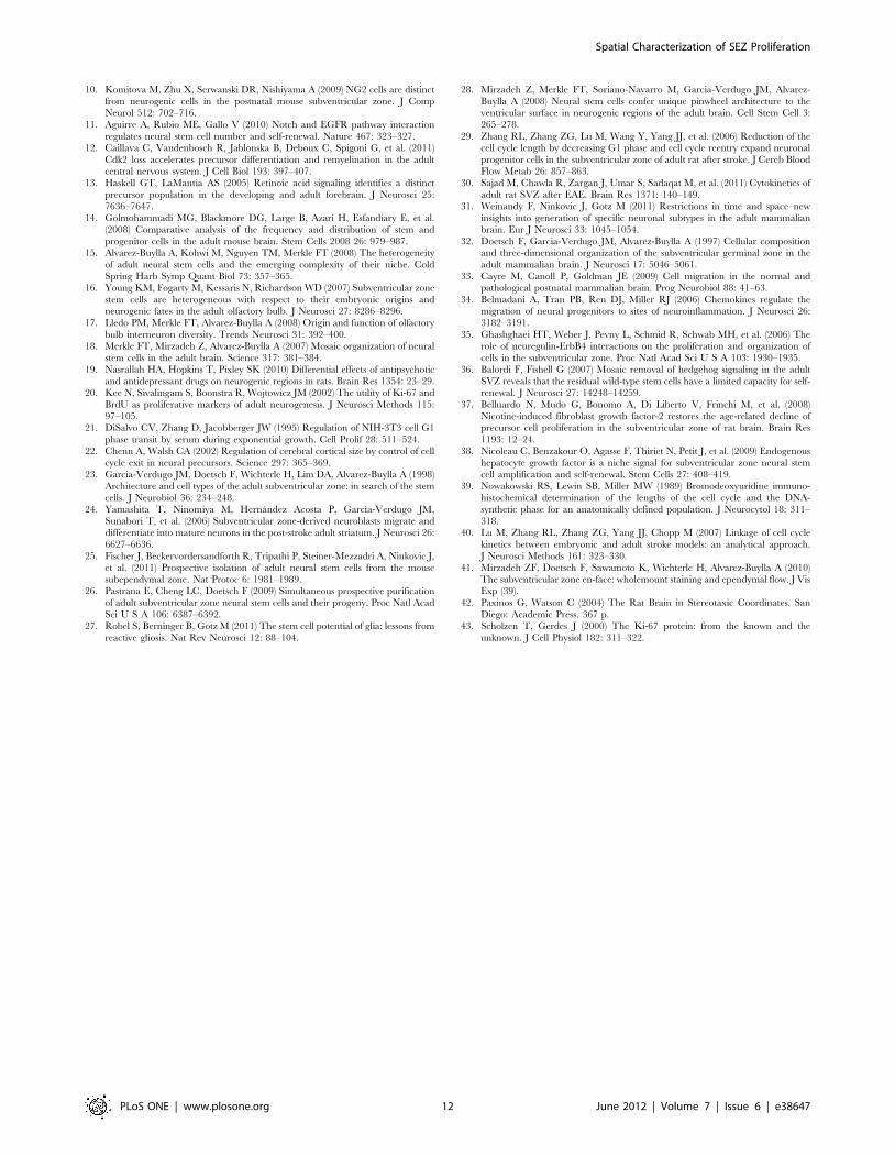
Table 1. Anterior-posterior axis landmarks of the SEZ divisions.

SEZ Bregma coordinates (mm) Anatomical references

Anterior [2.28; 1.44[ From the beginning to the end of the genu of the corpus callosum

Intermediate [1.44; 20.12[ From the end of the genu of the corpus callosum to the decussation of theanterior commissure

Posterior [20.12; 21.72[ From the decussation of the anterior commissure to the beginning of thehippocampus

Post Posterior [21.70; 23.60] From the beginning of the hippocampus to the fusion of the dorsal andventral parts of the lateral ventricle

Bregma coordinates are according to Paxinos & Watson (2004) [42].doi:10.1371/journal.pone.0038647.t001

Spatial Characterization of SEZ Proliferation

PLoS ONE | www.plosone.org 11 June 2012 | Volume 7 | Issue 6 | e38647

10. Komitova M, Zhu X, Serwanski DR, Nishiyama A (2009) NG2 cells are distinct

from neurogenic cells in the postnatal mouse subventricular zone. J CompNeurol 512: 702–716.

11. Aguirre A, Rubio ME, Gallo V (2010) Notch and EGFR pathway interaction

regulates neural stem cell number and self-renewal. Nature 467: 323–327.12. Caillava C, Vandenbosch R, Jablonska B, Deboux C, Spigoni G, et al. (2011)

Cdk2 loss accelerates precursor differentiation and remyelination in the adultcentral nervous system. J Cell Biol 193: 397–407.

13. Haskell GT, LaMantia AS (2005) Retinoic acid signaling identifies a distinct

precursor population in the developing and adult forebrain. J Neurosci 25:7636–7647.

14. Golmohammadi MG, Blackmore DG, Large B, Azari H, Esfandiary E, et al.(2008) Comparative analysis of the frequency and distribution of stem and

progenitor cells in the adult mouse brain. Stem Cells 2008 26: 979–987.15. Alvarez-Buylla A, Kohwi M, Nguyen TM, Merkle FT (2008) The heterogeneity

of adult neural stem cells and the emerging complexity of their niche. Cold

Spring Harb Symp Quant Biol 73: 357–365.16. Young KM, Fogarty M, Kessaris N, Richardson WD (2007) Subventricular zone

stem cells are heterogeneous with respect to their embryonic origins andneurogenic fates in the adult olfactory bulb. J Neurosci 27: 8286–8296.

17. Lledo PM, Merkle FT, Alvarez-Buylla A (2008) Origin and function of olfactory

bulb interneuron diversity. Trends Neurosci 31: 392–400.18. Merkle FT, Mirzadeh Z, Alvarez-Buylla A (2007) Mosaic organization of neural

stem cells in the adult brain. Science 317: 381–384.19. Nasrallah HA, Hopkins T, Pixley SK (2010) Differential effects of antipsychotic

and antidepressant drugs on neurogenic regions in rats. Brain Res 1354: 23–29.20. Kee N, Sivalingam S, Boonstra R, Wojtowicz JM (2002) The utility of Ki-67 and

BrdU as proliferative markers of adult neurogenesis. J Neurosci Methods 115:

97–105.21. DiSalvo CV, Zhang D, Jacobberger JW (1995) Regulation of NIH-3T3 cell G1

phase transit by serum during exponential growth. Cell Prolif 28: 511–524.22. Chenn A, Walsh CA (2002) Regulation of cerebral cortical size by control of cell

cycle exit in neural precursors. Science 297: 365–369.

23. Garcia-Verdugo JM, Doetsch F, Wichterle H, Lim DA, Alvarez-Buylla A (1998)Architecture and cell types of the adult subventricular zone: in search of the stem

cells. J Neurobiol 36: 234–248.24. Yamashita T, Ninomiya M, Hernandez Acosta P, Garcıa-Verdugo JM,

Sunabori T, et al. (2006) Subventricular zone-derived neuroblasts migrate anddifferentiate into mature neurons in the post-stroke adult striatum. J Neurosci 26:

6627–6636.

25. Fischer J, Beckervordersandforth R, Tripathi P, Steiner-Mezzadri A, Ninkovic J,et al. (2011) Prospective isolation of adult neural stem cells from the mouse

subependymal zone. Nat Protoc 6: 1981–1989.26. Pastrana E, Cheng LC, Doetsch F (2009) Simultaneous prospective purification

of adult subventricular zone neural stem cells and their progeny. Proc Natl Acad

Sci U S A 106: 6387–6392.27. Robel S, Berninger B, Gotz M (2011) The stem cell potential of glia: lessons from

reactive gliosis. Nat Rev Neurosci 12: 88–104.

28. Mirzadeh Z, Merkle FT, Soriano-Navarro M, Garcia-Verdugo JM, Alvarez-

Buylla A (2008) Neural stem cells confer unique pinwheel architecture to theventricular surface in neurogenic regions of the adult brain. Cell Stem Cell 3:

265–278.

29. Zhang RL, Zhang ZG, Lu M, Wang Y, Yang JJ, et al. (2006) Reduction of thecell cycle length by decreasing G1 phase and cell cycle reentry expand neuronal

progenitor cells in the subventricular zone of adult rat after stroke. J Cereb BloodFlow Metab 26: 857–863.

30. Sajad M, Chawla R, Zargan J, Umar S, Sadaqat M, et al. (2011) Cytokinetics of

adult rat SVZ after EAE. Brain Res 1371: 140–149.31. Weinandy F, Ninkovic J, Gotz M (2011) Restrictions in time and space–new

insights into generation of specific neuronal subtypes in the adult mammalianbrain. Eur J Neurosci 33: 1045–1054.

32. Doetsch F, Garcia-Verdugo JM, Alvarez-Buylla A (1997) Cellular compositionand three-dimensional organization of the subventricular germinal zone in the

adult mammalian brain. J Neurosci 17: 5046–5061.

33. Cayre M, Canoll P, Goldman JE (2009) Cell migration in the normal andpathological postnatal mammalian brain. Prog Neurobiol 88: 41–63.

34. Belmadani A, Tran PB, Ren DJ, Miller RJ (2006) Chemokines regulate themigration of neural progenitors to sites of neuroinflammation. J Neurosci 26:

3182–3191.

35. Ghashghaei HT, Weber J, Pevny L, Schmid R, Schwab MH, et al. (2006) Therole of neuregulin-ErbB4 interactions on the proliferation and organization of

cells in the subventricular zone. Proc Natl Acad Sci U S A 103: 1930–1935.36. Balordi F, Fishell G (2007) Mosaic removal of hedgehog signaling in the adult

SVZ reveals that the residual wild-type stem cells have a limited capacity for self-renewal. J Neurosci 27: 14248–14259.

37. Belluardo N, Mudo G, Bonomo A, Di Liberto V, Frinchi M, et al. (2008)

Nicotine-induced fibroblast growth factor-2 restores the age-related decline ofprecursor cell proliferation in the subventricular zone of rat brain. Brain Res

1193: 12–24.38. Nicoleau C, Benzakour O, Agasse F, Thiriet N, Petit J, et al. (2009) Endogenous

hepatocyte growth factor is a niche signal for subventricular zone neural stem

cell amplification and self-renewal. Stem Cells 27: 408–419.39. Nowakowski RS, Lewin SB, Miller MW (1989) Bromodeoxyuridine immuno-

histochemical determination of the lengths of the cell cycle and the DNA-synthetic phase for an anatomically defined population. J Neurocytol 18: 311–

318.40. Lu M, Zhang RL, Zhang ZG, Yang JJ, Chopp M (2007) Linkage of cell cycle

kinetics between embryonic and adult stroke models: an analytical approach.

J Neurosci Methods 161: 323–330.41. Mirzadeh ZF, Doetsch F, Sawamoto K, Wichterle H, Alvarez-Buylla A (2010)

The subventricular zone en-face: wholemount staining and ependymal flow. J VisExp (39).

42. Paxinos G, Watson C (2004) The Rat Brain in Stereotaxic Coordinates. San

Diego: Academic Press. 367 p.43. Scholzen T, Gerdes J (2000) The Ki-67 protein: from the known and the

unknown. J Cell Physiol 182: 311–322.

Spatial Characterization of SEZ Proliferation

PLoS ONE | www.plosone.org 12 June 2012 | Volume 7 | Issue 6 | e38647

61

3. The subependymal zone proliferative pattern and progenitor cell

distribution in mice: species matters!

62

63

The subependymal zone proliferative pattern and progenitor cell distribution in mice: species

matters!

Ana Mendanha Falcão1,2, Joana Almeida Palha1,2, Ana Catarina Ferreira1,2, Fernanda Marques1,2,

Nuno Sousa1,2, João Carlos Sousa1,2*

1 Life and Health Sciences Research Institute (ICVS), School of Health Sciences, University of

Minho, Braga, Portugal

2 ICVS/3B’s - PT Government Associate Laboratory, Guimarães, Braga, Portugal

Corresponding author: João Carlos Sousa. Life and Health Sciences Research Institute (ICVS),

School of Health Sciences, University of Minho, Campus Gualtar, 4710-057 Braga, Portugal.

Telephone: 351-253-604928 E-mail: [email protected]

64

65

Abstract

The subependymal zone (SEZ), the major neurogenic niche of the adult brain, responds to brain

insults and is modified in many neurodegenerative diseases, eliciting expectations for the

development of new endogenous regenerative therapies that ultimately envisage human

applications. Nevertheless, much remains to be done to fully understand the regulation and the

potential of the SEZ cells both in physiological and non-physiological conditions. So far, the most

widely used models to study adult neurogenesis in the SEZ are rodents. Interestingly, when

comparing the SEZ niche in different species, differences are observed. To translate studies from

animal models to humans for clinical applications, it is necessary to understand the SEZ species-

specific variations. In the past few years progress was made in understanding the SEZ niche

heterogeneity in rodents. Given our previous findings on the topographical mapping of adult rat

SEZ proliferation, we now extend the study to the adult mouse brain. We found no differences in

the proliferation in the anterior, intermediate and posterior SEZ and along the lateral wall.

However, we report relevant differences between two close related species, rats and mice that

should be taken into account when extrapolating data on the SEZ from mice to rats and vice-

versa.

Key words

Subependymal zone, mice, species-specific, variability

66

Introduction

The occurrence of neural stem cells (NSCs) in discrete zones of the adult brain has been

reported in several mammals (Barker et al 2011). Generally, NSCs are confined to two major

brain areas: the subependymal zone (SEZ) lining the lateral wall of the lateral ventricles and the

subgranular zone (SGZ) of the hippocampal dentate gyrus (Ming & Song 2005). The most well

recognized fate for SEZ born progenitor cells is the olfactory bulbs (OB) where they integrate into

circuitries mostly as GABAergic interneurons (Lois & Alvarez-Buylla 1994). Although the presence

of NSCs in the lateral ventricles is transversal to all mammalian species, the formation of new

functional adult olfactory neurons has only been unequivocally described in rodents (rat and

mice) and in some primates (Lindsey & Tropepe 2006). Importantly, the amount of proliferation

and new neurons integrated in the OBs is variable across species. When compared to rodents,

primates display a substantial decrease both in the number of NSCs in the SEZ and in the

olfactory neurogenesis (Azim et al 2012). Interestingly, a recent study relying on 14C birth dating

analysis of adult human olfactory neurons has demonstrated the limited, if any, existence of new

olfactory neurons in adult human OBs (Bergmann et al 2012). Additionally, other studies

demonstrated that adult neurogenesis in humans seems limited to the childhood (Sanai et al

2011). The major variation in NSCs occurrence and fate across species indicates its intrinsic

species-specific heterogeneity and ultimately may reflect distinct functional adaptations.

Regardless of species, SEZ alterations are observed in many pathophysiological conditions (Curtis

et al 2007). As an example, following ischemic stroke enhanced proliferation is observed in the

ipsilateral SEZ both in human and in rodents (Marti-Fabregas et al 2010). These observations

highlight an additional role for NSCs of the adult mammalian SEZ as a reservoir of cells for

regenerative purposes. Therefore, many efforts have been concentrated in understanding the

potentiality and modulation of NSCs in the adult brain.

Due to a short life span, together with high reproduction rates and known genetic background,

rodents are the most widely used models to study adult neurogenesis either in physiological

conditions or in the context of disease. Generally, it is expected that adult neurogenesis follow the

same rules between closely related species. For instance, rats and mice belong to the same sub-

family and results obtained from one species are often extrapolated to the other. However, there

is evidence for variability between NSCs from rat and mouse. Specifically, in vitro experiments

have revealed that neurospheres isolated from the entire brain displayed different properties

67

between mice and rat (Ray & Gage 2006). Furthermore, hippocampal derived neurospheres from

rat and mice exhibit significant differences in proliferative capacity and differentiation fate

(Steffenhagen et al 2011). Strengthening the in vitro data, in vivo work demonstrated differences

in adult hippocampal neurogenesis between rats and mice. Specifically, newly generated

hippocampal neurons matured more rapidly and were more frequent in rat than in mice (Snyder

et al 2009). Also of notice, radial glial tubes forming the rostral migratory stream (RMS), the path

of neuroblasts migration from the SEZ to the OBs, are more complex in rats than in mice (Peretto

et al 2005). Altogether, these findings raise questions about the differences across close related

species in the intrinsic heterogeneity and potential of NSCs, as well as their extrinsic regulation.

Despite some reports about distinctive hippocampal neurogenesis in rat and mouse, data is

missing on these potential differences in adult SEZ neurogenesis between these two species. We

have recently described that the proliferative profile of SEZ cells throughout the lateral ventricles

in rat varies in the anterior-posterior and dorsal-ventral axes and that the NSCs are

heterogeneously distributed in the SEZ axes (Falcao et al 2012) (Chapter 2). In the present

work, we characterized the proliferative pattern and NSCs distribution in mice.

68

Materials and Methods

Ethics statement

This study was approved by the Portuguese national authority for animal experimentation,

Direção Geral de Veterinária (ID: DGV9457). Animals were kept and handled in accordance with

the guidelines for the care and handling of laboratory animals in the Directive 2010/63/EU of

the European Parliament and of the Council.

Animals

All experiments were conducted in 10-weeks-old male C57BL/6 mice (Charles River, Barcelona,

Spain). Animals were maintained in 12 h light/dark cycles at 22 to 24ºC and 55% humidity and

fed with regular rodent’s chow and tap water ad libitum. To reduce stress-induced changes in the

hypothalamus–pituitary axis associated with 5-bromo-2’-deoxyuridine (BrdU) injections, all

animals were daily handled for 1 week until the day of the experimental procedure.

Administration of BrdU for proliferation assessment and for BrdU label retaining cells estimation

For the purpose of SEZ proliferation assessment, five animals were administered with BrdU (a

thymidine analogue that is incorporated in the DNA during the S phase; 50mg/Kg)

intraperitoneally (i.p.) and sacrificed 2h later. This protocol labels SEZ fast dividing cells. To

assess neuroblast proliferating cells an additional group of 5 animals was injected with BrdU and

sacrificed 2h later.

To label a quiescent pool of cells at the SEZ, a group of four animals was daily i.p. injected with

BrdU (50mg/Kg) for 2 weeks followed by another 2 weeks chase period. The neural stem cell

progeny that exit the cell cycle and retain the BrdU labelling leaves the SEZ during the chase

period.

Immunohistochemistry

Animals were anesthetized with ketamine hydrochloride (150mg/kg) plus medetomidine

(0.3mg/kg), and transcardially perfused with either cold saline for the stereological analysis of

the SEZ, or with 4% paraformaldehyde (PFA) in 0.01M PBS, a pre-fixation of the brain, for

fluorescence immunohistochemistry. Brains were removed, embedded in O.C.T. compound and

snap-frozen; serial coronal sections (20 µm) were cut in a cryostat and collected to slides for

69

immunohistochemistry.

For the stereological analysis of the SEZ, animals injected with a single pulse of BrdU were

analysed by immunohistochemistry. Briefly, slides were post-fixed in 4% PFA in 0.01M PBS for 30

min and BrdU or Ki67 antibodies, to assess proliferation, were used at a dilution of 1:100 (rat

Anti-Bromodeoxyurine, BU1/75 (ICR1) clone, Abcam, UK; Ki67 antigen, rabbit polyclonal

antibody, Novocastra, UK). For BrdU staining, a pre-treatment of 30 min in HCl 2N was required.

For Ki67 an additional antigen retrieval step (in 10 mM sodium citrate buffer at pH 6) was

performed. Primary antibodies were detected with the Ultravision Detection System (Lab Vision,

Freemont, CA, USA), and the reaction developed with 3,3'-diaminobenzidine substrate (Sigma,

St.Louis, MO, USA); sections were subsequently counterstained with hematoxylin.

Fluorescence immunohistochemistry was performed to label proliferating neuroblasts [double

BrdU/doublecortin (DCX) positive], neuroblasts (DCX positive cells) and neural stem cells [double

BrdU/ glial fibrillary acidic protein (GFAP) positive]. Antibodies against the following markers of

SEZ populations were used: DCX (rabbit polyclonal to doublecortin-neuroblast marker, Abcam) at

a dilution of 1:500 and GFAP (polyclonal rabbit anti-GFAP, DAKO, Spain) at a dilution of 1:100,

together with BrdU (Abcam) at a dilution of 1:100. Fluorescent secondary antibodies (Invitrogen,

Carlsbad, CA, USA), anti-rabbit and anti-rat were used to detect the primary antibodies at a

dilution of 1:1000. To label the nucleus, incubation with 4’,6-diamidino-2-phenylindole (DAPI;

Sigma) at a dilution of 1:1000 was performed. Primary and secondary antibodies were diluted in

PBS-0.5%Triton/10% FBS and incubated overnight at 4ºC, for the primary antibody, and 2h at

room temperature for the secondary antibody.

Wholemount staining

Wholemount staining for DCX and Ki67 was performed as previously described (Mirzadeh et al

2010). Briefly, the entire lateral wall from rat and mouse brains perfused with cold saline was

dissected under a stereomicroscope and incubated in 4% PFA-0.5% Triton overnight at 4ºC. For

Ki67 staining, wholemounts were incubated in 10 mM sodium citrate buffer (at pH 6) at 80ºC for

30 min. Primary (anti-DCX 1:250 Abcam, anti-ki67 1:100 Novocastra) and secondary (1:500,

Invitrogen) antibodies were each incubated for 2 days at 4ºC.

70

Stereology

Estimation of cell density in the different regions of the SEZ was performed as described before

(Falcao et al 2012). Coronal sections for proliferation analysis comprised SEZ between bregma

coordinates 1.18mm and -1.94mm. The number of BrdU or Ki67 positive cells and the areas

were estimated by means of the Visiopharm Integrator system (VIS) software in an Olympus

BX51 microscope (Olympus, Hamburg, Germany). Results were expressed as BrdU or Ki67

positive cells per area (in mm2). Every sixteenth section from the anterior SEZ, bregma 1.18mm,

until posterior SEZ, bregma -1.94mm, was analysed. The use of the VIS Software allowed

delimitation, at low magnification (40x), of the areas of interest in the SEZ and the counting of

BrdU positive cells within the defined areas at high magnification (400x). The divisions of the SEZ

in the anterior-posterior and dorsal-ventral axes were defined on the basis of established

anatomical references (Table 1), as previously described (Falcao et al 2012). Briefly, at the

anterior-posterior axis, the SEZ anterior division starts and finishes with the beginning and the

end of the genu of the corpus callosum, respectively (bregma 1.18mm to 0.74mm); intermediate

SEZ begins with the end of the genu of the corpus callosum and extends up to the decussation of

the anterior commissure (bregma 0.74mm to 0.14mm); the posterior division of the SEZ begins

at the decussation of the anterior commissure and extends to the beginning of the hippocampus,

bregma -0.94mm; the post-posterior division extends up to bregma -1.94mm (Table 1).

Confocal imaging and quantitative analysis

To estimate the number of neuroblasts (DCX positive cells) and proliferating neuroblasts (double

DCX/BrdU positive cells) along the anterior-posterior and dorsal-ventral axes, 6 sections per

animal were analysed. For each section, images were taken for the entire lateral wall of the SEZ

using a confocal microscope (FV1000; Olympus) and the total number of DCX positive cells and

the number of double labelled DCX/BrdU positive cells was counted. The rate for neuroblasts and

proliferating neuroblasts was estimated by dividing the number of positive and double positive

labelled cells for the corresponding area. The areas were determined using the Image J software.

The same analysis was performed to estimate rates of double GFAP/BrdU positive cells.

Confocal imaging of wholemount preparations of the lateral ventricle wall was performed to label

neuroblasts and proliferating cells. Images taken from the lateral wall were then assembled to

show the entire lateral wall staining.

71

Table 1: Anterior-posterior axis landmarks of the mice SEZ divisions. Bregma coordinates are according to Paxinos &

Franklin (2001).

Statistical analysis

All the data (presented as the mean ± SEM) was analysed with GraphPad PRISM 5 software

(GraphPad Software Inc., San Diego, CA). The analysis consisted of one-way analysis of variance

(ANOVA) with Bonferroni multiple comparison test post-test analysis for single-factor multiple

group comparisons to determine differences between three or more groups. Student’s t test was

performed for two-group comparisons. The threshold value for statistical significance was set at

p<0.05.

SEZ

Bregma coordinates

(mm) Anatomical references

Anterior 1.18; 0.74 From the beginning to the end of the

genu of the corpus callosum

Intermediate 0.74; -0.14 From the end of genu of the corpus callosum to the decussation of anterior commissure

Posterior -0.14; -0.94 From the decussation of anterior commissure to the beginning of the hippocampus

Post Posterior -0.94; -1.94 From the beginning of the hippocampus to the fusion of the dorsal and ventral parts of the lateral ventricle

72

Results

In the anterior-posterior axis, SEZ cell proliferation is lower in the post-posterior region

To assess proliferation along the anterior-posterior axis we have estimated the number of BrdU

and Ki67 positive cells per area in four SEZ levels: anterior, intermediate, posterior and post-

posterior (Figure 1). These regions were previously defined according to external anatomic cues

for the rat SEZ (Falcao et al 2012), and the same criteria for the definition of SEZ regions were

applied in this analysis for the mouse. Our results show no differences in cell proliferation

between anterior, intermediate and posterior SEZ, 2.37±0.08 x10³, 2.32±0.14 x10³ and

2.37±0.24 x10³ BrdU positive cells per mm2, respectively (Figure 1A). However, at post-posterior

levels, we found a reduction in proliferation to 1.51±0.18 x10³ BrdU positive cells per mm2.

Similar results were achieved when the same analysis was performed using the endogenous

proliferation marker Ki67 (Figure 1B).

In the dorsal-ventral axis, the dorsal SEZ displays the lowest cell proliferation

At the dorsal-ventral axis the SEZ was divided into four distinct parts: dorsal, RMS, dorsolateral

and ventral SEZ. Dorsal SEZ is at the dorsal wall of the SEZ, RMS constitutes the beginning of the

RMS, localized at the dorsal corner of the ventricular lateral wall, dorsolateral and ventral are

outlined from the lateral wall (Falcao et al 2012). The proliferation at the RMS, dorsolateral and

ventral SEZ was similar, 3.00±0.35 x10³, 2.60±0.28 x10³ and 2.51±0.14 x10³ BrdU positive

cells per mm2, respectively (Figure 1C). However, the dorsal SEZ exhibited the lowest proliferation

rates of the four regions, 0.36±0.08 x10³ BrdU positive cells per mm2, only 12-14% of the total

proliferation found in the other regions (Figure 1C). The same analysis was performed using the

Ki67 marker and the results were generally similar (Figure 1D).

The proliferation rates of the SEZ regions are similar along the anterior-posterior axis in the

lateral wall but not in the dorsal wall

To further investigate if the proliferation rates estimated for the four SEZ regions were preserved

along the anterior-posterior axis, we independently analysed cell proliferation within anterior,

intermediate and posterior levels of the SEZ. The proliferation, assessed by BrdU staining, in the

SEZ lateral wall did not change from intermediate to posterior positions (Figure 2). Also, no

significant differences were observed on the beginning of the RMS from anterior to intermediate

73

levels. However, when analysing the anterior SEZ, the proliferation rates for RMS were higher

than in undefined (und) region. Of notice, dorsal SEZ proliferation rate increases 4 times at

posterior levels from 0.12±0.12 x10³ to 0.58±0.06 x10³ BrdU positive cells per mm2 (Figure 2). A

similar increase in cell proliferation in the dorsal SEZ at posterior levels was found with the Ki67

marker (Figure 2).

Figure 1 - Proliferative pattern of the mice subependymal zone (SEZ) cells throughout the anterior-posterior and

dorsal-ventral axes. A, B. The SEZ proliferation, assessed by the number of BrdU and Ki67 positive cells, was

reduced at the post-posterior SEZ in the anterior-posterior axis. C, D. At the dorsal-ventral axis, the dorsal SEZ

displayed the lowest levels of proliferation. Proliferation was expressed as number of BrdU or Ki67 positive cells per

area (mm2). The threshold value for statistical significance was set at 0.05 (* p<0.05).

74

Figure 2 - Combined analysis of cell proliferation in the subependymal zone (SEZ) anterior-posterior and dorsal-

ventral axes. Proliferation pattern analysis was assessed by the number of BrdU and Ki67 positive cells in dorsal-

ventral SEZ regions along the anterior to posterior axis. The pattern of proliferation between different regions of the

SEZ was similar along anterior-posterior axis. However, the dorsal SEZ rates of proliferation were increased at the

posterior SEZ. Proliferation was expressed as number of BrdU and Ki67 positive cells per area (mm2). The threshold

value for statistical significance was set at 0.05 (* p<0.05).

Neural stem and precursor cells are homogeneously distributed in the lateral wall

Despite the observed relative homogeneity in proliferative patterns throughout the lateral SEZ

wall, we analysed the neuroblast distribution in the lateral wall. No major differences were

detected in the number of DCX positive cells and the double BrdU/DCX positive cells between

dorsolateral and ventral SEZ at the intermediate levels (Figure 3). Furthermore, when assessing

NSC rates by double staining of BrdU and GFAP, this population was homogeneously distributed

within the lateral wall (Figure 3). Since GFAP is a consensual marker for NSCs and the population

that constitutes label retaining cells (BrdU positive cells) are relatively quiescent cells, we

identified the NSCs in the SEZ by labelling cells with GFAP and BrdU (from label retaining cells).

75

These results suggest that neural stem and progenitor cells are uniformly distributed in the mice

SEZ.

Figure 3 - The distribution of neural stem cells and neuroblasts was similar in the dorsal-ventral axis. DCX and

double BrdU/DCX staining was used to assess neuroblasts and proliferating neuroblasts, respectively, at the

dorsolateral and ventral SEZ. Neural stem cells were detected by double staining BrdU retaining cells with GFAP and

assessed in the dorsolateral and ventral SEZ. All results are expressed as number of positive cells per area (in mm 2).

The threshold value for statistical significance was set at 0.05 (* p<0.05).

The BrdU/Ki67 ratios are similar throughout the SEZ

We have estimated the BrdU/Ki67 ratio which provides an estimation of the cell cycle lengths

between different dorsal-ventral regions and anterior-posterior divisions of the SEZ. These were

constant throughout both axes suggesting that there are no major differences in the cell cycle

length of the SEZ cells between the analysed regions (Figure 4).

Figure 4 - The BrdU/Ki67 ratio throughout the subependymal zone was constant. The ratios BrdU/Ki67 were

represented for the SEZ at anterior-posterior and dorsal-ventral axes. The threshold value for statistical significance

was set at 0.05 (* p<0.05).

76

The number of cells in the vicinity of the SEZ diminishes from anterior to posterior divisions

The estimation of the number of cells proliferating less than 100µm apart from the SEZ revealed

a remarkable reduction in the BrdU and Ki67 positive cells from anterior to posterior divisions.

The anterior division presented higher numbers of BrdU and Ki67 positive cells ectopically

placed, 9±1 and 27±3 respectively, when compared to intermediate and posterior divisions

(Figure 5).

Figure 5 - The number of cells proliferating in the vicinity of the subependymal zone decreased along the anterior-

posterior axis. The BrdU and Ki67 positive cells located within a distance of 100 µm apart from the SEZ were

estimated. Results were represented as number of BrdU or Ki67positive cells per section. The threshold value for

statistical significance was set at 0.05 (* p<0.05).

77

Discussion

By unravelling the topographical specificities in the proliferative rates in the rat SEZ, we have

previously emphasized the relevance to take into account the location within the SEZ when

studying the NSCs. Defining the exact coordinates in which analysis is performed is necessary to

make appropriate and valid associations and comparisons across studies. In this sense, it is not

surprising that similar studies on the SEZ dynamics give contradictory results, probably due to

the use of different anatomic clues or specific SEZ regions. Because we found significant

differences in the rat proliferation and neuroblast population throughout the SEZ axes, in this

study we have performed a topographical analysis of the adult SEZ NSC niche in mice, using a

similar approach to that described previously for the rat SEZ (Falcao et al 2012). The present

study gives further insights about the similarities and differences between the SEZ of two close

related species that must be taken into account for future analysis when extrapolating results

obtained from mice to rat and vice-versa.

So far, studies performed in mice shed light to what is becoming increasingly evident: the adult

NSC pool of the SEZ is heterogeneous regarding to its intrinsic properties and spatial locations

(Alvarez-Buylla et al 2008). Although in vitro cultures of SEZ derived cells support the existence of

multipotent NSCs by showing its self-renewal capacity and multipotentiality (Doetsch et al 2002),

in vivo studies show that NSCs are already pre-committed to form certain types of interneurons at

the OBs (Merkle et al 2007, Young et al 2007). Furthermore, NSCs from mice also express

different transcription factors (TFs) along the dorsal-ventral axis, which in part overlap its

embryonic counterpart’s identity and TFs expression pattern (Alvarez-Buylla et al 2008, Weinandy

et al 2011). In this sense, it was further suggested that combinations between TFs on the NSCs

determine their cellular fate (Hack et al 2005).

Despite the reported cellular heterogeneity in the SEZ, and contrary to the described for rats, we

found minor differences on mice proliferative patterns along the SEZ anterior-posterior and

dorsal-ventral axes. By using two different proliferation markers, the acutely administrated

exogenous marker BrdU, and the endogenous marker Ki67, we found a reduction in proliferation

only at post-posterior levels of the SEZ. Noteworthy, most studies disregard the analysis of SEZ at

post-posterior levels because it is generally assumed that new neurons at the OBs are born

exclusively from the SEZ regions corresponding to the herein defined anterior, intermediate and

posterior SEZ. Interestingly, a study performed by Merkle and colleagues (2007) revealed

78

additional sites of birth of new OB neurons (Merkle et al 2007). Amongst others, new neurons

arise both from post-posterior levels of the SEZ and from the most anterior part of the RMS.

Concerning the dorsal-ventral axis of the SEZ, we detected a sharp decline in proliferating cells at

the dorsal SEZ. In fact, dorsal SEZ is largely omitted in studies in the SEZ and only recently its

neurogenic potential was recognized (Brill et al 2009, Merkle et al 2007). Also, it was

documented that the medial wall hosts NSCs that originate new OB neurons but only at most

anterior levels (Merkle et al 2007), equivalent to the here defined anterior SEZ.

We have further analysed the BrdU/Ki67 ratios, an estimation of the cell cycle length, along the

SEZ axes and found no alterations throughout both anterior-posterior and dorsal-ventral axes.

This result is in accordance with the uniform proliferation rates between anterior, intermediate

and posterior SEZ concerning the anterior-posterior axis and between dorsolateral and ventral

SEZ in regards to the dorsal-ventral axis.

Recently, the NSC distribution profile throughout the mouse lateral wall was described by means

of pinwheels structures analysis in wholemounts (Mirzadeh et al 2008). Interestingly, type B1 cell

clusters (located in the middle of the pinwheels) were more numerous in anterior-ventral and

posterior-dorsal positions. Type B1 cells are generally accepted as NSCs that give rise to all

cellular populations at the SEZ (Doetsch et al 1999). These single cilium cells are identified by

GFAP and gamma-tubulin labelling surrounded by beta-catenin from ependymal cells, forming a

pinwheel-like structure. Altogether, data obtained from us and others (Mirzadeh et al 2008)

suggests that the heterogeneous distribution of type B1 cells does not impact on the proliferative

profile along the lateral wall.

It was suggested that the cells that comprise the pinwheel structure are heterogeneous namely

on the quiescence/mitotic activity (Shook et al 2012). Therefore, the inclusion of a mitotic

marker, such as PH3 or Ki67, in the estimations for the number of mitotic NSC in pinwheels

could considerably change the outcome. For instances, the aged SEZ displays fewer type B1 cells

but equal percentages for mitotic type B1 cells (Shook et al 2012). In the present work, we label

NSCs by co-labelling GFAP with label retaining cells, given by BrdU staining. The analysis of NSCs

using this approach revealed no statistically significant differences in the rates of NSCs between

dorsolateral and ventral SEZ. Similarly, the distribution of neuroblasts in the lateral wall was also

uniform, i.e., dorsolateral and ventral SEZ display similar rates of both neuroblasts and

proliferating neuroblasts. Taken together, these results further support the data from the

proliferation analysis. Although progenitors scattered from diverse parts of the lateral wall give

79

rise to different OB neuronal types, our data suggests that in what concerns the proliferation rate,

neuroblasts are similar along the lateral wall. Likewise, there was no spatial-specific distribution

of the general neuroblast population, as assessed by DCX staining.

We assessed the number of proliferating cells along the anterior-posterior axis and observed a

significant decrease in the number of cells proliferating in the vicinity of the SEZ at intermediate

and posterior levels. The migration of progenitor cells, namely neuroblast cells, out of SEZ

towards sites of injury has been described in the context of brain damage (Zhang et al 2004).

Although most of proliferating cells migrate tangentially, radial migration should not be excluded,

at least in response to injury.

A combined analysis of rat and mouse proliferation patterns along the SEZ highlights the

following inter-species discrepancies: i) the uppermost proliferation rate observed at the anterior

SEZ in rats is absent in mice, ii) the remarkable decline in proliferation from dorsolateral to

ventral SEZ present in the rat lateral wall does not occur in mice (Supplementary Figure 1), iii)

the differential cell cycle lengths of rat SEZ cells, namely between dorsolateral and ventral

progenitors, does not occur in mice, as given by similar BrdU/Ki67 ratios and iv) the differential

spatial distribution of rat neuroblasts (Supplementary Figure 1) and NSCs along the lateral wall is

homogeneous in mice. Nevertheless, some major similarities between the SEZ of both species

were found, such as the proliferative decay at post-posterior levels, the limited proliferation at the

dorsal SEZ, the augment of dorsal SEZ proliferation at posterior levels, and the decrease of

proliferating cells surrounding the SEZ at intermediate and posterior levels.

Commonly, many conclusions on numerous SEZ related studies in the rat are tacitly assumed to

hold true for mice, and vice-versa. The above outlined species-specific differences between these

two most common laboratory rodent models points to the need to revise and consider SEZ from

rats and mice as two distinct heterogeneous niches of neural stem and progenitor cells. Most

importantly, it may ultimately reflect distinct functional differences both in health and disease.

The existence of adult NSCs in the SEZ has been described in many mammalian species (Bedard

et al 2002, Kornack & Rakic 2001, Sawamoto et al 2011, Seaberg & van der Kooy 2002). The

closest to human the lesser extent is the formation of new neurons at the OBs. Thus, it is

reasonable to infer that species highly dependent on olfaction, such as rodents, require an

increased number of new OB neurons than species that do not depend as much on olfaction. In

this sense, nonhuman primates display a less developed SEZ-RMS-OB nexus than rodents.

In summary, although species-specific differences on hippocampal neurogenesis were

80

demonstrated, both in vivo and in vitro (Snyder et al 2009, Steffenhagen et al 2011) this is the

first report on the topic concerning the adult SEZ neurogenic niche.

81

References

Alvarez-Buylla A, Kohwi M, Nguyen TM, Merkle FT. 2008. The heterogeneity of adult neural stem

cells and the emerging complexity of their niche. Cold Spring Harb Symp Quant Biol 73:

357-65

Azim K, Zweifel S, Klaus F, Yoshikawa K, Amrein I, Raineteau O. 2012. Early decline in

progenitor diversity in the marmoset lateral ventricle. Cereb Cortex

Barker JM, Boonstra R, Wojtowicz JM. 2011. From pattern to purpose: how comparative studies

contribute to understanding the function of adult neurogenesis. Eur J Neurosci 34: 963-

77

Bedard A, Levesque M, Bernier PJ, Parent A. 2002. The rostral migratory stream in adult squirrel

monkeys: contribution of new neurons to the olfactory tubercle and involvement of the

antiapoptotic protein Bcl-2. Eur J Neurosci 16: 1917-24

Bergmann O, Liebl J, Bernard S, Alkass K, Yeung MS, et al. 2012. The age of olfactory bulb

neurons in humans. Neuron 74: 634-9

Brill MS, Ninkovic J, Winpenny E, Hodge RD, Ozen I, et al. 2009. Adult generation of

glutamatergic olfactory bulb interneurons. Nat Neurosci 12: 1524-33

Curtis MA, Faull RL, Eriksson PS. 2007. The effect of neurodegenerative diseases on the

subventricular zone. Nat Rev Neurosci 8: 712-23

Doetsch F, Garcia-Verdugo JM, Alvarez-Buylla A. 1999. Regeneration of a germinal layer in the

adult mammalian brain. Proc Natl Acad Sci U S A 96: 11619-24

Doetsch F, Petreanu L, Caille I, Garcia-Verdugo JM, Alvarez-Buylla A. 2002. EGF converts transit-

amplifying neurogenic precursors in the adult brain into multipotent stem cells. Neuron

36: 1021-34

Falcao AM, Palha JA, Ferreira AC, Marques F, Sousa N, Sousa JC. 2012. Topographical analysis

of the subependymal zone neurogenic niche. PLoS One 7: e38647

Hack MA, Saghatelyan A, de Chevigny A, Pfeifer A, Ashery-Padan R, et al. 2005. Neuronal fate

determinants of adult olfactory bulb neurogenesis. Nat Neurosci 8: 865-72

Kornack DR, Rakic P. 2001. The generation, migration, and differentiation of olfactory neurons in

the adult primate brain. Proc Natl Acad Sci U S A 98: 4752-7

Lindsey BW, Tropepe V. 2006. A comparative framework for understanding the biological

principles of adult neurogenesis. Prog Neurobiol 80: 281-307

82

Lois C, Alvarez-Buylla A. 1994. Long-distance neuronal migration in the adult mammalian brain.

Science 264: 1145-8

Marti-Fabregas J, Romaguera-Ros M, Gomez-Pinedo U, Martinez-Ramirez S, Jimenez-Xarrie E, et

al. 2010. Proliferation in the human ipsilateral subventricular zone after ischemic stroke.

Neurology 74: 357-65

Merkle FT, Mirzadeh Z, Alvarez-Buylla A. 2007. Mosaic organization of neural stem cells in the

adult brain. Science 317: 381-4

Ming GL, Song H. 2005. Adult neurogenesis in the mammalian central nervous system. Annu

Rev Neurosci 28: 223-50

Mirzadeh Z, Doetsch F, Sawamoto K, Wichterle H, Alvarez-Buylla A. 2010. The subventricular

zone en-face: wholemount staining and ependymal flow. J Vis Exp 39

Mirzadeh Z, Merkle FT, Soriano-Navarro M, Garcia-Verdugo JM, Alvarez-Buylla A. 2008. Neural

stem cells confer unique pinwheel architecture to the ventricular surface in neurogenic

regions of the adult brain. Cell Stem Cell 3: 265-78

Paxinos G, Franklin KB. 2001. The mouse brain in stereotaxic coordinates. USA: Academic Press

Peretto P, Giachino C, Aimar P, Fasolo A, Bonfanti L. 2005. Chain formation and glial tube

assembly in the shift from neonatal to adult subventricular zone of the rodent forebrain. J

Comp Neurol 487: 407-27

Ray J, Gage FH. 2006. Differential properties of adult rat and mouse brain-derived neural

stem/progenitor cells. Mol Cell Neurosci 31: 560-73

Sanai N, Nguyen T, Ihrie RA, Mirzadeh Z, Tsai HH, et al. 2011. Corridors of migrating neurons in

the human brain and their decline during infancy. Nature 478: 382-6

Sawamoto K, Hirota Y, Alfaro-Cervello C, Soriano-Navarro M, He X, et al. 2011. Cellular

composition and organization of the subventricular zone and rostral migratory stream in

the adult and neonatal common marmoset brain. J Comp Neurol 519: 690-713

Seaberg RM, van der Kooy D. 2002. Adult rodent neurogenic regions: the ventricular

subependyma contains neural stem cells, but the dentate gyrus contains restricted

progenitors. J Neurosci 22: 1784-93

Shook BA, Manz DH, Peters JJ, Kang S, Conover JC. 2012. Spatiotemporal changes to the

subventricular zone stem cell pool through aging. J Neurosci 32: 6947-56

Snyder JS, Choe JS, Clifford MA, Jeurling SI, Hurley P, et al. 2009. Adult-born hippocampal

neurons are more numerous, faster maturing, and more involved in behavior in rats than

83

in mice. J Neurosci 29: 14484-95

Steffenhagen C, Kraus S, Dechant FX, Kandasamy M, Lehner B, et al. 2011. Identity, fate and

potential of cells grown as neurospheres: species matters. Stem cell reviews 7: 815-35

Weinandy F, Ninkovic J, Gotz M. 2011. Restrictions in time and space-new insights into

generation of specific neuronal subtypes in the adult mammalian brain. Eur J Neurosci

33: 1045-54

Young KM, Fogarty M, Kessaris N, Richardson WD. 2007. Subventricular zone stem cells are

heterogeneous with respect to their embryonic origins and neurogenic fates in the adult

olfactory bulb. J Neurosci 27: 8286-96

Zhang R, Zhang Z, Wang L, Wang Y, Gousev A, et al. 2004. Activated neural stem cells contribute

to stroke-induced neurogenesis and neuroblast migration toward the infarct boundary in

adult rats. J Cereb Blood Flow Metab 24: 441-8

84

Supplementary figure 1 - En face views of proliferation and neuroblasts population in the lateral wall of rat and mice.

Wholemount staining for proliferation, Ki67 (green) and neuroblasts, DCX (red) revealed a different distribution of

cells in the mouse (A) and rat (B) lateral wall. Scale bar=1mm.

85

4. Acute peripheral inflammation impacts on subependymal zone,

choroid plexus and cerebrospinal fluid

86

87

Acute peripheral inflammation impacts on subependymal zone, choroid plexus and cerebrospinal

fluid

Ana Mendanha Falcão1,2, Fernanda Marques1,2, Ana Catarina Ferreira1,2, Ashley Novais1,2, Luísa

Pinto1,2, Nuno Sousa1,2, Joana Almeida Palha1,2, João Carlos Sousa1,2*

1 Life and Health Sciences Research Institute (ICVS), School of Health Sciences, University of

Minho, Braga, Portugal

2 ICVS/3B’s - PT Government Associate Laboratory, Guimarães, Braga, Portugal

Corresponding author: João Carlos Sousa, Life and Health Sciences Research Institute (ICVS),

School of Health Sciences, University of Minho, Campus Gualtar, 4710-057 Braga, Portugal.

Telephone: 351-253-604928 E-mail: [email protected]

88

89

Abstract

The subependymal zone (SEZ) is the major site of neurogenesis in the adult brain that originates

new neurons for the olfactory bulbs (OBs). The interplay between intrinsic and extrinsic factors

regulates the SEZ cell proliferation, fate and migration. The extrinsic cues that influence SEZ cells

in a paracrine manner are of diverse origins: ependymal cells, endothelial cells of the blood-brain

barrier, neurons, microglia and neural stem and precursor cells. Recently, we and others have

suggested that the cerebrospinal fluid (CSF), whose proteins are mostly synthetized and secreted

by the choroid plexus (CP), can also modulate the SEZ niche dynamics. Of notice, the CSF

composition is significantly altered in response to inflammation. Interestingly, inflammatory

conditions that considerably change the environment milieu of the SEZ niche have been shown to

trigger alterations in the proliferative and migratory profiles of the SEZ cells. In this study, we

challenged the peripheral immune system by administrating a single injection of

lipopolysaccharide (LPS) and analysed the CP expression profile of various genes, the CSF

composition and the SEZ cells. We observed an increase in proliferation of SEZ cells, namely in

the neuroblasts population, upon 12h of LPS stimulus. Furthermore, we report a rapid and

transient CP gene expression alteration of SEZ modulators, such as growth factors, that is also

reflected in the CSF. Our data suggest that alterations in the CP and thus in the CSF composition

triggered by LPS will likely impact on SEZ niche dynamics.

Key words

Subependymal zone, choroid plexus, cerebrospinal fluid, inflammation, neurogenesis

90

Introduction

In the past decades, the regenerative capabilities of the neurogenic areas of the adult brain, both

in physiological and in pathological conditions, have been extensively studied (Kaneko &

Sawamoto 2009, Vandenbosch et al 2009). The subependymal zone (SEZ), located in the lateral

wall of the lateral ventricles, is the major source of new neurons in the adult brain (Alvarez-Buylla

& Garcia-Verdugo 2002, Ming & Song 2011); SEZ born neuroblasts migrate anteriorly towards

the olfactory bulbs (OBs) where they differentiate into interneurons (Whitman & Greer 2009).

Moreover, stem cells of the SEZ are also capable of generating oligodendrocytes in vivo that in

turn migrate to the corpus callosum and the striatum (Menn et al 2006).

In the context of regeneration, the SEZ neural stem cell (NSC) niche is of particular interest given

the observed pronounced modifications upon brain injuries and in several disorders

(Vandenbosch et al 2009). In fact, progenitors from the SEZ display altered proliferative and

migratory profiles in a putative attempt to rescue the degenerating tissues (Curtis et al 2007).

Understanding the exact mechanisms underlying such responses will certainly originate novel

therapeutic targets for these conditions.

Of interest, many of these disorders and insults present an inflammatory component (Amor et al

2010), namely microglia activation and release chemokines and cytokines, which were shown to

modulate adult neurogenesis in the hippocampal dentate gyrus (Das & Basu 2008, Ekdahl et al

2009).

A growing number of studies suggest a key role for the choroid plexus (CP) in brain inflammation

(Marques et al 2009b, Marques et al 2009c) and in diseases such as multiple sclerosis

(Marques et al 2012, Reboldi et al 2009). The CP is a structure of the brain where the majority of

the cerebrospinal fluid (CSF) is produced (Speake et al 2001) and is part of the blood-CSF

barrier. The presence of receptors for several immune mediators, in both the apical and in the

basolateral CP membrane, and its secretory ability make it ideally positioned for transferring

signals into and out of the brain, through alterations in the CSF composition. Importantly, the

SEZ NSCs are in direct contact with the CSF and thus ideally positioned to be modulated by CP-

born molecules (Falcao et al 2012a, Lehtinen & Walsh 2011). In fact, the access of CSF proteins

to the SEZ was unequivocally demonstrated in studies with intracerebroventricular (ICV) injections

of growth factors that resulted in the modification of the SEZ cell population dynamics (Jin et al

2003, Kuhn et al 1997).

Whether alterations in the CSF composition, triggered by inflammation, have any impact on the

91

SEZ cell population remains unexplored. Here we show that a single acute peripheral

inflammatory stimulus is able to alter the CP expression of growth factors and other proteins

involved in stem cells dynamics, which correlates with alterations in cell proliferation in the SEZ

niche.

92

Materials and Methods

Ethics statement

This study was approved by the Portuguese national authority for animal experimentation,

Direção Geral de Veterinária (ID: DGV9457). Animals were kept and handled in accordance with

the guidelines for the care and handling of laboratory animals in the Directive 2010/63/EU of

the European Parliament and of the Council.

Animals and experimental groups

All experiments were conducted in 10-weeks-old male Wistar rats (Charles River, Barcelona,

Spain). Animals were maintained in 12h light/dark cycles at 22 to 24ºC and 55% humidity and

fed with regular rodent’s chow and tap water ad libitum.

For the analysis of the SEZ cell population, four groups of animals (n=5 per group) were

compared. Three groups received a single intraperitoneal (i.p.) injection of lipopolysaccharide

(LPS; Escherichia coli, serotype O26:B6; Sigma, St Louis, USA) at a 5mg/Kg body weight dose

for 6, 12 or 24h; a control group was injected with vehicle (0.9% NaCl) alone. Subsequent to

each of these periods of time, all groups received a single injection of 5-bromo-2’-deoxyuridine

(BrdU) at 50mg/kg, 2h before sacrifice (Figure 1A). To reduce stress-induced changes in the

hypothalamus–pituitary axis associated with the i.p. injections, animals were daily handled for 1

week until the day of sacrifice.

For CP gene expression and CSF protein analysis, two additional groups were included (n=5 per

group) for which LPS was administered 1 and 3h prior to sacrifice; the BrdU injection was not

performed in these animals. The inclusion of these two additional groups intended to perform the

kinetic profile of the changes in the CSF composition.

Immunohistochemistry

Animals were anesthetized with sodium pentobarbital and transcardially perfused with cold saline

and brains were removed, embedded in O.C.T. compound and snap-frozen. Serial coronal

sections (20 µm) were cut in a cryostat and collected to slides for immunohistochemistry. The

antibodies used against markers that evaluate cell proliferation were BrdU at a dilution of 1:50

(Mouse Anti-Bromodeoxyurine, Clone Bu20a, DAKO, Barcelona, Spain) and Ki67 at a dilution of

93

1:100 (Ki67 antigen, rabbit polyclonal ab, Novocastra, UK). For BrdU staining a pre-treatment

with HCl 2N for 30 min was required. Primary antibodies were detected by the Ultravision

Detection System (Lab Vision, Freemont, CA, USA), and the reaction developed with 3,3'-

diaminobenzidine substrate (Sigma); sections were subsequently lightly counterstained with

hematoxylin. Fluorescence immunohistochemistry was performed to label proliferating

neuroblasts [double BrdU/ doublecortin (DCX) positive] and neuroblasts (DCX positive cells). The

following antibodies against markers of SEZ populations were used: DCX (rabbit polyclonal to

doublecortin-neuroblast marker, Abcam) at a dilution of 1:500 together with BrdU (rat anti-BrdU,

BU1/75 clone, Abcam) at a dilution of 1:100. Fluorescent anti-rabbit and anti-rat secondary

antibodies (Invitrogen, Carlsbad, CA, USA) were used to detect the primary antibodies at a

dilution of 1:1000. To label the nucleus, incubation with 4’,6-diamidino-2-phenylindole (DAPI;

Sigma) at a dilution of 1:1000 was performed. Primary and secondary antibodies were diluted in

PBS-0.5%Triton/10% FBS and incubated overnight at 4ºC for the primary antibody, and 2h at

room temperature for the secondary antibody.

Stereology

Estimation of cell density in the different regions of the SEZ was obtained using the Visiopharm

Integrator system (VIS) software in an Olympus BX51 microscope (Olympus, Hamburg,

Germany). Coronal sections for proliferation analysis comprised SEZ between bregma

coordinates 2.28mm and -1.72mm. Proliferation in the SEZ was assessed by Ki67, an

endogenous marker expressed during all phases of mitosis, except for the resting phase G0; and

by the exogenous marker BrdU, a thymidine analogue that is incorporated in the DNA during the

S phase. The number of Ki67 and BrdU positive cells was counted and results expressed as Ki67

or BrdU positive cells per area (in mm2). The analysis was performed as we previously described

(Falcao et al 2012b). Briefly, every sixteenth section from the anterior SEZ, bregma 2.28mm until

posterior SEZ, bregma -1.72mm, was analysed. The VIS Software was used to delimitate, at low

magnification (40x), the areas of interest in the SEZ and to count the Ki67 or BrdU positive cells

within the defined areas at high magnification (400x). We have analysed the different regions of

the SEZ separately throughout the brain axis (Falcao et al 2012b).

CP and CSF collection

Animals were anesthetized with sodium pentobarbital (200mg/Kg) and transcardially perfused

94

with sterile RNAase free cold saline. Brains were removed and CP isolated under a

stereomicroscope (SZX7; Olympus), frozen in dry ice, and stored at -80ºC. CSF was collected

from the cisterna magna, an aliquot was kept to confirm the absence of blood contamination

(Huang et al 1995), and the remainder kept at -80ºC for later determination of specific protein

concentration.

RNA extraction, cDNA synthesis and qPCR analysis

Total RNA was isolated from the CP samples using Trizol reagent (Invitrogen). 500ng of total RNA

was amplified using the Superscript RNA amplification system (Invitrogen), according to the

manufacturer’s instructions. Subsequently, 1µg of CP RNA was reverse transcribed using

random primers of the superscript first-strand synthesis system for reverse transcription PCR

(Invitrogen). Quantitative real-time PCR analysis was used to measure the expression levels of

hypoxanthine guanine phosphoribosyl transferase (Hprt), basic fibroblast growth factor (bFgf),

nerve growth factor (Ngf), monocyte chemoattractant protein 1 (Mcp1), insulin growth factor 1

(Igf1), pigment epithelium-derived factor (Pedf), amphiregulin (Areg), bone morphogenetic protein

7 (Bmp7), chemokine (C-X-C motif) ligand 12 (Cxcl12), netrin1, ciliary neurotrophic factor (Cntf),

and slit homologue 2 (Slit2) mRNA transcripts. The reference gene, Hprt, was used as internal

standard for normalization since it has been previously shown that these conditions (i.p. injection

of LPS) do not interfere with the expression of Hprt (Marques et al 2007). The quantitative real-

time PCR reactions were conducted using equal amounts of cDNA from each sample and were

performed on a Bio-Rad CFX96TM using the QuantiTect SYBR Green RT-PCR reagent kit (Qiagen,

Hamburg, Germany). The oligonucleotide primers for bFgf, Ngf, Mcp1, Igf1, Pedf, Areg, Bmp7,

Cxcl12, Netrin1, Cntf and Slit2 (Table 1) were designed using the Primer3 software (Rozen &

Skaletsky 2000), on the basis of the respective GenBank sequences.

ELISA

The quantitative determination of MCP1 and NGF in the CSF was performed by enzyme-linked

immunosorbent assays (ELISA). Two microliters of CSF was used to detect the levels of these

proteins at the different LPS time points and in the control group. MCP1 was detected following

the manufacturer's instructions for ELISA kits, rat MCP-1 ELISA Set (BD Biosciences, Europe);

and NGF by a direct enzyme-linked immunosorbent assay using rabbit anti-NGF antibody

(ab66459, Abcam) at 1:600 dilution, followed by a biotinylated secondary anti-rabbit antibody at

95

a dilution of 1:500 (ab6720, Abcam) for detection with peroxidase-conjugated streptavidin

(Thermoscientific, UK) (1:1000 dilution) and 2,2'-azino-bis(3-ethylbenzothiazoline-6-sulphonic

acid) diammonium salt (ABTS) (Sigma). The reaction was stopped using 0.1mol/L citric acid and

read at an optical density of 405 nm. The standard curve was made with a NGF peptide

(ab66458, Abcam).

Table 1: List of qPCR primers sequence, forward and reverse, and the respective product size.

Gene Forward primer sequence

(5’ - 3’)

Reverse primer

sequence (5’ - 3’)

Product

size (bp)

Hprt gcagactttgctttccttgg tccactttcgctgatgacac 208

bFgf cacgtcaaactacagctcca aggcgttcaaagaagaaaca 149

Ngf ggacgcagctttctatcctg ctccctctgggacattgcta 131

Igf1 ggcattgtggatgagtgttg gtcttgggcatgtcagtgtg 128

Mcp1 tagcatccacgtgctgtctc tgctgctggtgattctcttg 122

Pedf atgaaggcgacgttaccaac gttccacttgggtgagcttc 129

Areg ctgctggtcttaggctcagg accacaagtccaccagcact 110

Bmp7 gaaaacagcagcagtgacca cagagggaaggcacactctc 144

Cxcl12 gctctgcatcagtgacggta cagccttgcaacaatctgaa 145

Netrin1 cactgccactactgcaagga ggtggtttgattgcaggtct 126

Cntf ggacctctgtagccgttctatctg ggtacaccatccactgagtcaagg 129

Slit2 tgtgtgtgcaaaccatcctt acagtgatatggggcaggag 147

Statistical analysis

Data (presented as the mean ± SEM) was analysed with GraphPad PRISM 5 software (GraphPad

Software Inc., San Diego, CA). The analysis consisted of a non-parametric Mann–Whitney test for

two-group comparisons between LPS treated animals and controls. The threshold value for

statistical significance was set at p < 0.05.

96

Results

12 h after LPS peripheral administration the neuroblast proliferation in the SEZ is increased

The peripheral inflammation triggered a rapid and transient 50% induction on SEZ cells BrdU

incorporation 12h after the LPS stimulus (from 1.5 ± 0.08 x10³ BrdU positive cells per mm2 in

saline injected animals to 2.24 ± 0.33 x10³ BrdU positive cells per mm2) (Figure 1B). This result

was further confirmed by the endogenous marker of proliferation Ki67; in accordance with the

results obtained for BrdU, the number of Ki67 positive cells in the SEZ was augmented in

animals challenged for 12h with LPS (Figure 1B). This increase was observed in the various

divisions of the SEZ (anterior, intermediate and posterior as well as in the RMS, ventral and

dorsolateral) (Figure 1C). We have further estimated the proliferating neuroblasts, by double

BrdU/DCX labeling, after 12h of LPS administration and found an increase in the double

BrdU/DCX positive cells from 0.95 ± 0.11 x10³ cells per mm2 to 1.30 ± 0.10 x10³ cells per mm2

after the inflammatory trigger (Figure 1D). Taken together, these results show a time specific and

transient enhancement of the SEZ cell proliferation in response to a peripheral stimulus.

The number of BrdU positive cells found in the vicinity of the SEZ is increased after the LPS

challenge

The number of cells proliferating in the vicinity of the SEZ (that could be migrating radially) up to

a maximal distance of 100μm were estimated. This analysis revealed an increase in the number

of BrdU positive cells surrounding SEZ at the time point of 12h subsequent to LPS administration

(Figure 1E). On the other hand, we did not find any difference in Ki67 positive cells migrating

away from the ventricles, when comparing controls and LPS groups (Figure 1E).

97

Figure 1: An acute peripheral inflammatory stimulus increases cell proliferation in the SEZ. A. Schematic

representation of experimental design. Animals were administrated with a single LPS injection for different periods of

time (6, 12 and 24h) or injected with saline. Subsequently, a single injection of BrdU was administrated to all groups

and after 2h animals were sacrificed. B. Analysis of BrdU positive cells in the SEZ show a sharp increase in BrdU

incorporation in the SEZ of 12h LPS. In agreement, the number of Ki67 positive cells also increased after 12h of LPS

injection. C. The increased proliferation in SEZ cells upon 12h of LPS stimulus was constant throughout the different

regions of the SEZ in both anterior-posterior and dorsal-ventral axes. D. The SEZ neuroblast population, double

98

BrdU/DCX cells, displayed increased proliferation in response to LPS challenge. E. The number of BrdU positive

cells in the vicinity of SEZ upon 12h of LPS stimulus was increased, but the number of Ki67 labeled cells was not

altered throughout time. The results are expressed as number of positive cells per SEZ area (mm2). The threshold

value for statistical significance was set at 0.05 (* p < 0.05). In all graphs, results are expressed as mean ± SEM

and n=5.

CP gene expression profile is significantly altered during an acute peripheral inflammation

We performed CP gene expression profile analysis on specific proteins that are synthesized in the

CP and simultaneously described to have an effect in the SEZ niche. We compared CP gene

expression on those proteins between control animals and animals injected with LPS for 1, 3, 6,

12 and 24h. In response to inflammation, CP exhibited an upregulation in the expression of

genes encoding growth factors such as bFgf and Ngf. The expression of bFgf displayed a 5 and 7-

fold increase after 6 and 12h of LPS injection, respectively (Figure 2A). Similarly, Nfg genes

displayed an increased expression from 1 to 12 h after the LPS challenge (Figure 2A). The gene

expression of the chemokine Mcp1 was upregulated after 3, 6, 12 and 24h of LPS injection,

peaking at 12h with a 95-fold increase relative to control (Figure 2A). On the contrary, Igf-1 was

down-regulated at 6, 12 and 24h upon inflammatory stimulus (Figure 2A). We further analysed

the expression levels of other genes and found that the mRNA levels of Areg, Netrin1, Cntf and

Slit2 were upregulated, while the levels of Pedf, Bmp7 and Cxcl12 mRNA were reduced as

depicted in Figure 2B. The time point where major differences were found on gene expression

was at 12h after LPS administration; at this time point 8 out of the 11 genes analysed were

altered.

The increased gene expression levels of Mcp1 is reflected in the CSF protein composition

We next evaluated if the CP gene expression changes upon an inflammatory stimulus were

reflected at the protein level in the CSF for some of the modulators. The MCP1 protein was below

the level of detection on saline injected animals but was detected after 3, 6, 12 and 24h after the

LPS systemic challenge. In fact, a robust increase in MCP1 protein levels was observed at 6 and

12h to 363 ± 107 ng/mL and 648 ± 112 ng/mL, respectively (Figure 3). On the other hand, the

levels of NGF were not altered in response to LPS despite the alterations previously found in the

Ngf expression levels in the CP.

99

Figure 2: Modulators of SEZ population that are synthesized by the CP display altered gene expression in response

to inflammation. A. The mRNA levels of the growth factors bFgf and Ngf were induced from 3h to 12h and 1h to 12h

after LPS stimulus, respectively. Furthermore, the expression of the chemokine Mcp1 displayed the highest fold

change increase upon 3, 6, 12 and 24h of LPS stimulus. Conversely, Igf-1 mRNA levels were reduced at 6, 12 and

24h after LPS injection. B. Schematic representation of the genes whose expression in the CP were altered in

response to peripheral inflammation triggered by LPS at different time points: 1, 3, 6, 12 and 24h. Associated with

each LPS time point are the genes found altered in CP, up (red) or down-regulated (green). Gene expression was

normalized by the housekeeping gene Hprt and expressed as fold change relative to the saline injected control

group. The threshold value for statistical significance was set at 0.05 (* p < 0.05). Results are expressed as mean

±SEM and n=5.

100

Figure 3: Determination of protein concentrations of MCP1 and NGF in the CSF after a peripheral inflammatory

stimulus. The CSF levels of MCP1 were drastically increased upon 3h of LPS stimulus and remained high until 24h

of LPS stimulus. These levels in the control group were below the detection limit. In contrast, the CSF levels of NGF

were unaltered upon LPS peripheral stimulus. The threshold value for statistical significance was set at 0.05 (* p <

0.05). In all graphs, results are expressed as mean ± SEM and n=5.

101

Discussion

In the present work we show that a single acute peripheral inflammatory stimulus results in

increased cell proliferation in the SEZ. By studying the CP expression profile of genes known to

modulate the SEZ and their CSF concentration, the data collected in this study suggests that

alterations in CP-born molecules, and thus in the CSF protein components, are likely to impact

on the SEZ population dynamics.

It has been reported that the SEZ responds to several injuries and that is altered in

neurodegenerative diseases, by either increasing or decreasing proliferation levels (Curtis et al

2007) and exhibiting abnormal progenitor migratory pathways towards sites of injury (Zhang et al

2004). Many, if not all of these insults to brain homeostasis share an inflammatory component

(Minghetti 2005). The role of neuroinflammation on adult neurogenesis is complex. Although

many molecules are commonly found as part of the inflammatory immune response in brain

diseases, there is still high heterogeneity on the inflammatory response namely due to spatial-

temporal specific secretion of the inflammatory mediators. In fact, the effect of the immune

system on adult neurogenesis has been debated for long (Carpentier & Palmer 2009, Gonzalez-

Perez et al 2010a). More specifically, the effects of LPS-triggered inflammation on adult

neurogenesis have been mostly reported for the subgranular zone of the hippocampal dentate

gyrus (Russo et al 2011b). Intracortical infusions of LPS for 28 days negatively regulate the

formation of new neurons in the hippocampus, but no differences were detected in the

proliferation rates (Ekdahl et al 2003). Conversely, acute intracerebroventricular injections of LPS

resulted in a decrease in proliferation but also survival and differentiation of hippocampal

progenitor cells (Russo et al 2011a). When administrated systemically, a single LPS injection

triggers a reduction in hippocampal proliferation (Fujioka & Akema 2010) and in neurogenesis

(Monje et al 2003). Many reports suggest that microglia activation and immune mediators

secreted by these and other cells are accountable for this effect (Ekdahl et al 2009). Conversely

to what was described for the hippocampus, we show, for the first time for the SEZ niche, that a

single LPS injection is able to trigger a specific and transient increase in the SEZ cell proliferation

pattern, namely in the neuroblast population. Because we have previously demonstrated that the

proliferative profile was heterogeneous throughout the axes in the rat SEZ (Falcao et al 2012b),

we further assessed proliferation in the various SEZ regions and observed a consistent increase

in proliferation in all analysed SEZ areas. In addition to the alteration in the proliferation of the

SEZ cells, we observed an increase in the number of BrdU positive cells in the vicinity of the SEZ.

102

Conversely, no differences were found in the number of Ki67 positive cells. Assuming that these

cells are SEZ-derived, these might mean that an abnormal migration of SEZ cells is occurring and

that the mismatch between BrdU and Ki67 suggests that cells are starting to exit the cell cycle to

differentiate.

The source and the identity of the molecule(s) that could trigger the herein observed differences

in the rate of proliferation of SEZ cells are diverse. On one hand, LPS might directly stimulate

NSCs due to a leaky blood-brain barrier (BBB). In fact, in vitro and in vivo studies using rat SEZ

derived NSCs show that toll-like receptors are expressed in the SEZ, namely the LPS receptor

TLR4 (Covacu et al 2009, Rolls et al 2007). Nevertheless, a role for a direct effect of LPS in the

SEZ is conflicting. Namely, TLR4 agonists, such as LPS, had no effect on proliferation and

differentiation of SEZ derived NSCs (Covacu et al 2009); in contrast, another study shows that

activation of TLR4 by LPS decreases the number of neurospheres formed (Rolls et al 2007). Of

notice, TLR4 deficient mice displayed enhanced NSC proliferation both in the dentate gyrus of

the hippocampus and in the SEZ (Rolls et al 2007). Another possible modulator of the SEZ

response to LPS is microglia. Microglia are the immune cells of the central nervous system and

are well described to play a role in response to neuroinflammation in the hippocampus, namely

in response to LPS (Ekdahl et al 2009). These cells produce and secrete several cytokines and

chemokines that influence NSC survival, proliferation, differentiation and migration (Gonzalez-

Perez et al 2010b). Other candidate that could be modulating the SEZ in response to a

peripheral insult is the blood brain barrier (BBB) itself. It has been reported that progenitor cells

in the SEZ lie close to blood vessels, and moreover, blood vessels at the SEZ are modified in a

way that allows the contact of NSCs in places that either lack the astrocytic endfeet or pericytes

in the BBB (Shen et al 2008, Tavazoie et al 2008). The BBB response to LPS, in other brain

areas, has also been described in several studies (Banks & Erickson 2010). When peripherally

administrated, LPS might induce a leakage in the BBB and induce the production of several

immune mediators such as interleukin 6 (IL-6) and interleukin 1beta (IL-1β) (Banks & Erickson

2010). In fact, these factors were also reported to be synthetized at the level of microglia in

response to LPS (Loughlin & Woodroofe 1996). Furthermore, these interleukins have both been

described to modulate NSC. In one hand, IL-6 was reported to stimulate self-renewal of NSCs

and inhibit the formation of new neurons (Covey et al 2011, Vallieres et al 2002). On the other

hand, a recent study shows that, when injected in the lateral ventricle, IL-1β decreased the

proliferating cells through upregulation of VCAM1 that maintains NSCs by inhibiting lineage

103

progression (Kokovay et al 2012). Although LPS can induce barrier breakdown, it was suggested

that LPS acts indirectly by stimulating cells constituting the barriers of the brain, rather than

entering into the brain parenchyma, since radioactive iodinated LPS, injected peripherally, did not

reach the brain parenchyma, even when repeatedly injected (Banks & Robinson 2010). Thus, the

impact of systemic LPS on SEZ cells is likely indirect through stimulus of the BBB and/or blood-

cerebrospinal fluid barrier (BCSFB), whose response will then impact on parenchymal cells, such

as microglia, ependyma, and neural stem and progenitor cells.

Recently, the role of the BCSFB in response to inflammation has been extensively examined,

namely upon an LPS acute inflammatory stimulus (Marques et al 2009a, Marques et al 2008,

Marques et al 2009b, Marques et al 2007). The gene expression profile of CP cells is profoundly

altered after an acute peripheral inflammatory stimulus triggered by LPS (Marques et al 2009b).

Many of the altered expressed molecules are immune modulators, such as cytokines and

chemokines, including IL-6 and IL1β. Furthermore, the CP gene expression alterations are

reflected in the CSF content (Marques et al 2007). The immune molecular players found altered

in response to LPS stimulus are similar between CP, BBB and microglia. More specifically, IL-1β,

IL-6 and tumor necrosis factor alpha (TNF-α) are commonly found induced in LPS mediated

immune responses. Importantly, these cytokines were reported to influence neural stem and

progenitor cell proliferation and differentiation in distinct manners, as referred above. The fact

that cytokines with different neurogenic actions are induced in several cellular types in response

to LPS and can be simultaneously present in the SEZ, does not allow us, in the present study, to

identify the precise origin of the molecules influencing the SEZ increased proliferation herein

described. It is however likely that molecules such as bFgf, Ngf, Igf1, Pedgf, Areg, BmP7,

Netrin1, Cntf, Slit2, Cxcl12 and Mcp1 whose transcription is altered in the CP upon the LPS

challenge, which in some cases is reflected in the CSF composition, are relevant SEZ

modulators. All of these molecules were described to play a role in proliferation and/or migration

of neural stem and progenitor cells (Emsley & Hagg 2003, Falk & Frisen 2002, Gonzalez-Perez et

al 2010a, Hurtado-Chong et al 2009, Imitola et al 2004, Lim et al 2000, Liu et al 2007, Murase

& Horwitz 2002, Ramirez-Castillejo et al 2006, Sawamoto et al 2006, Yan et al 2007). For

instances, the role of bFgf in stimulating proliferation of neural stem and progenitor cells has

been well described both in vitro and in vivo, namely the intracerebroventricular injection of

bFGF results in a massive induction of SEZ cell proliferation (Kuhn et al 1997). Of notice, NGF is

generally described to play a role on neuronal growth and differentiation. In particular, it has

104

been described as up-regulated, namely in the CSF of EAE rats (Micera et al 1995) and in the

CSF of multiple sclerosis patients (Laudiero et al 1992). Nevertheless, in this study although the

mRNA levels of NGF were altered in the CP, this effect was not detected at the level of the CSF,

which demonstrates that further mechanisms regulate the protein synthesis in the CP. Notably,

on the other hand, the CSF levels for MCP1 correlated with the CP gene expression. The CSF

should next be analysed for the above described SEZ modulators and the functional implications

of their increase/decrease in the CSF should be addressed, for instance by

intracerebroventricular injections.

In summary, in this study, we provide evidence, for the first time, that a single acute peripheral

inflammatory stimulus can impact in a specific and transient manner in SEZ cell proliferation. Of

interest, specific modulators of these altered dynamics might originate from the CP/CSF.

105

References

Alvarez-Buylla A, Garcia-Verdugo JM. 2002. Neurogenesis in adult subventricular zone. J

Neurosci 22: 629-34

Amor S, Puentes F, Baker D, van der Valk P. 2010. Inflammation in neurodegenerative diseases.

Immunology 129: 154-69

Banks WA, Erickson MA. 2010. The blood-brain barrier and immune function and dysfunction.

Neurobiol Dis 37: 26-32

Banks WA, Robinson SM. 2010. Minimal penetration of lipopolysaccharide across the murine

blood-brain barrier. Brain, behavior, and immunity 24: 102-9

Carpentier PA, Palmer TD. 2009. Immune influence on adult neural stem cell regulation and

function. Neuron 64: 79-92

Covacu R, Arvidsson L, Andersson A, Khademi M, Erlandsson-Harris H, et al. 2009. TLR

activation induces TNF-alpha production from adult neural stem/progenitor cells. J

Immunol 182: 6889-95

Covey MV, Loporchio D, Buono KD, Levison SW. 2011. Opposite effect of inflammation on

subventricular zone versus hippocampal precursors in brain injury. Ann Neurol 70: 616-

26

Curtis MA, Faull RL, Eriksson PS. 2007. The effect of neurodegenerative diseases on the

subventricular zone. Nat Rev Neurosci 8: 712-23

Das S, Basu A. 2008. Inflammation: a new candidate in modulating adult neurogenesis. J

Neurosci Res 86: 1199-208

Ekdahl CT, Claasen JH, Bonde S, Kokaia Z, Lindvall O. 2003. Inflammation is detrimental for

neurogenesis in adult brain. Proc Natl Acad Sci U S A 100: 13632-7

Ekdahl CT, Kokaia Z, Lindvall O. 2009. Brain inflammation and adult neurogenesis: the dual role

of microglia. Neuroscience 158: 1021-9

Emsley JG, Hagg T. 2003. Endogenous and exogenous ciliary neurotrophic factor enhances

forebrain neurogenesis in adult mice. Exp Neurol 183: 298-310

Falcao AM, Marques F, Novais A, Sousa N, Palha JA, Sousa JC. 2012a. The path from the

choroid plexus to the subventricular zone: go with the flow! Front Cell Neurosci 6: 34

Falcao AM, Palha JA, Ferreira AC, Marques F, Sousa N, Sousa JC. 2012b. Topographical

analysis of the subependymal zone neurogenic niche. PLoS One 7: e38647

106

Falk A, Frisen J. 2002. Amphiregulin is a mitogen for adult neural stem cells. J Neurosci Res 69:

757-62

Fujioka H, Akema T. 2010. Lipopolysaccharide acutely inhibits proliferation of neural precursor

cells in the dentate gyrus in adult rats. Brain Res 1352: 35-42

Gonzalez-Perez O, Jauregui-Huerta F, Galvez-Contreras AY. 2010a. Immune system modulates

the function of adult neural stem cells. Curr Immunol Rev 6: 167-73

Gonzalez-Perez O, Quinones-Hinojosa A, Garcia-Verdugo JM. 2010b. Immunological control of

adult neural stem cells. J Stem Cells 5: 23-31

Huang YL, Saljo A, Suneson A, Hansson HA. 1995. A new approach for multiple sampling of

cisternal cerebrospinal fluid in rodents with minimal trauma and inflammation. J

Neurosci Methods 63: 13-22

Hurtado-Chong A, Yusta-Boyo MJ, Vergano-Vera E, Bulfone A, de Pablo F, Vicario-Abejon C.

2009. IGF-I promotes neuronal migration and positioning in the olfactory bulb and the

exit of neuroblasts from the subventricular zone. Eur J Neurosci 30: 742-55

Imitola J, Raddassi K, Park KI, Mueller FJ, Nieto M, et al. 2004. Directed migration of neural

stem cells to sites of CNS injury by the stromal cell-derived factor 1alpha/CXC

chemokine receptor 4 pathway. Proc Natl Acad Sci U S A 101: 18117-22

Jin K, Sun Y, Xie L, Batteur S, Mao XO, et al. 2003. Neurogenesis and aging: FGF-2 and HB-EGF

restore neurogenesis in hippocampus and subventricular zone of aged mice. Aging cell

2: 175-83

Kaneko N, Sawamoto K. 2009. Adult neurogenesis and its alteration under pathological

conditions. Neurosci Res 63: 155-64

Kokovay E, Wang Y, Kusek G, Wurster R, Lederman P, et al. 2012. VCAM1 Is Essential to

Maintain the Structure of the SVZ Niche and Acts as an Environmental Sensor to

Regulate SVZ Lineage Progression. Cell Stem Cell 11: 220-30

Kuhn HG, Winkler J, Kempermann G, Thal LJ, Gage FH. 1997. Epidermal growth factor and

fibroblast growth factor-2 have different effects on neural progenitors in the adult rat

brain. J Neurosci 17: 5820-9

Laudiero LB, Aloe L, Levi-Montalcini R, Buttinelli C, Schilter D, et al. 1992. Multiple sclerosis

patients express increased levels of beta-nerve growth factor in cerebrospinal fluid.

Neurosci Lett 147: 9-12

Lehtinen MK, Walsh CA. 2011. Neurogenesis at the brain-cerebrospinal fluid interface. Annu Rev

107

Cell Dev Biol 27: 653-79

Lim DA, Tramontin AD, Trevejo JM, Herrera DG, Garcia-Verdugo JM, Alvarez-Buylla A. 2000.

Noggin antagonizes BMP signaling to create a niche for adult neurogenesis. Neuron 28:

713-26

Liu XS, Zhang ZG, Zhang RL, Gregg SR, Wang L, et al. 2007. Chemokine ligand 2 (CCL2) induces

migration and differentiation of subventricular zone cells after stroke. J Neurosci Res 85:

2120-5

Loughlin AJ, Woodroofe MN. 1996. Inhibitory effect of interferon-gamma on LPS-induced

interleukin 1 beta production by isolated adult rat brain microglia. Neurochem Int 29: 77-

82

Marques F, Falcao AM, Sousa JC, Coppola G, Geschwind D, et al. 2009a. Altered iron

metabolism is part of the choroid plexus response to peripheral inflammation.

Endocrinology 150: 2822-8

Marques F, Mesquita SD, Sousa JC, Coppola G, Gao F, et al. 2012. Lipocalin 2 is present in the

EAE brain and is modulated by natalizumab. Front Cell Neurosci 6: 33

Marques F, Rodrigues AJ, Sousa JC, Coppola G, Geschwind DH, et al. 2008. Lipocalin 2 is a

choroid plexus acute-phase protein. J Cereb Blood Flow Metab 28: 450-5

Marques F, Sousa JC, Coppola G, Falcao AM, Rodrigues AJ, et al. 2009b. Kinetic profile of the

transcriptome changes induced in the choroid plexus by peripheral inflammation. J

Cereb Blood Flow Metab 29: 921-32

Marques F, Sousa JC, Coppola G, Geschwind DH, Sousa N, et al. 2009c. The choroid plexus

response to a repeated peripheral inflammatory stimulus. BMC Neurosci 10: 135

Marques F, Sousa JC, Correia-Neves M, Oliveira P, Sousa N, Palha JA. 2007. The choroid plexus

response to peripheral inflammatory stimulus. Neuroscience 144: 424-30

Menn B, Garcia-Verdugo JM, Yaschine C, Gonzalez-Perez O, Rowitch D, Alvarez-Buylla A. 2006.

Origin of oligodendrocytes in the subventricular zone of the adult brain. J Neurosci 26:

7907-18

Micera A, De Simone R, Aloe L. 1995. Elevated levels of nerve growth factor in the thalamus and

spinal cord of rats affected by experimental allergic encephalomyelitis. Arch Ital Biol 133:

131-42

Ming GL, Song H. 2011. Adult neurogenesis in the mammalian brain: significant answers and

significant questions. Neuron 70: 687-702

108

Minghetti L. 2005. Role of inflammation in neurodegenerative diseases. Curr Opin Neurol 18:

315-21

Monje ML, Toda H, Palmer TD. 2003. Inflammatory blockade restores adult hippocampal

neurogenesis. Science 302: 1760-5

Murase S, Horwitz AF. 2002. Deleted in colorectal carcinoma and differentially expressed

integrins mediate the directional migration of neural precursors in the rostral migratory

stream. J Neurosci 22: 3568-79

Ramirez-Castillejo C, Sanchez-Sanchez F, Andreu-Agullo C, Ferron SR, Aroca-Aguilar JD, et al.

2006. Pigment epithelium-derived factor is a niche signal for neural stem cell renewal.

Nat Neurosci 9: 331-9

Reboldi A, Coisne C, Baumjohann D, Benvenuto F, Bottinelli D, et al. 2009. C-C chemokine

receptor 6-regulated entry of TH-17 cells into the CNS through the choroid plexus is

required for the initiation of EAE. Nat Immunol 10: 514-23

Rolls A, Shechter R, London A, Ziv Y, Ronen A, et al. 2007. Toll-like receptors modulate adult

hippocampal neurogenesis. Nat Cell Biol 9: 1081-8

Rozen S, Skaletsky H. 2000. Primer3 on the WWW for general users and for biologist

programmers. Methods Mol Biol 132: 365-86

Russo I, Amornphimoltham P, Weigert R, Barlati S, Bosetti F. 2011a. Cyclooxygenase-1 is

involved in the inhibition of hippocampal neurogenesis after lipopolysaccharide-induced

neuroinflammation. Cell Cycle 10: 2568-73

Russo I, Barlati S, Bosetti F. 2011b. Effects of neuroinflammation on the regenerative capacity of

brain stem cells. J Neurochem 116: 947-56

Sawamoto K, Wichterle H, Gonzalez-Perez O, Cholfin JA, Yamada M, et al. 2006. New neurons

follow the flow of cerebrospinal fluid in the adult brain. Science 311: 629-32

Shen Q, Wang Y, Kokovay E, Lin G, Chuang SM, et al. 2008. Adult SVZ stem cells lie in a

vascular niche: a quantitative analysis of niche cell-cell interactions. Cell Stem Cell 3:

289-300

Speake T, Whitwell C, Kajita H, Majid A, Brown PD. 2001. Mechanisms of CSF secretion by the

choroid plexus. Microsc Res Tech 52: 49-59

Tavazoie M, Van der Veken L, Silva-Vargas V, Louissaint M, Colonna L, et al. 2008. A specialized

vascular niche for adult neural stem cells. Cell Stem Cell 3: 279-88

Vallieres L, Campbell IL, Gage FH, Sawchenko PE. 2002. Reduced hippocampal neurogenesis in

109

adult transgenic mice with chronic astrocytic production of interleukin-6. J Neurosci 22:

486-92

Vandenbosch R, Borgs L, Beukelaers P, Belachew S, Moonen G, et al. 2009. Adult neurogenesis

and the diseased brain. Curr Med Chem 16: 652-66

Whitman MC, Greer CA. 2009. Adult neurogenesis and the olfactory system. Prog Neurobiol 89:

162-75

Yan YP, Sailor KA, Lang BT, Park SW, Vemuganti R, Dempsey RJ. 2007. Monocyte

chemoattractant protein-1 plays a critical role in neuroblast migration after focal cerebral

ischemia. J Cereb Blood Flow Metab 27: 1213-24

Zhang R, Zhang Z, Wang L, Wang Y, Gousev A, et al. 2004. Activated neural stem cells contribute

to stroke-induced neurogenesis and neuroblast migration toward the infarct boundary in

adult rats. J Cereb Blood Flow Metab 24: 441-8

110

111

5. General discussion and future perspectives

112

113

General discussion and future perspectives

Since the first discovery of mitotic activity in the adult brain, the number of studies relying on

adult neurogenesis has exponentially grown. The past four years comprised more than half (52%)

of the total papers published on adult neurogenesis (based on PubMed search on “adult” AND

“neurogenesis" OR "neural stem cells" from September 2008 up to September 2012 vs the total

number of papers until September 2012). This drastic increase on research in adult

neurogenesis reflects the enthusiasm of the neuroscientist community on the potential of adult

neural stem cells (NSCs) for the development of new therapies for central nervous system (CNS)

repair. Indeed, there are two possibilities to promote brain regeneration: cell transplantation

therapies and activation/modulation of the endogenous brain regenerative capacity. Regarding

the first, numerous studies report a successful therapeutic usage of cell transplantation in animal

models for spinal cord injury (McDonald et al 1999), stroke (Ishibashi et al 2004, Kelly et al

2004), Parkinson's disease (Sawamoto et al 2001) and demyelinating disorders (Pluchino et al

2003). Nevertheless, the clinical success of cell transplantation therapies in human patients, for

instances in Parkinson's disease, did not reach, thus far, the expectations (Freed et al 2001,

Olanow et al 2003). Of notice, it was suggested that the adult mammalian brain, per se, displays

regenerative capacity, however with low efficiency (Okano 2006, Okano & Sawamoto 2008). In

this context, the subependymal zone (SEZ) niche has a central role as the major source of adult

NSCs in the mammalian brain and given its response to brain damage. In fact, analysis

performed both in animal models and in postmortem tissue from human patients clearly

demonstrates that neural stem and precursor cells of the SEZ display abnormal proliferative and

migratory profiles following brain injury, possibly as an attempt to rescue neuronal loss. As an

example, patients who suffered an ischemic episode exhibit increased ipsilateral SEZ cell

proliferation (Marti-Fabregas et al 2010). Moreover, proliferating neuroblasts are found in human

ischemic-induced lesions, suggesting the existence of ectopic neurogenesis (Jin et al 2006,

Macas et al 2006). The functional significance of these new cells remains unexplored, but

certainly encourages further investigation on the brain endogenous regenerative capacity.

In order to enhance and modulate the endogenous regenerative capacity of adult NSCs towards

effective brain regeneration, it is essential to comprehend how the process of neurogenesis in

health and disease is intrinsically regulated.

In this sense, the present work gives further insights on the following themes:

114

i) The SEZ niche heterogeneity;

ii) The inter-species SEZ variability;

iii) The SEZ response to peripheral inflammation;

iv) The choroid plexus (CP) as a modulator of the SEZ niche.

These four topics and the contribution of the results herein presented to the general

understanding of the SEZ neural stem cell niche dynamics will next be discussed in detail.

i) The SEZ niche heterogeneity

The definition of a NSC encompasses two main characteristics: self-renewal capacity and

multipotency. Indeed, the existence of a NSC pool in the lateral wall of the lateral ventricles was

first demonstrated in vitro by collecting cells of the lateral wall and testing them for self-renewal

i.e., unlimited proliferative capacity and ability to generate progeny (through asymmetric

divisions), while maintaining an undifferentiated state, and the multipotentiality, i.e., the ability to

generate the three cellular types of the CNS: neurons, astrocytes and oligodendrocytes (Ming &

Song 2005). In this sense, it was for long tacitly assumed that the SEZ comprised a uniform pool

of NSCs that generated neuroblasts which would ultimately give rise to all types of interneurons

of the OBs. Additionally, it was shown that the SEZ NSCs can also give rise to Olig2 positive

progenitor cells that differentiated in oligodendrocytes in the corpus callosum and in

demyelinating areas (in response to demyelinating insults) (Menn et al 2006). In the past five

years, a new concept regarding SEZ NSCs heterogeneity has ascended based on in vivo studies

which showed that neuronal diversity comes from diverse and spatially confined stem cell

populations (Alvarez-Buylla et al 2008) instead of from a uniform pool of NSC, as previously

thought. The fact that NSCs are already in a pre-committed stage, and even when heterotopically

transplanted give rise to the same neuron type as intrinsically pre-determined, raises questions

about the multipotentiality of NSCs in vivo (Merkle et al 2007). Moreover, whether the SEZ

progenitor cells that generate oligodendrocytes in the corpus callosum are originated from a

different population of pre-committed NSCs remains unknown. Because it is becoming evident

that NSCs are heterogeneous throughout the anterior-posterior and dorsal-ventral axes, studies

addressing SEZ cell dynamics should be carefully analysed and take into consideration these

spatial differences. In fact, a brief examination of the literature reveals that many studies in the

SEZ do not specify the coordinates in which the analysis was performed, which may render

difficult the comparison between studies and yield contradictory results. Furthermore, the vast

115

majority of the studies do not perform an independent analysis of the different regions of the SEZ

and do not take into account that the topography of the SEZ is highly variable throughout the

brain axes. So far, no studies have independently addressed the proliferation and progenitor

population in the regions herein described. In this sense, the anatomical divisions that we

proposed herein, based on external landmarks, will help, per se, to standardize the analysis of

the SEZ. Specifically, we analysed the SEZ cells proliferative profile in the rat anterior-posterior

and dorsal-ventral axes and showed that:

- The SEZ cell proliferation decreases along the anterior-posterior axis and varies considerably

according to the position in the dorsal-ventral axis;

- These differences are associated with relevant gradients in the NSC population and in the

neuroblasts progenitor population throughout the dorsal-ventral axis;

- The neuroblasts display shorter cell cycles at the dorsolateral SEZ than in the ventral SEZ

These findings support the previously reported heterogeneity of the SEZ cells and further

reinforce the need to independently analyse the different regions of the SEZ.

The fact that different locations in the SEZ anterior-posterior and dorsal-ventral axes display

different rates of proliferation might be explained by the heterogeneous distributions of NSCs

throughout the axes since there was a correlation in the NSC density and the proliferation rates

between dorsolateral and ventral SEZ. Nonetheless, this raised the question of why are NSCs

preferentially located in the dorsolateral SEZ? Regardless of NSCs and neuroblasts distribution

throughout the axes, we also found differences on the cell cycle length between neuroblasts from

dorsolateral and ventral SEZ that clearly show the presence of two distinct neuroblast

populations. Whether similar differences on the cell distribution and cell cycle length also occur

in the TAP cells remains undetermined, and should next be analysed.

Together with the literature, our findings raise more questions: the existence of different

progenitor populations along the SEZ axes is exclusively determined intrinsically or is also

triggered by spatially restricted niche signals? For instances, is there a uniform distribution of

axon terminals (a source of neurotransmitters known to modulate SEZ cells) throughout the SEZ?

Do cerebrospinal fluid (CSF) protein concentration gradients play a role in this signalling? And,

most importantly, do cells in different anterior-posterior and dorsal-ventral regions display the

same potential to respond to brain damage? The evidence collected so far urges for further

investigation on the differences between both NSCs and neuroblasts isolated from distinct SEZ

regions. Specifically, it would be interesting to investigate the gene expression profiles and the

116

epigenetic states of cells from spatial confined populations. The stereotaxic method developed by

Merkle et al (2007) that allows the injection of viruses to label radial glia in a regionally specific

manner in P0 mice would be of great value to further isolate SEZ populations from specific

regions. The delivery of lentivirus to different populations of radial glia cells where the expression

of a fluorescent protein is under the control of the GFAP or DCX promoter will only label cells with

active expression of GFAP and DCX, and thus label NSCs and neuroblasts, respectively. These

cells could then be isolated by FACS sorting for further study with gene expression analysis,

epigenetics, and cell culture assays. Future studies should also address the response of spatially

confined populations to different brain injuries, by using the same labelling strategy described

above.

We performed the same aforementioned analysis for the rat SEZ in the mouse. By using the

same external landmarks, we determined the patterns of proliferation and neuroblast population

throughout the mice SEZ. Our main findings in the mouse SEZ topographical analysis are:

- At the anterior-posterior axis, the proliferation rates for anterior, intermediate and posterior SEZ

are similar, but at the post-posterior SEZ the proliferation is reduced comparatively to the

anterior, intermediate and posterior SEZ;

- At the dorsal-ventral axis, the dorsal SEZ displays the lowest proliferation rates of the dorsal-

ventral regions, while proliferation rates for RMS, ventral and dorsolateral SEZ are similar;

- The distribution of neuroblasts, proliferating neuroblasts and NSCs is similar between the

dorsolateral and the ventral SEZ.

Hence, the proliferation profile of the mouse SEZ, when comparing the dorsolateral and ventral

SEZ, is in accordance with the neuroblast and NSCs distribution.

In contrast with our data, Mirzadeh et al (2008) have showed, also in mice, that the distribution

and frequency of the type B1 cells, assumed as NSCs, was heterogeneous throughout the lateral

ventricle. For instances, more NSCs were allocated at the dorsal aspect of the lateral wall

(equivalent to the herein defined dorsolateral) when compared to more ventral parts (Mirzadeh et

al 2008). As the proliferating progeny is originated from NSCs, it would be expected to find more

progenitors in places with more NSCs. However, we found that the neuroblast progenitors and

the proliferation profile are relatively uniform. Moreover, we assessed NSC rates by staining label

retaining cells with GFAP, and found no difference between NSC frequency in the dorsolateral

and ventral SEZ. Together with the data by Mirzadeh et al (2008), our findings suggest that NSCs

assessed by pinwheels encompass different types of NSCs populations, active and quiescent,

117

and that the active population seems uniformly distributed in the lateral wall. This hypothesis can

be tested by determining the frequency and distribution of the NSCs in pinwheels, including the

mitotic marker Ki67.

We will next discuss the species-specific differences found in the rat and in the mouse.

ii) The species-specific SEZ variability

A major purpose of research in adult neurogenesis is to apply the discoveries generated in

laboratory animal models to the development of trials and studies in humans. A good

translational research project can impact on human health by enriching our understanding of

human pathology that ultimately leads to the raise of new therapeutic targets for brain

regeneration. In this sense, it is crucial to characterize and comprehend species-specific

variations on the SEZ neurogenic niche.

In this study, we report relevant species-specific differences in the adult SEZ neurogenic niche

between rat and mouse models, two closely related species and the most used laboratory animal

models for the study of brain pathological conditions. Conversely to what was observed in the rat

SEZ, no differences were detected between the proliferation at anterior, intermediate and

posterior levels, and between dorsolateral and ventral areas, in the SEZ of the mouse. In

agreement, no evidence was found for differences in the cell cycle length between SEZ cells

placed in different regions in the mouse. These findings raise several questions; for instances,

why does the ventral SEZ in rat display fewer progenitor cells than dorsolateral SEZ, when in

mice these two are similar? What is the functional implication of this species-specific difference?

Since cells expressing calbindin in the OBs are derived preferentially from ventral regions of the

adult mice SEZ (Merkle et al 2007), does the rat OBs have fewer periglomerular calbindin

positive interneurons when compared to mice OBs? The next step would be to compare in the rat

and mouse the amount and identity of new neurons, particularly calbindin positive interneurons

that integrate olfactory circuitries. Of relevance, differences between rat and mouse in the

number, maturation and functional properties of new hippocampal neurons have been previously

reported (Snyder et al 2009).

Also of interest is to compare the impact of the ablation of the SEZ-mediated neurogenesis in the

rat and in the mouse, by means of SEZ irradiation (Lazarini et al 2009) or infusion of anti-mitotic

drugs in the lateral ventricles (Doetsch et al 1999). Rats and mice would then be evaluated in

terms of behaviour in olfaction dependent tasks, since this is the major functional role described

118

for adult SEZ born neurons.

Our in vivo findings further support previous in vitro studies that show species-specific differences

between rat and mouse derived whole brain and hippocampal neurospheres (Ray & Gage 2006).

Future studies should also address in vitro species-specific differences between mouse and rat

SEZ-derived neurospheres regarding the cellular identity of the originated populations,

proliferative capacity, and response to mitogens and other subtracts. This in vitro analysis would

give us further insights on the in vivo function and potential of the NSCs.

In the adult brain, the presence of neural stem and progenitor cells in the SEZ was detected in all

the mammalian species studied so far (Barker et al 2011). Nonetheless, the formation of new

functional and mature neurons has only been well described in rodents and in nonhuman

primates (Lindsey & Tropepe 2006). However, it is known that in nonhuman primates the

formation of new neurons for the OBs is far less extended when compared to rodents.

Importantly, two recent studies report that the formation of new neurons in the OBs of the human

brain only occurs in childhood (Bergmann et al 2012, Sanai et al 2011). Moreover, no evidence

was found for the existence of a neuroblast chain migration towards the OBs in adult humans

(Sanai et al 2007). Despite the absence of new neurons in the OBs, NSCs could be successfully

isolated from the adult human brain. Also of interest, it was recently described a robust migratory

pathway to the pre-frontal cortex from the infant (4-6 months old) human SEZ (Sanai et al 2011).

Similarly, studies performed in early postnatal mice revealed a massive migration of SEZ-born

cells into numerous forebrain regions, including the cortex, striatum, and nucleus accumbens

(Inta et al 2008). Importantly, rodent models of many neurodegenerative diseases and brain

injuries display similar responses to damage by the SEZ (Curtis et al 2007, Sierra et al 2011).

Taken together, these findings demonstrate many common features of the SEZ dynamics

between animal models and humans, both in health and in disease. They also suggest an

evolutionary purpose of adult SEZ neurogenesis, since species depending on olfaction for daily

basis tasks exhibit greater formation of new neurons at the OBs, when compared to species that

do not depend on olfaction for survival, as it is the case of humans. The reason why rats and

mice, both dependent on olfaction, display a different profile for SEZ proliferation and NSC and

neuroblasts distribution remains unknown. Also unknown is whether there is a functional

implication for the observed dissimilarities.

119

iii) The SEZ response to peripheral inflammation

A growing number of studies identified an alteration in the proliferative and migratory profiles of

the SEZ cells in response to brain insults. Importantly, an inflammatory component is thought to

be part of this response. The contribution of the inflammatory mediators to neurogenesis seems

to depend on the context and/or on the modulator, since opposite effects have been reported.

On one hand, inflammation has been shown to negatively influence adult hippocampal

neurogenesis, a process that was restored upon treatment with anti-inflammatory drugs (Ekdahl

et al 2003). Conversely, ischemia induced SEZ neurogenesis was suppressed upon

administration of anti-inflammatory drugs, suggesting a positive role of inflammation on the SEZ

neurogenesis (Kim et al 2009). The role of neuroinflammation, by itself, has been discussed

mainly in the context of the formation of new hippocampal neurons, where it seems detrimental

(Ekdahl et al 2003). However, the impact of inflammation on SEZ cells is still poorly investigated.

In the present thesis, by studying the effect of an acute peripheral lipopolysaccharide (LPS)

stimulus, we found that:

- The proliferation of SEZ cells, namely of neuroblasts, is acutely and transiently enhanced after

the LPS stimulus;

- The number of cells proliferating in the vicinity of SEZ is enhanced 12h after the LPS stimulus.

Whether the increase in the neuroblast proliferation will originate additional new neurons remains

undetermined. Of interest, more cells were found proliferating in the vicinity of the SEZ. The

determination of the identity of these cells is crucial to understand if an ectopic migration is

occurring in response to LPS. Also, further studies are needed to investigate if there are any

changes in the identity and in the fate of the neurons formed during LPS stimulus. To address

this question, BrdU should be administrated during the peak of the SEZ response, LPS 12h,

followed, three weeks later, by assessment of new neurons at the OBs.

The mechanisms underlying the observed SEZ response to peripheral LPS remains unidentified.

In fact, it is known that inflammatory stimulus elicit the production and secretion of numerous

chemokines and cytokines from cells of the brain parenchyma, such as microglia, endothelial

cells of the blood-brain barrier and epithelial cells of the CP. Moreover, these immune mediators

have been described to influence neural stem and progenitor cells proliferation and migration,

some of them in opposite ways. Therefore, it is difficult to discriminate the exact mechanisms by

which inflammation is modulating the SEZ dynamics. We will focus our discussion on the putative

contribution of the CP derived molecules next.

120

iv) The CP as a modulator of the SEZ niche

The CP produces and secretes the vast majority of the CSF which, in turn, directly contacts the

NSCs of the SEZ. Interestingly, a recent study shows that adult SEZ-derived cells are able to form

neurospheres when cultured in adult CSF, suggesting a role for adult CSF molecules on SEZ

dynamics (Lehtinen et al 2011). In fact, it was demonstrated that CSF gradients of Slit2,

produced by the CP, direct neuroblast migration towards the OBs (Sawamoto et al 2006).

It was previously described that in response to an acute LPS stimulus, the CP changes its

transcriptome and secretome and thus the CSF composition (Marques et al 2007); the

molecules induced in the CP and increased in the CSF comprised mainly immune mediators,

such as interleukins (Marques et al 2009). Herein, we show that in response to peripheral

inflammation, the CP is able to change the expression levels of factors known to modulate the

SEZ. Specifically, our findings are:

- Gene expression of molecular modulators of the SEZ, such as bFgf, Igf2, Mcp1 and Ngf are

altered upon an acute peripheral inflammatory stimulus;

- The alterations in the CP gene expression are reflected in the CSF protein content of these SEZ

modulators.

Nevertheless, whether all the alterations detected in the CP gene expression of SEZ modulators

reflects in CSF composition needs to be determined. Furthermore, this work would benefit from

experiments with cultures of SEZ cells with physiological CSF and CSF collected from animals

stimulated with LPS, thus allowing to further unravel the role of the CP-CSF nexus on the SEZ

dynamics upon a peripheral inflammatory stimulus. In addition, intracerebroventricular injections

of candidate molecules, such as MCP1, would help to determine their function upon an

inflammatory challenge. On the other hand, the intracerebroventricular injections of neutralizing

antibodies for candidate proteins at the same time as the LPS trigger could be performed to

assess if the effect on the SEZ was suppressed. Alternatively, a lentivirus based approach (Regev

et al 2010) could be used to overexpress or knockdown candidate molecules specifically in the

CP, and hence in the CSF.

Our findings shed to light to the putative functions of the CP in modulating SEZ niche dynamics in

response to brain insults. In fact, CP gene expression and CSF composition are altered in

response to neurodegenerative diseases disorders (Emerich et al 2004, Marques et al 2012).

Future studies on brain insults and neurodegenerative diseases should analyse CP-CSF nexus

alterations and correlate with differences found in the SEZ niche.

121

In summary, the work presented herein gives novel perspectives on emerging subjects in the SEZ

adult neurogenesis and raises further questions regarding the complexity of the SEZ cells and the

SEZ niche modulation.

122

References

Alvarez-Buylla A, Kohwi M, Nguyen TM, Merkle FT. 2008. The heterogeneity of adult neural stem

cells and the emerging complexity of their niche. Cold Spring Harb Symp Quant Biol 73:

357-65

Barker JM, Boonstra R, Wojtowicz JM. 2011. From pattern to purpose: how comparative studies

contribute to understanding the function of adult neurogenesis. Eur J Neurosci 34: 963-

77

Bergmann O, Liebl J, Bernard S, Alkass K, Yeung MS, et al. 2012. The age of olfactory bulb

neurons in humans. Neuron 74: 634-9

Curtis MA, Faull RL, Eriksson PS. 2007. The effect of neurodegenerative diseases on the

subventricular zone. Nat Rev Neurosci 8: 712-23

Doetsch F, Garcia-Verdugo JM, Alvarez-Buylla A. 1999. Regeneration of a germinal layer in the

adult mammalian brain. Proc Natl Acad Sci U S A 96: 11619-24

Ekdahl CT, Claasen JH, Bonde S, Kokaia Z, Lindvall O. 2003. Inflammation is detrimental for

neurogenesis in adult brain. Proc Natl Acad Sci U S A 100: 13632-7

Emerich DF, Vasconcellos AV, Elliott RB, Skinner SJ, Borlongan CV. 2004. The choroid plexus:

function, pathology and therapeutic potential of its transplantation. Expert Opin Biol Ther

4: 1191-201

Freed CR, Greene PE, Breeze RE, Tsai WY, DuMouchel W, et al. 2001. Transplantation of

embryonic dopamine neurons for severe Parkinson's disease. N Engl J Med 344: 710-9

Inta D, Alfonso J, von Engelhardt J, Kreuzberg MM, Meyer AH, et al. 2008. Neurogenesis and

widespread forebrain migration of distinct GABAergic neurons from the postnatal

subventricular zone. Proc Natl Acad Sci U S A 105: 20994-9

Ishibashi S, Sakaguchi M, Kuroiwa T, Yamasaki M, Kanemura Y, et al. 2004. Human neural

stem/progenitor cells, expanded in long-term neurosphere culture, promote functional

recovery after focal ischemia in Mongolian gerbils. J Neurosci Res 78: 215-23

Jin K, Wang X, Xie L, Mao XO, Zhu W, et al. 2006. Evidence for stroke-induced neurogenesis in

the human brain. Proc Natl Acad Sci U S A 103: 13198-202

Kelly S, Bliss TM, Shah AK, Sun GH, Ma M, et al. 2004. Transplanted human fetal neural stem

cells survive, migrate, and differentiate in ischemic rat cerebral cortex. Proc Natl Acad

Sci U S A 101: 11839-44

123

Kim BJ, Kim MJ, Park JM, Lee SH, Kim YJ, et al. 2009. Reduced neurogenesis after suppressed

inflammation by minocycline in transient cerebral ischemia in rat. J Neurol Sci 279: 70-5

Lazarini F, Mouthon MA, Gheusi G, de Chaumont F, Olivo-Marin JC, et al. 2009. Cellular and

behavioral effects of cranial irradiation of the subventricular zone in adult mice. PLoS

One 4: e7017

Lehtinen MK, Zappaterra MW, Chen X, Yang YJ, Hill AD, et al. 2011. The cerebrospinal fluid

provides a proliferative niche for neural progenitor cells. Neuron 69: 893-905

Lindsey BW, Tropepe V. 2006. A comparative framework for understanding the biological

principles of adult neurogenesis. Prog Neurobiol 80: 281-307

Macas J, Nern C, Plate KH, Momma S. 2006. Increased generation of neuronal progenitors after

ischemic injury in the aged adult human forebrain. J Neurosci 26: 13114-9

Marques F, Mesquita SD, Sousa JC, Coppola G, Gao F, et al. 2012. Lipocalin 2 is present in the

EAE brain and is modulated by natalizumab. Front Cell Neurosci 6: 33

Marques F, Sousa JC, Coppola G, Falcao AM, Rodrigues AJ, et al. 2009. Kinetic profile of the

transcriptome changes induced in the choroid plexus by peripheral inflammation. J

Cereb Blood Flow Metab 29: 921-32

Marques F, Sousa JC, Correia-Neves M, Oliveira P, Sousa N, Palha JA. 2007. The choroid plexus

response to peripheral inflammatory stimulus. Neuroscience 144: 424-30

Marti-Fabregas J, Romaguera-Ros M, Gomez-Pinedo U, Martinez-Ramirez S, Jimenez-Xarrie E, et

al. 2010. Proliferation in the human ipsilateral subventricular zone after ischemic stroke.

Neurology 74: 357-65

McDonald JW, Liu XZ, Qu Y, Liu S, Mickey SK, et al. 1999. Transplanted embryonic stem cells

survive, differentiate and promote recovery in injured rat spinal cord. Nat Med 5: 1410-2

Menn B, Garcia-Verdugo JM, Yaschine C, Gonzalez-Perez O, Rowitch D, Alvarez-Buylla A. 2006.

Origin of oligodendrocytes in the subventricular zone of the adult brain. J Neurosci 26:

7907-18

Merkle FT, Mirzadeh Z, Alvarez-Buylla A. 2007. Mosaic organization of neural stem cells in the

adult brain. Science 317: 381-4

Ming GL, Song H. 2005. Adult neurogenesis in the mammalian central nervous system. Annu

Rev Neurosci 28: 223-50

Mirzadeh Z, Merkle FT, Soriano-Navarro M, Garcia-Verdugo JM, Alvarez-Buylla A. 2008. Neural

stem cells confer unique pinwheel architecture to the ventricular surface in neurogenic

124

regions of the adult brain. Cell Stem Cell 3: 265-78

Okano H. 2006. Adult neural stem cells and central nervous system repair. Ernst Schering Res

Found Workshop 215-28

Okano H, Sawamoto K. 2008. Neural stem cells: involvement in adult neurogenesis and CNS

repair. Philos Trans R Soc Lond B Biol Sci 363: 2111-22

Olanow CW, Goetz CG, Kordower JH, Stoessl AJ, Sossi V, et al. 2003. A double-blind controlled

trial of bilateral fetal nigral transplantation in Parkinson's disease. Ann Neurol 54: 403-14

Pluchino S, Quattrini A, Brambilla E, Gritti A, Salani G, et al. 2003. Injection of adult

neurospheres induces recovery in a chronic model of multiple sclerosis. Nature 422:

688-94

Ray J, Gage FH. 2006. Differential properties of adult rat and mouse brain-derived neural

stem/progenitor cells. Mol Cell Neurosci 31: 560-73

Regev L, Ezrielev E, Gershon E, Gil S, Chen A. 2010. Genetic approach for intracerebroventricular

delivery. Proc Natl Acad Sci U S A 107: 4424-9

Sanai N, Berger MS, Garcia-Verdugo JM, Alvarez-Buylla A. 2007. Comment on "Human

neuroblasts migrate to the olfactory bulb via a lateral ventricular extension". Science

318: 393; author reply 93

Sanai N, Nguyen T, Ihrie RA, Mirzadeh Z, Tsai HH, et al. 2011. Corridors of migrating neurons in

the human brain and their decline during infancy. Nature 478: 382-6

Sawamoto K, Nakao N, Kakishita K, Ogawa Y, Toyama Y, et al. 2001. Generation of

dopaminergic neurons in the adult brain from mesencephalic precursor cells labeled with

a nestin-GFP transgene. J Neurosci 21: 3895-903

Sawamoto K, Wichterle H, Gonzalez-Perez O, Cholfin JA, Yamada M, et al. 2006. New neurons

follow the flow of cerebrospinal fluid in the adult brain. Science 311: 629-32

Sierra A, Encinas JM, Maletic-Savatic M. 2011. Adult human neurogenesis: from microscopy to

magnetic resonance imaging. Front Neurosci 5: 47

Snyder JS, Choe JS, Clifford MA, Jeurling SI, Hurley P, et al. 2009. Adult-born hippocampal

neurons are more numerous, faster maturing, and more involved in behavior in rats than

in mice. J Neurosci 29: 14484-95

125

6. Annexes

126

REVIEW ARTICLEpublished: 09 August 2012

doi: 10.3389/fncel.2012.00034

The path from the choroid plexus to the subventricularzone: go with the flow!Ana Mendanha Falcão1,2, Fernanda Marques1,2, Ashley Novais1,2, Nuno Sousa1,2, Joana A. Palha1,2 and

João Carlos Sousa1,2*

1 School of Health Sciences, Life and Health Sciences Research Institute (ICVS), University of Minho, Braga, Portugal2 ICVS/3B’s—PT Government Associate Laboratory, Braga/Guimarães, Portugal

Edited by:

Arianna Maffei, State University ofNew York at Stony Brook, USA

Reviewed by:

Corette Wierenga, UtrechtUniversity, NetherlandsClaudia Lodovichi, Venetian Instituteof Molecular Medicine, Italy

*Correspondence:

João Carlos Sousa, School of HealthSciences, Life and Health SciencesResearch Institute (ICVS), Universityof Minho, Campus Gualtar,4710-057 Braga, Portugal.e-mail: [email protected]

In adult mammals, under physiological conditions, neurogenesis, the process ofgenerating new functional neurons from precursor cells, occurs mainly in two brain areas:the subgranular zone in the dentate gyrus of the hippocampus, and the subventricular zone(SVZ) lining the walls of the brain lateral ventricles. Taking into account the location of theSVZ and the cytoarchitecture of this periventricular neural progenitor cell niche, namely thefact that the slow dividing primary progenitor cells (type B cells) of the SVZ extend an apicalprimary cilium toward the brain ventricular space which is filled with cerebrospinal fluid(CSF), it becomes likely that the composition of the CSF can modulate both self-renewal,proliferation and differentiation of SVZ neural stem cells. The major site of CSF synthesisis the choroid plexus (CP); quite surprisingly, however, it is still largely unknown thecontribution of molecules specifically secreted by the adult CP as modulators of the SVZadult neurogenesis. This is even more relevant in light of recent evidence showing theability of the CP to adapt its transcriptome and secretome to various physiologic andpathologic stimuli. By giving particular emphasizes to growth factors and axonal guidancemolecules we will illustrate how CP-born molecules might play an important role in theSVZ niche cell population dynamics.

Keywords: choroid plexus, cerebrospinal fluid, subventricular zone, growth factors

INTRODUCTIONThe adult subventricular zone (SVZ) neural stem cell niche,also designated as subependymal zone to distinguish from theembryonic SVZ, is the major source of novel neurons in theadult brain (Whitman and Greer, 2009). The properties of thisneural progenitor cells niche are being increasingly studied, inlight of the potential usage of endogenous sources of regen-erative cells in disorders of the central nervous system. Theadult SVZ stem cell population is heterogeneous, in a region-specific manner, along the wall of the brain ventricles. This stemcells heterogeneity is a consequence of the pattern of transcrip-tion factors (intrinsic factors) they express (Alvarez-Buylla et al.,2008), and results in the generation of different types of novelneurons in the olfactory bulb (Lledo et al., 2008). In additionseveral extrinsic factors [other brain cells, blood vessels, andthe cerebrospinal fluid (CSF)] in the vicinity of the SVZ alsoparticipate in the regulation of the SVZ niche and in fate deter-mination of these progenitor cells. The CSF, whose compositionis mainly determined by the choroid plexus (CP) secretome,is a major source of proteins and smaller molecules that sig-nal the SVZ. Understanding the contribution of the CP in theinterplay between extrinsic factors and intrinsic properties of theSVZ neural progenitor cells is not only of biological relevance,but also of interest in pathological conditions that may alterthe CP transcriptome and/or secretome, and ultimately impacton the SVZ.

THE CP MORPHOLOGY AND FUNCTIONThe CPs are thin membranes that float in the CSF filled lateral,third and fourth brain ventricles. The CP is mainly composed of amonolayer of epithelial cells derived from the ependymal cells thatline the wall of the brain ventricles (Figure 1). Underneath thismonolayer of epithelial cells lays a stroma perfused with highlypermeable fenestrated blood vessels, fibroblasts, and immunecells such as dendritic cells and macrophages. The CP is a highlyvascularized structure with a 10 times fold higher blood flowwhen compared to the brain parenchyma (Keep and Jones, 1990).

CP epithelial cells display a clearly polarized cellular morphol-ogy bearing: (1) an apical surface (facing the brain ventricles,and hence the CSF) composed of a large number of microvilliof variable length that extensively increases the contact area withthe CSF; (2) a smother basolateral membrane (facing the CPconnective tissue, hence the blood side); and (3) lateral mem-branes, the surface contact area between adjacent epithelial cells.At the most apical portion of the lateral membranes the exis-tence of tight junctions limits the paracellular passage of bloodderived cells and proteins (Vorbrodt and Dobrogowska, 2003).Tight junctions, together with the expression of several basolat-eral and apical transporters, make CP epithelial cells the effectorsof the blood-CP-CSF barrier (Spector, 2010). The CP is responsi-ble for the generation of at least two-thirds of the CSF volume viathe secretion of water, ions, and macromolecules (Johanson et al.,2008). In fact, the CP epithelial cells display several transporters

Frontiers in Cellular Neuroscience www.frontiersin.org August 2012 | Volume 6 | Article 34 | 1

CELLULAR NEUROSCIENCE

Falcão et al. SVZ modulation by CP-born molecules

Subventricular zone

Lateral ventricle

Ependymal layer

Brain parenchyma

Astrocyte

microglia

neuron

CP epithelial cells

Fenestrated capillary

Dendritic cell

Macrophage

Fibroblast

B cell C cell A cell

Tight junctions

CSFCSF

Endothelial cells of the BBB

oligodendrocytes

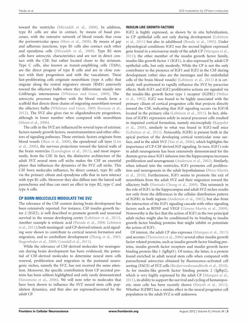
FIGURE 1 | The CP influences the CSF composition that baths the neural

progenitor cells in the SVZ. Due to its highly secretory capacity and itsparticular location facing the lateral wall of the brain ventricles, and hence theSVZ, the proteins being secreted by the CP rapidly flow in the CSF and enterin contact with the SVZ. Proteins and other molecules that are secretedtoward the CSF penetrate the interstitial space between the cells adjacent tothe wall of the ventricles. Being composed of a thin cell layer, the SVZ isinfluenced by the paracrine effect of the CP. In particular, type B1 neural stem

cells are in direct contact with the CSF by projecting a primary cilium towardthe ventricle. These cells are considered the stem cells of the adult SVZ andgive rise to type C cells (transit amplifying progenitors) that in turn originatethe type A cells (neuroblasts). Also in the SVZ are the resident astrocytes,microglia and endothelial cells of the blood-brain barrier (BBB). Together withthe ependymal cells and the CP-born molecules, these are the modulators ofthe adult SVZ cell niche. Cells of the brain parenchyma lay in the SVZneighborhood.

for water molecules and ions, transporters for small peptides andpolypeptides, and have the capacity to synthetize, and then secreteseveral proteins toward the CSF (Praetorius, 2007; Johanson et al.,2008). The necessary energy to feed this highly secretory capacityis provided by a high density of mitochondria (Redzic and Segal,2004). In addition, the CP epithelial cells have receptors, both inthe apical and basolateral sides, for molecules such as neurotrans-mitters, cytokines, bacterial toxins, amongst others; importantly,several of these receptors have been shown to signal down-stream cascades that ultimately influence the CP transcriptomeand secretome (Marques et al., 2009a, 2011; Johanson et al., 2011).

PROTEINS THAT ARE EXPRESSED BY THE CP ARE SECRETEDTOWARD THE CSFThe high secretory capacity of the CP is reflected in the compo-sition of the CSF (Chodobski and Szmydynger-Chodobska, 2001;Thouvenot et al., 2006) (Figure 1). Amongst the most abundantproteins in CSF are CP-secreted proteins, such as transthyretin(Sousa et al., 2007), transferrin and prostaglandin D2 synthase(Chodobski and Szmydynger-Chodobska, 2001). Reflecting theimportance of the CP-CSF nexus in the normal brain function-ing, these and other proteins have been independently exploredas unique biomarkers of psychiatric and neurological disorders.

In the last decade, the continuous improvement of largescreening proteomic techniques resulted in a more comprehen-sive characterization of the CSF protein content in several speciesand different ages (Parada et al., 2006; Zappaterra et al., 2007;Stoop et al., 2010), both in physiological and in neuropatho-logical conditions such as Alzheimer’s disease and depression(Ditzen et al., 2011; Menon et al., 2011; Ringman et al., 2012).However, changes in the CSF content may result not only fromalteration in the CP, but also (or rather) be the consequence froman altered brain parenchyma metabolism under the pathologicalcondition.

THE SVZ STEM CELL NICHE IS IN CLOSE CONTACTWITH THE CSFThe adult SVZ niche is located along the lateral walls of thelateral brain ventricles (Figure 1). It is composed of slow-dividing (type B) and fast-dividing (type C) stem cells, andneuroblasts (type A cells) (García-Verdugo et al., 1998). Theslow-dividing stem cells are divided in two distinct types, B1and B2 cells, based on cellular characteristics and position-ing in the SVZ. Type B1 cells are located closer to the ven-tricular space, with cell bodies immediately below the layerof ependymal cells, and are in direct contact with the CSFby a unique short non-motile primary cilium that extends

Frontiers in Cellular Neuroscience www.frontiersin.org August 2012 | Volume 6 | Article 34 | 2

Falcão et al. SVZ modulation by CP-born molecules

toward the ventricles (Mirzadeh et al., 2008). In addition,type B1 cells are also in contact, by means of basal pro-cesses, with the extensive network of blood vessels that crossthe periventricular space (Shen et al., 2008). By means of gapand adherens junctions, type B1 cells also contact each otherand ependyma cells (Mirzadeh et al., 2008). Type B2 stemcells have astrocytic characteristics and are not in direct con-tact with the CSF, but rather located closer to the striatum.Type C cells, also known as transit-amplifying cells (TAPs),are the direct progeny of type B cells and are in close con-tact with their progenitors and with the vasculature. Thesefast-proliferating cells originate neuroblasts (type A cells) thatmigrate along the rostral migratory stream (RMS) anteriorlytoward the olfactory bulbs where they differentiate mainly intoGABAergic interneurons (Whitman and Greer, 2009). Theastrocytic processes together with the blood vessels form ascaffold that directs these chains of migrating neuroblasts towardthe olfactory bulbs (Whitman and Greer, 2009; Bozoyan et al.,2012). The SVZ also gives rise to oligodendrocyte progenitors,although in lower number when compared with neuroblasts(Menn et al., 2006).

The cells in the SVZ are influenced by several types of extrinsicfactors namely growth factors, neurotransmitters and other effec-tors of signaling pathways. These extrinsic factors originate fromblood vessels (Shen et al., 2008), the ependymal cell layer (Limet al., 2000), the nervous projections toward the lateral walls ofthe brain ventricles (Lennington et al., 2011), and most impor-tantly, from the CSF. In fact, the distinctive architecture of theadult SVZ neural stem cell niche makes the CSF an essentialplayer that influences the dynamics of the SVZ cell niche. ThisCSF born molecules influence, by direct contact, type B1 cellsvia the primary cilium and ependyma cells that in turn interactwith type B1 cells. Moreover they also diffuse into the lateral wallparenchyma and thus can exert an effect in type B2, type C andtype A cells.

CP BORN MOLECULES MODULATE THE SVZThe relevance of the CSF content during brain development hasbeen extensively reported. For instance, CSF insulin-growth fac-tor 2 (IGF2), is well described to promote growth and neuronalsurvival in the mouse developing cortex (Lehtinen et al., 2011).Another example is retinoic acid (Parada et al., 2008; Lehtinenet al., 2011); both meningeal- and CP-derived retinoic acid signal-ing were shown to contribute to cortical neuron formation andmigration, and to cerebellum development (Zhang et al., 2003;Siegenthaler et al., 2009; Crandall et al., 2011).

While the relevance of CSF-derived molecules for neurogen-esis during brain development has been evidenced, the poten-tial of CSF-derived molecules to determine neural stem cellsrenewal, proliferation and migration in the postnatal neuro-genic niches, namely the SVZ, has not received the same atten-tion. Moreover, the specific contribution from CP secreted pro-teins has been seldom highlighted and only rarely demonstrated(Sawamoto et al., 2006). We will next refer to molecules thathave been shown to influence the SVZ neural stem cells pop-ulation dynamics, and that also are expressed/secreted by theadult CP.

INSULIN-LIKE GROWTH FACTORSIGF2 is highly expressed, as shown by in situ hybridization,in CP epithelial cells not only during development (Lehtinenet al., 2011) but also in adulthood (Bondy et al., 1992). Underphysiological conditions IGF2 was the second highest expressedgene found in a microarray study of the adult CP (Marques et al.,2011); the other member of the insulin growth factor family,insulin-like growth factor 1 (IGF1), is also expressed by adult CPepithelial cells, but only modestly. While the CP is not the onlycontributor to the presence of IGF1 and IGF2 in the CSF duringdevelopment (other sites are the meninges and the endothelialcells of the brain blood vessels) (Lehtinen et al., 2011) it is cer-tainly well positioned to rapidly influence the SVZ by paracrineeffects. Both IGF1 and IGF2 proliferative actions are signaled viathe insulin-like growth factor type 1 receptor (IGFR1) (Weberet al., 1992). IGF2 was found to be highly associated with theprimary cilium of cortical progenitor cells that projects directlytoward the CSF, indicating that IGF signaling occurs via IGF1Rlocated in the primary cilia (Lehtinen et al., 2011). In fact, abla-tion of IGFR1 expression solely in neural precursor cells resultedin impaired cortical formation, namely microcephaly (Kappeleret al., 2008), similarly to what was found in IGF2-null mice(Lehtinen et al., 2011). Noticeably, IGFR1 is present both in theapical portion of the developing cortical ventricular zone sur-face, and in the adult SVZ (Yan et al., 2006), which highlights theimportance of CP-CSF derived IGF signaling. In turn, IGF1’s rolein adult neurogenesis has been extensively demonstrated for thedentate gyrus since IGF1 infusion into the hippocampus increasesproliferation and neurogenesis (Anderson et al., 2002). Similarly,when infused into the ventricles, IGF1 promotes cell prolifera-tion and neurogenesis in the adult hypothalamus (Pérez-Martínet al., 2010). Furthermore, IGF1 seems to promote the exit ofneuroblasts from the adult SVZ and their migration toward theolfactory bulb (Hurtado-Chong et al., 2009). This mismatch inthe role of IGF1 in the hippocampus and adult SVZ niches resultsnot only from the differences in the cellular distribution patternof IGFR1 in both regions (Anderson et al., 2002), but also fromthe interaction of the IGF1 signaling cascade with other signalingfactors such as BDNF and VEGF (Llorens-Martín et al., 2009).Noteworthy is the fact that the action of IGF1 in the two principaladult niches might also be conditioned by its binding to insulingrowth factor binding proteins that might inhibit or potentiatethe action of IGF1.

Of interest, the adult CP also expresses (Marques et al., 2011)and secretes (Thouvenot et al., 2006) several other insulin growthfactor related proteins, such as insulin growth factor binding pro-teins, insulin growth factor receptors and insulin growth factorbinding protein-like 1 (Igfbpl1). Of notice, the latter, is a proteinfound enriched in adult neural stem cells when compared withparenchymal astrocytes obtained by fluorescence-activated cellsorting (FACS) of SVZ cells (Beckervordersandforth et al., 2010).As for insulin-like growth factor binding protein 2 (Igfbp2),which is very highly expressed by the adult CP (Marques et al.,2011), its ability to support the survival and cycling of hematopoi-etic stem cells has been recently shown (Huynh et al., 2011).Whether IGFBP2 has a similar effect in the neural progenitor cellpopulation in the adult SVZ is still unknown.

Frontiers in Cellular Neuroscience www.frontiersin.org August 2012 | Volume 6 | Article 34 | 3

Falcão et al. SVZ modulation by CP-born molecules

FIBROBLAST GROWTH FACTORSAnother important group of growth factors expressed by theadult CP (Marques et al., 2011) is the fibroblast growth fac-tor (FGF) family and related proteins. Their involvement inseveral processes of brain development (neural stem cell induc-tion, cell differentiation, brain regions patterning, and neuronalcircuit assembly) has been extensively demonstrated (Guillemotand Zimmer, 2011). In fact, the embryonic brain has severalsources of different FGF family members that contribute to thedetermination of brain regionalization; for example, the contri-bution of CSF-derived FGFs to embryonic brain development wasshown for FGF2 since it promotes precursor proliferation in chickembryos (Martín et al., 2006). FGF8, potentially derived from theCP-CSF, has also been described to participate in the pattern-ing of brain regions in the chick (Parada et al., 2005) and mouseembryos (Fukuchi-Shimogori and Grove, 2001).

As for the adult brain, FGF2 injected in the ventricles increasedproliferation in the SVZ and neurogenesis in the olfactory bulb(Kuhn et al., 1997). Furthermore subcutaneously injected FGF2,in both early post-natal and in young rats, crossed the brainbarriers and increased in the CSF while promoting proliferationin both the subgranular zone of the dentate gyrus and the SVZ(Wagner et al., 1999). The importance of FGF2 for neuronalproliferation is illustrated by the extensive use of FGF2 (alsoknown as bFGF) as a mitogen in SVZ neurosphere assays(Pastrana et al., 2011).

While less is known for the role of other FGF family membersin adult neurogenesis it is of notice that FGF family mem-bers, such as FGF3, FGF9 and FGF10, are expressed under basalphysiological conditions in the adult CP (Marques et al., 2011).Interestingly, FGF10 participation in the maintenance of theneurogenic potential of the adult SVZ was already suggested(Hajihosseini et al., 2008). Thus, under particular conditions,alterations in the expression of FGFs and their secretion towardthe CSF may impact in the SVZ population.

EPIDERMAL GROWTH FACTOR (EGF) AND TRANSFORMING GROWTHFACTOR ALPHA (TGFa)In vitro, when exposed to epidermal growth factor (EGF), adultSVZ derived cells form neurospheres that display multipotentand self-renewal properties (Pastrana et al., 2011). Althoughexpressed in a relative small number of type B1 cells, EGFreceptor (EGFR) expressing cells that form neurospheres in vitroare derived mainly from transit amplifying C cells (Doetsch et al.,2002). In vivo, it was shown that high levels of EGF administeredby intracerebroventricular infusion impacts on the SVZ byincreasing proliferation and generating progeny that occupies thesurrounding brain parenchyma, and also diverts SVZ cells fromthe neuronal lineage to the oligodendrocytic lineage (Doetschet al., 2002; Gonzalez-Perez et al., 2009). While it is believed(Doetsch et al., 2002) that the probable source of EGFR signalingoccurs via transforming growth factor alpha (TGFa), given itsexpression in the CP (Seroogy et al., 1993; Marques et al., 2011),we cannot exclude that this signaling pathway occurs throughEGF, also expressed by the CP (Marques et al., 2011). The rele-vance of TGFa/EGF for EGFR in this context is illustrated by theobservation that the decreased proliferation in the SVZ displayed

by the TGFa knockout mice can be corrected by supplementationwith EGF (Tropepe et al., 1997). Interestingly, TGFa signalingvia EGFR was also shown to influence the migratory propertiesof cells in the RMS (Kim et al., 2009) and of oligodendrocyteprecursors derived from SVZ cells (Gonzalez-Perez andQuiñones-Hinojosa, 2010). In addition, a role for TGFa/EGFRsignaling in promoting migration of cells derived from the SVZwas highlighted by TGFa infusion to the dopamine-depletedstriatum of rodent models of Parkinson’s disease (Cooper andIsacson, 2004; de Chevigny et al., 2008). Despite the interestingpotential of the CP as a source of TGFa/EGF for the modulationof the SVZ its specific physiological contribution has never beendemonstrated, which certainly deserves additional research.

PLATELET DERIVED GROWTH FACTORS (PDGF)PDGF signaling also occurs via the primary cilium and, interest-ingly, the PDGF signaling pathway modulates neural stem cellsand affects lineage fate. For instance, in vitro experiments showedthat PDGF increased neurosphere formation (Jackson et al.,2006). Whether SVZ GFAP-positive neural stem cells expressthe platelet derived growth factor receptor alpha polypeptide(PDGFRa) is disputable (Jackson et al., 2006; Chojnacki et al.,2011; Ihrie and Álvarez-Buylla, 2011), but infusion of PDGFinto the ventricle is known to bolster proliferation in the SVZ(Jackson et al., 2006). The endogenous source of this ligandhas not been determined (Ihrie and Álvarez-Buylla, 2011) butrecently we found that the CP expresses several PDGFs mRNAs(Marques et al., 2011), with particular emphasis to PDGFa. Onceagain, the physiological significance of this expression and itsinfluence over the SVZ neural stem cell niche remains to beestablished.

BONE MORPHOGENETIC PROTEINS (BMPs), SONIC HEDGEHOG(Shh) AND WntDuring the formation of the central nervous system, FGF sig-naling action is frequently aligned with and/or counteracted bysignaling from BMPs, Shh and Wnt pathways. The result ofthis interaction, impacting on cell proliferation and cell fate, isdependent of the highly dynamic spatiotemporal variation inthe expression of the various effector proteins (Guillemot andZimmer, 2011).

The role of BMP, Shh, and Wnt proteins derived from theCP-CSF during development has been shown. For instance, Shhexpression in the hindbrain CP is high and CSF Shh was demon-strated to be essential for cerebellar development by promotingproliferation of granule precursors (Huang et al., 2009, 2010).BMPs, that together with Wnt and FGF proteins, participatein cortical development (Shimogori et al., 2004), display a verydynamic presence in the CSF in an age dependent manner(Lehtinen et al., 2011).

In the adult SVZ, all these proteins participate in the regu-lation of the SVZ niche. Adult SVZ type B and C cells expressboth BMP2 and BMP4 and their respective receptors, and SVZependymal cells alter the activity of BMPs (Lim et al., 2000;Peretto et al., 2004). The CP origin of these ligands should alsobe considered since it expresses BMP1, BMP2, BMP4, BMP6and BMP7 under basal physiological conditions (Marques et al.,

Frontiers in Cellular Neuroscience www.frontiersin.org August 2012 | Volume 6 | Article 34 | 4

Falcão et al. SVZ modulation by CP-born molecules

2011). Noteworthy, the presence of some of these BMPs in theadult CSF was also recently confirmed (Lehtinen et al., 2011).Furthermore, growth differentiation factors 3 and 8 (Gdf3 andGdf8), known to modulate BMP signaling, are also expressed bythe adult CP (Marques et al., 2011) and are secreted toward theCSF (Lehtinen et al., 2011).

Shh and Wnts pathways are also active in the adult SVZ andwere implicated in the formation, maintenance, proliferation andmigration of adult neural stem cells. Shh is produced by ventralforebrain neurons that extend projections toward the SVZ, andpersistent Shh signaling determines a specific neural progeny(Ihrie et al., 2011). Shh is also expressed by the CP (Marques et al.,2011) but its levels in the adult CSF have not been determined. Asfor Wnt, evidence exists that it may participate in the regulationof the adult SVZ population, namely enhancing Wnt signalingvia beta-catenin in type C cells increases proliferation and resultsin a higher number of neurons in the olfactory bulb (Adachiet al., 2007). On the other hand, during embryonic developmentWnt may rather play a role in maintaining the SVZ stem cell pool(Piccin and Morshead, 2011). Similarly to Shh and BMPs, severalmembers (for instance Wnt5a, 5b, and 10b) of the Wnt familyare expressed in the adult CP, and specifically secreted by CPepithelial cells, under basal conditions (Thouvenot et al., 2006;Marques et al., 2011).

One of the most interesting features of the Shh and Wnt sig-naling pathways is that they are modulated by the primary cilium(Louvi and Grove, 2011), making the cilia projected by type B1cells toward the CSF a particularly well positioned route for CPderived “messages”.

EPHRINS, SEMAPHORINS AND SLITSNeuroblasts derived from the SVZ form a stream of moving cellsthat converge in the RMS; they are ensheated by a layer of astro-cyte processes and use each other as guides in the migrationprocess toward the olfactory bulb (Lledo et al., 2008). Severalproteins from the group of classical axon guidance moleculeswere implicated not only in the regulation of the migration ofneuroblasts, but also in the proliferation of type B cells. Forinstance, SVZ cells express the EphA and EphB receptors; theligands for these receptors are the transmembrane moleculesephrins. The relevance of these molecules in the modulationof the SVZ was shown by the infusion of the EphB2 ligandin the lateral ventricles, which disrupted the migratory chainof neuroblasts and increased the proliferation of type B1 cells(Conover et al., 2000). Interestingly, a role for EphB2 signalingwas suggested in the conversion of ependymal cells to astro-cytes after lesion of the ventricular wall (Nomura et al., 2010).Also, it has recently been shown that ephrinB3-EphB3 signal-ing in the SVZ is transiently inhibited to allow the expansionand survival of neural progenitor cells upon traumatic braininjury (Theus et al., 2010). EphrinB3 is one of the ephrinsexpressed by the CP at very low levels under basal physiologi-cal conditions (Marques et al., 2011). While the fact that thesemolecules are attached to the membrane might diminish the rel-evance of their expression in the CP in the modulation of theSVZ neural progenitors, the data provided by infusion experi-ments (Conover et al., 2000) suggest that the cleavage and release

of ephrins from the membrane of CP cells toward the CSF,under particular conditions, could influence the response of typeB1 cells.

Semaphorins and slits are guidance molecules that, contraryto ephrins, are secreted factors, and hence exert their effects to acertain distance, in a paracrine fashion. Several semaphorin fam-ily members and slits 1, 2, and 3 are expressed (Marques et al.,2011) and secreted by the CP (Hu, 1999; Sawamoto et al., 2006).Semaphorin signaling is usually associated with endothelial cells(Tamagnone and Mazzone, 2011); semaphorin-3a and its respec-tive receptor are highly expressed in the endothelial cells that arepresent along the RMS (Meléndez-Herrera et al., 2008), suggest-ing a role for semaphorin-3a in the migration of neuroblasts.Whether semaphorins specifically secreted by the CP influencethe migration of neuroblasts toward the olfactory bulb has notbeen determined.

On the other hand, a role for slits specifically derived fromthe CP in the modulation of SVZ neural progenitors niche wasclearly shown; for instance, slit 2 was shown to participate inthe regulation of neuronal migration of neurons in the devel-oping brain (Hu, 1999). Interestingly both the adult SVZ andthe RMS express the receptors Robo2 and Robo3 through whichslit1 and slit 2 exert their chemorepulsive activities (Nguyen-Ba-Charvet, 2004). Importantly, it was shown (Sawamoto et al.,2006) that ciliary beating from ependymal cells that line thewall of the brain ventricles contribute to the movement of theCSF, hence creating gradients of the chemorepelent slits secretedby the CP (Nguyen-Ba-Charvet, 2004; Marques et al., 2011)and thus contributing to the anterior migration of neuroblaststo the olfactory bulbs. Nevertheless, the role of slits in direct-ing the migration of future olfactory bulb neurons goes behindthe influence of the CP since type C and type A cells alsoexpress slit1 (Nguyen-Ba-Charvet, 2004), which seems to avoidthe invasion of the RMS by astrocytic processes (Kaneko et al.,2010).

PATHOLOGICAL CONDITIONS CAN ALTER THE CPTRANSCRIPTOME/SECRETOME AND IMPACT THE SVZBeing placed at the interface between the periphery and the cen-tral nervous system, the CP is particularly well positioned to sensealterations, and respond to, in both its basolateral side (the bloodside) and its apical interface (the CNS side). On the other hand,any response the CP mounts to external stimuli will ultimatelyreflect in its secretome, and hence in the CSF that surroundsthe brain parenchyma. In fact, since the CP epithelial cells areequipped with transporters for several proteins and metabolites,pathological damage to the CP itself will alter CSF compositionand ventricular volume, as in the case of hydrocephalus (Johansonet al., 2011).

One relevant example of a peripheral event that impactsin the CP transcriptome is peripheral inflammation. When asingle intraperitoneal injection of lipopolysaccharide, the mem-brane component of gram negative bacteria, was given, the CPdisplayed an acute and transient but profound change in itstranscriptome that reflected in the CSF composition (Marqueset al., 2007, 2009a). When a similar but repeated inflamma-tory stimulus was given, the result was still present but more

Frontiers in Cellular Neuroscience www.frontiersin.org August 2012 | Volume 6 | Article 34 | 5

Falcão et al. SVZ modulation by CP-born molecules

attenuated (Marques et al., 2009b). These alterations in the CPproperties and secretome may impact, even if transiently, inthe SVZ niche population. During development, it was recentlyreported that maternal peripheral inflammation alters fetal ven-tricular zone proliferation that reflects in cortical layers forma-tion. The effect was shown to involve the barriers of the brain,including the blood-CP-CSF barrier (Stolp et al., 2011). Whileseveral reports refer to the impact of neuroinflammation in theadult neurogenesis niches, as in multiple sclerosis, Alzheimer’sdisease and brain injury (Tepavcevic et al., 2011; Wood et al.,2011; L’Episcopo et al., 2012) (Hamilton and Holscher, 2012),the more subtle effects of inflammation-induced alterations inthe CP per se, and its impact in the adult SVZ, are presentlyunknown.

A physiological situation where alterations in the CP prop-erties, in the CSF content and in neurogenesis occur is aging.During aging, mainly at the later ages of life, the CP becomesprogressively less efficient in protein synthesis and CSF secre-tion (Redzic et al., 2005). This malfunction in CSF secretionand abnormal removal of CSF toxic compounds, such as theAlzheimer’s disease abeta peptide, might contribute to neu-ropathology (Preston, 2001; Carro et al., 2005). In fact, alterationsin the CP-CSF nexus properties might contribute to the alter-ations observed in the SVZ in rodents during aging and in modelsand Alzheimer’s disease (Jin et al., 2004; Sotthibundhu et al., 2009;Conover and Shook, 2011).

CONCLUDING REMARKSUnder basal normal physiological conditions the CP displaysthe ability to express several genes encoding for proteins knownto promote proliferation, differentiation, and survival of neuralprogenitor cells. These proteins are secreted toward the CSF,which is as a route for the delivery of CP-born proteins/moleculesto the SVZ. Per se this is physiologically relevant; yet under dis-ruptive conditions that alter regular CP homeostatic balance, theCSF protein content will be modified and this will lately impacton the SVZ. Whether this culminates in disease developing out-comes (such as is the case of glioblastomas), or might function aspotential rescue mechanisms for brain parenchyma lesions (suchas in stroke, Parkinson’s disease or multiple sclerosis), will be akey issue in adult neural stem cell research in the future. In fact,modulating the CP-CSF nexus in pathologies of the central ner-vous system could become an important aspect in the usage ofendogenous/exogenous neural progenitor cells for stem cell basedtherapies in the brain.

ACKNOWLEDGMENTSAna M. Falcão and Ashley Novais are recipients of Ph.D.fellowships from the Fundação para a Ciência e Tecnologia(FCT, Portugal). Fernanda Marques is a recipient of postdoc-toral fellowship from the Fundação para a Ciência e Tecnologia(FCT, Portugal). This work is supported by a grant from FCT(PTDC/SAU-OSM/104475/2008).

REFERENCESAdachi, K., Mirzadeh, Z., Sakaguchi, M.,

Yamashita, T., Nikolcheva, T., Gotoh,Y., Peltz, G., Gong, L., Kawase, T.,Álvarez-Buylla, A., Okano, H., andSawamoto, K. (2007). Beta-cateninsignaling promotes proliferation ofprogenitor cells in the adult mousesubventricular zone. Stem Cells 25,2827–2836.

Alvarez-Buylla, A., Kohwi, M., Nguyen,T. M., and Merkle, F. T. (2008). Theheterogeneity of adult neural stemcells and the emerging complexity oftheir niche. Cold Spring Harb. Symp.Quant. Biol. 73, 357–365.

Anderson, M. F., Aberg, M. A. I.,Nilsson, M., and Eriksson, P. S.(2002). Insulin-like growth factor-Iand neurogenesis in the adult mam-malian brain. Brain Res. Dev. BrainRes. 134, 115–122.

Beckervordersandforth, R., Tripathi, P.,Ninkovic, J., Bayam, E., Lepier,A., Stempfhuber, B., Kirchhoff, F.,Hirrlinger, J., Haslinger, A., Lie, D.C., Beckers, J., Yoder, B., Irmler,M., and Götz, M. (2010). In vivofate mapping and expression anal-ysis reveals molecular hallmarks ofprospectively isolated adult neu-ral stem cells. Cell Stem Cell 7,744–758.

Bondy, C., Werner, H., Roberts, C. T.,and LeRoith, D. (1992). Cellularpattern of type-I insulin-like

growth factor receptor gene expres-sion during maturation of the ratbrain: comparison with insulin-likegrowth factors I and II. Neuroscience46, 909–923.

Bozoyan, L., Khlghatyan, J., andSaghatelyan, A. (2012). Astrocytescontrol the development of themigration-promoting vasculaturescaffold in the postnatal brain viaVEGF signaling. J. Neurosci. 32,1687–1704.

Carro, E., Spuch, C., Trejo, J. L.,Antequera, D., and Torres-Aleman,I. (2005). Choroid plexus megalinis involved in neuroprotection byserum insulin-like growth factor I.J. Neurosci. 25, 10884–10893.

Chodobski, A., and Szmydynger-Chodobska, J. (2001). Choroidplexus: target for polypeptides andsite of their synthesis. Microsc. Res.Tech. 52, 65–82.

Chojnacki, A., Mak, G., and Weiss, S.(2011). PDGFR expression distin-guishes GFAP-expressing neuralstem cells from PDGF-responsiveneural precursors in the adultperiventricular area. J. Neurosci. 31,9503–9512.

Conover, J. C., Doetsch, F., García-Verdugo, J. M., Gale, N. W.,Yancopoulos, G. D., and Alvarez-Buylla, A. (2000). Disruption ofEph/ephrin signaling affects migra-tion and proliferation in the adult

subventricular zone. Nat. Neurosci.3, 1091–1097.

Conover, J. C., and Shook, B. A. (2011).Aging of the subventricular zoneneural stem cell niche. Aging Dis. 2,49–63.

Cooper, O., and Isacson, O. (2004).Intrastriatal transforming growthfactor alpha delivery to a modelof Parkinson’s disease inducesproliferation and migration ofendogenous adult neural progenitorcells without differentiation intodopaminergic neurons. J. Neurosci.24, 8924–8931.

Crandall, J. E., Goodman, T., McCarthy,D. M., Duester, G., Bhide, P. G.,Dräger, U. C., and McCaffery,P. (2011). Retinoic acid influ-ences neuronal migration fromthe ganglionic eminence to thecerebral cortex. J. Neurochem. 119,723–735.

de Chevigny, A., Cooper, O., Vinuela,A., Reske-Nielsen, C., Lagace, D.C., Eisch, A. J., and Isacson, O.(2008). Fate mapping and lineageanalyses demonstrate the produc-tion of a large number of stri-atal neuroblasts after transforminggrowth factor alpha and noggin stri-atal infusions into the dopamine-depleted striatum. Stem Cells 26,2349–2360.

Ditzen, C., Tang, N., Jastorff, A.M., Teplytska, L., Yassouridis,

A., Maccarrone, G., Uhr, M.,Bronisch, T., Miller, C. A., Holsboer,F., and Turck, C. W. (2011).Cerebrospinal fluid biomarkers formajor depression confirm relevanceof associated pathophysiology.Neuropsychopharmacology 37,1013–1025.

Doetsch, F., Petreanu, L., Caille, I.,Garcia-Verdugo, J. M., and Álvarez-Buylla, A. (2002). EGF convertstransit-amplifying neurogenic pre-cursors in the adult brain intomultipotent stem cells. Neuron 36,1021–1034.

Fukuchi-Shimogori, T., and Grove, E.A. (2001). Neocortex patterningby the secreted signaling moleculeFGF8. Science 294, 1071–1074.

García-Verdugo, J. M., Doetsch,F., Wichterle, H., Lim, D. A.,and Alvarez-Buylla, A. (1998).Architecture and cell types of theadult subventricular zone: in searchof the stem cells. J. Neurobiol. 36,234–248.

Gonzalez-Perez, O., and Quiñones-Hinojosa, A. (2010). Dose-dependent effect of EGF onmigration and differentiationof adult subventricular zoneastrocytes. Glia 58, 975–983.

Gonzalez-Perez, O., Romero-Rodriguez, R., Soriano-Navarro, M.,Garcia-Verdugo, J. M., and Álvarez-Buylla, A. (2009). Epidermal

Frontiers in Cellular Neuroscience www.frontiersin.org August 2012 | Volume 6 | Article 34 | 6

Falcão et al. SVZ modulation by CP-born molecules

growth factor induces the progenyof subventricular zone type B cellsto migrate and differentiate intooligodendrocytes. Stem Cells 27,2032–2043.

Guillemot, F., and Zimmer, C. (2011).From cradle to grave: the multi-ple roles of fibroblast growth factorsin neural development. Neuron 71,574–588.

Hajihosseini, M. K., De Langhe, S.,Lana-Elola, E., Morrison, H.,Sparshott, N., Kelly, R., Sharpe, J.,Rice, D., and Bellusci, S. (2008).Localization and fate of Fgf10-expressing cells in the adult mousebrain implicate Fgf10 in control ofneurogenesis. Mol. Cell Neurosci.37, 857–868.

Hamilton, A., and Holscher, C. (2012).The effect of ageing on neuroge-nesis and oxidative stress in theAPP(swe)/PS1(deltaE9) mousemodel of Alzheimer’s disease. BrainRes. 1449, 83–93.

Hu, H. (1999). Chemorepulsion ofneuronal migration by Slit2 in thedeveloping mammalian forebrain.Neuron 23, 703–711.

Huang, X., Ketova, T., Fleming, J. T.,Wang, H., Dey, S. K., Litingtung,Y., and Chiang, C. (2009). Sonichedgehog signaling regulates anovel epithelial progenitor domainof the hindbrain choroid plexus.Development 136, 2535–2543.

Huang, X., Liu, J., Ketova, T., Fleming,J. T., Grover, V. K., Cooper, M.K., Litingtung, Y., and Chiang,C. (2010). Transventricular deliveryof Sonic hedgehog is essential tocerebellar ventricular zone develop-ment. Proc. Natl. Acad. Sci. U.S.A.107, 8422–8427.

Hurtado-Chong, A., Yusta-Boyo, M.J., Vergaño-Vera, E., Bulfone, A.,De Pablo, F., and Vicario-Abejón,C. (2009). IGF-I promotes neu-ronal migration and positioning inthe olfactory bulb and the exit ofneuroblasts from the subventricularzone. Eur. J. Neurosci. 30, 742–755.

Huynh, H., Zheng, J., Umikawa, M.,Zhang, C., Silvany, R., Iizuka, S.,Holzenberger, M., Zhang, W., andZhang, C. C. (2011). IGF bindingprotein 2 supports the survival andcycling of hematopoietic stem cells.Blood 118, 3236–3243.

Ihrie, R. A., and Álvarez-Buylla, A.(2011). Lake-front property: aunique germinal niche by the lateralventricles of the adult brain. Neuron70, 674–686.

Ihrie, R. A., Shah, J. K., Harwell,C. C., Levine, J. H., Guinto, C.D., Lezameta, M., Kriegstein, A.R., and Álvarez-Buylla, A. (2011).Persistent sonic hedgehog signaling

in adult brain determines neuralstem cell positional identity. Neuron71, 250–262.

Jackson, E. L., Garcia-Verdugo, J. M.,Gil-Perotin, S., Roy, M., Quiñones-Hinojosa, A., VandenBerg, S., andÁlvarez-Buylla, A. (2006). PDGFRalpha-positive B cells are neuralstem cells in the adult SVZ that formglioma-like growths in response toincreased PDGF signaling. Neuron51, 187–199.

Jin, K., Galvan, V., Xie, L., Mao, X.O., Gorostiza, O. F., Bredesen,D. E., and Greenberg, D. A.(2004). Enhanced neurogenesisin Alzheimer’s disease trans-genic (PDGF-APPSw, Ind) mice.Proc. Natl. Acad. Sci. U.S.A. 101,13363–13367.

Johanson, C. E., Duncan, J. A., Klinge,P. M., Brinker, T., Stopa, E. G.,and Silverberg, G. D. (2008).Multiplicity of cerebrospinal fluidfunctions: new challenges in healthand disease. Cerebrospinal Fluid Res.5, 10.

Johanson, C., Stopa, E., McMillan, P.,Roth, D., Funk, J., and Krinke, G.(2011). The distributional nexusof choroid plexus to cerebrospinalfluid, ependyma and brain: tox-icologic/pathologic phenomena,periventricular destabilization, andlesion spread. Toxicol. Pathol. 39,186–212.

Kaneko, N., Marin, O., Koike, M.,Hirota, Y., Uchiyama, Y., Wu, J. Y.,Lu, Q., Tessier-Lavigne, M., Álvarez-Buylla, A., Okano, H., Rubenstein,J. L. R., and Sawamoto, K. (2010).New neurons clear the path ofastrocytic processes for their rapidmigration in the adult brain. Neuron67, 213–223.

Kappeler, L., De Magalhaes Filho, C.,Dupont, J., Leneuve, P., Cervera,P., Périn, L., Loudes, C., Blaise,A., Klein, R., Epelbaum, J., LeBouc, Y., and Holzenberger, M.(2008). Brain IGF-1 receptorscontrol mammalian growth andlifespan through a neuroendocrinemechanism. PLoS Biol. 6:e254. doi:10.1371/journal.pbio.0060254.

Keep, R. F., and Jones, H. C. (1990). Amorphometric study on the devel-opment of the lateral ventriclechoroid plexus, choroid plexus cap-illaries and ventricular ependyma inthe rat. Brain Res. Dev. Brain Res. 56,47–53.

Kim, Y., Comte, I., Szabo, G.,Hockberger, P., and Szele, F. G.(2009). Adult mouse subventric-ular zone stem and progenitorcells are sessile and epidermalgrowth factor receptor neg-atively regulates neuroblast

migration. PLoS ONE 4:e8122.doi: 10.1371/journal.pone.0008122

Kuhn, H. G., Winkler, J., Kempermann,G., Thal, L. J., and Gage, F.H. (1997). Epidermal growthfactor and fibroblast growthfactor-2 have different effectson neural progenitors in theadult rat brain. J. Neurosci. 17,5820–5829.

L’Episcopo, F., Tirolo, C., Testa,N., Caniglia, S., Morale, M. C.,Deleidi, M., Serapide, M. F.,Pluchino, S., and Marchetti, B.(2012). Plasticity of subventric-ular zone neuroprogenitors inMPTP (1-methyl-4-phenyl-1 2 3, 6-tetrahydropyridine) mouse modelof Parkinson’s disease involvescross talk between inflammatoryand Wnt/ -catenin signaling path-ways: functional consequencesfor neuroprotection and repair. J.Neurosci. 32, 2062–2085.

Lehtinen, M. K., Zappaterra, M. W.,Chen, X., Yang, Y. J., Hill, A. D., Lun,M., Maynard, T., Gonzalez, D., Kim,S., Ye, P., D’Ercole, A. J., Wong, E.T., LaMantia, A. S., and Walsh, C.A. (2011). The cerebrospinal fluidprovides a proliferative niche forneural progenitor cells. Neuron 69,893–905.

Lennington, J. B., Pope, S., Goodheart,A. E., Drozdowicz, L., Daniels, S.B., Salamone, J. D., and Conover,J. C. (2011). Midbrain dopamineneurons associated with reward pro-cessing innervate the neurogenicsubventricular zone. J. Neurosci. 31,13078–13087.

Lim, D. A., Tramontin, A. D., Trevejo,J. M., Herrera, D. G., García-Verdugo, J. M., and Alvarez-Buylla,A. (2000). Noggin antagonizesBMP signaling to create a nichefor adult neurogenesis. Neuron 28,713–726.

Lledo, P.-M., Merkle, F. T., and Álvarez-Buylla, A. (2008). Origin and func-tion of olfactory bulb interneu-ron diversity. Trends Neurosci. 31,392–400.

Llorens-Martín, M., Torres-Aleman, I.,and Trejo, J. L. (2009). Mechanismsmediating brain plasticity: IGF1and adult hippocampal neurogene-sis. Neuroscientist 15, 134–148.

Louvi, A., and Grove, E. A. (2011).Cilia in the CNS: the quiet organelleclaims center stage. Neuron 69,1046–1060.

Marques, F., Rodrigues, A.-J., Sousa, J.C., Coppola, G., Geschwind, D. H.,Sousa, N., Correia-Neves, M., andPalha, J. A. (2007). Lipocalin 2 isa choroid plexus acute-phase pro-tein. J. Cereb. Blood Flow Metab. 28,450–455.

Marques, F., Sousa, J. C., Coppola,G., Falcao, A. M., João Rodrigues,A., Geschwind, D. H., Sousa, N.,Correia-Neves, M., and Palha, J.A. (2009a). Kinetic profile of thetranscriptome changes induced inthe choroid plexus by peripheralinflammation. J. Cereb. Blood FlowMetab. 29, 921–932.

Marques, F., Sousa, J. C., Coppola,G., Geschwind, D. H., Sousa, N.,Palha, J. A., and Correia-Neves,M. (2009b). The choroid plexusresponse to a repeated periph-eral inflammatory stimulus. BMCNeurosci. 10, 135.

Marques, F., Sousa, J. C., Coppola,G., Gao, F., Puga, R., Brentani,H., Geschwind, D. H., Sousa, N.,Correia-Neves, M., and Palha, J. A.(2011). Transcriptome signature ofthe adult mouse choroid plexus.Fluids Barriers CNS 8, 10.

Martín, C., Bueno, D., Alonso, M. I.,Moro, J. A., Callejo, S., Parada,C., Martín, P., Carnicero, E., andGato, A. (2006). FGF2 plays akey role in embryonic cerebrospinalfluid trophic properties over chickembryo neuroepithelial stem cells.Dev. Biol. 297, 402–416.

Meléndez-Herrera, E., Colín-Castelán,D., Varela-Echavarría, A., andGutiérrez-Ospina, G. (2008).Semaphorin-3A and its receptorneuropilin-1 are predominantlyexpressed in endothelial cells alongthe rostral migratory stream ofyoung and adult mice. Cell TissueRes. 333, 175–184.

Menn, B., Garcia-Verdugo, J. M.,Yaschine, C., Gonzalez-Perez, O.,Rowitch, D., and Álvarez-Buylla,A. (2006). Origin of oligodendro-cytes in the subventricular zoneof the adult brain. J. Neurosci. 26,7907–7918.

Menon, K. N., Steer, D. L., Short, M.,Petratos, S., Smith, I., and Bernard,C. C. A. (2011). A novel unbi-ased proteomic approach to detectthe reactivity of cerebrospinal fluidin neurological diseases. Mol. CellProteomics 10, M110.000042.

Mirzadeh, Z., Merkle, F. T., Soriano-Navarro, M., Garcia-Verdugo, J.M., and Álvarez-Buylla, A. (2008).Neural stem cells confer unique pin-wheel architecture to the ventricu-lar surface in neurogenic regions ofthe adult brain. Cell Stem Cell 3,265–278.

Nguyen-Ba-Charvet, K. T. (2004).Multiple roles for slits in the controlof cell migration in the rostralmigratory stream. J. Neurosci. 24,1497–1506.

Nomura, T., Göritz, C., Catchpole,T., Henkemeyer, M., and Frisén, J.

Frontiers in Cellular Neuroscience www.frontiersin.org August 2012 | Volume 6 | Article 34 | 7

Falcão et al. SVZ modulation by CP-born molecules

(2010). EphB signaling controls lin-eage plasticity of adult neural stemcell niche cells. Cell Stem Cell 7,730–743.

Parada, C., Gato, A., Aparicio, M., andBueno, D. (2006). Proteome analy-sis of chick embryonic cerebrospinalfluid. Proteomics 6, 312–320.

Parada, C., Gato, A., and Bueno,D. (2008). All-trans retinol andretinol-binding protein fromembryonic cerebrospinal fluidexhibit dynamic behaviour dur-ing early central nervous systemdevelopment. Neuroreport 19,945–950.

Parada, C., Martín, C., Alonso, M. I.,Moro, J. A., Bueno, D., and Gato,A. (2005). Embryonic cerebrospinalfluid collaborates with the isthmicorganizer to regulate mesencephalicgene expression. J. Neurosci. Res. 82,333–345.

Pastrana, E., Silva-Vargas, V., andDoetsch, F. (2011). Eyes wideopen: a critical review of sphere-formation as an assay for stem cells.Cell Stem Cell 8, 486–498.

Peretto, P., Dati, C., De Marchis,S., Kim, H. H., Ukhanova, M.,Fasolo, A., and Margolis, F. L.(2004). Expression of the secretedfactors noggin and bone morpho-genetic proteins in the subependy-mal layer and olfactory bulb ofthe adult mouse brain. Neuroscience128, 685–696.

Pérez-Martín, M., Cifuentes, M.,Grondona, J. M., López-Ávalos,M. D., Gómez-Pinedo, U.,García-Verdugo, J. M., andFernández-Llebrez, P. (2010).IGF-I stimulates neurogenesis inthe hypothalamus of adult rats. Eur.J. Neurosci. 31, 1533–1548.

Piccin, D., and Morshead, C. M. (2011).Wnt signaling regulates symmetryof division of neural stem cells inthe adult brain and in response toinjury. Stem Cells 29, 528–538.

Praetorius, J. (2007). Water and solutesecretion by the choroid plexus.Pflugers Arch. 454, 1–18.

Preston, J. E. (2001). Ageing choroidplexus-cerebrospinal fluid system.Microsc. Res. Tech. 52, 31–37.

Redzic, Z., Preston, J., Duncan,J., Chodobski, A., andSzmydyngerchodobska, J. (2005).The choroid plexus−cerebrospinalfluid system: from development toaging. Curr. Top. Dev. Biol. 71, 1–52.

Redzic, Z. B., and Segal, M. B. (2004).The structure of the choroid plexusand the physiology of the choroidplexus epithelium. Adv. Drug Deliv.Rev. 56, 1695–1716.

Ringman, J. M., Schulman, H., Becker,C., Jones, T., Bai, Y., Immermann,F., Cole, G., Sokolow, S., Gylys, K.,Geschwind, D. H., Cummings, J. L.,and Wan, H. I. (2012). Proteomicchanges in cerebrospinal fluid ofpresymptomatic and affected per-sons carrying familial Alzheimerdisease mutations. Arch. Neurol. 69,96–104.

Sawamoto, K., Wichterle, H., Gonzalez-Perez, O., Cholfin, J. A., Yamada,M., Spassky, N., Murcia, N. S.,Garcia-Verdugo, J. M., Marin, O.,Rubenstein, J. L. R., Tessier-Lavigne,M., Okano, H., and Álvarez-Buylla,A. (2006). New neurons follow theflow of cerebrospinal fluid in theadult brain. Science 311, 629–632.

Seroogy, K., Lundgren, K., Lee, D.,Guthrie, K., and Gall, C. (1993).Cellular-localization of transform-ing growth factor-alpha messenger-rna in rat forebrain. J. Neurochem.60, 1777–1782.

Shen, Q., Wang, Y., Kokovay, E., Lin,G., Chuang, S.-M., Goderie, S. K.,Roysam, B., and Temple, S. (2008).Adult SVZ stem cells lie in a vas-cular niche: a quantitative analysisof niche cell-cell interactions. CellStem Cell 3, 289–300.

Shimogori, T., Banuchi, V., Ng, H.Y., Strauss, J. B., and Grove, E. A.(2004). Embryonic signaling cen-ters expressing BMP, WNT andFGF proteins interact to pattern thecerebral cortex. Development 131,5639–5647.

Siegenthaler, J. A., Ashique, A. M.,Zarbalis, K., Patterson, K. P., Hecht,J. H., Kane, M. A., Folias, A. E.,Choe, Y., May, S. R., Kume, T.,Napoli, J. L., Peterson, A. S., andPleasure, S. J. (2009). Retinoic acidfrom the meninges regulates cor-tical neuron generation. Cell 139,597–609.

Sotthibundhu, A., Li, Q.-X.,Thangnipon, W., and Coulson,E. J. (2009). Abeta(1-42) stimulatesadult SVZ neurogenesis throughthe p75 neurotrophin receptor.Neurobiol. Aging 30, 1975–1985.

Sousa, J. C., Cardoso, I., Marques,F., Saraiva, M. J., and Palha, J.A. (2007). Transthyretin andAlzheimer’s disease: where inthe brain? Neurobiol. Aging 28,713–718.

Spector, R. (2010). Nature and con-sequences of mammalian brainand CSF efflux transporters: fourdecades of progress. J. Neurochem.112, 13–23.

Stolp, H. B., Turnquist, C.,Dziegielewska, K. M., Saunders, N.

R., Anthony, D. C., and Molnár,Z. (2011). Reduced ventricularproliferation in the foetal cortexfollowing maternal inflammation inthe mouse. Brain 134, 3236–3248.

Stoop, M. P., Coulier, L., Rosenling, T.,Shi, S., Smolinska, A. M., Buydens,L., Ampt, K., Stingl, C., Dane,A., Muilwijk, B., Luitwieler, R. L.,Sillevis Smitt, P. A. E., Hintzen, R.Q., Bischoff, R., Wijmenga, S. S.,Hankemeier, T., van Gool, A. J., andLuider, T. M. (2010). Quantitativeproteomics and metabolomics anal-ysis of normal human cerebrospinalfluid samples. Mol. Cell Proteomics9, 2063–2075.

Tamagnone, L., and Mazzone, M.(2011). Semaphorin signals on theroad of endothelial tip cells. Dev.Cell 21, 189–190.

Tepavcevic, V., Lazarini, F., Alfaro-Cervello, C., Kerninon, C.,Yoshikawa, K., Garcia-Verdugo, J.M., Lledo, P.-M., Nait-Oumesmar,B., and Baron-Van Evercooren, A.(2011). Inflammation-induced sub-ventricular zone dysfunction leadsto olfactory deficits in a targetedmouse model of multiple sclerosis.J. Clin. Invest. 121, 4722–4734.

Theus, M. H., Ricard, J., Bethea, J.R., and Liebl, D. J. (2010). EphB3limits the expansion of neural pro-genitor cells in the subventricu-lar zone by regulating p53 dur-ing homeostasis and following trau-matic brain injury. Stem Cells 28,1231–1242.

Thouvenot, E., Lafon-Cazal, M.,Demettre, E., Jouin, P., Bockaert,J., and Marin, P. (2006). The pro-teomic analysis of mouse choroidplexus secretome reveals a highprotein secretion capacity ofchoroidal epithelial cells. Proteomics6, 5941–5952.

Tropepe, V., Craig, C. G., Morshead,C. M., and van der Kooy, D. (1997).Transforming growth factor-alphanull and senescent mice showdecreased neural progenitor cellproliferation in the forebrainsubependyma. J. Neurosci. 17,7850–7859.

Vorbrodt, A. W., and Dobrogowska,D. H. (2003). Molecular anatomyof intercellular junctions in brainendothelial and epithelial barriers:electron microscopist’s view. BrainRes. Brain Res. Rev. 42, 221–242.

Wagner, J. P., Black, I. B., and DiCicco-Bloom, E. (1999). Stimulation ofneonatal and adult brain neuro-genesis by subcutaneous injectionof basic fibroblast growth factor.J. Neurosci. 19, 6006–6016.

Weber, M. M., Melmed, S.,Rosenbloom, J., Yamasaki, H.,and Prager, D. (1992). Rat soma-totroph insulin-like growth factor-II(IGF-II) signaling: role of the IGF-I receptor. Endocrinology 131,2147–2153.

Whitman, M. C., and Greer, C. A.(2009). Adult neurogenesis and theolfactory system. Prog. Neurobiol.89, 162–175.

Wood, J. C., Jackson, J. S., Jakubs,K., Chapman, K. Z., Ekdahl, C.T., Kokaia, Z., Kokaia, M., andLindvall, O. (2011). Functionalintegration of new hippocam-pal neurons following insults tothe adult brain is determined bycharacteristics of pathologicalenvironment. Exp. Neurol. 229,484–493.

Yan, Y.-P., Sailor, K. A., Vemuganti, R.,and Dempsey, R. J. (2006). Insulin-like growth factor-1 is an endoge-nous mediator of focal ischemia-induced neural progenitor prolifer-ation. Eur. J. Neurosci. 24, 45–54.

Zappaterra, M. D., Lisgo, S. N., Lindsay,S., Gygi, S. P., Walsh, C. A., andBallif, B. A. (2007). A comparativeproteomic analysis of human andrat embryonic cerebrospinal fluid.J. Proteome Res. 6, 3537–3548.

Zhang, J., Smith, D., Yamamoto,M., Ma, L., and McCaffery, P.(2003). The meninges is a source ofretinoic acid for the late-developinghindbrain. J. Neurosci. 23,7610–7620.

Conflict of Interest Statement: Theauthors declare that the researchwas conducted in the absence of anycommercial or financial relationshipsthat could be construed as a potentialconflict of interest.

Received: 30 April 2012; accepted: 24 July2012; published online: 09 August 2012.Citation: Falcão AM, Marques F, NovaisA, Sousa N, Palha JA and Sousa JC(2012) The path from the choroid plexusto the subventricular zone: go with theflow! Front. Cell. Neurosci. 6:34. doi:10.3389/fncel.2012.00034Copyright © 2012 Falcão, Marques,Novais, Sousa, Palha and Sousa. This isan open-access article distributed underthe terms of the Creative CommonsAttribution License, which permits use,distribution and reproduction in otherforums, provided the original authorsand source are credited and subject to anycopyright notices concerning any third-party graphics etc.

Frontiers in Cellular Neuroscience www.frontiersin.org August 2012 | Volume 6 | Article 34 | 8


Recommended