Universidade Estadual de Campinas Faculdade de Odontologia de Piracicaba RENATA FERNANDES DE SOUZA LACERDA INFLUÊNCIA DA ARMAZENAGEM, CIMENTO RESINOSO, CICLAGEM TÉRMICA E MECÂNICA NA RESISTÊNCIA À MICROTRAÇÃO DA CERÂMICA VÍTREA REFORÇADA POR LEUCITA INFLUENCE OF STORAGE, RESIN CEMENT, THERMAL CYCLING AND MECHANICAL ON MICROTENSILE BOND STRENGHT OF LEUCITE REINFORCED GLASS CERAMIC Piracicaba 2017
Transcript
Universidade Estadual de Campinas
Faculdade de Odontologia de Piracicaba
RENATA FERNANDES DE SOUZA LACERDA
INFLUÊNCIA DA ARMAZENAGEM, CIMENTO
RESINOSO, CICLAGEM TÉRMICA E MECÂNICA NA
RESISTÊNCIA À MICROTRAÇÃO DA CERÂMICA
VÍTREA REFORÇADA POR LEUCITA
INFLUENCE OF STORAGE, RESIN CEMENT,
THERMAL CYCLING AND MECHANICAL ON
MICROTENSILE BOND STRENGHT OF LEUCITE
REINFORCED GLASS CERAMIC
Piracicaba
2017
RENATA FERNANDES DE SOUZA LACERDA
INFLUÊNCIA DA ARMAZENAGEM, CIMENTO RESINOSO,
CICLAGEM TÉRMICA E MECÂNICA NA RESISTÊNCIA À
MICROTRAÇÃO DA CERÂMICA VÍTREA REFORÇADA POR
LEUCITA
INFLUENCE OF STORAGE, RESIN CEMENT, THERMAL
CYCLING AND MECHANICAL ON MICROTENSILE BOND
STRENGHT OF LEUCITE REINFORCED GLASS CERAMIC
Tese apresentada à Faculdade de Odontologia de
Piracicaba, da Universidade Estadual de Campinas
como parte dos requisitos exigidos para obtenção do
Título de Doutora em Materiais Dentários.
Thesis presented to the Piracicaba Dental School of
the University of Campinas in partial fulfillment of the
requirements for the degree of Doctor in Dental
Materials.
Orientador: Prof. Dr. Luis Roberto Marcondes Martins
Este exemplar corresponde à versão final da Tese
de doutorado defendida pela aluna Renata
Fernandes de Souza Lacerda, e orientada pelo
Prof. Dr. Luis Roberto Marcondes Martins.
Piracicaba
2017
Dedicatória
Agradeço primeiramente à Deus, e aos meus pais Marco Antônio
Martins de Lacerda e Vilma Rosa Fernandes de Souza Lacerda que se
dedicaram imensamente, com muita paciência e amor para que eu concluísse toda a
minha trajetória na Odontologia, e que me deram toda força e respaldo necessários
para finalizar mais esta etapa.
Agradeço também as minhas tias e primas, que me apoiaram,
acompanharam e me aconselharam em muitos momentos. E ao meu irmão (gêmeo)
Ivan Fernandes de Souza Lacerda e a minha cunhada Gabriele Nadal Lacerda, que
me deram o maior e melhor presente, a minha afilhada Laura, que me proporcionou
momentos de muita alegria nesta fase.
Agradecimentos
À Deus por todo amparo e proteção em todos os momentos de minha
vida.
Ao meu namorado Pedro Milano, que me acompanhou nos 2 últimos e
decisivos anos de doutorado, oferecendo palavras amigas, conselhos, e me
ensinando a ser paciente e perseverante diante das dificuldades presentes no
caminho.
À Faculdade de Odontologia de Piracicaba - UNICAMP, na pessoa do seu
Diretor Prof. Dr. Guilherme Elias Pessanha Henriques pela oportunidade da
realização do Curso de Doutorado nesta instituição.
À Coordenadoria da Pós-Graduação em nome da Profa. Dra. Cínthia
Pereira Machado Tabchoury e ao Programa de Pós-Graduação em Materiais
Dentários em nome da coordenadora Profa. Dra. Regina Maria Puppin Ronatini.
À Coordenação de Aperfeiçoamento de Pessoal de Nível Superior -
CAPES pela concessão da bolsa de doutorado por 3 anos.
Aos docentes do Curso de Pós-Graduação em Materiais Dentários, pelos
ensinamentos e experiências cotidianas fundamentais para minha formação.
Aos técnicos do departamento de Materiais Dentários, engenheiro Marcos
Blanco Cangiani e Selma Segalla pela disponibilidade e auxílio quando solicitado.
Aos amigos de doutorado: Tales Garcia, Eveline Soares, Marina Moreno
Renally Wanderley, Camila Sobral, Raquel Viana, Rafael Pacheco, Caio Vinícius e
Isabela, Gabriel Nima, Paulo Campos, Henrique Heringer, Fabian Murillo.
A todos os amigos da área de Materiais Dentários e também amigos de
outras áreas. A nossa amizade e troca de experiências foi essencial para o
crescimento pessoal e profissional de cada um.
Às companheiras e amigas de Pós-Graduação que me cederam suas
residências em muitos momentos, Marina, Renally, Eveline, e Letícia. Obrigada por
todo o carinho com que me acolheram e pela amizade eterna.
A todos os meus amigos, estivessem eles perto ou longe, presentes ou
apenas em pensamento. Vocês alegraram meus dias e me ajudaram a tornar
possível a conclusão de mais esta etapa.
Obrigada!
Resumo
O objetivo neste estudo foi avaliar a influência da ciclagem térmica e
mecânica na resistência de união (µTBS) de dois cimentos resinosos à cerâmica
vítrea reforçada por leucita (IPS Empress Esthetic) após 24 h e 1 ano de
armazenamento. Setenta e dois blocos (8mm x 8mm x 3mm) de cerâmica vítrea
reforçada por leucita foram confeccionados e a superfície da cerâmica condicionada
com ácido fluorídrico a 10% por 60s, seguida da aplicação do silano. Os blocos
cerâmicos foram unidos a blocos similares de compósito resinoso (Filtek Z250)
utilizando o cimento resinoso dual (Variolink II) ou o cimento resinoso dual auto
adesivo (RelyX U200). Os espécimes foram armazenados em estufa a 37°C por 24h.
Em seguida, os grupos foram submetidos a diferentes tratamentos: ciclagem térmica
(3,000 ciclos térmicos entre 5 e 55°C), ciclagem termo-mecânica (250,000 ciclos
mecânicos e 3000 ciclos térmicos) ou permaneceram sem tratamento, armazenados
em água destilada (controle). Após os tratamentos, os espécimes foram seccionados
perpendicularmente à área de união, em ambos os eixos x e y, para obtenção de
palitos de aproximadamente 1mm² de área de união, os quais foram armazenados
em água destilada por 24h ou 1 ano. Os palitos obtidos de cada grupo foram
submetidos ao ensaio de microtração, imediato (24h) e após 1 ano de
armazenamento, a velocidade de 0,5mm/min, totalizando 12 grupos experimentais
(n=6). Os resultados obtidos foram submetidos à análise de variância (3 fatores) e
teste de Tukey (α = 0,05). A µTBS do grupo controle foi significativamente maior (p <
0,05) que os grupos submetidos à ciclagem térmica e à ciclagem mecânica,
independente do cimento resinoso utilizado e do tempo de armazenamento. Os
grupos submetidos à ciclagem térmica apresentaram valores significativamente
maiores (p < 0,05) que os grupos submetidos à ciclagem termo mecânica,
independente do cimento resinoso utilizado ou do tempo de armazenamento. O
cimento convencional apresentou maior µTBS que o cimento auto-adesivo (p< 0,05)
independente do tratamento ou do tempo de armazenamento. O modo de fratura
predominante para os grupos após 24h de armazenamento foi a fratura mista
envolvendo o cimento resinoso, cerâmica e compósito e após 1 ano, a fratura
adesiva. Pode-se concluir que a ciclagem térmica e termo mecânica diminuiu
consideravelmente a µTBS em ambos os tempos de armazenamento. O cimento
resinoso convencional mostrou desempenho superior ao cimento auto adesivo e o
armazenamento por 1 ano reduziu a µTBS de todos os grupos.
Palavras - chave: Cerâmica, Cimentos de resina, Fadiga.
Abstract
The objective of this study was to evaluate the influence of thermal and
thermomechanical cycling on microtensile bond strength (µTBS) of two resin
cements to leucite reinforced glass ceramic (IPS Empress Esthetic) after 24 h and 1
year of storage. Seventy-two blocks (8mm x 8mm x 3mm) of leucite reinforced glass
ceramic were prepared and the ceramic surface was conditioned with 10%
hydrofluoric acid followed by silane application. The ceramic blocks were attached to
similar blocks of composite resin (Filtek Z250) using the dual resin cement (Variolink
II) or self-adhesive dual resin cement (RelyX U200, 3M ESPE). The specimens were
stored in an oven at 37 ° C for 24h. Then, the groups were submitted to different
treatments: thermal cycling (3,000 thermal cycles between 5 and 55 ° C),
thermomechanical cycling (250,000 mechanical cycles and 3000 thermal cycles) or
remained untreated, stored in distilled water (control). After the treatments, the
specimens were sectioned perpendicularly to the union area, in both x and y axes, to
obtain sticks of approximately 1mm² of union area, which were stored in distilled
water for 24h or 1 year. The sticks obtained from each group were submitted to the
microtensile strength test, immediately (24h) and after 1 year of storage, at a speed
of 0.5mm / min, totaling 12 experimental groups (n = 6). The results were submitted
to Analysis of variance (3 factors) and Tukey test (α = 0.05). The µTBS of the control
group was significantly higher (p <0.05) than the groups submitted to thermal cycling
and thermomechanical cycling, regardless of the resin cement used and storage
time. The groups submitted to thermal cycling showed significantly higher values (p
<0.05) than the groups submitted to thermomechanical cycling, regardless of the
resin cement used or storage time. Conventional cement presented higher µTBS
than self-adhesive cement (p <0.05) regardless of treatment or storage time. The
predominant fracture mode for the groups after 24h of storage was the mixed fracture
involving the resin cement, ceramic and composite, and after 1 year, the adhesive
fracture. It can be concluded that thermal and thermomechanical cycling significantly
decreased µTBS in both storage times. Conventional resin cement showed superior
performance to self-adhesive resin cement; and storage for 1 year reduced the µTBS
of all groups.
Keywords: Ceramic, resin cement, fatigue.
SUMÁRIO
1 INTRODUÇÃO 11
2 ARTIGO: Effect of resin cement and aging on bonding to ceramic 14
3 CONCLUSÃO 33
REFERÊNCIAS* 34
APÊNDICE 38
APÊNDICE 1: Obtenção das amostras em cerâmica 39
APÊNDICE 2: Confecção das amostras em resina e Cimentação 41
APÊNDICE 3: Ciclagem térmica 46
APÊNDICE 4: Ciclagem térmico/mecânica 47
APÊNDICE 5: Ensaio de tração 47
APÊNDICE 6: Preparo dos espécimes para análise do padrão de fratura 49
APÊNDICE 7: Análise do padrão de fratura 24h 51
APÊNDICE 8: Análise do padrão de fratura 1ano 52
ANEXO 1 : Comprovante de submissão do artigo 53
11
1 INTRODUÇÃO
Com o crescimento da demanda por tratamentos estéticos, sistemas
cerâmicos e de cimentação têm sido desenvolvidos e aperfeiçoados, buscando
maiores melhorias na resistência mecânica destes materiais, e maior capacidade de
união aos substratos dentais (Rigolin et al., 2014).
Técnicas restauradoras minimamente invasivas têm sido preconizadas
para conservar a estrutura dental, reduzindo danos à polpa durante o preparo (Lürs
et al., 2013, Rigolin et al., 2014). As cerâmicas odontológicas são usadas
rotineiramente como materiais restauradores estéticos na Odontologia e apresentam
in distilled water for 5 min, plus 5min in ethyl alcohol 92%, and dried with compressed
free oil and water spray. Two layer of Monobond S silane coupling agent (Ivoclar
Vivadent) was applied onto all ceramic surface and allowed to air dry for 60 s.
18
Table 1: Division of the groups according to type of cement, cycling and storage.
Groups
Resin cements
Cycling
Storage
GR24-U2C
Rely X U200
Control (without cycling)
24 Hours
GR24-U2T
Thermal cycling
GR24-U2M
Thermal/mechanical cycling
GR24-VC
Variolink II
Control (without cycling)
24 Hours
GR24-VT
Thermal cycling
GR24-VM
Thermal/mechanical cycling
GR1-U2C
Rely X U200
Control (without cycling)
1 Year
GR1-U2T
Thermal cycling
GR1-U2M
Thermal/mechanical cycling
GR1-VC
Variolink II
Control (without cycling)
1 Year
GR1-VT
Thermal cycling
GR1-VM
Thermal/mechanical cycling
Ceramic to composite cementation
The composite surface of the blocks was etched for 30 s with 37% phosphoric
acid gel (3M ESPE), rinsed for 30 s, and dried for 30 s. One coat of Single Bond
Universal (3M ESPE) was applied on all surface of the composite blocks, air dried for
5 s, and light activated for 10 s (UltraLume 5, Ultradent). Variolink II (Ivoclar Vivadent,
19
Shade A3) or RelyX U200 (3M ESPE, shade A3) resin cements were manipulated
according to the manufacturers’ instructions and applied to ceramic block surface.
The ceramic blocks were bonded to the composite blocks, and submitted to a static
load of 500 g for 1 minute. The resin cement excess was removed using microbrush.
Four light-activations were performed for 40 s each one at IPS Empress esthetic
/Z250 interface, and for 40 s light exposure at the top surface using a LED source
(UltraLume LED 5, Ultradent), totaling 200 seconds.
Thermal Cycling and Thermal Mechanical cycling
The specimens of the groups GR24-U2T, GR24-VT, GR1-U2T and GR1-VT
were submitted to the thermal cycling (3,000 cycles) in thermal cycler (MSCT 3 -
Marnucci ME, Sao Carlos, SP, Brazil) with deionized water between 5oC and 55°C
(dwell time of 30 s) and transfer time between baths of 10 s. Specimens of groups
GR24-U2M, GR24-VM, GR1-U2M and GR1-VM were placed in a stainless steel box,
and stabilized with a layer of polyether impression material (Impregun F, 3M ESPE).
The specimens were submitted to the mechanical fatigue test (ER37000 - ERIOS,
Sao Paulo, SP, Brazil) with 250,000 sine-wave cycles of 80 N. The loading was
accomplished by a stainless steel sphere with 8 mm-diameter applied on the ceramic
central area at 2 Hz in wet environment prior to microtensile bond test, and
additionally thermal cycled (3,000 cycles) in deionized water between 5oC and 55°C
(dwell time of 30 s) and transfer time of 10 s between baths.
Microtensile bond strength (µTBS)
After the experimental procedures, the ceramic/resin blocks were cut
perpendicular to bonded interface to obtain sticks with 1x1 mm cross-sections using
a water-cooled diamond blade (EXTEC, Enfield, CT, USA) in a low speed saw
machine (Isomet 1000, Buehler, Lake Bluff, IL, USA). An average of 10 sticks was
obtained by block, totaling about 60 sticks per group. Then, the sticks obtained to
groups GR24-U2C, GR24-U2T, GR24-U2M, GR24-VC, GR24-VT and GR24-VM, were
stored in deionized water at 37°C for 24h and the sticks of groups GR1-U2C, GR1-
U2T, GR1-U2M, GR1-VC, GR1-VT and GR1-VM, for 1 year. After periods of storage, the
20
cross-sectional area of the bond interface of each stick was measured using a digital
caliper (Mitutoyo, Tokyo, Japan). Each stick was fixed to the grips using a
cyanoacrylate adhesive (Super Bonder Gel, Loctite Ltd, São Paulo, Brasil) and the
µTBS was conducted a testing machine (EZ Test, EZS, Shimadzu, Tokyo, Japan) at
0.5 mm/min cross-head speed until failure.
Fractured specimens were examined under optical microscopy (Stemi
DV4 - Zeiss) at 32x magnification. The failure mode was classified into: 1 - adhesive;
2 - cohesive within ceramic; 3 - cohesive within resin cement; 4 - cohesive within
resin; and 5 – mixed, involving resin cement, ceramic and composite.
Statistical Analysis
Statistical Analysis Values of µTBS were calculated and the data supplied
in megapascals. For each group, all the specimens were tested, and the average
value of all them sticks was recorded as the bond strength for each specimen. A total
of about sixty sticks was recorded for each group. µTBS data were submitted to
three-way analysis of variance, and multiple comparisons were performed using the
Tukey post hoc test (α = 0.05).
Results
The means of µTBS are shown in Tables 2 and 3. Significant differences
for resin cements (p<0.00001), storage times (p<0.00001), and treatments
(p<0.00001) were detected. The interaction between storage times vs treatments
(p<0.00001) factors was significant. There were no significant interactions between
storage times vs resin cement (p=0.57142), and treatments vs resin cement
(p=0.57073). The triple interaction between factors was not significant (p=0.73170).
Table 2 shows that the mean value for Variolink II resin cement was
significantly higher when compared to the RelyX U200 resin cement (p<0.05).
21
Table 2 – Means of Microtensile Bond Strength (± Standard Deviation (MPa) for the resin
cements.
Resin Cement Microtensile Bond Strength (MPa)
Variolink II 25.2 ± 3.2 a
RelyX U200 23.1 ± 3.7 b
Means followed by different lowercase letters in the column indicate significant differences at p< 0.05 (Tukey test).
Table 3 shows that the µTBS mean value for storage time of 24 h was
significantly higher than 1 year (p<0.05) for all treatments. For all treatments, the
µTBS for Control was significantly higher than that for thermal cycling, and
thermomechanical cycling groups (p<0.05). The µTBS for thermal cycling was
significantly higher than thermomechanical cycling (p<0.05) for both storage times
(24 h and 1 year).
Table 3 – Means of Microtensile Bond Strength ± Standard Deviation (MPa) in accordance to
treatment and water storage regardless of resin cement.
Treatment Microtensile Bond Strength (MPa)
After 24 hours Water Storage
After 1 year Water Storage
Control group 30.9 ± 1.7 a,A 25.3 ± 1.4 a,B
Thermocycled 25.2 ± 1.5 b,A 21.32 ± 1.6 b,B
Thermal/mechanical cycling
22.3 ± 2.8 c,A 19.53 ± 1.2 c,B
Means followed by different capital letters in the same row and/or lowercase letters in the column are significantly different at p<0.005 (Tukey test).
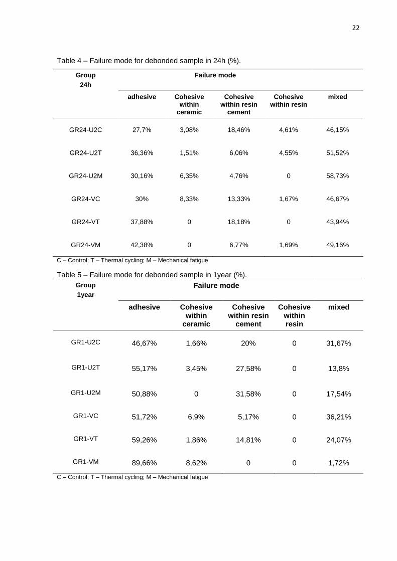
Predominant mixed failure was found for all groups at storage for 24 h, in
accordance treatment and resin cement (Table 4). Predominant adhesive failure was
shown for sticks stored for 1 year, irrespective of treatment and resin cement (Table
5).
22
Table 4 – Failure mode for debonded sample in 24h (%).
Group
24h
Failure mode
adhesive Cohesive within
ceramic
Cohesive within resin
cement
Cohesive within resin
mixed
GR24-U2C
27,7%
3,08%
18,46%
4,61%
46,15%
GR24-U2T
36,36%
1,51%
6,06%
4,55%
51,52%
GR24-U2M
30,16%
6,35%
4,76%
0
58,73%
GR24-VC
30%
8,33%
13,33%
1,67%
46,67%
GR24-VT
37,88%
0
18,18%
0
43,94%
GR24-VM
42,38%
0
6,77%
1,69%
49,16%
C – Control; T – Thermal cycling; M – Mechanical fatigue
Table 5 – Failure mode for debonded sample in 1year (%).
Group
1year
Failure mode
adhesive Cohesive within
ceramic
Cohesive within resin
cement
Cohesive within resin
mixed
GR1-U2C
46,67%
1,66%
20%
0
31,67%
GR1-U2T
55,17%
3,45%
27,58%
0
13,8%
GR1-U2M
50,88%
0
31,58%
0
17,54%
GR1-VC
51,72%
6,9%
5,17%
0
36,21%
GR1-VT
59,26%
1,86%
14,81%
0
24,07%
GR1-VM
89,66%
8,62%
0
0
1,72%
C – Control; T – Thermal cycling; M – Mechanical fatigue
23
Grafic 1 – Representative and comparative graphical illustration of failure mode for all
groups, stored for 24hours and 1 year.
Discussion
The first hypothesis, which stated the aging methods would not affect the
µTBS was rejected. The results showed that the groups submitted to thermal cycling,
thermomechanical cycling, and water storage decreased significantly the µTBS,
when compared to the control. When in clinical service, resin-retained all-ceramic
dental restorations are subjected to intraoral temperature changes as thermal
mechanical cycling that induce stresses, and exposition to fluids and bacterial
products present in saliva (Hernandez et al., 2008). The moist of the oral
environment associated with thermal mechanical cycling and thermal cycling may
affect the ceramic restorations and reduce resistance to catastrophic fracture (White
et al., 1995; Umemoto et al., 1995).
0%
20%
40%
60%
80%
100%
U2C U2T U2M VC VT VM U2C U2T U2M VC VT VM
Failu
re M
od
e (
%)
adhesive Cohesive within ceramicCohesive within resin cement Cohesive within resinmixed
24
The samples submitted to cycling load in the wet environment can lead to
propagation of small cracks within ceramic (Stokes et al., 1988) or at the interface
between resin cement and ceramic, influencing negatively the bond strength (Guarda
et al., 2013). The water at the crack ends can influence the crack spread, (Taskonak
et al., 2008) and the stress corrosion will facilitate the crack growth and fracture
occurance (Stokes et al., 1988). In this study, the thermal mechanical cycling
significantly decreased the µTBS values, which is in agreement with previous studies
(Borges et al., 2009; Guarda et al., 2013).
Thermal cycling effect on µTBS was determined in this study.
Temperature changes in the bonded materials with different thermal expansion
coefficients and thermal conductivities cause thermal stresses at the interface
(Fonseca et al., 2004). Besides, the stress produced by load and thermal cycling in
the interface between different materials with different elastic modulus, contributes
with degradation of the bond integrity (Aguiar et al., 2014). Previous study shown
that the durability of the bond between resin cement and silaned ceramic surfaces
decreases with thermal cycling or water storage (Furukawa et al., 2002).
The µTBS reduction can occur due the alterations in the mechanical
properties of resin cements by the continuous action of water at the resin cement-
ceramic interface (Guarda et al., 2013). Depending of silane type, storage time, and
thermal cycling can decrease the µTBS of the resin-ceramic interface (White et al.,
1995; Furukawa et al., 2002; Abo-Hamar et al., 2005; Salvio et al., 2007; Hernandez
et al., 2008). Previous study showed that a degree of hydrolysis is responsible by the
silane efficacy, the higher the degree of hydrolysis, the better the bond provided by
the silane agent (Umemoto et al., 1995). Silane permeability is the result of the
silicon-oxygen bonds hydrolysis at the ceramic–silane interface by the water
absorption (Stokes et al., 1988). Probably, this fact may also be responsible for the
degradation level of the bond strength at the resin-ceramic interface during water
storage.
Water storage is another factor that can reduce the mechanical properties
of composite resins. Previous studies shown that the reduction in the mechanical
properties of composite resins water stored may occur within 2 to 6 months
(Ferracane et al., 1995; Ferracane et al., 1998; Drummond et al., 1998; Taskonak et
al., 2008). Another study showed significant reduction in the mechanical properties of
25
the composite resins water stored for 1 year (Carrilho et al., 2005). Others studies
demonstrated that the long-term stability of the resin/dentin (De Munck et al., 2005),
and resin/ceramic interfaces (El Zohairy et al., 2004) may be compromised over time,
suggesting that interface degradation may have a role in the long-term survival of
adhesively-luted all-ceramic restorations (Hernandez et al., 2008). Probably, this
reduction is due to continuous action of the water on the material structure. The
mechanism of water transport and its effects on the mechanical properties of
polymers depend on several factors such as composition and monomer ratio that
change according to specific applications and manufacturer’s goals, and the
variability defines the chemical stability of resin in a specific environment (Ruyter et
al., 1987; Van Landingham et al., 1999; Soles et al., 2000; Santerre et al., 2001).
When the resin cement factor was analyzed, Variolink II showed higher
µTBS compared to RelyX U200 rejecting the second hypothesis. Variolink II is a resin
cement with adequate bond strength and hardness values (Fonseca et al., 2004;
Abo-Hamar et al., 2005) that requires phosphoric acid etching pretreatment and
adhesive system. On the other hand, RelyX U200 self-adhesive resin cement
contains methacrylated phosphoric acid esters, which promoted the reactions with
the basic fillers and calcium ions from hydroxyapatite (De Munck et al., 2004; Hikita
et al., 2007).
This findings are in accordance to previous studies that showed similar
results, in which the conventional dual-cure resin cement exhibited higher bond
strength performance to the different surface treatments after artificial aging, even
when compared with self-adhesive resin cement (Kumbuloglu et al., 2005; Baldissara
et al., 2006; Phark et al., 2009). The conventional dual-cure resin cement seems to
have superior micromechanical interlocking, probably because it is able to flow better
into the surface-treated ceramic microporosity filling the microspaces between matrix
and filler particles (Phark et al., 2009).
Despite the luting procedure with Variolink II resin cement be complex in
relation to RelyX U200, the µTBS was higher. Resin sensibility depends on the
polymer cross-linking level, volumetric fraction of nanometer-sized pores, and
quantity of fillers (Soles et al., 2000), and degree of conversion (Ferracane et al.,
1994). Previous studies showed that an increase in water sorption was observed
when the triethyleneglycol dimethacrylate (TEGDMA) and urethane dimetacrylate
26
ratio to bisphenol-A-glycidyl dimetacrylate was increased (Beatty et al., 1993; Venz et
al., 1991). In this study, the presence of TEGDMA in the Variolink II and RelyX U200
resin cements probably contributed for water sorption acceleration and decreased the
mechanical properties of the resin cement after thermal cycling, thermal mechanical
cycling and storage time.
The failure mode correlated directly with the bond-strength results, as is
shown in Table 4 and 5. Even though the groups submitted to µTBS after 24 h
showed bond strengths that were higher than the groups analyzed after 1 year, the
mixed mode failure was predominant. For the groups analyzed after 1 year was
verified a predominance of adhesive failures. This reduction in µTBS in the
mechanical properties of the resin cements would be due to a continuous action of
water on resin cement-ceramic interface (Guarda et al., 2013).
Data of the present study demonstrated that thermal cycling, mechanical
fatigue and storage time affected the µTBS values. Further studies should be carried
out using different cementation loads and different number of thermal cycling and
mechanical fatigue.
Conclusion
The following conclusions can be drawn:
1 - Thermal cycling and mechanical fatigue procedures significantly
decreased the µTBS for both storage times in relation to control group;
2 - Variolink II showed higher bond strength values compared to RelyX
U200;
3 – Water storage time reduced the µTBS for all treatments.
Referências*1
Abo-Hamar SE, Federlin M, Hiller KA, Friedl KH, Schmalz G. Effects of temporary
cements on the bond strength of ceramic luted to dentin. Dent Mater.
2005;21(9):794-803.
1 *
De acordo com a norma da UNICAMP/FOP, baseadas na padronização do Internaticonal Committee of Medical Journal
Editors – Vancouver Group. Abreviatura dos periódicos em conformidade com o Pubmed.
27
Aguiar TR, André CB, Correr-Sobrinho L, Arrais CAG, Ambrosano GMB, Giannini M.
Effect of storage times and mechanical load cycling on dentin bond strength of
conventional and self-adhesive resin luting cements. J Prosthet Dent.
2014;111(5):404-410.
Aguiar TR, Di Francescantonio M, Arrais CA, Ambrosano GM, Davanzo C, Giannini
M. Influence of curing mode and time on degree of conversion of one conventional
and two self-adhesive resin cements. Oper Dent. 2010;35(3):295-299.
Baldissara P, Valandro LF, Monaco C, Ferrari M, Bottino MA, Scotti R. Fatigue
resistance of the bond of a glass-infiltrated alumina ceramic to human dentin. J
Adhes Dent. 2006;8(2):97-104.
Barghi N, Fischer DE, Vatani L. Effects of porcelain leucite content, types of etchants,
and etching time on porcelain-composite bond. J Esthet Restor Dent. 2006;18(1):47-
52.
Beatty MW, Swartz ML, Moore BK, Phillips RW, Roberts TA. Effect of crosslinking
agent content, monomer functionally, and repeat unit chemistry on properties of
unfilled resins. J Biomed Mater Res. 1993;27(3):403-413.
Blatz MB, Sadan A, Martin J, Lang B. In vitro evaluation of shear bond strengths of
resin to densely-sintered high-purity zirconium-oxide ceramic after long-term storage
and thermal cycling. J Prosthet Dent. 2004;91(4):356-362.
Borges GA, Spohr AM, De Goes MF, Correr-Sobrinho L, Chan DNC. Effect of etching
and airborne particle abrasion on the microstructure of different dental ceramics. J
Prosthet Dent. 2003; 89(5):479-488.
Borges GA, Caldas D, Taskonak B, Yan J, Correr-Sobrinho L, de Oliveira WJ.
Fracture loads of all-ceramic crowns under wet and dry fatigue conditions. J
Prosthodont. 2009; 18(8):649–655.
28
Canay S, Hersek N, Ertan A. Effect of different acid treatments on a porcelain
Sundfeld Neto D, Naves LZ, Costa AR, Correr AB, Consani S, Borges GA. The effect
of hydrofluoric acid concentration on the bond strength and morphology of the
surface and interface of glass ceramics to a resin cement. Oper Dent. 2015;
40(5):470-47.
Umemoto K, Kurata S. Effects of mixed silane coupling agent on porcelain tooth
material and various dental alloys. Dent Mat. 1995;14(2):135-142.
Vásquez V, Ozcan M, Nishioka R, Souza R, Mesquita A, Pavanelli C. Mechanical
and thermal cycling effects on the flexural strength of glass ceramics fused to
titanium. Dent Mater J. 2008;27(1):7-15.
Taskonak B, Griggs JA, Mecholsky JJ Jr, Yan JH. Analysis of subcritical crack growth
in dental ceramics using fracture mechanics and fractography. Dent Mater. 2008;
24(5):700-707.
White SN, Zhao XY, Zhaokun Y, Li ZC. Cyclic mechanical fatigue of a feldspathic
dental porcelain. Int J Prosthodont. 1995; 8(5):413-420.
38
APÊNDICE
Materiais e Métodos
Os materiais que foram utilizados neste estudo estão descritos no
Quadro1.
Quadro 1 – Descrição dos materiais e respectivos fabricantes.
Materiais Fabricante Composição
Cimento Resinoso Variolink II
Ivoclar-Vivadent, Schaan,
Liechtenstein
A matriz monomérica é composta de Bis-GMA, uretano dimetacrilato, e trietileno
glicol dimetacrilato. As partículas inorgânicas são vidro de bário, ytterbium trifluorídrico, Ba-Al-fluorsilicato de vidro, e
and partículas mistas de óxidos, catalisadores, estabilizadores, e
pigmentos.
Cimento Resinoso RelyX U200
3M ESPE, St Paul, MN, EUA
Pasta base: Pó de vidro tratado com silano, ácido 2 propenóico, 2metil,1,1-[1-(hydroximetil)-1,2-ethanodiyl] éster, TEG-DMA, sílica tratada com silano,nfibra de vidro, persulfato de sódio, eper-3,5,5-
trimetil-hexanoato t-butila. Pasta catalisadora: Pó de vidro tratado
com silano, dimetacrilato substituto, sílica tratada com silano, p-toluenosulfonato de sódio, 1-benzil-5-feni-ácido bárico, sais de
cálcio, 1,12-dodecano dimetacrilato, hidróxido de cálcio e dióxido de titânio.
Silano – Monobond-S
Ivoclar-Vivadent, Schaan,
Liechtenstein
Solução alcoólica de silano metacrilato, ácido fosfórico metacrilato, e metacrilato
de sulfureto.
Sistema adesivo Scothbond Universal
3M ESPE, St Paul, MN, EUA
BIS-GMA, metacrilato de 2-hidroxietila, sílica tratada com silício, álcool etílico,
de acrílico e ácido itacônico, canforoquinona, N,N-demotibenzocaína, metacrilato de 2-dimetilamonoetilo, metil
etil cetona.
Resina Composta Filtek Z250
3M ESPE, St Paul, MN, EUA
Cerâmica tratada com silano, BIS-GMA, BIS-EMA, sílica tratada com silano,
UDMA, TEGDMA.
39
APÊNDICE 1: OBTENÇÃO DAS AMOSTRAS EM CERÂMICA
Foram confeccionados 72 blocos cerâmicos com a cerâmica a base de
leucita IPS Empress Esthetic (Ivoclar-Vivadent),dentina, cor A2, com dimensões
8mm de comprimento,8mm de largura e 3mm de altura, seguindo as recomendações
do fabricante. A resina acrílica autopolimerizável Duralay (Reliance) na cor vermelha,
foi inserida numa matriz de silicone de mesma dimensão para confecção dos
padrões (figura 1). Em seguida, o conduto de alimentação com 3mm de diâmetro por
6mm de comprimento foi colocado no padrão de resina acrílica e fixado num cilindro
plástico com 12,2mm de diâmetro por 30mm de altura (figura 2). Após, foi adaptado
na base plástica do cadinho e um cilindro de silicone (Ivoclar Vivadent) foi
posicionado na base formadora do cadinho (figura 3). O padrão de resina acrílica foi
incluído com revestimento à base de fosfato IPS Empress Esthetic Speed (Ivoclar
Vivadent), na proporção de 100g de pó para 16ml de líquido, próprio do material, e
11ml de água destilada e espatulado por 2minutos (figura 4). Após a presa do
revestimento (figura 5), o cilindro de silicone, o formador do conduto e a base foram
removidos (figura 6).
Então, o bloco de revestimento foi levado ao forno elétrico (Vulcan A-550,
Degussa Ney, Yucaipa, Califórnia, USA) na temperatura de 850°C, e mantidos por
90 minutos para remoção da resina e expansão do revestimento. Decorrido esse
período de aquecimento, o bloco de revestimento foi removido do forno e 2 pastilhas
da cerâmica IPS Empress Esthetic foram posicionadas no conduto juntamente com o
êmbolo de óxido de alumínio do sistema IPS Empress com 12mm de diâmetro por
37mm de altura, e levado ao forno EP600 (Ivoclar Vivadent). O conjunto foi mantido
por 20 minutos a 1075°C, seguido de uma pressão de 5bar por 15 minutos (figura 7).
Quando o bloco de revestimento atingiu a temperatura ambiente (figura 8), as
amostras foram removidas seccionando o bloco de revestimento ao meio.
Posteriormente as amostras de cerâmica foram removidas com jateamento
(Renfert,Hilzingen, Germany) usando partículas de vidro com 50µm de diâmetro
(Bio-Art, São Carlos, SP, Brasil), com pressão de 2bar para remoção do
revestimento próximo as amostras. A seguir as amostras em cerâmicas foram limpas
por 10 minutos utilizando ultra-som (Ultrasonic Cleaner 1440 D), seguido de lavagem
em água corrente, secagem com jato de ar. O conduto de alimentação foi removido
40
com disco diamantado (KG Sorensen, Cotia, SP, Brasil) e a região do conduto
submetida ao acabamento com broca cilíndrica de diamante.
Após a desinclusão, os blocos cerâmicos foram submetidos ao
acabamento e polimento com lixas de carbeto de silício de granulação 320,600 e
1200 (Norton S.A., São Paulo, SP, Brazil) manualmente (figura 9) e limpos em ultra-
som por 5minutos em água destilada e 5minutos em álcool etílico 92% e secos com
jato de ar.
1 2
3
4 5
Figura 2 - padrão de resina acrílica fixado no cilindro plástico da base formadora de cadinho Figura 1 – matriz de silicone
Figura 3 - um cilindro de silicone foi posicionado na base formadora do cadinho
Figura 4 - O padrão de resina acrílica foi incluído com revestimento
Figura 5 – aguardando a presa do revestimento
41
APÊNDICE 2: CONFECÇÃO DAS AMOSTRAS EM RESINA E CIMENTAÇÃO
Para cimentação dos blocos cerâmicos, foram confeccionados 72 blocos
de resina composta Filtek Z250, cor A3 (3M ESPE), nas mesmas dimensões dos
blocos cerâmicos. Estes blocos em resina foram confeccionados em matriz de
silicone, preenchidas em incrementos de 2,0mm de espessura (figura 10), tendo
cada incremento fotoativado por 40s com o aparelho LED-UltraLume 5 (Ultradent,
Salt Lake City, UT, USA), com irradiância de 1100mW/cm2 (figura 11).
6 7 8
9
Figura 6 – Presa do revestimento. O cilindro de silicone, o formador do conduto e a base foram removidos
Figura 7 - O conjunto foi mantido por 20 minutos a 1075°C, seguido de uma pressão de 5bar por 15 minutos
Figura 8 - o bloco de revestimento atingiu a temperatura ambiente
Figura 9 - Após a desinclusão, os blocos cerâmicos foram submetidos ao acabamento e polimento com lixas de carbeto de silício de granulação 320,600 e 1200
42
Os conjuntos blocos de cerâmica/resina composta foram separados em
12 grupos (n=6), conforme o quadro 2.
10 11
Figura 10 - blocos em resina confeccionados em matriz de silicone
Figura 11 - cada incremento foi fotoativado por 40s com o aparelho LED-UltraLume 5
43
Quadro 2 - Divisão dos grupos de acordo com o cimento resinoso, ciclagem térmica,
mecânica, térmico-mecânica e armazenagem
Grupos
Cimentos
Resinosos
Ciclagem
Armazenagem
GR24-U2C
Rely X U200
Sem Ciclagem
Controle (24Horas)
GR24-U2T
Ciclagem Térmica
GR24-U2M
Ciclagem Térmico/Mecânica
GR24-VC
Variolink II
Sem Ciclagem
Controle (24Horas)
GR24-VT
Ciclagem Térmica
GR24-VM
Ciclagem Térmico/Mecânica
GR1-U2C
Rely X U200
Sem Ciclagem
1 Ano
GR1-U2T
Ciclagem Térmica
GR1-U2M
Ciclagem Térmico/Mecânica
GR1-VC
Variolink II
Sem Ciclagem
1 Ano
GR1-VT
Ciclagem Térmica
GR1-VM
Ciclagem Térmico/Mecânica
As superfícies dos blocos cerâmicos foram condicionadas por 60 s,
utilizando gel de ácido fluorídrico 10% (Dentsply), seguindo recomendações do
fabricante (figura 12). O condicionamento da superfície do bloco foi realizado da
seguinte forma: o gel foi aplicado e distribuído sobre a superfície do bloco cerâmico
utilizando microbrush. Após aplicação, os blocos foram lavados com jato água/ar por
44
60s, limpos em ultra-som por 5min em água destilada e 5 min em álcool etílico 92%
seguido da secagem com jato de ar por 30s. Após a limpeza e secagem, duas
camadas do agente de silanização Monobond-S (Ivoclar-vivadent) foram aplicadas e,
após 1 min, a superfície foi seca com jato de ar por 30s (figura 13). A superfície do
bloco de resina foi condicionada com ácido fosfórico 37% (Condac) por 30s (figura
14). Após aplicação, os blocos de resina foram lavados com jato água/ar por 30s,
secos com jato de ar por 30s. Em seguida, foi aplicado sobre a superfície do bloco
de resina, uma camada do adesivo Single Bond (3M ESPE) (figura 15), jato de ar por
5s e fotoativado por 10 s, com o aparelho LED-UltraLume 5 (Ultradent).
Em seguida, uma porção de cimento resinoso RelyX U200 (figura 17) (3M
ESPE) e Variolink II (figura 16) (Ivoclar-vivadent) foram aplicados sobre o centro da
12 13
14 15
Figura 12 - As superfícies dos blocos cerâmicos foram condicionadas por 60 s, utilizando gel de ácido fluorídrico 10%
Figura 13 - duas camadas do agente de silanização Monobond-S foram aplicadas, por 1 min
Figura 14 - A superfície do bloco de resina foi condicionada com ácido fosfórico 37% (Condac) por 30s
Figura 15 - foi aplicado sobre a superfície do bloco de resina, uma camada do adesivo Single Bond
45
superfície dos blocos cerâmicos de seus respectivos grupos (figura 18) e estes
posicionados sobre os blocos de resina composta, mantendo sob pressão constante
de 500g por 1 minuto (figura 19). Após remoção do excesso de agente cimentante
utilizando microbrush, cada interface do conjunto cerâmica/resina composta foi
fotoativada por 40s com o aparelho LED-UltraLume 5 (Ultradent), com irradiância de
1100mW/cm², com fotoativação final na superfície de topo, totalizando 160 segundos
(figura 20). Todos os conjuntos (12 grupos) bloco cerâmica/resina composta foram
armazenados em água deionizada numa estufa a 37°C por 24h.
16 17
18 19
Figura 16 - Variolink II Figura 17 - RelyX U200
Figura 18 - um volume de cimento resinoso RelyX U200 e Variolink II foram aplicados sobre o centro da superfície dos blocos cerâmicos dos respectivos grupos
Figura 19 - os blocos de cerâmica foram posicionados sobre os blocos de resina composta, mantendo sob pressão constante de 500g por 1 minuto
46
APÊNDICE 3: CICLAGEM TÉRMICA
Os conjuntos bloco cerâmica/ resina composta dos grupos GR24-U2T,
GR24-VT, GR1-U2T e GR1-VT foram submetidos a 3.000 ciclos térmicos na máquina
(MSCT 3 - Marnucci ME, São Carlos, SP, Brasil) com temperatura entre 5°C e 55°C,
com banhos de 30s em cada temperatura (Figura 21).
20
Figura 20 - Após remoção do excesso de agente cimentante utilizando microbrush, cada interface do conjunto cerâmica/resina composta foi fotoativada por 40s com o aparelho LED-UltraLume 5
Figura 21 - Máquina de termociclagem (MSCT 3 - Marnucci ME, São Carlos, SP, Brasil)
operando com temperatura entre 5°C e 55°C, com banhos de 30s em cada temperatura.
47
APÊNDICE 4: CICLAGEM TÉRMICO/MECÂNICA
Os conjuntos bloco cerâmica/resina composta dos grupos GR24-U2M,
GR24-VM, GR1-U2M e GR1-VM foram submetidos à ciclagem térmico/mecânica. Uma
extremidade em forma de cinzel de 25mm de comprimento, acoplada à máquina de
ensaio de fadiga (Erios, São Paulo, Brazil), (Figura 22), foi posicionada no centro do
bloco cerâmico e os blocos foram submetidos a 250.000 ciclos em máquina de
ciclagem mecânica, com 2 HZ de frequência. Durante o ensaio de ciclagem
mecânica foi realizada ao mesmo tempo a ciclagem térmica com temperatura entre
5°C e 55°C, com tempo de 30s em cada temperatura.
APÊNDICE 5: ENSAIO DE TRAÇÃO
Após armazenagem, ciclagem térmica e ciclagem térmica/mecânica, os
conjuntos bloco cerâmica/resina composta, foram seccionados em cortadeira
metalografica Isomet 1000 (Buehler) (Figura 23) utilizando um disco diamantado de
dupla-face sob refrigeração de água. Inicialmente, os conjuntos bloco
Figura 22 - Máquina de ensaio de fadiga (Erios, São Paulo, Brazil) onde os blocos
cerâmica/resina foram submetidos a 250.000 ciclos, 2 HZ de frequência e banhos de 30s
entre 5°C e 55°C em cada temperatura.
48
cerâmica/resina composta foram fixados em uma base de acrílico com cera
pegajosa. Os blocos foram seccionados nos planos X e Y com ângulo de 90 graus
entre os cortes de modo a obter amostras em forma de palito, com
aproximadamente 1mm² de área da interface adesiva. Uma média de 10 palitos foi
obtido por bloco, totalizando 60 palitos por grupo.
Em seguida, os palitos dos grupos GR24-U2C, GR24-U2T, GR24-U2M,
GR24-VC, GR24-VT e GR24-VM, foram armazenados em água deionizada numa
estufa a 37 °C, por 24h, e os palitos dos grupos GR1-U2C, GR1-U2T, GR1-U2M,
GR1-VC, GR1-VT e GR1-VM foram armazenados em água deionizada numa estufa a
37 °C, por 1 ano.
Após os períodos de armazenagem, os palitos foram fixados com adesivo
de cianoacrilato em gel (Super Bonder Gel, Loctite Ltd, São Paulo, Brasil), em
dispositivo para microtração, mantendo a interface adesiva livre (Figura 24). Este
dispositivo foi fixado na máquina de ensaio EZ Test (EZS, Shimadzu, Tóquio,
Japão), e submetido ao ensaio de resistência à microtração a velocidade constante
de 0,5 mm/min, até ocorrer fratura. Os valores de resistência de união foram obtidos
em kgf e suas médias expressas em MegaPascal (MPa) desta forma: Valor indicado
pela máquina em kgf multiplicado por 9,82 para obtenção do valor em Newton,
sendo este valor dividido pela área do espécime, obtendo-se o valor em MPa. A
média por grupo foi calculada a partir da média das parcelas (espécime) por bloco.
49
APÊNDICE 6: PREPARO DOS ESPÉCIMES PARA ANÁLISE DO PADRÃO DE
FRATURA
Após a realização do ensaio da resistência da união, os homólogos de
cada espécime fraturado foram fixados em folha de papel com auxílio de fita
adesiva, juntamente com informações relativas a sua área de superfície e força
exibida em kgf pela máquina de ensaio. Em seguida, os homólogos de cada
espécime foram fixados em stubs de alumínio com o auxílio de resina composta em
suas bases, inseridos emparelhadamente sobre fita dupla face de carbono (Electron
Microscopy Sciences, Washington 19034 – USA) e desidratadas por duas horas no
interior de um recipiente plástico hermeticamente fechado contendo sílica gel. Em
seguida, todos os espécimes foram observados em estereomicroscópio
(Stemi DV4 - Zeiss) com aumento de 32 X. As superfícies das amostras que
geraram dúvidas, receberam cobertura de ouro/paládio (Balzers, modelo SCD 050
sputter coater, Balzers Union Aktiengesellschaft, Fürstentum Liechtenstein, FL-9496
– Germany) e o modo de falha foi confirmado usando o microscópio eletrônico de
varredura (SEM; LEO 435 VP; LEO Electron Microscopy Ltd., Cambridge, UK),
operado a 20 Kv, pelo mesmo operador. A distância de trabalho utilizada estava