Università degliStudi di Bologna Facoltà di Agraria Dipartimento di Colture Arboree Dottorato di Ricerca in Colture Arboree (XV Ciclo) CHANGES IN PLANT METABOLISM INDUCED BY DIOXYGENASE INHIBITORS AND THEIR EFFECT ON THE EPIPHYTIC MICROBIAL COMMUNITY AND FIRE BLIGHT (ERWINIA AMYLOVORA) CONTROL Alterazioni del metabolismo della pianta indotte dagli inibitori delle diossigenasi e loro influenza sulla biocenosi microbica epifitica e sull’infezione da Colpo di Fuoco Batterico (Erwinia amylovora). TUTORE: Chiarissimo Prof. Guglielmo Costa Co-tutore: Dr. Joel L. Vanneste COORDINATORE: Chiarissimo Prof. Silviero Sansavini TESI DI DOTTORATO DI: Dr. Francesco Spinelli Anno accademico 2001-2002
Transcript
Università degliStudi di Bologna
Facoltà di Agraria Dipartimento di Colture Arboree
Dottorato di Ricerca in Colture Arboree (XV Ciclo)
CHANGES IN PLANT METABOLISM INDUCED BY DIOXYGENASE INHIBITORS AND THEIR EFFECT ON THE
EPIPHYTIC MICROBIAL COMMUNITY AND FIRE BLIGHT (ERWINIA AMYLOVORA)
CONTROL Alterazioni del metabolismo della pianta indotte dagli inibitori delle diossigenasi e
loro influenza sulla biocenosi microbica epifitica e sull’infezione da Colpo di Fuoco Batterico (Erwinia amylovora).
TUTORE: Chiarissimo Prof. Guglielmo Costa Co-tutore:Dr. Joel L. Vanneste COORDINATORE:Chiarissimo Prof. Silviero Sansavini
TESI DI DOTTORATO DI: Dr. Francesco Spinelli
Anno accademico 2001-2002
Ai miei genitori e a mia sorella a cui tanto devo
Contents
CONTENTS PREFACE VIII ACKNOLEDGMENTS X INTRODUCTION 1 FIRE BLIGHT 2 1. INTRODUCTION 2 2. THE PATHOGEN: ERWINIA AMYLOVORA 3
2.1. Biology and metabolism 3 2.2. Plasmids 6 2.3. Outer membrane and capsule of Erwinia
amylovora 7
2.4. Virulence factors of Erwinia amylovora 11 2.4.1. EPS and amylovoran 11 2.4.2. Hrp genes. 11
2.6.3. Bacterial mixtures 39 2.7. SELECTION OF A BIOLOGICAL CONTROL AGENT 40 2.8. DELIVERY OF BACTERIAL ANTAGONISTS 41 2.9. CONTEMPORARY USE OF BACTERIAL ANTAGONIST AND
ANTIBIOTICS
43 2.10. CONTEMPORARY USE OF BACTERIAL ANTAGONIST AND
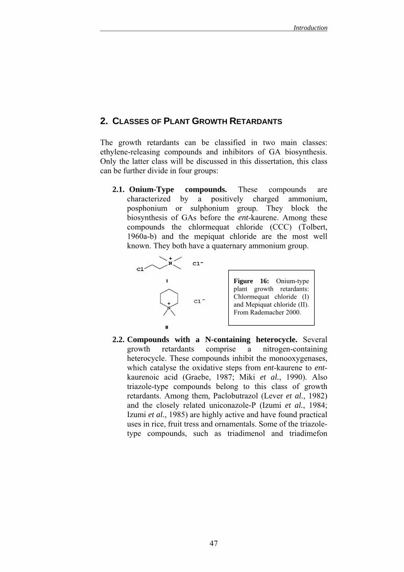
2.1. Onium-Type compounds 47 2.2. Compounds with a N-containing heterocycle 47 2.3. Structural mimic of 2-oxoglutaric acid 48 2.4. 16,17- Dihydro-GA5 and related structures 49
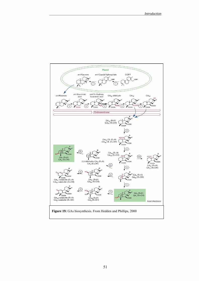
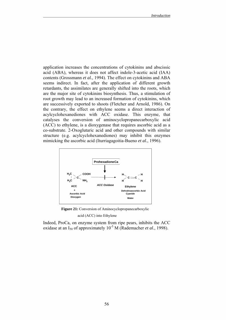
3.1. EFFECT ON GIBBERELLIN BIOSYNTHESIS 53 3.2. EFFECT ON OTHER PHYTOHORMONES LEVELS 55 3.3. EFFECT ON FLAVONOID METABOLISM 57
II
Contents
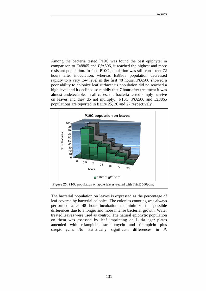
3.4. PROHEXADIONE-Ca 57 3.4.1. Influence of ProCa on flavonoids biosynthesis 60
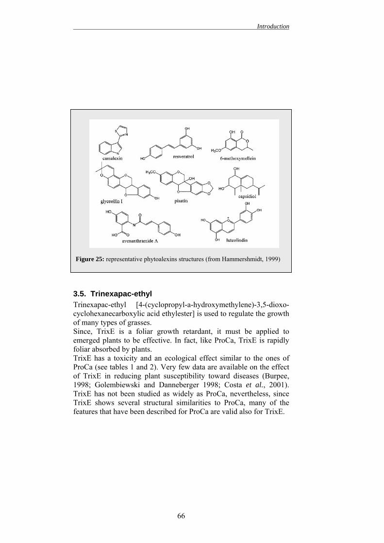
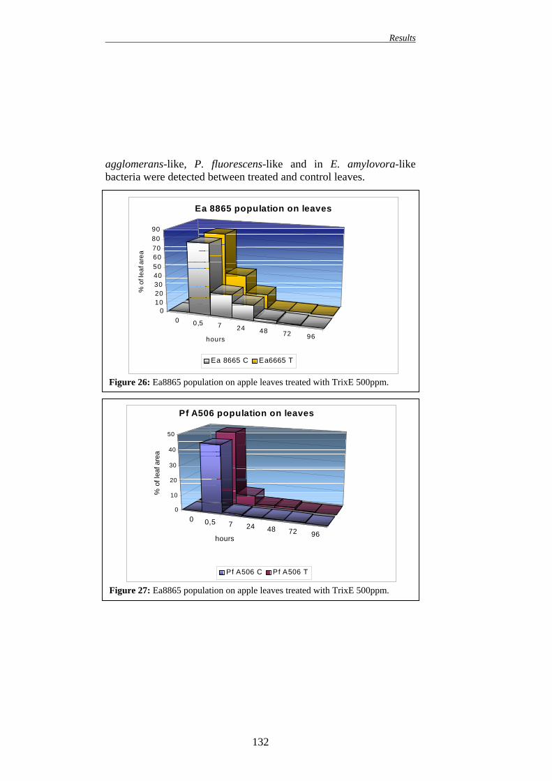
INSERT 3: A GENERAL OVERVIEW OF FLAVONOIDS 62 3.5. TRINEXAPAC-ETHYL 66
SAR INDUCERS 68 3.6. BENZOTHIADIAZOLE (BTH) 68
INSERT 4: SAR. 69 AIM OF THE STUDY 73 MATERIALS AND METHODS 76 1. ISOLATION, IDENTIFICATION AND SELECTION OF A VIRULENT ERWINIA AMYLOVORA STRAIN
77
1.1. BERESWILL ET AL., (1992) PROTOCOL 78 1.2. AMPLIFICATION OF 16SRDNA USING PRIMERS
DESIGNED BY WEISBURG ET AL., (1991)
79 2. ISOLATION, IDENTIFICATION AND SELECTION OF BACTERIAL
ANTAGONISTS AGAINST FIRE BLIGHT
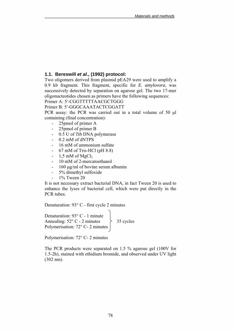
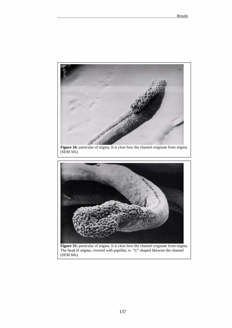
80 2.1. INHIBITION TEST AGAINST E. AMYLOVORA 80 2.2. HR TEST ON TOBACCO LEAVES 81 2.3. IMMATURE PEAR FRUIT TEST (IPF TEST) 82 2.4. CONTROL OF FIRE BLIGHT ON DETACHED FLOWERS 83 2.5. CONTROL OF FIRE BLIGHT ON DETACHED FLOWERING
BRANCHES
83 3. EFFICACY OF DIOXYGENASE INHIBITORS IN REDUCING SHOOT BLIGHT INCIDENCE (SECONDARY INFECTION)
84
4. EFFICACY OF DIOXYGENASE INHIBITORS IN REDUCING SHOOT GROWTH
84
5. EFFICACY OF DIOXYGENASE INHIBITORS IN REDUCING BLOSSOM BLIGHT INCIDENCE (PRIMARY INFECTION)
85
6. EFFECT OF DIOXYGENASE INHIBITORS APPLICATION ON APPLE AND PEAR NECTAR COMPOSITION
85
6.1. DETERMINATION METHODS OF NECTAR SUGAR
III
Contents
CONTENT BY GASCHROMATOGRAPHY (GC) (BAGDANOV ET AL., 1997)
86
6.2. DETERMINATION OF NECTAR PHENOLIC COMPOUNDS CONTENT BY HPLC
87
7. EFFECT OF NECTAR SUGAR COMPOSITION ON BACTERIAL GROWTH
88
7.1. SERIAL DILUTIONS METHOD TO ASSESS BACTERIAL GROWTH
89
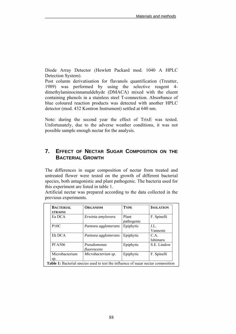
8. EFFECT OF DIOXYGENASE INHIBITORS APPLICATION ON FLOWERS AND NECTAR ATTRACTIVENESS TO HONEYBEES (APIS MELLIFERA)
90 9. EFFECT OF DIOXYGENASE INHIBITORS APPLICATION ON
THE NATURAL MICROBIAL COMMUNITY ON APPLE AND PEAR BLOSSOMS
91 9.1. SHANNON-WEINER INDEX 91
10. EFFECT OF DIOXYGENASE INHIBITORS APPLICATION ON E. AMYLOVORA, P. AGGLOMERANS AND P. FLUORESCENS POPULATION ON APPLE BLOSSOMS
92 11. EFFECT OF DIOXYGENASE INHIBITORS APPLICATION ON
NATURAL MICROBIAL COMMUNITY ON APPLE LEAVES
94 11.1. LEAF IMPRINTING 94
12. EFFECT OF DIOXYGENASE INHIBITORS APPLICATION ON E. AMYLOVORA, P. AGGLOMERANS AND P. FLUORESCENS POPULATION ON APPLE LEAVES
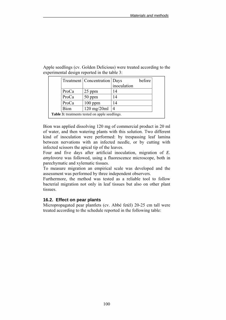
94 13. SUGAR ON LEAVES 95 14. Effect of Trixe on Bacterial Endophytic Population
IN APPLE TISSUES
96 15. MICROSCOPICAL INVESTIGATION I: EFFECT OF DIOXYGENASE INHIBITORS ON PRIMARY INFECTION OF BLOSSOMS
97 15.1. GFP- AND RFP-LABELLED BACTERIA:
TRANSFORMATION BY ELECTROPORATION
97 15.2. SCANNING ELECTRON MICROSCOPE EQUIPMENT (SEM)
15.4. FLUORESCENCE MICROSCOPE 99 16. MICROSCOPICAL INVESTIGATION II: EFFECT OF DIOXYGENASE INHIBITORS AND SAR INDUCER ON E. AMYLOVORA MIGRATION INSIDE PLANT TISSUES
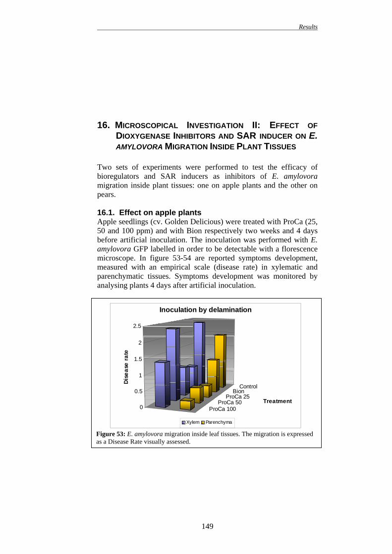
99 16.1. EFFECT ON APPLE PLANTS 99 16.2. EFFECT ON PEAR PLANTS 100
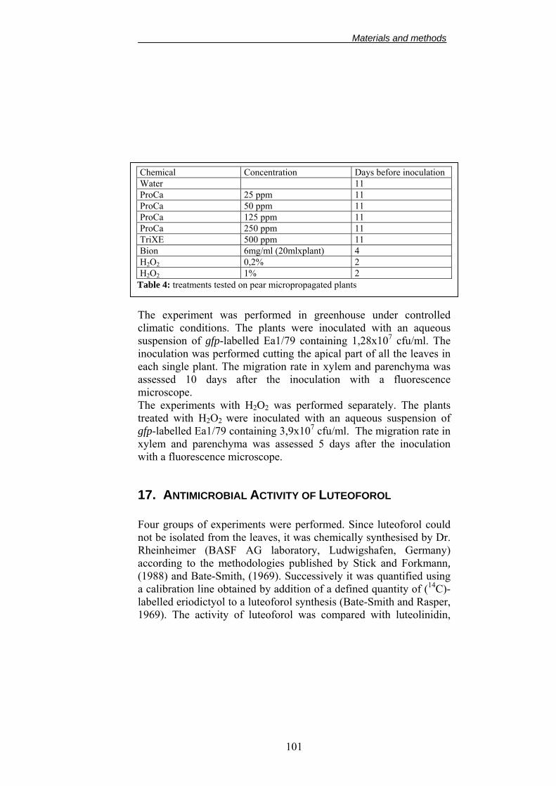
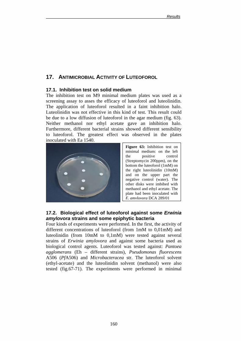
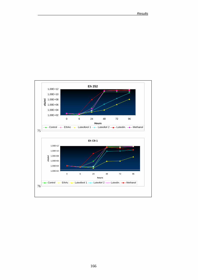
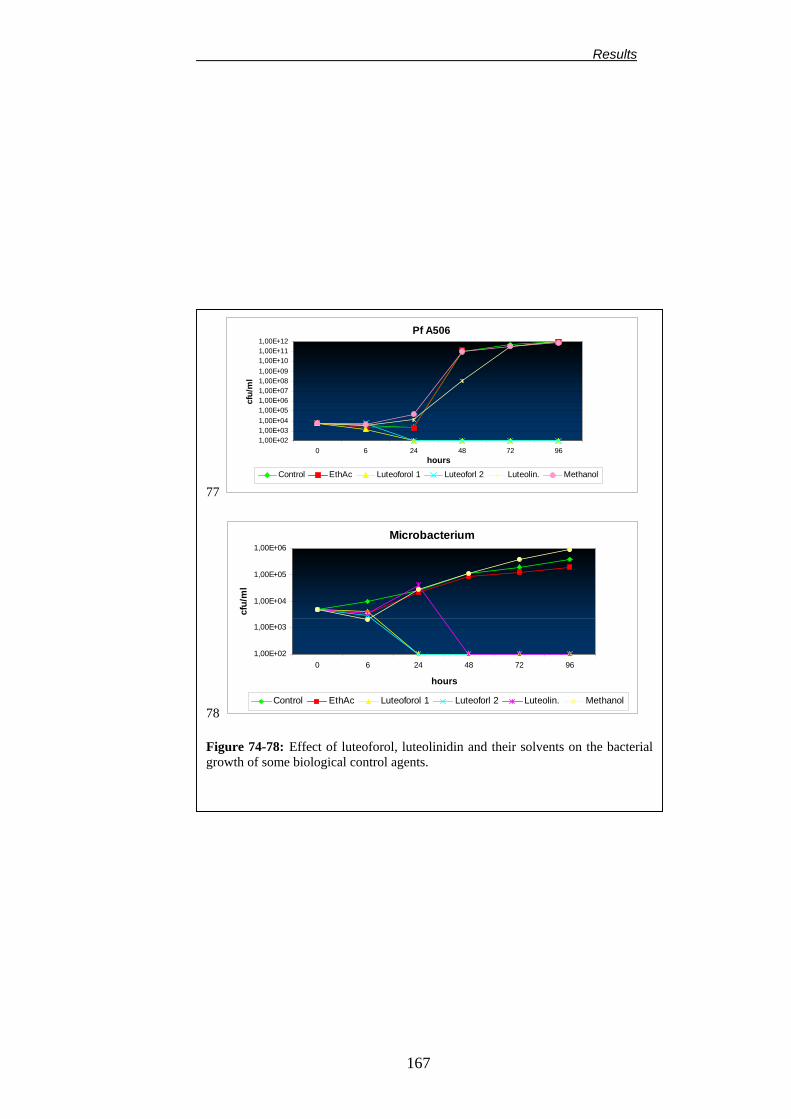
17. ANTIMICROBIAL ACTIVITY OF LUTEOFOROL 101 17.1. INHIBITION TEST ON SOLID MEDIUM. 102 17.2. BIOLOGICAL EFFECT OF LUTEOFOROL AGAINST SOME ERWINIA AMYLOVORA STRAINS AND EPIPHYTIC BACTERIA
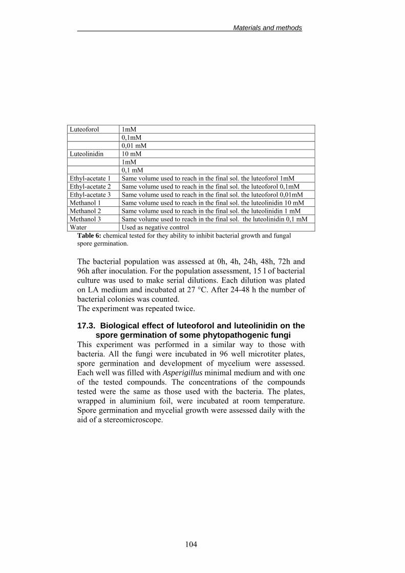
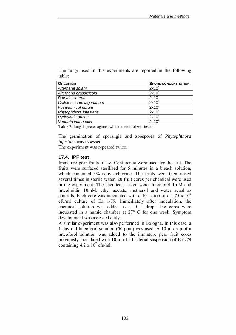
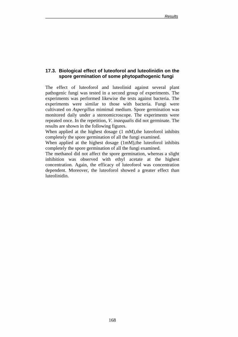
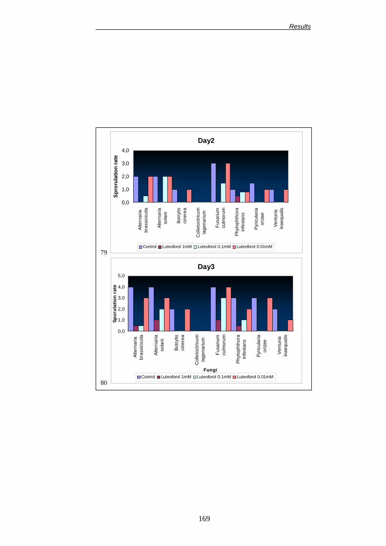
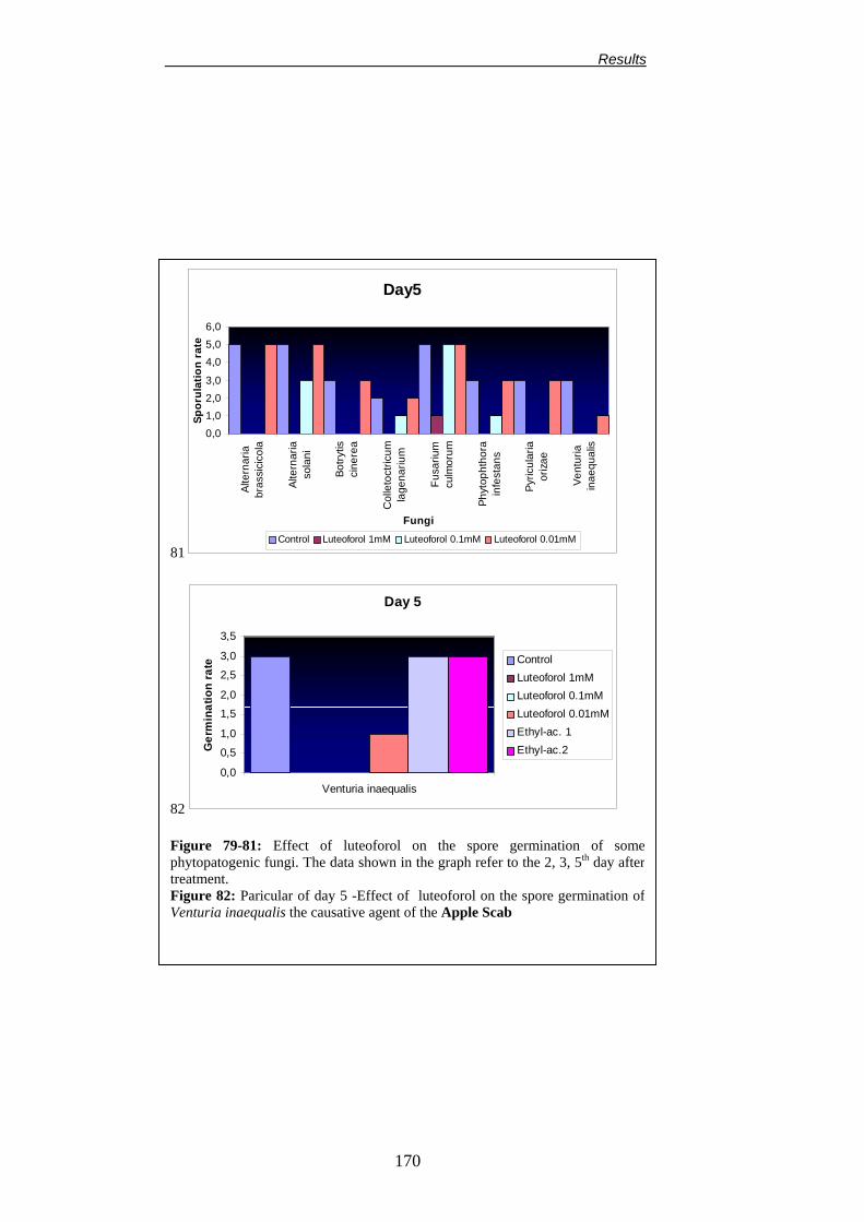
103 17.3. BIOLOGICAL EFFECT OF LUTEOFOROL AND LUTEOLINIDIN ON THE SPORE GERMINATION OF SOME PHYTOPATHOGENIC FUNGI
104 17.4. IPF TEST 105 17.5. BIOLOGICAL EFFECT OF LUTEOFOROL AND LUTEOLINIDIN ON MICROPROPAGATED PLANTS
106
RESULTS
107
1. ISOLATION, IDENTIFICATION AND SELECTION OF A VIRULENT ERWINIA AMYLOVORA STRAIN
108
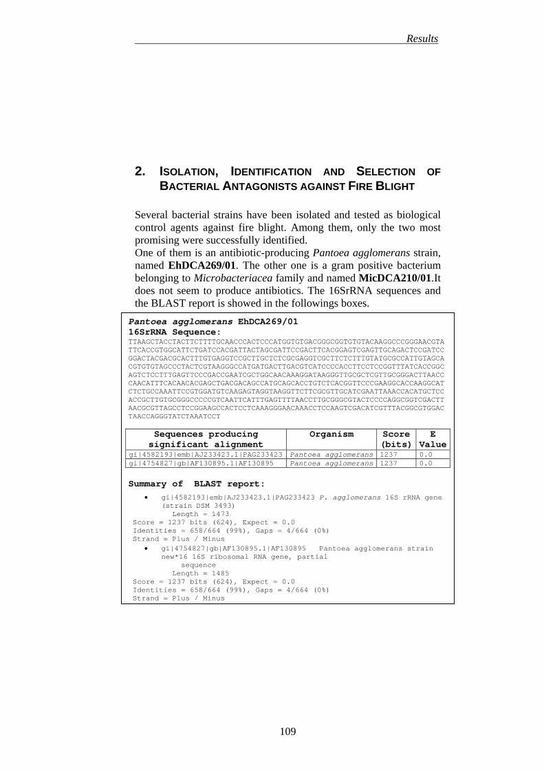
2. ISOLATION, IDENTIFICATION AND SELECTION OF BACTERIAL ANTAGONISTS AGAINST FIRE BLIGHT
109
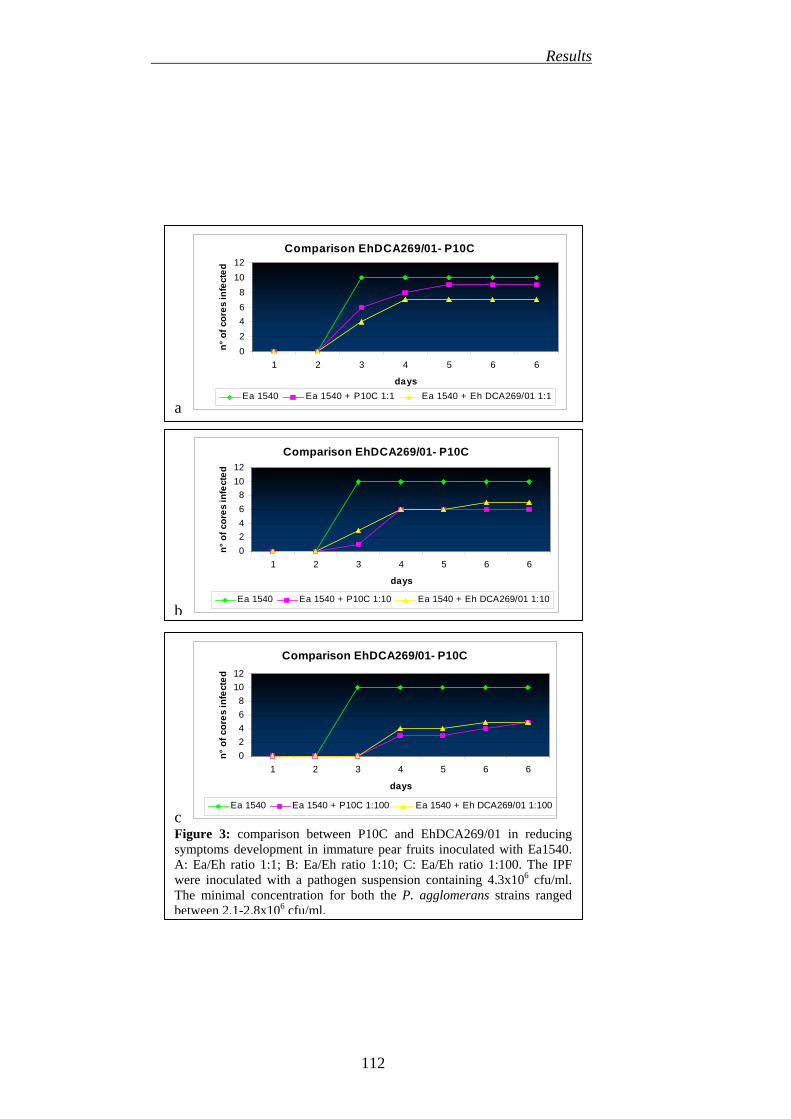
3. EFFICACY OF DIOXYGENASE INHIBITORS IN REDUCING SHOOT BLIGHT INCIDENCE (SECONDARY INFECTION)
113
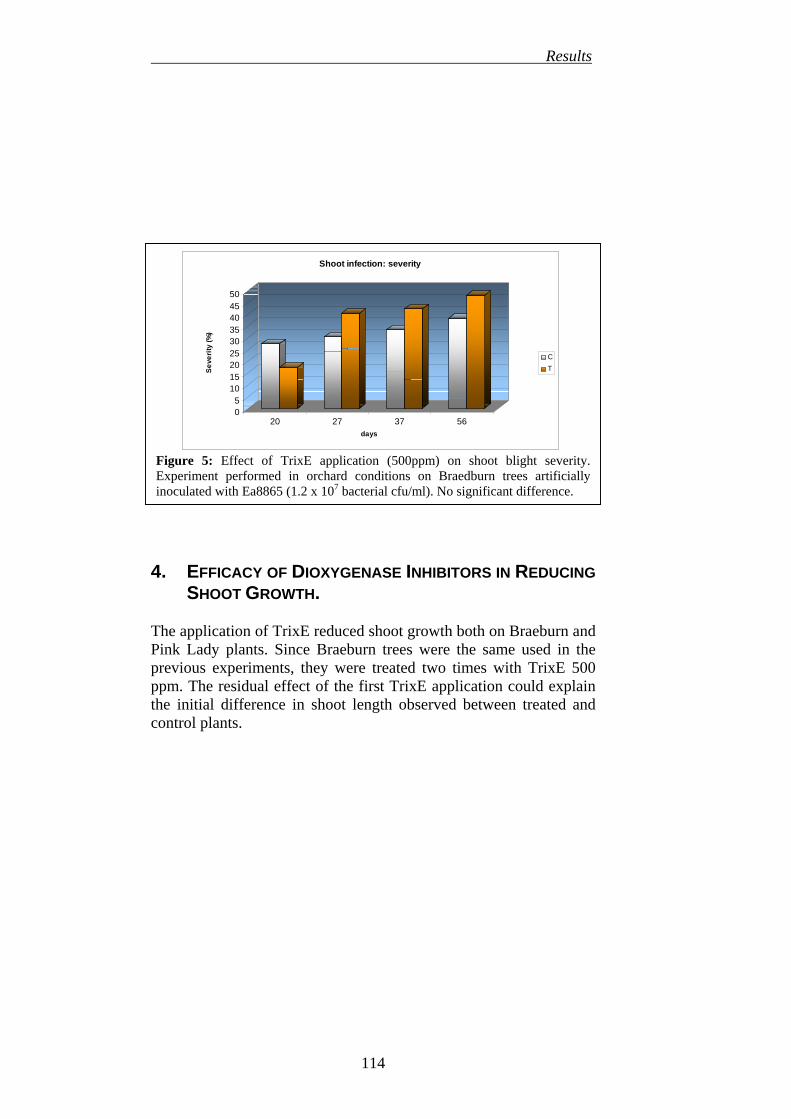
4. EFFICACY OF DIOXYGENASE INHIBITORS IN REDUCING SHOOT GROWTH
114
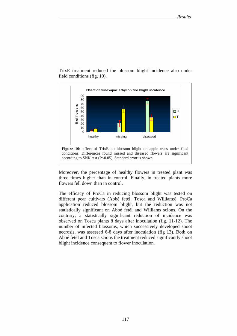
5. EFFICACY OF DIOXYGENASE INHIBITORS IN REDUCING BLOSSOM BLIGHT INCIDENCE (PRIMARY INFECTION)
115
6. EFFECT OF DIOXYGENASE INHIBITORS APPLICATION ON APPLE AND PEAR NECTAR COMPOSITION
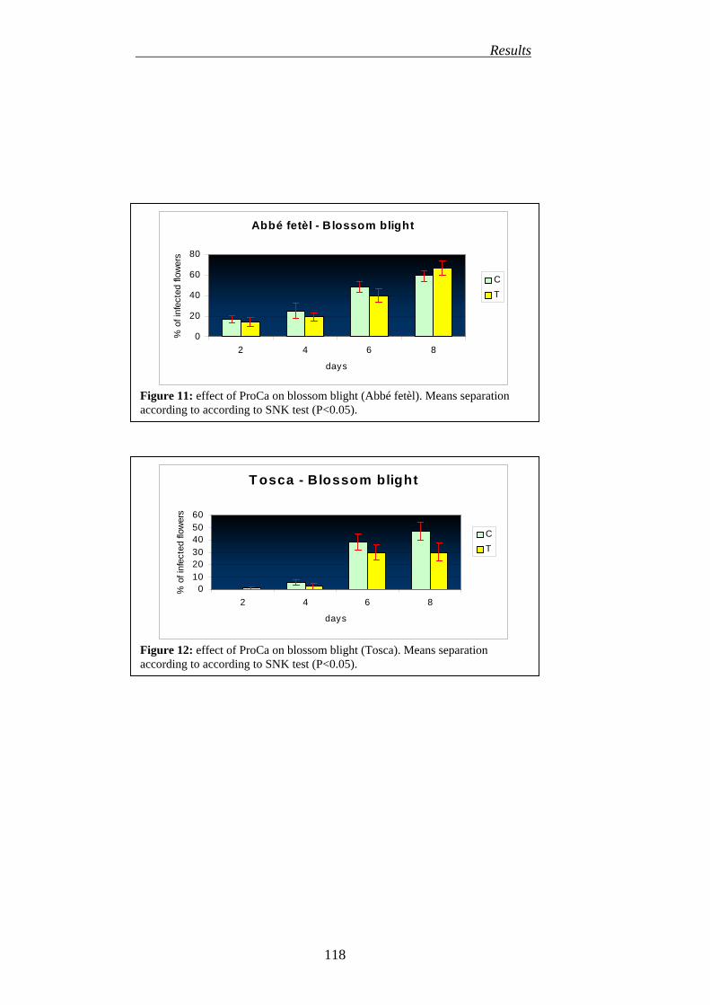
119
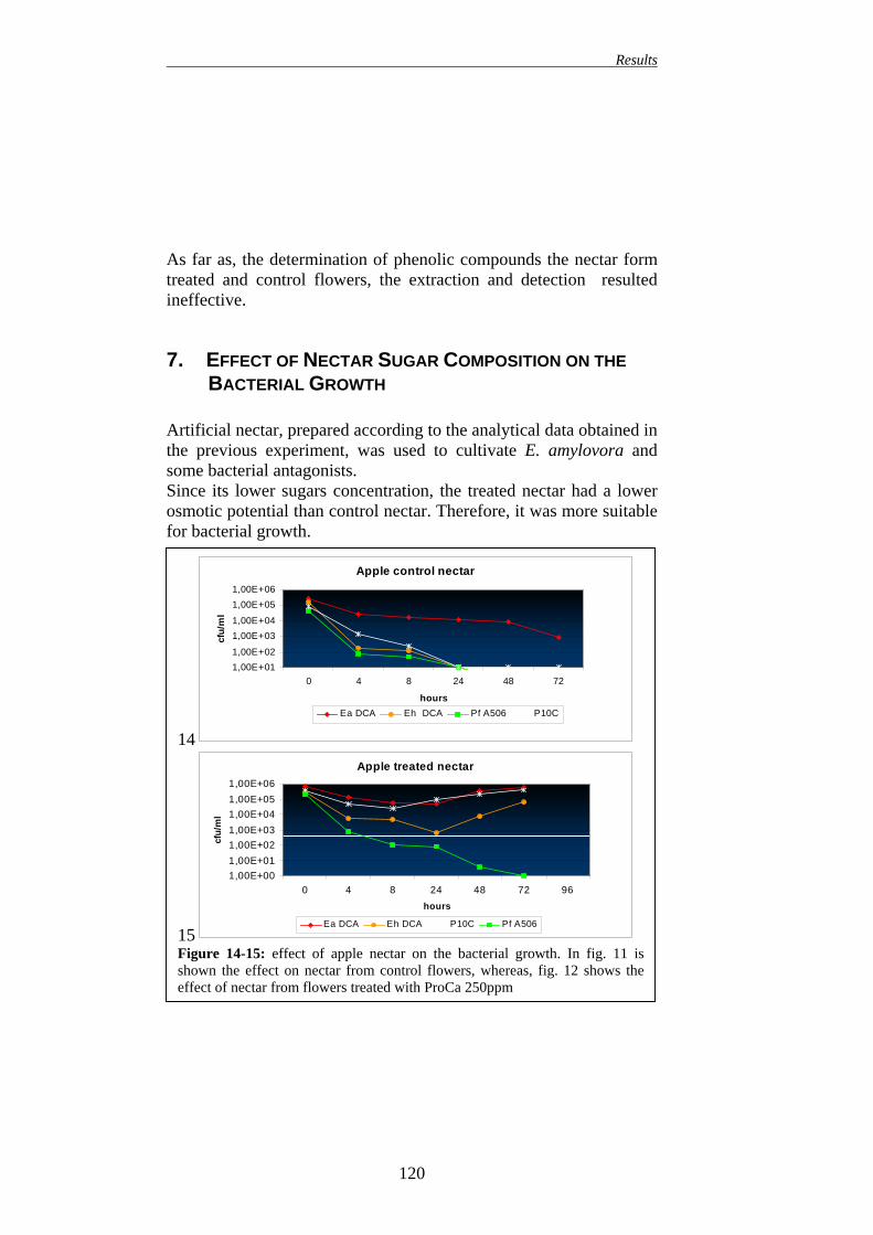
7. EFFECT OF NECTAR SUGAR COMPOSITION ON BACTERIAL GROWTH
120
8. EFFECT OF DIOXYGENASE INHIBITORS APPLICATION ON
V
Contents
FLOWERS AND NECTAR ATTRACTIVENESS TO HONEYBEES (APIS MELLIFERA)
122
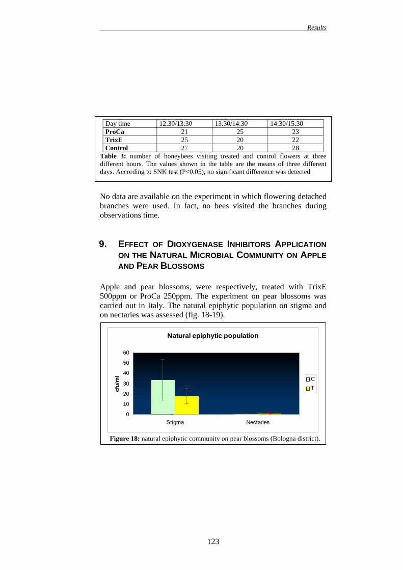
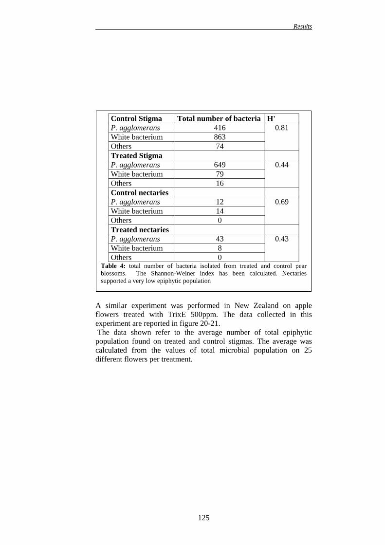
9. EFFECT OF DIOXYGENASE INHIBITORS APPLICATION ON THE NATURAL MICROBIAL COMMUNITY ON APPLE AND PEAR BLOSSOMS
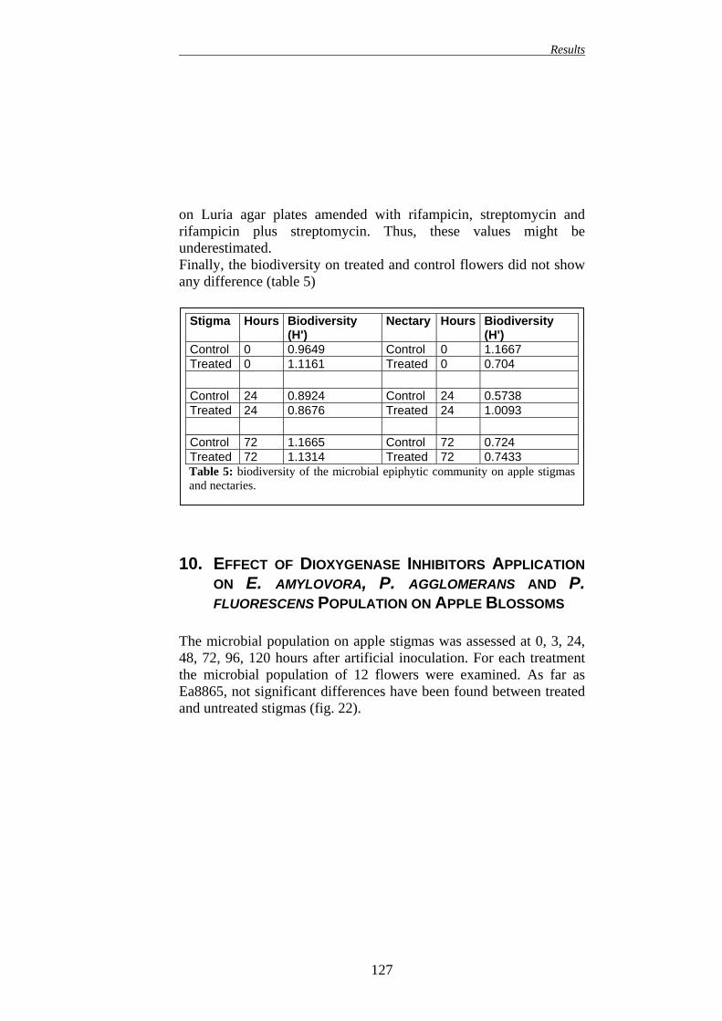
123 10. EFFECT OF DIOXYGENASE INHIBITORS APPLICATION ON E.
AMYLOVORA, P. AGGLOMERANS AND P. FLUORESCENS POPULATION ON APPLE BLOSSOMS
127 11. EFFECT OF DIOXYGENASE INHIBITORS APPLICATION ON
NATURAL MICROBIAL COMMUNITY ON APPLE LEAVES
130 12. EFFECT OF DIOXYGENASE INHIBITORS APPLICATION ON E.
AMYLOVORA, P. AGGLOMERANS AND P. FLUORESCENS POPULATION ON APPLE LEAVES
130 13. SUGAR ON LEAVES 134 14. EFFECT OF TRIXE ON BACTERIAL ENDOPHYTIC
POPULATION IN APPLE TISSUES
135 15. MICROSCOPICAL INVESTIGATION I: EFFECT OF DIOXYGENASE INHIBITORS ON PRIMARY INFECTION OF BLOSSOMS
136 16. MICROSCOPICAL INVESTIGATION II: EFFECT OF DIOXYGENASE INHIBITORS AND SAR INDUCER ON E. AMYLOVORA MIGRATION INSIDE PLANT TISSUES
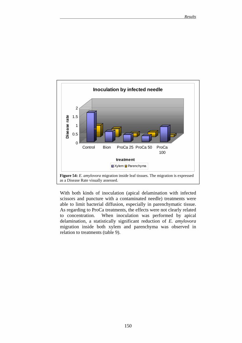
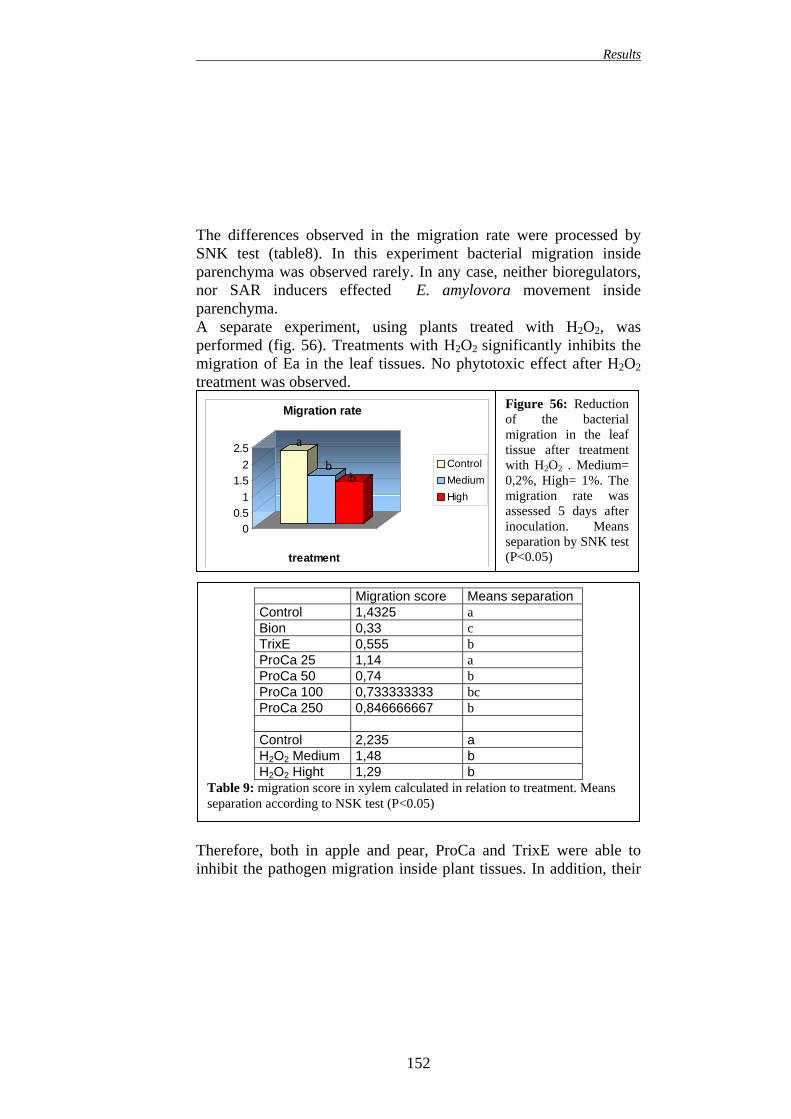
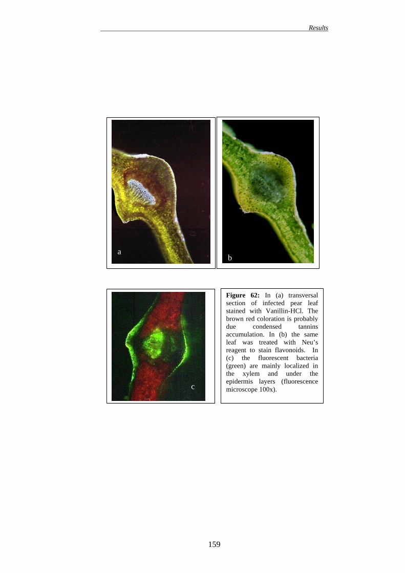
149 16.1. EFFECT ON APPLE PLANTS 149 16.2. EFFECT ON PEAR PLANTS 151
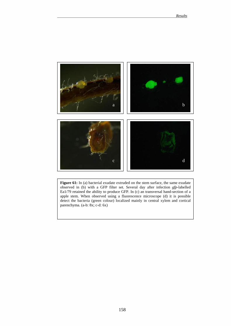
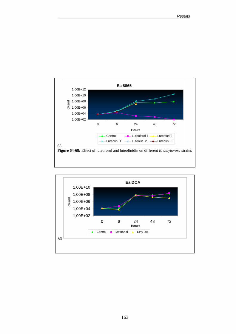
17. ANTIMICROBIAL ACTIVITY OF LUTEOFOROL 160 17.1. INHIBITION TEST ON SOLID MEDIUM 160 17.2. BIOLOGICAL EFFECT OF LUTEOFOROL AGAINST SOME ERWINIA AMYLOVORA STRAINS AND EPIPHYTIC BACTERIA
160 17.3. BIOLOGICAL EFFECT OF LUTEOFOROL AND LUTEOLINIDIN ON THE SPORE GERMINATION OF SOME PHYTOPATHOGENIC FUNGI
168 17.4. IPF TEST 171 17.5. BIOLOGICAL EFFECT OF LUTEOFOROL AND LUTEOLINIDIN ON MICROPROPAGATED PLANTS
172
VI
Contents
DISCUSSION
173
FINAL REMARKS
183
REFERENCES 187
ANNEX 230
VII
Preface
PREFACE Fire blight, caused by the gram negative bacterium Erwinia amylovora, is one of the most destructive bacterial diseases of Pomaceous plants. Therefore, the development of reliable methods to control this disease is desperately needed. This research investigated the possibility to interfere, by altering plant metabolism, on the interactions occurring between Erwinia amylovora, the host plant and the epiphytic microbial community in order to obtain a more effective control of fire blight. Prohexadione-calcium and trinexapac-ethyl, two dioxygenase inhibitors, were chosen as a chemical tool to influence plant metabolism. These compounds inhibit the 2-oxoglutarate-dependent dioxygenases and, therefore, they greatly influence plant metabolism. Moreover, dioxygenase inhibitors were found to enhance plant resistance to a wide range of pathogens. In particular, dioxygenase inhibitors application seems a promising method to control fire blight. From cited literature, it is assumed that these compounds increase plant defence mainly by a transient alteration of flavonoids metabolism. We tried to demonstrate, that the reduction of susceptibility to disease could be partially due to an indirect influence on the microbial community established on plant surface. The possibility to influence the interactions occurring in the epiphytic microbial community is particularly interesting, in fact, the relationships among different bacterial populations on plant surface is a key factor for a more effective biological control of plant diseases. Furthermore, we evaluated the possibility to combine the application of dioxygenase inhibitors with biological control in order to develop an integrate strategy for control of fire blight.
VIII
Preface
The first step for this study was the isolation of a pathogenic strain of E. amylovora. In addition, we isolated different epiphytic bacteria, which respond to general requirements for biological control agents. Successively, the effect of dioxygenase inhibitors treatment on microbial community was investigated on different plant organs (stigmas, nectaries and leaves). An increase in epiphytic microbial population was found. Further experiments were performed with aim to explain this effect. In particular, changes in sugar content of nectar were observed. These changes, decreasing the osmotic potential of nectar, might allow a more consistent growth of epiphytic bacteria on blossoms. On leaves were found similar differences as well. As far as the interactions between E. amylovora and host plant, they were deeply investigated by advanced microscopical analysis. The influence of dioxygenase inhibitors and SAR inducers application on the infection process and migration of pathogen inside different plant tissues was studied. These microscopical techniques, combined with the use of gpf-labelled E. amylovora, allowed the development of a bioassay method for resistance inducers efficacy screening. The final part of the work demonstrated that the reduction of disease susceptibility observed in plants treated with prohexadione-calcium is mainly due to the accumulation of a novel phytoalexins: luteoforol. This 3-deoxyflavonoid was proven to have a strong antimicrobial activity. Keywords: fire blight, induced resistance, biological control, dioxygenase inhibitors, confocale laser scanning microscope, gfp-labelled bacteria, prohexadione-calcium, luteoforol.
IX
Acknowledgments
ACKNOWLEDGMENTS These investigations were supported by the EU Commission (QLK5-CT-1999-01583). I firstly thank prof. Guglielmo Costa for his indispensable support and for the useful suggestions and criticisms I received from him during all these three years. I thank all the persons who allow me to complete this research and in particular, Dr. Joel Vanneste that guided me at the very beginning of my microbiological researches and taught to me basis of phytopathology. I thanks all the scientists in his lab for their patience when I messed up their labs and especially I whish to thanks my friend Deirdre Cornish, Janet Yu and Darrienne Voyle. I also wish to thank Prof. Klaus Geider for the hospitality in his laboratories and especially for all his constructive suggestions. A special thanks to Dr. Fabrizio Ciampolini and to prof. Mauro Cresti: only their help and suggestions permitted me to carry out my investigations on the microscopic interaction between the pathogen and the host plant. I also wish to thank Dr. Wilhelm Rademacher and Dr. John-Bryan Speakman for the hospitality in their lab, for the great support I received from them and especially for their cooperation in our joint research. I thank Dr. Rheinheimer (BASF) and Mrs. Paul (BASF) for carrying out the luteoforol synthesis and Birgit Hoffmann for excellent technical assistance. I should thank all my colleagues for their help and their useful suggestions and in particular I thank Carlo Andreotti, Mirco Montefiori, Massimo Noferini, Anna Maria Bregoli and Giovanni Fiori. A very special thank is for my friend and colleague Emidio Sabatini that helped me so often and taught to me, a biologist, all the basis for agronomic research.
X
Acknowledgments
A very special thank to my parents and my sister for their immense patience and to all my friends: Davide, Serena, Martina, Claudio, Matteo, Valentina, Fabio, Piero, Alessio, Barbara, Samir and Manuela for their friendship. Finally a very special thank to Mara for her priceless friendship. I should thank you all.
XI
INTRODUCTION
1
Introduction
FIRE BLIGHT
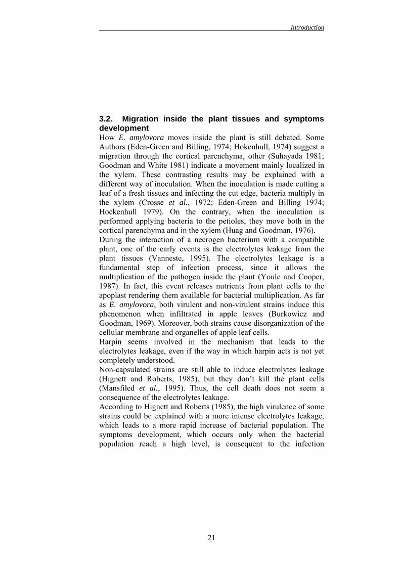
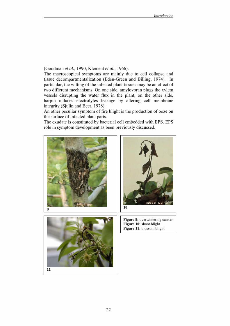
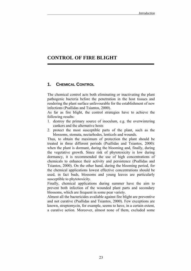
1. INTRODUCTION Erwinia amylovora, a gram negative bacterium, is the causative agent of fire blight (Burrill 1883). This bacterium infects most of the plants belonging to family of Rosaceae and in particularly to the subfamily of Pomoideae such as Cotoneater, Crateagus and Pyracantha, even if, economically, the most important host plants are apple and pears (Eden-Green and Billing, 1974). The bacterium penetrates in the plant trough flowers, but it can also enter via wounds and trough natural openings in the plant cuticle. E. amylovora can invade the whole tree solely by internal progression through the host tissues; thus, a single infection can potentially kill a tree (Vanneste, 1995). This aspect renders fire blight the most devastating bacterial disease of apples and pears. Production of bacterial exudate on the surface of infected tissues is the most characteristic symptom of fire blight (Bennet and Billing, 1980b). Other typical symptoms are the wilting and the consequent necrosis of the infected tissues. The symptomatology of fire blight is rather complicate with different symptoms in relation to different plant parts (blossom blight, shoot blight, overwintering cankers…). The severity of fire blight is dependent on the host plant and on the environmental conditions. Generally, pears are more susceptible than apple (Eastgate, 2000). High humidity and warm temperature are favourable to the disease development. The blossoms are particularly sensitive to infection, even if in high susceptible host plants, the bacterium can infect also mature tissues (Eden-Green and Billing, 1974). The economic impact of fire blight is rather high, in fact a severe outbreak can disrupt the production for several years, moreover it
2
Introduction
limits the areas where most susceptible and economically interesting apple and pear varieties can be grown. Furthermore, its economical importance is likely to increase for several reasons. Firstly, fire blight is still spreading geographically into new apple- and pear- growing areas (Vanneste, 2000). Secondly, with the exception of streptomycin, there is no registered product that can effectively control fire blight. Finally, today, apple plantings are mainly constituted by susceptible cultivars grafted on susceptible rootstock planted in high-density orchards (Longstroth, 2001). These changes, together with the ever-increasing developments of streptomycin resistant strains of E. amylovora, stimulate the research of reliable strategies to control fire blight.
2. THE PATHOGEN: ERWINIA AMYLOVORA 2.1. Biology and metabolism According to the Bergey’s Manual of Systematic Bacteriology, 8th edition (Krieg and Holt, 1984), Erwinia amylovora is a Gram-negative bacteria belonging to the class of Enterobacteriaceae. It was the first bacterium identified as a plant pathogen (Burrill, 1883). The bacterial cell is a rod with 2-7 perithricous flagella and it may be surrounded by an exopolysaccharide capsule (EPS),microscope (Bennet and Billing, using these flagella and the moinfection process. In fact, Bayot apple blossoms sprayed with a sucell developed a higher incidence
Figure 1: E. amylovora cell (TEM100000 x)
which is visible with the electron 1978) (fig.1). E. amylovora moves tility seems important during the and Ries (1986) demonstrate that spension of motile E. amylovora than blossom sprayed with non-
3
Introduction
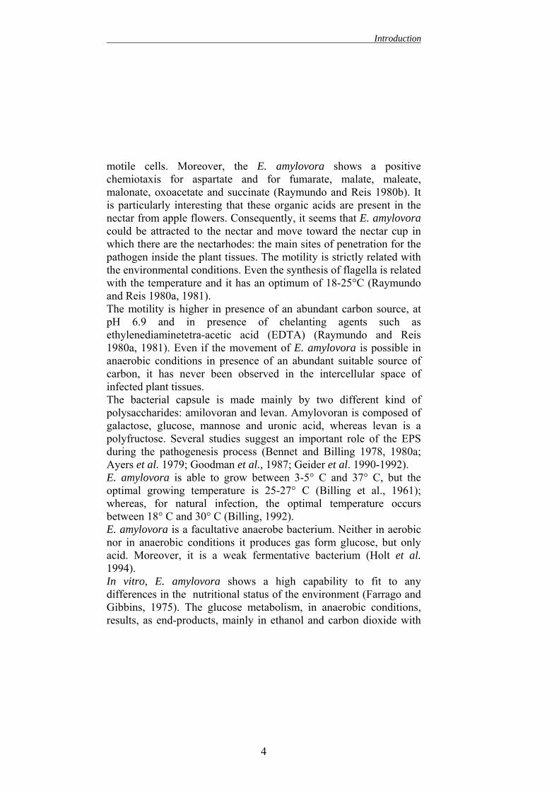
motile cells. Moreover, the E. amylovora shows a positive chemiotaxis for aspartate and for fumarate, malate, maleate, malonate, oxoacetate and succinate (Raymundo and Reis 1980b). It is particularly interesting that these organic acids are present in the nectar from apple flowers. Consequently, it seems that E. amylovora could be attracted to the nectar and move toward the nectar cup in which there are the nectarhodes: the main sites of penetration for the pathogen inside the plant tissues. The motility is strictly related with the environmental conditions. Even the synthesis of flagella is related with the temperature and it has an optimum of 18-25°C (Raymundo and Reis 1980a, 1981). The motility is higher in presence of an abundant carbon source, at pH 6.9 and in presence of chelanting agents such as ethylenediaminetetra-acetic acid (EDTA) (Raymundo and Reis 1980a, 1981). Even if the movement of E. amylovora is possible in anaerobic conditions in presence of an abundant suitable source of carbon, it has never been observed in the intercellular space of infected plant tissues. The bacterial capsule is made mainly by two different kind of polysaccharides: amilovoran and levan. Amylovoran is composed of galactose, glucose, mannose and uronic acid, whereas levan is a polyfructose. Several studies suggest an important role of the EPS during the pathogenesis process (Bennet and Billing 1978, 1980a; Ayers et al. 1979; Goodman et al., 1987; Geider et al. 1990-1992). E. amylovora is able to grow between 3-5° C and 37° C, but the optimal growing temperature is 25-27° C (Billing et al., 1961); whereas, for natural infection, the optimal temperature occurs between 18° C and 30° C (Billing, 1992). E. amylovora is a facultative anaerobe bacterium. Neither in aerobic nor in anaerobic conditions it produces gas form glucose, but only acid. Moreover, it is a weak fermentative bacterium (Holt et al. 1994). In vitro, E. amylovora shows a high capability to fit to any differences in the nutritional status of the environment (Farrago and Gibbins, 1975). The glucose metabolism, in anaerobic conditions, results, as end-products, mainly in ethanol and carbon dioxide with
4
Introduction
small amounts of lactic acid, acetic acid, succinic acid, formic acid, acetoin and 2,3-butanediol (Sutton and Starr 1959-60). As far as nitrogen metabolism, E. amylovora differs from most of the Erterobacteriacae, since it does not reduce nitrate to nitrite. E. amylovora can use the aspartate as nitrogen source, interestingly the aspartate represents the 58% of total amino acids in apple shoots (Lewis and Tolbert, 1964). Moreover, for growth E. amylovora needs nicotinic acid (Starr and Mandel 1950) and no other growth factors are needed. Only some strains, cured of pEA29 plasmid need also thiamine as growth factor. In fact, this plasmid, almost ubiquitous in E. amylovora strains(Falkenstein et al. 1989, Laurent et al. 1989), is involved in thiamine metabolism (Bennet and Billing, 1978; Laurent et al. 1989; Bereswill et al. 1992). This plasmid seems also play a quantitative role in pathogenicity (Laurent et al. 1989). E. amylovora secretes different kinds of extracellular enzymes: a β-glucosidase, proteases and some hydrolytic enzymes. E. amylovora shows only a weak β-glucosidase activity (Hildebrant and Schroth, 1965). This enzyme catalyses the reaction from arbutin, a compounds common in pears, to glucose and hydroquinone and the end-product is toxic to several bacteria (Hildebrant and Schroth, 1963) and to E. amylovora as well (Berg and Gibbins, 1983). The presence of exogenous β-glucoside, such arbutin in pear tissues, could act, after transformation in hydroquinone as a plant defence mechanism. A similar role is supposed for phloridzin in apple tissues (Gibbins 1972). Furthermore, E. amylovora, as the most part of “necrogens” bacteria, is able to produce a detectable amount of hydrolytic enzymes (Seemuller and Beer 1976). Nevertheless, no cellulolytic, pectolytic or xylolitic activity was detected. The role of these enzymes in the infection process has not been yet established. E. amylovora produces also two neutral proteases. These proteases have been isolated from the ooze and from infected plant tissues (Seemuller and Beer 1977). Finally, E. amylovora produces also two different molecules putatively identified as toxic factors. The first molecule, which is 6-thioguanine, does not show any toxic effect on pear cell cultures (Feistner and Staub, 1986). The second is
5
Introduction
(L)-2,5-dihydrophenylalanine (DHP) that is a real necrotoxin (Feistner, 1988). The mode of action of DHP is not completely clear. It can act killing directly the plant cells or it can block the hypersentitive reaction (HR). According to Schwartz et al., (1991), the toxicity of DHP is due to its inhibition of the shikimic acid pathway in plant cells Since not all the E. amylovora strains produce this toxin, it seems an incidental virulence factor more than a key factor necessary for pathogenesis (Geider et al., 90). 2.2. Plasmids None of the several plasmids found in E. amylovora (Marçais et al.1990; Laurent et al., 1989; Panopoulos, 1978; Merckaert et al., 1982) seem strictly involved in pathogenicity. Only one plasmid of ca. 30 Kb, named pEA29, is almost ubiquitous in all E. amylovora strains (Marçais et al.1990, Laurent et al., 1989; Merckaert et al., 1982; Vanneste et al., 1985; Falkenstein et al., 1988). This plasmid can not be transmitted by conjugation (Verdonck et al., 1987) and it is particularly stable and resistant to the classical physical or chemical methods for curing (Laurent et al., 1989).
Figure 2: the structure of the ubiquitousplasmid pEA29 (from www.plantpathology.msu.edu)
In E. amylovora, the resistance to streptomycin is, in most of cases, chromosomally encoded (Panopoulos, 1978, Chiou and Jones, 1991; Thomson et al., 1993; Schroth et al., 1979; Minsavage et al., 1990; Vanneste and Yu, 1993) even if, it could be plasmid encoded as well (Chiou and Jones, 1991). In fact, the resistance to streptomycin can be encoded by a 34 Kb plasmid, known as pEA34 (Chiou and Jones, 1991). The plasmid pEA34 carries the genes strA and strB which are
6
Introduction
part of a transposon called Tn5393 (Chiou and Jones, 1993). The streptomycin resistance genes are homologous to the ones isolated in several other bacterial species (Chiou and Jones, 1993; Norelli et al., 1991). 2.3. Outer membrane and capsule of Erwinia amylovora Outer membrane of E. amylovora is formed by lipopolysaccharide (LPS). LPS is characteristic of all the gram-negative bacteria. Since it is part of the outer membrane (fig. 3), it might be involved in the plant-microbe interaction (Vanneste, 1995). In addition, E. amylovora outer membrane in protected by a capsule, which is formed mainly by two exopolysaccharides (EPS): the homopolymer levan and the heteopolymer amylovoran. Most of the structural genes involved in EPS synthesis are located in the ams region of bacterial chromosome (Burget and Geider, 1995). Numerous evidences suggest that the ams gene cluster is regulated in response to environmental stimuli (Burgert and Geider, 1995, Burgert and Geider, 1997, Ilan et al., 1999). EPS capsule may allow the bacteria to elude the plant defence mechanisms and it seem involved in obtainment of nutrients from the plant cells (Belleman et al. 1990, Coplin e Cook, 1990) even if, as demonstrated by Brisset and Paulin (1992), E. amylovora strains EPS deficient have still the ability to induce electrolyte leakage from plant cells. This capacity is a key step in pathogenesis, in fact, it allows the bacteria to multiply inside the plant environment.
7
Introduction
Figure 3: general structure of the cell envelope of a Gram-negative bacterium(from www.arches.uga.edu)
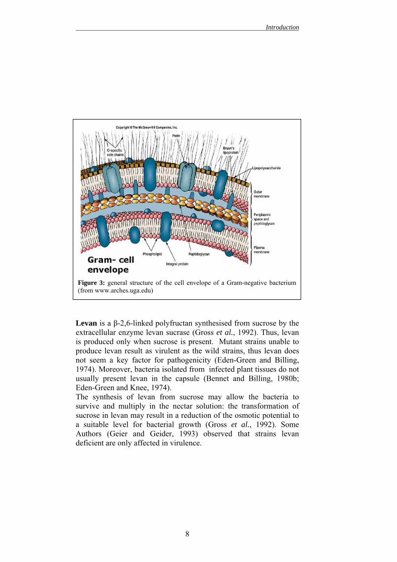
Levan is a β-2,6-linked polyfructan synthesised from sucrose by the extracellular enzyme levan sucrase (Gross et al., 1992). Thus, levan is produced only when sucrose is present. Mutant strains unable to produce levan result as virulent as the wild strains, thus levan does not seem a key factor for pathogenicity (Eden-Green and Billing, 1974). Moreover, bacteria isolated from infected plant tissues do not usually present levan in the capsule (Bennet and Billing, 1980b; Eden-Green and Knee, 1974). The synthesis of levan from sucrose may allow the bacteria to survive and multiply in the nectar solution: the transformation of sucrose in levan may result in a reduction of the osmotic potential to a suitable level for bacterial growth (Gross et al., 1992). Some Authors (Geier and Geider, 1993) observed that strains levan deficient are only affected in virulence.
8
Introduction
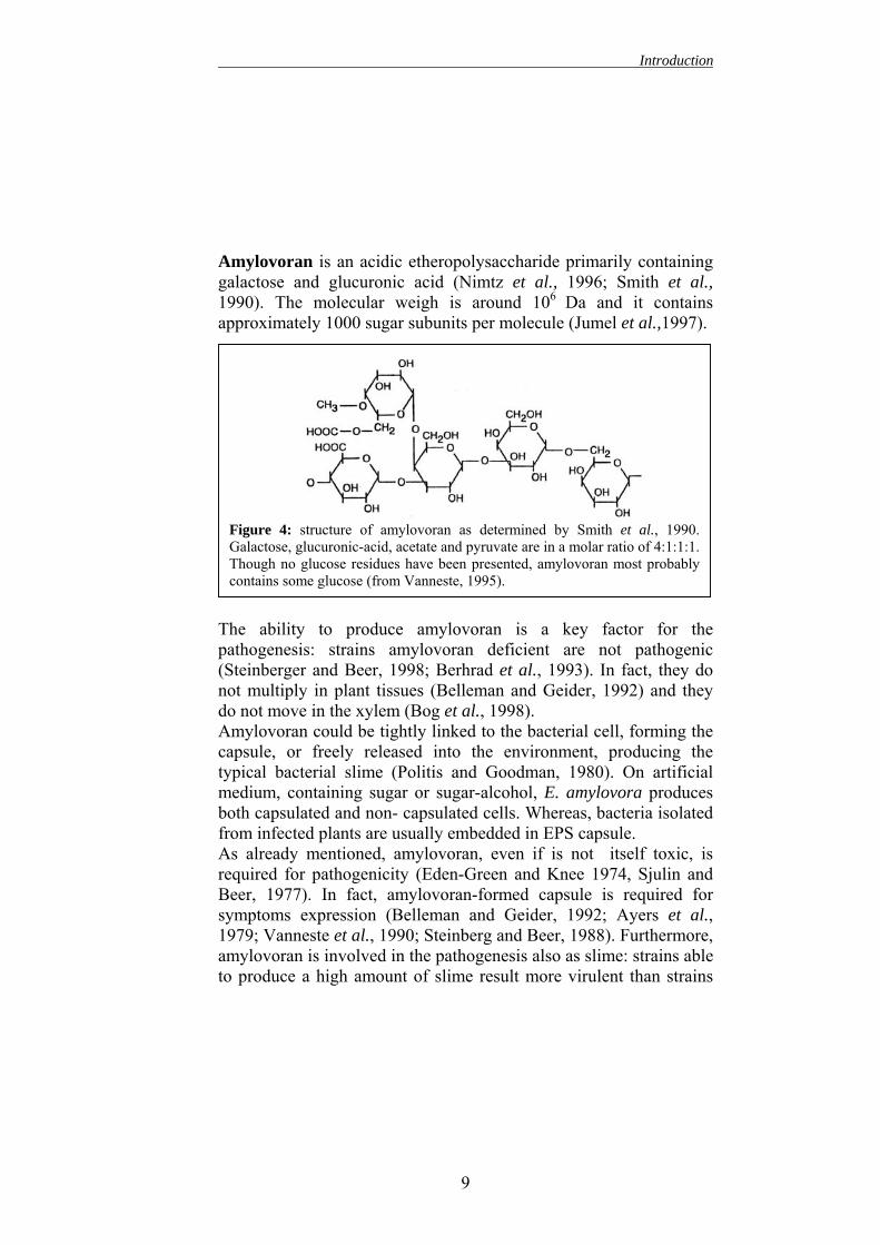
Amylovoran is an acidic etheropolysaccharide primarily containing galactose and glucuronic acid (Nimtz et al., 1996; Smith et al., 1990). The molecular weigh is around 106 Da and it contains approximately 1000 sugar subunits per molecule (Jumel et al.,1997).
Figure 4: structure of amylovoran as determined by Smith et al., 1990.Galactose, glucuronic-acid, acetate and pyruvate are in a molar ratio of 4:1:1:1.Though no glucose residues have been presented, amylovoran most probablycontains some glucose (from Vanneste, 1995).
The ability to produce amylovoran is a key factor for the pathogenesis: strains amylovoran deficient are not pathogenic (Steinberger and Beer, 1998; Berhrad et al., 1993). In fact, they do not multiply in plant tissues (Belleman and Geider, 1992) and they do not move in the xylem (Bog et al., 1998). Amylovoran could be tightly linked to the bacterial cell, forming the capsule, or freely released into the environment, producing the typical bacterial slime (Politis and Goodman, 1980). On artificial medium, containing sugar or sugar-alcohol, E. amylovora produces both capsulated and non- capsulated cells. Whereas, bacteria isolated from infected plants are usually embedded in EPS capsule. As already mentioned, amylovoran, even if is not itself toxic, is required for pathogenicity (Eden-Green and Knee 1974, Sjulin and Beer, 1977). In fact, amylovoran-formed capsule is required for symptoms expression (Belleman and Geider, 1992; Ayers et al., 1979; Vanneste et al., 1990; Steinberg and Beer, 1988). Furthermore, amylovoran is involved in the pathogenesis also as slime: strains able to produce a high amount of slime result more virulent than strains
9
Introduction
producing less amylovoran (Ayers et al., 1979). Several functions for amylovoran have been proposed. Firstly, it may act as a physical barrier avoiding the agglutination inside plant tissues (Romeiro et al., 1981a-b). In fact, in comparison with the capsulated cell, the non-capsulated ones are agglutinated more frequently by malin, which is a small protein found in apple tissues (Romeiro et al., 1981b). Malin seems to interact with the LPS that, in non-capsulated cells, is exposed on the bacterial surface (Romeiro et al., 1981 a). This phenomenon could explain the absence of non-capsulated cells in plant tissues. Secondly, amylovoran may be involved in the migration inside plant tissues. In fact, it can absorb water and swell up, pushing the bacteria inside the plant tissues through the path of less resistance (Eden-Green and Billing, 1974). This would explain the mass migration of bacteria in the cortical tissues (Vanneste, 1995). Also the production of exudates on the surface of the infected plants can be due to this mechanism (Schouten 1988-89). Thirdly, the wilting of shoots seems due to the disruption of the water flux in the xylem due to E. amylovora accumulation (Goodman et al., 1987). The localization in xylematic vessels can produce bacterial aggregates that stuck in the vessel obstructing the water flux (Sjulin and Beer, 1977; Goodman et al., 1987). Occlusion of the xylem is a consequence both of bacterial multiplication (with a increased bacterial density) and EPS interaction with xylogucan and pectine, which are linked to plant cell walls (Goodman et al., 1987). After the fissuring xylematic vessel walls, the bacteria can be forced in the parenchyma and finally they are extruded on plant surface. These droplets of the exudate are important in the diffusion of bacteria and in epidemiology of fire blight (Bennet and Billing, 1978; Ayers et al., 1979).
10
Introduction
2.4. Virulence factors of Erwinia amylovora Differently from other necrogenic bacteria, Erwinia amylovora does not secret any pectinolytic enzymes (Seemuller and Beer, 1976). Moreover, it does not produce any important toxin, nevertheless, two main factors are certainly involved in pathogenesis: the EPS (Bennet and Billin, 1980a) and the hypersensitive response proteins encoded by the hrp gene cluster (Steinberg and Beer, 1988). Several factors, even if not directly involved in pathogenesis, are required for host tissues colonisation. These factors permit E. amylovora to overcome the lack of nutrients in the plant apoplast allowing the rapid bacterial multiplication necessary to permeate plant defences (Eastgate et al., 1997).
2.4.1. EPS and amylovoran The functions of EPS have been already mentioned in section 2.3. The effective role of EPS during the infection process has not been completely understood. EPS may play several functions: - probably it is involved in the trick out of plant defences by
masking the bacterial cell surface elicitors. - It has been supposed to be an external storage system for water
(Langlotz and Geider, unpublished) and energy - It could be responsible for the plant cells collapse and tissue
distruption (Vanneste 1995) - EPS, after hydratation and reaction with some plant compounds,
can act a pressure that facilitates bacterial migration and extrusion of the bacteria in plant parenchyma.
2.4.2. Hrp genes The hrp genes are common is several erwinias (Coplin et al. 1992; Laby and Beer 1992; Bauer et al., 1994; Cui et al., 1996; Nizan et al. 1997) and some Authors supposed that they are basic components of erwinias pathogenicity (Kim and Beer, 2000). Hrp genes are located in a ca. 20-25 Kb region of DNA. The cluster consists in 8 complementation groups involved in the production and secretion of a HR elicitor protein known as harpin (Wei and Beer,
11
Introduction
1993). The proteins encoded by these genes are necessary to induce both the hypersensitive reaction in non-host plants and symptoms development in susceptible plants (Beer et al., 1991). Insert 1: Hypersensitive Reaction (HR) Most of the gram negative bacteria, when infiltrate in the intercellular space of a non-host plant, give a hypersensitive reaction (Klement 1982; Goodman Novacky 1994). The HR is a rapid localized defence response characterized by the collapse and death of cells in the plant tissue surrounding the infection site. The reaction is due to rapid K+/H+ exchange leading to the cell death and consequent release of toxic compounds. Macroscopically this reactions leads to withered area at the infiltration site. HR to occur needs a high number of bacteria ( at least 5x10⁶ cfu/ml), even if a single bacterium can induce HR on a single plant cell (Turner and Novacky, 1974). The contact between the bacterial and the plant cells is needed (Holliday et al,. 1981). Several studies proved that the same genes involved in the HR are needed to develop the symptomatology in host plants (Lindergren et al., 1986). The proteins encoded by the hrp genes are involved in the production and secretion of harpin. Some of them encode for proteins necessary for a type III secretion pathway and several of them have been characterized in E. amylovora. This secretion pathway is used to export the virulence-associate molecules, such as harpin, directly into the plant cell. The type III secretion apparatus consists in a pilus-like structure extruded on the bacterial surface (Bogdanove et al., 1996). Some of the hrp genes characterized in E. amylovora are reported in the following list: - HrpV probably encodes for an inner membrane component of
the type III apparatus and it is essential for its functions (Alfano and Collmer, 1997; Wei and Beer, 1993).
12
Introduction
- HrpC, J and T encode for proteins forming the outmembrane apparatus of the type III secretion. In particular, hrpC seems involved in the translocation of molecules across the outer membrane. It probably acts by assembling a multimeric channel trough the bacterial membrane (Alfano and Collmer, 1997; Kim et al. 1997). HrpJ may act as an extracellular sensor important in a contact-dependent expression and secretion of virulence factors (Bogdanove et al., 1996).
- HrpL encodes for a regulatory protein needed for the expression of the other hrp loci (Wei and Beer; 1995). HrpL belongs to the ECF (extracytoplasmatic functions) subfamily of eubacterial σ factors. This factor is able to recognize the hrp boxes, which are conserved promoter sequences. As a σ factors, the HrpL bound with the RNA polymerase and induces the expression of the sequences promoted by the hrp boxes. HrpL is regulated, in response to the environmental stimuli, via the σ54/hrpS system (Frederick et al., 1993; Wei and Beer 1995). HrpS is a σ54-dependent enhancer protein, which modulates the expression of hrpL. Moreover, two other regulatory proteins, HrpX and HrpY, activate expression of hrpL (Wei et al., 2000). It has been suggested activation, via phospsorylation, of HrpY by HrpX that is a sensor kinase associated to the cell membrane. According to this model, HrpS is a positive regulator of HrpL transcription, whereas HrpX and Y modulate the expression levels (Wei et al., 2000).
- HrpN encodes for harpin, which is necessary to induce the HR in incompatible plants (Wei et al. 1992a).
Harpin, which is a 37 kDa, glycine-rich, heat stable protein that lacks in cysteine, is encoded by hrpN. Since Harpin is responsible of HR in non-host plant (Wei et al., 1992a-b), it elicits the rapid K+/H+
exchange (Popham et al., 1995) and the production of active oxygen species (Baker et al., 1993), which lead to disruption of mitochondrial functions with the consequent programmed cell death (Xie and Chen, 2000). Even if the role of harpin during the HR is clear, it has not been established its function in the pathogenesis. In
13
Introduction
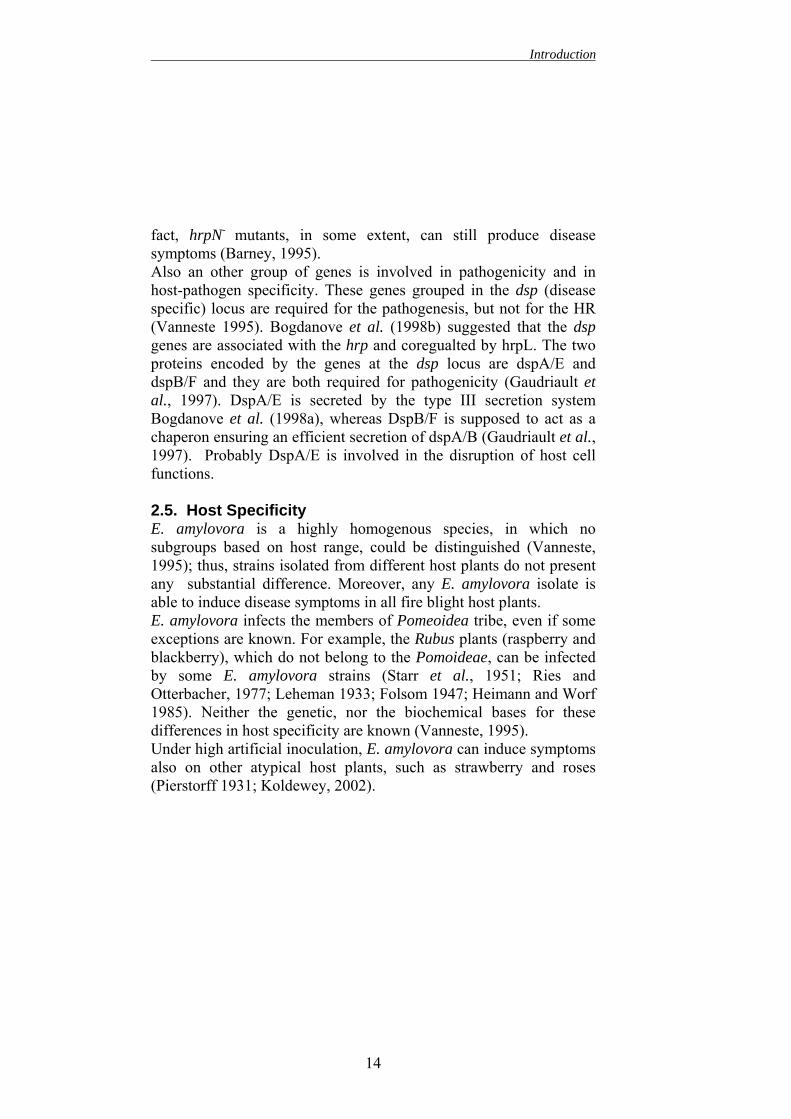
fact, hrpN- mutants, in some extent, can still produce disease symptoms (Barney, 1995). Also an other group of genes is involved in pathogenicity and in host-pathogen specificity. These genes grouped in the dsp (disease specific) locus are required for the pathogenesis, but not for the HR (Vanneste 1995). Bogdanove et al. (1998b) suggested that the dsp genes are associated with the hrp and coregualted by hrpL. The two proteins encoded by the genes at the dsp locus are dspA/E and dspB/F and they are both required for pathogenicity (Gaudriault et al., 1997). DspA/E is secreted by the type III secretion system Bogdanove et al. (1998a), whereas DspB/F is supposed to act as a chaperon ensuring an efficient secretion of dspA/B (Gaudriault et al., 1997). Probably DspA/E is involved in the disruption of host cell functions. 2.5. Host Specificity E. amylovora is a highly homogenous species, in which no subgroups based on host range, could be distinguished (Vanneste, 1995); thus, strains isolated from different host plants do not present any substantial difference. Moreover, any E. amylovora isolate is able to induce disease symptoms in all fire blight host plants. E. amylovora infects the members of Pomeoidea tribe, even if some exceptions are known. For example, the Rubus plants (raspberry and blackberry), which do not belong to the Pomoideae, can be infected by some E. amylovora strains (Starr et al., 1951; Ries and Otterbacher, 1977; Leheman 1933; Folsom 1947; Heimann and Worf 1985). Neither the genetic, nor the biochemical bases for these differences in host specificity are known (Vanneste, 1995). Under high artificial inoculation, E. amylovora can induce symptoms also on other atypical host plants, such as strawberry and roses (Pierstorff 1931; Koldewey, 2002).
14
Introduction
3. DISEASE CYCLE The disease cycle is exemplified in figure 5. The overwintering cankers are the most probable origin of inoculum to start the spring cycle. From them the bacteria spread to open flowers (Thomson, 2000). The bacterium may be disseminated by rain, insects and also birds (Meijneke, 1974; Seidel et al., 1994). The arrival of the pathogen on flowers allows a rapid multiplication and dispersion to other flowers. Also in secondary dissemination, both rain and insect play an important role (Thomson, 2000). Successively, the bacterium can infect plants also through wounds and natural opening on the plant surface.
Figure 5: disease cycle (from Thomson 2000, In: Vanneste (ed.) Fire blight. Thedisease and its causative agent Erwinia amylovora. CABI publishing.
15
Introduction
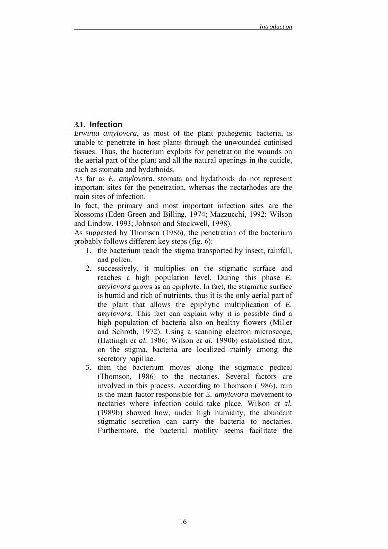
3.1. Infection Erwinia amylovora, as most of the plant pathogenic bacteria, is unable to penetrate in host plants through the unwounded cutinised tissues. Thus, the bacterium exploits for penetration the wounds on the aerial part of the plant and all the natural openings in the cuticle, such as stomata and hydathoids. As far as E. amylovora, stomata and hydathoids do not represent important sites for the penetration, whereas the nectarhodes are the main sites of infection. In fact, the primary and most important infection sites are the blossoms (Eden-Green and Billing, 1974; Mazzucchi, 1992; Wilson and Lindow, 1993; Johnson and Stockwell, 1998). As suggested by Thomson (1986), the penetration of the bacterium probably follows different key steps (fig. 6):
1. the bacterium reach the stigma transported by insect, rainfall, and pollen.
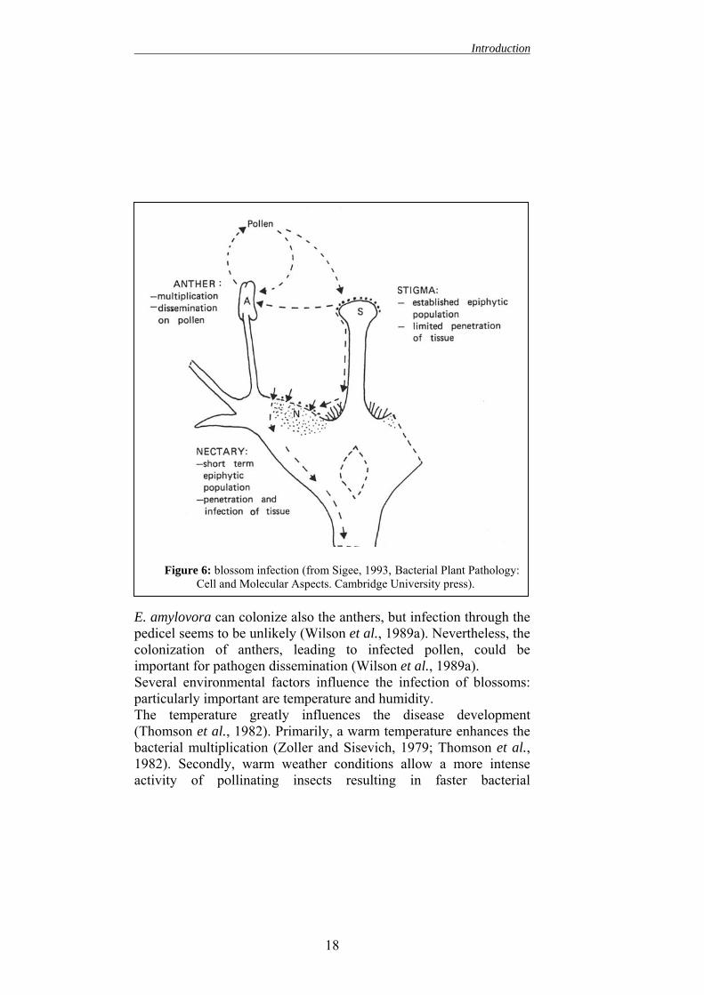
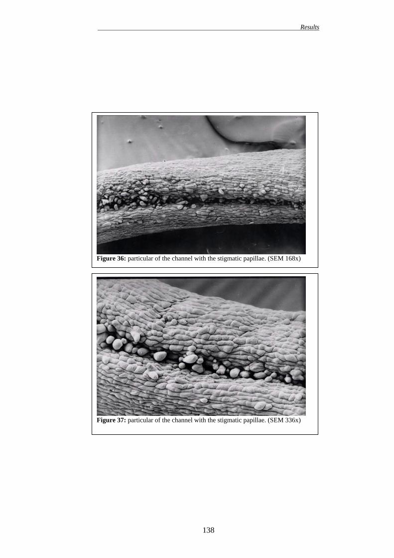
2. successively, it multiplies on the stigmatic surface and reaches a high population level. During this phase E. amylovora grows as an epiphyte. In fact, the stigmatic surface is humid and rich of nutrients, thus it is the only aerial part of the plant that allows the epiphytic multiplication of E. amylovora. This fact can explain why it is possible find a high population of bacteria also on healthy flowers (Miller and Schroth, 1972). Using a scanning electron microscope, (Hattingh et al. 1986; Wilson et al. 1990b) established that, on the stigma, bacteria are localized mainly among the secretory papillae.
3. then the bacterium moves along the stigmatic pedicel (Thomson, 1986) to the nectaries. Several factors are involved in this process. According to Thomson (1986), rain is the main factor responsible for E. amylovora movement to nectaries where infection could take place. Wilson et al. (1989b) showed how, under high humidity, the abundant stigmatic secretion can carry the bacteria to nectaries. Furthermore, the bacterial motility seems facilitate the
16
Introduction
movement along the stigmatic pedicel (Bayot et al., 1986). These Authors demonstrated that flowers sprayed with motile E. amylovora strain showed a higher incidence than the ones sprayed with non-motile mutants. According to Raymundo et al. (1980a), E. amylovora has also a positive chemotaxis to some of the organic acid, such as malic acid and tartaric acid, present in the apple nectar. Only few Authors (Pierstorff, 1931; Rosen 1936) suggested that the bacterium penetrates in the stigma and moves inside it to the nectaries.
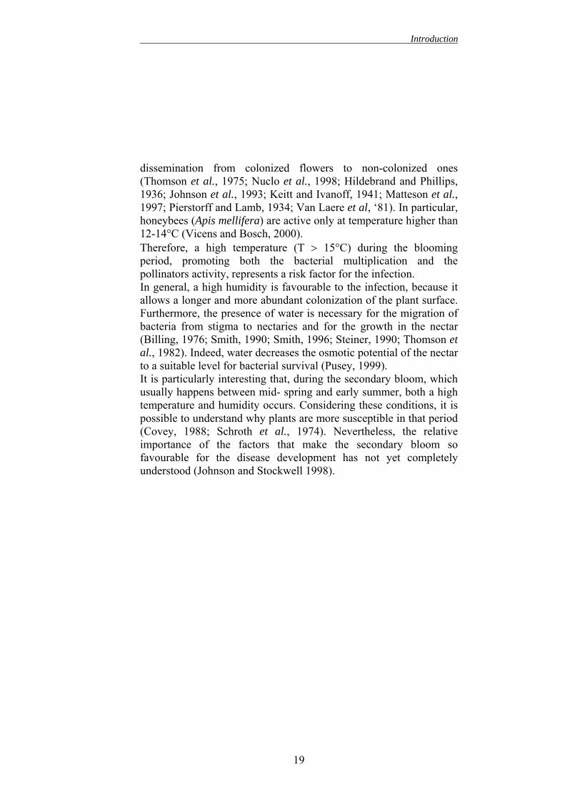
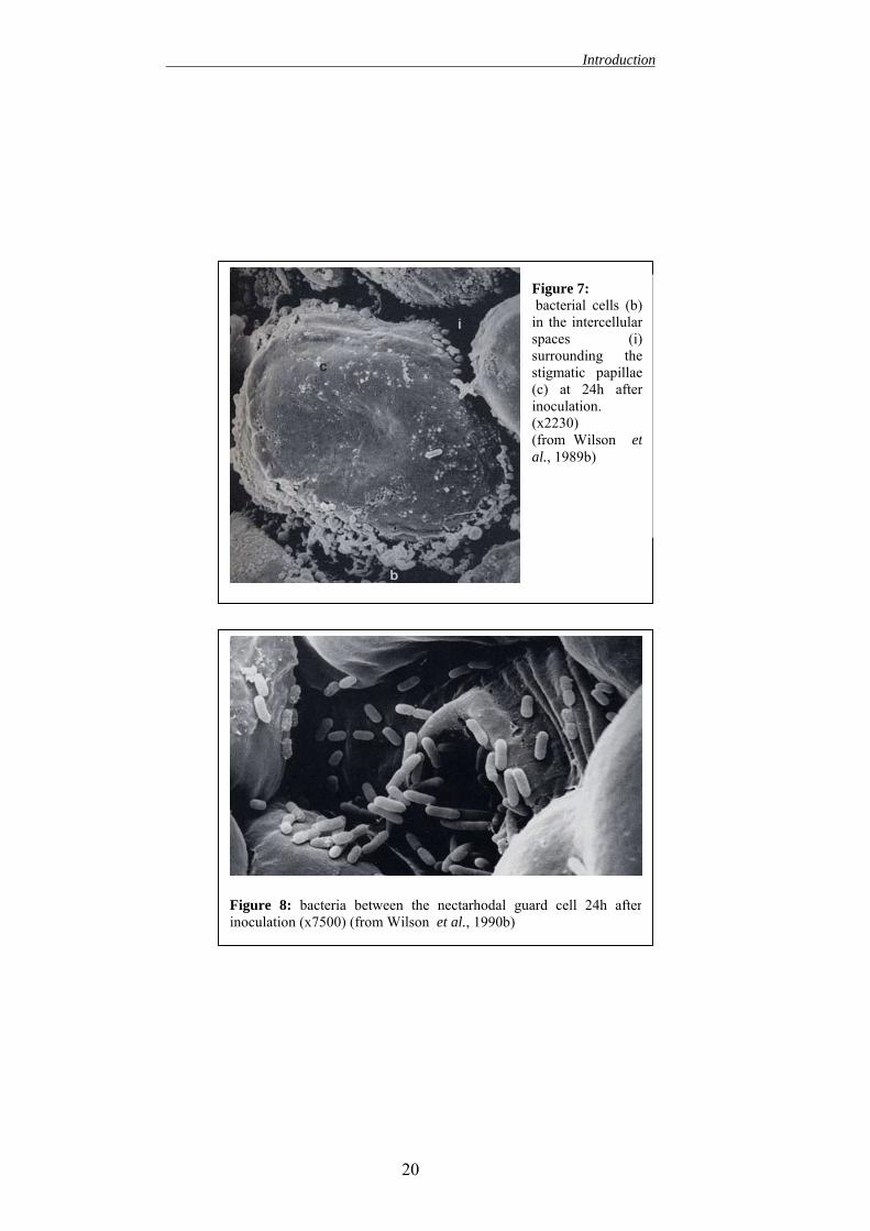
4. Finally, the bacterium penetrates through the nectarhodes (Wilson et al., 1990b). The nectarhodes are stomate-like openings, from where the nectar is secreted. Therefore, they allow direct access to plant internal tissues. Several studies suggest that in the nectaries the readily availability of nutrients increases the E. amylovora growth (Wilson et al., 1989). This further bacterial multiplication in the nectaries seems needed for infection (Pierstorff 1931, Rosen, 1936; Hildedrand, 1937; Thomson, 1986; Wilson et al., 1990b; Campbell et al., 1991; Ivanoff and Kiett, 1941). In pear, the nectaries are full exposed and easily reachable for bacteria, whereas in apple, they are protected by trichomes and thus, they are much less accessible (Rosen, 1936). Finally, even if most of the studies (Baker, 1971; Wilson et al., 1989 a-b, Thomson, 1986) showed that the main penetration sites for E. amylovora are the nectarhodes, different Authors (Rosen, 1936; Hildebrand, 1937; Wilson et al., 19898a) suggested that the pathogen could enter through other parts of the flower. Hence, E. amylovora is probably able to exploit all the weakness present in the tissues of a particular flower (Vanneste, 1995).
Cell and Molecular Aspects. Cambridge University press).
E. amylovora can colonize also the anthers, but infection through the pedicel seems to be unlikely (Wilson et al., 1989a). Nevertheless, the colonization of anthers, leading to infected pollen, could be important for pathogen dissemination (Wilson et al., 1989a). Several environmental factors influence the infection of blossoms: particularly important are temperature and humidity. The temperature greatly influences the disease development (Thomson et al., 1982). Primarily, a warm temperature enhances the bacterial multiplication (Zoller and Sisevich, 1979; Thomson et al., 1982). Secondly, warm weather conditions allow a more intense activity of pollinating insects resulting in faster bacterial
18
Introduction
dissemination from colonized flowers to non-colonized ones (Thomson et al., 1975; Nuclo et al., 1998; Hildebrand and Phillips, 1936; Johnson et al., 1993; Keitt and Ivanoff, 1941; Matteson et al., 1997; Pierstorff and Lamb, 1934; Van Laere et al, ‘81). In particular, honeybees (Apis mellifera) are active only at temperature higher than 12-14°C (Vicens and Bosch, 2000). Therefore, a high temperature (T > 15°C) during the blooming period, promoting both the bacterial multiplication and the pollinators activity, represents a risk factor for the infection. In general, a high humidity is favourable to the infection, because it allows a longer and more abundant colonization of the plant surface. Furthermore, the presence of water is necessary for the migration of bacteria from stigma to nectaries and for the growth in the nectar (Billing, 1976; Smith, 1990; Smith, 1996; Steiner, 1990; Thomson et al., 1982). Indeed, water decreases the osmotic potential of the nectar to a suitable level for bacterial survival (Pusey, 1999). It is particularly interesting that, during the secondary bloom, which usually happens between mid- spring and early summer, both a high temperature and humidity occurs. Considering these conditions, it is possible to understand why plants are more susceptible in that period (Covey, 1988; Schroth et al., 1974). Nevertheless, the relative importance of the factors that make the secondary bloom so favourable for the disease development has not yet completely understood (Johnson and Stockwell 1998).
19
Introduction
Figure 7: bacterial cells (b)in the intercellularspaces (i)surrounding thestigmatic papillae(c) at 24h afterinoculation. (x2230) (from Wilson etal., 1989b)
Figure 8: bacteria between the nectarhodal guard cell 24h afterinoculation (x7500) (from Wilson et al., 1990b)
20
Introduction
3.2. Migration inside the plant tissues and symptoms development How E. amylovora moves inside the plant is still debated. Some Authors (Eden-Green and Billing, 1974; Hokenhull, 1974) suggest a migration through the cortical parenchyma, other (Suhayada 1981; Goodman and White 1981) indicate a movement mainly localized in the xylem. These contrasting results may be explained with a different way of inoculation. When the inoculation is made cutting a leaf of a fresh tissues and infecting the cut edge, bacteria multiply in the xylem (Crosse et al., 1972; Eden-Green and Billing 1974; Hockenhull 1979). On the contrary, when the inoculation is performed applying bacteria to the petioles, they move both in the cortical parenchyma and in the xylem (Huag and Goodman, 1976). During the interaction of a necrogen bacterium with a compatible plant, one of the early events is the electrolytes leakage from the plant tissues (Vanneste, 1995). The electrolytes leakage is a fundamental step of infection process, since it allows the multiplication of the pathogen inside the plant (Youle and Cooper, 1987). In fact, this event releases nutrients from plant cells to the apoplast rendering them available for bacterial multiplication. As far as E. amylovora, both virulent and non-virulent strains induce this phenomenon when infiltrated in apple leaves (Burkowicz and Goodman, 1969). Moreover, both strains cause disorganization of the cellular membrane and organelles of apple leaf cells. Harpin seems involved in the mechanism that leads to the electrolytes leakage, even if the way in which harpin acts is not yet completely understood. Non-capsulated strains are still able to induce electrolytes leakage (Hignett and Roberts, 1985), but they don’t kill the plant cells (Mansfiled et al., 1995). Thus, the cell death does not seem a consequence of the electrolytes leakage. According to Hignett and Roberts (1985), the high virulence of some strains could be explained with a more intense electrolytes leakage, which leads to a more rapid increase of bacterial population. The symptoms development, which occurs only when the bacterial population reach a high level, is consequent to the infection
21
Introduction
(Goodman et al., 1990, Klement et al., 1966). The macroscopical symptoms are mainly due to cell collapse and tissue decompartmentalization (Eden-Green and Billing, 1974). In particular, the wilting of the infected plant tissues may be an effect of two different mechanisms. On one side, amylovoran plugs the xylem vessels disrupting the water flux in the plant; on the other side, harpin induces electrolytes leakage by altering cell membrane integrity (Sjulin and Beer, 1978). An other peculiar symptom of fire blight is the production of ooze on the surface of infected plant parts. The exudate is constituted by bacterial cell embedded with EPS. EPS role in symptom development as been previously discussed.
1. CHEMICAL CONTROL The chemical control acts both eliminating or inactivating the plant pathogenic bacteria before the penetration in the host tissues and rendering the plant surface unfavourable for the establishment of new infections (Psallidas and Tsiantos, 2000). As far as fire blight, the control strategies have to achieve the following results: 1. destroy the primary source of inoculum, e.g. the overwintering
cankers and the alternative hosts 2. protect the most susceptible parts of the plant, such as the
blossoms, stomata, nectarhodes, lenticels and wounds. Thus, to obtain the maximum of protection the plant should be treated in three different periods (Psallidas and Tsiantos, 2000): when the plant is dormant, during the blooming and, finally, during the vegetative growth. Since risk of phytotoxicity is low during dormancy, it is recommended the use of high concentrations of chemicals to enhance their activity and persistence (Psallidas and Tsiantos, 2000). On the other hand, during the blooming period, for the chemical applications lowest effective concentrations should be used, in fact buds, blossoms and young leaves are particularly susceptible to phytotoxicity. Finally, chemical applications during summer have the aim to prevent both infection of the wounded plant parts and secondary blossoms, which are frequent in some pear variety. Almost all the bactericides available against fire blight are preventive and not curative (Psallidas and Tsiantos, 2000). Few exceptions are known, streptomycin, for example, seems to have, in a certain extent, a curative action. Moreover, almost none of them, excluded some
23
Introduction
exceptions, such as fosetyl-Al and BTH, can penetrate inside the plant tissues and thus have a systemic effect. Thus, they are effective to prevent fire blight infection, but they are inactive when the disease is already established. Nevertheless, some chemicals, without a direct bactericidal activity can have systemic action. Some of them, such as benzothiadiazole (BTH) or harpin, belong to the class of SAR inducers, others, such Prohexadione-Ca and Trinexapac-ethyl, are biregulators characterized by the ability to increase plant resistance against pathogens. The main problem of these compounds is that they should be applied several days before the risk period to allow the plant to build up its defences.
1.1. Efficacy of chemical control Today, since none of the antibacterial treatments is at the same time totally effective, environmental safe, non-phytotoxic and systemic, it is not yet available a completely reliable chemical method to control fire blight (Psallidas and Tsiantos, 2000). Moreover, the chemical treatments present other weak points. In fact, the bactericides should be sprayed before the inoculum reaches the susceptible plant. Then, they should be effective during all the period in which the pathogen is present. Thus they have to be sprayed several times during the year according to a reliable prediction model. Moreover, the efficacy of a treatment is influenced by several factors:
- The environmental conditions, such as high humidity, temperature, rain or hail, can drastically decrease the efficacy of the chemicals used.
- The time of spraying, that can determine if a chemical will be effective or not.
- The method of application that influences the covertures of all the susceptible plant parts.
- The physiological state of the host plant at the time of treatment
- The plant species and cultivars
24
Introduction
- The inoculum consistence: higher is the pathogen population lesser is the effectiveness of the chemical applied (Koistra and de Gruyter, 1984; Tsiantos and Psallidas, 1996a).
Since all these factors are involved in determining the efficacy of a chemicals, the efficiency of a certain compound results very difficult to assess, in field conditions experiments. Finally, to increase the efficacy of chemical treatments, it is necessary combine them with other control methods (for instance cultural measures, proper irrigation, fertilization and pruning) in an integrated management programme (Psallidas and Tsiantos, 2000).
1.2. Current methodologies of chemical control According to Van der Zwet and Keil (1979), the chemical tested against fire blight may be grouped in 4 categories:
1. Copper compounds 2. Antibiotics 3. Carbamates 4. Other compounds
More recently, different kinds of compounds have been developed. Among them, SAR inducers and bioregulators seem the more promising. This dissertation will focus on these compounds in the chapters “Growth Regulators” and “SAR Inducers”. Since their low efficacy, the carbamates will not be treated in this dissertation.
1.3. Copper compounds The active ingredient of these compounds is the copper ion, which is extremely toxic both to bacterial cell and to all plant life (Psallidas and Tsiantos, 2000). Since its high toxicity, copper solutions can not be used per se as a foliar pesticide. Thus, the copper is usually sprayed either in an insoluble form, such as copper hydroxide, copper oxychloride or cuprous oxide, or as CuSO4 mixed with lime Ca (OH)2 to form the Bordeaux mixture. The effectiveness of the Bordeaux mixture as a bactericide is greatly affected by the ingredients proportion used. In particular, the composition of the dry
25
Introduction
deposit, formed on leaf surface after treatment, seems the main factor that determines the efficacy of the mixture (Gremlyn, 1990). Some of the major disadvantages of the Bordeaux mixture are the difficulty in the preparation and the residual phytoxicity towards some host plants, such as pears (Martin and Woodcock, 1983). On the other hand, the insoluble copper compounds have the advantage to be easier to prepare and to apply than the Bordeaux mixture. The formulations, to be effective, have to release the toxic copper ion. According to Psallidas and Tsiantos (2000), the formation of soluble copper ion from an insoluble structure could be due to the reaction with: - CO2 or ammonium salt dissolved in the rainwater - Microbial secretion on the plant surface - Plant secretion form healthy or wounded surface The copper formulations commercially available and most frequently used are: ammoniacal copper sulphate, copper hydroxide, copper oxide and copper oxychloride. All these copper formulations were more effective than Bordeaux mixture in controlling fire blight, even if they resulted phytotoxic for blossoms and young leaves at the dosages recommended for pathogen control (Psallidas and Tsiantos, 2000). Finally, even if resistance to copper has not been yet detected, it is widely spread among other phytopathogenic bacteria (Cooksey, 1990).
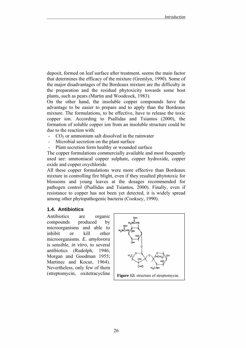
1.4. Antibiotics Antibiotics are organic compounds produced by microorganisms and able to inhibit or kill other microorganisms. E. amylovora is sensible, in vitro, to several antibiotics (Rudolph, 1946; Morgan and Goodman 1955; Martinec and Kocur, 1964). Nevertheless, only few of them (streptomycin, oxitetracycline
2
Figure 12: structure of streptomycin.
6
Introduction
and kasugamycin) are valuable for practical field application (Ark, 1949). Among them, streptomycin is the most effective against fire blight. Streptomycin was initially isolated by Schatz et al., (1944) from Sterptomyces griseus. Streptomycin belongs to the class of aminoglycoside antibacterial compounds. Also amikacin, gentamicin, tobramycin, kanamycin and netilmicin belong to the same class of compounds. These compounds inhibit the protein synthesis in bacterial cells by affecting ribosome. In fact, all the aminoglycosides irreversibly bind to specific proteins of the ribosomal 30S subunit. Thus they interfere with the initiation complex and they cause the misreading of mRNA, which produces, by insertion of incorrect amino acids into the polypeptide, nonfunctional or toxic peptides (Gottliebe and Show, 1970). Finally they break up polysomes into nonfunctional monosomes. Streptomycin is also toxic to the plants: it inhibits the chlorophyll synthesis, thus high concentration of this antibiotic may lead to chlorosis and death of the plant. Streptomycin is usually foliar applied. In fact, even if it is easily up taken by the roots, its concentration inside the plant tissues is too low to be effective against bacterial pathogen (Anderson and Nienow, 1947). Several experimental studies proved the efficacy of streptomycin in controlling fire blight (Heuberger and Poulos, 1953; Ark and Scott, 1954; van der Zwet and Keil, 1979). According to Van der Zewt and Keil (1979), a concentration of 100-150 ppm applied 3-5 times during the blooming period is enough to achieve good results. However, the effect of streptomycin is not long lasting: the plants sprayed with the antibiotic result protected from fire blight for 4 days, whereas injured plants are protected only within 6h from application (Van der Zwet and Keil, 1972). An other problem connected with streptomycin is the built up of resistance among sensitive bacteria. For this reason the use of streptomycin is prohibited in several countries. Streptomycin resistant strains of E. amylovora were firstly reported in California in 1972 (Miller and Schroth 1972; Moller et al., 1972). After these first
27
Introduction
reports E. amylovora streptomycin resistant strains have been found in several countries (Coyer and Convey, 1975; Chiou and Jones, 1991; El-Goorani et al., 1989; Thomson et al., 1993). Bacteria can avoid the lethal effect of streptomycin in three different ways: altering the ribosomal proteins, producing enzymes able to modify and inactivate streptomycin or preventing the streptomycin access to the target site (Amyes and Gemmell, 1992). In E. amylovora, resistance to streptomycin could be due both to a mutation on the chromosomal DNA and to an acquired plasmid or transposon. As far as the mutation of the chromosomal DNA, since streptomycin binds to a single site on the ribosomal 30S subunit, resistance is conferred by a single base pair mutation at codon 43 of the rpsL gene, which results in a lysine to arginine conversion in ribosomal protein S12 (Jones et al., 1996). Trasposon Tn5393, which carries the gene strA and strB and, therefore, confers streptomycin resistance, is widely distributed among gram-negative bacteria isolated from apple orchards (Minsavage et al., 1990; Jones et al., 1991; Norelli et al., 1991; Burr et al., 1993). Moreover, Chiou and Jones (1991) demonstrated that this plasmid has a high frequency of transfer between the donor and recipient strains. Thus, streptomycin resistance among phytopatogenic gram-negative bacteria is an ever increasing problem that reduces the possibility to use this antibiotic.
1.5. Other compounds Flumequin. It is a non-antibiotic, non-sulphamide bactericide active against both gram-positive and gram-negative bacteria. Its chemical name is (1H-5H)-dihydro-6,7-fluoro-9-methyl-5oxo-1-benzo (I,j)-quinolizin carboxilic acid-2, and it is commercially known as Fire StopTM and FructilTM. It interferes with the DNA gyrase, thus blocking DNA replication (Psallidas and Tsiantos, 2000). This chemical has given promising results and seems having no phytotoxic effect (Psallidas and Tsiantos, 2000). Oxolinic acid. It is a syntethic bactericide belonging to the family of quinoline. Its chemical name is 5-ethyl-5,8-dihydro-8-oxo-(1,3)-
28
Introduction
dioxolo-(4,5g) quinoline-7-carboxilic acid and it is commercially known as StarnerTM. Its mode of action has not been yet understood completely. It does not seem to be phytotoxic. Some results indicate that this compound can have also a curative action against fire blight (Tsiantos and Psallidas, 1993 a, b, 1996b). Fosetyl-aluminium (fosetyl-Al). It is a systemic fungicide commercially known as AllietteTM. This compounds seems to act by inducing plant resistance more than with a direct antimicrobial effect (Farih et al., 1981; Guest, 1984). The plant can absorb this compound both by leaves and roots and, successively, it moves systemically inside the plant enhancing the defence mechanisms (Guest, 1986). More recently it has been hypothesised that fosetyl-Al have a double mechanism: on one hand, it act directly against microrganisms slowing down their growth, on the other hand, it stimulates the plant defences allowing the overwhelming of the pathogen (Fenn and Goffey 1984; Guest 1986, Chase 1993). However, AllietteTM does not seem effective in controlling fire blight (Norelli and Aldwinckle, 1993; Clarke et al., 1993; Tsiantons and Psallidas, 1993 a, b, 1996a) even if the results from different laboratories are contrasting. Also plant extracts and essential oils have been used against fire blight. Some of them have a direct bactericidal effect (Mosch et al., 1989; Vanneste 1996). Whereas others, such the extract from leaves of Reynutria sachalinensis, Hedera helix, Viscum album and Alchemilla vulgaris, could induce plant resistance mechanisms (Mosch et al., 1993 and 1996). Bioregulators and SAR inducers will be treated in the in the relative chapters.
2. BIOLOGICAL CONTROL The biological control is a manipulation of the biotic community with the aim to influence the pathogen population using other non-pathogenic organisms. The manipulation of the biotic community is
29
Introduction
an extremely complicate process in which a high number of variables is involved. Thus, the results are often difficult to predict. The objective of the biological control is not to eradicate the pathogen population, but to reduce its population under the risk threshold. Hence, it has the aim to prevent the host infection by the pathogen, but, after infection occurred, it usually has no curative efficacy. Biological control agents are effective and they can provide for the deficiencies of reliable chemical control methods. Furthermore, they are harmless to human beings and animals, and, in some cases, they could be cheaper than pesticides and highly effective throughout the crop growth period (Nakkeeran et al., 2002). The microbial antagonists act through antibiosis, secretion of volatile toxic metabolites, mycolytic enzymes production, parasitism and competition for space and nutrients. The mode of action of biological control agents can be grouped in three main categories:
- competition for space and nutrients - antibiosis - parasitism/ predation
As far as fire blight, production of antibiotics and competitive exclusion for sites and nutrients are considered to be the principal mechanisms used by bacterial antagonists to control E. amylovora (Wilson and Lindow, 1993; Vanneste 1996; Johnson and Stockwell, 2000).
2.1. Competition All the ecological requirements of a species constitute its ecological niche. When two species share overlapping ecological niches, they may be forced into competition for the common resources of those niches. More deeply the two niches overlap, more intense is the competition. This interspecific competition is a density-dependent check on the growth of one or both populations.
30
Introduction
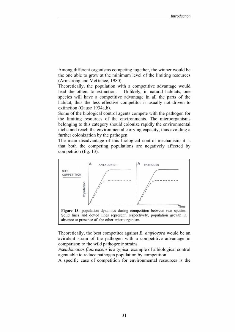
Among different organisms competing together, the winner would be the one able to grow at the minimum level of the limiting resources (Armstrong and McGehee, 1980). Theoretically, the population with a competitive advantage would lead the others to extinction. Unlikely, in natural habitats, one species will have a competitive advantage in all the parts of the habitat, thus the less effective competitor is usually not driven to extinction (Gause 1934a,b). Some of the biological control agents compete with the pathogen for the limiting resources of the environments. The microorganisms belonging to this category should colonize rapidly the environmental niche and reach the environmental carrying capacity, thus avoiding a further colonization by the pathogen. The main disadvantage of this biological control mechanism, it is that both the competing populations are negatively affected by competition (fig. 13).
TacPaA
Figure 13: population dynamics during competition between two species.Solid lines and dotted lines represent, respectively, population growth inabsence or presence of the other microorganism.
heoretically, the best competitor against E. amylovora would be an virulent strain of the pathogen with a competitive advantage in omparison to the wild pathogenic strains. seudomonas fluorescens is a typical example of a biological control gent able to reduce pathogen population by competition. specific case of competition for environmental resources is the
31
Introduction
competition for iron. The competition for iron could be the mode of action of some biological control agents. Thus the production of high effective siderophores may represent a competitive advantage.
2.2. Antibiosis Some biological control agents, such as Pantoea agglomerans, produce antibiotics able to inhibit or to kill E. amylovora. A particular kind of antibiosis occurs when an antagonistic organism does not produce any kind of toxic compounds by itself, but it modifies the plant environment thus leading to the production and release of toxic substances. For example, the antagonist may trigger out the production of phytoalexins or other plant defence compounds. Also in this case, the antagonist, to successfully inhibit the pathogen, need to share the same habitat. In fact, the antibiotic compounds have usually a low diffusibility in the environment and they can be easily degraded. In comparison to competition, the main advantage of
antibiosis is that the antagonist population is not negatively affected by the presence of the pathogen (fig. 14).
Figure 14: population dynamics during competition between a antibiotic-producing antagonist and a plant pathogenic microorganism. Solid lines anddotted lines represent, respectively, population growth in absence or presenceof the other microorganism.
32
Introduction
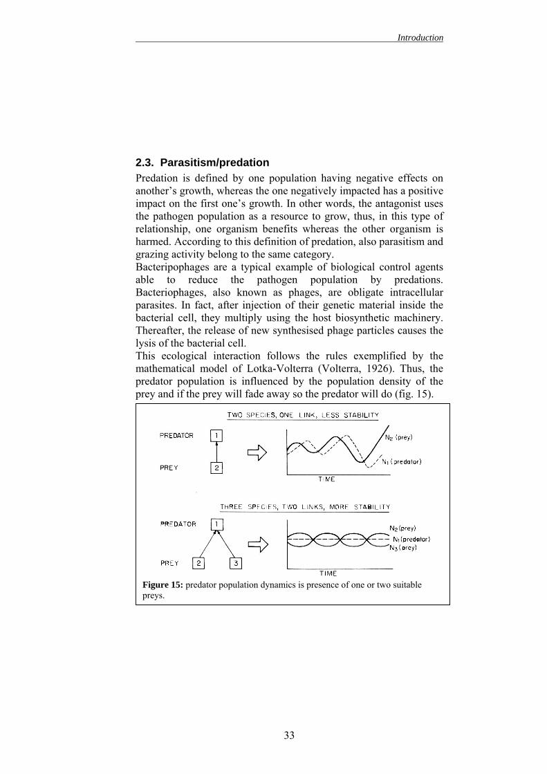
2.3. Parasitism/predation Predation is defined by one population having negative effects on another’s growth, whereas the one negatively impacted has a positive impact on the first one’s growth. In other words, the antagonist uses the pathogen population as a resource to grow, thus, in this type of relationship, one organism benefits whereas the other organism is harmed. According to this definition of predation, also parasitism and grazing activity belong to the same category. Bacteripophages are a typical example of biological control agents able to reduce the pathogen population by predations. Bacteriophages, also known as phages, are obligate intracellular parasites. In fact, after injection of their genetic material inside the bacterial cell, they multiply using the host biosynthetic machinery. Thereafter, the release of new synthesised phage particles causes the lysis of the bacterial cell. This ecological interaction follows the rules exemplified by the mathematical model of Lotka-Volterra (Volterra, 1926). Thus, the predator population is influenced by the population density of the prey and if the prey will fade away so the predator will do (fig. 15).
Figure 15: predator population dynamics is presence of one or two suitable preys.
33
Introduction
2.4. Biological control of fire blight Several kinds of antagonists have been evaluated for fire blight control: gram-negative bacteria, non virulent strains of E. amylovora (Tharaud et al., 1997), yeasts (Mercier and Lindow, 1996), gram-positive bacteria (Jock et al., 2002) and bacteriophages specific to E. amylovora (Ritchie and Klos, 1977; Palmer et al., 1997). The biological control to be effective has to interfere with the infection process. As suggested by Maxsons-Stein (2002), the best strategy for control fire blight is to prevent infection during the blooming period before wind-driven rain and pollinating insects spread the disease throughout the orchard. The first step of the infection is the pathogen multiplication on the stigma. Biological control aims to prevent this key step of pathogenesis. In fact, an effective interaction on stigma and hypanthial surface prevents the floral infection and thus the further damage to the plant. Therefore a better understanding of the biology of the pathogen and of its behaviour on the stigma is necessary to optimise biological control methods. Moreover, suppression of floral infections reduces the inoculum of E. amylovora available for other phase and cycle of disease, including shoot infections during the same season and floral infections in the following seasons (Johnson and Stockwell, 2000).
2.5. Biological interaction on stigmatic surface The stigmatic surface is the site where the biological control agents must interact with the pathogen to successfully reduce the disease incidence (Hattingh et al., 1986; Thomson 1986; Wilson et al., 1989b, Vanneste 1995). The stigmatic surface is characterized by particular epidermal cells known as stigmatic papillae. Among these papillae there are large intercellular spaces where bacteria can reside (Johnson and Stockwell, 2000). Since the stigma is rich of nourishing exudates, it is one of the few parts of the plant that allows epiphytical growth of E. amylovora (Wilson et al., 1989b). At the first opening of blossoms, stigmas are almost sterile (McLaughlin et al., 1992). Thereafter, the exposed stigmas are soon colonised by several microorganisms.
34
Introduction
The biological control of fire blight, reducing E. amylovora population on the stigma reaches two main aims. The first is the decrease of pathogen population on the stigma: less is the pathogen population, less the probability to successfully infect the flower (Hirano and Upper, 1983; Johnson et al., 1993b) Secondly, it causes a reduction of the inoculum available to be spread from a flower to an other by rain (van der Zwet and Keil, 1979) and pollinating insects (Pierstorff and Lamb, 1934; Hildebrand and Phillips, 1936; Keit and Ivanoff 1941; Van Laere et al., 1981). A requirement for a successful biological control is the establishment of a large population of antagonistic organisms on the stigma before its colonization by E. amylovora (Wilson et al., 1992; Johnson et al., 1993b; Wilson and Lindow, 1993). A second requirement is the colonization of most of the flowers in orchards by a large population of the antagonistic organisms (Johnson et al., 1993b; Lindow et al., 1996). The biological control agent population should almost reach the carrying capacity of the apple and pear stigma that has been estimated to be around 106 cfu/ml (Wilson et al., 1992; Wilson and Lindow, 1993). Thus to be effective the antagonistic population on stigma should range between 105 -106 cfu/ml. During the second step of infection, after the stigmatic multiplication, the pathogen, washed by rain or heavy dew, reaches the nectaries trough which it penetrates. Therefore, to enhance biological control effectiveness the microbial antagonist should be able to rapidly colonise also the nectar cup surface. Fire blight is a good candidate for biological control because the bacterial antagonists need to persist on the nutrient-rich, stigmatic surface for only about a week to suppress blossom infection effectively (Johnson and Stockwell, 2000).
2.6.1. Pseudomonas fluorescens A506 PfA506 is available on the USA market since 1996 (BlightBanTM A506, Plant Health Technologies; Boise, Idaho). The application of this bacterium in orchard could results in a 40-60% reduction in incidence of fire blight on blossoms (Johnson et al. 1993b; Lindow et al., 1996). Thus, its efficacy is comparable with that obtained using chemical agents. Furthermore, its application can suppress the severity of frost injuries caused by ice nucleation-active strains of Pseudomonas syringae (Lindow et al., 1996; Johnson and Stockwell, 2000). This bacterium is a good colonizer of apple and pear stigma especially during early spring when the temperature ranges between 10-12° C (Johnson et al., 1993b; Stockwell et al., 1996a). Under field conditions, the population established on flowers is usually rather high and it is around 105-106 cfu/blossom, in addition, the percentage of colonized flowers ranges between the 50-70% of the treated blossoms (Stockwell et al., 1992, 1998). The remaining
36
Introduction
flowers are not colonized by the antagonistic bacteria and thus they remain unprotected and vulnerable to infection. As already mentioned, PfA506 antagonizes E. amylovora simply by competition for sites and nutrients (Hattings et al., 1986; Vanneste 1996; Wilson et al., 1992; Wilson and Lindow, 1993). In fact, PfA506 can inhibit E. amylovora growth on the stigma only if it is inoculated several hours (24-72h) before the pathogen, whereas it is ineffective if co-inoculated with the pathogen (Wilson and Lindow, 1993). PfA506 population exceeds, 70-80 hours after inoculation, the carrying capacity of the stigma (106 cfu). Thus PfA506 suppress the pathogen by a pre-emptive sequestration of sites and resources on the stigma (Wilson and Lindow, 1993). Moreover, PfA506 colonizes also the nectaries, thus reducing the probability of a successful infection by E. amylovora (Wilson and Lindow, 1993). PfA506 does not seem to produce, in vivo, any kind of antibiotic or toxic compounds. Nevertheless, other mechanisms of inhibition have been proposed for P. fluorescens. Among them, the production of siderophore and the stimulation of plant defence are particularly interesting. Wilson and Lindow (1993) observed that pear blossoms (cv. Comice) reddened after treatment with P. fluoresecens A506. According to these Authors, the reddening could be due to the production by P. fluorescens of a β-glucosidase, which catalyses the transformation of arbutin in new phenolic compounds able to inhibit the pathogen. In addition, when P. fluoresecens A506 is applied on plant kept in a grow chamber, the bacteria reduce the nectar secretion (Wilson and Lindow, 1993). This reduction in nectar secretion may successively increase its sugar concentration thus rendering its osmotic potential to high for E. amylovora survival (Wilson and Lindow, 1993). In this case, the application of the antagonist reduces the fire blight incidence suppressing E. amylovora just before the penetration through the nectarhodes (Wilson and Lindow, 1993). In spite of all these possible influences on plant metabolism, the treatment with P. fluorescens A506 does not affect either fruit production or flower attractiveness for pollinating insects (Wilson and Lindow, 1993).
37
Introduction
2.6.2. Pantoea agglomerans (Gavini et al., 1989) - formerly Erwinia herbicola (Löhnis, 1911) Also different strains of Pantoea agglomerans have been widely investigated as possible biological control agents against fire blight. In particular, the strain EhC9-1 (Ishimaru et al., 1988) has been tested for long time in USA. It is an excellent colonizer of apple and pear stigma even more effective than P. fluorescens. Several studies demonstrated that the application of EhC9-1 could result in a 50-80% reduction in fire blight incidence on blossoms (Johnson et al., 1993b). Therefore, the protective level reached with EhC9-1 is comparable to that obtained with streptomycin and usually exceeds the level of control provided by Pf A506 (Johnson and Stockwell, 2000). Similarly to PfA506, EhC9-1 population on blossoms reach 104-106 cfu (Stockwell et al., 1992, 1996a, 1998; Johnson et al. 1993b) and the 40-70% of treated blossom have a detectable EhC9-1 population (Stockwell et al., 1992, 1998). Several other strains of P. agglomerans, such as Eh252 (Vanneste et al., 1992), Eh318 (Wright and Beer, 1996), Eh112Y (Wodzinski et al., 1994), Eh1087 (Kearns and Hale, 1996), EhHl9N13 (Wilson et al., 1990a) and Eh325 (Pusey, 1997), have been tested with promising results as biological control agents against fire blight. Moreover, P10C, a P. agglomerans strain, has been register for use in New Zealand (Vanneste et al., 2002b). The efficacy of all P. agglomerans strains is related to production of antibiotics able to inhibit E. amylovora. For example Eh252 produces a putative microcin (Vanneste and et al., 2002a), whereas EhC9-1 produces two different β-lactams antibiotics called herbicolin O and herbicolin I (Ishimaru et al., 1988). Herbicolin O is effective against a wide range of bacterial genera, whereas, herbicolin I is toxic to E. amylovora, Bacillus cereus and Staphylococcus aureus (Ishimaru et al., 1988). Even if antibiotic production is important for the inhibition of the pathogen multiplication, it is not, per se, sufficient to completely antagonize E. amylovora: competition for site and nutrient also
38
Introduction
contributes to the overall effectiveness of biological control (Johnson and Stockwell, 2000).
2.6.3. Bacterial mixtures To maximize the effectiveness of fire blight biological control, the use of bacterial mixtures of antagonists have been investigated in several laboratories (Stockwell et al., 1992; Vanneste and Yu, 1996; Nuclo et al., 1998). The aims of these mixtures are:
- combine different mechanisms of biocontrol (antibiosis, exclusive competition…)
- expand the range of environmental conditions suitable for multiplication of the antagonists
- enhance the total antagonistic population - increase the number and type of ecological niches colonized
by antagonists Using a mixture that follows these criteria it is, theoretically, possible achieve a large and stable community of bacterial antagonists on the plant surface. A mixture of EhC9-1 and PfA506 responds to the criteria previously listed. In fact, these two bacteria have different and complementary mechanisms of antagonisms (Ishimaru et al., 1988; Wilson and Lindow, 1993), they have different maximal temperatures for growth in vitro (27°C for PfA506 and 37°C for EhC9-1), they differ in tolerance to desiccation stress and UV radiation1 and in ability to utilize various carbon and nitrogen sources (Wilson and Lindow, 1994). Moreover, PfA506 used alone is a good colonizer of apple and pears flower especially during period characterized by frequent rainfall and moderate daytime temperature (10-12°C). Whereas, during period with limited rainfall and warm daytime temperature (16-22°C), EhC9-1 colonizes the majority of flowers. For these reasons, the application of a mixture of these two bacteria might result in a constant coverage of the plant surface almost independently of the climatic conditions. When applied as a mixture
1 1 V.O. Stockwell unpublished data reported in Johnson and Stockwell, 2000
39
Introduction
the percentage of treated flowers colonized by at lest one of the antagonistic bacteria ranges between the 80-90% (Stockwell et al., 1992). Thus the application of a mixture of bacterial antagonists reaches the following purposes: a higher and well-established population of antagonistic bacteria on flower, and a higher percentage of flowers colonized by biocontrol agent (Johnson and Stockwell, 2000). Nevertheless, the application of a mixture of antagonistic bacteria does not achieve a greater reduction of fire blight incidence, than the application of a single biocontrol agent (Vanneste and Yu, 1996). Several hypotheses have been proposed to explain this unexpected result that has not yet completely understood. It is possible that the interspecific competition between the bacteria applied as biocontrol agents vanishes any possible synergic effect. For example the antibiotic production typical of P. agglomerans strains could negatively affect PfA506 population, whereas, the latter bacterium could reduce P. agglomerans growth by pre-emptive sequestration of mutually required growth-limiting resources.
2.7. Selection of a biological control agent A biological control agent has to responds to several criteria: it should be a good epiphyte in a wide range of environmental conditions, it should share the same epiphytic niche of the pathogen, and, of course, it should be harmless for plants, animals and human. Several screening tests, such as media-based test and immature pear fruits test, have been used to assess the effectiveness of a possible bacterial antagonist (Wrather et al., 1973; Beer and Rundle 1983; Isenbeck and Schultz, 1985; Ishimaru et al., 1988; Nicholson et al., 1990; Vanneste et al., 1992; Kearn and Mahanty, 1993). Unfortunately, these tests are mainly suitable for antibiotic producing bacteria, whereas they might underestimate the efficacy of bacteria, such as PfA506, which inhibits E. amylovora by competition (Mercier and Lindow, 1996). Thus, testing the effectiveness of antagonistic bacteria directly on blossoms is considered by most researchers a more reliable method (Andrews, 1985; Mercier and Lindow, 1996; Pusey, 1997).
40
Introduction
To verify the efficacy of antagonistic strains, detached flowers with the pedicel placed into vials containing a 10-25% sucrose solution (Pusey, 1997) can be used or alternatively it is possible use detached flowering branches. In any case the blossoms used for the test have to be closed until the experiment is performed, thus the stigma remains almost sterile till the experimental inoculation. Finally, the effectiveness of a possible biological control agent should be evaluated in orchard conditions. In fact, plants grown in greenhouse, or in controlled environment, usually support a higher epiphytic population than plants grown under field conditions (O’Brien and Lindow, 1989; Loper and Lindow, 1993; Beattie and Lindow, 1994). In fact, under field conditions, several factors, such as physiology and growth of plants, weather conditions and competition with indigenous microorganisms, may influence the establishment of a consistent population of the antagonist and therefore the efficacy of biological control.
2.8. Delivery of bacterial antagonists The populations of bacterial antagonists artificially introduced in orchards are relatively resilient (Lindow et al., 1996; Stockwell et al., 1996a). Moreover, they can partially self-sustain by spreading naturally from blossom to blossom (Johnson et al., 1993b; Lindow et al., 1996; Nuclo et al., 1998). The initial establishment of a consistent and widely spread population of bacterial antagonists is the key factor that determines the efficacy of further biological control. Among the several factors, which influence the initial establishment of artificially introduced bacterial populations, the methods of preparation and application of inoculum, the insect activity, the bloom stage and the daytime temperature play a primary role (Johnson and Stockwell, 2000). As far as inoculum preparation, Stockwell et al. (1998) demonstrated that inoculum consisting of cells of PfA506 and EhC9-1 that had been freeze-dried and resuspended in water established epiphytic population in blossoms more consistently (large populations on a
41
Introduction
greater proportion of flowers) than suspension of cells prepared from cultures actively growing on artificial medium. A possible explanation of this observation has been proposed by Stockwell et al. (1998). They suggested that the freeze-drying process, exposing bacteria to freezing and desiccation stresses, selects a subset of cells with a higher fitness to the unreceptive environment of plant surface. For delivery of bacterial antagonists, different methods have been proposed. The bacteria can be sprayed likewise chemical treatments, but they can be also dispersed by natural vectors, such as pollinating insects. Indeed, the dissemination by honeybees has been proposed by different laboratories as a method to implement the biological control in commercial orchards (Thomson et al., 1992; Johnson et al., 1993a; Vanneste, 1996). For this purpose a pollen insert, fulfilled with the bacterial inoculum, is placed at the entrance of the beehive (fig. 16).
Tal
Figure 16: cross section of a pollen insert (from Vanneste. (1996). Biocontrol News and Information 17: 67N-78N).
hus all the insects became contaminated by the bacterial inoculum s they exit from the beehive. The inoculum can consist in either yophilised bacterial cells (Johnson et al., 1993a) or pollen grains
42
Introduction
coated with the biological control agent (Thomson et al., 1992). In the latter case, the dissemination of pollen could also enhances the flower pollination and thus the fruit production in the orchard. The delivery of antagonistic bacteria by honeybees could improve the secondary dissemination of bacteria from colonized blossoms to non-colonized ones, thus allowing the establishment of a consistent population on a great proportion of flowers (Nuclo et al., 1998). Nevertheless, the orchard sprayers are more effective and reliable, for the initial establishment of bacterial antagonists, than dissemination via pollinator insects (Johnson et al., 1993a). In fact, the dissemination of bacterial antagonists by honeybees is influenced by several environmental factors, such as temperature and presence of other flowering plants; these factors introduce a high variability in the pre-emptive establishment of biological control agents on flowers. The time of application of bacterial antagonists is also important. The application at early bloom provides more time for the bacteria to grow to large populations. Conversely it protects only the small proportion of early-opened blossoms. Thus, a multiple application, at 25% and 90% bloom, covering both early-and late-opening blossoms, seems to provide the best protective effect (Johnson and Stockwell, 2000).
2.9. Contemporary use of bacterial antagonists and antibiotics According to Lindow et al. (1996) and Stockwell et al. (1996a), biological control agents, to be effective, should be integrated with conventional orchard management strategies. Thus, their populations should remain consistent also after the use of other control methods, such as the chemical applications, which are usually employed in commercial orchards. Several Authors proved the compatibility of bacterial antagonists and traditional control methods (Lindow et al., 1996; Stockwell et al., 1996a; Vanneste et al., 2001b). For example, the application of streptomycin together with resistant bacterial antagonists may even increase the antagonists populations
43
Introduction
on blossoms by suppressing indigenous bacterial epiphytes sensitive to the antibiotic, which compete with (Stockwell et al., 1996a). Therefore, biological control of fire blight should be viewed as a complementary disease control strategy, where the benefits from its use will be most significant when integrated with orchard sanitation and the application of antibiotics during periods of high infection risk (Johnson and Stockwell, 2000).
2.10. Contemporary use of bacterial antagonists and bacteriophages Billing (1960) found a difference in the bacteriophage sensitivity between virulent and avirulent strains of E. amylovora. The susceptibility was related with the presence of the bacterial capsule. In 1985, Billing demonstrated that the LPS could be responsible for the bacteriophage susceptibility. Therefore, the absence of EPS capsule in some E. amylovora strains, exposing the LPS membrane, could increase the susceptibility to bacteriophage infection. In 1973, Erskine considered the possible use of bacteriophages as biological control agent against fire blight. He isolated a specific phage, named S1, able to lyse both E. amylovora and a yellow amylovora-like saprophyte bacteria classified as Y. Moreover, this bacteria showed and inhibitory activity against the virulent strain of E. amylovora. It is likely that this yellow saprophytic bacterium was a Pantoea agglomerans strain susceptible to the same bacteriophage of E. amylovora. Similarly, to other bacteriophages, also this phage presented two different life cycles: the lytic and the lysogenic cycle. During the lytic cycle, new phage particles are produced within the infected bacterium. Thereafter, viruses usually lyse the infected host bacterium in order to be released. Instead, in the lysogenic life cycle, the phages can incorporate their DNA into the bacterial chromosome thus becoming noninfectious prophages. Hence, in a lysogenic cycle the viral genome is replicated along with the host DNA. Bacteriophages capable of a lysogenic life cycle are termed temperate phages, whereas bacterial cells carrying prophages are called lysogenic cells. Several environmental stimuli, such as ultraviolet light, can cause the reversion to a lytic cycle and then the
44
Introduction
destruction of the bacterial host cell. Sometimes temperate viruses become lytic spontaneously. Erskine observed that the phage released by the lysogenic form of Y, named Y (S1), were able to lyse the E. amylovora cells and showed a greater lysogenic ability for Y. Erskine proposed that the release of phage particles from the lysogenic form of the yellow bacteria was responsible of a modulation of the disease severity and incidence by controlling the E. amylovora population sensitive to the S1 phage. Finally, the lysogenic form of E. amylovora resulted less virulent that the uninfected one. According to this result, it is likely that the presence S1 could select ipo-virulent strains of E. amylovora. This, together with the use of bacterial antagonists such as Pantoea agglomerans might represent an effective method to control fire blight. The use of bacteriophage as biological control organism was proposed also by Chatterjee and Gibbins (1971).
45
Introduction
GROWTH RETARDANTS 1. INTRODUCTION The plant growth retardants are compounds, which are used to control vegetative growth in a desired way without changing developmental patterns or being phytotoxic. Their principal aim is the reduction of unwanted shoot elongation without lowering plant productivity. They reduce the shoot growth by inhibiting cells elongation and division. In fact, they antagonize the functions of gibberellins and auxins; the plant hormones that are primarily involved in shoot elongation. However, it is possible control shoot growth using compounds other than growth retardants, such as chemicals with a low herbicidal activity. In this case, even if any phytotoxicity can be shown, a reduction in plant productivity has to be expected (Rademacher, 2000). The plant growth regulators can be applied for several purposes. For example, their application keeps fruit trees more compact, thus allowing high-density plantation. Moreover, they enhance canopy compactness, thus increasing light and air penetration in the canopy, which could lead to higher photosynthesis and to a better chemical spray penetration. Finally, they reduce pruning costs and they produce a better ratio between vegetative growth and fruit production (Fletcher et al., 1999; Giafagna, 1995; Nickell, 1983; Rademacher, 1991; Schott and Walter, 1991). Since plant organs compete for nutrients, a reduction of shoot growth might enhance the amount of water and photo-assimilates available for the growth of other plant parts such as fruits (Costa et al., 1986) and roots (De et al., 1982). Growth retardants influence the hormone balance in plants; therefore they have, directly or indirectly, numerous effects on plant metabolism. Some of these effects will be discussed in the following paragraphs.
46
Introduction
2. CLASSES OF PLANT GROWTH RETARDANTS The growth retardants can be classified in two main classes: ethylene-releasing compounds and inhibitors of GA biosynthesis. Only the latter class will be discussed in this dissertation, this class can be further divide in four groups:
2.1. Onium-Type compounds. These compounds are characterized by a positively charged ammonium, posphonium or sulphonium group. They block the biosynthesis of GAs before the ent-kaurene. Among these compounds the chlormequat chloride (CCC) (Tolbert, 1960a-b) and the mepiquat chloride are the most well known. They both have a quaternary ammonium group.
2.2. Compounds with a N-contagrowth retardants comprisheterocycle. These compounds which catalyse the oxidative stkaurenoic acid (Graebe, 1987triazole-type compounds beloretardants. Among them, Pacloand the closely related uniconIzumi et al., 1985) are highly acuses in rice, fruit tress and ornamtype compounds, such as t
ining heterocycle. Several e a nitrogen-containing inhibit the monooxygenases, eps from ent-kaurene to ent-; Miki et al., 1990). Also
ng to this class of growth butrazol (Lever et al., 1982) azole-P (Izumi et al., 1984; tive and have found practical entals. Some of the triazole-
riadimenol and triadimefon
Introduction
(Buchenauer and Röhner, 1981), are fungicides presenting some growth regulating effect as a side activity.
Also the paclobutrazol has a fungicidal side activity that is probably due to its effect on sterol formation (Sugavanam, 1984).
Figure 16: Plant growthretardants with an N-containing heterocycle: - I acymidol - II flurprimidol - III uniconazole-P - IV paclobutrazol - V inanbenfide - VI tetcyclacis
From Rademacher, 2000.
2.3. Structural mimic of 2-oxoglutaric acid. These compounds inhibit the formation of active GAs by mimicking the 2-oxoglutaric acid that is the co-substrate of dioxigenases that catalyse late steps of GA biosynthesis. Acylcyclohexanediones, such as Prohexadione-calcium (ProCa) and trinexapac-ethyl (TrixE), belong to this class of compounds. Also daminozide is a structural mimic of 2-oxoglutaric acid, but it is active only toward few plant species, such as apple and chrysanthemums (Rademacher, 1993; Brown et al., 1997). Moreover, it has a high toxicity against animal (Brown and Casida, 1988). This dissertation will focus mainly on prohexadione-calcium and trinexapac-ethyl.
48
Introduction
O
O
O
CaO
O -
++
-
O
O
O
O
OH O
O
O
HO
HO
Figure 17: Structural mimic of 2-oxoglutaric acid from the left: ProCa, TrixE and oxoglutaric acid.
2.4. 16,17- Dihydro-GA5 and related structures. These
compounds represent the most recent group of growth retardants. Several different structures, mostly GA5 derivates, have been described to reduce shoot elongation in graminaceous plants (Evans et al., 1994; Mander et al., 1998a-b; Mander et al., 1995; King et al., 1997). These compounds inhibit the dioxygenases, and particularly the 3β-hydroxilases, which catalyse the late steps of GA metabolism (Foster et al., 1997; Junttila et al., 1997; Tagaki et al., 1994). These compounds seem to compete with the natural GA substrate, such as GA20, for the respective enzymatic sites (Tagaki et al., 1994). Their effect on GA metabolism is similar to the one obtained with acylcyclohexanediones (Junttila et al., 1997; Foster et al., 1997). Exo-16,17- dihydro-GA5-13-acetate is the most active growth retardants ever known for graminaceous plants (Rademacher et al., 1999), although it is virtually inactive in reducing shoot elongation in any other plant species tested (Rademacher et al., 1999).
To understand the mode of action of the growth retardants, it is necessary briefly introduce the GA biosynthesis.
Insert 2: Gibberellin Biosynthesis GAs are diterpenoids and consist of 19 or 20 carbon atoms. At the present, 125 different GAs are known to occur in higher plants and/or GA-producing fungi (Rademacher, 2000). GAs have several biological effects: they influence stem elongation, flower induction, anther development, seed and pericarp growth and seed germination by inducing hydrolytic enzymes. Furthermore, they mediate environmental stimuli, which modify the flux through the GA-biosynthetic pathway. Regulation of GA biosynthesis is therefore of fundamental importance to plant development and adaptation to the environment (Hedden and Kamiya, 1997). The biosynthesis of GAs can de divided into three stages according to the nature of enzymes involved and to the corresponding localization in the cell:
1. Proplastidial phase: the terpene cyclases 2. Endoplasmatic reticum phase: the monooxigenases 3. Cytosolic phase: the dioxigenases
50
Introduction
Figure 19: GAs biosynthesis. From Hedden and Phillips, 2000
51
Introduction
52
Plastidial phase: formation of ent-Kaurene GAs are exclusively formed from C5 compound isopentenil diphosphate (IPP). The IPP is synthesised both from mevalonic acid (MVA) (Chappell, 1995) in cytosol and via non-mevalonate pathway in plastids (Lichtenthaler, 1999; Rohemer, 1999). In this pathway, D-glyceraldheide 3-phoshate plus pyruvate yields 1-deoxy-D-xylulose 5 phosphate, which is converted into IPP. GAs precursors are formed primarily via 1-deoxy-D-xylulose 5-phosphate pathway in plastids of green tissues although this has not yet been conclusively demonstrated (Rademacher, 2000). IPP is transformed into dimethylallyl-PP by an isomerase, successively three molecules of IPP are sequentially added to this compound to form geranyl diphosphate (GPP), farnesyl diphosphate (FPP) and the C20 geranylgeranyl diphosphate (GGPP). Finally, the GGPP is cyclized via copalyl diphoshate (CPP) to ent-kaurene. The ent-kaurene is primarily produced in rapidly dividing cells (Aach et al., 1997).
The endoplasmatic reticulum phase: oxidation of ent-kaurene to GA12-Aldehyde The reactions of this phase are catalysed by monooxigenase, which require O2 and NADPH for activity and involve cytochrome P450. ent-Kaurene is oxidized stepwise at C-19 via ent-kaurenol and ent-kaurenal to ent-kaurenoic acid. Successively, ent-kaurenoic acid is hydroxylated to ent-7α-hydroxykaurenoic acid. After an oxidative ring contraction with extrusion of C-7, GA12-aldehyde, which is the first intermediate specific for GAs, is formed (Rademacher, 2000).
The cytosolic phase: further oxidation of GA12-Aldehyde to different GAs Most of the reactions during this phase are catalysed by soluble dioxygenases, which require 2-oxoglutarate as co-substrate and FeII and ascorbate as co-factors for activity. Hydroxylations at position 13 and 12α may involve both dioxygenases and monooxygenases. GA12, the first GA, is obtained from GA12-aldehyde by the oxidation,
Introduction
at position 7, of the aldehyde function into a carboxylic acid group. In higher plants, the GA12 is commonly transformed, through the 13- hydroxylation pathway, in GA53. Successively, a stepwise oxidation of C-20, and the lactone formation from C-19, with the loss of C-20 as CO2 is catalysed by the multifunctional GA 20-oxidase. These reactions lead to GA20, a C19-GA (Rademacher, 2000). Considerable biological activity can be found only among C19-GA, which are further hydroxylated at position 3β, e.g. GA1, which is formed from GA20 (Rademacher, 2000). Whereas, the hydroxylation at position 2β (e.g. conversion of GA1 into GA8) drastically reduces biological activity. These steps, together with further oxidative reactions and conjugation with sugars, have the function of terminating the mission of a GA (Rademacher, 2000). Several kinds or isoenzymes of GA 20-oxidase, 3β-hydroxilase and 2β-hydroxilase exist in plant tissues. Moreover, since their specificity of substrate is relatively low, it is probable that they have overlapping activities (Rademacher, 2000).
Dioxygenases involved in GA biosynthesis The enzymes involved in the third stage of the pathway are soluble oxidases that use 2-oxoglutarate as a co-substrate (Hedden and Kamiya, 1997). These 2-oxoglutarate-dependent dioxygenases belong to the family of nonheme Fe-containing enzymes (De Carolis and De Luca, 1994; Prescott, 1993; Proebsting et al., 1992). These enzymes are inhibited by acylcyclohexanediones.
3. ACYLCYCLOHEXANEDIONES
3.1. Effect on gibberellin biosynthesis As mentioned previously, prohexadione-calcium (ProCa) (Nakayama et al., 1990a) and trinexapac-ethyl (TrixE) (Adams et al., 1992) belong to this class of growth retardants. These two acylcyclohexanediones have structural similarity to 2-oxoglutaric
53
Introduction
acid, therefore, they inhibit the formation of active GAs by mimicking 2-oxoglutaric acid, which is the co-substrate of dioxigenases that catalyse late steps of GA biosynthesis (Griggs et al., 1991; Nakayama et al., 1991a). Virtually all higher plants react with a reduced shoot growth after treatment (Rademacher, 2000).
Figure 20: GAs biosynthesis steps inhibited by acylcyclohexanediones (from Nakayama et al., 1990b)
Several studies with cell-free preparations demonstrated that acylcyclohexanediones block most of the steps after GA12-aldehyde formation (Graebe et al., 1992; Griggs et al., 1991; Hedden, 1991; Kamiya et al., 1992; Nakayama et al., 1990b, Nakayama et al., 1991a; Rademacher et al. 1992). They inhibit both 3β-hydroxilases, which are involved, for example, in the formation of active GA1 from inactive GA20, and 2β-hydroxilases, which catalyse the conversion of GA1 into inactive GA8 (Grossmann et al., 1989; Nakayama et al., 1991a). In fact, the application of acylcyclohexanediones results in lowered levels of the biologically active GA1 and its metabolite GA8, which is accompanied by an increased concentration of GA20 and earlier precursors of GA1 (Adams et al., 1992; Brown et al., 1997;
54
Introduction
Junttila et al., 1997; Kamiya et al., 1992; Lee et al., 1998; Nakayama et al., 1991a; Rademacher et al. 1992; Zeevaart et al., 1993). Since, the application of GAs, which need to be metabolically activated, does not reverse the growth retardant effect of acylcyclohexanediones, it has been suggested that also the late stages of GA formation are blocked by these compounds (Junttila et al., 1991; Nakayama et al., 1991b; Zeevaart et al., 1993). In some cases, simultaneous application of active GAs, such as GA1, and acylcyclohexanediones can results in an increased GA-like activity since the acylcyclohexanediones protect the exogenous GAs from inactivation by 2β-hydroxilases (Nakayama et al., 1990a; Sponsel and Reid, 1992). As far as acylcyclohexanediones structure, the presence of a free carboxylic acid group enhances the similarity to 2-oxoglutaric acid, thus it results in higher activity as compared to the corresponding methyl or ethyl ester. Also longer acyl side-chains lead to increased inhibitory activity as compared to shorter ones, but too long chains cause phytotoxicity. Therefore, substitutes such as ethyl or cyclopropil, as in ProCa and TrixE respectively, appear to be optimal for practical uses (Rademacher, 2000). Even if acylcyclohexanediones with a free carboxylic acid group are more active, the ones with an esteric group in their structure are often more easily up taken by leaves than the ionised forms (Rademacher, 2000). Successively, in the plant cell, the acid can be formed again by saponification. Under practical conditions, ProCa (a salt of an acid) and TrixE (an ester) have a comparable effect on graminaceous plants. However, on dicotyledonous plants, ProCa generally gives better results than TrixE (Rademacher, 2000). This can indicate that TrixE is easily saponificated into its active acidic form in grasses, whereas this process is not as pronounced in dicotyledonous plants.
3.2. Effect on the levels of other phytohormones Acylcyclohexanediones seem to have a side effect on other plant hormones. For example, ProCa and TrixE reduce ethylene levels in sunflower cell suspensions and in leaf disks of wheat (Grossmann, 1992). Moreover, in shoots of wheat and oilseed rape, ProCa
55
Introduction
application increases the concentrations of cytokinins and abscissic acid (ABA), whereas it does not affect indole-3-acetic acid (IAA) contents (Grossmann et al., 1994). The effect on cytokinins and ABA seems indirect. In fact, after the application of different growth retardants, the assimilates are generally shifted into the roots, which are the major site of cytokinins biosynthesis. Thus, a stimulation of root growth may lead to an increased formation of cytokinins, which are successively exported to shoots (Fletcher and Arnold, 1986). On the contrary, the effect on ethylene seems a direct interaction of acylcyclohexanediones with ACC oxidase. This enzyme, that catalyses the conversion of aminocyclopropanecarboxylic acid (ACC) to ethylene, is a dioxygenase that requires ascorbic acid as a co-substrate. 2-Oxoglutaric acid and other compounds with similar structure (e.g. acylcyclohexanediones) may inhibit this enzymes mimicking the ascorbic acid (Iturriagagoitia-Bueno et al., 1996).
ACC +
Ascorbic Acid Dioxygen
C COOH NH 2
CH
H
H
HC
Ethylene+Dehydroascorbic Acid
Cyanide
Water
Prohexadione-Ca
ACC Oxidase
H2C H2 C
ACC +
Ascorbic Acid Dioxygen
C COOH NH 2
CH
H
H
HC
Ethylene+Dehydroascorbic Acid
Cyanide
Water
Prohexadione-Ca
ACC OxidaseACC +
Ascorbic Acid Dioxygen
C COOH NH 2
CH
H
H
HC
Ethylene+Dehydroascorbic Acid
Cyanide
Water
Prohexadione-Ca
ACC Oxidase
H2C H2 C
Figure 21: Conversion of Aminocyclopropanecarboxylic
acid (ACC) into Ethylene
Indeed, ProCa, on enzyme system from ripe pears, inhibits the ACC oxidase at an I50 of approximately 10-5 M (Rademacher et al., 1998).
56
Introduction
3.3. Effect on flavonoid metabolism Since the biosynthesis of flavonoids and other phenylpropanoids comprises steps that are catalysed by cytochrome P450-dependent monooxygenases and by 2-oxoglutarate-dependent dioxygenases (2-ODDs), it may be affected by growth retardants belonging to both compounds with a N-containig heterocycle and structural mimic of 2-oxoglutaric acid (Forkmann and Heller, 1999; Weisshaar and Jenkins, 1998). High dosages of acylcyclohexanediones (ProCa and TrixE) inhibit anthocyanidines formation in flowers and other parts of higher plants (Rademacher et al., 1992). Most probably, these growth retardants inhibit the 2-oxoglutarate-dependent dioxygenases and, in particular the flavanon 3-hydroxylase (FHT), which are involved in anthocyanins biosynthesis (Rademacher et al. 1992). These changes in flavonoids and other phenylpropanoids metabolism seem related to the decrease of susceptibility to pathogen observed in treated plants. In fact, plants treated with ProCa or TrixE are significantly less affected by several diseases, such as fire blight (Erwinia amylovora) (Fernando and Jones, 1999; Momol et al., 1999; Costa et al., 2000-2001; Winkler 1997; Yoder et al., 1999) or apple scab (Venturia inaequalis) (Costa et al., 2001; Spinelli et al., 2002). Moreover, the application of ProCa may also reduce the grey mould (Botrytis cinerea) incidence on grapevine (Rademacher, 1999 unpublished data).
3.4. Prohexadione-Ca Prohexadione-Ca (calcium, 3-oxido-4-propionyl-5-oxo-cyclohexene carboxylate) is a bioregulator that has been developed by BASF Corporation and Kumiai Chemical Industry Company, Ltd. for controlling vegetative growth in apple (Winkler, 1997). ProCa is a promising tool to control vegetative growth on apple (Owens and Stover, 1999; Unrath 1999) and pear (Elfving, 1999). In addition to its beneficial effects in controlling shoot growth and optimising fruit production, it is effective in decreasing host susceptibility to diseases. According to Guak et al., (2001), the treatment with ProCa increases the concentration of non-structural carbohydrates (TNC) in
57
Introduction
all the plant parts treated without altering the allocation pattern. This increment is mainly due to increased levels of starch rather than soluble sugars. The effects due to ProCa application on shoot growth, non-structural sugars concentration and N allocation can be reverted by foliar application of GA 4+7. Treatments with ProCa result in a reduction in number of leaves and therefore of total leaf area of the canopy (Guak et al., 2001). According to these Authors, the total leaf area was reduced of 18%. In addition, in treated leaves the dry matter per unit of leaf area increased after treatment. Also the root dry weight is increased, therefore suggesting a modification in the sinks competition for nutrients. As already mentioned, ProCa increase the ratio of TNC to N in leaves and stem. This effect, together with the increase of carbohydrate content, could leads to enhanced cold hardiness, bloom and fruit set (Guak et al., 2001). Moreover, also growth patterns of apple can be manipulated by altering the supply of gibberellins (Guak et al., 2001). ProCa is absorbed via green tissues and translocated acropetally within the plant. In fact, foliar absorption is the only significant means of plant up take. ProCa uptake is generally complete within eight hours following the application (Evans et al., 1997). Vegetative growth control is manifested by reduction in internodal length. Treatments are most effective at spray concentration of 125 to 250 ppm when new vegetative growth is between 10-20 cm. Biological activity continues for three to four weeks, at which time a second treatment may be desirable. Thus, ProCa does not persist in the plant or effect vegetative growth the following season (Evans et al., 1997). The optimum rate of ProCa required to provide effective control of vegetative growth depends on tree vigor, age, training system, crop load and other factors that influence vegetative growth (Evans et al., 1997). According to the same Authors, earlier applications (petal fall) have provided more effective control of vegetative growth than later applications (second cover). Moreover, ProCa has a very low toxicity to mammalian and a low propensity for crop residues (Winkler, 1997).
Table 1: toxicity ofProCa on rats. (from BAS 125 10Wtechnical data sheet)
There is no other hazard associated with the compound and no health risk for user or consumer is indicated. In chronic toxicity, no carcinogenic, mutagenic or teratogenic effects were observed. ProCa rapidly dissipates in soil by microbial community and causes no detrimental ecological effects. In fact, in soil it is rapidly metabolised to CO2 and its half-life is less than one day. Since ProCa reduces canopy density, its use reduces the tree row spray volumes of other pesticides up to 25% (Winkler, 1997). Thus, ProCa fits exceptionally well into the Integrated Pest Management (IPM) programs approved by the US Environmental Protection Agency (EPA) (Winkler, 1997). Test method Test subject Results LC50 - 96 h Fish (Oncharynchus mykiss) > 100 mg/l EC50 - 48 h Water flea (Daphia magna) > 100 mg/l EC50 - 120 h Algae (Selenastrum capricornutum) > 100 mg/l LC50 - 14 days Earthworm (Eisenia foetida) > 100 mg/l
Table 2: ProCa ecological effects: it is no harmful to fish, algae and earthworm (from BAS 125 10W technical data sheet)
As mentioned before, ProCa reduces host susceptibility to different plant diseases such as fire blight and apple scab. Since ProCa has no direct antimicrobial activity (Römmelt et al., 1999), several mechanisms have been proposed to explain the reduction of host susceptibility. ProCa may protect the plant from pathogens by a more rapid hardening off of new shoots and leaves (Winkler, 1997). Moreover, the reduction of canopy density may lead to a different microclimatic conditions (less humidity, more air circulation)
59
Introduction
unfavourable to plant pathogens. Nevertheless, ProCa seems to reduce plant susceptibility mainly by altering flavonoids metabolism. These compounds, in fact, are involved plant defence mechanisms (Römmelt et al., 1999). For example, in apple plants, scab resistance is correlated with flavan-3-ol content in plant tissues (Treutter and Feucht, 1990; Mayr et al., 1997). It is also known that plant tissues in proximity to the infection site, are rich in flavonoids (Feucht et al., 1998). In particular, flavonoids accumulate, together with other phenolic compounds, in apple shoots infected by E. amylovora (Römmelt et al., 1999-2000). Finally, several phenolic compounds have an antimicrobial activity. The effect of ProCa on flavonoids biosynthesis it probably due to the inhibition of the 2-ODDs, involved in flavonoids biosynthesis.
3.4.1. Influence of ProCa on flavonoids biosynthesis According to Römmelt et al., (2000), the treatment with ProCa induces, on apple leaves, the biosynthesis of distinct flavonoids, which usually do not occur: two eriodictyol derivatives (eriodictyol 7-glucoside and eriodictyol 3′-glucoside) and luteoliflavan (3-deoxycatechin). The content of these compounds reaches the highest level in the youngest apple leaves within 12 to 21 days after treatment (Römmelt et al., 1999). The formation of eriodictyolderivatives and luteoliflavan could be explained by a possible inhibition of FHT (flavanone 3-hydroxylase), which is a 2-oxoglutarate dependent dioxygenase (Heller and Forkmann. 1993). FHT catalyses the hydroxylation of flavanones to dihydroflavonols (Heller and Forkmann, 1993), therefore ProCa interferes also with the synthesis of naturally formed flavonols, catechins, proanthocyanidins and anthocyanidins. In fact, also inhibition of anthocyanins formation by ProCa has been described (Rademacher, 1993). Moreover, ProCa treatment stimulates the accumulation of simple phenols, phenolic acids, flavan-3-ols as epicatechin, and of some procyanidins. After ProCa treatment, the presence of new biosynthesis 3-deoxyflavonoids, naturally absent in apple leaves, indicates that an alternative pathway is induced. This pathway leads
60
Introduction
to the synthesis of flavan 4-ols and derived compounds, which might be responsible of the enhanced resistance of treated plants to pathogens. The identification of 3-deoxyanthocyanins as phytoalexins in Sorghum (Lo Sze-Chung et al., 1999) corroborates this hypothesis.
Phenylalanine
Cinnamic Acid
4-Coumaroyl-CoA
Naringenin
Eriodictyol
Dihydro-quercetin
Leuco-cyanidin
Catechin
Quercetin
Cyanidin
Proantho-cyanidins
Other Phenolics
PAL
3x Malonyl-CoACHS CHI
F3’H
FHT
DFR
LAR
Prohexadione-Ca
Luteoforol
Luteoliflavan
Polymer?
alternativepathwayinduced byprohexadione-Ca
basicpathway FNR/(DFR?)
Figure 22: effect of ProCa on flavonoids biosynthesis (from Stich et al., 2000).
61
Introduction
Figure 23: Reaction generally catalysed by FNR and luteoforol structure (form Stich et al., 2000)
Insert 3: a general overview of flavonoids Flavonoids belong to plant secondary metabolites, and they can be found in all higher plants and also in some mosses (Forkmann and Heller, 1999). More than 4000 different flavonoids have been isolated and identified from thousands of plant species (Forkamann and Martens, 2001). Flavonoids have numerous biological functions. For example, the flower coloration that is involved in attraction of pollinators is due to flavonoids such as anthocyanins. The flavonoids are also responsible of the coloration of other plants parts such as fruits: in this case the colour due to flavonoid pigments attract animal, therefore facilitating seeds distributions. Moreover, the flavonoid pigments are involved in protection against UV light. Flavonoids are also involved in plant-microbe interactions. For example, they are implicated in plant defence mechanisms. In fact, several phytoalexins, such as pterocarpans, which structurally derive from 3-deoxyanthocyanins and from 5-deoxyanthocyanins, belong to the class of flavonoids. Moreover, flavonoids are present in root
62
Introduction
63
exudates and they modulate interactions between host plant and symbiotic bacteria and fungi in the soil. Some flavonoids and other phenolic compounds probably stimulate the growth of various arbuscular mycorrhizal fungal species while inhibiting others (Bécard et al., 1992; Poulin, et al., 1993). Therefore, flavonoid derivatives can influence the initial stages of the fungal life cycle and micorrhizal symbiosis establishment. Finally, they are involved in fertility and germination of pollen and in regulating plant growth and enzyme activity. Fore example, in some cases, flavonoids act as auxin transport inhibitors (Jacobs et al., 1988) and for that reason they alter the hormone balance in the plant parts.
Biosynthesis of Flavonoids The flavonoids biosynthetic pathway is showed in fig. 24. The reactions to the anthocyanins are catalysed by chalcone synthase (CHS), chalcone isomerase (CHI), flavanone 3-hydroxylase (FHT), dihydroflavonol 4-reductase (DFR), anthocyanidin synthase (ANS) and flavonoids 3-O-glucosyltransferase (FGT). The other flavonoid classes are derived from intermediates in anthocyanidin formation, and the respective reactions are catalysed by leucoanthocyanidin reductase (LAR), flavone synthase I or II (FNS I, FNS II), flavonol synthase (FLS) and isoflavone synthase (IFS). Proanthocyanidins are most probably synthesised by condensing leucoanthocyanidins (flavan 3,4-diols) with flavan 3-ols. 5-Deoxyflavonoids are synthesised through the combined action of CHS and chalcone ketide reductase (CHKR). The formation of 3-deoxyflavonoids is initiated by flavanone 4-reductase (FNR) through a reaction similar to that of DFR. Flavonoids can be extensively modified by hydroxylation, methylation of hydroxyl groups, glycosylation, acylation, and a number of other reactions. Flavonoid 3′-hydroxylase (F3′H) and flavonoid 3′,5′-hydroxylase (F3′5′H) are of particular interest; these enzymes catalyse the introduction of B-ring hydroxyl groups at the appropriate positions to provide the precursors for forming many different flavonoids, including luteolin and tricetin (flavones), quercetin and myricetin (flavonols), and cyanidin and delphinidin
Introduction
(anthocyanidins). The main substrates for B-ring hydroxylation are naringenin and dihydrokaempferol (Fig. 24).
Figure 24: Scheme of the individual steps in the flavonoid pathway leading to themost important classes and the hydroxylation of ring B (shaded region shows therespective hydroxylated compounds from some flavonoid classes in the left box).ANS, anthocyanidin synthase; CHI, chalcone isomerase; CHKR, chalcone ketidereductase; CHS, chalcone synthase; DFR, dihydroflavonol 4-reductase; FGT,flavonoid 3-O-glucosyltransferase; FHT, flavanone 3-hydroxylase; FLS, flavonolsynthase; FNR, flavanone 4-reductase; FNS, flavone synthase; IFS, isoflavone synthase; LAR, leucoanthocyanidin reductase (from Forkmann andMartens, 2001).
64
Introduction
2-ODDs involved in flavonoids metabolism: a possible target for acylcyclohexanediones These enzymes catalyse different reactions, such as the hydroxylation, desaturation, epoxidation or cyclization of substrates. Their activities depend on ferrous iron and molecular oxygen, which is reduced during catalysis by two electrons provided by decarboxylation of the co-substrate (Prescott, 1993; DeCarolis and DeLuca, 1994; Prescott and John, 1996). Intermolecular dioxygenases catalyse numerous reactions in plants, e.g. in the formation of gibberellins (Hedden and Graebe, 1982) and of secondary metabolites such as various flavonoids (Forkmann et al., 1980; Britsch et al., 1981; Lukačin et al., 2000). Different 2-oxoglutarate-dependent dioxygenases have been identified in flavonoid biosynthesis, which include the widely distributed anthocyanidin synthase (ANS) (Menssen et al., 1990), flavanone 3b-hydroxylase (FHT) (Forkmann et al., 1980; Britsch et al., 1981; Lukačin et al., 2000) and flavonol synthase (FLS) (Britsch et al., 1981; Holton et al., 1993). These enzymes might be a possibly influenced by ProCa.
Flavonoid phytoalexins Phytoalexins are chemically diverse (Bailey and Mansfield, 1982; Hammerschmidt and Schultz 1996). Several of them belong to simple phenylpropanoid derivatives, flavonoid- and isoflavonoid-derived phytoalexins, sesquiterpenes, and polyketides; representative structures are found in figure 25. Phytoalexins may be biosynthetically derived from one or several primary biosynthetic pathways. The isoflavonoids are the best understood class of phytoalexins in term of biosynthesis, enzymology and molecular biology. Furthermore, their role in disease resistance has been addressed trough many studies correlating timing and localization of their appearance with inhibition of microbial ingress (Rhodes, 1985).
3.5. Trinexapac-ethyl Trinexapac-ethyl [4-(cyclopropyl-a-hydroxymethylene)-3,5-dioxo-cyclohexanecarboxylic acid ethylester] is used to regulate the growth of many types of grasses. Since, TrixE is a foliar growth retardant, it must be applied to emerged plants to be effective. In fact, like ProCa, TrixE is rapidly foliar absorbed by plants. TrixE has a toxicity and an ecological effect similar to the ones of ProCa (see tables 1 and 2). Very few data are available on the effect of TrixE in reducing plant susceptibility toward diseases (Burpee, 1998; Golembiewski and Danneberger 1998; Costa et al., 2001). TrixE has not been studied as widely as ProCa, nevertheless, since TrixE shows several structural similarities to ProCa, many of the features that have been described for ProCa are valid also for TrixE.
66
Introduction
On creeping bentgrass (Agrostis palustris) TrixE reduces the severity of Sclerotina homoeocarpa infection (Golembiewski and Danneberger 1998). Moreover, on apple, the application of TrixE might prevent scab (Venturia inaequalis) infection (Costa et al., 2001), but, to be effective, both on shoot control and disease suppression, TrixE should be used at higher dosages than ProCa (Costa et al., 2001; Wertheim S.J. 1993; Rademacher unpublished data). The reason for TrixE having less biological activity in fruit trees lies, most probably, in the fact that it has to be saponified into its acid form. This reaction is very efficient in graminaceous plants but not in fruit trees and many other dicots (Rademacher, 2000). The use of high dosages leads to several problems: the reduction in the numbers of blossoms, and therefore of fruits, in the following season is the major of them (Rademacher 2000).
67
Introduction
SAR INDUCERS
1. Benzothiadiazole (BTH)1 BTH, also known as acibenzolar-S-methyl, is a SAR inducer and it is commercialised by Syngenta as Actigard 50WTM, BlockadeTM and Plant ActivatorTM for leafy vegetables. BTH can be absorbed both by green tissues and roots. This compound has no direct effect on pathogens, instead it stimulates plant natural defences by triggering out the Systemic Acquired Resistance (SAR). In most plants, low levels of the pathogen inoculum naturally trigger a resistance response.
Figure 26: molecular structure ofsalicylic acid (SA) and BTH (fromWendehenne et al., 1998, modified)
BTH does not belong to growth retardants; therefore it seems to have no affects the plant growth (Anfoka, 2000). Nevertheless, Godard et al., (1999) observed a growth reduction affecting seedlings height of cauliflower plants treated with BTH; in addition this reduction was dose-dependent. Similarly to ProCa and TrixE, also BTH showed a very low toxicity. As far as the risks for human health, EPA has concluded that there are no risks of concern from BTH use. There was no significant acute toxicity in a battery of acute toxicity studies and no dermal sensitivity was detected. Furthermore, in mutagenicity studies on rats and mice, BTH was found negative for carcinogenicity. As far as the 1 In this dissertation I will refer to BHT also with its former commercial name BION® from NOVARTIS company.
68
Introduction
environmental risks, BTH does not seem to cause detrimental ecological effects. In fact, BTH readily degrades under environmental conditions by abiotic and biotic processes. Biotically, BTH rapidly degraded aerobically on soil (5hours) and water (less than one day), and was somewhat slower in water anaerobically indicating a biphasic pattern of decline (4 days followed by 95 days). Moreover, it is practically non-toxic to terrestrial animals on an acute and subacute basis, practically non-toxic to insects, moderately or highly toxic to freshwater and estuarine aquatic animal on an acute basis. Due to its low toxicity and low environmental risks (see table 1 and 2), BTH fits completely into IPM programs approved by EPA. To understand BTH mode of action it is necessary introduce briefly the systemic acquired resistance and its role in plant defence mechanisms.
Insert 4: SAR After infection, plants react to pathogen activating several defence mechanisms. In resistant plant, the hypersensitive response (HR), that is a rapid localized defence response, leads to the collapse and death of cells in the plant tissue surrounding the infection site, thus blocking further development of the infection process. In addition, the distal uninfected parts of the plant usually develop systemic acquired resistance (SAR) which results in enhanced long-lasting resistance against pathogens (Ryals et al., 1996). SAR confers quantitative protection against a broad spectrum of microorganisms in a manner comparable to immunization in mammals, although underlying mechanisms differ (Sticher et al., 1997). Both HR and SAR trigger out the enhanced expression of genes involved in plant defence that encode for phytoalexin biosynthetic enzymes, proteinase inhibitors, peroxidase, antiviral factors, hydrolytic enzymes and pathogenesis-related (PR) proteins (Wendehenne et al., 1998). Several efforts have been made in the identification of signals involved in initiation and regulation of HR and SAR (Bent, A.F.,
69
Introduction
1996; Hammond and Jones 1996; Durner et al., 1997). One of these factors, salicylic acid (SA), plays a key role in the induction and maintenance of plant resistance against pathogens (Hammond and Jones 1996; Durner et al., 1997). It inhibits catalase and ascorbate peroxidase, which are two of major H2O2-sacavenging enzymes (Conrath et al., 1995; Durner and Klessing, 1995). Thus, it leads, in infected tissues, to an accumulation of H2O2, which might activate, jointly with changes in the cellular redox state, the defence response, and the PR genes induction (Conrath et al., 1995; Durner and Klessing, 1996; Chen et al., 1993). This mechanism seems not to act in uninfected tissues in which SA level is too low to effectively inhibit catalase or APX (By et al., 1995; Summermatter et al., 1995). When applied exogenously to tobacco plants, SA induces PR genes and enhances plant resistance to tobacco mosaic virus (TMV) (White, 1979). Moreover, it induces the same set of genes that are systemically activated by TMV infection (Ward et al., 1991). Finally, the development of host resistance correlates with an increasing of endogenous SA in plant tissues (Malamy and Klessing, 1992; Métraux et al., 1990). BTH is a structural analogous of SA (fig. 26). Thus, it has been suggested that BTH might activate the SAR pathway by mimicking the endogenous SA signal. The resistance induction obtained with BTH is associated with activation of several pathogenesis-related protein genes (Görlach et al., 1996; Lawton et al., 1996; Friedrich et al., 1996) and phenolics compounds as well as β-glucoside residues (Benhamou and Bélarnger, 1998). In tobacco plants, Wendehenne et al. (1998) demonstrated that BTH is effective in inhibition of both APX and catalase. Moreover, BTH is more efficient than SA in inhibition of catalase and induction of defence genes expression. BTH was shown to activate plant defences in different plant species against viral, bacterial and fungal pathogens (Friedrich et al., 1996; Görlach et al., 1996; Lawton et al., 1996; Anfoka, 2000; Godard et al., 1999; Maxson-Stein et al., 2002).
70
Introduction
The application of BTH could reduce both incidence and severity of disease (Anfoka, 2000; Maxson-Stein et al., 2002). In some cases, after the application of BTH on tobacco plants, a reduction of disease up to 80% was observed (Friedrich et al., 1996). The maximum induction of resistance occurs few days (2-4) after treatment with BTH (Godard et al., 1999, Thomson et al., 1999). Nevertheless, the protection obtained with BTH is durable up to 15-30 days after treatment (Godard et al., 1999). BTH has been also tested for controlling fire blight disease in apple and pear (Maxson-Stein et al., 2002; Brisset et al., 2000; Thomson et al., 1999). Maxson-Stein et al. (2002) reported that the level of PR-1 and PR-8 in apple seedlings was increased 10-fold in 2-7 days after treatments, whereas the level of PR-2 increased 100 folds. According to the same Authors, under field conditions, apple trees treated with BHT at weekly interval from pink bottom stage showed a significantly decrease on fire blight incidence both from natural infection and following artificial inoculation with the pathogen. However, its effect was less consistent than streptomycin applied weekly, which resulted in 95% of control of natural infections. Moreover, the disease severity decreased with increasing dosages of BTH. Finally, it is possible combine streptomycin and BTH to achieve better results in controlling fire blight incidence and severity (Maxson-Stein et al., 2002). The Authors observed a reduction of fire blight incidence in flower under natural infection. In addition, they found that a weekly application of BTH three times before and three times after artificial inoculation significantly reduced fire blight incidence also on shoots. Syngenta Corporation, on the base of experiences on leafy vegetables, recommends dosages of BTH ranging from 37.5 mg a.i./l to 75 mg a.i./l. Nevertheless, as suggested by Brisset et al. (2000), to control fire blight on apple higher dosages might be necessary. The most effective dosages tested range from 150 mg a.i./l to 300 mg a.i./l. Moreover, several applications both before and after the inoculation may be required (Brisset et al., 2000; Maxson-Stein et al., 2002).
71
Introduction
Even if on herbaceous plants the protective effect of BTH has been described as durable at least two weeks, apple trees, under field conditions, sprayed with biweekly application intervals showed a higher incidence than plants treated with weekly intervals. For all these reasons, BTH should be considered as a supplement rather than as a replacement for the bactericides currently used to control fire blight (Maxson-Stein et al., 2002).
72
AIM OF THE STUDY
73
Aim of the study
AIM OF THE STUDY The primary aim of this study was to investigate the possibility to interfere both on host-pathogen relationship and epiphytic microbial community by altering plant metabolism. More in details, this research intended to elucidate how the modifications induced in plant metabolism influence Erwinia amylovora infection and the antagonistic bacteria used to control fire blight. Two growth retardants, prohexadione-calcium and trinexapac-ethyl, were chosen as a chemical tool to influence plant metabolism. These compounds inhibit the 2-oxoglutarate-dependent dioxygenases (2-ODDs), which are enzymes involved in several metabolic pathways. In fact, some of the key enzymes of the gibberellin and other phytohormones metabolism are 2-ODDs and they also play an important role in flavonoids metabolism. This research focused also on elucidation of the mechanisms underlying the reduction of plant susceptibility observed after dioxygenase inhibitors applications. From cited literature, it is assumed that these compounds increase plant defence mainly by a transient alteration of flavonoids metabolism. This research had the aim to demonstrate that the reduction of susceptibility to disease could be partially due to an indirect influence on the microbial community established on plant surface. In fact, the possibility to interfere on the interrelations occurring among different bacterial populations on plant surface is particularly interesting since it is key factor for a more effective biological control of plant diseases. Therefore, the effect of dioxygenase inhibitors treatment on microbial community was investigated on different plant organs (stigmas, nectaries and leaves). Furthermore, to explain their effect on epiphytic microbial population, the composition of the nectar and the solutions secreted by the plant both on flower and leaf surface were studied. These
74
Aim of the study
secretions can be an important nutritional resource for bacterial growth. In addition, this research intended to evaluate, as a practical application, the possibility to combine the use of dioxygenase inhibitors with biological control in order to develop an integrate strategy for fire blight control. To study the influence of induced modifications in plant metabolism on the multiplication and migration of Erwinia amylovora inside plant tissues, the infection process was deeply investigated by means of advanced microscopical analysis combined with molecular biology methodologies. The influence of dioxygenase inhibitors and SAR inducers application on E. amylovora colonization of host tissues was studied and correlated with the histological accumulation of phenolic compounds, which can act as antimicrobial substances. In addition, this research aimed to evaluate these techniques as a novel bioassay method for screening of resistance inducers efficacy. The final part of the work intended to demonstrate that the reduction of disease susceptibility observed in apple plants treated with prohexadione-calcium is mainly due to the accumulation of a novel phytoalexins: luteoforol. This 3-deoxyflavonoid, which, in apple, is exclusively biosynthesised after treatment, was proven to be a highly active antimicrobial compound.
75
MATERIALS AND METHODS
76
Materials and methods
MATERIALS AND METHODS
1. ISOLATION, IDENTIFICATION AND SELECTION OF A VIRULENT ERWINIA AMYLOVORA STRAIN
The first step necessary for all the successive experiments was the selection of local virulent strain of E. amyolovora. Infected pear shoots were collected from a commercial orchard in Altedo (Ferrara district). The infected plant material was incubated in a humid camber at 20°C, until droplets of bacterial exudates occurred on shoot surface. These droplets were sampled with a sterile needle and diluted in sterile MgSO4 (10mM pH 7.3); 50 µl aliquots were successively plated on Miller and Schroth medium and on CCT medium (See Annex). Both these media are selective for E. amylovora. The putative E. amylovora colonies were picked up and spread on fresh prepared Luria Agar. After 24 hours incubation at 27° C, single colonies were picked up with a sterile toothpick and identified according to Bereswill et al., (1992) protocol. The isolate obtained in this way was named EaDCA289/01. To confirm the identification, a molecular classification based on amplification of 16SrDNA was performed using the primers designed by Weisburg et al., (1991). According to this methodology, 16S ribosomal DNA was amplified by PCR and sequenced. Successively, the sequences were compared with corresponding fragments available in Genbank (NCBI). Finally, the virulence of EaDCA289/01 was tested on immature pear fruits and on apple seedlings.
77
Materials and methods
1.1. Bereswill et al., (1992) protocol: Two oligomers derived from plasmid pEA29 were used to amplify a 0.9 kb fragment. This fragment, specific for E. amylovora, was successively detected by separation on agarose gel. The two 17-mer oligonucteotides chosen as primers have the following sequences: Primer A: 5′-CGGTTTTTAACGCTGGG Primer B: 5′-GGGCAAATACTCGGATT PCR assay: the PCR was carried out in a total volume of 50 µl containing (final concentration):
- 25pmol of primer A - 25pmol of primer B - 0.5 U of Tth DNA polymerase - 0.2 mM of dNTPS - 16 mM of ammonium sulfate - 67 mM of Tris-HCl (pH 8.8) - 1,5 mM of MgCl2 - 10 mM of 2-mercatoethanol - 160 µg/ml of bovine serum albumin - 5% dimethyl sulfoxide - 1% Tween 20
It is not necessary extract bacterial DNA, in fact Tween 20 is used to enhance the lyses of bacterial cell, which were put directly in the PCR tubes. Denaturation: 93° C - first cycle 2 minutes Denaturation: 93° C - 1 minute Annealing: 52° C - 2 minutes 35 cycles Polymerisation: 72° C- 2 minutes Polymerisation: 72° C- 2 minutes The PCR products were separated on 1.5 % agarose gel (100V for 1.5-2h), stained with ethidium bromide, and observed under UV light (302 nm).
78
Materials and methods
1.2. Amplification of 16SrDNA using primers designed by Weisburg et al., (1991): Primer fD1 (forward): 5′-AGAGTTTGATCCTGGCTCAG Primer rP2 (reverse): 5′-ACGGCTACCTTGTTACGACTT Primer fD2 (forward): 5′-AGAGTTTGATCATGGCTCAG (Of course, it is possible combine only forward primers with reverse ones) PCR assay: the PCR was carried out in a total volume of 30 µl containing:
- 0.4 µl of forward primer (5 pM/µl) - 0.4 µl of reverse primer (5 pM/µl) - 0.4 µl dNTPS (10mM) - 0.1 µl of TAQ (5 U/µl) (Thermoprime Plus - ABgenes) - 3 µl ReddyMix Reaction Buffer (10x concentration -
ABgenes) - 1.8 µl of MgCl2 25mM - 23,9 µl of MilliQ - 0.2 µl of a cloudy bacterial suspension (~ 109 cfu/ml)
Denaturation: 94 °C - first cycle 3 minutes Denaturation: 94 °C - 1 minute Annealing: 60 °C - 1 minute 35 cycles Polymerisation: 72° C- 1 minute Polymerisation: 72 °C- 7 minutes PCR products were separated on 0.7% agarose gel (147V for 1-1.30h), stained with ethidium bromide, and observed under UV light (302 nm). The expected 1.6 Kb band was cut from the gel, and the DNA eluted and extracted using QUIAquick Gel Extraction Kit (Quiagen). The
79
Materials and methods
DNA fragment was successively sequenced and compared with corresponding sequences available in Genbank (NCBI).
2. ISOLATION, IDENTIFICATION AND SELECTION OF BACTERIAL ANTAGONISTS AGAINST FIRE BLIGHT
Flowers and leaves from several apple and pear cultivars were sampled in Bologna District, Germany and France. Single flowers and leaves were put in 1.5 ml eppendorf tubes filled with 1ml of sterile MgSO4 (10mM pH 7.3). The tubes were vortexed for 10 seconds. Successively, 25 µl aliquots were picked up and dropped on Petri dishes filled with Luria Agar amended with cycloheximide (50 mg/L). On each quadrant of plate, 4 drops, from different plant samples, were placed. After 24h incubation at 27° C, each morphologically different colony was picked up and spread on a freshly prepared LA plate. A further 24h incubation and spread was needed to purify the bacteria. Bacteria, from pure cultures obtained as described, were tested as possible biological control agents against fire blight. The bacteria were submitted to the following test:
- Inhibition test against E. amylovora. The bacteria negative to this test were discarded.
- HR test on tobacco leaves. The bacteria positive to this test were discarded.
- Immature pear fruit test - Control of fire blight on detached flowers and branches.
Finally, the bacteria selected by these test were identified by amplification of 16SrDNA and fragments sequencing 2.1. Inhibition test against E. amylovora For the experiments plate of Ceria 132 minimal medium were prepared. Each plate was overlaid with a thin layer (2-3mm) of Ceria 132 minimal medium infiltrated with a pure culture of E. amylovora - Ea1540. After solidification, a loop of each possible antagonists
80
Materials and methods
was placed on the agar surface. For the inoculation, the bacteria were picked up from 24h-old cultures on Luria agar. As positive control a strain of Pantoea agglomerans, named P10C, was used; as negative control Ea1540, the same strain infiltrated in the Ceria 132, was used. After, a 24-48 hours incubation at 27° C, the plates were observed to assess the presence of an inhibition halo around the
antagonistic bacteria. Almost all the isolates negative to this test were discarded. Among them, only bacteria characterized by a very fast growth were used for the following tests. They were successively plated on KB medium for detection of fluorescent Pseudomonads.
Figure 1: inhibition halo produced by Eh252 on a plate infiltrated with E.amylovora (from Vanneste)
2.2. HR test on tobacco leaves The bacteria positive to the inhibition test were good candidates for biological control of fire blight. This test had the aim to check if these bacteria were phytopatogenic or not. In fact, phytopathogenic bacteria induce HR on non-host plants. Fresh cultures of bacteria were used to prepare a cloudy water suspension. The bacterial concentration of the suspension was assessed with a visual method to be approximately higher than 108 cfu/ml. To infiltrate the bacteria into plant tissues a syringe without needle was used. The bacterial suspension was infiltrated on the inferior surface of tobacco leaf lamina. As positive control Ea1540 was used, as negative water. The development of a hypersensitive reaction was assessed 2-3 days after infiltration. All bacteria positive to this test were supposed to be phytopathogenic and therefore discarded.
81
Materials and methods
Bacteria negative to the HR test were stored in glycerol stock. To prepare glycerol stock, bacteria, collected from 24 hours-old pure cultures, grown on Luria agar, were used. 2.3. Immature pear fruit test (IPF Test) Immature pear fruits were surface sterilized by immersion for 5 minutes in a bleach solution containing 3% active chloride. The fruits were successively rinsed in water until all trace of hypochloride disappeared. Only cores of 5 mm diameter were used for the experiment. The cores were placed in empty Petri dishes. A 10 µl drop of a MgSO4 (10 mM) suspension of antagonistic bacteria was pipetted on the top of the cores. Soon after a 10 µl drop of a MgSO4 (10 mM) suspension of Ea1540 was added. Different ratio pathogen/antagonist were tested: 1:1, 1:10, 1:100. Three replicates of 10 fruit cores per treatments were used. The minimal pathogen concentration used was 3x10⁶ cfu/ml. After inoculation, the infected cores were incubated at 27°C in a disinfected humid chamber for 5 to 7 days. Production of exudate on core surface was assessed daily.
82
Materials and methods
2.4. AppleSingleppen20°Caqueoapprowas scontr 2.5. This floweconstthe ex24 hoplasti
Figure 2: immature pear fruit cores used for IMP test. This test is useful to check the virulence of E. amylovora strains, the efficacy of biological control agents or antimicrobial activity of chemical forcontrolling fire blight.
Control of fire blight on detached flowers blossoms at popcorn stage were collected from the orchard.
e flower, still closed, were placed with the pedicel in a 1.5 ml dorf filled with water an incubated, in a disinfected chamber at
, until opening. The opened flowers were sprayed with an us suspension of antagonistic bacteria, which contained ximately 1 x 10⁸ cfu/ml. Successively, E. amylovora - Ea8865, prayed (ca. 5 x 10⁶ cfu/ml). Water was sprayed as negative
ol.
Control of fire blight on detached flowering branches experiment was performed likewise the one on single detached rs, the only difference was that the plant material was ituted by detached flowering braches. Using this plant material, periment can be performed for longer period. During the first urs after inoculation the branches were closed in transparent
c bags to increase humidity and favour the infection.
83
Materials and methods
Finally, some of the most promising bacteria selected by these tests were identified by amplification and sequencing of the 16SrDNA.
3. EFFICACY OF DIOXYGENASE INHIBITORS IN REDUCING SHOOT BLIGHT INCIDENCE (SECONDARY INFECTION)
The experiments were performed in an experimental orchard of HortResearch Ruakura Research Center (Hamilton - New Zealand) Apple plants cv. Braedburn were hand sprayed with TrixE 500 ppm until run off. TrixE was sprayed as Moddus that is a commercial formulation containing 250 g/L of active principle. Fifteen days after treatment, experimental inoculation was performed. Five to ten shoots per plant were inoculated with E. amylovora Ea8865 by cutting the apex of the three youngest leaves with scissors dipped in a suspension containing 1.2 x 10⁷ bacterial cell/ml. Deionised water was used as control. Symptoms development was monitored at 7, 21, 27, 37, 56 days after inoculation. Both incidence and severity were assessed. Note: A similar experiment was designed to assess the efficacy of ProCa. But, because of the changes in New Zealand pesticide regulation, it was not possible to perform the experiment. Since in Italy it is not allowed fire blight experimental inoculation in orchard condition, data on ProCa effect are missing.
4. EFFICACY OF DIOXYGENASE INHIBITORS IN REDUCING SHOOT GROWTH
The same apple plants (cv. Braedburn) used in the previous experiment were pruned to cut off infected shoots and treated once again with TrixE 500 ppm until ruff off. Apple plants of cv. Pink lady were treated as well. The shoot growth was monitored at 1, 14, 21, 60 and 120 days after treatment.
84
Materials and methods
5. EFFICACY OF DIOXYGENASE INHIBITORS IN REDUCING BLOSSOM BLIGHT INCIDENCE (PRIMARY INFECTION)
All these experiments were performed on apple plant materials cv. Royal Gala. At pink button stage, Royal Gala trees were treated with TrixE 500 ppm until run off. The efficacy of TrixE was assessed on detached flower, detached flowering braches and entire trees. Inoculation was performed spraying a water suspension of Ea8865 containing ca. 5 x 10⁵ cfu/ml. Entire trees were inoculated 6 days after treatments, single flowers at 14 days, and finally, branches at 21days. An other set of experiments was performed to assess ProCa efficacy in controlling blossom blight on pear under controlled conditions. Potted scions of different pear cultivars (Abbé fetèl, Tosca and Williams) were sprayed until run off with a ProCa solution (250ppm). The artificial inoculation was performed 12 days after treatment by spraying an E. amylovora suspensions containing 1.8 x 10⁷ viable cells/ml of EaDCA289/01. Number of infected blossoms and infected blossoms developing in shoot infections was assed from 2 to 8 days after inoculation.
6. EFFECT OF DIOXYGENASE INHIBITORS APPLICATION ON APPLE AND PEAR NECTAR COMPOSITION
The experiments were conducted for two consecutive years, in the experimental orchard of Bologna University. Apple plants (cv. Golden deliciuos) and pear plants (cv. Abbé fetèl) were treated at pink button stage, with 250 ppm of ProCa until run off. For each treatment, 3 randomised replicates of 8 trees each were considered. After 15 days, the nectar was collected from the nectaries using a glass pipette especially designed for the nectar sampling. The nectar was collected in two different days with similar weather conditions: moderate maximal temperature (18-22) and humidity (60-70%). To minimize the possible differences due to differential
85
Materials and methods
secretion of nectar during the day, nectar was collected from control and treated plants contemporary. Daily production of nectar, total sugar concentration, ratio between sugars and dry matter were determined by gaschromatographic and rifractometric techniques. Moreover, the penolic compounds content was assessed by HPLC. 6.1. Determination methodgaschromatography (GC) (B A nectar aliquot of 100 µl was mixed with 40 µl a 10% mannFluka). 100 µl of the solution was transand dried out in a current of nitroSuccessively 200 µl of oxime r12 mg/ml hydroxylamine hydrosealed with a screw-on plug. Aft75 °C for 30 min. When the samµl of hexamethyldisilazane andadded, then the tubes were well m
Figure 3: pipette especially designedfor collecting nectar. Pipette designhas the aim to minimize the dilution ofnectar with humidity presents inhuman breath.
s of nectar sugar content by agdanov et al., 1997)
added to 4 ml of distilled water and itol solution (internal standard from
fered to a conica-bottomed test tube gen at 50°C temperature.
eagent (pyridine solution containing chloride) were added and the tube
er mixing, the tube was heated at 70-ple cooled to room temperature, 100 10 µl of trifluoroacetic acid were ixed and let stand for 30 minutes.
86
Materials and methods
Afterwards the sample was centrifuged at 5000 rpm for several second. For the GC analysis 0.6 – 1 µl of sample were injected. The equipment used for analysis was a gaschromatograph fitted with an SE 52 capillary column (or equivalent) and a flame ionisation detector. The fused silica gel capillary column was 25 m, 0.32 mm id, 0.1-0.15 µm film thickness. A carrier gas flow of 4 ml/min of hydrogen was used. 6.2. Determination of nectar phenolic compounds content by HPLC Nectar samples were lyophilised and 100 mg dry weight were exhaustively extracted with 500 ml methanol containing the internal standard (flavon 0.1 mg/ml in methanol pro analysis) in a cooled water bath during sonification. The crude extract was centrifuged (10000 g for 10 minutes at -10 °C), the supernatant collected and directly ready for HPLC analysis. Separation and identification was performed following the HPLC/CRD technique previously described by Treutter, 1989 and Treutter et al., 1994. The HPLC equipment consisted of two pumps (mod. 422, Kontron Instrument) and a gradient programmer (mod. M800, Kontron Instrument). A reverse phase column (25 cm long, 4 mm internal diameter) prepacked with Shandon Hypersil ODS (3 mm diameter) was employed. Solvent system was composed by 5% formic acid (A) and methanol (B) with a flow rate of 0,5 ml/min. Gradient profile was the following: 0-5 min., isocratic, 5% B in A; 5-10 min., 5-10% B in A; 10-15 min., isocratic, 10% B in A; 15-35 min., 10-15% B in A; 35-55 min, isocratic, 15% B in A; 55-70 min., 15-20% B in A; 70-80 min., isocratic, 20% B in A; 80-95 min., 20-25% B in A; 95-125 min., 25-30% B in A; 125-145 min, 30-40% B in A; 145-160 min., 40-50% B in A; 160-175 min., 50-90% B in A; 175-195 min., isocratic, 90% B in A; 195-210 min., 90-5% B in A; 210-235 min., isocratic, 5% B in A. After column, absorbance determination was performed at 280 nm with an HPLC detector (mod. 432 Kontron Instrument) and single compounds UV spectra were detected among 250 and 400 nm with a
87
Materials and methods
Diode Array Detector (Hewlett Packard mod. 1040 A HPLC Detection System). Post column derivatisation for flavanols quantification (Treutter, 1989) was performed by using the selective reagent 4-dimethylaminocinnamaldehyde (DMACA) mixed with the eluent containing phenols in a stainless steel T-connection. Absorbance of blue coloured reaction products was detected with another HPLC detector (mod. 432 Kontron Instrument) settled at 640 nm. Note: during the second year the effect of TrixE was tested. Unfortunately, due to the adverse weather conditions, it was not possible sample enough nectar for the analysis.
7. EFFECT OF NECTAR SUGAR COMPOSITION ON THE BACTERIAL GROWTH
The differences in sugar composition of nectar from treated and untreated flower were tested on the growth of different bacterial species, both antagonistic and plant pathogenic. The bacteria used for this experiment are listed in table 1. Artificial nectar was prepared according to the data collected in the previous experiments.
able 1: Bacterial species used to test the influence of sugar nectar composition
88
Materials and methods
The different sugars were dissolved in phosphate buffer 50 mM pH 7.3. Due to the high sugar content, the solutions were sterilized by filtration. Each bacterial strain was cultivated in 30 ml glass vials containing 5ml of artificial nectar; 10 µl of an overnight culture were use as inoculum for the nectar solution. The bacterial cultures were incubated for 5 days at 27°C with moderate shaking. The bacterial population was assessed at 0, 4, 24, 48, 72, 96 hours after inoculation by tenfold sequential dilutions. The experiment was repeated 4 times. 7.1. Serial dilutions method to assess the bacterial growth
This method is a particularly useful to asses bacterial population. The concentration of bacteria in a culture could be assessed in this way: pick up 100 µl aliquot of bacterial suspension and mix it with 900 µl of sterile MgSO4 10 mM.
RtcsFaµsinp4
Figure 4: tenfold dilutions plated on LA plate.
epeat this operation with the solution obtained: each step is a enfold dilution. In this way repeating the dilution 5 times the final oncentration obtained is 105 times less concentrated than the initial uspension. our 10 µl drops per dilutions can be pipetted on the dry surface of n appropriate agar medium. This is a further 100 times dilution (10 l drop from 1 ml). After a 24-48h incubation it is possible count the ingle colonies per dilutions. To calculate the concentration of the nitial bacterial suspension, it is necessary multiply the average umber of colonies counted per the 100 (first dilution) and, finally er 10 to the dilution number. For example, in the case shown in fig , the average of colonies at the second dilution is 16.5. This number
89
Materials and methods
has to be multiplied per 10 to the dilution number (102). Since the first dilution, as mentioned, is a 100 times dilution, the number obtained has to be multiplied again for 102. Thus the initial concentration of viable bacteria was 16.5 x 10⁴, that is 1.65 x 10⁵.
8. EFFECT OF DIOXYGENASE INHIBITORS APPLICATION ON FLOWERS AND NECTAR ATTRACTIVENESS TO HONEYBEES (APIS MELLIFERA)
Since pollinating insects show preference to different types of nectar in relation to the sugar concentration, sugar ratio and presence of volatile compounds, this experiment had the aim to verify if the changes found in nectar composition, after treatment with ProCa and TrixE, might affect the attractiveness of flowers. The experiment was performed in the experimental orchard of Bologna University. Golden deliciuos apple plants were treated, at pink button stage, with ProCa 250 ppm, TrixE 500 ppm or with water as control. After 15 days the number of honeybees visiting treated and untreated trees was counted. The count of honeybees was performed each hour, from 12:30pm to 3:30pm, during three consecutive days characterized by moderate temperature (18-22°C) and humidity (60-70%). In addition, an other experiment was performed using flowering braches collected from the same trees and put in water containing pots, which were placed 7 m far from 5 beehives.
9. EFFECT OF DIOXYGENASE INHIBITORS APPLICATION ON THE NATURAL MICROBIAL COMMUNITY ON APPLE AND PEAR BLOSSOMS
The experiment was performed in an experimental orchard of HortResearch Ruakura Research Center (Hamilton - New Zealand). Apple plants cv. Royal Gala were treated, at pink button stage, with 500 ppm of TrixE or water, as control, until run off. Treated and
90
Materials and methods
untreated flowers were sampled at 0, 24, 72 and 120 hours after treatment. From each flower the stigma and the nectar cup were collected aseptically. Successively, they were washed separately in 1 ml of MgSO4 10 mM. Four aliquots (25 µl) of the washing solution were spread on Luria Agar plates amended with cycloheximide (50 mg/L). The plates were incubated for 24-48 hours at 27°C. Total number of colonies was assessed. In addition, the colonies were separated in relation to colony morphology. Using these data the biodiversity index of Shannon-Weiner was calculated. A similar experiment was performed in the experimental orchard of Bologna University. In this case, pear plants (cv. Abbé fetèl) were sprayed with ProCa 250 ppm or water, as negative control, until run off. Changes in the natural bacterial community were determined both on stigma and on the nectaries 20 days after treatment. 9.1. Shannon-Weiner index This index of diversity is derived from the information theory. It considers the number of species in the environment and their relative abundance. Therefore, this index does not contemplate the ecological importance of the single species. Shannon-Winner index can be expressed by the following mathematical formula:
H΄ = Σ j=1,s [(n j /N) log (n j /N)] In this formula:
- s: number of species in the community - n j: individuals of a single species - N: Σ n j total number of individuals The theoretical maximum for H' is log(s). The minimum value (when N>>s) is log[N / (N-s)]. Note: base conversion with Logarithms To convert from known log bases to any other log base use:
logb = loge (x)/loge (b)
91
Materials and methods
where b is the base value, loge is the natural logarithm, and x is the value to be transformed. For example to take a log base 2 you would use:
log2 = loge (x)/loge (2)
10. EFFECT OF DIOXYGENASE INHIBITORS APPLICATION ON E. AMYLOVORA, P. AGGLOMERANS AND P. FLUORESCENS POPULATION ON APPLE BLOSSOMS
The experiment was performed in an experimental orchard of HortResearch Ruakura Research Center (Hamilton - New Zealand). Apple plants cv. Royal Gala were treated, at pink button stage, with TrixE 500 ppm or water, as control, until run off. Each treatment consisted of 16 trees. These trees were divided in 4 groups consisting in 4 plants each. Six day after treatments, 30 freshly opened blossoms per tree were marked with water resistant paint. Only flowers with the anthers not jet matured were chosen. The 4 groups of trees were sprayed with different bacteria or with water as control. The inoculation was performed using a hand sprayer. Particular attention was paid to the treatment of marked blossoms. The bacterial strains used in this experiment are reported in table 2.
Table 2: bacterial species used for artificial inoculation of apple blossoms in field conditions.
92
Materials and methods
Ea8865 inoculum was prepared diluting in tap water overnight culture. Also P10C was prepared in the way. Whereas, PfA506 inoculum was prepared from freeze-dried cells, following the commercial package instructions. The bacterial population on the stigma was assessed at 0.5 h, 3h and 1, 2, 3, 4, 5 days after artificial inoculation. Before the inoculation was checked the natural microbial community on the treated and control stigmas. To assess the bacterial population stigmas were collected aseptically from flowers and washed in 1 ml of sterile MgSO4 10 mM. Tenfold sequential dilutions were made and plated in the appropriate agar medium amended with cycloheximide (50 mg/L): Ea8865 was plated on Luria Agar amended with rifampicin (100 µg/mg), P10C on Luria agar plus rifampicin and streptomycin both 100 µg/mg, finally PfA506 was plated on Luria agar plus streptomycin 100 µg/mg. Using these selective plates was possible assess only population of artificially introduced bacteria. The washing solution from water treated stigmas was plated on standard Luria agar, and on Luria agar amended with all the antibiotic combinations. In this way was possible check the natural occurrence of antibiotic resistant microorganisms.
11. EFFECT OF DIOXYGENASE INHIBITORS APPLICATION ON NATURAL MICROBIAL COMMUNITY ON APPLE LEAVES
This experiment was substantially similar to the experiment described in paragraph 9. and it was performed in an experimental orchard of HortResearch Ruakura Research Center (Hamilton - New Zealand). Apple plants cv. Royal Gala and Pink Lady were treated with 500ppm of TrixE or water, as control, until run off. Treated and untreated leaves were sampled at 0, 24, 72 hours after treatment. To assess the consistence of the epiphytic microbial community leaf imprinting, both of the superior and inferior leaf lamina, were taken. The prints were taken on Luria agar plates amended with 50 mg/L of
93
Materials and methods
cycloheximide. Total number of colonies was assessed. In addition, bacterial colonies were separated in relation to their colony morphology. Using these data the biodiversity index of Shannon-Weiner was calculated. 11.1. Leaf imprinting Leaf imprinting is a simple technique to assess the epiphytic microbial population. This method could be applied also to other plant parts such as stigma and nectaries. Leaves were sampled using a disinfected forceps and placed in single plastic bags. The leaf surface is then press on the surface of dry agar. After incubation it is possible count the number of colonies and assess the biodiversity inside the microbial community.
12. EFFECT OF DIOXYGENASE INHIBITORS APPLICATION ON E. AMYLOVORA, P. AGGLOMERANS AND P. FLUORESCENS POPULATION ON APPLE LEAVES
This experiment was similar to the experiment described in paragraph 10. Apple plants cv. Pink Lady were treated, at pink button stage, with TrixE 500 ppm or water, as control, until run off. Each treatment consisted of 16 trees. These trees were divided in 4 groups consisting in 4 plants each. The 4 groups of trees were sprayed with different bacteria or with water as control. The inoculation was performed using a hand sprayer. The bacterial strains used in this experiment are reported in table 2. Inoculum, for all the bacteria used, was prepared diluting in tap water overnight culture. Bacterial population on leaves was assessed at 0.5, 8, 24, 48, 72 and 96 hours after artificial inoculation. Before inoculation, the natural microbial community on treated and control leaves was also checked. To assess the bacterial population leaf imprinting technique was used. Ea8865 was plated on Luria Agar amended with rifampicin (100 µg/mg) and cycloheximide (50 mg/L),
94
Materials and methods
P10C on Luria agar plus cycloheximide (50 mg/L), rifampicin and streptomycin both 100 µg/mg, finally PfA506 was plated on Luria agar plus cycloheximide (50 mg/L) and streptomycin 100 µg/mg. Using these selective medium plates was possible assessing only the population of artificially introduced bacteria.
13. SUGAR ON LEAVES This experiment had the aim to explain the differences found in the epiphytical community between treated and untreated leaves. For the experiment apple plants cv. Royal Gala were treated with ProCa 250 ppm, TrixE 500 ppm and water as control. After 15 day several leaves were randomly sampled. To assess the sugar content on leaves 20 gr of fresh leaves were washed in 20 ml of extraction solution. This solution was prepared by mixing 80 % of absolute ethanol with 20% of imidazole (0.1 M pH 7). The washing solution was centrifuged at 8000 rpm for 1 minute and the supernatant transferred in a new vial. After the addition of 500 µl of silanising agent plus internal standard, the samples were dried out under a current of air at room temperature. The silanising agent consisted in: 9 parts of pyridine + 3 parts of hexamethyldisilazane + 1 part of trimethylchlorosilane. The internal standard, consisting in 1 mg of phenil β-glucoside, was added to each sample. For each treatment, total leaf area, and dry weight were determined as well. The GC methodologies used have been described in paragraph 6.1.
14. EFFECT OF TRIXE ON BACTERIAL ENDOPHYTIC POPULATION IN APPLE TISSUES
Also this experiment was performed in an experimental orchard of HortResearch Ruakura Research Center (Hamilton - New Zealand). Apple plants cv. Pink Lady and Braeburn were treated with 500 ppm
95
Materials and methods
of TrixE or water, as control, until run off. After 15 days, treated and untreated leaves and flowers were sampled. As far as flowers, only nectar cup and stigma were considered. The cut edge of pedicel, both for leaves and flowers was sealed with paraffin. The plant material was surface sterilized. For sterilization the plant material was immersed 1 minute in ethanol 95%, 3 minutes in bleach solution (14% free chloride), 30 seconds ethanol 95% and, finally, rinsed several times in sterile deionised water (Johnston, 1998). To verify if the sterilization method was effective, leaves artificially sprayed with P10C were analysed. After sterilization, no. P10C colonies ware detected on selective medium. Moreover, from sterilized leaves and flowers no bacterial colonies was detected also on standard Luria agar plates. Finally, to verify if the sterilization affects also bacterial endophytic population a qualitative test was performed. Leaves were artificially infected with Ea8865. After surface sterilization, it was still possible find Ea8865 from the internal leaf tissues. A comparable sterilization was obtained also by 5 minutes immersion in bleach solution (3% free chloride) followed by several rinse in sterile water. After surface sterilization, the plant material was grounded in the plastic bag with the addition of 2ml of MgSO4 10 mM. Immediately after, 1ml of juice was picked up, placed in a 1.5 ml eppendorf, and centrifuged at 5000 rpm for 30 seconds. Approximately all the supernatant was transferred to a clean tube and centrifuged again at 8000 rpm for 3 minute. The supernatant was discarded and the pellet resuspended in 200 µl of sterile MgSO4 10 mM. This suspension was diluted and plated on Luria agar amended with cycloheximide (50 mg/L). The bacteria isolated were putatively identified by colony morphology. The most frequently isolated ones were identified by amplification and sequencing of 16SrDNA or alternatively with Biolog GN2 MicroPlateTM. This Biolog system is designed for identification and characterization of a very wide range of aerobic gram-negative bacteria. The Biolog GN2 MicroPlate performs 95 discrete tests simultaneously and gives a characteristic reaction pattern called a “metabolic fingerprint”. The metabolic fingerprint
96
Materials and methods
patterns are compared and identified using the MicroLog™ database software.
15. MICROSCOPICAL INVESTIGATION I: EFFECT OF DIOXYGENASE INHIBITORS ON PRIMARY INFECTION OF BLOSSOMS
Different kinds of techniques were used for these investigations. The morphology of apple, pear, Pyracantha sp. and Crataegus sp. stigma was investigated with a fluorescent stereomicroscope. Further analyses were conducted using a scanning electron microscope (SEM). Finally, the infection process was investigated by merging two powerful techniques: confocal laser scanning microscopy and the gfp-labelling of E. amylovora. In this it way was possible observing the pathogen migration on thick, viable plant tissues without any kind of manipulation of the samples. The main aim of these experiments was to check if dioxygenase inhibitors application could affect the infection process and in particular the migration of bacteria from the stigmatic surface to the nectaries. Furthermore, the interaction on stigma between the antagonists and the pathogen was investigated. Finally, the accumulation of phenolic compounds in relation to infection was studied. The blossoms were sprayed with ProCa 250 ppm. After 9 days, artificial inoculation was performed by spraying an E. amylovora suspension containing approximately 5x10⁶ cfu/ml. The strains used were gfp-Ea 1/79, rfp-Ea1/79; gfp-Ea 286 and rfp-Ea286. The inoculated blossoms were observed from 1 to 5 days after artificial inoculations. 15.1. gfp- and rfp-labelled bacteria: transformation by electroporation The competent cells were prepared by removal of ions through several rounds of rinsing with cool-sterile water. 1 ml overnight culture was transferred in 1.5 ml sterile eppendorf. Cells were collected by centrifugation at 8000 g for 5 min, washed several times
97
Materials and methods
with large volume of bidistilled sterile water and, finally, resuspended in 100 µl of sterile water. The competent cells were used immediately. For electroporation, 1 - 2 µl of plasmid DNA (pfdC1Z´-gfp or pfdC1Z´-rfp) was added to the bacterial suspension and mixed. The suspension was then transferred to a 2 mm electroporation cuvette (Eurogenetics, Belgium) and pulsed at 800 Ohms, 2.5 kV, 2.5 µFD with a BioRad gene pulser. After electroporation, 1 ml of SOC medium was added instantly, incubated 60 min at 28 °C with slight shaking and then spread the selective agar plate with proper dilution. Actively growing cultures were observed under fluorescence microscope to confirm expression of the inserted plasmids.
15.2. Scanning electron microSample collected for scanning elecwere fixed in 5 % glutaraldeyde aAfter critical-point drying and golanalysed by SEM Philips 501B
98
Figure 6: plasmid pfdC1Z'; It wascommercially available at Clontech. Theplasmid confers rifampicin resistance
scope equipment (SEM) tron microscope (SEM) analysis nd dehydrated in 100% ethanol. d-sputter coating, samples were
Materials and methods
15.3. Confocal laser scanning microscope equipment (CSLM) Optical section were obtained with a Confocal Laser Scanning Microscope (CLSM)(Microradiance, Bio-Rad Instruments, Henel, UK) mounted on Nikon Optiphot microscope and equipped with an Argon laser. A 60x objective and the BHS (GHS) filterset were used for imaging. All images were collected using a stepper motor to make Z-series. 15.4. Fluorescence microscope For observation, a Zeiss Axiophot optical microscope (Neofluar 100x oil immersion objective), with BHS (GHS) filterset was used. BHS: excitation light λ 543 nm, emission λ 555-700 nm; GHS: excitation light λ 488 nm, emission λ 500-535 nm.
16. MICROSCOPICAL INVESTIGATION II: EFFECT OF DIOXYGENASE INHIBITORS AND SAR INDUCER ON E. AMYLOVORA MIGRATION INSIDE THE PLANT TISSUES
For these experiments gfp-labelled Ea 1/79 was used. Plant tissues were observed both with fluoresce microscope and CSLM. Pathogen migration was correlated with phenolic compounds accumulation in plant tissues. Phenolic compounds and/or flavonoid accumulation was detected, in fresh viable plant tissues, exploiting the autofluorescence of these compounds or using specific staining techniques based on Neu’s reagent, vanillin HCl, ammonia and Wilson reagent. These methodologies are described in detail in Annex part 3. 16.1. Effect on apple plants ProCa and Bion (BTH content 50%) efficacy was also tested by monitoring symptoms development with labelled E. amylovora. The bacterial strain used for inoculation was gfp-Ea1/79 (5.5x10⁷ cfu/ml)
99
Materials and methods
Apple seedlings (cv. Golden Delicious) were treated according to the experimental design reported in the table 3:
Table
Bion wasof water, kind of between infected sFour andamylovorparechymTo measuassessmenFurthermbacterial tissues. 16.2. EfMicroprotreated ac
applied dissolving 120 mg of commercial product in 20 ml and then watering plants with this solution. Two different inoculation were performed: by trespassing leaf lamina nervations with an infected needle, or by cutting with cissors the apical tip of the leaves. five days after artificial inoculation, migration of E. a was followed, using a fluorescence microscope, both in atic and xylematic tissues. re migration an empirical scale was developed and the t was performed by three independent observers.
ore, the method was tested as a reliable tool to follow migration not only in leaf tissues but also on other plant
fect on pear plants pagated pear plantlets (cv. Abbé fetèl) 20-25 cm tall were cording to the schedule reported in the following table:
100
Materials and methods
Chemical Concentration Days before inoculation Water 11 ProCa 25 ppm 11 ProCa 50 ppm 11 ProCa 125 ppm 11 ProCa 250 ppm 11 TriXE 500 ppm 11 Bion 6mg/ml (20mlxplant) 4 H2O2 0,2% 2 H2O2 1% 2
Table 4: treatments tested on pear micropropagated plants
The experiment was performed in greenhouse under controlled climatic conditions. The plants were inoculated with an aqueous suspension of gfp-labelled Ea1/79 containing 1,28x10⁷ cfu/ml. The inoculation was performed cutting the apical part of all the leaves in each single plant. The migration rate in xylem and parenchyma was assessed 10 days after the inoculation with a fluorescence microscope. The experiments with H2O2 was performed separately. The plants treated with H2O2 were inoculated with an aqueous suspension of gfp-labelled Ea1/79 containing 3,9x10⁷ cfu/ml. The migration rate in xylem and parenchyma was assessed 5 days after the inoculation with a fluorescence microscope.
17. ANTIMICROBIAL ACTIVITY OF LUTEOFOROL Four groups of experiments were performed. Since luteoforol could not be isolated from the leaves, it was chemically synthesised by Dr. Rheinheimer (BASF AG laboratory, Ludwigshafen, Germany) according to the methodologies published by Stick and Forkmann, (1988) and Bate-Smith, (1969). Successively it was quantified using a calibration line obtained by addition of a defined quantity of (14C)-labelled eriodictyol to a luteoforol synthesis (Bate-Smith and Rasper, 1969). The activity of luteoforol was compared with luteolinidin,
Ea 1/79 Erwinia amylovora Plant pathogenic K. Geider Ea 286 Erwinia amylovora Plant pathogenic K. Geider Ea 1540 Erwinia amylovora Plant pathogenic D.V. Dye Ea 8865 Erwinia amylovora Plant pathogenic S. V. Thomson Ea DCA289/01 Erwinia amylovora Plant pathogenic F. Spinelli Eh 252 Pantoea agglomerans Epiphytic S. Beer Eh C9-1 Pantoea agglomerans Epiphytic C.A. Ishimaru Eh DCA269/01 Pantoea agglomerans Epiphytic . F. Spinelli Pf A506 Pseudomonas
fluorescens Epiphytic S.E. Lindow
MicDCA210/01 Microbacteriaceae str.. Epiphytic F. Spinelli Streptococcus mutans
Streptococcus sobrinus Human pathogenic
DMSZ
able 5: bacterial species against which luteoforol was tested
The chemicals used were: streptomycin sulphate (200 ppm) as positive control, sterile water as negative control, luteolinidin 10 mM, methanol (luteolinidin solvent), luteoforol 1mM and ethyl acetate (luteoforol solvent). The plates were incubated for 24-48h at 27° C. Thereafter, the presence of inhibition haloes was assessed. 17.2. Biological effect of luteoforol against some Erwinia amylovora strains and Epiphytic bacteria Different concentrations of luteoforol, luteolinidin and their solvents were tested in liquid cultures of several bacterial strains. The bacterial strains used were the same as in the inhibition test on solid medium. To test the effect of the examined compounds, the bacterial strains were cultured in 96-well microtiter plates. The Streptococcus strain, one of the causative agents of human dental caries, was cultured on CasoY at 37° C. The remaining bacterial strains were grown in M9 liquid minimal medium maintained at 27° C. The final volume in each well was 150 µl. Each well was inoculated with an aliquot from an overnight culture on LB of the respective bacterial species. The compounds tested were:
103
Materials and methods
Table 6: chemical tested for they ability to inhibit bacterial growth and fungal spore germination.
Luteoforol 1mM 0,1mM 0,01 mM Luteolinidin 10 mM 1mM 0,1 mM Ethyl-acetate 1 Same volume used to reach in the final sol. the luteoforol 1mM Ethyl-acetate 2 Same volume used to reach in the final sol. the luteoforol 0,1mM Ethyl-acetate 3 Same volume used to reach in the final sol. the luteoforol 0,01mM Methanol 1 Same volume used to reach in the final sol. the luteolinidin 10 mM Methanol 2 Same volume used to reach in the final sol. the luteolinidin 1 mM Methanol 3 Same volume used to reach in the final sol. the luteolinidin 0,1 mM Water Used as negative control
The bacterial population was assessed at 0h, 4h, 24h, 48h, 72h and 96h after inoculation. For the population assessment, 15 l of bacterial culture was used to make serial dilutions. Each dilution was plated on LA medium and incubated at 27 °C. After 24-48 h the number of bacterial colonies was counted. The experiment was repeated twice. 17.3. Biological effect of luteoforol and luteolinidin on the
spore germination of some phytopathogenic fungi This experiment was performed in a similar way to those with bacteria. All the fungi were incubated in 96 well microtiter plates, spore germination and development of mycelium were assessed. Each well was filled with Asperigillus minimal medium and with one of the tested compounds. The concentrations of the compounds tested were the same as those used with the bacteria. The plates, wrapped in aluminium foil, were incubated at room temperature. Spore germination and mycelial growth were assessed daily with the aid of a stereomicroscope.
104
Materials and methods
The fungi used in this experiments are reported in the following table:
Table 7: fungal species against which luteoforol was tested
The germination of sporangia and zoospores of Phytophthora infestans was assessed. The experiment was repeated twice. 17.4. IPF test Immature pear fruits of cv. Conference were used for the test. The fruits were surfaced sterilised for 5 minutes in a bleach solution, which contained 3% active chlorine. The fruits were then rinsed several times in sterile water. 20 fruit cores per chemical were used in the experiment. The chemicals tested were: luteoforol 1mM and luteolinidin 10mM; ethyl acetate, methanol and water acted as controls. Each core was inoculated with a 10 l drop of a 1,75 x 10⁴
cfu/ml culture of Ea 1/79. Immediately after inoculation, the chemical solution was added as a 10 l drop. The cores were incubated in a humid chamber at 27° C for one week. Symptom development was assessed daily. A similar experiment was also performed in Bologna. In this case, a 1-day old luteoforol solution (50 ppm) was used. A 10 µl drop of a luteoforol solution was added to the immature pear fruit cores previously inoculated with 10 µl of a bacterial suspension of Ea1/79 containing 4.2 x 10⁷ cfu/ml.
105
RESULTS
107
Results
RESULTS
1. ISOLATION, IDENTIFICATION AND SELECTION OF A VIRULENT ERWINIA AMYLOVORA STRAIN
The putative E. amylovora colonies isolated on MS and CCT medium were transferred on Luria agar plates. Using the Bereswill et al., (1992) protocol an E. amylovora strain was successfully isolated. The isolate obtained in this way was named EaDCA289/01. The identification was confirmed by amplification and sequencing of 16S ribosomal DNA. Finally, the virulence of EaDCA 289/01 was successfully tested on immature pear fruits and on apple and pear seedlings. Using an IPT test, this strain was compared for virulence with Ea1540 and Ea8865, two E. amylovora strains from New Zealand (table 1).
N° of core with exudates Day 1 Day 2 Day 3 Day 4 Day 5 Day 6 Control 0 0 0 0 0 0EaDCA 289/01 0 3 10 10 10 10Ea1540 0 0 10 10 10 10Ea8865 0 0 9 10 10 10
Table 1: virulence of EaDCA289/01 in comparison to Ea1540 and Ea8865. For the inoculation a 10 µl drop of a MgSO4 (10 mM) suspension containing 3.5x10⁶ cfu/ml of each pathogenic strain was pipetted on the top of the cores.
108
Results
2. ISOLATION, IDENTIFICATION AND SELECTION OF BACTERIAL ANTAGONISTS AGAINST FIRE BLIGHT
Several bacterial strains have been isolated and tested as biological control agents against fire blight. Among them, only the two most promising were successfully identified. One of them is an antibiotic-producing Pantoea agglomerans strain, named EhDCA269/01. The other one is a gram positive bacterium belonging to Microbacteriacea family and named MicDCA210/01.It does not seem to produce antibiotics. The 16SrRNA sequences and the BLAST report is showed in the followings boxes.
All the bacterial isolates able to inhibit E. amylovora on minimal medium were stored glycerol stock for further test and for identification. In the following figures (1-3), the efficacy of EhDCA269/01 in reducing fire blight symptoms development in IPF test is shown. The results achieved with EhDCA269/01 were compared with the ones obtained with P10C, a P. agglomerans strain registered in New Zealand as fire blight control agent.
110
Results
Immature pear fruit
0
2
4
6
8
10
12
Day 1 Day 2 Day 3 Day 4
n° o
f cor
e in
fect
ed
Control Ea 8865 Ea ItalyEa 8865 + P10C 1:1 Ea 8865 + EhDCA269/01 1:1 Ea 8865 + EhDCA269/01 1:10Ea 8865 + P10C 1:10 Ea 8865 + EhDCA269/01 1:100 Ea 8865 + P10C 1:100
Figure 1: EhDCA269/01 inhibits Ea8865 in IPT test with an efficacycomparable P10C effect. The IPF were inoculated with a pathogen suspensioncontaining 3.5x10⁶ cfu/ml. The minimal concentration for both the antagonistswas 2.2x10⁶ cfu/ml.
Immature pear fruit
0
2
4
6
8
10
12
Day 1-2 Day 3 Day 4 Day 5 Day 6 Day 7
n° o
f cor
e in
fect
ed
Watere control Ea 1540 + P10C 1:100 Ea 1540 Ea 1540 + EhDCA269/01 1:100Ea 1540 + P10C 1:10 Ea 1540 + EhDCA269/01 1:100 Ea 1540 + EhDCA269/01 1:1 Ea 1540 + P10C 1:1Ea I1540 + Pf A506 1:100 Ea 1540 Ea 1540 + Microb. 1:100
Figure 2: effect of EhDCA 269/01 and MicDCA 210/01 on Ea1540. Theirefficacy is compared with P10C effect. The IPF were inoculated with apathogen suspension containing 4.3x10⁶ cfu/ml. The minimal concentration foreach antagonists ranged between 2.1-2.8x10⁶ cfu/ml.
111
Results
a
Comparison EhDCA269/01- P10C
02468
1012
1 2 3 4 5 6 6
days
n° o
f cor
es in
fect
ed
Ea 1540 Ea 1540 + P10C 1:1 Ea 1540 + Eh DCA269/01 1:1
b
Comparison EhDCA269/01- P10C
02468
1012
1 2 3 4 5 6 6
days
n° o
f cor
es in
fect
ed
Ea 1540 Ea 1540 + P10C 1:10 Ea 1540 + Eh DCA269/01 1:10
c
Comparison EhDCA269/01- P10C
02468
1012
1 2 3 4 5 6 6
days
n° o
f cor
es in
fect
ed
Ea 1540 Ea 1540 + P10C 1:100 Ea 1540 + Eh DCA269/01 1:100
Figure 3: comparison between P10C and EhDCA269/01 in reducingsymptoms development in immature pear fruits inoculated with Ea1540.A: Ea/Eh ratio 1:1; B: Ea/Eh ratio 1:10; C: Ea/Eh ratio 1:100. The IPFwere inoculated with a pathogen suspension containing 4.3x10⁶ cfu/ml. The minimal concentration for both the P. agglomerans strains ranged between 2.1-2.8x10⁶ cfu/ml.
112
Results
3. EFFICACY OF DIOXYGENASE INHIBITORS IN REDUCING SHOOT BLIGHT INCIDENCE (SECONDARY INFECTION)
Under field conditions, TrixE, applied on at a dosage of 500 ppm and 15 day before artificial inoculation, reduced both fire blight incidence and severity. The results are reported in figure 4 and 5. The treatment reduced the incidence of almost 50%, in comparison to control. Moreover, this effect was long lasting, since the incidence reduction was still detectable almost 2 month after inoculation. Severity has been calculated as the ratio between shoot necrosis length and total shoot length: Severity = (Ln/Lt)x100 Ln: necrosis length Lt: shoot length Therefore, the severity increase, observed in treated shoots 27 days after inoculation, might be explained with a reduction in total shoot length due to TrixE growth retardant effect (see experiment 4).
0
10
20
30
40
50
60
% o
f inf
ecte
d sh
oots
20 27 37 56days
Shoot infection: incidence
CT
Figure 4: fire blight incidence reduced by TrixE (500ppm) application.Experiment performed in orchard conditions on Braedburn trees artificiallyinoculated with Ea8865 (1.2 x 10⁷ bacterial cfu/ml). The differences observed aresignificant according to SNK (p<0.05)
113
Results
05
101520253035404550
Sev
erity
(%)
20 27 37 56days
Shoot infection: severity
CT
Figure 5: Effect of TrixE application (500ppm) on shoot blight severity.Experiment performed in orchard conditions on Braedburn trees artificiallyinoculated with Ea8865 (1.2 x 10⁷ bacterial cfu/ml). No significant difference.
4. EFFICACY OF DIOXYGENASE INHIBITORS IN REDUCING
SHOOT GROWTH. The application of TrixE reduced shoot growth both on Braeburn and Pink Lady plants. Since Braeburn trees were the same used in the previous experiments, they were treated two times with TrixE 500 ppm. The residual effect of the first TrixE application could explain the initial difference in shoot length observed between treated and control plants.
114
Results
Shoot growth
y = 1,69x2 - 3,84x + 13,416R2 = 0,9613
y = 0,4636x2 - 1,0044x + 10,77R2 = 0,94810
51015202530354045
1 14 21 60 120
days
cm
C
T
Poli. (C)
Poli. (T)
Figure 6: effect of TrixE application on shoot growth of Pink Lady trees. The differences observed are significant according to SNK (p<0.05)
Shoot growth
y = 2,23x + 37,65R2 = 0,9269
y = 1,18x + 23,44R2 = 0,989810
152025303540455055
1 14 21 60 120
days
cm
CTLineare (C)Lineare (T)
Figure 7: effect of TrixE application on shoot growth of Braedburn trees. Thedifferences observed are significant according to SNK (p<0.05)
5. EFFICACY OF DIOXYGENASE INHIBITORS IN REDUCING BLOSSOM BLIGHT INCIDENCE (PRIMARY INFECTION)
All these experiments were performed on apple plant materials cv. Royal Gala.
115
Results
TrixE application reduced fire blight incidence both in controlled and field conditions. On flowering branches the incidence reduction, in comparison to control was about 50% (fig. 8). On detached flowers, TrixE was ineffective, and treated flowers showed a slightly higher incidence than control ones (fig. 9).
Detached flowers
0.00%
5.00%
10.00%
15.00%
20.00%
25.00%
1 2 3 4 5
days
% o
f dis
ease
d flo
wer
s
CT
Figure 9: effect of TrixE on blossom blight incidence on detached flowers. Nosignificant differences were observed.
Fire blight control on detached shoots
0,010,020,030,040,050,060,070,0
6 7 9 10 11Days after inoculation
% o
f inf
ecte
d flo
wer
s
CT
Figure 8: effect of TrixE on blossom blight incidence on flowering braches.Differences are significant at day 9-10 according to Duncan’s test (P<0.05).Standard error is shown.
116
Results
TrixE treatment reduced the blossom blight incidence also under field conditions (fig. 10).
Effect of trinexapac ethyl on fire blight incidence
0102030405060708090
healthy missing diseased
% o
f flo
wer
s
C
T
Figure 10: effect of TrixE on blossom blight on apple trees under filedconditions. Differences found missed and diseased flowers are significantaccording to SNK test (P<0.05). Standard error is shown.
Moreover, the percentage of healthy flowers in treated plant was three times higher than in control. Finally, in treated plants more flowers fell down than in control. The efficacy of ProCa in reducing blossom blight was tested on different pear cultivars (Abbé fetèl, Tosca and Williams). ProCa application reduced blossom blight, but the reduction was not statistically significant on Abbé fetèl and Williams scions. On the contrary, a statistically significant reduction of incidence was observed on Tosca plants 8 days after inoculation (fig. 11-12). The number of infected blossoms, which successively developed shoot necrosis, was assessed 6-8 days after inoculation (fig 13). Both on Abbé fetèl and Tosca scions the treatment reduced significantly shoot blight incidence consequent to flower inoculation.
117
Results
Abbé fetèl - Blossom blight
0
20
40
60
80
2 4 6 8
days
% o
f inf
ecte
d flo
wer
s
CT
Figure 11: effect of ProCa on blossom blight (Abbé fetèl). Means separation according to according to SNK test (P<0.05).
T osca - Blossom blight
0102030405060
2 4 6 8
days
% o
f inf
ecte
d flo
wer
s
CT
Figure 12: effect of ProCa on blossom blight (Tosca). Means separation according to according to SNK test (P<0.05).
118
Results
Shoot blight
0
10
20
30
40
50
Abbé fètel Tosca
days
% o
f inf
ecte
d br
anch
es
CT
Figure 13: effect of ProCa on shoot blight after flower inoculation. Means separation according to SNK test (P<0.05).
6. EFFECT OF DIOXYGENASE INHIBITORS APPLICATION ON APPLE AND PEAR NECTAR COMPOSITION
Even if apple and pear nectar is mainly composed by sucrose, fructose and glucose, the relative ratio of these sugars differs between the two species. Pear nectar is much less concentrated than apple one. Sugar composition of apple and pear nectar is shown in table 2. Both in apple and pear the ProCa application reduced the nectar sugar content significantly. Moreover, ProCa treatment enhanced the daily production of nectar per flower.
PePeAppApp
TabsugMe
Fructose %
Glucose%
Sucrose %
TOTAL %
N° flowers/100 µl nectar
ar T. 5,8821 a 6,6300 a 0,8355 a 13,348 a 197 ar C. 7,7298 a 8,6431 b 1,0109 a 17,556 b 273 le T. 6,3811 a 6,6756 a 27,059 a 40,116 a 331 le C. 9,2564 b 9,5769 b 30,895 b 50,338 b 442
le 2: effect of ProCa on sugar concentration of apple and pear nectar. The ar concentration is expressed as a percentage on the total weigh of nectar. ans separation by Duncan’s test (P ≤ 0,05).
119
Results
As far as, the determination of phenolic compounds the nectar form treated and control flowers, the extraction and detection resulted ineffective.
7. EFFECT OF NECTAR SUGAR COMPOSITION ON THE BACTERIAL GROWTH
Artificial nectar, prepared according to the analytical data obtained in the previous experiment, was used to cultivate E. amylovora and some bacterial antagonists. Since its lower sugars concentration, the treated nectar had a lower osmotic potential than control nectar. Therefore, it was more suitable for bacterial growth.
Figure 14-15: effect of apple nectar on the bacterial growth. In fig. 11 isshown the effect on nectar from control flowers, whereas, fig. 12 shows theeffect of nectar from flowers treated with ProCa 250ppm
Figure 16-17: effect of pear nectar on the bacterial growth. In fig. 13 is shownthe effect on nectar from control flowers, whereas, fig. 14 shows the effect ofnectar from flowers treated with ProCa 250ppm
EaDCA289/01, the E. amylovora strain used in this experiment, was less sensitive than the bacterial antagonists to the nectar sugar concentration. Whereas, PfA506 resulted the most sensitive bacterium. When cultivated in apple nectar from control flowers, the population of all the bacterial antagonists tested decreased rapidly, and it was not detectable 24 hours after inoculation. In apple treated nectar, P. agglomerans (EhDCA269/01 an P10C) populations stabilized to a consistent level (~105 cfu/ml). PfA506 population decreased and it was not detectable anymore 72 hours after
121
Results
inoculation. Therefore, these bacteria can survive longer in the treated nectar and they might even multiply. The pear nectar is less concentrated than the apple one. Therefore, its effect on the bacterial growth is less strong. Nevertheless, the nectar from treated flowers supported a higher bacterial population than the one from control flowers. In treated pear nectar, EhDCA269/01 and P10C multiplied, whereas they just survive in control nectar. As far as EaDCA289/01, it can multiply indifferently on treated or control pear nectar. The effect of treated and control nectar was tested also on MicDCA210/01 and on a not-identified white bacterium, which was very often isolated from apple blossoms. The results obtained with these two bacteria are substantially similar to the ones obtained with the other epiphytic bacteria previously tested and they are not shown in this dissertation. As far as the white bacteria, which will be mentioned also in following experiments, none of our several efforts of identification succeeded.
8. EFFECT OF DIOXYGENASE INHIBITORS APPLICATION ON FLOWERS AND NECTAR ATTRACTIVENESS TO HONEYBEES (APIS MELLIFERA)
ProCa and TrixE treatment did not affect honeybee’s preference for apple flowers. Therefore, the differences in nectar sugar content did not influence honeybees feeding choice. The data were collected from12:30pm and 3:30pm, during three consecutive days characterized by moderate temperature (18-22°C) and humidity (60-70%).
122
Results
Tdd
No data are available on the experiment in which flowering detached branches were used. In fact, no bees visited the branches during observations time. 9. EFFECT OF DIOXYGENASE INHIBITORS APPLICATION
ON THE NATURAL MICROBIAL COMMUNITY ON APPLE AND PEAR BLOSSOMS
Apple and pear blossoms, were respectively, treated with TrixE 500ppm or ProCa 250ppm. The experiment on pear blossoms was carried out in Italy. The natural epiphytic population on stigma and on nectaries was assessed (fig. 18-19).
.
Natural epiphytic population
0
10
20
30
40
50
60
Stigma Nectaries
cfu/
ml
CT
Figure 18: natural epiphytic community on pear blossoms (Bologna district)
Day time 12:30/13:30 13:30/14:30 14:30/15:30 ProCa 21 25 23 TrixE 25 20 22 Control 27 20 28 able 3: number of honeybees visiting treated and control flowers at three ifferent hours. The values shown in the table are the means of three different ays. According to SNK test (P<0.05), no significant difference was detected
123
Results
IscNoiacwopmdrr
CT
Natural occurence of Eh on pear blossoms
0
5
10
15
20
25
Stigma Nectaries
cfu/
ml
Figure 19: natural occurrence of P. agglomerans on pear flowers.
n these graphics the average of epiphytic population per flower is hown. The untreated stigmas supported more consistent microbial ommunity than treated ones. evertheless, the putative autochthonous P. agglomerans population n treated flower was higher than on control ones (fig.19). Moreover, n treated blossom, P. agglomerans is the predominant bacterium, nd its population represent the 84-87% of all the microbial ommunity. On the contrary, in control flowers an unidentified hite bacterium it is the most frequently isolated bacterium (54-63% f all the microbial community). P. agglomerans colonies were utatively identified by colony morphology on Miller and Schroth edium. In any case, according to SNK test (P<0.05), no statistical
ifference was found between treated and control blossom. These esults and the biodiversity index of Shannon-Weiner (H') are eported in table 4.
124
Results
A similar experiment was performed in New Zealand on apple flowers treated with TrixE 500ppm. The data collected in this experiment are reported in figure 20-21. The data shown refer to the average number of total epiphytic population found on treated and control stigmas. The average was calculated from the values of total microbial population on 25 different flowers per treatment.
Control Stigma Total number of bacteria H' P. agglomerans 416 White bacterium 863 Others 74
0.81
Treated Stigma P. agglomerans 649 White bacterium 79 Others 16
0.44
Control nectaries P. agglomerans 12 White bacterium 14 Others 0
0.69
Treated nectaries P. agglomerans 43 White bacterium 8 Others 0
0.43
Table 4: total number of bacteria isolated from treated and control pear blossoms. The Shannon-Weiner index has been calculated. Nectaries supported a very low epiphytic population
125
Results
0,00E+00
5,00E+02
1,00E+03
1,50E+03
2,00E+03
2,50E+03
0 24 72 120
hours
cfu/
ml
ct
Natural epiphitic population on stigma
Figure 20: average number of total epiphytic bacteria on apple stigma. Standarderror is shown. Means separation according to SNK test (p<0.05)
Figure 21: average number of total epiphytic bacteria on apple nectaries.Standard error is shown. Means separation according to SNK test (p<0.05)
Both treated stigmas and nectaries supported a higher bacterial population than control ones. No differences were found on the population of P. agglomerans-like bacteria. The same trees used to assess the natural bacterial population on blossoms, were inoculated, 120 hours after treatment, with Ea8865, PfA506 and P10C to perform experiment n. 10. Therefore, the microbial community on stigmas at 120 hours (fig. 20) was assessed just before the artificial inoculation. The values reported result from sum of colony counting
126
Results
on Luria agar plates amended with rifampicin, streptomycin and rifampicin plus streptomycin. Thus, these values might be underestimated. Finally, the biodiversity on treated and control flowers did not show any difference (table 5)
1
T4tEa
Stigma Hours Biodiversity (H')
Nectary Hours Biodiversity (H')
Control 0 0.9649 Control 0 1.1667 Treated 0 1.1161 Treated 0 0.704 Control 24 0.8924 Control 24 0.5738 Treated 24 0.8676 Treated 24 1.0093 Control 72 1.1665 Control 72 0.724 Treated 72 1.1314 Treated 72 0.7433 Table 5: biodiversity of the microbial epiphytic community on apple stigmas and nectaries.
0. EFFECT OF DIOXYGENASE INHIBITORS APPLICATION ON E. AMYLOVORA, P. AGGLOMERANS AND P. FLUORESCENS POPULATION ON APPLE BLOSSOMS
he microbial population on apple stigmas was assessed at 0, 3, 24, 8, 72, 96, 120 hours after artificial inoculation. For each treatment he microbial population of 12 flowers were examined. As far as a8865, not significant differences have been found between treated nd untreated stigmas (fig. 22).
127
Results
Ea8865
1,00E+03
1,00E+04
1,00E+05
1,00E+06
1,00E+07
1,00E+08
0 3 24 48 72 96 120
hours after inoculation
cfu/
ml C
T
Figure 22: Ea8865 population on treated and control stigmas. No significantdifferences were found
P10C
1,00E+03
1,00E+04
1,00E+05
1,00E+06
1,00E+07
0 3 24 48 72 96 120
hours after inoculation
cfu/
ml C
T
Figure 23: P10C population on treated control stigmas. The differences at 48and 72 hours are significant according to Duncan’s test (P < 0.05)
128
Results
PfA506 population was not detectable 24 hours after artificial inoculation. P10C population on treated stigmas was higher than on control ones, and, at 48 and 72 hours after inoculation this diversity was, according to Duncan’s test, statistically significant for P < 0.05. Water treated blossoms were used as control. The natural epiphytic population on them was assessed by plating the washing solution of stigmas on Luria agar plates amended with rifampicin, streptomycin and rifampicin plus streptomycin. No significant differences in P. agglomerans-like and in E. amylovora-like bacteria were detected between treated and control blossoms. Nevertheless, a white bacterium was often isolated (Experiment n. 7). This bacterium resulted streptomycin and rifampicin resistant. Its colony morphology resembled E. amylovora, but it did not produce HR on tobacco. The population of this bacterium was assessed both on water and PfA506 treated plant (fig. 21). Also in this case, treated stigmas supported a higher bacterial population than control ones.
White bacterium
1,00E+01
1,00E+02
1,00E+03
1,00E+04
1,00E+05
0 3 24 48 96 120
hours after inoculation
cfu/
ml C
T
Figure 24: natural population of an unidentified white bacterium on treated andcontrol stigmas. Means separations according SNK test (P<0.05)
129
Results
11. EFFECT OF DIOXYGENASE INHIBITORS APPLICATION ON NATURAL MICROBIAL COMMUNITY ON APPLE LEAVES The aim of this experiment was to test if the differences, found in the natural microbial community on blossoms, occurred also on leaves. The microbial population was assessed, by leaf imprinting, both for the superior and inferior leaf page. Both in treated and control leaves no significant differences were found between the superior and inferior leaf page. The natural epiphytic population on leaves showed such a high degree of variability that no clear statement could be drawn. Nevertheless, the biodiversity Index of Shannon-Weiner was calculated at 3 days after treatment (table 6).
Tabpag
12. ON EFLUOR Similarpopulagraphicwith a
Pink Lady (H') Braedburn (H') Inferior Control 1.3874 Control 1.0790 Treated 0.3883 Treated 1.4424 Superior Control 1.1467 Control 0.3883 Treated 1.2710 Treated 1.4597
le 6: Shannon-Weiner Index calculated for inferior and superior leaf e.
EFFECT OF DIOXYGENASE INHIBITORS APPLICATION . AMYLOVORA, P. AGGLOMERANS AND P. ESCENS POPULATION ON APPLE LEAVES
ly to treated blossoms, also treated leaves supported a higher tion of all the bacteria sprayed. As shown in the following s, on treated leaves the bacteria survived for a longer time and
higher population than on control ones.
130
Results
Among the bacteria tested P10C was found the best epiphyte: in comparison to Ea8865 and PfA506, it reached the highest and more resistant population. In fact, P10C population was still consistent 72 hours after inoculation, whereas Ea8865 population decreased rapidly to a very low level in the first 48 hours. PfA506 showed a poor ability to colonize leaf surface: its population did no reached a high level and it declined so rapidly that 7 hour after treatment it was almost undetectable. In all cases, the bacteria tested simply survive on leaves and they do not multiply. P10C, PfA506 and Ea8865 populations are reported in figure 25, 26 and 27 respectively.
0 0,5 7 24 48 72 96
P10C CP10C T0
102030405060708090
100
% o
f lea
f are
a
hours
P10C population on leaves
P10C C P10C T
Figure 25: P10C population on apple leaves treated with TrixE 500ppm.
The bacterial population on leaves is expressed as the percentage of leaf covered by bacterial colonies. The colonies counting was always performed after 48 hours-incubation to minimize the possible differences due to a longer and more intense bacterial growth. Water treated leaves were used as control. The natural epiphytic population on them was assessed by leaf imprinting on Luria agar plates amended with rifampicin, streptomycin and rifampicin plus streptomycin. No statistically significant differences in P.
131
Results
agglomerans-like, P. fluorescens-like and in E. amylovora-like bacteria were detected between treated and control leaves.
0 0,5 7 24 48 72 96
Ea 8665 C
Ea6665 T0102030405060708090
% o
f lea
f are
a
hours
Ea 8865 population on leaves
Ea 8665 C Ea6665 T
Figure 26: Ea8865 population on apple leaves treated with TrixE 500ppm.
0 0,5 7 24 48 72 96
Pf A506 CPf A506 T0
10
20
30
40
50
% o
f lea
f are
a
hours
Pf A506 population on leaves
Pf A506 C Pf A506 T
Figure 27: Ea8865 population on apple leaves treated with TrixE 500ppm.
132
Results
29 28
30 31
32
133
Figure 28: control leaf imprinting 7h after inoculation with P10C Figure 29: treated leaf imprinting 7h after inoculation with P10C Figure 30: control leaf imprinting 7h after inoculation with Ea8865 Figure 31: treated leaf imprinting 7h after inoculation with Ea8865 Figure 32: treated leaf imprinting 24hafter inoculation with P10C
Results
13. SUGAR ON LEAVES This experiment had the aim to explain the differences observed in the epiphytical community of treated and untreated leaves. Sugar were extracted from leaf surface and analysed by GC. The results found are mainly qualitative. Therefore it was not possible compare the sugar content found on treated and control leaves.
succinmalicitriquin
xya-xb-xsucfrucα-glβ-gl
sorbitol +manma
Table 7: organic acid or ProCa.
Nevertheless, we mainly constituteamount of sugars than in leaves spmainly due to a hicompounds were fthem were undete
s and sugars on apple leaf surface treated with water, TrixE
found that the sugars present on leaf surface are d by mannose, α-and β-glucose. Moreover, the on the surface of water treated leaves seems higher rayed with ProCa and TrixE. This difference is gher level of β-glucose. Finally, all the investigate ound on ProCa treated leaves, whereas, several of ctable on control leaves. Also on TrixE treated
134
Results
leaves an higher number of compounds were detected in comparison to control.
14. EFFECT OF TRIXE ON BACTERIAL ENDOPHYTIC POPULATION IN APPLE TISSUES
An effective method to determine endophytic microorganisms was developed. This method confers a complete surface sterilization of plant materials (flowers and leaves), but it does not affect the bacterial population inside plant tissues. Using this method was possible a qualitative determination of endophytic microorganisms in apple tissues. The bacteria were divided, according to colony morphology, in different groups. No significant difference in the consistence of endophytic microbial population was detected between treated tissues and control ones. The average number of microorganisms inside apple leaf tissues ranges between 8 to 41 cfu per gram of dry matter. The bacteria most frequently isolated were tested as potential biocontrol agents. Among them, different isolates presented a morphology similar to P. agglomerans. All the P. agglomerans-like bacteria resulted positive in the inhibition test against E. amylovora. These bacteria were stored in glycerol-stock for further test and identification. As far as the other endophytic bacteria, it was not possible the identification by amplification and sequencing of the 16S ribosomal DNA. Therefore, they were processed with the Gram stain. Then, Biolog System was used to identify the unknown bacteria. Also this method was ineffective.
135
Results
15. MICROSCOPICAL INVESTIGATION I: EFFECT OF DIOXYGENASE INHIBITORS ON PRIMARY INFECTION OF BLOSSOMS
Using gfp-labelled E. amylovora and CLSM investigate the infection process on blossoms without altering specimens by staining or sectioning was possible. In addition, the SEM observation allowed the discovery of a particular anatomical feature of stigma of plants belonging to Pomoideae subfamily. Observing pear, apple, Pyracantha sp. and Crataegus sp. stigmas, a channel, originating from the distal part of the stigma and continuing along the pedicel to the nectar cup, was observed. Moreover, the epidermis of this channel is constituted by stigmatic papillae.
Figure 33: apple stigma. It is possible observe a channel all along the pedicel.This channel is covered with stigmatic papillae and originates from stigma.(SEM 28x)
136
Results
Figure 34: particular of stigma. It is clear how the channel originate from stigma.(SEM 50x)
Figure 35: particular of stigma. It is clear how the channel originate from stigma.The head of stigma, covered with papillae, is “U” shaped likewise the channel (SEM 84x)
137
Results
Figure 36: particular of the channel with the stigmatic papillae. (SEM 168x)
Figure 37: particular of the channel with the stigmatic papillae. (SEM 336x)
138
Results
Figure 38: on the left the base of pedicel surrounded with trichomes. Stigmatic papillae from the channel are evident (SEM 168x).
This channel was found in all the plant material observed: apple ((fig.30-38, 40), pear (fig. 39), Phyracantha sp. and Crataegus sp.(fig. 41-44). Furthermore, observing the migration of gfp-labelled E. amylovora, it was possible prove that the bacterium preferentially exploits this channel during the movement from the stigmatic surface to the nectaries (fig. 35, 36, 37, 38, 39, 40). Neither, treatment with ProCa, nor with TrixE negatively affects the pathogen migration along the stigma. Similarly, the colonization of stigma by E. amylovora and P. agglomerans was investigated. Bacteria localized mainly among the papillae (fig. 41-43). The penetration of E. amylovora through the nectarhodes and the infection of nectaries was investigated (fig 44-47). E. amylovora was found moving mainly in parenchyma.
139
Results
Finally, using different techniques for staining phenolic compounds, accumulation of these substances in the tissues surrounding the infection sites was detected. Nevertheless, no differences between treated and control blossoms were found. Using Neu’s reagent, tissues stained with a green-yellow colour were putatively identified as flavonoids-rich tissues.
Figure 39: pear stigma heavily infected. A great number of bacteria (green) is visible in the stigmatic channel (red: plant tissues) (CLSM x1000).
140
Results
Figure 40: pedicel of apple stigma. It is evident the channels, several stigmatic papillae and the bacteria (green) moving inside the channel (CLSM 1500x).
141
Results
142
Figure 41: Crataegus sp. stigmas observed with a stereomicroscope (x 60). The arrow indicates the stigmatic channel.
Figure 42: Crataegus sp. stigmas observed with a fluorescence (GHS filter set) stereomicroscope (x 60). In green the bacteria moving inside the channel
Results
143
Figure 43: E. amylovoramoving mainly inside thechannel from Crataegus sp.stigma to nectar cup.Fluorescence stereomicroscope (170x)
Figure 44: Crataegus sp.stigmas observed with afluorescence (GHS filter set)stereomicroscope (x 60). Ingreen the bacteria movinginside the channel (170x)
Results
Figure 45: E. amylovora multiplying among the stigmatic papillae of a Crataegus sp. stigma (120x)
144
Results
Figure 46 E. amylovora multiplying among the stigmatic papillae of a Pyracantha sp. stigma (1500x).
145
Results
Figure 47 P. agglomerans multiplying among the stigmatic papillae of a Pyracantha sp. stigma (1550x)
146
Results
48
s
49 b
51
s
50
b
Figure 48-51: infection of nectaries. E.amylovora (green) localised inparechymatic tissues. In yellow (fig. 47)phenolic compounds accumulation . Thespecimen was treated with Neu’sreagent. p: stigmatic pedicel; b: bacteria.Respectively 100, 200, 400, 50x
147
Results
a
c
148
b
d
Figure 52: in (a-b) greenautofluorescence of stigmatic papillae(CSLM 600x-1200x). In (c-d)transversal section of an infected appleovary. In green the bacteria, in yellowaccumulation of flavonoid after Neu’sreagent staining. (fluorescencemicroscope 100x –200x)
Results
16. MICROSCOPICAL INVESTIGATION II: EFFECT OF DIOXYGENASE INHIBITORS AND SAR INDUCER ON E. AMYLOVORA MIGRATION INSIDE PLANT TISSUES
Two sets of experiments were performed to test the efficacy of bioregulators and SAR inducers as inhibitors of E. amylovora migration inside plant tissues: one on apple plants and the other on pears.
16.1. Effect on apple plants Apple seedlings (cv. Golden Delicious) were treated with ProCa (25, 50 and 100 ppm) and with Bion respectively two weeks and 4 days before artificial inoculation. The inoculation was performed with E. amylovora GFP labelled in order to be detectable with a florescence microscope. In figure 53-54 are reported symptoms development, measured with an empirical scale (disease rate) in xylematic and parenchymatic tissues. Symptoms development was monitored by analysing plants 4 days after artificial inoculation.
ControlBion
ProCa 25ProCa 50
ProCa 1000
0.5
1
1.5
2
2.5
Dis
ease
rate
Treatment
Inoculation by delamination
Xylem Parenchyma
Figure 53: E. amylovora migration inside leaf tissues. The migration is expressed as a Disease Rate visually assessed.
149
Results
0
0.5
1
1.5
2
Dise
ase
rate
Control Bion ProCa 25 ProCa 50 ProCa100
treatment
Inoculation by infected needle
Xylem Parenchyma
Figure 54: E. amylovora migration inside leaf tissues. The migration is expressed as a Disease Rate visually assessed.
With both kinds of inoculation (apical delamination with infected scissors and puncture with a contaminated needle) treatments were able to limit bacterial diffusion, especially in parenchymatic tissue. As regarding to ProCa treatments, the effects were not clearly related to concentration. When inoculation was performed by apical delamination, a statistically significant reduction of E. amylovora migration inside both xylem and parenchyma was observed in relation to treatments (table 9).
150
Results
Tabsepa
16.2. Effect on pear plants Also on Williams micropropagated plants, ProCa treatment reduced E. amylovora migration inside leaf tissues. Also in this experiment, the effect of ProCa seems not strictly dependent on the concentration (figure 55).
Figure 55: migration of the gfp-labelled Ea 1/79 in the leaf parenchyma inrelation to different treatments. The migration rate was assessed 10daysafter inoculation.
qumpeated
Xylem Parenchyma Migration Migration Control 2.37 a Control 1.925 a Bion 0.85 b Bion 1.14 b ProCa25 0.95 b ProCa25 0.39 b ProCa50 2.35 a ProCa50 0.49 b ProCa100 1.39 b ProCa100 0.267 b
le 8: migration of E. amylovora after inoculation by delamination. Means ration according to NSK test for P<0.05.
ite low dose (50ppm) didn’t show any relevant difference in arison with the highest dosage. Bion, a SAR inducers, showed a er effect than any ProCa concentrations, whereas the TrixE, at high dosage (500ppm), was as effective as ProCa.
151
Results
The differences observed in the migration rate were processed by SNK test (table8). In this experiment bacterial migration inside parenchyma was observed rarely. In any case, neither bioregulators, nor SAR inducers effected E. amylovora movement inside parenchyma. A separate experiment, using plants treated with H2O2, was performed (fig. 56). Treatments with H2O2 significantly inhibits the migration of Ea in the leaf tissues. No phytotoxic effect after H2O2 treatment was observed.
00.5
11.5
22.5
Migration
treatment
Migration rate
ControlMediumHigh
b b
a
Migration score MeanControl 1,4325 a Bion 0,33 c TrixE 0,555 b ProCa 25 1,14 a ProCa 50 0,74 b ProCa 100 0,733333333 bc ProCa 250 0,846666667 b Control 2,235 a H2O2 Medium 1,48 b H2O2 Hight 1,29 b
Table 9: migration score in xylem calculated in relatioseparation according to NSK test (P<0.05)
Therefore, both in apple and pear, ProCa aninhibit the pathogen migration inside plant tiss
152
Figure 56: Reductionof the bacterialmigration in the leaftissue after treatmentwith H2O2 . Medium=0,2%, High= 1%. Themigration rate wasassessed 5 days afterinoculation. Meansseparation by SNK test(P<0.05)
s separation
n to treatment. Means
d TrixE were able to ues. In addition, their
Results
efficacy was comparable to results obtained with SAR inducers (BTH and H2O2). Furthermore, it was demonstrated that this method is a simple and effective tool to test the efficacy of chemicals in reducing disease progression of fire blight. Using this method was also possible investigate how E. amylovora migrates inside different plant organs, such as leaves and shoots, without altering sample by fixation and staining (fig 57-58-61). Different infection pathway were observed: inside the leaf, after infection by apical delamination, E. amylovora moved indifferently inside xylem (fig 57 b-c-d; 58 a-b) or parenchyma (Fig. 57 a-e). Whereas, in shoots, the pathogen moved preferentially inside xylematic vessels (fig. 58c) even if, in heavly infected shoots also the cortical parenchyma is colonized (fig. 57d). Migration from xylem to parenchyma were also observed. Occlusion of xylematic vessels by bacterial accumulation was noted. The accumulation of phenolic compounds in infected plant tissues was investigated (fig. 59-60). Infected tissues, observed after treatments with Neu’s reagent presented a yellow-green fluorescence which indicate the presence of flanonoids. The same sections, stained with vanillin-HCl (specific for condensed tannins), showed a brown-red colouration (Fig. 61). Nevertheless, no clear differences between treated plants and control were observed.
153
Results
a
c
e
154
b
d
Figure 57: migration in apple leaf ofgfp-labelled Ea 1/79. A-D: inoculationby delamination; E inoculation bypuncture. In (a) infected edge cut.Migration trough parenchyma. In (b)infected edge cut. Migration troughxylem. In (c) Ea1/79 moving from axylematic vessel to the parenchyma.In (d) bacterial accumulation inside axylematic vessel. In (e) Bacteriapenetrating mainly via parenchyma.(a-c) 50x, (d-e) 100x
Results
a b b
c
Figure 58: migration inside leaf and stemmoving inside a xylematic vessel are evideof viable leaves was observed. In (a) alsbacteria are moving from xylem to parenchof infected pear stem. The bacteria are mvessels. Some of them (c) are completelycoloured tissue is sclerenchyma In (d) rsections bacteria full filled cells of corticalc-d fluorescence microscope (50x and 150x
155
d
. In (a) and (b) the bacteria (green)nt. As in fig. 57, the superior leaf pageo a collateral vein is infected an theyma. In (c) and (d) transversal sectionsigrating mainly in medullary xylematic full of bacterial cells. In (c) the blueed corpuscles are chloroplasts, in this parechyma. a-b CSLM (50x and 150x))
Results
a
c
Figure 59: (a) transversal section of infectedand epidermis were yellow coloured after filterset 100x). (b) Infected parechymatic ce430x). Surrounding cell presents a green fluothe cell wall. (c-d) infected cells from a pearpresent a yellow colouration after treatment colouration is mainly localize near cell wallsthe section shown in (d)
156
b
d
pear shoot. Cells of cortical parenchymaNeu’s reagent treatment (CSLM, BHSll infected with (CSLM, GHS filter setrescence mainly localize in proximity ofr leaf (CSLM, BHS filter set 430x). Cellswith Neu’s reagent. Also in this case the. The section shown in (c) is 4 µm beside
Results
Figure 60:
a
b
transversal sections ofpear shoot observedusing a fluorescentmicroscope (33x). In(a) using a GFPfilterset it is possibledetect the bacteria(green) localizedmanly in medullaryxylem. In (b) the same sectionafter Vanillin-HClstain. The red-browncolour is in elation tothe presence ofcondensed tannins.Finally in (c) the samesection is observedunder UV light: theblue colour is duemainly to lignin andother cell wall boundphenols (such asferulic acid).
a
157
Results
a
c
Figure 61: In (a) bacterial exudate extrudeobserved in (b) with a GFP filter set. SEa1/79 retained the ability to produce GFPapple stem. When observed using a fluodetect the bacteria (green colour) localizeparenchyma. (a-b: 8x; c-d: 6x)
158
b
d
d on the stem surface, the same exudateeveral day after infection gfp-labelled. In (c) an transversal hand-section of arescence microscope (d) it is possibled mainly in central xylem and cortical
Results
a b
Figure 62: In (a) transversal
section of infected pear leafstained with Vanillin-HCl. Thebrown red coloration is probablydue condensed tanninsaccumulation. In (b) the sameleaf was treated with Neu’sreagent to stain flavonoids. In(c) the fluorescent bacteria(green) are mainly localized inthe xylem and under theepidermis layers (fluorescencemicroscope 100x). c
159
Results
17. ANTIMICROBIAL ACTIVITY OF LUTEOFOROL 17.1. Inhibition test on solid medium The inhibition test on M9 minimal medium plates was used as a screening assay to asses the efficacy of luteoforol and luteolinidin. The application of luteoforol resulted in a faint inhibition halo. Luteolinidin was not effective in this kind of test. This result could be due to a low diffusion of luteoforol in the agar medium (fig. 63). Neither methanol nor ethyl acetate gave an inhibition halo. Furthermore, different bacterial strains showed different sensibility to luteoforol. The greatest effect was observed in the plates inoculated with Ea 1540.
Figure 63: Inhibition test onminimal medium: on the leftthe positive control(Streptomycin 200ppm), on thebottom the luteoforol (1mM) onthe right luteolinidin (10mM)and on the upper part thenegative control (water). Theother disks were imbibed withmethanol and ethyl acetate. Theplate had been inoculated withE. amylovora DCA 289/01
17.2. Biological effect of luteoforol against some Erwinia amylovora strains and some epiphytic bacteria Four kinds of experiments were performed. In the first, the activity of different concentrations of luteoforol (from 1mM to 0,01mM) and luteolinidin (from 10mM to 0,1mM) were tested against several strains of Erwinia amylovora and against some bacteria used as biological control agents. Luteoforol was tested against: Pantoea agglomerans (Eh – different strains), Pseudomonas fluorescens A506 (PfA506) and Microbacteracea str. The luteoforol solvent (ethyl-acetate) and the luteolinidin solvent (methanol) were also tested (fig.67-71). The experiments were performed in minimal
160
Results
liquid medium M9. The bacterial population was assessed at 0h, 6h, 24h, 48h, 72h, 96h after the inoculation. The results are reported in the following graphs (fig. 64-68). The experiments were repeated two times. Luteoforol showed a greater activity against bacteria than luteolinidin. The effect of luteoforol is concentration dependent, but at the lowest concentration, activity could be observed. In some cases, luteoforol reduced the bacterial population to a non-detectable level. Methanol (luteolinidin solvent), it did not show any effect on the bacteria multiplication. Ethyl-acetate (luteoforol solvent) gave a slightly inhibition when applied at the highest concentration, even if its effects is not comparable with the results obtained with luteoforol. Finally, we found that different strains of E. amylovora present different susceptibilities toward luteoforol.
Figure 69-73: Effect of the highest concentration of lutoforol and luteolinid solvents (ethyl acetate and methanol respectively) on different E. amylovora strains.
74
Eh DCA
1,00E+02
1,00E+04
1,00E+06
1,00E+08
1,00E+10
1,00E+12
0 6 24 48 72 96
Hours
cfu/
ml
Control EthAc Luteoforol 1 Luteoforl 2 Luteolin. Methanol
165
Results
75
Eh 252
1,00E+02
1,00E+04
1,00E+06
1,00E+08
1,00E+10
1,00E+12
0 6 24 48 72 96
Hours
cfu/
ml
Control EthAc Luteoforol 1 Luteoforl 2 Luteolin. Methanol
76
Eh C9-1
1,00E+02
1,00E+04
1,00E+06
1,00E+08
1,00E+10
1,00E+12
0 6 24 48 72 96
hours
cfu/
ml
Control EthAc Luteoforol 1 Luteoforl 2 Luteolin. Methanol
Control EthAc Luteoforol 1 Luteoforl 2 Luteolin. Methanol
78
Microbacterium
1,00E+02
1,00E+03
1,00E+04
1,00E+05
1,00E+06
0 6 24 48 72 96
hours
cfu/
ml
Control EthAc Luteoforol 1 Luteoforl 2 Luteolin. Methanol
Figure 74-78: Effect of luteoforol, luteolinidin and their solvents on the bacterialgrowth of some biological control agents.
167
Results
17.3. Biological effect of luteoforol and luteolinidin on the spore germination of some phytopathogenic fungi
The effect of luteoforol and luteolinid against several plant pathogenic fungi was tested in a second group of experiments. The experiments was performed likewise the tests against bacteria. The experiments were similar to those with bacteria. Fungi were cultivated on Aspergillus mimimal medium. Spore germination was monitored daily under a stereomicroscope. The experiments were repeated once. In the repetition, V. inaequalis did not germinate. The results are shown in the following figures. When applied at the highest dosage (1 mM),the luteoforol inhibits completely the spore germination of all the fungi examined. When applied at the highest dosage (1mM),the luteoforol inhibits completely the spore germination of all the fungi examined. The methanol did not affect the spore germination, whereas a slight inhibition was observed with ethyl acetate at the highest concentration. Again, the efficacy of luteoforol was concentration dependent. Moreover, the luteoforol showed a greater effect than luteolinidin.
168
Results
79
Day2
0,0
1,0
2,0
3,0
4,0
Alte
rnar
iabr
assi
cico
la
Alte
rnar
iaso
lani
Botry
tisci
nere
a
Col
leto
ctric
umla
gena
rium
Fusa
rium
culm
orum
Phyt
opht
hora
infe
stan
s
Pyric
ular
iaor
izae
Vent
uria
inae
qual
is
Spro
rula
tion
rate
Control Luteoforol 1mM Luteoforol 0.1mM Luteoforol 0.01mM
80
Day3
0,0
1,0
2,0
3,0
4,0
5,0
Alte
rnar
iabr
assi
cico
la
Alte
rnar
iaso
lani
Botry
tisci
nere
a
Col
leto
ctric
umla
gena
rium
Fusa
rium
culm
orum
Phyt
opht
hora
infe
stan
s
Pyric
ular
iaor
izae
Vent
uria
inae
qual
is
Fungi
Spor
ulat
ion
rate
Control Luteoforol 1mM Luteoforol 0.1mM Luteoforol 0.01mM
169
Results
170
81
Day5
0,01,02,03,0
4,05,06,0
Alte
rnar
iabr
assi
cico
la
Alte
rnar
iaso
lani
Botry
tisci
nere
a
Col
leto
ctric
umla
gena
rium
Fusa
rium
culm
orum
Phyt
opht
hora
infe
stan
s
Pyric
ular
iaor
izae
Vent
uria
inae
qual
is
Fungi
Spor
ulat
ion
rate
Control Luteoforol 1mM Luteoforol 0.1mM Luteoforol 0.01mM
82
Day 5
0,0
0,5
1,0
1,5
2,0
2,5
3,0
3,5
Venturia inaequalis
Ger
min
atio
n ra
te ControlLuteoforol 1mMLuteoforol 0.1mMLuteoforol 0.01mMEthyl-ac. 1Ethyl-ac.2
Figure 79-81: Effect of luteoforol on the spore germination of somephytopatogenic fungi. The data shown in the graph refer to the 2, 3, 5th day aftertreatment. Figure 82: Paricular of day 5 -Effect of luteoforol on the spore germination ofVenturia inaequalis the causative agent of the Apple Scab
Figure 83: Effect of luteoforol, luteolinidin and their solvents on the sporegermination of some phytopatogenic fungi. The data shown in the graph refer tothe 6th day after treatment. The second column of Phytophthora refers to the
17.4. IPF Test The efficacy of luteoforol and luteolinidin to control fire blight symptom development was assessed in the IPF (Immature Pear Fruit) test. Neither the application of luteoforol nor luteolinidin was effective. The results on the IPF test are not conclusive: 5-6 days after inoculation, the pear cores were colonised by fungi and the experiment was therefore terminated. In a previous experiment performed in Bologna, the efficacy of luteoforol in reducing incidence in IPF test was assessed using a 1-day old luteoforol. Since this compound is supposed to be highly instable, the results obtained are not conclusive. The result are shown in figure 84
IPF test
0
20
40
60
80
100
Control Streptomycin Luteoforol
% o
f inf
ecte
d co
res
Figure 84: effect of luteoforol in reducing symptoms development in IPF test
171
Results
17.5. Biological effect of luteoforol and luteolinidin on micropropagated plants When applied to micropropagated plantlets, luteoforol resulted in a phytotoxic effect. None of the other compounds showed a similar effect. Luteoforol was applied at 1 mM. The treated plantlets died after 12h.
Figure 85: Effect ofluteoforol (1mM) andluteolinidin (10 mM) 12hours after application.Ethyl acetate andmethanol were alsotested. Each vial contained 2-3micropropagated pearplantlets (cv. Williams)
172
DISCUSSION
173
Discussion
DISCUSSION
1. Dioxygenase inhibitors as a tool to decrease plant
susceptibility to diseases. According to literature cited, efficacy of dioxygenase inhibitors in reducing shoot blight was confirmed. TrixE and ProCa reduced clearly shoot blight incidence. However, also a reduction in severity was observed. This effect is transient: fire blight severity, in treated plants, rapidly exceeds the one of control trees. These observations are probably imputable to the method used for severity calculation. In fact, severity is inversely proportional to shoot length. Since ProCa and TrixE are growth retardants, treated shoots grow slower than control ones, therefore a slower shoot growth leads to a more rapid severity increase. The shoot length reduction was also demonstrated in treated apple plants. On the two different cultivars tested (Braedburn and Pink Lady) TrixE showed a differential efficacy. In particular, on Pink Lady trees the growth retardant effect was very clear, whereas on Braedburn plants shoot control was not so strong. A possible explanation could be that Braedburn plants were previously treated with TrixE. At the beginning of experiment, treated shoots were already shorter than control ones and during all the experiment these difference increased. Dioxygenase inhibitors applications reduced blossom blight incidence as well. Nevertheless, on detached flowers, TrixE was ineffective. Since the incidence reduction is supposed to be related to an increase of plant defences after treatment, the ineffectiveness of TrixE could be explained by supposing that detached flowers do not have the possibility to react readily to infection. In fact, blossoms in these conditions simply survive. Detached flowering branches and entire trees were found a more reliable material to test the
174
Discussion
effectiveness of dioxygenase inhibitors. In field conditions, on treated apple plants, a statistically significant increment in fallen blossoms was noted. Since a phytotoxic effect of ProCa and TrixE seems unlikely, a possible explanation could reside in their inhibitory effect on GAs biosynthesis. In apple, the fruit setting activity of GAs in well known: an increase of GAs level (mainly GA3 and GA4-7) enhances the number of blossoms that develop in fruits. In fact, in flowers, GAs delay the formation of the pedicel-stem abscission zone which results in the shedding of blossoms. Therefore, the application of acylcyclohexanediones, reducing GAs level on plant tissues, might increase flower abscission. Nonetheless, application of dioxygenase inhibitors might also have a positive effect on blossoming. Indeed, acylcyclohexanediones are structural analogous of 2-oxoglutaric and ascorbic acid and, thus, they inhibit ACC oxidase and ethylene formation (Iturriagagoitia-Bueno et al., 1996). It is well known that ethylene is involved in flower senescence and abscission processes (Bleecker and Kende 2000; Bartoli et al., 1997). However, thinning is an important problem in apple plants, hence, the increased number of fallen flowers in treated plants probably should not affect yield. In fact, on apple plants, dioxygenase inhibitors application at pink button stage does not reduce yield and it even enhances the average fruit production per tress (Costa et al., 2001). The increased abscission of flowers after TrixE treatment might also be responsible of the reduced blossom blight incidence. According to this hypothesis, TrixE treatment, increasing plant reactivity to diseases, reduces blossom blight incidence simply inducing a rapid fall of infected flowers. In treated blossoms, the release of a toxic compound, as a response to pathogens (paragraph 4), might leads to plant cells death and to detachment of flowers. Moreover, in treated flowers, E. amylovora migration is supposed to be slower (paragraph 3). Consequently, the desegregation of plant tissues produced by infection might results in detachment of blossoms before the penetration of E. amylovora inside the branch tissues. The
175
Discussion
combination of these factors could explain the reduction of fire blight incidence on shoot resulting from flower inoculation. 2. Plants treated with dioxygenase inhibitors support a more
consistent microbial community. Plants treated with dioxygenase inhibitors are less sensitive to fire blight infection. The transient modifications in flavonoids metabolism induced by treatment are the main factors implicated in reduction of susceptibility to E. amylovora (Römmelt et al., 2000). Nevertheless, an effect on the epiphytic microbial community might also be involved. In fact, treated plants can sustain higher populations of both natural epiphytic bacteria and artificially introduced antagonists. After treatment, a higher microbial population was found on different plant organs, such as stigmas, nectaries and leaves. The increased consistence of the beneficial bacterial community on blossom might play an important role on fire blight control. Moreover, the reduction in sugar concentration observed in treated nectar allows the multiplication of antagonists in nectaries. On the contrary, E. amylovora seem less affected by sugar concentration changes. As mentioned previously, interaction among the bacterial antagonists and the pathogen occurs primarily on stigma. Thus, the stigma is the site for biological control of fire blight. Nevertheless, colonization of nectaries by antagonists might represent a further step for biological control (Pusey, 1999). Since bacterial antagonists can survive in treated nectar, they can colonize nectaries and consequently reduce the possibility of infection by E. amylovora. Since acylcyclohexanediones have several effects on plant metabolism and not all of them have been deeply investigated, the reason for the sugar content reduction in treated flowers is still unknown. Nevertheless a possible explanation could be suggested. In fact, both ethylene and gibberellins seem involved in flower development and nectar secretion. After application of gibberellin (1500 ppm) to Cucumis sativus flowers, Kamler and Tronickova (1980-1982) observed an increase sugar content of nectar. In addition, ethylene promotes mobilization
176
Discussion
of substrates and an efflux of material from petals to the gynaecium, nectar and stem (Nichols and Ho, 1975). These Authors observed that levels of ethylene, which caused irreversible wilting of petals, also promoted an accelerated transfer of sucrose to the nectar. Therefore, it is likely that the application of acylcyclohexanediones, reducing both ethylene and gibberellins level in plant tissues, might decrease sugar content of nectar. According to our results ProCa application decreases sugar concentration of nectar, but increases daily secretion. This increase might be due to a higher availability of water. In fact, after ProCa application, the assimilates are generally shifted into the roots, which are stimulate to grow (Fletcher and Arnold, 1986; Guak et al., 2001). Biological control agents can colonize also leaf surface. The colonization of leaves by bacterial antagonists is important for two main reasons: firstly, it can protect plants from secondary infection, and, secondly, it can facilitate the dissemination from colonized plant surface to non-colonized ones, thus allowing the establishment of a resistant and resilient population of antagonistic bacteria. Treatments with ProCa result in a reduction of number of leaves and therefore of total leaf area of the canopy (Guak et al., 2001). Therefore, treatment with dioxygenase inhibitors, altering the canopy structure (Winkler, 1997), can change microclimatic conditions inside foliage. These changes might influence the epiphytic microbial community. Even if treated leaves can sustain a higher epiphytic community, the differences observed between treated and untreated plants were not statistically significant. Saprophytic bacteria can epiphytically multiply on leaves only in presence of organic nutrients. Consequently, the occurrence of sugars and other organic compounds on leaves was investigated. The results obtained are mainly qualitative. The sugars present on leaf surface are mainly constituted by mannose and α-and β-glucose. Moreover, the amount of sugars on the surface of water treated leaves seems higher than in leaves sprayed with ProCa and TrixE. This difference is mainly due to a higher level of β-glucose. Finally, all the investigate compounds were found on ProCa treated leaves, whereas, several of them were undetectable on control leaves. Also
177
Discussion
on TrixE treated leaves a higher number of compounds was detected in comparison to control. Thus, dioxygenase treatments, seems to reduce the amount of sugar on leaf surface, but it increases the diversity in number of substances secreted. As already mentioned a decrease in sugar content was also observed on nectar. The metabolic reasons of these phenomena are still unknown. However, a possible explanation can be suggested. According to Guak et al., (2001), the treatment with ProCa increases the concentration of non-structural carbohydrates (TNC) in all the plant parts treated. Therefore, a higher availability of TNC might lead to secretion of sugars not usually present on leaf surface. Instead the lower amount of total sugars on leaf surface might be due to the accumulation of TNC as starch (Guak et al., 2001). Finally, an other hypothesis could be suggested: the reduction in sugar content observed in treated leaves might be due only to a higher epiphytic microbial population and thus, to an increased feeding activity. According to this hypothesis, the higher variability among organic compounds found on leaf surface might be essentially due to the metabolism of microbial community. Even if plant organs treated with TrixE and ProCa can sustain a more consistent beneficial microbial community, no synergic effect was observed combining dioxygenase inhibitors treatment with bacterial antagonists. The experiments performed are not conclusive: a small number of plants was used under glasshouse conditions, which are more favourable to infection than field conditions. Moreover, under natural conditions the application of ProCa and TrixE increasing microbial population on all plant organs may enhance the spread of antagonists from colonized plants to non-colonised ones allowing the establishment of a more resistant and resilient beneficial microbial community. The possible positive effects of these factors can be pointed out only on long-term experiments. 3. Infection process and migration inside plant tissues: effect
of dioxygenase inhibitors and SAR inducers. The use of CLSM combined with gfp-labelled bacteria, is a powerful method to study the microscopical interaction between host plants
178
Discussion
and bacterial pathogens. These techniques are especially interesting since plant tissues are not exposed to any kind of treatments or manipulations: no fixation, sectioning or stain is needed. Therefore, these methodologies allow in vivo observation of infection process, migration inside host tissues and disease development. The first steps of infection process, such as the epiphytic multiplication of E. amylovora on the stigma and migration from stigma to nectaries seem not substantially influenced by dioxygenase inhibitors application. On the contrary, dioxygenase inhibitors and SAR inducers greatly influence the migration inside plant tissues: underling the importance of their effect on plant defences. Until the pathogens was localised on plant surface, where plant defences are ineffective, no influence due dioxygenase inhibitors application was detected: in fact, the activation of plant resistance mechanisms occurs only when the pathogen penetrates inside plant tissues and interacts directly with viable plant cells. On stigmatic surface, E. amylovora seems to exploit the nutrients produced by stigmatic papillae. In very few cases, a desegregation of stigmatic tissues integrity, subsequent to E. amylovora colonization, was observed. Therefore, no electrolytes leakage on stigmatic cells is induced by E. amylovora and penetration through stigmatic tissues seems unlikely. In all Pomaceous plants investigated, a stigmatic channel was observed. It originates from stigma and reaches, dwelling along all stigmatic pedicel, the nectaries. E. amylovora migration toward nectaries preferentially occurs in this channel. It is noteworthy that the channel epidermis is completely constituted by stigmatic papillae, which provide a nutrient-rich and protected environment for the pathogen. The presence of bacteria within the channel, which deeply penetrates inside the pedicel, might mislead to the impression of an improbable bacterial infection through stigmatic tissues. Since the channel is covered with stigmatic papillae, it allows the multiplication of bacterial antagonists, and thus, within the channel biological control agents and the pathogen might interact.
179
Discussion
As far as the effect of dioxygenase inhibitors and SAR inducers in reducing bacterial migration inside green tissues, their efficacy was higher inside parenchyma than in xylem. This observation corroborates the hypothesis that their protective effect is mainly due to a strengthening of plant defences. In addition, secondary colonization of parenchyma by bacteria, which come out from xylematic vessels, is reduced in treated plants. Xylem, in fact, is constituted by death cells, whereas the parenchyma by viable and metabolic active cells, therefore, it is parenchyma that plant defence mechanisms occur. A concentration dependent effect of ProCa was not observed. Since the plant material used was constituted by seedling or young and still herbaceous plants, the lowest dosage tested probably reached a saturating effect. As a result, the expected dose-response effect of ProCa was not observed. The inoculation method greatly determines the migration pathway inside the leaves. After inoculation by puncture with an infected needle, bacteria move preferentially in xylem, whereas inoculation by apical delamination leads to bacterial migration almost indifferently through xylem or parenchyma. Thus, even if with the methodologies used the artificial influences were minimized, the observations performed were affected by inoculation methods. As regarding to shoots infection, the pathogen migrates mainly in the medullar xylematic vessels. In heavily infected shoots the vessel are completely occluded by bacteria. This observations confirms the disruption of water flux due to bacterial accumulation in xylem as noted by Goodman et al., (1987), Sjulin and Beer, (1977). However, E. amylovora was often observed also in cortical parenchyma. Even if bacteria move in the apoplast between intercellular spaces, the bacteria localize in the intercellular spaces, they were frequently noted inside cells. Phenolic compounds accumulation was observed in the tissues surrounding infection. No differences were found among treated and control tissues. Probably, the methodologies used were not sensitive enough to visualize changes in flavonoids content between treated
180
Discussion
and untreated tissues. These changes, in fact, are mainly qualitative (Andreotti, 2000) and not quantitative. Finally, these methodologies can be used to perform a simple screening test to evaluate the efficacy of investigated compounds in increasing plant resistance against pathogens. 4. Luteoforol: a novel phytolexin induced by ProCa
application is involved in increased resistance to E. amylovora infection.
Chemical analyses of the treated apple tissues showed the occurrence of flavonoids, which are not commonly formed in apple. In particular, the accumulation of the 3-deoxycatechin luteoliflavan was observed, which has never been detected in Rosaceous species so far (Röemmelt et al., 1999). Luteoliflavan accumulation is imputable to FHT-inhibition by 2-Oxoglutarate analogues (ProCa and TrixE). Luteoliflavan, a 3-deoxycatechin, derives from eriodictyol, which is a substrate for FHT. 3-Deoxycatechins belong to the rare class of 3-deoxyflavonoids, which are found only in a few plant species (Styles and Ceska, 1975; Lo Sze-Chung et al., 1999; Stich and Forkmann, 1988; Ramesh et al., 2001). The most prominent 3-deoxyflavonoids are the 3-deoxyanthocyanidins, which are plant pigments (Stich and Forkmann, 1988). In Sorghum bicolor, however, they act as phytoalexins (Lo et al., 1999) and an extract from Bridelia crenulata showed inhibiting effects on human pathogenic bacteria (Ramesh et al., 2001). Luteoliflavan, the 3-deoxycatechin induced by ProCa treatment, and a number of constitutive as well as induced phenolic compounds such as flavanone 7-O-glucosides, phloretin derivatives, p-coumaroylglucose, caffeic acid, chlorogenic acid and 6’’-O-trans-p-coumaroyl-eriodictyol 3’-O-glucosid did not show sufficient inhibitory effects in vitro to provide an explanation for the enhanced fire blight resistance. A more precise understanding of the novel biosynthetic pathway induced by ProCa showed that luteoliflavan is formed from luteoforol. This is a rather unstable intermediate and shows strong chemical reactivity which impedes analytical detection. The experiments performed showed that luteoforol is the active compound responsible for the enhanced resistance after
181
Discussion
prohexadione-Ca treatment. Due to the high efficacy, even the low concentrations of luteoforol present in the leaves after treatment may cause the effects observed. Since luteoforol could not be isolated from the leaves, it was chemically synthesised (Bate-Smith, 1969) and tested in vitro for its effect against E. amylovora and, in general, against a large number of different bacterial and fungal pathogens. Even low concentrations (0.1mM to 1mM) of luteoforol showed strong antimicrobial effect, which was appreciably more pronounced than that observed with luteolinidin, the 3-deoxyanthocyanidin phytoalexin in Sorghum bicolor. Spore germination of Venturia inaequalis and other plant pathogenic fungi was inhibited by concentrations of 0.01 mM upwards. The effect observed was not just bacteriostatic or fungistatic, but luteoforol clearly revealed fungicidal and bactericidal activity. The broad toxic effect is an important feature since it should not easily be overcome by pathogen variants. Unexpectedly, luteoforol was found phytotoxic at low dosages. Therefore, luteoforol is supposed to be compartmentalised inside plant cell organelles, such as vacuole, to avoid its toxic effect. During the infection, E. amylovora induces electrolytes leakage by altering the cell membrane integrity (Sjulin and Beer, 1978; Youle and Cooper, 1987). The subsequent cell collapse might release luteoforol. In the apoplast, luteoforol might act both against plant and bacterial cells. Thus, luteoforol is supposed to reduce further disease development firstly by killing directly pathogen cells and, secondarily, by mimicking a hypersensitive reaction. Luteoforol is also very active against the bacterial antagonists tested. To combine ProCa treatment with biological control the selection of a luteoforol-resistant bacterial antagonists is not necessarily needed, since luteoforol is not secreted on plant surface. Nevertheless, we found that some antagonistic bacteria, such as P. agglomerans, can survive as endophyte. In this case, a luteoforol-resistant antagonist might enhance biological control effectiveness.
182
FINAL REMARKS
183
Final Remarks
FINAL REMARKS Dioxygenase inhibitors application resulted a useful tool to reduce fire blight incidence both on blossoms and shoots. Incidence reduction obtained with ProCa and TrixE ranged between 50 to 66% in comparison to control. Furthermore, this research demonstrated that the decrease of plant susceptibility to disease is mainly due to the treatment-induced accumulation of luteoforol, which is a new phytoalexin of apple. In fact, this compounds has a very high antimicrobial activity and even at the low concentrations founded in apple leaves it can be active against pathogens. In addition, the incidence reduction can be partially explained with the plant-mediated effect of dioxygenase inhibitors on epiphytic microbial populations. In fact, when ProCa and related compounds are applied, a higher beneficial microbial community can be found both on blossoms and leaves. Furthermore, after dioxygenase application a reduction in the sugar content of nectar was observed. It was demonstrated that the resulting lowered osmotic potential allows the growth of bacterial antagonists in the nectar and thus they can reduce E. amylovora colonization of nectar cup and penetration in host tissues. As far as practical applications, it is likely that, combining dioxygenase inhibitors treatment with other control methods, higher reduction of fire blight incidence and severity would be achieved. In particular, the combination of ProCa and related compounds with environmentally safe control methods, such as biological control, seems a promising strategy. In fact, since ProCa showed a very low toxicity and environmental risks, it would completely fit in an integrated pest management system. Moreover, ProCa and TrixE application reduces canopy density and thus a reduced amount of
184
References
L.C. and Vreugdenhil D. (eds). Progress in Plant Growth Regulation. Kluwer, Dordrecht pp: 571-577.
Ramesh N., Viswanathan M. B., Saraswathy A., Brindha P., Balakrishna K. and Lakshmanaperumalsamy P. (2001). Antibacterial activity of luteoforol from Bridelia crenulata. Fitoterapia, 72(4): 409-11.
Raymundo A.K. and Reis S.M. (1980a). Chemotaxis of Erwinia amylovora. Phytopathology 70: 1066-1069.
Raymundo A.K. and Reis S.M. (1980b). Motility of Erwinia amylovora. Phytopathology 70: 1062-1065.
Raymundo A.K. and Reis S.M. (1981). Factors affecting the motility of Erwinia amylovora. In : Lonzano J.C. (ed.) Proceeding of Fifth Conference on Plant Pathogenic Bacteria. Cali, Columbia 1981. Centro International de Agricultura Tropical, Cali, Columbia, pp:308-322.
Rhodes M.J.C. (1985). The physiological significance of plant phenolic compounds. Annual proceedings of Phytochemistry Society of Europe 25: 99-117.
Ries S.M. and Otterbacher A.G. (1977). Occurrence of fire blight on thornless blackberry in Illinois. Plant Disease Reporter 61: 232-235.
Ritchie D.F., Klos E.J. (1977). Isolation of Erwinia amylovora bacteriofage from aerial parts of apple trees. Phytopathology, 67: 101-104.
Rohmer M. (1999). The discovery of a mevalonate-independent pathway for isoprenoid iosynthesis in bacteria, algae and higher plants. Natural Product Reports 16: 565-574.
Romeiro R.S., Karr J.S. and Goodman R.N. (1981a). Erwinia amylovora cell wall receptor for apple agglutinin. Physiological Plant pathology 19: 383-390.
Romeiro R.S., Karr J.S. and Goodman R.N. (1981b). Isolation of a factor from apple that agglutinates Erwinia amylovora. Plant Physiology 68: 772-777.
Römmelt S., Rademacher W. and Treutter D., (2000). Changes in phenylpropanoid biosynthesis of apple induced by the dioxygenase inhibitor prohexadione-Ca and its role in
217
Final Remarks
other chemicals is needed. All these observations open a promising and environmental safe method for resistance induction in pomefruit trees against fire blight and other bacterial and fungal diseases. Moreover, dioxygenase inhibitors present some advantages in comparison to BTH and SAR inducers. In fact, they are plant growth retardants and they can be used also to control vegetative growth and fruit production in commercial orchards, thus allowing high-density plantation fields. Finally, since ProCa and TrixE stimulate plant resistance, their application likely prevents resistance development both in phytopathogenic fungi and bacteria. Despite of all these considerations, our preliminary data on the combination of biological control and dioxygenase inhibitors did not confirm the hypothesis of their synergic effect. Therefore, further researches are needed both to investigate this hypothesis and the possible combination of dioxygenase inhibitors with other fire blight control strategies. Moreover, this research deeply investigated how the infection process occurs and how it is influenced by plant metabolism. Several aspects of E. amylovora penetration and migration in host plant tissues were elucidated. Furthermore, the practical application of the techniques used for these investigations allowed the development of a new and reliable method for resistance inducers efficacy screening. Further researches are also needed to investigate more deeply ProCa and TrixE influence on plant metabolism. Because of the observed influence of dioxygenase inhibitors on nectar and foliar secretions, similar effects have been hypothesised also for stigmatic secretions. Since we observed a general reduction in the amount of sugars secreted on nectar and on leaf surface, it likely that also the nutrient rich stigmatic secretion might be affected by dioxygenase application. These possible differences might explain the higher bacterial population found on treated stigmas. However, the mechanisms underlying all these phenomena are still unknown. Moreover, it could be interesting a more precise quantitative determination of the organic compounds secreted on nectar cup and
185
Final Remarks
leaves. It could also be interesting investigate the presence of phenolic compound in the nectar. In fact, since many of them have, in some extent, an antimicrobial activity, they can protect blossoms from fire blight infection. Finally also possible hysto-anatomical changes on leaves and blossom due to dioxygenase inhibitors application should be further studied.
186
REFERENCES
187
References
REFERENCES
Aach H., Bode H., Robinson D.G. and Graebe J.E. (1997). ent-Kaurene synthase is located in proplastids of meristematic shoot tissues. Planta 202: 211-219.
Adams R., Kerber E., Pfister K. and Weiler E.W. (1992). Studies on the action of the new growth retardant CGA 163’0935 (cimectacarb). In: Karssen C.M., van Loon L.C. and Vreugdenhil D. (eds). Progress in Plant Growth Regulation. Kluwer, Dordrecht pp: 818-827.
Alfano J.R. and Collmer A. (1997). The type III secretion pathway of plant pathogenic bacteria: trafficking harpins, Avr proteins and death. Journal of Bacteriology 179: 5655-5662.
Amyes, S.G.B. and Gemmel C.G. (1992). Antibiotic resistance in bacteria. Journal of Medical Microbiology 36:4-24.
Anderson H.W. and Nienow I. (1947). Effect of streptomycin on higher plants. Phytopathology 37: 1 (Abstr.)
Andreotti C., 2001. I composti della specie Pyrus communis: caratterizzazione varietale e risposta a fattori di stress di natura diversa. Tesi di Dottorato di Ricerca in Colture Arboree, XIV ciclo, a.a. 2000-2001, Bologna.
Andrews J.H. (1985). Strategies for selecting antagonistic micoorganisms from the phylloplane. In: Windels C. and Lindow S.E. (eds) Biological Control on the Phylloplane. APS Press, St Paul, Minnesota, pp: 31-44.
Ark P.A. (1949). Use of streptomycin in Agriculture. In: Waksman S.A. (ed.) Streptomycin: Nature and Practical Applications. Williams and Wilkins, Baltimore, Maryland, USA pp: 607-612.
Ark P.A. and Scott C.E. (1954). Antibiotics as protection against fire
188
References
blight. Phytopathology 44: 481 (Abstr.). Armstrong R.A. and McGehee, R. (1980). Competitive exclusion.
American Naturalist 115, 151-70. Ayers A.R., Ayers S.B. and Goodman R.N. (1979). Extracellular
polysaccharide of Erwinia amylovora: correlation with virulence. Applied and Environmental Microbiology 38: 659-666.
Bagdanov S., Martin P. ad Luellmann C. (1997). Harmonised methods of the European Honey Commission “Determination of sugar by GC” – Apidologie – Extra issue, 1-59.
Bailey J.A. and Mansfield J.W. (1982). Phytoalexins. Blackie, Glasgow. 334 pp
Baker C.J., Roberts I.S. and Eastgate J.A. (1998). Identification and characterisation of the Erwinia amylovora rpoS gene: RpoS is not involved in induction of fireblight disease symptoms. Journal of Bcateriology 180: 6789-6792.
Baker K.F. (1971). Fire blight of pome fruits: the genesis of concept that bacteria can be pathogenic to plants. Hilgardia 40: 603-633.
Baldwin C.H. and Goodman R.N. (1963). Prevalence of Erwinia amylovora in apple buds as detected by phage typing. Phytopathology 53: 1299-1303.
Barny M.-A. (1995). Erwinia amylovora hrpN mutants, blocked in harpin synthesis, express a reduced virulence on host plants and elict variable hypersensitive rection on tocacco. European Journal of Plant Pathology 101:333-340.
Barny M.-A., Guinebretière M.H., Marçais B., Coissac E., Paulin J.-P. and Laurent J. (1990). Cloning of a large gene cluster involved in Erwinia amylovora CFBP 1430 virulence. Molecular Microbiology 4: 777-786.
Bartoli C.G., Guiamet J.J and Montaldi E., (1997). Ethylene production and response to ethylene in senescing petals of Chrisantemum morifolium RAM cv. Unsei. Plant Science 124: 15-21.
Bartholomew J. W. and Finkelstein H. (1958). Relationship of cell wall staining to Gram differentiation. Journal of Bacteriology
in Sorghum vulgare. Phytochemistry 8: 1803-1810. Bayot R.G. and Ries S.M. (1986). Role of motility in apple blossom
infection by Erwinia amylovora and studies of fire blight control with attractant and repellent compounds. Phytopathology 76: 441-45.
Beattie G.A. and Lindow S.E. (1994). Comparison of the behaviour of epiphytic fitness mutants of Pseudomonas syringae under controlled and fiel conditions. Applied and Environmental Microbiology 60: 3799-3808.
Bécard G., Douds D.D. and Pfeffer P.E. (1992). Extensive in vitro hyphal growth of vesicular-arbuscular mycorrhizal fungi in the presence of CO2 and flavonols. Applied and Environmental Microbiology 58: 821-825.
Beer S.V. and Rundle J.R. (1983). Suppression of Erwinia amylovora by Erwinia herbicola in immature pear fruits. Phytopathology 73: 1346.
Beer S.V., Bauer D.W., Jiang Y.H., Laby R.J., Sneath B.J., Wei Z.M., Wilcox D.A. and Zumoff C.H. (1991). The hrp gene cluster of Erwinia amylovora. In Hennecke H. (ed.) Proceeding 5th International Symposium on Molecular Genetics of Plant-Microbe Interaction. Interlaken, Swizzerland pp: 53-60.
Bell R.L., Janik J., Zimmermann R.H. and van der Zwet T. (1997). Estimation of heritability and combining ability for fire blight resistance in pear. Journal of American Society of Horticultural Science 102: 133-38.
Belleman P. and Geider K., (1992). Localization of trasposon insertions in pathogenicity mutants of Erwinia amylovora and their biochemical characterization. Journal of General Microbiologt 138: 931-940.
Belleman P., Janh N., Theiler R. and Geider K., (1990). Trasposon mutagenesis of Erwinia amylovora. Acta Horticulture 273: 233-237.
Benhamou N. and Bélarnger R. (1998). Induction of systemic acquired resistance in Pythium damping-off in cucumber plants
190
References
by benzothiadizole: ultrastructure and cytochemistry of the host response. The Plant Journal 14: 13-21.
Bennet R.A. and Billing E. (1978).Capsulation and virulence in Erwinia amylovora. Annals of applied biology 89: 41-45.
Bennet R.A., and Billing E. (1980a). Evidence of two virulence determinats in the fireblight pathogen Erwinia amylovora. Journal of General Microbiology 116: 351-56.
Bennet R.A., and Billing E. (1980b). Origin of polysaccharide component of ooze from plants infected with Erwinia amylovora. Journal of General Microbiology 116: 341-49.
Bent A.F. (1996). Plant disease resistance genes: function meets structure. Plant Cell 8: 1757-1771.
Bent A.F. (1996). Plant disease resistance genes: function meets structure. Plant Cell 8:1757-1771.
Benveniste P. (1986). Sterol biosynthesis. Annual Review of Plant Physiology 37: 275-308.
Bereswill S., A. Pahl, Belleman P., Zeller W. and Geider K. (1992). Sensitive and species-specific detection of Erwinia amylovora by Polymerase Chain Reaction analysis. Applied And Environmental Microbiology, 58: 3522-3526.
Berg C.W. and Gibbins L.N. (1983). Impairment of substrate oxidation in the cytoplasm membrane of th fire blight organism E. amylovora. Canadian Journal of Plant Pathology 5: 1-6.
Bernhard E., Choplin D.L. and Geider K (1993). A gene cluster of amylovoran synthesis in Erwinia amylovora: characterization and relationship to the cps genes in Erwinia stewartii. Molecular General Genetics 239: 158-168.
Bi Y.-M., Kenton P., Mur L., Darby R. and Draper J. (1995). Hydrogen peroxide does not function downstream of salicylic acid in the induction of PR protein expression. Plant Journal 8: 235-245.
Billing E. (1960). An association between capsulation and phage sensitivity in Erwinia amylovora. Nature, 186: 819-20.
Billing E. (1976). Weather and fire blight in England. Annals of Applied. Biology 82: 259-266.
191
References
Billing E. (1992). Billing revised system (BRS) for fireblight risk assessment. EPPO Bulletin 22: 1-102.
Billing E., (1985). Avirulent mutants of Erwinia amylovora: relationship between phage sensitivity and biological properties. In Civerolo E.L., Collier A., Davis R.E. and Gillespie A.G. (eds) Plant Pathogenic Bacteria. Martinus Nijhoff, Dordrecht, pp: 617-622.
Billing E., Crosse J.E. and Garret C.M.E. (1960). Laboratory diagnosis of the fire blight and bacterial blossom blight of pear. Plant pathology 9: 19-25.
Billing E., Crosse J.E. and Garret C.M.E. (1961). Characteristic of English isolate of Erwinia amylovora (Burrill) Wilson et al. Journal of Applied Bacteriology 24: 195-211.
Bleecker A.B. and Kende H. (2000) Ethylene: a gaseous signal molecule in plants. Annual Review of Cell Development and Bioliology 16: 1-18.
Bogdanove A.J, Bauer D.W., and Beer S.V. (1998a). Erwinia amylovora secretes DspE, a pathogenic factor and functional AvrE homolog, through the Hrp (type III secretion) pathway. Journal of Bacteriology 180: 2244-2247.
Bogdanove A.J, Kim J.F., Wei Z., Kolchinsky P., Charkowski A.O., Colin A.K., Collmer A. and S.V. (1998b). Homology and functional similarity of the hrp-linked pathogenicity locus, dspEF, of Erwinia amylovora and the virulence locus avrE of Pseudomonas syringae pathovar tomato. Proceeding of the National Academy of Science of United State of America 95: 1325-1330.
Bogdanove A.J., Zhong-Min W., Liping Z. and Beer S.V. (1996). Erwinia amylovora secretes harpin via a typeIII pathway and contains a homolog of yopN of Yersinia spp. Journal of Bacteriology 178/6: 1720-1730.
Bogs J., Bruchmüller I., Erbar C. and Geider K. (1998).Colonization of host plant by the fire blight pathogen Erwinia amylovora labelled with genes for bioluminescence and fluorescence. Phytopathology 88: 416-421.
192
References
Brisset M.-N. and Paulin J.-P. (1992). A reliable strategy for the study of disease and hypersentive reaction induced by Erwinia amylovora. Plant Science 85:171-77.
Brisset M.-N., Cesbron S., Thomson S.V. and Paulin J.-P. (2000). Acibenzolar-S-methyl induces the accumulation of defence-related enzymes in apple and protects from fire blight. European Journal of Plant Pathology 106: 529-536.
Britsch L. Heller W. and Grisebach, H. (1981). Conversion of flavanone to flavone, dihydroflavonol and flavonol with an enzyme system from cell cultures of parsley. Zeitschrift für Naturforschung 36c: 742-750.
Brown M.A. and Casida J.E. (1988). Oxidative conversion of daminozide to methylating and acylating agents. Journal of Agricultural and Food Chemistry 36: 819-822.
Brown R.G.S., Kawaide H., Yang Y.Y., Rademacher W. and Kamiya Y. (1997). Daminozide and Prohexadione have similar modes of action as inhibitors of late stages of gibberellin metabolism. Physiologia Plantarum 101:309-313
Buchenauer H., Röhner E. (1981). Effect of triadimefon and triadimenol on growth of various plant species as well as on gibberellin content and sterol metabolism in shoots of barley seedlings. Pesticides Biochemistry and Physiology 15:58-70.
Burden R.S., Cooke D.T. and Carter G.A. (1989). Inhibitors of sterol biosynthesis and growth in plants and fungi. Phytochemistry 28: 1791-1804.
Burgert P. and Geider K. (1995). Molecular analysis of the ams operon required for exopolysaccharide synthesis of Erwinia amylovora. Molecular Microbiology 15: 917-933.
Burgert P. and Geider K. (1997). Characterisation of the amsI gene product as a low molecular weight acid phosphatase controlling exopolysaccharide production in Erwinia species, Escherichia coli and Salmonella typhimurium. FEBS letters 400: 252-256
Burkowicz A. and Goodman R.N. (1969). Permeability alterations induced in apple leaves by virulent and avirulent strains of Erwinia amylovora. Phytopathology 59: 314-318.
193
References
Burpee L.L. (1998). Effects of plant growth regulators and fungicides on Rhizoctonia blight of tall fescue. Crop Protection 17: 503-507.
Burril T.J. (1883). New species of Micrococcus. American Naturalist 17: 319.
BurrT.J., Norelli J.L., Reid C.L., Clapton L.K., Nelson L.S. and Aldwinkle H.S. (1993). Streptomycin-resistant bacteria associated with fire blight infections. Plant Disease 77:63-66.
Campbell R.J., Fell R.D. and Marini R.P. (1991). Canopy position, defoliation and girdling influence apple nectar production. Hort-Science 26: 531-532.
Chappell J. (1995). Biochemistry and molecular biology of isoprenoid biosynthetic pathway in plants. Annual Review of Plant Physiology 46: 521-547.
Chase A.R. (1993). Efficacy of fosetyl-Al for control of some bacterial disease on ornamentals. Plant Disease 77: 771-776.
Chatterjee A.K. and Gibbins L.N. (1971). Induction of nonpigmented variants of Erwinia herbicola by incubation at supraoptimal temperatures. Journal of Bacteriology 105: 107-112.
Chen Z.X.; Silva H. and Klessig D.F. (1993). Active oxygen species in the induction of plant systemic acquired resistance by salicylic acid. Science 262: 1883-1886.
Chiou C.S. and Jones A. L. (1991). The analysis of the plasmid mediated streptomycin resistance in Erwinia amylovora. Phytopathology 81: 710-714.
Chiou C.S. and Jones A. L. (1993). Nucleotide sequence analysis of a transposon (Tn5393) carrying streptomycin resistance genes in Erwinia amylovora and other gram-negative bacteria. Journal of Bacteriology 175: 732-740.
Civerolo E.L. and Keil H.L. (1969). Inhibition of bacterial spot of peach foliage by Xanthomonas pruni bacteriophage. Phytopathology 59: 1966-67.
Clarke G.G., Travis J.W. and Hickey K.D. (1993). Efficacy of fosetyl-aluminium and copper for control of fire blight on blossoms and shoots of apple. Acta Horticulture 338: 281-288.
Conrath U., Chen Z.X., Ricigliano J.R. and Klessig D.F. (1995). Two
194
References
inducers of plant defense responses, 2,6-dichloroisonicotinic acid and salicylic acid, inhibit catalase activity in tobacco. Proceedings of the National Academy of Science of the United States of America 92: 7143-7147.
Cooksey D.A. (1990). Genetic of bactericide resistance in plant pathogenic bacteria. Annual Review of Phytopathology 28:201-214.
Coplin D.L. and Cook D. (1990). Molecular genetics of extracellular polysaccharide biosynthesis in vascular phytopathogenic bacteria. Molecular Plant-microbe Interaction 3: 271-279.
Costa G., Sabatini E., Spinelli F., Andreotti C., Bomben C. and Vizzotto G. (2001). Two Years of Application of Prohexadione-Ca on Apple: Effect on Vegetative and Cropping Performance, Fruit Quality, Return Bloom and Residual Effect. 9th International Symposium on Plant Bioregulators in Fruit Production, Seoul, Korea, 19-22/8/2001.
Costa G., Andreotti C., Bucchi F., Sabatini E. , Bazzi C., et al. (2000). Prohexadione-Ca (Apogee): growth regulation and reduced fire blight incidence in pear. HortScience 36: 931-933.
Costa G., Pisani P.L. and Ramina A. (1986). Il controllo ormonale del ciclo della fruttificazione negli alberi da frutto. Rivista di Ortoflorofrutticoltura Italiana. 70: 5-23.
Costa G., Spinelli F., Sabatini E. and Rademacher W. (2001). Incidence of scab (Venturia inaequalis) in apple as affected by different plant. Proceeding of Bioregulators. 9th International Symposium on Plant Bioregulators in Fruit Production, Seoul, Korea, 19-22/8/2001.
Covey R.P. (1988).The significance of the secondary bloom to fire blight development on Bartlett pears in eastern Washington. Plant Disease 72: 9-11.
Crosse J.E., Goodman R.N. amd Shaffer W.H. Jr. (1972). Leaf damage as predisposing factor in the infection of apple shoots by Erwinia amylovora. Phytopathology 62:176-182.
De Carolis E. and De Luca V. (1994). 2-Oxoglutarate-dependent dioxygenases and related enzymes : biochemical characterization. Phytochemistry 36: 1093-1107.
195
References
De R., Giri G., Saran G., Singh R.K. and Chatuvedi G.S. (1982). Modification of water balance of dryland wheat through the use of chlormequat chloride. Journal of Agricultural Science 98: 593-597.
Dellagi A., Brisset M.-N., Paulin J.-P. and Expert D. (1998). Dual role of desferrioxamine in Erwinia amylovora pathogenicity. Molecular Plant-Microbe Interaction 11: 734-742.
Dellagi A., Reis D., Vian B. and Expert D. (1999). Expression of desferrioxamine receptor gene of Erwinia amylovora CFBP 1430 during pathogenesis. Molecular Plant-Microbe Interaction 12: 463-466.
Dixon R.A., Harrison M.J. and Paiva N.L. (1995). The isoflavonoid phytoalexins pathway: from enzymes to gene to transcriptional factors. Physiologia Plantarum 93: 385-992.
Durner J. and Klessig D.F. (1995). Inhibition of ascorbate peroxidase by salicylic acid and 2,6-dichloroisonicotinic acid, two inducers of plant defense responses. Proceedings of the National Academy of Science of the United States of America 92: 11312-11316.
Durner J. and Klessig D.F. (1996). Salicylic acid is a modulator of tobacco and mammalian catalases. Journal of Biological Chemistry 271: 28492-28501.
Durner J., Shah J. and Klessig D.F. (1997). Salicylic acid and disease resistance in plants. Trends-in-Plant-Science 2: 266-274.
Eastgate J.A. (2000). Erwinia amylovora: the molecular basis of fireblight disease. Molecular Plant Pathology 1(6): 325-329.
Eastgate J.A., Taylor N., Coleman M.J., Healy B., Thompson L. and Robert I.S. (1995). Cloning, expression and characterisation of the Ion gene of Erwinia amylovora: Evidence for a heat-shock response. Journal of Bacteriology 117: 932-937.
Eastgate J.A., Thompson L., Milmer J.,Cooper R.M., Pollit C.E. and Robert I.S. (1997). Identification of non-pathogenic Erwinia amylovora guaB mutant. Plant Pathology 46: 594-599.
196
References
Edden P. and Kamiya Y. (1997). Giberrelin biosynthesis: enzymes, genes and their regulation. Annual Review of Plant Physiology and Plant Molecular Biology 48: 431-460.
Eden-Green S.J. and Billing E. (1974). Fireblight. Review of Plant Pathology 53: 353-365.
Eden-Green S.J. and Knee M. (1974). Bacterial polysaccharide and sorbitol in fireblight exudate. Journal of General Microbiology 81: 509-512.
Elfving D. (1999). ApogeeTM: new regulator for pear? Proceeding of the 95th Washington State Horticultural Association Annual Meeting: 139-143.
El-Goorani M.A., El-Kasheir H.M., Shoeib A.A. and Hassanein F.M. (1989). Distribution od streptomycin resistant strains of Erwinia amylovora in Egypt during 1988. Journal of Phytopathology 127: 69-74.
Erskine J.M. (1973). Characteristics of Erwinia amylovora bacteriophage and its possible role in epidemiology of fire blight. Canadian Journal Microbiology, 19: 837-45.
Evans L.T., King R.W., Mander L.N., Pharis R.P. and Duncan K.A. (1994). The differential effects of C-16,17-dihydro gibberellins and related compounds on stem elongation and flowering in Lolium temulentum. Planta 193:107-114.
Evans R.R., Evans J. and Rademacher W. (1997). Prohexadione-Calcium for suppression of vegetative growth in easter apples. Acta Horticulturae 451: 663-666.
Falkestein H., Belleman P., Walter S., Zeller W. and Geider K. (1988). Identification of Erwinia amylovora, the fire blight pathogen, by colony hybridisation with DNA from plasmid pEA29. Applied and Environmental Microbiology 54: 2798-2802.
Falkestein H., Zeller W., Geider K. (1989). The 29kb plasmid common in strains of Erwinia amylovora, modulates developments of fire blight symptoms. Journal of General Microbiology 135: 2643-2650
Farago D.A and Gibbins L.N. (1975). Variation in the activity level of selected enzymes of Erwinia amylovora 595 in response to
197
References
change in dissolved oxygen tension and growth rate of D-glucose limited chemostat cultures. Canadian Journal of Microbiology 21(3): 343-352.
Farih A., Tsao P.H. and Menge J.A. (1981). Fungotoxic activity of efosite aluminium on growth, sporulation, and germination of Phytophthora parasitica and P. citrophthora. Phytopathology 71: 934-936..
Feistner G.J. (1988). (L)-2,5-Dihydrophenylalanine from the fire blight pathogen Erwinia amylovora. Phytochemistry 27: 3417-3422.
Feistner G.J. and Staub C.M. (1986). 6-Thioguanine from Erwinia amylovora. Current Microbiology 13: 95-101.
Fenn M.E. and Goffey M.D. (1984). Studies on the in vitro and in vivo anti-fungal activity of fosetyl-Al and phosphorous acid. Phytopathology 74: 606-611.
Fernando W.G.D. and Jones A.L. (1999). Prohexadione-Calcium – a tool for reducing secondary fire blight infection. Acta Horticulturae 489: 397-400.
Feucht W., Treutter D. and Schwalb P. (1998). Principles of barrier formation of scab-infected apple fruits. Journal of Plant Disease Protection 105: 394-403.
Fletcher R.A. and Arnold V. (1986). Stimulation of cytokinins and chlorophyll synthesis in cucumber cotyledons by triadimefon. Physiologia. Plantarum 66: 197-201.
Fletcher R.A., Gilley A., Sankhla N. and Davis T.D. (1999). Triazoles as plant growth regulators and stress protectants. Horticicultural Review 23: 55-138.
Folsom D. (1947). Bacterial twig on blossom blight of raspberry in Maine. Plant Disease Reporter 31: 324- 329.
Forkamann G. and Martens S. (2001). Metabolic engineering and applications of flavonoids. Current Opinion on Biotechnology 12: 155-160.
Forkmann G, Heller W. (1999). Biosynthesis of flavonoids. In: Barton D., Nakanishi K., Meth-Cohn O. (eds) Comprehensive Natural Products Chemistry. Elsevier, Amsterdam pp: 713-748.
198
References
Forkmann G., Heller W. and Grisebach H. (1980). Anthocyanin biosynthesis in flowers of Matthiola incana. Flavanone 3- and flavonoid 3’-hydroxylases. Zeitschrift für Naturforschung 35c: 691-695.
Foster K.R., Lee I.J., Pharis R.P. and Morgan P.W. (1997). Effects of ring D-modified gibberellinson gibberellin levels and development in selected Sorghum bicolor maturity genotypes. Journal of Plant Growth Reguulators 16: 79-87.
Fredeick R.D., Majerczak D.R. and Coplin D.L. (1993). Erwinia stewardii WtsA, a possible regulator of pathogenicity gene expression, is similar to Pseudomonas syringae pv. phaseolicola HrpS. Molecular Microbiology 9:477-485.
Friedrich L., Lawton K.A., Ruess W., Manster P., Specker N., Gut-Rella M., Meier B., Dincher S., Staub T., Uknes S., Métraux J.P., Kessmann H., and Rayals J. (1996). A benzothiadizole derivative induces systemic acquired resistance in tobacco. Plant Journal 10:61-70
Gaudriault S., Brisset M.-N. and Barney M.-A. (1998). HrpV of Erwinia amylovora, a new Hrp-secreted protein. FEBS letter 428: 224-228.
Gaudriault S., Malandrin L., Paulin J.-P. and Barney M.-A. (1997). DspA, an essential pathogenicity factor, of Erwinia amylovora showing homology with AvrE of Pseudomonas syringae, is via the Hrp secretion pathway in a DspB dependent way. Molecular Microbiology 26: 1057-1069.
Gause G.F. (1934a). The Struggle for Existence. Williams and Wilkins, Baltimore.
Gause G.F. (1934b). Experimental analysis of Vito Volterra's mathematical theory of the struggle for existence. Science 79 16-7.
Gavini F., Mergaert J., Beji A., Mielcarek C., Izard D., Kersters K. and De Ley. J. (1989). Transfer of Enterobacter agglomerans (Beijerinck 1888) Ewing & Fife 1972 to Pantoea gen. nov. as Pantoea agglomerans comb. nov. and description of Pantoea dispersa sp. nov. International Journal of Systematic Bacteriology 39: 337-345.
199
References
Geider K., Falkenstein H., Belleman P., Janh N., Schwartz T., Theiler R. and Bernhard F. (1990). Virulence factors of Erwinia amylovora. Acta Horticulture, 273: 227-231.
Geider K., Geier G., Bernhard F., Belleman P., Bugert P., Scmidt A., Chang J.R. and Metzger M. (1992). Exopolysaccharides in pathogenicity of Erwinia amylovora in Plant Pathogenic Bacteria, Verrsailles (France), June 9-12, 1992 Ed. INRA, Paris 1994 (Les Colloques, n° 66): 485-489.
Geier G. and Geider K. (1993). Chraracterization and influence on virulence of the levansucrase gene from the fire blight pathogen Erwinia amylovora. Physiological Molecular Plant Pathology 42: 387-404.
Gianfagna T. (1995). Natural and synthetic growth regulators and their use in horticultural and agronomic crops. In: Davies P.J. (ed) Plant Hormones. Kluwer, Dordrecht pp: 751-773.
Gibbins L.N. (1972). Relationship between pathogenic and non-pathogenic bacterial inhabitants in aerial plant surface. In: Maas Geesteranus, H.P. (ed.) Proceeding of the Third International Conference on Plant Pathogenic Bacteria. Wagheningen 14-21 April 1971. Pudoc Wagheningen, pp: 15-24.
Godard J.-F., Ziadi S., Monot C., Le Corre D and Silué D. (1999). Benzothiadizole (BTH) induces resistance in cauliflower (Brassica oleracea var botrytis) to downy mildew of crucifers caused by Peronospora parasitica. Crop protection 18: 397-405.
Golembiewski R.C., Danneberger T.K. (1998). Dollar spot severity as influenced by trinexapac-ethyl, creeping bentgrass cultivar, and nitrogen fertility. Agronomy Journal 90: 466-470.
Goodman R.N. and Novacky A.J. (1994). The Hypersenditive Reaction in Plants to Pathogens: a Resistance Phenomenon. APS Press, St Paul, Minnesota 256 pp.
Goodman R.N. and White J. A. (1981). Xylem parenchyma plasmolysis and vessel wall disorientation caused by Erwinia amylovora. Phytopathology, 71: 844-852.
200
References
Goodman R.N., Butrov D., Gidley M. (1987). Structure and proposed mode of action for amylovorin. Acta Horticulture, 217: 157-160.
Goodman R.N., Chun W. and Feistner G. (1990). Bioassay for necrogenic response to DHP and virulence of Erwinia amylovora. Acta Horticulturae 273: 221-229.
Gòrlach J.F., Volrath S., Knauf-Beiter G., Hengy G., Beckhove J., Kogel K.H., Oostendorp M., Staub T., Ward E., Kessmann H. and Rayals J. (1996). Benzothiadizole, a novel class of inducers of systemic acquired resistance, activates gene expression and disease resistance in wheat. Plant Cell 8: 629-643.
Gottliebe D. and Show P.D. (1970). Mechanism of action os antifungal antibiotics. Annual Review of Phytopathology 8: 371-402.
Graebe J.E. (1987). Gibberellin biosynthesis and control. Annual Review of Plant Physiology 38: 419-465.
Graebe J.E., Böse G., Grosselindemann E., Hedden P., Aach H., et al. (1992). The biosynthesis of ent-kaurene in germinating seeds and the function of 2-oxoglutarate in gibberellin biosynthesis. In: Karssen C.M., van Loon L.C. and Vreugdenhil D. (eds). Progress in Plant Growth Regulation. Kluwer, Dordrecht pp: 545-554
Gremlyn R.J. (1990). Agro-chemicals – Preparation and Mode of Action. John Wiley and Sons, Chichester.
Griggs D.L., Hedden P., Temple-Smith K.E. and Rademacher W. (1991). Inhibition of gibberellin 2ß-hydroxylases by acylcyclohexanedione derivatives. Phytochemistry 30: 2513-2517.
Gross M., Geier G., Rudolph K. and Geider K. (1992). Levan and levasucrase synthetised by the fire blight pathogen Erwinia amylovora. Physiological Molecular Plant Pathology 40: 371-381.
Grossmann K. (1992). Plant growth retardants: their mode of action and benefit for physiological research. In: Karssen C.M., van Loon L.C. and Vreugdenhil D. (eds). Progress in Plant Growth Regulation. Kluwer, Dordrecht pp: 788-797.
201
References
Grossmann K., Höauser C., Sauerbrey E., Fritsch H., Schmidt O., et al. (1989). Plant growth retardants as inhibitors of ethylene production. Journal of Plant Physiology 134: 538-543.
Grossmann K., König-Kranz S. and Kwiatkowski J. (1994). Phytohormonal changes in intact shoots of wheat and oilseed rape treated with the acylcyclohexanedione growth retardant prohexadione calcium. Physiologia. Plantarum 90: 139-143.
Guak S., Neilsen D. and Looney N.E. (2001). Growth, allocation of N and carbohydrates, and stomatal conductance of greenhouse grown apple treated with prohexadione-Ca and gibberellins. Journal of Horticultural Science and Biotechnology 76: 746-752.
Guest D.I. (1984). The influence of cultural factors on the direc anti-fungal activities of fosetyl-Al, propanocarp, methalaxyl, SN 75196 and Dowco 444. Phytopathlogische Zeitschrift 111: 155-164.
Guest D.I. (1986). Evidence from light microscopy of living tissues that fosetyl-Al modifies the defence response in tobacco seedlings following inoculation with Phytophthora nicotina var. nicotinae. Physiological and Molecular Plant Pathology 26: 251-261.
Halbwirth H., Martens S., Wienand U., Forkmann G. and Stich K. (2003). Biochemical formation of anthocyanins in silk tissue of Zea mays. Plant Science (in press).
Hammerschmidt R. and Schultz J. (1996). Multiple defenses and signals in plant defense against pathogens and herbivores. Recent Advice on Phytochemistry. 30: 121-154.
Hammond-Kosack K.E. and Jones J.D.G. (1996). Resistance gene-dependent plant defense responses. Plant Cell 8: 1773-1791.
Harborne J.B. (1966a). Comparative biochemistry of flavonoids. II. 3-Deoxyanthocyanins and their systematic distribution in ferns and gesneriads. Phytochemistry 5: 589-600.
Harborne J.B. (1966b). Comparative biochemistry of the flavonoids VI. Flavonoid patterns in the Bignoniaceae and the Gesneriaceae. Phytochemistry 6: 16431-1651.
202
References
Hartmann M.A. (1998). Plant sterols and the membrane environment. Trends in Plant Science 3: 170-175.
Hartmann M.A. (1998). Plant sterols and the membrane environment. Trends in Plant Science 3: 170-175.
Hartung J.S., Fulbright J.W. and Klos E.J. (1988). Cloning of a bacteriophage polysaccharide depolymerase gene and its expression in Erwinia amylovora. Molecular Plant-Microbe Interaction 1: 87-93.
Hattingh M.J., Beer S.V. and Lawson E.W. (1986). Scanning electron microcsopy of apple blossom colonized by Erwinia amylovora and Erwinia herbicola. Phytopathology, 76: 900-904.
Hedden P. (1991). Gibberellin biosynthetic enzymes and the regulation of gibberellin concentrations. Takahashi N., Phinney B.O., MacMillan J. (eds). Gibberellins. Springer-Verlag, New York pp: 94-105.
Hedden P. and Graebe J.E. (1982). Cofactor requirements for the soluble oxidases in the metabolism of the C20-gibberellins. Journal of Plant Growth Regulation 1: 105-116.
Heimann M.F. and Worf G.L. (1985). Fire blight of raspberry caused by Erwinia amylovora in Wisconsin. Plant Disease 69: 360.
Heller W. and Forkmann G. (1993). Biosynthesis of flavonoids. In: Harborne J.B. (ed) Flavonoids: Advances in Research since 1986. Chapman and Hall, London, pp: 499-536.
Hendry A.T., Carpenter J.A. and Garrard E.H. (1967). Bacteriophage studies of isolates from fire blight sources. Can Journal of Microbiology, 13: 1357-1364.
Heuberger J.W. and Poulos P.L. (1953). Control of fire blight and frog-eye leaf spot (black rot) disease of apple in Delaware 1952. Plant Disease Reporter 37: 81-83.
Hignett R.C. and Roberts A.L. (1985). A possible regulatory function for bacterial outer surface components in fireblight disease. Physiological Plant Pathology 27: 235-243..
Hildebrand E.M. (1937). The blossom-blight phase of fire blight and methods of control. Cornell University Agriculture Experiment Station Memoirs 207, Ithaca, New York: 40pp
203
References
Hildebrand E.M. and Phillips E.F. (1936). The honeybees and beehive in relation to fire blight. Journal of Agricultural Research 52: 789-810.
Hildebrant D.C. and Schroth M.N. (1963). Relation of arbution-hydroquinone in pears blossomto invasion by Erwinia amylovora. Nature 197: 153.
Hildebrant D.C. and Schroth M.N. (1965). β-Glucosidase of in Erwinia amylovora and Pseudomonas syringae. Phytopathology 55: 31-33.
Hirano S.S and Upper C.D. (1983). Ecology and epidemiolgy of foliar bacterial plant pathogens. Annual Review of Phytopathology 21: 243-269.
Hockenhull J. (1974). Some anatomical and pathological features of healthy and diseased hawthorn (Crateagus monogyna) naturally infected by the fireblight pathogen Erwinia amylovora.In: Royal Veterinary Agriculture University Yearbook: 125-136.
Hockenhull J. (1979). In situ detection of Erwinia amylovora antigen in syptomless petiole and stem tissue by means of fluorescent antibody technique. In: Royal Veterinary Agriculture University Yearbook: 1-14.
Holliday M.J., Keen N.T. and Long M. (1981). Cell death patterns and accumulation of fluoresecent material in the hypersensitive response in soybean leaves to Pseudomonas syringae pv. glycinea. Physiological Plant Pathology 18: 279-287.
Holt J.G., Krieg N.R., Sneath P.H.A., Staley J.T. and Williams S.T. (1994). Bergey’s Manual of Determinative Bacteriology, 9th
edition. Williams and Wilkins , Baltimore, Maryland, USA: 787pp.
Holton T.A., Brugliera F. and Tanaka, Y. (1993). Cloning and expression of flavonol synthase from Petunia hybrida. Plant Journal 4: 1003-1010.
Huag P.Y and Goodman R.N. (1976). Ultrastructural modification in apple stem induced by Erwinia amylovora and the fire blight toxin. Phytopathology 75: 269-276.
Ilan O, Bloch Y., FrankelG., Ulrich H., Geider K. and Rosenshine I. (1999). Proteine tytosine kinases in bacterial pathogens are
204
References
associated with virulence and production of exopolysaccharade. EMBO Journal 18: 3241-3218.
ISCO (1982). ISCOTABLES: A Handbook of Data for Biological and Physical Scientists (8th Edition). ISCO, Inc., Lincoln, Nebraska.
Isenbeck M. and Schultz F.A. (1985). Biological control fo fire blight on ornamentals, I. Control of the pathogen by antagonistic bacteria. Journal of phytopathology 113: 324-333.
Ishimaru C. and Klos R.J. (1984). New medium for detecting Erwinia amylovora and its use in epidemiological studies. Phytopathology 74: 1342-1345.
Ishimaru C.A., Klos E.J. and Brubaker R.R. (1988). Multiple antibiotic production by Erwinia herbicola. Phytopathology 78: 746-750.
Iturriagagoitia-Bueno T., Gibson E.J., Scho-field C.J., and John P. (1996). Inhibition of 1-aminocyclopropane-1-carboxylate oxidase by 2-oxoacids. Phytochemistry 43: 343-349.
Ivanoff S.S. and Kiett G.W. (1941). Relations of nectar concentration to growth of Erwinia amylovora and fire blight infection of apple and pear blossom. Journal of Agricultural Research 52: 746-750.
Izumi K., Kamiya Y., Sakurai A., Oshio H. and, Takahashi N. (1985). Studies of sites of action of a new plant growth retardant (E)-1-(4-chlorophenyl)-4,4-dimethyl-2-(1,2,4-triazol-l-yl)-1-penten-3-ol (S-3307) and comparative effects of its stereoisomers in a cell-free system from Cucurbita maxima. Plant Cell Physiology 26: 821-827.
Izumi K., Yamaguchi I., Wada A., Oshio H. and Takahashi N. (1984). Effects of a new plant growth retardant (E)-1-(4-chlorophenyl)-4,4-dimethyl-2-(1,2,4-triazol-1-yl)-penten-3-ol (S-3307) on the growth and gibberellin content of rice plants. Plant Cell Physiology 25: 611-617.
Jacobs M. and Rubery P.H. (1988). Naturally occurring auxin transport regulators. Science 241: 346-349.
Jock S., Völksch B., Mansvelt L. and Geider K. (2002). Characterization of Bacillus strains from apple and pear trees in
205
References
South Africa antagonistic to Erwinia amylovora, FEMS Microbiology Letter 211: 247-252.
Johnson K.B., Stockwell V.O. (1998). Managment of fire blight: a Case Study in Microbial Ecology. Annual Review of Phytopathology 36: 227-248
Johnson K.B., Stockwell V.O. (2000). Biological Control of Fire Blight. In: Vanneste J.L. (ed.) Fire Blight: the Disease and its Causative Agent, Erwinia amylovora. CAB International Wallingford, Oxon, United Kingdom.
Johnson K.B., Stockwell V.O., Burgett D.M., Sugar D. and Loper J.E. (1993a). Dispersal of Erwinia amylovora and Pseudomonas fluorescens by honey bees from hive to apple and pear blossoms. Phytopathology 83: 479-484.
Johnson K.B., Stockwell V.O., McLaughlin M.J., Loper J.E. and Roberts R.G. (1993b). Effects of bacterial antagonists on establishment of honey bee-dispersed Erwinia amylovora in pear blossom and on fire blight control. Phytopathology, 83: 995-1002.
Johnston P.R. (1998). Leaf endophytes of manuka (Leptospermum scoparium). Mycological Research 102: 1009-1016.
Jones A.L., McManus P.S. and Chiou, C.-S. (1996). Epidemiology and genetic diversity of streptomycin resistance in Erwinia amylovora in Michigan. Acta Horticulturae 411:327-330.
Jones A.L., Norelli J.L. and Ehret G.R. (1991). Detection of streptomycin-resistant Pseudomonas syringae pv. populans in Michigan apple orchard. Plant Disease 75: 529-531.
Jumel K., Geider K. and Harding S.E. (1997). The solution molecular weight and shape of the bacterial exoplysaccharades amylovoran and stewartan. International Journal of Macromolecules 20: 251-258.
Junttila O., Jensen E. and Ernstsen A. (1991). Effects of prohexadione (BX-112) and gibberellins on shoot growth in seedlings of Salix pentandra. Physiologia. Plantarum 83: 17-21.
Junttila O., King R.W., Poole A., Kretschmer G., Pharis R.P., et al. (1997). Regulation in Lolium temulentum of the metabolism of
206
References
gibberellin A20 and gibberellin A1 by 16,17-dihydro GA5 and by the growth retardant, LAB198 999. Australian Journal of Plant Physiology 24: 359-369.
Kamiya Y., Nakayama I. and Kobayashi M. (1992). Useful probes to study the biosynthesis of gibberellins. In: Karssen C.M., van Loon L.C. and Vreugdenhil D. (eds). Progress in Plant Growth Regulation. Kluwer, Dordrecht pp: 555-565.
Kamler F. and Tronickova E. (1982). Nectar production by pickling cucumber genotypes with different types of flowering. Sbornik-UVTIZ,-Zahradnictvi. 1980, 7: 2, 117-126 (Abstr.).
Kamler F. and Tronickova E. (1982). Nectar production of different genotypes of cucumber, Cucumis sativus. Zapylanie roslin warzywnych. III Seminarium, 28 II - 1 III 1979. 1982, 113-134 (Abstr.).
Kearns L.P. and Hale C.N. (1993). Biological control of fire blight by Erwinia herbicola: survival of applied bacteria in orchard and glasshouse trials. Acta Horticulture 338: 333-339
Kearns L.P. and Hale C.N. (1996). Partial characterization of an inhibitory strain of Erwinia herbicola with potential as a biocontrol agent of for Erwinia amylovora, the fire blight pathogen. Journal of Applied Bacteriology 81: 369-374.
Kearns L.P. and Mahanty H.K. (1993). Identification and cloning of partial characterization of an inhibitory strain of Erwinia herbicola DNA responsible for suppression of Erwinia amylovora. Acta Horticulture 338: 249-253.
Kearns L.P. and Mahanty H.K. (1998). Antibiotic production by Erwinia herbicola Eh1087: Its role in inhibition of Erwinia amylovora and partial characterization of antibiotic biosynthesis genes. Applied and Environmental Microbiology 64: 1837-1844.
Keitt G.W. and Ivanoff S.S. (1941). Transmission of fire blight by bees and its relation to nectar concentration of apple and pear blossom. Journal Agricultural Research 62: 745-753.
Kim J.F. and Beer S.V. (1998). HrpW of Erwinia amylovora, a new harpin that contains a domain homologous to pectate lyases of distinct class. Journal of Bacteriology 180: 5203-5210.
207
References
Kim J.F., Wei Z.-M. and Beer S.V. (1997). The hrpA and hrpC of Erwinia amylovora encode componets of a type III pathway that secretes harpin. Journal of Bacteriology 179: 1690-1697.
King, E.O. Ward, M.K. and Raney D.E. (1954). Two simple media for the demonstration of pyocyanin and fluorescein. Journal of Laboratory and Clinical Medicine 44:301-307.
Klement Z. (1982). Hypersensitivity. In: Mount M.S. and Lacy G.S. (eds.) Phytoopathogenic Prokriotes Vol. 2 Academic Press, New York, pp: 149-147.
Klement Z. and Goodman R.N. (1966). Hypersensitive reaction induced in apple shoots by an avirulenat form of Erwinia amylovora. Acta Phytopathologica Academiae Scientiarum Hungaricae 1: 177-182.
Koistra T.S.J. and de Gruyter J. (1984). Chemical control of Erwinia amylovora under artificial conditions. Acta Horticulture 151:223-232
Koldewey M. (2002). Untersuchungen zur Resistenz gegen Feuerbrand (Erwinia amylovora) an nicht in der deutschen Feuerbrandverordnung deklarierten Pflanzen wie z. B. Rose. Universität Hannover Fachbereich Gartenbau -Diploma thesis
Krieg N.R. and Holt J.G. (1984). Bergey’s Manual of Systematic Bacteriology, Vol.1, 8th edition. Williams and Wilkins , Baltimore, Maryland, USA: 964pp.
Laplaze L., Gherbi H., Frutz T., Pawlowski K., Franche C., Macheix J.-J., Auguy F., Bogusz D. and Duhoux E. (1999). Flavan-containing cells delimit Frankia-infected compartments in Casuarina glauca nodules. Plant physiology 121:113-122.
Laurent J., Barny M.A., Kotoujansky A., Dufriche P. and Vanneste J.L. (1989). Characterization of an ubiquitous plasmid Erwinia amylovora. Molecular Plant-Microbe Interaction 2: 160-164.
Lawton K.A., Friedrich L., Hunt M., Weymann K., Delaney T., Kessmann H., Staub T., and Rayals J. (1996). Benzothiadizole induces disease resistance in Arabidopsis by activation of systemic acquired resistance signal transduction pathway. Plant Journal 10:71-82.
208
References
Lee I.J., Foster K.R. and Morgan P.W. (1998). Effect of gibberellin biosynthesis inhibitors on native gibberellin content and floral initiation in Sorghum bicolor. Journal of Plant Growth Regululators 17: 185-195.
Lehman D. (1933). Bacterial twig on blossom blight of raspberry. Phytopathology 23: 21-26.
Lever B.G., Shearing S.J., Batch J.J. (1982). PP 333 - a new broad spectrum growth retardant. In: Brighton Crop Protection Conference-Weeds, 1: 3-10. Croydon: Brighton Crop Protection Council
Lewis L.N. and Tolbert N.E. (1964). Nitrogen requirement and metabolism of E. amylovora Physiologia Plantarum 17: 44-48.
Lichtenthaler H.K. (1999). The 1-deoxy-D-xylulose-5-phosphate pathway of isoprenoid biosynthesis in plants. Annual Review of Plant Physiology and Plant Molecular Biology 50: 47-65
Lindengren P.B., Peet R.C. and Panopoulos N.J. (1986). Gene cluster of Pseudomonas syringae pv. “phaseolicola” controls pathogenicity on beans plants and hypersensitivity on nonhost plants. Journal of Bacteriology 186: 512-22.
Lindow S.E., McGourty G. and Elskins R. (1996). Interaction of antibiotics with Pseudomonas fluorescens A506 in the control of fire blight and frost injury of pear. Phytopathology 86: 841-848.
Lo Sze-Chung C., De Verdier K. and Nicholson R.L. (1999). Accumulation of 3-deoxyanthocyanidin phytoalexins and resistance to Colletotrichum sublineolum in sorghum. Physiological and Molecular Plant Pathology 55: 263-273.
Löhnis F. (1911). Landwirtschaftlich-bakteriologisches Praktikum. Gebrüder Borntraeger, Berlin, Germany.
Longstroth M. (2001). The 2000 fire blight epidemic in southwest Michigan apple orchards. Compact Fruit Trees 34: 16-19.
Loper J.E. and Lindow S.E. (1993). Role of competition and antibiosis in suppression of plant disease by bacterial biological control agents. In: Lumsden R.D. and Vaughn J.L. (eds) proceeding of the Beltsville Symposium XVIII: Pest
209
References
Management: Biologically based Technologies. American Chemical Society, Washington, DC, pp: 114-155.
Loper J.E., Henkels M.D., Robert R.G., Grove G.G., Willet M.J. and Smith T.J. (1991). Evaluation of streptomycin, oxitetracycline, and copper resistance of Erwinia amylovora isolated from pear orchards in Washington state. Plant Disease 75: 287-90.
Lotka A. J. (1925). Elements of physical biology. Baltimore: Williams & Wilkins, Baltimore, Maryland, USA.
Lukačin R., Gröning I., Schiltz E., Britsch L. and Matern U. (2000). Purification of recombinant flavanone 3b-hydroxylase from Petunia hybrida and assignment of the primary site of proteolytic degradation. Archive of Biochemistry and Biophysics 375: 364-370.
Malamy J. and Klessig-D.F. (1992). Salicylic acid and plant disease resistance. Plant Journal. 2: 643-654.
Mander L.N., Adamson G., Bhaskar V.K., Twitchin B., Camp D., et al. (1998a). Effects of 17-alkyl-16,17-dihydrogibberellin A5 derivatives on growth and flowering in Lolium temulentum. Phytochemistry 49: 1509-1515.
Mander L.N., Camp D., Evans L.T., King R.W., Pharis R.P., et al. (1995). Designer gibberellins: the quest for specific activity. Acta Horticiculturae 394: 45-55.
Mander L.N., Sherburn M., Camp D., King R.W., Evans L.T., et al. (1998b). Effects of Dring modified gibberellins on flowering and growth of Lolium temulentum. Phytochemistry 49: 2195-2206.
Mansfield J., Brown I. and Maroofi A. (1995). Bacterial pathogenicity and the plant’s response: Ultrastructural biochemical and physiological perspectives. In: Bliss D., King S.D. (eds.) Bacterial Pathogenesis and Disease Resistance. Butterworth-Heinemann, Boston.
Marçais B., Zutra D., Shabi E. and Steinberg E. (1990). Variabilité de Erwinia amylovora en Israel. Presented at Congres Società Pathologie Vegetale. Rennes.
Markham KR. 1982. Techniques of flavonoid identification. (Biological Technique Series), London: Academic Press.
210
References
Martens S., Forkmann G., Matern U. and Lukačin R. (2000). Cloning of parsley flavone synthase I. Phytochemistry 58: 43-46.
Martin H. and Woodcock D. (1983). The Scientific Principles of Crop Protection. 7th edn. Edward Arnold, London.
Martinec T. and Kocur M. (1964). A taxonomic study of Erwinia amylovora (Burril, 1882) Wilson et al., 1920. International Bullettin of Bacteriological Nomenclature and Taxonomy 14: 5-14.
Matteson Heidenreich M.A., Corral- Garcia M.R., Momol E.A. and Burr T.J. (1997). Russet of apple fruit caused by Aureobacterium pupulans and Rhodotorula glutinis. Plant Disease 81: 337-342.
Maxson-Stein K.M., He S.-Y., Hammerschmidt R. and Jones A.L. (2002). Effect of treating apple trees with acibenzolar-S-methyl ester on fire blight and expression of pathogenesis-related proteins genes. Plant Disease 86: 785-790.
Mayr U., Michalek S., Treutter D. and Feucht W.(1997). Phenolic compounds of apple and their relationship to scab resistance. Journal of Phytopathology 145:69-75.
Mazzucchi U. (1992). L’agente causale in Giornate di studio sul Colpo di Fuoco Batterico. Facoltà di Agraria. Istituto di Patologia Vegetale., Università degli Studi di Bologna. Bologna 1-2 Aprile, 1992.
McLaughlin R.J., Roberts R.G., Stocwell V.O., Loper J.E. and Sugar D. (1992). Natural colonization of pear flower tissues by bacterial during primary bloom. Phytopathology 84: 1067 (Abstr.).
McManus P.S. and Jones A.L. (1994). Epidemiology and genetic analysis of streptomycin-resistant Erwinia amylovora from Michigan and evaluation of oxytetracycline for control. Phytopathology, 84: 627-33.
Meijneke C.A.R. (1974). The 1971 outbreak of fire blight in the Netherlands. In: 19th International Horticultural Congress, Vol. 2, pp: 373-382.
Menssen A., Höhmann S., Martin W., Schnable P.S., Peterson P.A., Saedler H. and Gierl A. (1990). The En/Spm transposable
211
References
element of Zea mays contains splice sites at the termini generating a novel intron from a dSpm element in the A2 gene. EMBO Journal 9: 3051-3057.
Mercier J. and Lindow S.E. (1996). A method involving ice nucleation for the identification of microorganisms antagonistic to Erwinia amylovora on pear flowers. Phytopathology 86: 940-945.
Merkaert C., Thiry M.E., Thiry G.C. and Ledoux L. (1982). Characterization of plasmids present in Erwinia amylovora. Archives of International Journal Physiology and Biochememistry 90: B54
Metraux J.P., Signer H., Ryals J., Ward E., Wyss-Benz M., Gaudin J., Raschdorf K., Schmid E., Blum W. and Inverardi B. (1990). Increase in salicylic acid at the onset of systemic acquired resistance in cucumber. Science. 250: 1004-1006.
Miki T., KamiyaY., Fukazawa M., Ichikawa T. and Sakurai A. (1990). Sites of inhibition by a plant-growth regulator, 4’-chloro-2’-(α-hydroxybenzyl)-isonicotinanilide (inabenfide), and its related compounds in the biosynthesis of gibberellins. Plant Cell Physiology 31:201-206.
Miller T.D. and Schroth M.N. (1972). Monitoring the epiphytic population of Erwinia amylovora on pear with a selective medium. Phytopathology 62: 1175-1182.
Miller T.D., Schroth M.N. (1972). Monitoring the epiphytic population of Erwinia amylovora on pear with a selective medium. Phytopathology 62: 1175-1182.
Minsavage G.V., Canteros B.I. and Stall R.E. (1990). Plasmid mediated resistance to streptomycin in Xanthomonas campestris pv. vesicatoria. Phytopathology 80: 719-723.
Moller W.J., Beutel J.A., Reil W.O. and Zoller B.G. (1972). Fire blight resistance to streptomycin in California. Phytopathology 62: 779 (Abstr.).
Momol, M.T., Ugine, J.D., Norelli, J.L. and Aldwinckle, H.S., 1999. The effect of prohexadione calcium, SAR inducers and calcium on the control of shoot blight caused by Erwinia amylovora on apple. Acta Horticulturae 489: 601-605.
212
References
Morgan B.S. and Goodman R.N. (1955). In vitro sensitivity of plant bacterial pathogens to antibiotics and antibacterial substances. Plant Disease Reporter 39: 487-490.
Mosh J., Klingauf F. and Zeller W. (1989). On the effect of plant extracts against fireblight (Erwinia amylovora). Acta horticulturae 273: 355-361.
Mosh J., Mende A., Zeller W., Rieck M. and Ullrich W. (1993). Plant extracts with a resistance induction effect against fireblight (Erwinia amylovora). Acta Horticulturae 338: 389-395
Mosh J., Zeller W., Rieck M. and Ullrich W. (1996). Further studies on plant extracts with a resistance induction effect against fireblight (Erwinia amylovora). Acta horticulturae 411: 361-366.
Nakayama I., Kamiya Y., Kobayashi M., Abe H. and Sakurai A. (1990a). Effect of a plant growth regulator, prohexadione, on the biosynthesis of gibberellins in cell-free systems derived from immature seeds. Plant Cell Physiology 31: 1183-1190.
Nakayama I., Miyazawa T., Kobayashi M., Kamiya Y., Abe H, et al. (1991a). Studies on the action of the plant growth regulatorsBX-112, DOCHC, and DOCHC-Et. In: Takahashi N., Phinney B.O. and MacMillan J. (eds). Gibberellins. Springer-Verlag, New York, pp: 311-319.
Nakayama I., Miyazawa T., Kobayashi M., Kamiya Y., Abe H., et al. (1990b). Effects of a new plant growth regulator prohexadione-calcium (BX-112) on shoot elongation caused by exogenously applied gibberellins in rice (Oryza sativa L.) seedlings. Plant Cell Physiology 31: 195-200.
Nakayama M., Yamane H., Murofushi N., Takahashi N., Mander L.N., et al. (1991b). Gibberellin biosynthetic pathway and the physiologically active gibberellin in the shoot of Cucumis sativus L. Journal of Plant Growth Regulators 10:115-119.
Nakkeeran S., Krishnamoorthy A.S., Ramamoorthy V. and Renukadevi P. (2002).Microbial inoculants in plant disease control. Journal of Ecobiology 14: 83-94.
213
References
Nes W.D. and Venkatramesh M. (1999). Enzymology of ohytosterol transformation. Critical Review of Biochemistry and Molecular Biology 34: 81-93.
Nichols R. and Ho L.C. (1975). An effect of ethylene on the distribution of 14C-sucrose from the petals to other flower parts in the senescent cut inflorescence of Dianthus caryophyllus. Annals of Botany 39: 433-438.
Nicholson S.L., Sigee D.C. and Epton H.A.S. (1990). Biological control of fire blight of perry pear: comparative evaluation of antagonists on immature fruit slices, micropropagated shoots and orchard blossom. Acta Horticultura 273: 397-399.
Nimtz M., Mort A., Domke T., Wray V., Zhang Y., Qui F., Coplin D. and Geider K. (1996). Structure of amylovoran, the capsular exoplysaccharide from the fire blight pathogen Erwinia amylovora. Carbohydrate Reseach 287: 59-76.
Norelli J.L and Aldwinckle H.S. (1993). Orchard evaluation of chemical and biological spray materials to control fireblight. Acta Horticultura 338: 363-368.
Norelli J.L., Aldwinckle H.S. and Beer S.V. (1984). Different host-pathogen interactions among cultivars of apple and strains of Erwinia amylovora. Phytopathology 74: 136-139.
Norelli J.L., Burr T.J., Lo Cicero A.M., Gilbert M.T. and Katz B.H. (1991). Homologous streptomycin resistance gene present among diverse gram-negative bacteria in New York apple orchards. Applied and Environmental Microbiology 7:486-491.
Nuclo R.L. (1997). Natural spread and competition between two bacterial antagonist and the fire blight pathogen, Erwinia amylovora, on blossom of Bartlett pear. MSc thesis. Oregon State Univ., Corvallis, 67pp.
Nuclo R.L., Johnson K.B., Stockwell V.O. and Sugar D. (1998). Secondary colonization of pear blossom by two bacterial antagonists of the fire blight pathogen. Plant Disease 82: 661-668.
214
References
O’Brien R.D. and Lindow S.E. (1989). Effect of plant species and environmental conditions on epiphytic population size of Pseudomonas syringae and other bacteria. Phytopathology 79: 619-627.
Owens and Stover, (1999). Vegetative growth and flowering of young apple trees in response to prohexadione-Ca. HortScience 34: 1194-1196.
Palmer E.L., Fernando W.G.D. and Jones A.L. (1997). Control of Erwinia amylovora by mixture of bacteriophages. Phytopathology 87: S73-S74.
Panopoulos N.J. (1978). Genetic nature of streptomycin resistance in Erwinia amylovora. In: Gilbert-Clarey (ed.) Proceeding of the 4th International Conference on Plant Pathology and Bacteriology Vol.2: p.467-469, Station de Pathologie Vegetale et Phytobacteriologie INRA, Angers, France.
Pierstorff A.L. (1931). Studies on the fire-blight organism, Bacillus amylovorus. Cornell University Agricultural Experiment Station Memoir 136, Ithaca, New York.
Pierstorff A.L. and Lamb H. (1934). The honey bee in relation to the overwintering and primary spread of fire blight organism. Phytopathology 24: 1347-1357.
Politis D.J. and Goodman R.N. (1980). Fine structure of extracellulare polysaccharide of Erwinia amylovora. Applied Environmental Microbiology 40: 596-607.
Popham P.L., Pike S.M. and Novacky A. (1995). The effect of harpin from Erwinia amylovora on the plasmalemma of suspension-cultured tobacco cells. Physiological Molecular Plant Pathology 47: 39-50.
Poulin M.-J., Bel-Rhlid R., Piché Y. and Chenevert R. (1993). Flavonoids released by carrot (Daucus carota) seedlings stimulate hyphal development of vesicular-arbuscular mycorrhizal fungi in the presence of optimal CO2 enrichment. Journal of Chemical Ecology 19: 2317-2327.
Prescott A.G. (1993). A dilemma of dioxygenases (or where biochemistry and molecular biology fail to meet). Journal of Experimental Botany 44:849-861.
215
References
Proebsting W.M., Hedden P., Lewis M.J., Croker S.J. and Proebsting N. (1992). Gibberellin concentration and transport in genetic lines of pea: effects of grafting. Plant Physiology 100: 1354-1360.
Psallidas P.G and Tsiantos J. (2000). Chemical Control of Fire Blight. In: Vanneste J.L. (ed.) Fire Blight: the Disease and its Causative Agent, Erwinia amylovora. CAB International Wallingford, Oxon, United Kingdom.
Pusey P.L. (1997). Crab apple blossoms as a model system for fire blight biocontrol research. Phytopathology, 87: 1096-1102.
Pusey P.L. (1999). Effect of nectar on microbial antagonists evaluated for use in control of fire blight of pome fruits. Phytopathology, 89: 39-46.
Rademacher W. (1991). Inhibitors of gibberellin biosynthesis: applications in agriculture and horticulture. In: Takahashi N., Phinney B.O. and MacMillan J. (eds). Gibberellins. Springer-Verlag, New York, pp: 296–310.
Rademacher W. (1993). On the mode of action of acylcyclohexanediones – new type of plant growth retardant with possible relationship to daminozide. Acta Horticulturae 329: 31-34.
Rademacher W. (2000). Growth retardants: effects on giberellin biosynthesis and other metabolic pathways. Annual Review of Plant Physiology and Plant Molecular Biology 51: 501-531.
Rademacher W., Pharis R.P. and Mander L.N. (1999). Agricultural use of new GA derivatives. Japanese Journal of Crop Science 68: 362-367.
Rademacher W., Speakman J.-B., Evans R.R., Evans J.R., Römmelt S, et al. (1998). Prohexadione-Ca - a new plant growth regulator for apple with interesting biochemical features. In: Shafer W.E. (ed) Proceedings of the Annual Meeting of Plant Growth Regulators Society of America, 25th, pp: 113-18. LaGrange: PlantGrowth Regulators Society of America.
Rademacher W., Temple-Smith K.E., Griggs D.L., Hedden P. (1992). The mode of action of acylcyclohexanediones - a new type of plant growth retardant. In: Karssen C.M., van Loon
216
References
resistance against pathogens. Polyphenols Communications 2000, Freesing-Weihenstephan (Germany). September 11-15, 2000 pp: 589-590.
Römmelt S., Treutter D., Speakman J.-B. and Rademacher W. (1999). Effects of prohexadione-Ca on the flavonoid metabolism of apple with respect to plant resistance against fire blight. Acta Horticulturae 489: 359-363.
Rosen H.R. (1936). Mode of penetration and progressive invasion of fire-blight bacteria into apple and pear blossoms. University of Arkansas College of Agriculture, Agricultural Experiment Station Bulletin n° 331.
Rudolph B.A. (1946). Attempts to control fire blight with penicillin. Phytopathology 36:717-725.
Sambrook J., Fritsch E.F. and Maniatis T. (1989). Molecular Cloning: a Laboratory Manual (Second edition). Cold Spring Harbor, N. Y., Cold Spring Harbor laboratory press, 1989
Schatz A., Bugie E. and Waksman S.A. (1944). Streptomycin: a substance exhibiting antibiotic activity against Gram positive and Gram negative bacteria. Proceeding of the Society for Experimental Biology and Medicine 55: 66-69.
Schnabel E.L. and Jones A.L. (2001). Isolation and characterization of five Erwinia amylovora bacteriophage and assessment of phage resistance in strains of Erwinia amylovora. Applied and Environmental Micorbiology 67: 59-64.
Schnabel E.L., Fernando W.G.D., Meyer M.P. and Jones A.L. (1999). Bacteriophage of Erwinia amylovora and their potential for biocontrol. Acta Horticulture 489: 649-653.
Schott P.E. and Walter H. (1991). Bioregulators: present and future fields of application. In: Gausman H.W. (ed) 1991. Plant Biochemical Regulators. Marcel Dekker, NewYork pp: 247-321.
218
References
Schouten H.J. (1988). Notes on the role of water potential in the pathogenesis of fire blight, caused by Erwinia amylovora. Netherlands Journal of Plant Pathology 94: 213-220.
Schouten H.J. (1989). A possible role in pathogenesis for swelling of extracellular slime of Erwinia amylovora of increasing water potential. Netherlands Journal of Plant Pathology 95: 169-174.
Schroth M.N., Thomson S.V. and Moller W.J. (1979). Streptomycin resistance in Erwinia amylovora . Phytopathology 69:565-568.
Schroth M.N., Thomson S.V., Hildebrand D.C. and Moller W.J. (1974). Epidemiology and control of fire blight. Annual Review of Phytopathology 12: 389-412.
Schwartz T., Bernhard F., Theiler R. and Geider K. (1991). Diversity of the fire blight pathogen in production of dihydrophenylalanine, a virulence factor of some Erwinia amylovora strains. Phytopathology 81:873-
Seemuller E.A. and Beer S.V. (1976). Absence of cell wall polysaccharide degradation by Erwinia amylovora. Phytopathology 66: 433-436.
Seemuller E.A. and Beer S.V. (1977). Isolation and partial characterization of two neutral protease of Erwinia amylovora. Phytopathologische Zeitachrift 90: 12-21.
Seidel M., Steffen E., Seidel D. and Walter A. (1994). Surival of Erwinia amylovora (Burril) Winslow et al. on bird feet. Archives of Phytopathology and Plant Protection 29: 25-27
Sharkar S.K. and Howarth R.E. (1976). Specificity of vanillin test for flavanols. Journal of Agriculture and Food Chemistry 24: 317-320.
Sigee D.C. (1993). Bacterial Plant Pathology: Cell and Molecular Aspects. Cambridge University press.
Sjulin T.M. and Beer S.V. (1977). Mechanism of wilt induction by amylovorin in cotoneaster shoots and its relation to wilting of shoots infected by Erwinia amylovora. Phytopathology 68: 89-94.
Smith A.R.W., Rastall R.A., Rees N.H., Hignett R.C. and Wait R. (1990). Structure of the extracellular polysaccharide of Erwinia
219
References
amylovora: a preliminary report. Acta Horticulture 273: 211-219.
Smith T.J. (1990). Frontiers in fire blight management. Good Fruit Grower 41(7): 16-18.
Smith T.J. (1996). A risk assessment model for fire blight of apple and pear. Acta Horticulture 411: 97-104.
Spinelli F., Bini F. and Brunelli A. (2002). Utilizzo di vari regolatori di crescita per il controllo della ticchiolatura di melo. Atti VI Giornate Scientifiche SOI, Spoleto 23-25 aprile 2002. Vol. 1: 127-128.
Sponsel V.M., Reid J.B. (1992). The effect of the growth retardant LAB198 999 and its interaction with gibberellins A1, A3, and A20 in fruit growth of tall and dwarf peas. In: Karssen C.M., van Loon L.C. and Vreugdenhil D. (eds). Progress in Plant Growth Regulation. Kluwer, Dordrecht pp: 578-584.
Starr M.P. and Mandel M. (1950). The nutrition of phytopathogenic bacteria. IV. Minimal nutritive requirements of genus Erwinia. Journal of Bacteriology, 60: 669-972.
Starr M.P., Cardona C. and Folsom D. (1951). Bacterial fire blight of raspberry. Phytopathology 41: 915-919.
Steinberger E.M and Beer S.V. (1988). Creation and complementation of pathogenicity mutants of Erwinia amylovora. Molecular Plant –Microbe Interaction 1:135-144.
Steiner P.W. (1990). Predicting apple blossom infections by Erwinia amylovora. Microbiology 143: 625-632.
Stich K. and Forkmann G. (1988). Biosynthesis of 3-deoxyanthocyanins with flower extracts from Sinningia cardinalis. Phytochemistry 27: 785-789.
Stich K., Halbwirth H., Kampan W., Treutter D., Römmelt S., Peterek S., Forkmann G., Fisher T.C., Martens S., Meisel B., Gosta G., Andreotti C., Sabatini E., Bazzi C. and Rademacher W. (2000). Induction of pathogen resistance in pear by transiently altering the flavonoid metabolism with specific enzyme inhibitors. Acta Horticulturae 596: 513-521.
220
References
Sticher L., Mauch-Mani B. and Métraux J.P. (1997). Systemic acquired resistance. Annual review of Phytopathology 35:235-270.
Stockwell V.O., Johnson K.B. and Loper J.E. (1996a). Compatibility of bacterial antagonists of Erwinia amylovora with antibiotics used for fire blight control. Phytopathology 86: 834-840.
Stockwell V.O., Johnson K.B. and Loper J.E. (1996b). Recovery of streptomycin-resistant isolates of Erwinia amylovora from Oregon orchards. Phytopathology 86: S50 (Abstr.).
Stockwell V.O., Johnson K.B. and Loper J.E. (2001). Enhancement of biocontrol of fire blight by combining Pseudomonas fluorescens A506 with the iron chelate FeEDDHA. Abstr. Presented at the 9th International Workshop on Fire Blight, Napier, Hawke’s Bay, New Zealand.
Stockwell V.O., Johnson K.B., Sugar D. and Loper J.E. (2001). Optimising of mixtures of bacterial antagonists for fire blight suppression. Abstr. Presented at the 9th International Workshop on Fire Blight, Napier, Hawke’s Bay, New Zealand.
Stockwell V.O., Loper J.E. and Johnson K.B. (1992). Establishment of bacterial antagonist on blossom pear. Phytopathology 82: 1128 (Abstr.).
Stockwell V.O., Sugar D., Spotts R., Johnson K.B. and Loper J.E. (1996). Recovery of streptomycin-resistant isolates of Erwinia amylovora from Oregon orchards. Phytopathology 86: S50 (Abstr.).
Stockwell V.O., Sugar D., Spotts R., Johnson K.B. and Loper J.E. (1998). Establishment of bacterial antagonists of Erwinia amylovora on pear and apple blossoms as influenced by inoculum preparation. Phytopathology 88: 506-513.
Styles E.D. and Ceska O.C.(1975). Genetic control of 3-hydroxy- and 3-deoxyflavonoids in Zea mays, Phytochemistry 14: 413-/415.
Sugavanam B. (1984). Diastereoisomers and enantiomers of paclobutrazol: their preparation and biological activity. Pesticides Science 15: 296-302.
221
References
Suhayada C.G. and Goodman R.N. (1981). Early proliferation and migration and subsequent xylem occlusion by Erwinia amylovora and the fate of its extracellular polysaccharide (EPS) in apple shoots. Phytopathology 71: 697-707.
Summermatter K., Sticher L. and Metraux J.P. (1995). Systematic responses in Arabidopsis thaliana infected and challenged with Pseudomonas syringae pv. syringae. Plant-Physiology 108: 1379-1385.
Sutton D.D. and Star M.P. (1959). Anaerobic dissimilation of glucose by Erwinia amylovora. Journal of Bacteriology 78: 427-431.
Sutton D.D. and Star M.P. (1960). Intermediary metabolism of carbohydrate by Erwinia amylovora. Journal of Bacteriology 80: 104-110.
Takagi M., Pearce D.W., Janzen L.M. and Pharis R.P. (1994). Effect of exo-16,17-dihydro-gibberellin A5 on gibberellin A20 metabolism in seedlings of dwarf rice (Oryza sativa L. cv. Tan-ginbozu). Plant Growth Regulators 15: 207-213.
Tang Y.W., Ellis N.M., Hopkins M.K., Dodge D.E. and Persing D.H. (1998). Comparison of phenotypic and genotypic techniques of identification of unusual aerobic pathogenic gram negative bacilli. Journal of Clinical Microbiology: 3674-3679
Tharaud M., Laurent J. and Paulin J.-P. (1996). Are hrp genes of Erwinia amylovora involved in early suppression of host defence mechanisms? Acta Horticulturae 411: 267.
Thomas T.M. and Jones A.L. (1997). Severity of fire blight on apple cultivars and strains in Michigan. Plant Disease 76: 1049-52.
Thomson S.V. (1986). The role of the stigma in fire blight infections. Phytopathology 76: 476-482.
Thomson S.V. (2000). Epidemiology of fire blight. In: Vanneste J.L. (ed.) Fire Blight: the Disease and its Causative Agent, Erwinia amylovora. CAB International Wallingford, Oxon, United Kingdom.
Thomson S.V., Gouk S.C. and Paulin J.P. (1999). Efficay of Bion (Actigard) to control fire blight in pear and apple orchards in
222
References
USA, New Zealand and France. Acta Hortuculturae 489: 589-603.
Thomson S.V., Gouk S.C., Vanneste J.L., Hale C.N. and Clark R. (1993). The presence of streptomycin resistant strains of Erwinia amylovora in New Zealand. Acta Hortuculturae 338: 223-230.
Thomson S.V., Hansen D.R., Flint K.M., and Vandenbergh P.A. (1992). Dissemination of bacteria antagonistic to Erwinia amylovora by honey bees. Plant Disease 76: 1052-1056.
Thomson S.V., Schroth M.N., Moller W.J. and Reil W.O. (1975). Occurrence of fire blight of pears in relation to weather and epiphytic populations of Erwinia amylovora. Phytopathology 65: 353-358.
Thomson S.V., Schroth M.N., Moller W.J. and Reil W.O. (1982). A forecasting model for fire blight of pear. Plant Disease 66: 576-579.
Tolbert N.E. (1960a). (2-Chloroethyl)-trimethylammoniumchloride and related compounds as plant growth substances. I. Chemical structure and bioassay. Journal of Biological Chemistry 235: 475-479.
Tolbert N.E. (1960b). (2-Chloroethyl)-trimethylammoniumchloride and related compounds as plant growth substances. II. Effect on growth of wheat. Plant Physiology 35: 380-385.
Treutter D. (1989). Chemical reaction detection of catechins and proanthocyanidins with 4-dimethylaminocinnamaldehyde. Journal of Cromatography, 467: 185-193.
Treutter D., Santos-Buelga C., Gutmann M. and Kolodziej H. (1994). Identification of flavan-3-ols and proanthocyanidins by high-performance liquid chromatography and chemical reaction detection. Journal of Cromatography, 667: 290-297.
TreutterD. And Feucht W. (1990). The pattern of flavan-3-ols in reaction to scab resistance of apple cultivars. Journal of Horticultural Science 65: 511-517.
Tsiantos J. and Psallidas P.G. (1993a). Experiments on chemical control of fire blight (Erwinia amylovora) in Greece. Phytopathologia Mediterranea 32:201-205.
223
References
Tsiantos J. and Psallidas P.G. (1993b). Chemical control of fire blight (Erwinia amylovora)under natural and artificial conditions. Acta Horticulturae 338: 305-307.
Tsiantos J. and Psallidas P.G. (1996a). Chemical control of fire blight (Erwinia amylovora). Phytopathologia Mediterranea 35:224 (abstr.).
Tsiantos J. and Psallidas P.G. (1996b). Trials with different chemicals for the control of fireblight (Erwinia amylovora). Phytopathologia Mediterranea 35:235 (abstr.).
Turner J.G. and Novacky A. (1974). The quantitative relation between plant and bacterial cells in the hypersensitive reaction. Phytopathology 64: 885-890.
Unrath C.R. (1999). Prohexadione-Ca: a promising chemical for controlling vegetative growth of apples. HortScience 34: 1197-1200.
van der Zwet T. (1993). Manipulation of the epiphytic microbial community to promote biological control of Erwinia amylovora on pear and apple. Acta Horticulturae 338: 351-360.
van der Zwet T. and Keil H.L. (1972). Importance of pear-tissue injury to infection by Erwinia amylovora and control with streptomycin. Canadian Journal of Microbiology 18:893-900.
van der Zwet T. and Keil H.L. (1979). Fire Blight – a Bacterial Disease of Rosaceous Plants. Agricultural Handbook n° 510. US Department of Agriculture, Washington, DC, 200 pp.
van Laere O., De Greef M. and De Wael L. (1981). Influence of the honey bee on fire blight trasmission. Acta Horticulture 117: 131-141.
Vandenbergh P.A. and Cole R.L. (1986). Cloning and expression in Escherichia coli of the polysacharide depolymerase associated with bacteriophage-infected Erwiniaa amylovora. Applied Environmental Microbiology 51: 862-864.
Vanneste J.L. (1995). Erwinia amylovora. In: Singh U.S., Singh R.P. and Kohomoto K. (eds.) Pathogenesis and Host Specificity in Plant Disease: Histopahological, Biochemical, Genetic and Molecular Basis,Vol.1, Prokaryotes, Pergammon Press, Oxford and London, pp:21-46.
224
References
Vanneste J.L. (1996). Honey bees and epiphytic bacteria to control fire blight, a bacteria disease of apple and pears. Biocontrol News and Information 17: 67N-78N.
Vanneste J.L. and Paulin J.P. (1990). Isolation of lytic phages of Erwinia amyolovora. Acta Horiculture 273: 95-98.
Vanneste J.L. and Yu J. (1993). Significance of the presence of streptomycin resistant plant pathogenic and epiphytic bacteria in New Zealand. In: Popay A.J. (ed.) Proceedings of the 46th of the New Zealand Plant Protection Conference. Palmerston North, New Zealand pp:171-173.
Vanneste J.L. and Yu J. (1996). Biological control of fire blight using Erwinia herbicola Eh252 and Pseudomonas fluorescens A506 separately or in combination. Acta Horticulturae 411: 351-353.
Vanneste J.L., Cornish D.C., Yu J. and Voyle M.D. (2002a). The peptide produced by Erwinia herbicola Eh252 is a microcin. Acta Horticulturae 590: 2852-290.
Vanneste J.L., Cornish D.C., Yu J., Voyle M.D. and Boyd R.J. (2002b). A new biological control agent for control of fire blight on the New Zealand market, which can be sprayed or distributed using honey bees. Acta Horticulturae 590: 231-235.
Vanneste J.L., Paulin J.-P. and Expert D. (1990). Bacteriophage Mu as a genetic tool to study Erwinia amylovora pathogenicity and hypersensitive reaction in tobacco. Journal of Bacteriology 172: 932-941.
Vanneste J.L., Yu J. and Beer S.V. (1992). Role of antibiotic production by Erwinia herbicola Eh252 in biological control of Erwinia amylovora. Journal of Bacteriology 174: 2785-2796.
Verdonck L., Margaert J., Rijckaert C., Swings J., Kesters K. and De Ley J. (1987). Genus Erwinia, numerical analysis of phenotypic features. International Journal of Systematic Bacteriology 37: 4-18.
Vicens N. and Bosch J. (2000) .Weather-dependent pollinator activity in an apple orchard, with special reference to Osmia cornuta and Apis mellifera (Hymenoptera: Megachilidae and Apidae). Environmental-Entomology 29: 413-420.
225
References
Volterra V. (1926). Variazioni e fluttuazioni del numero d'individui in specie animali conviventi. Memorie della Reale Accademia Nazionale dei Lincei. Ser. VI, vol. 2.
Ward E.R., Uknes S.J., Williams S.C., Dincher S.S., Wiederhold D.L., Alexander D.C., Ahl-Goy P., Metraux J.P. and Ryals J.A. . (1991). Coordinate gene activity in response to agents that induce systemic acquired resistance. Plant Cell, 3: 1085-1094.
Wei Z., Kim J.F. and Beer S.V. (2000). Regulation of hrp genes and type III protein secretion in Erwinia amylovora by HrpX/HrpY, a novel two component system, and HrpS. Molecular Plant Microbe Interaction 13: 1251-1262.
Wei Z.-M. and Beer S.V. (1993). HrpI Erwinia amylovora Functions in Secretion of Harpin and Is Member of a New Protein Family. Journal of Bacteriology 175/24: 7958-7967.
Wei Z.-M. and Beer S.V. (1995). HrpL activates Erwinia amylovora hrp gene transcription and is a member of the EFC subfamily of σ factors. Journal of Bacteriology 177: 6201-6210.
Wei Z.-M. and Beer S.V. (1996). Harpin from Erwinia amylovora induces plant resistance. Acta Horticulturae 411: 223-225.
Wei Z.-M., Laby R.J., Zumoff C.H., Bauer D.W., He S.Y., Collmer A. and Beer S.V. (1992a). Harpin, elicitor of hypersensitive response produced by the plant pathogen Erwinia amylovora. Science 257: 85-88.
Wei Z.-M., Sneath B.J. and Beer S.V. (1992b). Expression of Erwiniaa amylovora hrp genes in response to environmental stimuli. Journal of Bacteriology 174: 1875-1882.
Weisburg W.G., Barns S.M., Pelletier D.A. and Lane D. (1991). 16S ribosomial DNA amplification for phylogenetic study. Journal of Bacteriology 173:697-703.
Weisshaar B. and Jenkins G.I. (1998). Phenylpropanoid biosynthesis and its regulation. Current Opinions on Plant Biology 1: 251-257.
Wendehenne D., Durner J., Chen Z., and Klessing D.F. (1998). Benzothiadiazole, an inducer of plant defences, inhibits catalase and ascorbate peroxidase. Phytochemistry 47: 651-657.
226
References
Wertheim S.J. (1993). Annual report 1993. Research station for fruit growing Wilhelminadorp, the Netherlands.
White R.F. (1979). Acetyl salicylic acid (aspirin) induces resistance to tobacco mosaic virus in tobacco. Virology 99: 410-412.
Willis D.K., Rich J.J. and Hrabak E.M. (1991). hrp genes of phytopathogenic bacteria. Molecular Plant-Microbe Interaction 4: 132-138.
Wilson M. and Lindow S.E. (1993). Interactions between the biological control agent Pseudomonas fluorescens strain A506 and Erwinia amylovora in pear blossom. Phytopathology, 83: 117-123.
Wilson M. and Lindow S.E. (1994). Coexistence among epiphytic bacterial populations mediate through nutritional resources partitioning. Applied and Environmental Microbiology 60: 4468-4477.
Wilson M., Epton H.A.S. and Sigee D.C. (1992). Interactions between Erwinia herbicola and Erwinia amylovora on the stigma of hawthorn blossom. Phytopathology, 82: 914-18.
Wilson M., Epton H.A.S. and Sigee D.C., (1989a). Erwinia amylovora infection of hawthorn blossom: I. The anther. Journal of Phytopathology, 127:1-14.
Wilson M., Epton H.A.S. and Sigee D.C., (1989b). Erwinia amylovora infection of hawthorn blossom: II. The stigma. Journal of Phytopathology, 127: 15-28.
Wilson M., Epton H.A.S. and Sigee D.C., (1990a). Biological control of fire blight of hawthorn (Crateagus monogyna) with Erwinia herbicola under protected conditions. Plant Pathology, 39: 301-308.
Wilson M., Epton H.A.S. and Sigee D.C., (1990b). Erwinia amylovora infection of hawthorn blossom: III. The nectary. Journal of Phytopathology, 187: 62-74.
Winkler V.W. (1997). Reduced risk concept for prohexadione-calcium, a vegetative growth control plant growth regulator in apples. Acta Horticulturae 451: 667-671.
Wodzinski R.S., Umholtsz T.E., Rundle J.R. and Beer S.V. (1994). Mechanisms of inhibition of Erwinia amylovora by E.
227
References
herbicola in vitro and in vivo. Journal of Applied Bacteriology 76: 22-29.
Wrather J.A., Kuc J. and Williams E.B. (1973). Protection of apple and pear fruit tissue against fire blight with non-pathogenic bacteria. Phytopathology 63: 309-311.
Wright S.A.I. and Beer S.V. (1996). The role of antibiotics in biological control of fire blight by Erwinia herbicola strain Eh318. Acta Horticulturae 411: 309-311.
Xie Z. and Chen Z. (2000). Harpin-induced hypersensitivity cell death is associated with altered mitochondrial functions in tobacco cells. Molecular Plant-Microbe Interaction 13:183-190.
Yoder K.S., Miller S.S. and Byers R.E. (1999). Suppression of fireblight in apple shoots by prohexadione-calcium following experimental and natural inoculation conditions. HortScience 34: 1202-1204.
Youle D. and Cooper R.M. (1987). Possible determinants of pathogenicity of Erwinia amylovora; evidence for an induced toxin. Acta Horticulture 217: 161-166.
Zaccardelli M., Saccardi A., Gambin E., Minardi P. and Mazzucchi U. (1992). Xanthomonas campestris pv. pruni bacteriophages on peach trees and their potential use for biological control. Plant Pathogenic Bacteria, Versailles (France), June 9-12, 1992. Ed. INRA, Paris 1994 (Les Colloques, n°66).
Zeevaart J.A.D., Gage D.A. and Talon M. (1993). Gibberellin A1 is required for stem elongation in spinach. Proceeding of the National Academy of Science of United States of America 90:7401-7405.
Zhong-Min W. and Beer S. V. (1995). hrpL activates Erwinia amylovora hrp gene transcription and is a member of the ECF subfamily of σ factors. Journal of Bacteriology 177: 6201-6210.
Zoller B.G. and Sisevich J. (1979). Blossom populations of Erwinia amylovora in pear orchards vs accumulated degree hours over 18,3 °Celsius. Phytopathology 69: 1050 (Abstract)
228
References
Zweller W. and Wolf B. (1996). Studies on biological control of Fire Blight. Acta Horticulturae 411: 341-345.
229
Annex
ANNEX 1. General Purpose Media and Reagents Aspergillus nidulans minimal medium Solution A: NaNO3 6.00 g KH2PO4 1.52 g KCl 1.52 g Distilled water 500.0 ml Adjust pH to 6.5 with 2 N NaOH. Solution B: MgSO4 x 7 H2O 0.52 g FeSO4 x 7 H2O trace ZnSO4 x 7 H2O trace Agar 15.0 g Distilled water 250.0 ml Solution C: Glucose 10.0 g Distilled water 200.0 ml Sterilize solution C by filtration Autoclave separately at 121°C for 15 min. Mix A, B and C before pouring plates. (by DSMZ-Deutsche Sammlung von Mikroorganismen und Zellkulturen GmbH, Braunschweig, Germany)
230
Annex
Ceria 132 minimal medium Agar 18.0 g 20x Ceria 132 salts 50 ml Distilled water 950 ml Dissolve agar in water, sterilize at 121°C for 15 minutes. Cool the autoclaved medium to 50ºC and add 50 ml of 20x salts filter sterilized. Ceria 132 salts (20x) K2HPO4 35.1 g KH2PO4 15.1 g L-Asparagine 15.0 g (NH4)2 SO4 10.0 g Nicotinic Acid 2.50 g Glucose 20.0 g Na Citrate (tri-Na) 2.50 g MgSO4x7H20 0.05 g Distilled water 500 ml Filter-sterilise and store at -20. King’s B medium (KB) (King et al. 1954) Proteose peptone (Difco No.3/Oxoid L46) 20.0 g K2HPO4 1.5 g MgSO4 x 7H2O 1.5g Agar 15.0 g Glycerol 10.0 ml Distilled water 1.0 L This general purpose medium is particularly useful for detecting fluorescent pseudomonads, but other bacteria also grow easily on it. Autoclave dextrose separately from the other ingredients. Cool the autoclaved medium to 50ºC and mix well before pouring the plates so the calcium carbonate remains suspended. Xanthomonads produce large, yellow, domed, mucoid colonies on this medium, which can be used for general isolation from plant tissues.
231
Annex
Luria broth (LB) Tryptone 10.0 g Yeast extract 5.0 g NaCl 10.0 g Distilled water 1.0 L Shake until the solutes have dissolved. Adjust the pH to 7.0 with 5N NaOH (~ 0.2 ml). Adjust the volume of the solution to 1 liter with deionised H2O and autoclave for 15 minutes at 121 °C. Luria agar is LB plus 15 g of agar. From Sambrock et al., 1989. M9 minimal medium Per liter: To 750 ml of sterile deionised H2O (cooled to 50°C or less), add: 5x M9 salts 200.0 ml sterile deionised H2O to 1 liter 20% solution of glucose* 20.0 ml (*or of an other appropriate carbon source) If necessary, supplement the M9 medium with stock solution of the appropriate amino acids. M9 salts (5x) Na2HPO4 x 7 H2O 64.0 g KH2PO4 15.0 g NaCl 2.5 g NH4Cl 5.0 g M9 salts is made by dissolving these salts in deionised H2O to a final volume of 1 liter. The salts solution is divided into 200 ml aliquots and sterilized by autoclaving for 15 minutes at 121°C. From Sambrock et al., 1989
232
Annex
Nutrient agar (NA) Beef extract 3.0 g Peptone 5.0 g Agar 15.0 g Distilled water 1.0 L Prepared formulations of NA are commercially available and usually preferred. Nutrient broth is NA without agar. (by DSMZ-Deutsche Sammlung von Mikroorganismen und Zellkulturen GmbH, Braunschweig, Germany) Sodium Phosphate Buffer (50 mM, pH 7.3) Na2HPO4x12H2O 5.75 g Na2HPO4x2H2O 2.0 g Distilled water 1 L Calculated from ISCO (1982). SOC medium (for electroporation) Bacto tryptone 10 g Bacto yeast extract 2.5 g NaCl 292.1 mg KCl 94.4 mg MgCl2 476.1 mg MgSO4 601.5 mg Glucose 1.98 g Distilled water 500 ml Dissolve agar in water, sterilize at 121°C for 15 minutes. 2. Selective Media for E. amylovora CCT medium (Ishimaru and Klos, 1984) Sucrose 100 g Sorbitol 10.0 g 1% acqueous solution of tergitol anionic 7 30.0 ml Crystal violet (0.1% in absolute ethanol) 2.0 ml Nutrient Agar (Difco) 23.0 g Distilled water 970 ml
233
Annex
Autoclave for 15 minutes at 121 °C and cool at 50°C, then add 2ml of thallium nitrate (1% w/v in water) and 50 mg of cycloheximide. After poured in Petri dishes, CCT could be stored in the dark at 5° C for 2-3 weeks. Longer storage is not recommended since CCT can become toxic to E. amylovora upon prolonged storage. Using this medium, E. amylovora colonies appear, after 3 days incubation, large (4.0-7.0 mm), with entire margins, pulvinate, light blue and opalescent. Glycerol stock Luria broth 0.85 ml Glycerol 100% 0.2 ml The bacteria in glycerol stock can be stored at –20°C or –70°C for a long period. To prepare glycerol stock 24h-old culture on solid medium should be used. Collect the bacteria from the agar surface and dissolve them in the Luria broth until the suspension become very concentrated (108-109 cfu/ml). Add the glycerol, mix the tube vigorously and store at –20°C or -70°C. Miller and Schroth medium (MS) (Miller and Schroth, 1972) Mannitol 10 g Nicotinic acid 0.5 g L-asparagine 3.0 g K2HPO4 2.0 g MgSO4 x 7H2O 0.2 g Sodium taurocholate 2.5 g Tergitol anionic 7 (Sodium heptadecyl sulfate) 0.1 ml Nitrilotriacetic acid (NTA) (2% acqueous solution1) 10 ml Bromytol blue (0.5 % aqueous solution) 9 ml Neutral red (0.5 % aqueous solution) 2.5 ml Agar 20 g Distilled water 1.0 L Thallium nitrate (1 % aqueous solution) 1.75 ml Cycloheximide 50 mg 1: the NTA aqueous solution should be neutralized with ca. 0.73 g of KOH/g NTA
234
Annex
Add single compounds in the order listed. Adjust the medium to pH 7.2-7.3 with ca 5 ml of NaOH (1N). Autoclave for 15 minutes at 121 °C and cool at 50°C, then add 1.75 ml of thallium nitrate (1% w/v in water) and 50 mg of cycloheximide. The pH of the medium after autoclaving should be ca. 7.4. Erwinia genus shows, on this medium, reddish-orange coloured colonies. Colonies of Pseudomonads and other bacteria appear green to blue in colour. Observing the colonies morphology with a 10x dissecting microscope it is possible discern among the different Erwinia species. E. amylovora colonies are smooth, with dark orange centres, and have entire translucent margins 3. Staining Techniques
3.1. Gram Stain Principle Bacteria can be recognized as gram positive (blue-black/purple) if they retain the primary dye complex of crystal violet and iodine in the face of attempted decolourization, or as gram negative (pink) if decolourization occurs as shown by the cell accepting the counterstain safranin. Generally the mechanism of the Gram stain is: The fixed bacteria are stained with the triphenylmethane dye, crystal violet. Next the smear is flooded with Grams solution which oxidatively forms an insoluble complex with the crystal violet. The smear is then flooded with the organic solvent, acetone-alcohol. Depending on cell permeability the crystal violet-iodine complex will be washed from Gram negative bacteria in solvent but not from Gram positive bacteria. Upon counterstaining with safranin, organisms which had been discolorized by the ethanol (Gram negative) will stain pink. Gram positive organisms which retained the crystal violet will appear blue-black/purple microscopically.
235
Annex
Materials - Crystal violet solution - Grams Iodine solution - Acetone alcohol - Safranin solution Procedure 1. Prepare the film on the slide and allow to air dry. Do not heat to
dry film. 2. When film is dry, place slide on heating block for several
minutes. Slide should be just warm to your hand. Do not overheat.
3. Allow slide to cool - this will happen quickly - in just a few seconds. Do not add stain to hot slide.
4. Flood slide with crystal violet - leave 1 minute. 5. Wash gently with water. 6. Flood slide with Grams Iodine - leave 1 minute. 7. Wash iodine from slide with acetone-alcohol mixture. Add a few
more drops of acetone alcohol until no more colour comes from film - usually 30 seconds. 8. Wash gently with water. 9. Flood slide with safranin - leave 1 minute. 10. Wash gently with water. Clean back of slide with tissue and
place slide in tray. Precaution 1. At no time should the film (smear) be exposed to too much heat. When the specimen is still wet, heat causes coagulation of the protein resulting in heavy overstaining which cannot be removed by the decolourizer. A thick smear will also show more tendency to "lift off" during staining. 2. Rinsing the Grams Iodine off with the decolorizer gives more stability to the CV-GI complex and false over decolorizing will not take place. 3. Flooding a hot slide with crystal violet will cause the stain to precipitate and make decolourizing much more difficult.
236
Annex
Quality Control It is recommended that controls be run concurrently with unknowns or at least run on a daily basis using known smears containing Gram positive and Gram negative bacteria. [by Prof. Nam Sun Wang - Department of Chemical Engineering University of Maryland, and form Bartholomew and Finkelstein (1958)].
3.2 Staining techniques for flavonoids and phenolic compounds Phenolic compounds can be visualized with the following staining method for are based mainly. Most of these techniques imply the observation with a fluorescence microscope. The filter commonly used (UV, BLU and GREEN filter stets) are described in the NA methods and in Material and Methods, paragraph 15. Autofluorescence Flavonoids and phenolic compoud can be observed with fluorescence microscope without any king of specifical stain. These compound infact show a certain degree of autofluorescence (Laplaze et al., 1999). Ammonium hydroxide An increase in pH, obtained with 0.1 M ammonium hydroxide (pH 10.3) treatment, causes the ionisation of the phenolic OH groups, and a subsequent change in fluorescence colour to intense green. It is possible distinguish between lignin and other phenols bound to cell wall. Lignin, in fact, fluoresces with a blue colour also after treatments with ammonium hydroxide, whereas the fluorescence of other phenols, such as ferulic acid, changes from blue to green. Also a NH4OH (0,5% w/v) could be used. NA or Neu’s Reagent This reagents is obtained by dissolving 1% (w/v) diphenylboric acid 2-aminoethyl ester (Naturestoffreagenz A or NA) in absolute
237
Annex
methanol. The specimens were immersed in this solution for 1-5 minutes. After staining the section were mounted in glycerine-water (15% v/v) and observed with epifluorescence. This reagent induces a secondarye autofluorescence in flavonoids (Markham, 1982). . Two filter set were used: The first (UV filter) with a 340- to 380- nm excitation and 425- nm barrier filter The second (BLU filter) with a 450- to 490- nm excitation filter and a 515- nm barrier filter. Flavonoids shows a weak orange fluorescence after excitation at 365 nm and a bright yellow-green fluorescence after excitation at 420 nm (Laplaze et al., 1999). Wilson’s reagent The reagent is prepared by mixing (5:5 w/w) citric acid and boric acid (Prolabo) in 100 ml of absolute methanol. The sections should be immersed in Wilson’s reagent for 15 minutes, successively mounted in glycerine-water and observed using a fluorescent microscope. The secondary fluorescence induced by this method is similar to the one obtained with NA. Vanillin HCL (Sharkar and Howarth, 1976) This staining techniques is used to visualize condensed tannins and catechins. The reagent is prepared by mixing vanillin 10% (w/v) in a solution containing absolute ethanol and concentrated HCl (1:1). The specimens are mounted directly in this reagent an observed with a normal light microscope. Flavonols, catechins, and condensed tannins assume a brown-red colour.