25
UNIVERSITI PUTRA MALAYSIA Evaluation of vacA and cagA Genotypes of Helicobacter pylori in Iranian Patients with Peptic Ulcer Disease ALI SABER HOSSEIN ABADI FPSK(M) 2009 4
UNIVERSITI PUTRA MALAYSIA
Evaluation of vacA and cagA Genotypes of Helicobacter pylori in
Iranian Patients with Peptic Ulcer Disease
ALI SABER HOSSEIN ABADI
FPSK(M) 2009 4

Evaluation of vacA and cagA Genotypes of Helicobacter pylori in
Iranian Patients with Peptic Ulcer Disease
By
ALI SABER HOSSEIN ABADI
Thesis Submitted to the School of Graduate Studies, Universiti Putra Malaysia, in
Fulfilment of the Requirement for the Degree of Master of Science
August 2009
In the Name of God the Compassionate the Merciful
I dedicate this thesis to, My beloved Father, Mother and Wife for their invaluable love, tolerance,
generosity, moral and financial support
ii
Abstract of thesis presented to the Senate of Universiti Putra Malaysia in fulfilment of the requirement for the degree of Master of Science
Evaluation of vacA and cagA Genotypes of Helicobacter pylori in Iranian Patients with Peptic Ulcer Disease
By
ALI SABER HOSSEIN ABADI
August 2009
Chairman: Professor Patimah Ismail, PhD
Faculty: Medicine and Health Sciences
Helicobacter pylori infection occurs all over the world, and more than half of the world
population is infected by this microorganism. Research on the variety of H. pylori genes
is valuable from two perspectives; first, for predicting the outcome of the infection and
second, for better understanding of its distribution in the world and the evolutionary
origins of this organism.
It has been suggested that Helicobacter pylori strains containing cagA gene and the
s1/m1 genotype of vacuolating cytotoxin gene A (vacA) may be associated with peptic
ulcer diseases. Some studies have also shown that allele s1 of the vacA gene is
associated with gastroduodenal diseases.
iii

In order to investigate the cagA and vacA genes, biopsies of the antrum and corpus of the
stomachs of patients were obtained. To detect H. pylori infection, the
phosphoglucosamine mutase gene (glmM) was amplified through the PCR method and
observed on 2% (w/v) agarose gel electrophoresis. All the H. pylori-positive samples
were subjected to further PCR amplification to determine different alleles of the vacA
gene. The PCR products were separated on 2% (w/v) agarose gels electrophoresis. 37,
15 and 32 out of 84 specimens were duodenal ulcer (DU), gastric ulcer (GU) and
gastritis (GT), respectively. Seventy-seven (91.7%, χ2= 58.333, p < 0.05) out of 84
samples were H. pylori-positive. cagA gene was detected in 80% (χ2= 12.6, p < 0.001),
76.9% (χ2= 3.769, p > 0.05), and 48.3% (χ2= 0.034 p > 0.05) from DU, GU and GT
samples, respectively. It was found that 66% (23/35) of DU samples, 62% (8/13) of GU
samples and none of 29 GT samples were s1/m1. 17% (6/35) of DU samples, 15%
(2/13) of GU samples and 52% (16/29) of GT samples were s1/m2. 17% (6/35) of DU
samples, 23% (3/13) of GU samples and 48% (13/29) of GT samples were s2/m2.
This study demonstrates that the presence of the m2 allele of vacA is strongly associated
with gastritis and the presence of allele s1 is associated with peptic ulcers. Helicobacter
pylori strains with vacA-s1/m2-cagA+ genotype are associated with peptic ulceration
diseases.
iv

Abstrak tesis yang dikemukakan kepada Senat Universiti Putra Malaysia sebagai memenuhi keperluan untuk ijazah Master Sains
Penilaian Genotaip vacA dan cagA Daripada Helicobacter pylori ke Atas Pesakit Iran Yang Dijangkiti Radang Peptik
Oleh
ALI SABER HOSSEIN ABADI
Ogos 2009
Pengerusi: Profesor Patimah Ismail, PhD
Fakulti: Perubatan dan Sains Kesihatan
Infeksi Helicobacter pylori berlaku di seluruh dunia dan lebih separuh daripada populasi
penduduk dunia dijangkiti dengan mikroorganisma ini. Kajian terhadap berbagai-bagai
jenis gen adalah bernilai daripada dua perspektif; pertama, untuk menjangka hasil
daripada jangkitan. Kedua, untuk mendapat kefahaman yang lebih mendalam terhadap
taburan kepelbagaiannya di dunia dan permulaan evolusi organisma ini.
Adalah dicadangkan bahawa jenis Helicobacter pylori mengandungi gen cagA dan
genotaip s1/m1 sitotoksin genA bervakuol (vacA) yang berkemungkinan berkaitan
dengan penyakit radang perut. Sesetengah kajian ada menunjukkan alel s1 gen vacA
adalah berkaitan dengan penyakit radang pangkal usus.
v

Dalam mengenalpasti gen cagA dan vacA, biopsi antrum dan korpus perut pesakit
diperolehi. Untuk mengesan infeksi, gen phosphoglucosamine mutase (glmM) telah
diamplikasikan menggunakan kaedah PCR dan diperhatikan pada elektroforesis 2%
(w/v) gel agar. Semua sampel positif H. Pylori ditentukan untuk amplifikasi PCR
selanjutnya atau menentukan pelbagai alel gen vacA. Hasil daripada PCR telah
diasingkan pada elektroforesis 2% (w/v) gel agar. Daripada 84 spesimen, 37 adalah
radang duodenum (DU),15 adalah radang gastrik (GU) dan 32 ialah gastritis (GT) .
Sebanyak tujuh puluh tujuh (91.7%, χ2= 58.333, p < 0.05) daripada 84 sampels adalah
positif H. Pylori. Gen cagA telah dikesan dalam 80% (χ2= 12.6, p < 0.001) daripada
sampel DU, 76.9% (χ2= 3.769, p > 0.05) daripada sampel GU dan 48.3% (χ2= 0.034 p >
0.05) dari sampel GT. Didapati 66% (23/35) dari sampel DU, 62% (8/13) dari sampel
GU adalah s1/m1 dan tiada pada sampel 29 GT dikesan. Sebanyak 17% (6/35) daripada
DU sampel, 15% (2/13) adalah sampel GU dan 52% (16/29) sampel GT adalah s1/m2.
Sebanyak 17% (6/35) dari DU sampel, 23% (3/13) adalah sampel GU dan 48% (13/29)
sampel GT adalah s2/m2.
Kajian ini menunjukkan alel m2 dari vacA adalah sangat berkait rapat dengan gastritis
dan kehadiran alel s1 adalah berkaitan dengan radang penghadaman. Jenis Helicobacter
pylori dengan genotaip vacA-s1/m2-cagA+ adalah berkaitan dengan penyakit radangan
penghadaman.
vi

ACKNOWLEDGEMENTS
I would like to express my sincere thanks to Professor Dr. Patimah Ismail, Department
of Biomedical Science, Faculty of Medicine and Health Sciences, Universiti Putra
Malaysia for her comments through the process of my research. I am also thankful to my
co-supervisor, Associate Professor Dr. Zivar Salehi, Department of Biology, Faculty of
Science, University of Guilan, for providing facilities, for her very helpful guidance and
moral support on the perfect time. I would also like to appreciate the other members of
my supervisory committee, Dr. Cheah Yoke Kqueen.
I would like to record my appreciation to Dr. Moheb Ahaki, Dr. Abbas Mollaghanbari
and Mrs. Modirahmadi who provided all the specimens for the present study. I would
also like to express my special thanks to Mr. Mohammad Halimi, Dr. Behnam Kamali,
Dr. Hassan Moeini, Dr. Fatemeh Jahanshiri, Dr. Mirsaed Mirinargesi, Miss Somaye
Hooshmand and Miss Naghmeh Ghiasi who always supported me in this research.
I would like to express my appreciation to my parents, Mr. Azim Saber and Mrs.
Fatemeh Gholamnia, my wife Mrs. Matin Mehrban, my older brother Mr. Mohammad
Saber and my sister Mrs. Maryam Saber for their valuable moral and financial support..
I am also thankful to my parents in law, Mr. Mohammadali Mehrban and Mrs. Monireh
Rousta for their valuable moral support.
vii

I certify that an Examination Committee has met on 17-8-2009 to conduct the final examination of Ali Saber Hossein Abadi on his Master of Science thesis entitled “Evaluation of vacA and cagA Genotypes of Helicobacter pylori in Iranian Patients with Peptic Ulcer Disease” in accordance with Universiti Pertanian Malaysia (Higher Degree) Act 1980 and Universiti Pertanian Malaysia (Higher Degree) Regulation 1981. The Committee recommends that the candidate be awarded the relevant degree. Members of the Examination Committee are as follows: Rozita Rosli , PhD Associate Professor Faculty of Medicine and Health Sciences Universiti Putra Malaysia (Chairman) Asmah Rahmat, PhD Professor Faculty of Medicine and Health Sciences Universiti Putra Malaysia (Internal Examiner) Chong Pei Pei, PhD Associate Professor Faculty of Medicine and Health Sciences Universiti Putra Malaysia (Internal Examiner) Noraziah Mohamad Zin, PhD Associate Professor Faculty of Allied Health Sciences Universiti Kebangsaan Malaysia (External Examiner)
Prof. Dr. Bujang Kim Huat Deputy Dean School of Graduate Studies Universiti Putra Malaysia
Date:
viii
This thesis submitted to the Senate of Universiti Putra Malaysia and has been accepted as fulfillment of requirement for degree of Master of Science .The members of the Supervisory Committee were as follows: Patimah Ismail, PhD Professor Faculty of Medicine and Health Sciences Universiti Putra Malaysia (Chairman) Cheah Yoke Kqueen, PhD Lecturer Faculty of Medicine and Health Sciences Universiti Putra Malaysia (Member) Zivar Salehi, PhD Associate Professor Faculty of Science University of Guilan, Iran (Member)
HASANAH MOHD. GHAZALI, PhD Professor and Dean
School of Graduate Studies Universiti Putra Malaysia
Date: 16 November 2009
ix

DECLARATION
I declare that the thesis is based on my original work except for quotations and citations which have been duly acknowledged. I also declare that it has not been previously, and is not concurrently submitted for any other degree at Universiti Putra Malaysia or at any other institutions.
ALI SABER HOSSEIN ABADI
Date: 21 September 2009
x

TABLE OF CONTENTS
Page ACKNOWLEDGEMENTS ii ABSTRACT iii ABSTRAK v ACKNOWLEDEGEMENTS vii APPROVAL ix DECLARATION xi LIST OF TABLES xv LIST OF FIGURES xvi LIST OF ABBREVIATIONS
xvii
CHAPTER
1 GENERAL INTRODUCTION
1
2 LITERATURE REVIEW 7 2.1 History of H. pylori 7 2.2 Microbiology 8 2.2.1 Microbiologic Characteristics 8 2.2.2 General Features of the Genome 9 2.2.3 Cell Division and Protein Secretion 10 2.2.4 Recombination, Repair and Restriction Systems 11 2.2.5 Transcription and Translation 11 2.2.6 Regulation of Gene Expression 12 2.3 Pathogenesis 12 2.3.1 Pathogenicity and Virulence Factors 12 2.4 Epidemiology 15 2.4.1 Descriptive Studies and Prevalence 15 2.4.2 Risk Factors 16 2.5 Possible Routes of Transmission 21 2.5.1 Person-to-Person Transmission 21 2.5.2 Waterborne Transmission 23 2.5.3 Zoonotic or Vectorborne Transmission 23 2.5.4 Iatrogenic Transmission 23 2.6 Diagnostic Tests 24 2.6.1 Histology 24 2.6.2 Culture 25 2.6.3 Polymerase Chain Reaction 25 2.6.4 Rapid Urease Testing 26 2.6.5 Urea Breath Test 26 2.6.6 Serologic Tests 27 2.6.7 Stool Antigen Testing 27 2.7 Management 28
xi

2.7.1 General Treatment Principles 28 2.7.2 Emerging Therapies 29 2.7.3 Duration of Therapy 29 2.7.4 Potential Benefits of Treatment 30 2.8 Peptic Ulcer Disease 31 2.8.1 Risk Factors 32 2.8.2 Associated Complications 32 2.9 Gastritis 33 2.10 Helicobacter pylori infection in Iran 34 2.11 H. pylori Infection and Gastrointestinal Diseases 34 2.11.1 H. pylori Infection and Peptic Ulcer Disease 34 2.11.2 H. pylori Infection and Gastritis 35 2.11.3 H. pylori Infection and Gastric Cancer 36 2.11.4 H. pylori Infection and Nonulcer Dyspepsia 36 2.11.5 H. pylori Infection and Gastroesophageal Reflux
Disease (GERD) 37
2.12 H. pylori Infection and Other Disease Associations
37
3 METHODOLOGY 38 3.1 Samples 38 3.2 Sample Size Determination 38 3.3 DNA Extraction 39 3.4 Controls 40 3.5 Polymerase Chain Reaction (PCR) 40 3.5.1 Amplification of glmM Gene by PCR 40 3.5.2 Amplification of cagA Gene by PCR 41 3.5.3 Amplification of vacA Gene by PCR 41 3.6 Optimization of Annealing Temperature 43 3.7 Detection and Analysis of Amplified DNA Products by Agarose Gel
Electrophoresis 44
3.8 Detection and Analysis of Amplified DNA Products by Polyacrylamide Gel Electrophoresis (PAGE) System
44
3.9 Silver Staining 44 3.10 Purification of PCR Products from 2% Agarose Gel 45 3.11 Statistical analysis
45
4 RESULTS 47 4.1 Patients 47 4.2 Detection of H. pylori Infection 47 4.3 Detection of cagA Gene Status 49 4.4 Detection of Different Alleles of vacA 51 4.5 Relationship between Presence of H. pylori and Gender 53 4.6 Relationship between Signal Sequence and Middle Region 54 4.7 Relationship between vacA Typing and cagA Status 54 4.8 Relationship between vacA Typing with DU, GU, and GT 56
xii

4.9 Association between Different Genotypes of H. pylori with Peptic Ulcer and Gastritis
58
DISCUSSION 60
5 CONCLUSIONS 72 6 RECOMMENDATIONS
73
7 REFERENCES 75 APPENDICES 91
BIODATA OF STUDENT 105 LIST OF PUBLICATION 106 CONFERENCE
107
xiii
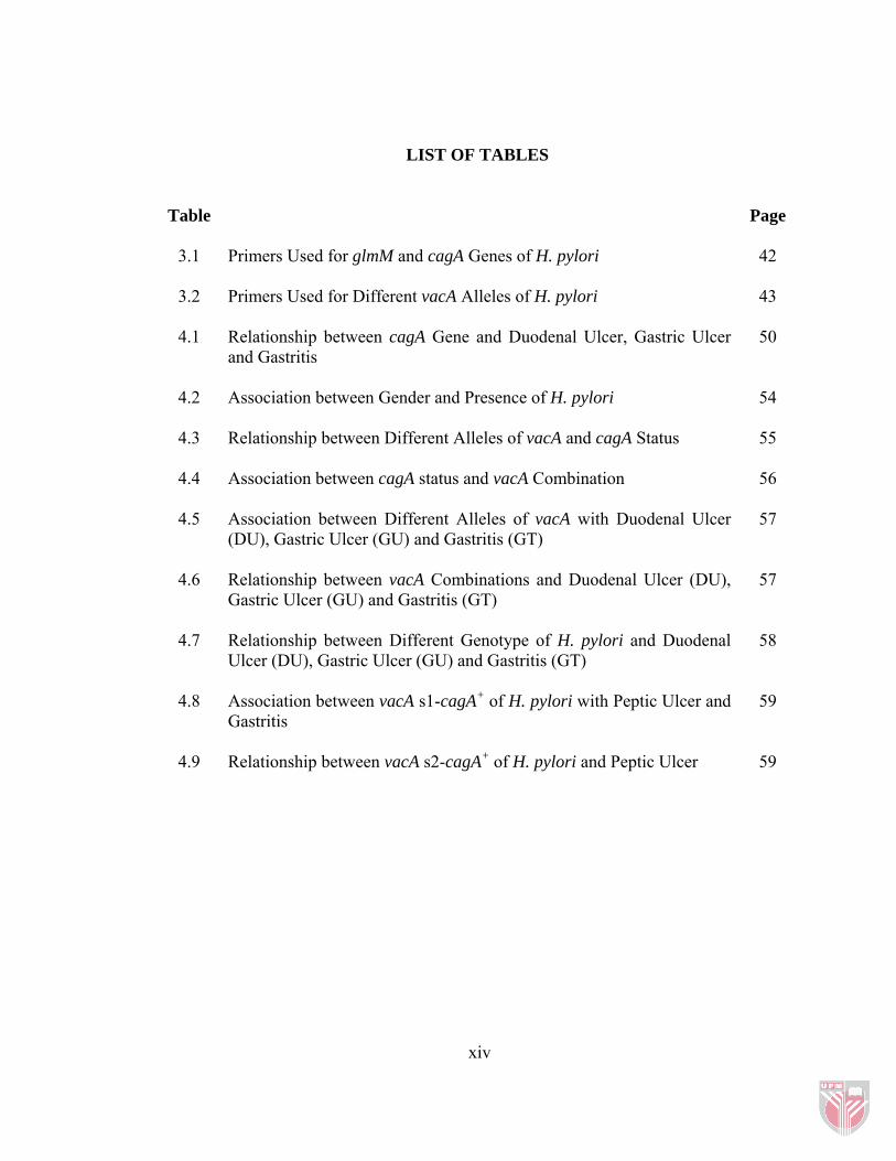
LIST OF TABLES
Table
Page
3.1 Primers Used for glmM and cagA Genes of H. pylori
42
3.2 Primers Used for Different vacA Alleles of H. pylori
43
4.1 Relationship between cagA Gene and Duodenal Ulcer, Gastric Ulcer and Gastritis
50
4.2
Association between Gender and Presence of H. pylori 54
4.3 Relationship between Different Alleles of vacA and cagA Status
55
4.4 Association between cagA status and vacA Combination
56
4.5
Association between Different Alleles of vacA with Duodenal Ulcer (DU), Gastric Ulcer (GU) and Gastritis (GT)
57
4.6 Relationship between vacA Combinations and Duodenal Ulcer (DU), Gastric Ulcer (GU) and Gastritis (GT)
57
4.7 Relationship between Different Genotype of H. pylori and Duodenal Ulcer (DU), Gastric Ulcer (GU) and Gastritis (GT)
58
4.8 Association between vacA s1-cagA+ of H. pylori with Peptic Ulcer and Gastritis
59
4.9 Relationship between vacA s2-cagA+ of H. pylori and Peptic Ulcer 59
xiv

LIST OF FIGURES
Figure
Page
2.1 Helicobacter pylori on the Epithelial Cells of the Stomach
8
2.2 vacA Polymorphism
14
4.1 Distribution of H. pylori in Different Gastroduodenal Diseases
48
4.2 Identification of H. pylori Infection in the Gastric Biopsy Specimens by PCR of glmM Gene on 2% (w/v) Agarose Gel Electrophoresis
49
4.3 Detection of the cagA Gene by PCR on 2% (w/v) Agarose Gel
51
4.4 Detection of s1 and s2 Alleles of vacA Gene of H. pylori by PCR on 8% (w/v) Polyacrylamide Gel Electrophoresis
52
4.5 Detection of m1 and m2 Alleles of vacA Gene of H. pylori by PCR on 2% (w/v) Agarose Gel Electrophoresis
53
xv

LIST OF ABBREVIATIONS
APS - Ammonium Persulfate
ASR - Age Standardized Ratio
ATP - Adenosin Tri-Phosphate
bp - Base pair
cagA - Cytotoxin Associated GeneA
CAD - Coronary Artery Disease
CT - Computerized tomography
DMSO - Dimethyl Sulfoxide
DNA - Deoxyribonucleic acid
DU - Duodenal Ulcer
EDTA - Ethylenediamine Tetra-Acetic acid
EtOH - Ethanol
GU - Gastric Ulcer
GT - Gastritis
GERD - Gastroesophageal Reflux Disease
H. pylori - Helicobacter pylori
kbp - kilo base pair
kDa - kiloDalton
KAc - Potassium Acetate
xvi
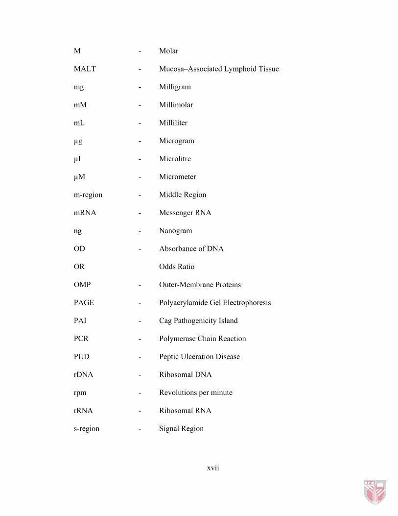
M - Molar
MALT - Mucosa–Associated Lymphoid Tissue
mg - Milligram
mM - Millimolar
mL - Milliliter
µg - Microgram
µl - Microlitre
µM - Micrometer
m-region - Middle Region
mRNA - Messenger RNA
ng - Nanogram
OD - Absorbance of DNA
OR Odds Ratio
OMP - Outer-Membrane Proteins
PAGE - Polyacrylamide Gel Electrophoresis
PAI - Cag Pathogenicity Island
PCR - Polymerase Chain Reaction
PUD - Peptic Ulceration Disease
rDNA - Ribosomal DNA
rpm - Revolutions per minute
rRNA - Ribosomal RNA
s-region - Signal Region
xvii
xviii
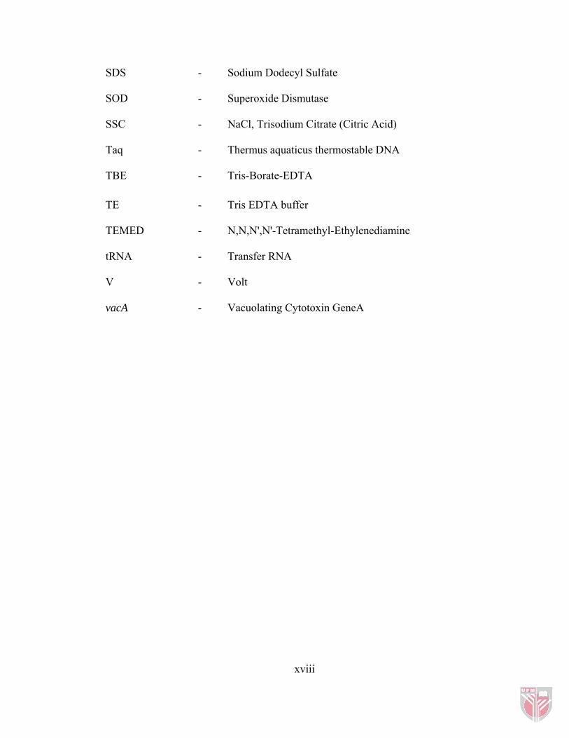
SDS - Sodium Dodecyl Sulfate
SOD - Superoxide Dismutase
SSC - NaCl, Trisodium Citrate (Citric Acid)
Taq - Thermus aquaticus thermostable DNA
TBE - Tris-Borate-EDTA
TE - Tris EDTA buffer
TEMED - N,N,N',N'-Tetramethyl-Ethylenediamine
tRNA - Transfer RNA
V - Volt
vacA - Vacuolating Cytotoxin GeneA

CHAPTER 1
INTRODUCTION
Peptic ulcer is a gastrointestinal disease which is characterized by mucosal damage
secondary to pepsin and excess gastric acid secretion. It usually occurs in the
proximal duodenum and stomach. It can also happen in the lower esophagus, the
jejunum, and the end of duodenum (Ramakrishnan et al., 2007).
Many different factors contribute to gastrointestinal diseases, such as old age,
tobacco smoking (Luo et al., 2002), the use of nonsteroidal anti-inflammatory drugs
(NSAIDs) and Helicobacter pylori infection (Kurata et al., 1997). A collection of
other infections such as tuberculosis, Crohn’s disease, myeloproliferative disorder,
chronic renal failure, and sarcoidosis can be associated with a larger risk of peptic
ulce. Critical illness, surgery, or hypovolemia can result in gastroduodenal corrosions
or ulcers; these may have some complications such as bleeding or perforation and
can be silent (Ziegler 2005).
Active chronic gastritis is the most common type of chronic gastric inflammation
that H. pylori has been detected in 70% - 80% of cases of this disease (Cover &
Blaser 1995). The disease influences the antrum and fundus of the stomach,
frequently showing permeation of the lymphocytes and eosinophils. The
inflammatory process is more considerable in the antrum than other parts of the body
(Atherton et al., 1996).

H. pylori can settle in the gastric mucosa and remain there for many years with
minimal symptoms and complications in the majority of cases (Blaser et al., 1995). It
infected about 50% of the world’s population (Souto et al., 1998). However, it has
been reported that some cases have shown significant morphological changes in the
gastric mucosa-inflammation to ulceration. Others indicated the progression of
chronic gastritis to cancer through chronic atrophic gastritis, intestinal metaplasia,
dysplasia and carcinoma (Blaser et al., 1995).
The high incidence of H. pylori infection may cause around 40 percent of all gastric
cancer cases all over the world (Parkin et al., 2001). The International Agency for
Research on Cancer (IARC) in 1994 reported that there is a relationship between H.
pylori infection and gastric cancer. They found sufficient evidences that introduced
H. pylori as a carcinogen among human populations. They classified H. pylori as
group I definite carcinogen (Chen et al., 2007). Yamagata et al. (2000) reported a
significant relationship between H. pylori infection and a succeeding incidence of
gastric cancer for men in the Japanese population.
Torres et al. showed that about 80% of population is infected by H. pylori at an
average age of 10 years and 20% of one-year-old children in Mexico had antibodies
against H. pylori (Torres et al., 1998). H. pylori Infection has significantly different
rates between developing and developed countries. For instance, the annual
prevalence of H. pylori infection in the United States is 0.5% to 1% in children less
than 10 years of age and 50% in adults around 60 years old, respectively. Different
ethnic groups, for example Hispanics, Afro-Americans, and Native Americans are
2

infected at an early period of aging (Everhart et al., 2000). Generally, low prevalence
of H. pylori infection has been observed in the United States, Canada, northern and
western Europe, whereas high prevalence of the infection has been reported in India,
Africa and Latin America (Torres et al., 2000).
The pathogenic effect of H. pylori in duodenal ulcer and gastric ulcer is obvious.
Eighty percent of patients with gastric ulcer and 95% of patients with duodena ulcer
have H. pylori infection in the United States (Breuer et al., 1998) which is the major
cause of peptic ulcer disease in 48% of cases in the U.S. (Ramakrishnan 2007).
However, the prevalence of H. pylori infection has been reported in about 90% of
patients with gastroduodenal diseases in Spain (Arroyo et al., 2004).
A seroepidemiological study in Iran demonstrated that 42.7% of cases between 10 to
25 years old were seropositive for H. pylori infection. There was an age-related raise
in H. pylori infection although there was no significant relationship between H.
pylori infection and gender (Pirouz et al., 2000). Many studies have reported that
85% of the Iranian adult population has H. pylori infection (Malekzadeh et al.,
2004).
Helicobacter pylori is a Gram-negative, spiral, microaerophilic bacterium that
chronically infects the gastric mucosa of more than half of the world population.
(Parsonnet 1995.) Two putative virulence agents of H. pylori have been recognized
in ulcerogenic strains. The first one is cytotoxin associated geneA (cagA) and the
second is vacuolating cytotoxin geneA (vacA) (Hsu et al., 2002).
3

A 120-140 kDa protein is encoded by the cagA gene and is present in about 60-70%
of H. pylori strains. This gene is part of the cag pathogenicity island (PAI), a 40-kbp
fragment with a number of genes contributing to cytokine production (Hsu et al.,
2002). Strains that do not produce the CagA protein normally lack the whole cag
PAI. Current studies have shown that the frequency of H. pylori strains with the
cagA gene is higher in patients with peptic ulcer disease than in patients with chronic
gastritis (Guerrero et al., 2000).
The vacuolating cytotoxin gene (vacA) is another virulence factor in that its product,
VacA protein, can damage epithelial cells (Hsu et al., 2002). The presence of the
vacA gene has been proven in almost all H. pylori strains and contains two variable
parts, the s-region and m-region (Atherton et al., 1997). Each of these two regions
has the different allelic types, s1 and s2 for the s-region and m-region containing m1
and m2 allelic forms (Van Doorn et al., 1998).
Some reports show that type s1 is associated with peptic ulcer disease and it also has
a significant relationship with the presence of the cagA gene (Rudi et al., 1999). m1
allele has been observed more in severe gastric epithelial than m2 allele and it is
associated with higher levels of toxin activity (Atherton et al.,1997).
There are four combinations of vacA; therefore, the genotype s2/m1 was not
identified in many studies (Castillo-Rojas et al., 2004). Type s1/m1 and s1/m2 strains
produce high and moderate levels of toxin, respectively, while strains with the s2/m2
genotype produce little or no toxin (Forsyth et al., 1998). Some studies in a number
4
of countries have demonstrated that cagA+ and vacA type s1 are associated with H.
pylori causing peptic ulcer disease (Catalano et al., 2001). Many publications in
Brazil have also proven this relationship (Brito et al., 2000). A recent study in Iran
revealed that duodenal ulcer has a strong association with H. pylori infection (Rasmi
et al., 2009), whereas another study in Tehran, capital city of Iran, showed no
significant relationship between different genotype of H. pylori and outcomes (Jafari
et al., 2008).
Research on the variety of H. pylori genes is valuable from two perspectives; first,
for predicting the outcome of the infection and second, for better understanding of its
distribution in the world and the evolutionary origins of this microorganism. The
detection of vacA and cagA, virulence markers described in several clinical
outcomes, can be used to help the treatment and prevention of H. pylori infection in
Iran. This study investigated different genotypes of vacA and cagA genes of
Helicobacter pylori isolated from patients with peptic ulcers and their relationship
with this disease.
5
The objectives of this study were to:
1- Detect H. pylori infection by the PCR method in patients with peptic ulcers and
gastritis
2- Identify the vacA and cagA genes in patients
3- Determine the distribution of cagA and vacA genes among patients with peptic
ulcer and gastritis.
4- Investigate the association between cagA gene with vacA gene and their
relationship with gastroduodenal diseases
6


