25
UNIVERSITI PUTRA MALAYSIA THE EXPRESSION OF CHICKEN ANEMIA VIRUS VP3 GENE AND INDUCTION OF APOPTOSIS IN TRANSFORMED AND TUMOR CELLS MOHAMED GHRICI FPV 2001 9
UNIVERSITI PUTRA MALAYSIA
THE EXPRESSION OF CHICKEN ANEMIA VIRUS VP3 GENE AND INDUCTION OF APOPTOSIS IN TRANSFORMED AND TUMOR
CELLS
MOHAMED GHRICI
FPV 2001 9
THE EXPRESSION OF CHICKEN ANEMIA VIRUS VP3 GENE AND INDUCTION OF APOPTOSIS IN TRANSFORMED AND TUMOR CELLS
By
MOHAMED GHRICI
Thesis Submitted in Fulfilment of the Requirement for the Degree of Master of Science in the Faculty of Veterinary Medicine
Universiti Putra Malaysia
October 2001

To my father GHRlCI MOHAMED and my mother KRIDER YAMINA
ii
Abstract of thesis presented to the Senate of Universiti Putra Malaysia in fulfilment of the requirement for the degree of Master of Science.
THE EXPRESSION OF CmCKEN ANEMIA VIRUS VP3 GENE AND INDUCTION OF APOPTOSIS IN TRANSFORMED AND TUMOR CELLS
By
MOHAMED GHRICI
October 2001
Chairman : Associate Professor Dr Mohd Azmi Mohd Lila, Ph.D.
Faculty : Veterinary Medicine
The pathogenicity of chicken anemia virus (CA V), as shown in previous studies, is
the function of apoptotic mechanism as observed in chicken thymocytes and
transformed chicken-Iymphoblastoid T cells. Thus the present study aimed to
investigate the gene and its gene product responsible for apoptosis in as such that
leads to the destruction of affected cells. It is known that apoptosis process is an
important natural physiological mechanism that induces killing of cancer cells.
Therefore, in theory, the gene or its gene product that is responsible for the apoptotic
mechanism could be exploited especially in cancer therapy. In this study, a complete
open reading frame (ORF) encoding VP3 protein was obtained from the DNA
extracted from archival paraffin-embedded tissues and cloned into a plasmid vector.
The sequencing of the full length of the ORF encoding VP3 protein showed that it
was 363 base pairs long, which is similar in size to that of the reference CA V Cux-l
strain. Comparison of the nucleotide sequences of this VP3 gene with that of the
CA V Cux-l strain exhibited 98% sequence homology indicating that these two
viruses are closely related. The functional characteristics of VP3 gene were further
investigated by using an eukaryotic expression system, plasmid pcDNA 3.1/Zeo+.
iii
The purified recombinant pcDNA-VP3 plasmid was used to transfect mammalian
cells and cancer cells via electroporation. The induction of apoptosis, upon
expression of VP3 gene, was studied by identifying and detecting morphological and
biochemical changes due to apoptosis. Upon electron microscopic examination,
typical apoptotic features were observed, which includes nuclear margination and
formation of apoptotic bodies, as early as two days after transfection. Morphological
changes developed due to apoptosis were obvious which were later confirmed by
means of the apoptotic biochemical staining and DNA fragmentation assay. The
differential activities of VP3 were further investigated by the use of the
immunofluorescence technique. The VP3 protein was expressed and detected only in
the cytoplasm of normal cells. In contrast, the expression of VP3 protein was
localized particularly in the nucleus of the transformed cell lines (Vero and rat
embryo fibroblast cells) and human derived breast cancer cells (MCF-7 and MDA
MB 231). This differential cellular localization of VP3 protein in normal versus
tumorigenic and transformed cells was the reason of VP3 specifically inducing
apoptosis in transformed and cancerous cells but not in normal cells. The effects of
VP3 protein expression in these three types of cells were also confirmed upon
examination by confocal microscopy analysis with cells stained in propidium iodide
and acridine orange stains. In conclusion, VP3 protein expression alone was able to
induce apoptosis in transformed cells and in human derived cancer cells but had no
effect on normal cells. The substantial evidences indicated that the DNA construct
containing the VP3 gene under the control of human cytomegalovirus (hCMV)
promoter, alias in the form of a DNA vaccine is the new potential candidate for anti
cancer therapy.
iv
Abstrak tesis yang dikemukakan kepada Senat Universiti Putra Malaysia sebagai memenuhi keperluan ijazah Master Sains
EKSPRESI GEN VP3 VIRUS ANEMIA A YAM YANG MENGARUH APOPTOSIS DALAM SEL-SEL YANG TERUBAH DAN SEL-SEL KANSER
Oleh
MOHAMED GHRICI
Oktober 2001
Pengerusi: Profesor Madya Mohd Azmi Mohd Lila, Ph.D.
Fakulti: Perubatan Veterinar
Sebagaimana yang telah ditunjukkan pada kajian terdahulu, permulaan
pegembangan penyakit Virus Anemia Ayam (CA V) yang mempunyai fungsi
mekanisma apoptotik yang terdapat dalam timosit-timosit ayam dan limphoblastoid
sel-sel T ayarn yang terubah. Kajian ini bermatlarnat untuk menyiasat gen dan hasil
keluaran gen yang bertanggung-jawab terhadap apoptosis seperti meningkatnya
kerosakkan kepada sel-sel yang terjangkit. Sebagaimana yang diketahui, proses
apoptosis adalah mekanisma fisiologikal semulajadi yang penting dalam mengaruh
pembunuhan sel-sel kanser. Oleh itu secara teori, gen atau hasilan gen yang
bertindakbalas bagi mekanisma apoptosis boleh dipergunakan terutamanya dalam
pengubatan kanser. Dalam kajian ini, rangka bacaan terbuka yang lengkap CORP)
yang mengekod protin VP3 telah diperolehi daripada DNA yang telah diekstrak dari
tisu-tisu ditanam parafin dan diklonkan ke dalam vektor plasmid. Pada ketika
pengklonan dan penurutan keseluruhan jarak gen ia telah menunjukkan rangka
bacaan terbuka yang lengkap itu mengandungi 363 nukleotid dan menpunyai saiz
yang sarna dengan strain rujukan Cux-l CAV. Gen yang didapati adalah 98 %
homolgus (sarna siri) kepada strain rujukkan dan juga tinggi pengekalan dengan gen
v
VP3 dari lain-lain strain CA V. Ciri-ciri fungsi gen VP3 juga disiasat dangan
menggunakan sistem expressi eukaryotik, pcDNA 3.11 Zeo + plasmid. Rekombinan
plasmid pcDNA-VP3 yang telah ditulinkan telah digunakan terhadap sel mamalia
transfeksi dan sel kanser melalui electroporasis. Apoptosis yang diaruhkan pada
ketika expressi gen VP3 telah dikaji dengan mengenalpasti dan mengesan secara
morfologi dan perubahan biokima yang disebabkan oleh apoptosis. Pada masa
pemeriksaan microskop elektron transmisi, gambaran apoptosis yang tipikal telah
dilihat termasuk pinggiran nuklear dan pembentukkan badan-badan apoptotik
seawal-awal hari ke 2 selepas transfeksi. Perubahan morphologi terhasil disebabkan
apoptosis kemudian ditentukan dengan pewamaan secara biokimia apoptotik dan
asai pemotongan DNA. Aktiviti-aktiviti VP3 yang berbeza kemudiannya diselidik
dengan menggunakan teknik immunofluoresen. Menarik sekali, dalam sel-sel
normal, protin VP3 yang dikesan hanya bersetempat dalam sitoplasm dan tidak
dalam lain-lain bahagian sel-sel transfeksi. Sebaliknya, pada ketika transfeksi,
protein VP3 terdapat dalam nukleusnya (Vero dan Fibroblas embrio tikus) dan sel
sel kanser payudara manusia. Kesan-kesan expresi protin VP3 dalam 3 jenis sel-sel
ini juga telah di kenalpasti menerusi pemeriksaan analisa mikroskop konfokal
dengan sel-sel yang diwamakan menerusi propidium iodida dan acridine oren.
Kesimpulannya, expressi protin VP3 sahaja mampu untuk mengaruh apoptosis
dalam sel-sel yang transfeksi dan sel payudara manusia tetapi tidak berkesan ke-atas
sel-sel normal. Bukti-bukti yang kuat menunjukkan. DNA yang dibentuk
mengandungi gen VP3 dibawah kawalan pemaju sitomegalovirus manusia (hCMV)
dan dalam bentuk sebagai vaksin DNA adalah mempunyai potensi baru dalam
pengubatan anti-kanser.
vi
ACKNOWLEDGEMENTS
Firstly, I would like to express my thanks and deepest gratitude to Associate
Professor Dr Mohd Azmi Mohd Lila as a supervisor for his guidance, suggestions,
discussion and patience throughout the course of this study.
Secondly, I would like to thank my co-supervisors Professor Dr Aini Ideris
and Professor Dr Abdul Rani Bahaman for their critical readings, comments and
suggestions.
Additionally, I would like to acknowledge Mr. Kamaruddin and Mr Abdul
Rahim Osman for their help and Mr. Ho and Ms. Zulika for their assistance in the
electron microscopy processing sections.
I am grateful to all my friends at the virology laboratory for their friendship.
I would like to express my deepest gratitude to all my brothers and sisters and
special thanks to my wife Thu Zar Than and her family for their encouragement,
which had helped me to undertake and complete these studies.
Finally, I would like to acknowledge my financial sponsors, the Malaysian
Ministry of Science, Technology and Environment IRP A grant without those
assistance this research would not have been possible.
vii
I certify that an Examination Committee met on the 5th October 200 1 to conduct the final examination of Mohamed Ghrici on his Master of Science thesis entitled "The Expression of Chicken Anemia Virus VP3 Gene and Induction of Apoptosis in Transfonned and Tumor Cells" in accordance with Universiti Pertanian Malaysia (Higher Degree) Act 1980 and Universiti Pertanian Malaysia (Higher Degree) Regulations 1981. The committee recommends that the candidate be awarded the relevant degree. Members of the Examination Committee are as follows:
HASSAN HJ MOHD DAUD, Ph.D. Faculty of Veterinary Medicine Universiti Putra Malaysia (Chainnan)
MOHD AZMI MOHD LILA, Ph.D. Associate Professor Faculty of Veterinary Medicine Universiti Putra Malaysia (Member)
AINI BINTI IDERIS, Ph.D. Professor Faculty of Veterinary Medicine Universiti Putra Malaysia (Member)
ABDUL RANI BAHAMAN, Ph.D. Professor Faculty of Veterinary Medicine Universiti Putra Malaysia (Member)
GHAZALI MOHA YIDIN, Ph.D . Profes orlDeputy Dean of Graduate School Universiti Putra Malaysia
Date: 1 2 NOV 2001
viii
This thesis submitted to the Senate of Universiti Putra Malaysia has been accepted as fulfilment of the requirement for the degree of Master of Science.
ix
AINI BT IDERlS, Ph.D. Professor Dean of Graduate School Universiti Putra Malaysia
Date: 1 0 JAN 2002
DECLARATION
I hereby declare that the thesis is based on my original work except for quotations and citations which have been duly acknowledged. I also declare that it has not been previously or concurrently submitted for any other degree at UPM or other institutions.
Mohamed Ghrici
Date: Z'.\\.�I
x

TABLE OF CONTENTS
DEDICATION ABSTRACT ABSTRAK ACKNOWLEDGEMENTS APPROVAL DECLARATION LIST OF FIGURES LIST OF ABBREVIATIONS
CHAPTER
1
2
3
INTRODUCTION
LITERATURE REVIEW 2.1 Physico-Chemical Characteristics of CA V 2.2 Morphological Characteristics of CA V 2.3 Genome Organisation of CA V
2 .3. 1 Structure of the Genome 2.3.2 Promoter and Enhancer 2.3.3 Transcription of the CA V Genome
2.4 CA V Proteins 2.4. 1 VP1 Protein 2.4.2 VP2 Protein 2.4.3 VP3 Protein
2.5 Virus Growth and Replication 2.6 Immunosuppression 2.7 How CA V Induce Apotosis 2 .8 Mechanisms of Cell Death 2.9 Molecular Mechanisms of Apotosis 2. 1 0 Virus and Apoptosis 2. 1 1 Characterization of Apoptotic Pathway 2. 1 2 Understanding Apoptosis by the use of VP3 Gene 2. 1 3 Anti-Tumor Therapies
CLONING AND SEQUENCING OF CA V VP3 GENE 3. 1 Introduction 3.2 Materials and Methods
3.2. 1 Total DNA Extraction From Paraffin-Embedded Tissues
3.2.2 Agarose Gel Electrophoresis 3.2.3 Primers for Polymerase Chain Reaction (PCR) 3.2.4 Amplification of the Big CAY DNA Fragment 3.2.5 Amplification of Small CA V DNA Fragment 3.2.6 Amplification of VP3 Gene 3.2.7 Direct Purification of DNA 3.2.8 Cloning of PCR Product into a Plasmid Vector 3.2.9 Bacterial Cell Transformation
xi
Page 11
III
V
Vll
Vlll
x xiv xix
1
5 5 6 6 6 8 1 2 13 1 5 1 6 1 7 1 8 1 9 2 1 22 23 25 25 26
29 30
30 31 32 32 33 34 34 35 37
3.2.10 Miniprep Plasmid Extraction 37 3.2.11 Restriction Enzyme Analysis 39 3.2.12 Sequencing ofthe VP3 Gene 39 3.2.13 Computer Analysis 39
3.3 Results 40 3.3. 1 DNA Preparations Extracted from Paraffin-
Embedded Tissues 40 3.3.2 In vitro Amplification of CA V VP3 Gene by
the Polymerase Chain Reaction 40 3.3.3 Cloning of the VP3 Gene 4 1 3.3.4 The DNA Sequence of VP3 Gene 46
3.4 Discussion 48
4 THE CLONING OF VP3 GENE INTO EUKARYOTIC EXPRESSION VECTOR 4.1 Introduction 56 4.2 Materials and Methods 57
4.2.1 Plasmid Vector 57 4.2.2 Double Restriction Digestion of pc DNA 3. l IZeo +
with XbaI and HindUI 58 4 .2.3 Restriction Digestion of VP3 60 4.2.4 Plasmid-VP3 DNA Ligation 62 4.2.5 Screening of Recombinant Transformants 63 4.2.6 Large Scale Preparation and Purification of
pcDNA-VP3 65
4.3 Results 66 4.3. 1 Recombinant pcDNA-VP3 Plasmid Construct 66 4 .3.2 Selection of Positive Recombinants by Restriction
Enzyme Analysis 67
4.4 Discussion 69
5 THE EXPRESSION OF VP3 GENE IN AN ESTABLISHED-' TRANSFORMED CELL LINE 5 . 1 Introduction 73 5.2 Materials and Methods 75
5.2.1 General Procedures 75 5.2.2 Cell Culture 75 5.2.3 Tolerance of Cell Lines to Zeocin 76 5.2.4 Transformation of Cells by Electroporation 77
5.2.5 Selection of Transformants 78 5.2.6 Electron Microscopic Analysis of Transfected Cells 79 5.2.7 Genomic DNA Isolation 80 5.2.8 Detection of Genomic DNA Fragmentation 8 1
5.3 Results 84 5.3. 1 Optimum Concentration of Zeocin in Cell Culture 84
xii
5.3.2 In Vitro VP3 Gene Expression 88 5.3.3 Morphological Changes Induced by VP3 gene
Expression 88 5.3.4 DNA Fragmentation Induced by VP3 Expression 9 1
5 .4 Discussion 94
6 IN- VITRO EXPRESSION OF VP3 PROTEIN IN PRIMARY CELL CULTURE AND HUMAN DERIVED CANCER CELLS 6.1 Introduction 1 04 6.2 Materials and Methods 1 05
6 .2 . 1 Adherent Continuous Cell Lines 1 05 6.2.2 Human Derived Cancer Cell Lines: MCF-7 and
MDA-MB 23 1 1 06 6.2.3 Preparation of Primary Cell Culture:
Chicken Embryo Fibroblast (CEF) 1 06 6.2.4 Plasmids and Cloning Vector used for
Calcium Phosphate Transfection 1 07 6.2.5 Plasmid Purification and Quantification 1 08 6.2.6 DNA Transfection by Calcium Phosphate 1 09 6.2.7 Immunofluorescence Staining of Transformed
Cells 1 1 1 6.2.8 Confocal Microscopy of the Transfected Cells III
6.3 Results 6.3 . 1 DNA Transfection 1 12 6 .3 .2 Analysis of VP3 Gene Expression by Indirect
Immunofluorescence Staining 1 1 3 6 .3 .3 Confocal Microscopy Analysis of Transfected
Cells 1 1 7
6.4 Discussion 124
7 GENERAL DISCUS SION AND CONCLUSION 136
REFERENCES 142 APPENDICES 158 BIODATA OF THE AUTHOR 1 61
xiii
LIST OF FIGURES
Figure
2.1 A scheme of the CA V life cycle. The closed arrows represent the transcription steps of the CA V genome showing the CA V transcript with a cap site (*) and the poly(A) tail (A)n followed by the synthesis of CA V proteins. The open arrows represent the steps for the production of encapsidated CA V ssDNA. (Source:
Page
Notebom et ai., 1998) 9
2.2 Genomic organization of the chicken anemia virus (CA V). Diagrammatic representation of the CA V genome illustrating the three major overlapping open reading frames (ORFs) for VP l , VP2 and VP3 and the non-coding regulatory regIOn (promoter/enhancer). (Source: Phenix et ai., 1994) 11
3.1 Physical map of the pCR-Blunt vector. Note the multi cloning site for the insertion ofVP3 gene. (Source: Invitrogen, USA). 3 5
3.2 Isolation of total DNA from paraffin-embedded tissue. DNA was extracted from paraffin-embedded tissues of suspected CA Vinfected chickens and electrophoresed into agarose/ethidium bromide stained gel and photographed under UV light. Lane 1: lKb DNA marker; Lane 2: no DNA extraction from paraffinembedded liver; Lane 3 : total DNA sample extracted from paraffin-embedded thymus. 41
3.3 Agarose gel electrophoresis of CA V specific DNA fragments amplified by peR. The amplified products were sized by ethidium bromide agarose gel lectrophoresis, showing the big fragment (1.5Kb) (Lane 1) and the small fragment (0. 8 Kb) (Lane 3) . Lanes 2 and 4: negative control containing no template. Lane 6: 100 bp DNA ladder. 42
3.4 PCR amplification of VP3 gene from the big fragment of CA V DNA. Amplification product was electrophoresed on 1 % agarose gel. Stained with ethidium bromide and photographed under UV light. Lane 1: 100 bp DNA marker; Lane 2: specific amplification of VP3 gene at 0.4 Kb; Lane 3 : big fragment used as template for the amplification of VP3 gene; Lane 4: negative control containing no template. 43
3.5 Restriction endonuclease (EcoRI) analysis of pCR-Blunt-VP3 . The recombinant plasmid DNAs were separated on 1 % agarose gel after EcoRI digestion of the pCR-Blunt-VP3 which released the VP3 fragment (Lane 3 and 4). Lane 1: 1 Kb DNA marker. 44
xiv
3.6
3.7
4.1
4.2
5.1
5.2
5.3
5.4
5.5
5.6
The DNA sequence of VP3 (UPMlma.I) as compared to the established CA V strain Cux-I.
The complete nucleotide sequence of VP3 gene and its deduced amino acid sequence.
Physical map of pcDNA 3 . 1 -Zeo+ for cloning of VP3 gene. Note that the multi cloning site for the insertion of the VP3 DNA fragment. (Source: Invitrogen, USA).
Restriction endonuclease analysis of the recombinant plasmid pcDNA-VP3 . The pcDNA-VP3 was digested with Xbal and Hind III and electrophoresed in 1 % agarose-ethidiumbromide stained gel. Lane 1: 1Kb DNA marker; Lane 2: undigested pcDNA-VP3; Lane 3,4 and 5: Restriction pattern of the pcDNA-VP3 cleaved with Xbal and Hind III releasing the VP3 from the recombinant plasmid.
Schematic diagram of the ApoAlert LM PCR assay. The diagram shows the different steps of the ApoAlert LM-PCR assay from the ligation of dephosphorylated adaptors to the ends of DNA break generated during apoptosis, followed by PCR amplification of these DNA fragments. The resulting nucIeosomal ladder can be visualized on an agarose lethidium bromide gel. (Source: CLONTECH, USA).
Sensitivity of Vero cell lines to Zeocin. 1: untreated Vero cells; 2: Vero cells treated with 0.5 mg/ml of Zeocin and 3: Vero cells treated with 1.5 mg/ml of Zeocin.
Sensitivity of BHK cell lines to Zeocin. 1: non-treated BHK cells; 2: BHK cells treated with 0.5 mg/mI, and 3: BHK cells treated with 1 mg/ml of Zeocin.
Sensitivity ofBT cell lines to Zeocin. 1: non treated BT cells; 2: BT cells treated with 0.250 mg/ml of Zeocin, and 3: BT cells treated with 0.5 mg/ml of Zeocin.
Electron microscopical examination of normal Vero cell lines. Note the normal distribution of euchromatin and heterochromatin and the round shape of the cell with complete integrity of all the organelles and plasma membrane.
Electron microscopic appearance of VP3-induced apoptosis in Vero cells, three days after electroporation. Early nuclear changes with chromatin condensation and margination together with cytoplasm condensation
xv
46
47
5 8
67
8 1
82
83
84
87
87

5.7 Ultrastructure of transfected Vero cells undergoing apoptosis at day four after electroporation. The nucleus is deeply convoluted and broken up into a number of small fragments. Translucent vacuoles are observed in the cytoplasm of apoptotic cells.
5.8
5.9
6.1
6.2
Ultrastructural morphological changes observed between day four and five after electroporation. Late stage of apoptosis where apoptotic bodies were already formed following the packaging of nuclear fragments and remnants of the cytoplasm surrounded by plasma membrane.
Amplification of DNA strand breaks associated with apoptosis using LM-PCR Ladder assay. Total DNA was extracted from the transfected Vero cells and amplified by LM-PCR ladder assay. The products of amplification were analyzed on 1 .2% agarose gel electrophoresis. Lane 1 : 1 00 bp DNA marker; Lane 2: Total DNA extracted from transfected Vero cells; Lane 3:LM-PCR amplification of total DNA extracted from non transfected Vero cells. Intemucleosomal DNA fragmentation amplified by LM-PCR ladder assay at day three (Lane 4), day four (Lane 5) and at day five (Lane 6) post-transfection.
Localization of VP3 protein expression in chicken embryo fibroblast (CEF) transiently transfected with 1 5 ug of pcDNAVP3. Three days after transfection, chicken embryo fibroblast were fixed and analyzed by indirect immunofluorescence. 1 : Non-transfected CEF. 2: Transfected CEF exhibiting a complete fluorescence staining of the nucleus. 3: The fluorescence is mainly concentrated around the nucleus in small granules and in larger aggregates in the cytoplasm.
Indirect immunoflourescence of transformed-cells Vero and REF transfected with 10 ug of pcDNA-VP3. Both cells were harvested 4 days post-transfection and were reacted with monoclonal antibody, anti-VP3. 1 . Transfected Vero cells at early stage of apoptosis in which the nucleus is weakly stained. 2. transfected REF cells at the late stage of apoptosis where the nucleus is fragmented into numerous dispersed flourescence structure forming apoptotic bodies.
xvi
88
88
89
1 1 0
1 1 1
6.3
6.4
6.5
6.6
6.7
Indirect immunofluorescence staining of human breast tumor derived cells, MCF-7 and MDA-MB 231 transfected with 5 ug of pcDNA-VP3. The cells were fixed four days after transfection and were stained with VP3 specific monoclonal antibody. 1: Mock-transfected MCF-7 cells with no sign of apoptosis. 2: Fluorescence concentrated III the nuclear fragments in the transfected MCF-7. 3: Widely scattered fluorescence reflecting an extensive nuclear fragmentation in the transfected MDA-MB-231.
Confocal microscopy analysis of the morphological changes induced by VP3 expression in Vero cell lines. 1: Normal Vero cells showing abnormal high uptake of dyes due to loss of plasma membrane integrity. 2: Nuclear fragmentation and apoptotic bodies formation in Vero cells transfected with 10 ug of pcDNA-VP3. 3: Three dimensional structure of Vero cells transfected with 10 ug of pcDNA, the red fluorescence represent a DNA droplets of different sizes, scattered throughout the cells which is correlated with an extensive nuclear fragmentation.
Morphological changes of RK-13 cell lines undergoing apoptosis. Three days after transfection with 10 ug of pcDNAVP3, RK-13 cells were harvested, stained with propidium iodide/ acridine orange staining and analyzed by confocal microscopy. 1: Mock-transfected RK-13 cells with an initial dyes uptake. 2: Bright orange staining of the condensed chromatin in the form of moon shape. 3: RK-13 cells with nuclear fragmentation.
Confocal laser scanning microscopic images of transfected REF cell lines. REF cells were transfected with 10 ug of pcDNAVP3 and stained with propidium iodide/acridine orange before being examined by confocal microscopy. 1: Early stage of apoptosis in transfected REF cells. 2: The whole nucleus IS
strongly stained and the beginning of nuclear fragmentation.
Confocal microscopy analysis of the transfected human breast tumor derived cells, MCF-7. MCF-7 cells were analyzed three days after transfection and were stained with propidium iodide/acridine orange. 1: No sign of apoptosis in the MCF-7 cells transfected with 10 ug of pcDNA 3.lIZeo +. 2: Transfection of MCF-7 cells with 10 ug of pCMV-HBS did not induce any apoptotic features and the cells were less fluorescent.
xvii
112
115
116
117
118
6.8 Confocal laser scanning microscopic images of transfected MCF-7 cells. MCF-7 cells were transfected with 10 ug of pcDNA-VP3 and were stained with propidium iodide/acridine orange at day three of post-transfection before being analyzed by confocal microscopy. 1 : Early stage of apoptosis with weak fluorescence . 2: Condensation of the chromatin at the periphery of the nucleus with strong fluorescence. 3 : Fragmentation of the nucleus at advanced apoptotic phase.
xviii
119
ADP AO AIP ATV A260 BCL-2 BCR-ABL BGHpA BHK bp BSA BI °C CaCh CalMg cAMP CAT CAY ccdB CEF CNRS CPE CnnA CsCl Da ddH20 DMEM Dnase d NTP ds E.coli EDTA e.g ELISA EM EMEM FBS FITC g xg g/ml H202 HBS Hbs HEPES hCMV hr HSV/tk
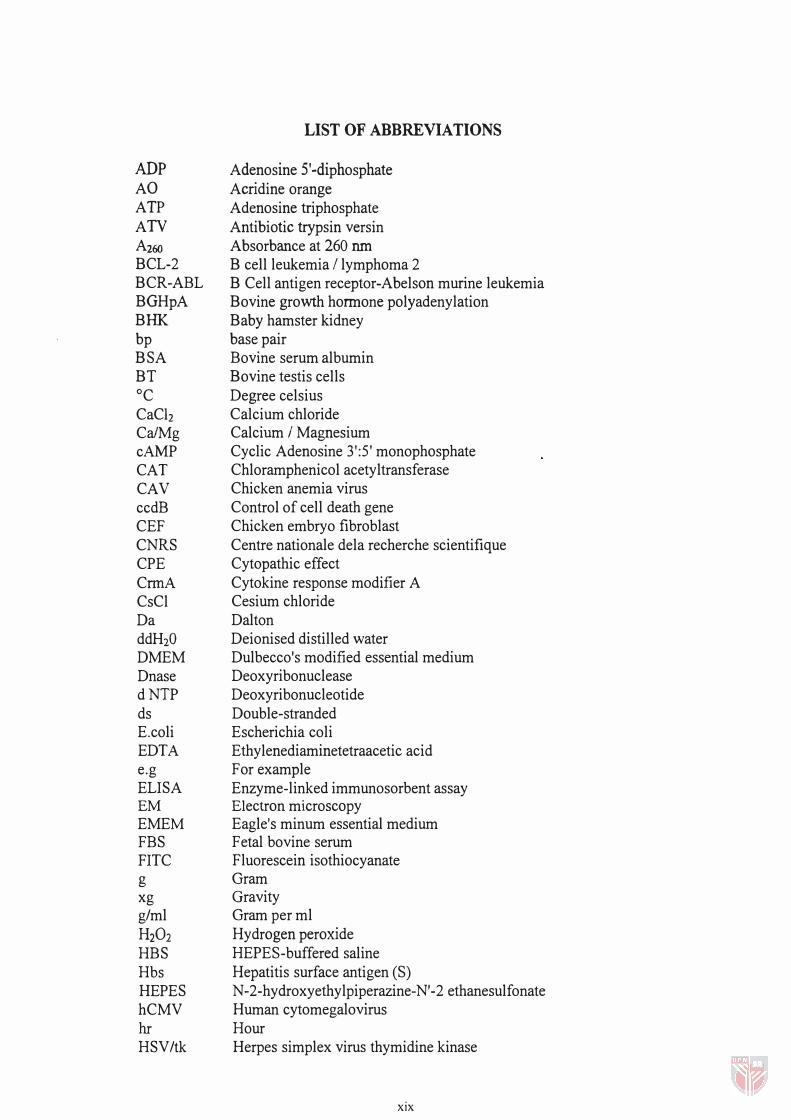
LIST OF ABBREVIATIONS
Adenosine 5'-diphosphate Acridine orange Adenosine triphosphate Antibiotic trypsin versin Absorbance at 260 run B cell leukemia I lymphoma 2 B Cell antigen receptor-Abelson murine leukemia Bovine growth hormone polyadenylation Baby hamster kidney base pair Bovine serum albumin Bovine testis cells Degree celsius Calcium chloride Calcium I Magnesium Cyclic Adenosine 3':5 ' monophosphate Chloramphenicol acetyltransferase Chicken anemia virus Control of cell death gene Chicken embryo fibroblast Centre nationale dela recherche scientifique Cytopathic effect Cytokine response modifier A Cesium chloride Dalton Deionised distilled water Dulbecco's modified essential medium Deoxyribonuclease Deoxyribonucleotide Double-stranded Escherichia coli Ethylenediaminetetraacetic acid For example Enzyme-linked immunosorbent assay Electron microscopy Eagle's minum essential medium Fetal bovine serum Fluorescein isothiocyanate Gram Gravity Gram per ml Hydrogen peroxide HEPE8-buffered saline Hepatitis surface antigen (8) N-2-hydroxyethylpiperazine-N'-2 ethanesulfonate Human cytomegalovirus Hour Herpes simplex virus thymidine kinase
xix
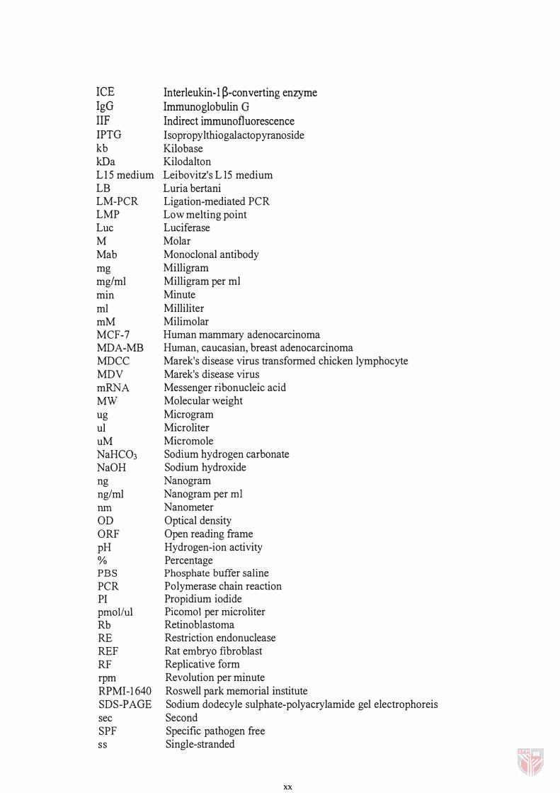
ICE IgG IIF IPTG kb kDa LI5 medium LB LM-PCR LMP Luc M Mab mg mg/ml min ml mM MCF-7 MDA-MB MDCC MDV mRNA MW ug ul uM NaHC03 NaOH ng ng/ml nm
OD ORF pH % PBS
PCR PI pmoi/ul Rb RE REF RF rpm RPMI-1640 SDS-PAGE sec SPF ss
Interleukin-l �-converting enzyme Immunoglobulin G
Indirect immunofluorescence Isopropy lthiogalactopyranoside Kilobase Kilodalton Leibovitz's L 15 medium Luria bertani Ligation-mediated PCR Low melting point Luciferase Molar Monoclonal antibody Milligram Milligram per ml Minute Milliliter Milimolar Human mammary adenocarcinoma Human, caucasian, breast adenocarcinoma Marek's disease virus transformed chicken lymphocyte Marek's disease virus Messenger ribonucleic acid Molecular weight Microgram Microliter Micromole Sodium hydrogen carbonate Sodium hydroxide Nanogram Nanogram per ml Nanometer Optical density Open reading frame Hydrogen-ion activity Percentage Phosphate buffer saline Polymerase chain reaction Propidium iodide Picomol per microliter Retinoblastoma Restriction endonuclease Rat embryo fibroblast Replicative form Revolution per minute Roswell park memorial institute Sodium dodecyle sulphate-polyacrylamide gel electrophoreis Second Specific pathogen free Single-stranded
xx
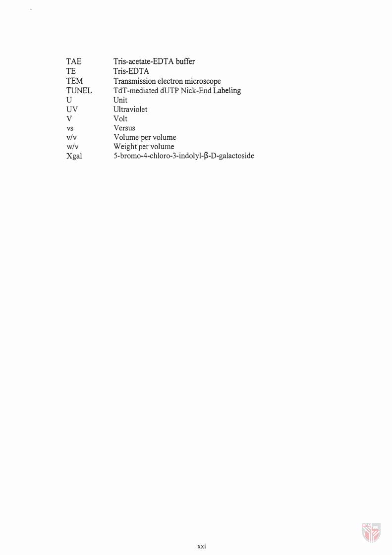
TAE TE TEM TUNEL U UV v vs v/v w/v Xgal
Tris-acetate-EDTA buffer Tris-EDTA Transmission electron microscope TdT-mediated dUTP Nick-End Labeling Unit Ultraviolet Volt Versus Volume per volume Weight per volume 5-bromo-4-chloro-3-indolyl-�-D-galactoside
xxi

CHAPTER!
INTRODUCTION
The living organisms and the maintenance of their wellbeing and health are
tribute to the equilibrium between cell division and apoptosis. The failure or
dysregulation of one of these two fundamental mechanisms can be fatal or have serious
pathological consequences (Singh et a!., 1994). Cellular death may occur either via
apoptosis or necrosis. Necrosis is non-physiological or pathological type of cell death
and it is uncontrolled in nature. It occurs following an extreme cell damage and is
characterized by swelling and lysis of the cell causing the release of its content into the
surrounding environment leading to inflammation (Trump et ai., 1991).
Apoptosis differ from necrosis particularly by two dominant markers:
morphological marker characterizing the structural changes of apoptotic cell and the
biochemical marker characterizing the cleavage of DNA into domain-sized fragments
(Walker et aI., 1993). Apoptosis is a programmed cell death and is a genetically
controlled cellular mechanism of cell death to eliminate unwanted cells (Earnshaw,
1995). Apoptosis is a widespread cellular mechanism involved in numerous processes
such as the embryonic and neural development (Sanders and Wride, 1995), the
regulation of the immune system (Williams, 1994), organogenesis and tissue
homeostasis (Wyllie et aI., 1980). Some of the molecular and biochemical mechanisms
of apoptosis have been elucidated and the research in this area continues to expand at an
2
extremely rapid rate following the development of new techniques and equipment (Hale
et aI., 1996).
The interest in apoptosis rise from the findings that apoptosis was involved in
many important diseases such as neurodegenerative disorders, AIDS and particularly
cancer, which affect millions of people around the globe. The growing amount of data
generated through a continuous investigations of the mechanisms of apoptosis has
proved to be very valuable in understanding the basic cell biology which will path the
way to the development of very sensitive diagnostic tools and the design of therapies for
various diseases. The suppression or failure andlor excessive rate of apoptosis can
trigger the development of tumors as well as render the tumor cells resistant to the
current chemotherapy and radiotherapy (Williams, 1994; Kerr et aI., 1994; Thompson,
1995).
The elucidation of the molecular mechanisms underlying apoptosis cannot be
achieved without developing appropriate methods for detecting and characterizing
apoptosis. Classically, the detection and characterization of apoptosis was based on the
examination of morphological changes at the cellular level by light-electron-microscopy
in combination with vital fluorescent dyes (McGahon et al., 1995). With the
advancement of the technology new tools were designed to detect apoptosis at the
molecular level, such as the use of Annexin V to monitor the loss of membrane
phospholipid asymetry during apoptosis (Koopmans et al., 1993) or assays to detect
DNA fragmentation by agarose gel electrophoresis (Wyllie. 1980), or by in-situ nick-
3
end labeling (Gavrieli et al., 1992). Assays to measure the disruption of the
mitochondrial transmembrane potential and the measurement of DNA and RNA content
by flow cytometry were developed later (Darzynkiewicz et al., 1 992). Flow cytometry
and its various alternatives are the most powerful methods to detect and quantitate
apoptotic cells (Darzynkiewicz et aI., 1997)
The important contribution of apoptosis research was made particularly in the
field of oncology, where it was demonstrated that tumor was a consequence of failure or
suppression of apoptosis, which also may render tumor cells more aggressive and
resistant to chemotherapy and radiotherapy (Williams, 1991). Chemotherapeutic drugs
and ionizing radiation, which are the only available cancer treatment, destroy the
cancerous cells via apoptosis usually via the action of wild type p53 (Dive and Hikman,
1991; Fisher, 1994; Thompson, 1995). These current therapies become hopeless when a
mutation strike the p53 gene and/or by the over-expression of bcl-2 (Lowe et aI., 1 994).
This problem may be solved by the use of a novel cancer therapy that would be
independent to the present of prerequisite factors. VP3 protein is encoded by an avian
virus (CAV) and has a length of 121 amino acids with an estimated size of 1 4 kDa
(Notebom et at., 1 994). The chicken anemia virus (CA V) was first isolated in Japan in
1979 (Yuasa et al., 1979) and was characterized as a new viral pathogen belonging to
Circoviridae family (Studdert et aI., 1993). In young chickens, CA V transiently cause
severe anemia and immunodeficiency due to depletion of cortical thymocytes (Jeurissen
et al., 1989; Yuasa et al., 1979). The depletion of thymocytes was demonstrated to


