University of Groningen Dengue and Chikungunya virus van Duijl-Richter, Mareike IMPORTANT NOTE: You are advised to consult the publisher's version (publisher's PDF) if you wish to cite from it. Please check the document version below. Document Version Publisher's PDF, also known as Version of record Publication date: 2016 Link to publication in University of Groningen/UMCG research database Citation for published version (APA): van Duijl-Richter, M. (2016). Dengue and Chikungunya virus: Cell entry mechanisms and the impact of antibodies on infectivity. University of Groningen. Copyright Other than for strictly personal use, it is not permitted to download or to forward/distribute the text or part of it without the consent of the author(s) and/or copyright holder(s), unless the work is under an open content license (like Creative Commons). The publication may also be distributed here under the terms of Article 25fa of the Dutch Copyright Act, indicated by the “Taverne” license. More information can be found on the University of Groningen website: https://www.rug.nl/library/open-access/self-archiving-pure/taverne- amendment. Take-down policy If you believe that this document breaches copyright please contact us providing details, and we will remove access to the work immediately and investigate your claim. Downloaded from the University of Groningen/UMCG research database (Pure): http://www.rug.nl/research/portal. For technical reasons the number of authors shown on this cover page is limited to 10 maximum. Download date: 01-01-2022
Transcript
University of Groningen
Dengue and Chikungunya virusvan Duijl-Richter, Mareike
IMPORTANT NOTE: You are advised to consult the publisher's version (publisher's PDF) if you wish to cite fromit. Please check the document version below.
Document VersionPublisher's PDF, also known as Version of record
Publication date:2016
Link to publication in University of Groningen/UMCG research database
Citation for published version (APA):van Duijl-Richter, M. (2016). Dengue and Chikungunya virus: Cell entry mechanisms and the impact ofantibodies on infectivity. University of Groningen.
CopyrightOther than for strictly personal use, it is not permitted to download or to forward/distribute the text or part of it without the consent of theauthor(s) and/or copyright holder(s), unless the work is under an open content license (like Creative Commons).
The publication may also be distributed here under the terms of Article 25fa of the Dutch Copyright Act, indicated by the “Taverne” license.More information can be found on the University of Groningen website: https://www.rug.nl/library/open-access/self-archiving-pure/taverne-amendment.
Take-down policyIf you believe that this document breaches copyright please contact us providing details, and we will remove access to the work immediatelyand investigate your claim.
Downloaded from the University of Groningen/UMCG research database (Pure): http://www.rug.nl/research/portal. For technical reasons thenumber of authors shown on this cover page is limited to 10 maximum.
Chapter 8Strongly Neutralizing Antibodies against Chikungunya Virus Inhibit Infection at Multiple Stages of the Viral Life Cycle
Mareike K. S. van Duijl-Richter1,¤, Jelle S. Blijleven2,¤, Julie M. Fox3, Michael S. Diamond3,4,5,6, Antoine M. van Oijen2,7,#, Jolanda M. Smit1,#
1: Department of Medical Microbiology, University Medical Center Groningen, University of Groningen, The Netherlands, 2: Centre for Synthetic Biology, Zernike Institute of Advanced Materials, University of Groningen, 9747 AG Groningen, The Netherlands3: Department of Medicine, 4: Department of Pathology and Immunology, 5: Department of Molecular Microbiology, 6: Center for Human Immunology and Immunotherapy Programs, Washington University School of Medicine, USA7: School of Chemistry, University of Wollongong, Wollongong, NSW 2522, Australia
¤,# These authors contributed equally to this work.
Manuscript in preparation
Chapter 8
8
164
Strongly neutralizing antibodies against Chikungunya virus inhibit infection at multiple stages of the viral life cycle
Abstract
Chikungunya virus (CHIKV) is a rapidly spreading arbovirus causing febrile illness and polyarthralgia. There is no vaccine or specific antiviral drug available to treat or prevent infection. In recent years, numerous monoclonal antibodies (MAbs) against CHIKV were isolated and characterized. Several of these MAbs were found highly neutralizing in vitro and decreased symptoms and disease in in vivo animal studies. Hence, antibody-based therapy represents an attractive treatment strategy in patients. Here, we investigated the mode of action of CHIKV neutralization by four MAbs targeting different epitopes on the viral envelope proteins. We show that all MAbs prevent infection at a pre-attachment and post-attachment step. All MAbs influenced the capacity of the virus to mediate membrane fusion. We found clear differences in the efficiency of neutralization of infection and fusion inhibition, which probably reflects different MAb-specific working mechanisms. Furthermore, we demonstrate that all four MAbs inhibit viral egress if administered post infection, revealing an additional way of neutralization by strongly neutralizing MAbs.
Mechanisms of CHIKV neutralization
8
165
Introduction
Chikungunya virus (CHIKV) is a single-stranded positive-sensed RNA virus. It belongs to the alphavirus genus, which amongst others comprises the well-studied Semliki Forest virus (SFV), Sindbis virus (SINV) and Eastern and Venezuelan Equine Encephalitis virus (EEEV & VEEV) [1]. CHIKV, which is transmitted by mosquitoes of the Aedes species, re-emerged in 2004 and caused since then millions of infections in the tropical and subtropical regions of the world [2]. The majority of individuals that are infected with CHIKV develop Chikungunya fever, with symptoms including fever, rash, headache, muscle pain, and joint pain [3]. Most of the initial symptoms usually clear within 7-10 days after infection. However, a large group of patients (estimates range from 12%-49%) experiences severe joint pain that can persist from months to years [4,5]. There is no vaccine or antiviral treatment available, and therapy is therefore confined to the treatment of symptoms [6,7]. Antibodies are key in controlling CHIKV infection and disease [8-10]. In recent years, multiple highly neutralizing monoclonal antibodies (MAbs) against CHIKV were isolated and characterized. In animal studies, several of these MAbs were found to protect mice and primates from disease and tissue injury [8,11-18]. Therefore, the use of anti-CHIKV antibodies has been suggested as a prophylactic therapy for individuals at risk like neonates or patients with underlying co-morbidities [9,10]. Functional studies revealed that the majority of strongly neutralizing antibodies identified to date interacts with the viral envelope glycoprotein E2 and interferes with the infectious properties of the virus [13,15,17,19-22]. CHIKV, like other alphaviruses, enters the host cell via receptor-mediated endocytosis, which is followed by fusion from within acidic endosomes [23-25]. Receptor binding is facilitated by the E2 protein and the membrane fusion process is mediated by the envelope glycoprotein E1 [26-28]. Fusion is triggered by the acidic pH inside the endosomal lumen. The E1 protein is structurally divided in three domains (DI, DII, and DIII). The fusion loop is located on the DII domain at the distal end of the protein. At neutral pH, E1 forms a heterodimer with the E2 glycoprotein, and three of these heterodimers are arranged as a one spike on the virion (see also figure 1). Each virion has 60 spikes. The E2 protein consists of three immunoglobulin-fold domains (A, B, and C) which are connected by β-ribbons. Domain B, located at the tip of the spike, carries one of the putative receptor-binding sites and shields the fusion loop on E1 [21,26,29]. Upon exposure to low pH, the E2/E1 heterodimer destabilizes, and the E2 domain B moves out. This transition leads to exposure of the E1 fusion loop, which subsequently inserts into the target membrane. Then, E1 trimerization occurs and the concerted folding of several E1 trimers into a hairpin-like conformation leads to fusion pore formation. Upon membrane fusion, the nucleocapsid is released into the cytosol and viral translation is initiated [21,30,31]. Antibodies targeting the viral spike proteins have been shown to inhibit virus binding to the host cell, membrane fusion, and viral release. Moreover, antibodies
Chapter 8
8
166
were found to induce complement-mediated lysis and phagocytosis of the viral particle [32,33]. In case of CHIKV, interference with receptor binding is possibly target-cell specific, as CHIKV is able to use various receptors and attachment factors for entry [34]. In contrast, fusion-inhibitory antibodies can prevent infection in all target cells regardless of receptor usage. Indeed, some of the most potent murine and human monoclonal E2 antibodies (MAbs) were shown to inhibit CHIKV fusion [13,17]. The ultrapotent fusion-inhibiting murine antibody CHK-152 is thought to lock the viral spike proteins such that the virion cannot undergo the conformational changes that are required for fusion [35]. In this study, we aimed to elucidate the mode of action of four highly neutralizing CHIKV antibodies. We selected three E2 antibodies (CHK-65, CHK-88, CHK-152) and one E1 antibody (CHK-180). Using cell-based assays, we show that the neutralizing activity of our MAbs was the highest when added before attachment to the cells. Yet, all four antibodies also prevented infection at a post-attachment step. Indeed, both the extent and rate of fusion were affected by the antibodies. Low pH-induced binding and E1 trimerization were inhibited to varying degrees by the MAbs. To gain more insight into the mechanism and kinetics of fusion inhibition, we next investigated fusion of antibody-opsonized CHIKV on a single-particle basis. We found that sub-optimal levels of CHK-152 lead to an increase in viral fusion lag time, which likely reflects the inhibition of a subset of viral fusion protein trimers. Next to neutralizing CHIKV early in infection, we discovered that all four MAbs inhibited viral egress, representing an additional mechanism of action.
Figure 1 Binding epitope of selected neutralizing MAbs.Ribbon diagram of a CHIKV spike consisting of three E2/E1 heterodimers from the side (A) and the top (B). For clarity, binding epitopes and domain colors are only depicted for one E2/E1 heterodimer. E1 domain I, II, and III are shown in red, yellow, and blue, respectively. E2 domain A, B, and C are depicted in cyan, green, and pink, respectively. The β-ribbon connector is shown in magenta. The other two heterodimers are shown in black (E2) and grey (E1). Binding epitopes are indicated as spheres. CHK-65 epitopes (green & dark green) and CHK-88 epitopes (blue & dark green) are partially overlapping epitopes located on domain B on E2. CHK-65 also binds additional residues on domain A and the protein stem. CHK-152 epitopes (pink) are located on domain A and the β-ribbon connector between domain B and domain C of E2. The CHK-180 epitope (red) is located on domain II of E1 in close proximity of the fusion loop (orange). This figure was prepared using the program PyMOL; PDB 2XFC.
Mechanisms of CHIKV neutralization
8
167
Results
Selection of strongly neutralizing monoclonal antibodies In a previous study, 36 broadly neutralizing anti-CHIKV MAbs were described. From this panel, we selected three highly neutralizing (EC50 < 7 ng/ml for all CHIKV lineages) E2 antibodies that bind to different epitopes (table 1, figure 1) and one neutralizing E1 antibody (EC50 = < 140 ng/ml for all CHIKV lineages) [13]. The binding epitopes were mapped by alanine scanning mutagenesis [36]. As depicted in figure 1, CHK-65 has a large footprint spanning several amino acids along the E2 stem, domain A, and the tip of domain B. CHK-88 binds to the tip of domain B, and has partial overlap in binding site with CHK-65. CHK-152 binds to domain A and the β-ribbon connector between domain B and domain C. CHK-180 binds to domain II on E1 close to the fusion loop.
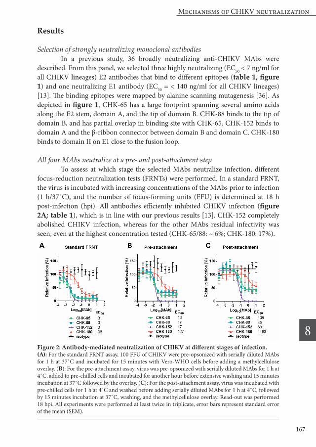
All four MAbs neutralize at a pre- and post-attachment step To assess at which stage the selected MAbs neutralize infection, different focus-reduction neutralization tests (FRNTs) were performed. In a standard FRNT, the virus is incubated with increasing concentrations of the MAbs prior to infection (1 h/37˚C), and the number of focus-forming units (FFU) is determined at 18 h post-infection (hpi). All antibodies efficiently inhibited CHIKV infection (figure 2A; table 1), which is in line with our previous results [13]. CHK-152 completely abolished CHIKV infection, whereas for the other MAbs residual infectivity was seen, even at the highest concentration tested (CHK-65/88: ~ 6%; CHK-180: 17%).
Figure 2: Antibody-mediated neutralization of CHIKV at different stages of infection. (A): For the standard FRNT assay, 100 FFU of CHIKV were pre-opsonized with serially diluted MAbs for 1 h at 37˚C and incubated for 15 minutes with Vero-WHO cells before adding a methylcellulose overlay. (B): For the pre-attachment assay, virus was pre-opsonized with serially diluted MAbs for 1 h at 4˚C, added to pre-chilled cells and incubated for another hour before extensive washing and 15 minutes incubation at 37˚C followed by the overlay. (C): For the post-attachment assay, virus was incubated with pre-chilled cells for 1 h at 4˚C and washed before adding serially diluted MAbs for 1 h at 4˚C, followed by 15 minutes incubation at 37˚C, washing, and the methylcellulose overlay. Read-out was performed 18 hpi. All experiments were performed at least twice in triplicate, error bars represent standard error of the mean (SEM).
Chapter 8
8
168
To investigate if the MAbs interfere with CHIKV infection at a pre- or post-attachment stage, distinct FRNT assays were carried out. In the pre-attachment assay, CHIKV is pre-incubated with antibodies for 1 h at 4˚C after which the complexes are incubated with cells for 1 h at 4˚C to allow virus adsorption. Upon removal of unbound virus, infection is initiated and the number of FFU is determined at 18 hpi. In the post-attachment assay, the antibodies are added after virus binding to the cell. The results of the pre-attachment assay are in line with the standard FRNT assay and shows that all MAbs neutralize CHIKV infectivity (figure 2B, table 1). Overall, higher EC50 values were observed in the pre-attachment assay, which likely reflects decreased opsonization and/or adsorption at lower temperature. The EC50 values were 16-17 ng/ml for the E2 antibodies and 127 ng/ml for the E1 antibody CHK-180. The EC50 values determined for the post-attachment assay were in turn higher than the pre-attachment values (figure 2C, CHK-65: 108 ng/ml; CHK-88: 45 ng/ml; CHK-152: 60 ng/ml; and CHK-180: 1183 ng/ml). Also the fraction resistant to neutralization was highest in the post-attachment assay. For CHK-65 and CHK-88, the fraction of virions that remained infectious increased from around 25% to ~ 40% if opsonization was performed at a post-attachment step. For CHK-180, this fraction was considerably higher and increased from ~ 35% (pre attachment) to ~55% (post attachment). CHK-152 completely neutralized CHIKV in all assays albeit with different efficiency.
Fusion inhibitory capacity of the neutralizing MAbs We next investigated whether the MAbs directly interfere with membrane fusion. To this end, we biosynthetically labeled the virus with the fluorescent probe pyrene, as described before [30]. The specific infectivity of pyrene-labeled CHIKV was comparable to that of unlabeled virus (300 vs. 340, respectively), indicating that pyrene-labeling does not affect the functional properties of the virus. As the membrane fusion characteristics have not been described for the CHIKV-LR2006 OPY1 strain, we first defined the pH dependency of fusion. Like for other strains [23,30], low pH induced rapid fusion of CHIKV-LR2006 OPY1 with liposomes. High extents of fusion were seen between pH 4.7 and pH 5.4 (figure S1). To assess the effect of MAbs on membrane fusion, CHIKV particles were pre-incubated with serial dilutions of MAbs for 10 minutes at 37˚C and fusion was measured at pH 5.1. In figure 3A, representative curves of all MAb conditions and two pH-
Table 1. Neutralizing capacity and binding epitope of the 4 selected MAbsMAb EC50
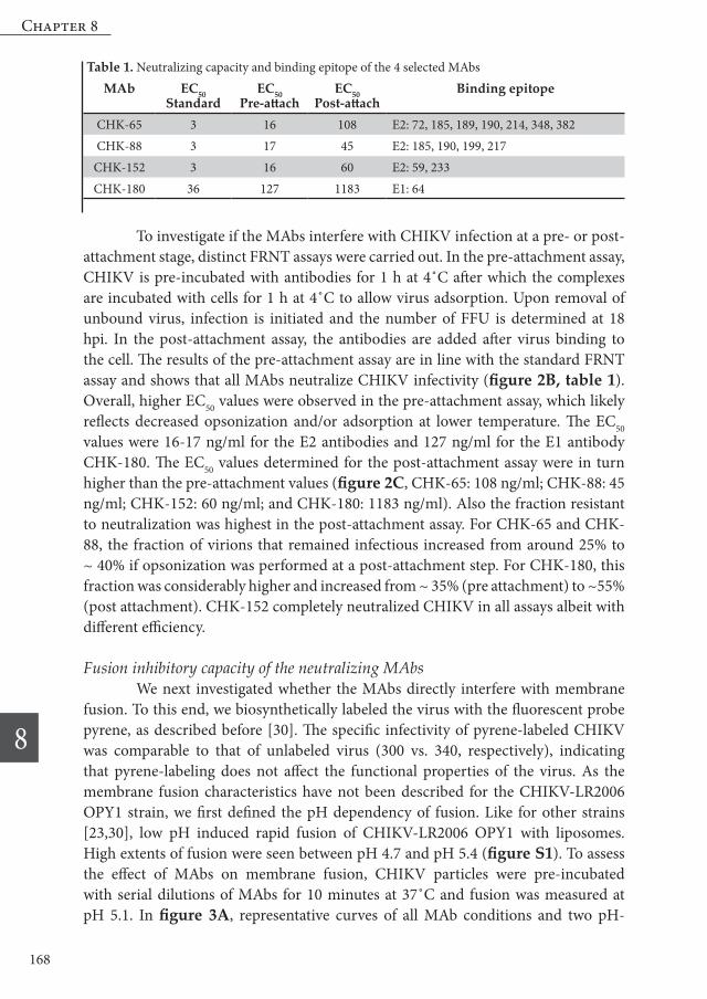
control conditions are shown. In figure 3 B-F, fusion extents (calculated as the average fusion extent between 50-60 s after acidification) and rates (calculated as the inverse time point at which half of the particles fused) are plotted. In the absence of antibodies, fusion occurred quickly as half of the virus particles fused within 2 seconds after acidification. The total extent of fusion was on average 76% (figure 3). Neither the rate nor the extent of fusion was significantly influenced in presence of an isotype control antibody (figure 3B). After addition of a pH buffer pre-titrated to pH 7.4, a total fusion extent of on average 3% was observed, which can be attributed to bleaching of the probe (figure 3A). Importantly, all four MAbs inhibited membrane
Figure 3 Fusion inhibition of CHIKV by MAb CHK-65, CHK-88, CHK-152 and CHK 180 as measured in the bulk fusion assay. Pyrene-labeled virus was pre-opsonized with MAbs at different concentrations and mixed with liposomes in HNE buffer. Fusion was triggered by addition of a low-pH buffer at t=0. Total fusion extent is represented by filled circles/solid lines and was determined as the average fusion extent at 50-60 s after acidification. Fusion rate is represented by open squares/dashed lines and was calculated as the inverse time point at which half of the particles fused. (A) shows representative fusion curves of CHIKV pre-opsonized with 10 nM of the respective antibody. (B-F) show quantification of the fusion rate and the total fusion extent for each antibody, including an isotype control.
Chapter 8
8
170
fusion in a dose-dependent way, albeit with different efficiency. In line with previous data with the African prototype strain S27 [13], CHIKV-LR fusion was efficiently inhibited by CHK-152 (figure 3A,E). At 10 nM and 5 nM, the total fusion extent was reduced by 85% and 75%, respectively. At both concentrations, very low fusion rates were observed. At 1 nM (which is equal to an antibody/epitope ratio of 0.17), the extent of CHIKV membrane fusion was inhibited with 12% and little to no effect was seen on the rate of fusion. CHK-65 and CHK-88 exhibited similar neutralization profiles as in the cell-based assays. However, at 10 nM, CHK-88 was more efficient in inhibiting CHIKV fusion than CHK-65. The extent of fusion was reduced by 84% by CHK-88; compared to a reduction of 52% by CHK-65 (figure 3A,C,D). At an antibody concentration of 5 nM, the antibody-specific differences were more visible. For CHK-65, a reduction in fusion extent of 28% was detected. Moreover, a low fusion rate was observed, with half of the particles having fused after more than 5 seconds. In contrast, in case of CHK-88, the fusion extent decreased by 57%, with half of the particles fused after about 3 seconds. These results suggest the mechanism of fusion inhibition might be different despite partial overlap in binding sites. The E1 antibody CHK-180 had the lowest inhibitory effect on membrane fusion (figure 3A,F). Although at 10 nM, a clear reduction in fusion rate was achieved (half of the particles fused after more than 4 seconds), inhibition of the total fusion extent was only 34%. Interestingly, however, at 1 nM, the extent of fusion inhibition was similar to that of CHK-152 and better than the inhibition of fusion extent by CHK-65 and CHK-88. To investigate if fusion inhibition is a general feature of neutralizing CHIKV antibodies, we next measured the fusion inhibitory effect of 14 additional neutralizing MAbs described by Pal and co-workers [13]. Approximately one third of the antibodies tested efficiently inhibited fusion (>70%). Another one third partially inhibited fusion (30-63% fusion inhibition), and the remaining antibodies did not or only marginally inhibited fusion (≤11% inhibition) (table S1). Hence, inhibition of fusion is a frequent, but not imperative working mechanism of highly neutralization antibodies.
Molecular mechanism of antibody-mediated fusion inhibition The observation that all four selected MAbs inhibited fusion with different inhibition profiles prompted us to take a closer look at the mechanism of action of these antibodies. Viral fusion is preceded by a number of conformational rearrangements in the envelope spike proteins, and neutralizing antibodies targeting different epitopes might act at different stages of this process. The first step in viral fusion is destabilization of the E2/E1 heterodimer complex. Dissociation of the E2/E1 heterodimer can be investigated using sedimentation analysis, which separates protein monomers, dimers and trimers based on molecular weight in a sucrose gradient [37]. We successfully implemented this assay system for CHIKV and demonstrated that E2/E1 dissociation and E1 trimer formation occurs upon exposure of the virus to low
Mechanisms of CHIKV neutralization
8
171
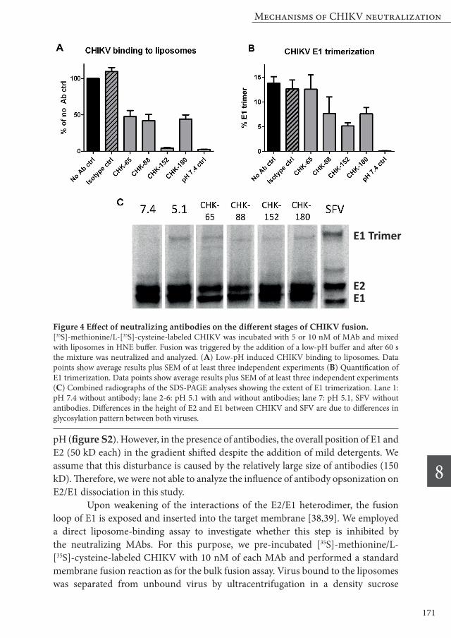
pH (figure S2). However, in the presence of antibodies, the overall position of E1 and E2 (50 kD each) in the gradient shifted despite the addition of mild detergents. We assume that this disturbance is caused by the relatively large size of antibodies (150 kD). Therefore, we were not able to analyze the influence of antibody opsonization on E2/E1 dissociation in this study. Upon weakening of the interactions of the E2/E1 heterodimer, the fusion loop of E1 is exposed and inserted into the target membrane [38,39]. We employed a direct liposome-binding assay to investigate whether this step is inhibited by the neutralizing MAbs. For this purpose, we pre-incubated [35S]-methionine/L-[35S]-cysteine-labeled CHIKV with 10 nM of each MAb and performed a standard membrane fusion reaction as for the bulk fusion assay. Virus bound to the liposomes was separated from unbound virus by ultracentrifugation in a density sucrose
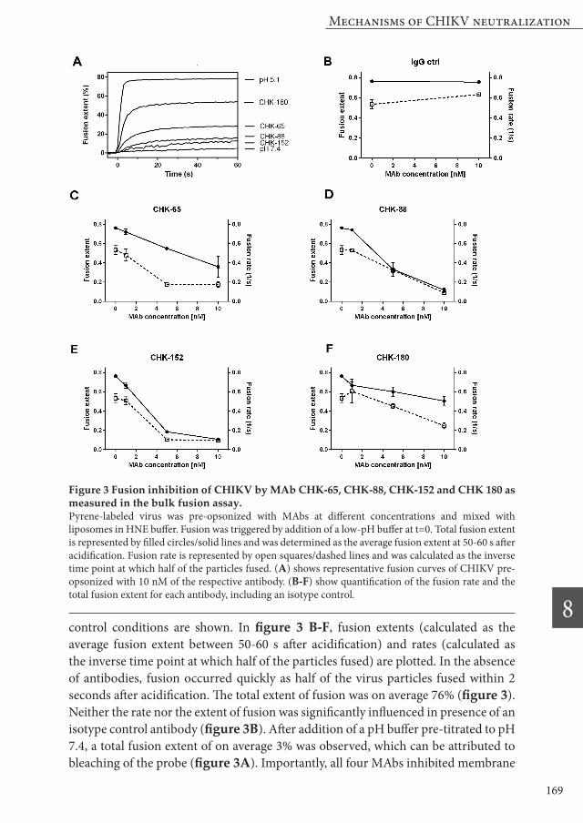
Figure 4 Effect of neutralizing antibodies on the different stages of CHIKV fusion.[35S]-methionine/L-[35S]-cysteine-labeled CHIKV was incubated with 5 or 10 nM of MAb and mixed with liposomes in HNE buffer. Fusion was triggered by the addition of a low-pH buffer and after 60 s the mixture was neutralized and analyzed. (A) Low-pH induced CHIKV binding to liposomes. Data points show average results plus SEM of at least three independent experiments (B) Quantification of E1 trimerization. Data points show average results plus SEM of at least three independent experiments (C) Combined radiographs of the SDS-PAGE analyses showing the extent of E1 trimerization. Lane 1: pH 7.4 without antibody; lane 2-6: pH 5.1 with and without antibodies; lane 7: pH 5.1, SFV without antibodies. Differences in the height of E2 and E1 between CHIKV and SFV are due to differences in glycosylation pattern between both viruses.
E1 Trimer
E2E1
Chapter 8
8
172
gradient. During centrifugation, the bound virus co-flotates with the liposomes to the top of the gradient. In the absence of antibodies, on average 55% of the virus particles were bound to liposomes upon exposure to pH 5.1. As can be seen in figure 4A, virtually no binding (2% of pH 5.1 control) of CHIKV to liposomes takes place if the pH is maintained at 7.4. Likewise, incubation of CHIKV with CHK-152 leads to almost complete abrogation of binding to the liposomes. Opsonization with either CHK-65, CHK-88 or CHK-180 lead to a significant reduction of virus-liposome binding (~ 50% for each antibody). Next, we evaluated the extent of E1 trimerization in the presence and absence of MAbs. E1 trimerization was analyzed by SDS-PAGE (figure 4B,C). Under optimal fusion conditions, 14% E1 trimerization was detected. This is much lower than the extent of E1 trimerization observed in literature for SFV [37]. To rule out potential technical issues we also determined the extent of E1 trimerization for SFV and in line with literature, almost half of the E1 proteins rearranged to E1 trimers under optimal fusion conditions (figure 4C). One explanation for this difference can be that the E1 trimer of CHIKV is less stable and therefore less resistant against SDS-PAGE analysis. However, using solubilization buffer with lower amounts of SDS did not result in an increased detection of the trimer (data not shown). Alternatively, it is possible that CHIKV E1 trimerization is less efficient than SFV E1 trimerization. Incubation with CHK-65 does not significantly inhibit E1 trimerization when compared to the isotype control antibody or no antibody control. The other three antibodies did inhibit trimer formation, with CHK-152 being most effective, followed by CHK-180 and CHK-88.
Single-particle analysis of inhibition of CHIKV fusion To receive detailed insight in the inhibition of CHIKV fusion by our MAbs, we next evaluated the membrane fusion properties at the single-particle (SP) level using total internal reflection microscopy (TIRF-M). By using this assay, improved kinetic information of R18-labeled CHIKV fusion can be obtained due to the higher time resolution. Moreover, dephasing effects are avoided as the population is not observed as a whole, but as single virions. As the SP assay is technically challenging at 37˚C, we first analyzed whether the fusion kinetics are temperature dependent. Bulk fusion measurements were performed at five different temperatures ranging from ~ 19˚C to ~ 37˚C. The temperature data were fitted well by the Arrhenius equation, indicating that throughout this temperature range, the same transition is rate limiting (figure S3). Therefore, all experiments were subsequently performed at room temperature (RT). In the SP assay, CHIKV binds to the lipid bilayer in a non-specific manner, possibly due to electrostatic interactions. We however noticed that in presence of antibodies (5 and 10 nM) the ability of the virus to interact with the lipid bilayer is drastically reduced (figure S4). As there is no CHIKV specific receptor in the lipid bilayer, the comparatively large antibodies likely shield the virion surface thereby
Mechanisms of CHIKV neutralization
8
173
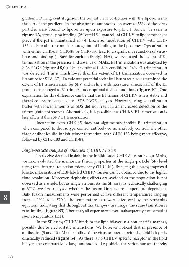
preventing binding. To be able to measure fusion kinetics at the single particle level, we reduced the antibody concentration to 1 nM. At this concentration, a low, but sufficient number of particles bound to the lipid bilayer to perform the analysis (Figure S4). At this condition, CHK-65, CHK-88 and CHK-180 did not inhibit the rate or fusion extent of the bound particles (figure 5A). This finding is not surprising, as in the bulk fusion assay, only very limited fusion inhibition was found at this concentration as well. Opsonization with CHK-152 at 1 nM did not inhibit the total fusion extent significantly either. However, we did observe a trend towards a reduced fusion extent (figure 5A). In addition, both the median and the mean fusion times were increased upon opsonization with CHK-152 (figure 5B,C).
Figure 5 Influence of antibody-opsonization on CHIKV fusion as measured in the single particle assay R18-labeled virus was pre-opsonized with MAbs at 1 nM and docked to a lipid-bilayer in a flow cell channel for 3 minutes. The aqueous environment was acidified by flowing in citric acid buffer. Viral fusion and the time of acidification was observed with total internal fluorescence microscopy and the fusion behavior of individual virions was analyzed. Total fusion extent (A), mean fusion time (B) and median fusion time (C) of CHIKV particles pre-incubated with 1 nM CHK-65, CHK-88, CHK-152, CHK-180 or an IgG control binding to the bilayer despite opsonization are shown. The total number of analyzed fused virions was 149, 96, 33, and 219 for particles opsonized with CHK-65, CHK-88, CHK-152, and CHK-180, respectively.
Chapter 8
8
174
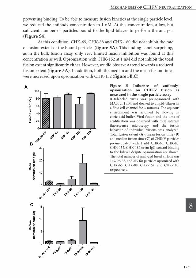
Other mechanisms of CHIKV neutralization: Inhibition of viral egress The experiments so far showed that the MAbs can exert their function both at a pre- and post-binding level. For viruses budding at the plasma membrane, including CHIKV, inhibition of egress has been described as an additional neutralization mechanism [40-44]. As a small plaque phenotype can be a consequence of egress inhibition, we first analyzed CHIKV plaque formation in the presence of the four MAbs by a standard plaque-reduction neutralization test. All four antibodies (at 10 µg/ml) influenced the plaque size of CHIKV (figure 6A). To assess whether these MAbs indeed inhibit virus egress we next investigated the release of CHIKV progeny RNA in the absence and presence of MAbs. For this purpose, CHIKV infection was allowed for 1 h at 37˚C, after which serially diluted antibodies were added to the cells and incubation was continued for another 6 h. Thereafter, both the intracellular and supernatant viral RNA levels were assessed. Figures 6B and 6C clearly show that the addition of the MAbs decreased the levels of viral RNA in the supernatant without affecting viral replication. These results indicate that in addition to inhibiting attachment and fusion, all four MAbs also exert their neutralizing capacity by preventing viral egress.
Figure 6 CHIKV egress inhibition by four neutralizing MAbs(A): For the analysis of the plaque phenotype, 75 PFU of CHIKV were pre-opsonized with serially diluted MAbs for 1 h at 37˚C and incubated for 1.5 h with Vero-WHO cells before adding an 1% agarose overlay. Read-out was performed at 43 hpi. (B+C): Release of viral progeny was assessed with qRT-PCR. Vero-WHO cells were infected with CHIKV at MOI 1. At 1 hpi, the cells were washed thoroughly and medium with serially diluted MAbs was added. At 6 hpi, viral RNA was quantified in the supernatant (B) and the infected cells (C). Data points show averages of at least two independent experiments performed in triplicate. Error bars represent standard error of the mean (SEM).
Mechanisms of CHIKV neutralization
8
175
Discussion
In this chapter, we show that neutralization of CHIKV by four different strongly neutralizing antibodies takes place at a pre- and post-binding stage, with membrane fusion being inhibited by all MAbs tested. When focusing in detail on the mechanism of fusion neutralization, we found that all antibodies (partially) inhibited low-pH induced binding of the CHIKV to liposomes, and that also E1 trimerization is partially inhibited by three MAbs. In addition to preventing viral entry, all MAbs investigated inhibit viral egress. In the cell-based studies, all four MAbs displayed lower EC50 values for pre-binding inhibition than for post-binding inhibition. Hence, prevention of virus attachment to the cell and/or uptake represents an important neutralization mechanism. At high antibody concentration, infection will be likely neutralized at this early step. Antibody-binding presumably interferes with virus-receptor binding by steric hindrance or by directly blocking one of the receptor binding sites located on E2. Yet, as it is known that CHIKV can bind to various receptors and attachment factors [34], it remains to be determined whether binding and infection are inhibited equally well in other cell lines. Neutralization of infection through inhibition of membrane fusion is very powerful as this step is independent of cell type. In this case, also antibody-bound particles internalized by Fc-receptor expressing cells will be neutralized. All four antibodies inhibited membrane fusion, albeit to a different extent. The level of inhibition is likely defined at the epitope level. Yet inhibition of fusion is not imperative for neutralization. Indeed, one-third of the panel of antibodies tested did not inhibit membrane fusion. Future studies should unravel whether antibodies that interfere with receptor binding are also neutralizing in Fc-receptor expressing macrophages, which are thought to be permissive to CHIKV [45-47]. CHK-152 is one of the most potent and also the best investigated CHIKV antibody so far. It binds to epitopes located on E2 domain A and the β-ribbon connector. Importantly, the latter is located in the acid-sensitive region (ASR) of the E2 protein, a region that becomes disordered at low pH, thereby facilitating the exposure of the fusion loop [21,28,35]. Here we show that opsonization of CHIKV with CHK-152 almost completely abrogates low-pH induced binding of the virus to liposomes and E1 trimerization. Furthermore, opsonization of CHIKV with CHK-152 lead to a rapid decrease in the rate and extent of membrane fusion in the bulk fusion assay. In the single-particle assay, an increase in the lag time between acidification and fusion was seen at sub-optimal concentrations of CHK-152. It has been suggested before for antibody-mediated neutralization of influenza virus [48] that the increase of this lag time is due to the reduced number of available fusogenic trimers. Upon binding of CHK-152 to CHIKV at a low concentration, a subset of E1 proteins may not re-arrange into fusogenic trimers. This stochastically increases the time until multiple trimers can act simultaneously to induce fusion [49]. At increasing
Chapter 8
8
176
antibody concentrations, more fusion proteins are “frozen” and slower fusion rates are observed. Taken together, based on the structural and functional data on CHK-152, we hypothesize that CHK-152 stabilizes the pH-neutral conformation of the ASR, thereby preventing dissociation of the E2/E1 heterodimer and insertion of the fusion loop in the target membrane. For the other three MAbs, more subtle effects were seen. CHK-65, CHK-88 and CHK-180 reduced low-pH dependent virus-liposome binding by approximately 50%. At the same antibody concentration, CHK-65, CHK-88 and CHK-180 inhibited the total fusion extent of CHIKV in the bulk fusion assay by 52%, 84% and 34%, respectively. This suggests that for CHK-65 and CHK-180 the membrane fusion reaction is affected prior to fusion loop insertion into the target membrane. For CHK-88, we propose that also additional mechanisms are involved, though a direct comparison between the binding assay and the bulk fusion assay is difficult. We found that CHK-88 reduced E1 trimerization by ~ 50%. It is possible that the critical number of E1 trimers that fold-back simultaneously cannot be not achieved thereby limiting the chance to induce membrane fusion. In this respect it is important to note that also the less potent CHK-65 and CHK-180 inhibited trimerization with 50%. Therefore, it is more likely that the MAbs “lock” the virus particle by cross-linking viral proteins, a phenomenon that has been shown before for flaviviruses [50,51]. Indeed, antibodies binding to the tip of the B domain (like CHK-88 and CHK-65) were found to cross-link the viral spike proteins [52]. For fusion to occur, opposing protein-free membrane patches of the virus and the host membrane need to be in close contact to form the fusion pore [53]. Cross-linking of multiple spikes on the virus particle might fix the envelope proteins in a rigid conformation, thereby preventing the formation of a protein-free membrane patch on the virion surface needed for fusion. Importantly, CHK-180 exhibited only mediocre fusion inhibition, although E1 trimerization and liposome binding was inhibited by around 50%. The observation that fusion can take place despite profound inhibition of E1 trimerization further indicates that only a subset of trimers is needed to mediate fusion. Interestingly, at concentrations of 5 nM and 10 nM, we observed a clear delay in fusion time, suggesting that antibody binding disables some but not all fusion proteins. CHK-180 has been shown to interact with E1. The rather low fusion inhibition even at high concentrations might be due to the low accessibility of the binding epitope. Although fusion inhibition by 1 nM of CHK-180 was comparable with inhibition by the other antibodies at 1 nM, at higher concentrations fusion inhibition by CHK-180 did not significantly increase. This might reflect that only few epitopes are accessible on the virion, with the majority of available epitopes being occupied at a 1 nM concentration already. Not only for CHK-180, but also for the other three MAbs a resistant fraction of virus particles that fused was noted in the bulk fusion assay. Likewise, in the cell-based assays, for all antibodies except CHK-152, virus particles were found
Mechanisms of CHIKV neutralization
8
177
resistant to neutralization despite high antibody concentrations. As for CHK-180, we hypothesize that virions that fuse are not sufficiently opsonized by antibodies. This could be related to structural differences between individual particles. The fraction of particles resistant to neutralization for each antibody likely reflect epitope accessibility and affinity. As the four investigated antibodies bind different epitopes, it is likely that also the stoichiometry of neutralization is different. Based on the number of epitopes, a theoretical full occupancy (120 antibodies per virion) is achieved at a concentration of 5.7 nM. Due to the relatively big size of an antibody (approximately 11×12 nm) compared to the size of the virion (approximately 70 nm), only a fraction of this number is required to completely “coat” the virion. Using the formula provided by Burton and colleagues [54], we calculated that a complete coating would be achieved at ~ 51 antibodies per virus particle, which corresponds to a concentration of 2.4 nM. Indeed, a considerable increase in fusion inhibitory capacity was seen from 1 nM to 5 nM of concentration for all four antibodies. This suggests that neutralization is a “multiple hit” phenomenon requiring several antibodies, which has been described earlier for other viruses [48,54,55]. The further increase in fusion inhibition upon increasing the concentration further to 10 nM is probable due to the specific affinity and/or avidity of the respective antibodies. Upon elevating the concentration to 20 nM, no increase in inhibitory capacity was seen (data not shown), indicating that saturation of the available epitopes occurs at 10 nM. In the bulk assay, we are able to calculate theoretical occupancies, but no information about the stoichiometry can be obtained on a per particle basis. Therefore, we are currently investigating the stoichiometry of neutralization with labeled antibodies in the single-particle assay. Since several fusion trimers need to assemble to mediate fusion, it has been proposed that inhibiting only a part of the active fusion trimers is sufficient to prevent fusion. Indeed, we showed before that only half of the stoichiometrically available epitopes need to be bound to efficiently inhibit fusion of influenza virus [48], and we are now testing if a similar neutralization mechanism can be observed for CHIKV. Though all four antibodies bind different epitopes, they all inhibit viral egress. Also here, CHK-65, CHK-88 and CHK-152 were more efficient than CHK-180. During egress inhibition, binding of antibodies probably prevents the correct orientation and transmembrane interactions of the E2/E1 heterodimers, which is required for budding of the viral particles [40,56,57]. Inhibition of egress is of special importance in an in vivo situation, as not only circulating particles, but also already infected cells are targeted. In summary, with this study we contributed to the current knowledge on antibody-mediated neutralization of CHIKV inhibition. Our experiments show that the highly neutralizing antibodies described in this study prevent infection at various stages, including cell binding, viral fusion, and even release of viral progeny. Of these stages, fusion inhibition is an important neutralization mechanism, as it
Chapter 8
8
178
prevents infection independent from cell type at a post-attachment step. Moreover, neutralization can be achieved even at low concentrations [13,17]. The most potent antibody of this study, CHK-152 was previously shown to reduce infection in a murine and non-human primate model in a combination therapy approach [12]. To prevent the emergence of a resistant virus population during therapy, it desirable to select several highly neutralizing antibodies that bind different epitopes and preferentially act at various stages of infection. Profound understanding of the working mechanism of the MAbs of this study and other antibodies can further guide the compilation of a balanced cocktail of different antibodies for combination therapy or the development of epitope-based vaccines.
Materials & Methods
Cell culture & monoclonal antibodies Vero-WHO cells were maintained in Dulbecco’s minimal essential medium (DMEM, PAA laboratories) supplemented with 5 % fetal bovine serum (FBS), 100 U/ml penicillin, and 100 mg/ml streptomycin. Baby hamster kidney (BHK)-21 cells were maintained in RPMI medium (Life Technologies) supplemented with 10 % FBS, 100 U/ml penicillin, and 100 mg/ml streptomycin. The cell cultures were maintained at 37˚C and with 5 % CO2. Monoclonal antibodies were obtained from hybridoma cell lines as described before [13] and purified by protein G affinity and size exclusion chromatography. Epitope mapping was performed by alanine scanning mutagenesis as described before [17,36].
Production, labeling & inactivation of viruses CHIKV strain LR2006 OPY1 was kindly provided by Prof. Andres Merits. CHIKV strain S27 was kindly provided by Prof. Stephan Günther. Virus stocks were prepared as described before [30]. Briefly, CHIKV seed stocks were prepared by infection of Vero-WHO cells at a multiplicity of infection (MOI) of 0.01. The supernatant was harvested at 48 hpi, cleared from cell debris by low-speed centrifugation and frozen in liquid nitrogen. Pyrene-labeled virus was produced in BHK-21 cells cultured beforehand in the presence of 15µg/ml 1-pyrenehexadecanoic acid (Invitrogen). Purified virus was prepared like pyrene-labeled virus, but in absence of pyrene. Before freezing, the virus was UV-inactivated as the single-particle fusion assay was performed outside the BSL-3 facility [30]. The purified CHIKV particles were subsequently labelled with the octadecyl rhodamine B chloride (R18; Invitrogen) fluorophore. For this purpose, 7.2×10¹² particles of purified and inactivated CHIKV were diluted in PBS (10 mM phosphate, 140 mM NaCl, 0.2 mM EDTA) and R18 (dissolved in DMSO) was added to a final concentration of 1 µM. Subsequently, the virus solution was kept on ice for 1 h. A gel-filtration column (PD-10 desalting column; GE Healthcare) was used to separate the virus from unincorporated dye. The most concentrated fractions were combined and used in the experiment.
Mechanisms of CHIKV neutralization
8
179
For the production of [35S]-methionine/L-[35S]-cysteine labeled virus (CHIKV and as a control SFV) a confluent monolayer of BHK-21 cells was infected at an MOI of 10 (T75 cm2 flask). After 2.5 h at 37˚C, the medium was replaced with 5.5 ml of DMEM without cystine/methionine (Gibco). 1.5 h after medium replacement, 200µC EasyTag™ EXPRESS35S Protein Labeling Mix (PerkinElmer) was added to the culture medium. At 20 hpi, the supernatant was harvested and cleared from cell debris by low-speed centrifugation. The supernatant was layered on top of a two-step sucrose gradient (20%/50% w/v in HNE) and centrifuged for 2 h at 154.000 × g at 4˚C in a SW41 rotor (Beckman Coulter). Fractions containing the radioactive virus were collected at the 20%/50% sucrose interface and pooled based on radioactivity counts as measured by liquid scintillation analysis. The infectivity of all virus preparations was determined by standard plaque assay on Vero-WHO cells, the number of genome-containing particles was determined by qRT-PCR using the same method and primer set as described before [30].
Neutralization assays
(i) Plaque reduction neutralization test (PRNT) PRNT assays were performed as described before for West Nile virus [58]. Briefly, serially diluted MAbs were mixed with 102 PFU of CHIKV and incubated for 1 h at 37°C. CHIKV-MAb mixtures were added to Vero-WHO cells and incubated for 1 h at 37°C, followed by an overlay with 1% low-melt agarose (SeaPlaque) in α-modified Eagle medium and 4% FBS. After solidification, plaques were visualized at 43 hpi. First, cells were fixed with 10% formaldehyde, then agarose plugs were removed, and staining was performed with 1% (wt/vol) crystal violet in 20% (vol/vol) ethanol.
(ii) Focus reduction neutralization test – standard (FRNT) FRNT assays were performed essentially as described before [13]. Briefly, serially diluted MAbs were incubated with 102 FFU of CHIKV-LR for 1 h at 37°C. MAb-virus mixtures were added to Vero cells and incubated for 1 h at 37°C. Cells were washed four times with media and incubated for 15 minutes (min) at 37°C followed by addition of 1% methylcellulose/MEM mixture supplemented with 2% heat inactivated (HI)-FBS. At 18 hpi, cells were fixed with 1% paraformaldehyde (PFA) in PBS. Foci were stained using chCHK-9 followed by HRP-conjugated anti-human IgG and developed with TrueBlue substrate (KPL). For counting, a biospot plate reader was used (Cellular Technology, Inc). MAb containing wells were compared to wells with no MAb. The EC50 value was determined using non-linear regression.
(iii) Pre- and post-attachment neutralization assays Pre- and post-attachment assays were performed as previously described [13]. Briefly, for pre-attachment assays, serially diluted MAbs were incubated with 102 FFU of CHIKV-LR for 1 h at 4°C. MAb-virus mixtures were added to pre-chilled Vero
Chapter 8
8
180
cells for 1 h at 4°C. Cells were washed four times with media and incubated at 37°C for 15 min followed by addition of 1% methylcellulose/MEM mixture supplemented with 2% HI-FBS. For post-attachment assays, 102 FFU of CHIKV-LR was adhered to pre-chilled Vero cells at 4°C for 1 h. Cells were rinsed four times with media to remove non-adherent virus. Serially diluted MAbs were added and incubation was continued for 1 h at 4°C. Cells were rinsed four times with media, incubated at 37°C for 15 min, and overlaid and processed as described above.
Egress inhibition assays Vero-WHO cells were washed once with PBS and infected with virus (MOI of 1) for 1 h at 37°C. Then, cells were washed six times with medium and replaced with media containing serially diluted MAbs and supplemented with 25 mM NH4Cl to prevent de novo infection. 6 h after initial infection, cell supernatant was harvested and RNA was isolated using the QiaAMP viral RNA mini kit (Qiagen). Cells were washed two times with PBS and intracellular RNA was isolated using the RNeasy mini kit (Qiagen). The quantity of CHIKV RNA was determined by qRT-PCR using the TaqMan RNA to CT 1-step kit (Applied Biosystems) with an E1 specific primer/probe set [59] and compared to a standard curve generated from RNA isolated from a CHIKV stock to determine FFU equivalents. The values were normalized to volume of supernatant used for extraction or quantity of total RNA.
Preparation of liposomes Large unilamellar vesicles (liposomes) were prepared by freeze-thaw extrusion as described before [30,60]. Liposomes were extruded through a polycarbonate membrane with 200 nm pores and consisted of phosphatidylcholine (PC) from egg yolk, phosphatidylethanolamine (PE) prepared from transphosphatidylation of egg PC, sphingomyelin (SPM) from porcine brain and cholesterol from ovine wool in a molar ratio of 1:1:1:1.5. Liposomes that were used to prepare the lipid bilayer for the single-particle assay were extruded through a polycarbonate membrane with 100 nm pores and consisted of 1,2-dioleoyl-sn-glycero-3-phosphocholine (DOPC), 1,2-dioleoyl-sn-glycero-3-phosphoethanolamine (DOPE), porcine brain sphingomyelin (SPM), ovine wool cholesterol and 1,2-dioleoyl-sn-glycero-3-phosphoethanolamine-N-(biotinyl) (Biotin-PE) in a molar ratio of 1:1:1:1.5:2×10⁻⁵. All lipids and polycarbonate membranes were purchased from Avanti Polar Lipids.
Fusion assays & antibody mediated inhibition of fusion
(i) Bulk fusion assay Fusion of CHIKV with liposomes was assessed as described before [30]. Briefly, pyrene-labeled CHIKV (2x10¹⁰ viral particles, corresponding to 0.75 µM viral phospholipid) was mixed with 200 µM liposomes (corresponding to 3x10¹⁰
particles) in a total volume of 665 µl HNE buffer (5 mM HEPES, 150 mM NaCl, 0.1 mM EDTA). After 60 s of incubation under constant magnetic stirring, the pH was lowered by the addition of 35 µl of a pre-titrated buffer (0.1 MES, 0.2M acetic acid,
Mechanisms of CHIKV neutralization
8
181
NaOH to achieve desired pH). The 100 % fusion value was determined by adding 35 µl of 0.2 M octaethyleneglycol monododecyl ether (C12E8; Sigma-Aldrich), which caused an infinite dilution of the probe. The total fusion extent was calculated as the average value between 50 s and 60 s after lowering the pH. For the antibody-inhibition studies, pyrene-labeled CHIKV was incubated for 10 min at 37˚C with the indicated antibody dilutions in HNE before proceeding with a fusion measurement.
(ii) Single-particle fusion assay. Single-particle fusion experiments were performed at room temperature as reported before [30,49]. Glass microscope coverslips (24 × 50 mm, No. 1.5; Marienfeld) were cleaned using 30 min sonications in acetone and ethanol, followed by 10 min sonication with 1 M potassium hydroxide and finally 30 min cleaning in an oxygen plasma cleaner. The last step was performed on the day of measurement. Polydimethylsiloxane (PDMS) flow cells with a channel cross-section of 0.1 mm² were prepared as before [48]. Imaging was performed with total internal reflection fluorescence microscopy (TIRF-M), using an inverted microscope (Olympus IX-71) and a high numerical aperture, oil-immersion objective (NA 1.45, 60×; Olympus). Liposomes were flushed into the flow cell and a planar lipid bilayer was allowed to form for >50 min. Virions were docked non-specifically to the lipid bilayer for 3 min at 50 µL/min. Fluorescein-labelled streptavidin (Life Technologies) was introduced into the flow cell at 0.2 µg/mL for 5 min at 10 µL/min, as a pH drop proxy. Then, PBS with 2 mM Trolox ((±)-6-Hydroxy-2,5,7,8-tetramethylchromane-2-carboxylic acid, Sigma-Aldrich) was flown in for 2 min at 100 µL/min to remove unbound virions and fluorescein. The presence of Trolox prevented laser intensity dependent fusion-inactivation, presumably by reducing oxidative damage of the fluorescent dye. The aqueous environment was acidified by flowing in citric acid buffer (10 mM, 140 mM NaCl, 0.2 mM EDTA) of pH 5.1 at 600 µL/min. The fluorophores were excited using 488 nm and 561 nm lasers (Coherent Inc.). Viral membrane fluorescence (red) and fluorescein pH drop fluorescence (green) were projected on different halves of an EM-CCD camera (Hamamatsu). Capturing exposure time was 300 ms. Opsonization was performed for 15 min at 37 degrees with appropriate concentration of antibody and 10× diluted labeled virus. Home-written software in MATLAB was used to extract the fluorescence signals corresponding to the pH drop signal and individual virions. The fluorescein pH-drop signal was integrated over the entire field of view and the t=0 of the experiment defined from this data as before.
Sedimentation analysis of solubilized CHIKV membrane proteins The influence of acidification on E2/E1 dissociation and E1 trimerization was assessed by sedimentation analysis, essentially as described before [37,61,62]. Briefly, 0.75 µM viral phospholipid of [35S]-methionine/L-[35S]-cysteine labeled CHIKV particles was mixed with 200 µM liposomes in HNE buffer. The mixture was acidified by adding a pre-titrated amount of low pH buffer (0.1 MES, 0.2M acetic acid, NaOH
Chapter 8
8
182
to achieve desired pH). 60 s after acidification, the mixture was neutralized to pH 8.0 by NaOH and placed on ice. The virus was solubilized by adding the detergent Igepal® CA-630 (Sigma Aldrich) to a final volume of 1%. The sample was layered on top of a continuous 5 to 20% (wt/wt) gradient in HNE buffer and 0.1% Igepal® CA-630. After overnight centrifugation in a SW55 Ti rotor (Beckman Coulter) at 192,000 × g, the gradient was fractionated in 20 parts. Radioactivity in each fraction was determined by liquid scintillation analysis. For the antibody-inhibition studies, [35S]-methionine/L-[35S]-cysteine labeled CHIKV was incubated for 10 min at 37˚C with the 10 nM of the respective MAb in HNE before proceeding with a fusion measurement as described above.
Liposomal binding assay The influence of antibody-opsonization of CHIKV on low-pH induced liposome-binding was assessed using a liposomal binding assay described before for SFV and SINV [60,61,63]. Briefly, a fusion reaction as described for the sedimentation analysis was performed in a total volume of 140 µl, neutralized to pH 8.0 and placed on ice. 100 µl of this fusion reaction was added to 1.4 ml of 50% sucrose in HNE (w/v). A sucrose density gradient was prepared consisting of 60% sucrose in HNE, followed by 50% sucrose in HNE including the fusion mixture, 20% sucrose in HNE and 5% sucrose in HNE on top. Gradients were centrifuged in a SW55 Ti rotor (Beckman Coulter) for 2 h at 150,000 × g. The gradient was fractionated in ten parts and radioactivity in each fraction was determined by liquid scintillation analysis. The relative radioactivity in the top four fractions compared to total radioactivity in the gradient was taken as the measure for CHIKV that were bound to liposomes. For the antibody-inhibition studies, [35S]-methionine/L-[35S]-cysteine labeled CHIKV was incubated for 10 min at 37˚C with the 10 nM of the respective MAb in HNE before proceeding with a fusion measurement as described above.
Analysis of the conformational changes in the viral envelope proteins The conformational changes in the CHIKV spike proteins were analyzed by sodium dodecyl sulfate polyacrylamide gel electrophoresis (SDS-PAGE), essentially as described before [60]. Briefly, a fusion reaction as described for the sedimentation analysis was performed, neutralized to pH 8.0 and placed on ice. Samples were solubilized by 4x Laemmli buffer (Biorad) and analyzed by SDS-PAGE on 10% Mini-PROTEAN® TGX™ Precast Protein Gels (Biorad). Gels were fixed in 1 M sodium salicylate for 30 min and dried. Viral protein bands were visualized in a Cyclone Plus Phosphor Imager (PerkinElmer) and radiographs were further analyzed using ImageJ. Quantification of the trimeric form of E1 was performed using the following formula: E1trimer/(E1trimer+((E1+E2)*0.9818)/2) which includes correcting the contribution of E2 based on the amount of methionine and cysteine residues in the E2 and E1 proteins. For the antibody-inhibition studies, [35S]-methionine/L-[35S]-cysteine labeled CHIKV was incubated for 10 min at 37˚C with 10 nM of the respective MAb in HNE before proceeding with a fusion measurement as described above.
Mechanisms of CHIKV neutralization
8
183
1. Powers AM, Brault AC, Shirako Y, Strauss EG, Kang W, Strauss JH, et al. Evolutionary relationships and systematics of the alphaviruses. J Virol. 2001;75: 10118-10131.
2. Centers for Disease Control and Prevention. Countries and territories where chikungunya cases have been reported. 2015. Available: http://www.cdc.gov/chikungunya/geo/
3. Thiberville SD, Moyen N, Dupuis-Maguiraga L, Nougairede A, Gould EA, Roques P, et al. Chikungunya fever: epidemiology, clinical syndrome, pathogenesis and therapy. Antiviral Res. 2013;99: 345-370.
4. Kam YW, Ong EK, Renia L, Tong JC, Ng LF. Immuno-biology of Chikungunya and implications for disease intervention. Microbes Infect. 2009;11: 1186-1196.
5. Dupuis-Maguiraga L, Noret M, Brun S, Le Grand R, Gras G, Roques P. Chikungunya disease: infection-associated markers from the acute to the chronic phase of arbovirus-induced arthralgia. PLoS Negl Trop Dis. 2012;6: e1446.
6. Michault A, Staikowsky F. Chikungunya: first steps toward specific treatment and prophylaxis. J Infect Dis. 2009;200: 489-491.
7. Ahola T, Courderc T, Ng LF, Hallengard D, Powers A, Lecuit M, et al. Therapeutics and vaccines against chikungunya virus. Vector Borne Zoonotic Dis. 2015;15: 250-257.
8. Lum FM, Teo TH, Lee WW, Kam YW, Renia L, Ng LF. An essential role of antibodies in the control of Chikungunya virus infection. J Immunol. 2013;190: 6295-6302.
9. Gasque P, Couderc T, Lecuit M, Roques P, Ng LF. Chikungunya virus pathogenesis and immunity. Vector Borne Zoonotic Dis. 2015;15: 241-249.
10. Couderc T, Khandoudi N, Grandadam M, Visse C, Gangneux N, Bagot S, et al. Prophylaxis and therapy for Chikungunya virus infection. J Infect Dis. 2009;200: 516-523.
11. Hawman DW, Stoermer KA, Montgomery SA, Pal P, Oko L, Diamond MS, et al. Chronic joint disease caused by persistent Chikungunya virus infection is controlled by the adaptive immune response. J Virol. 2013;87: 13878-13888.
12. Pal P, Fox JM, Hawman DW, Huang YJ, Messaoudi I, Kreklywich C, et al. Chikungunya viruses that escape monoclonal antibody therapy are clinically attenuated, stable, and not purified in mosquitoes. J Virol. 2014;88: 8213-8226.
13. Pal P, Dowd KA, Brien JD, Edeling MA, Gorlatov S, Johnson S, et al. Development of a highly protective combination monoclonal antibody therapy against Chikungunya virus. PLoS Pathog. 2013;9: e1003312.
14. Goh LY, Hobson-Peters J, Prow NA, Gardner J, Bielefeldt-Ohmann H, Pyke AT, et al. Neutralizing monoclonal antibodies to the E2 protein of chikungunya virus protects against disease in a mouse model. Clin Immunol. 2013;149: 487-497.
15. Selvarajah S, Sexton NR, Kahle KM, Fong RH, Mattia KA, Gardner J, et al. A neutralizing monoclonal antibody targeting the acid-sensitive region in chikungunya virus E2 protects from disease. PLoS Negl Trop Dis. 2013;7: e2423.
16. Fric J, Bertin-Maghit S, Wang CI, Nardin A, Warter L. Use of human monoclonal antibodies to treat Chikungunya virus infection. J Infect Dis. 2013;207: 319-322.
17. Smith SA, Silva LA, Fox JM, Flyak AI, Kose N, Sapparapu G, et al. Isolation and Characterization of Broad and Ultrapotent Human Monoclonal Antibodies with Therapeutic Activity against Chikungunya Virus. Cell Host Microbe. 2015;18: 86-95.
18. Fong RH, Banik SS, Mattia K, Barnes T, Tucker D, Liss N, et al. Exposure of epitope residues on the outer face of the chikungunya virus envelope trimer determines antibody neutralizing efficacy. J Virol. 2014;88: 14364-14379.
19. Lee CY, Kam YW, Fric J, Malleret B, Koh EG, Prakash C, et al. Chikungunya virus neutralization antigens and direct cell-to-cell transmission are revealed by human antibody-escape mutants. PLoS Pathog. 2011;7: e1002390.
20. Strauss JH, Strauss EG. The alphaviruses: gene expression, replication, and evolution. Microbiol Rev. 1994;58: 491-562.
21. Voss JE, Vaney MC, Duquerroy S, Vonrhein C, Girard-Blanc C, Crublet E, et al. Glycoprotein
References
Chapter 8
8
184
organization of Chikungunya virus particles revealed by X-ray crystallography. Nature. 2010;468: 709-712.
22. Kam YW, Lum FM, Teo TH, Lee WW, Simarmata D, Harjanto S, et al. Early neutralizing IgG response to Chikungunya virus in infected patients targets a dominant linear epitope on the E2 glycoprotein. EMBO Mol Med. 2012.
23. Hoornweg TE, van Duijl-Richter MKS, Ayala Nuñez NV, van Hemert MJ, Smit JM. Dynamics of Chikungunya virus cell entry unraveled by single virus tracking in living cells. Manuscript in preparation. 2015.
24. Bernard E, Solignat M, Gay B, Chazal N, Higgs S, Devaux C, et al. Endocytosis of chikungunya virus into mammalian cells: role of clathrin and early endosomal compartments. PLoS One. 2010;5: e11479.
25. Gay B, Bernard E, Solignat M, Chazal N, Devaux C, Briant L. pH-dependent entry of chikungunya virus into Aedes albopictus cells. Infect Genet Evol. 2012;12: 1275-1281.
26. Smith TJ, Cheng RH, Olson NH, Peterson P, Chase E, Kuhn RJ, et al. Putative receptor binding sites on alphaviruses as visualized by cryoelectron microscopy. Proc Natl Acad Sci U S A. 1995;92: 10648-10652.
27. Ashbrook AW, Burrack KS, Silva LA, Montgomery SA, Heise MT, Morrison TE, et al. Residue 82 of the Chikungunya virus E2 attachment protein modulates viral dissemination and arthritis in mice. J Virol. 2014;88: 12180-12192.
28. Li L, Jose J, Xiang Y, Kuhn RJ, Rossmann MG. Structural changes of envelope proteins during alphavirus fusion. Nature. 2010;468: 705-708.
29. Jose J, Snyder JE, Kuhn RJ. A structural and functional perspective of alphavirus replication and assembly. Future Microbiol. 2009;4: 837-856.
30. van Duijl-Richter MK, Blijleven JS, van Oijen AM, Smit JM. Chikungunya virus fusion properties elucidated by single-particle and bulk approaches. J Gen Virol. 2015;96: 2122-2132.
31. Kielian M, Chanel-Vos C, Liao M. Alphavirus Entry and Membrane Fusion. Viruses. 2010;2: 796-825.
32. Burton DR. Antibodies, viruses and vaccines.
Nat Rev Immunol. 2002;2: 706-713.
33. Law M, Hangartner L. Antibodies against viruses: passive and active immunization. Curr Opin Immunol. 2008;20: 486-492.
34. van Duijl-Richter MK, Hoornweg TE, Rodenhuis-Zybert IA, Smit JM. Early Events in Chikungunya Virus Infection-From Virus CellBinding to Membrane Fusion. Viruses. 2015;7: 3647-3674.
35. Sun S, Xiang Y, Akahata W, Holdaway H, Pal P, Zhang X, et al. Structural analyses at pseudo atomic resolution of Chikungunya virus and antibodies show mechanisms of neutralization. Elife. 2013;2: e00435.
36. Davidson E, Doranz BJ. A high-throughput shotgun mutagenesis approach to mapping B-cell antibody epitopes. Immunology. 2014;143: 13-20.
37. Waarts BL, Smit JM, Aneke OJ, McInerney GM, Liljestrom P, Bittman R, et al. Reversible acid-induced inactivation of the membrane fusion protein of Semliki Forest virus. J Virol. 2005;79: 7942-7948.
38. Cao S, Zhang W. Characterization of an early-stage fusion intermediate of Sindbis virus using cryoelectron microscopy. Proc Natl Acad Sci U S A. 2013;110: 13362-13367.
39. Gibbons DL, Erk I, Reilly B, Navaza J, Kielian M, Rey FA, et al. Visualization of the target-membrane-inserted fusion protein of Semliki Forest virus by combined electron microscopy and crystallography. Cell. 2003;114: 573-583.
40. Masrinoul P, Puiprom O, Tanaka A, Kuwahara M, Chaichana P, Ikuta K, et al. Monoclonal antibody targeting chikungunya virus envelope 1 protein inhibits virus release. Virology. 2014;464-465: 111-117.
42. Kajihara M, Marzi A, Nakayama E, Noda T, Kuroda M, Manzoor R, et al. Inhibition of Marburg virus budding by nonneutralizing antibodies to the envelope glycoprotein. J Virol. 2012;86: 13467-13474.
43. Orvell C, Kristensson K. The effects of monoclonal antibodies against the hemagglutinin-neuraminidase and fusion
Mechanisms of CHIKV neutralization
8
185
protein on the release of Sendai virus from infected cells. Arch Virol. 1985;86: 1-15.
44. Dowdle WR, Downie JC, Laver WG. Inhibition of virus release by antibodies to surface antigens of influenza viruses. J Virol. 1974;13: 269-275.
45. Solignat M, Gay B, Higgs S, Briant L, Devaux C. Replication cycle of chikungunya: a re-emerging arbovirus. Virology. 2009;393: 183-197.
46. Sourisseau M, Schilte C, Casartelli N, Trouillet C, Guivel-Benhassine F, Rudnicka D, et al. Characterization of reemerging chikungunya virus. PLoS Pathog. 2007;3: e89.
47. Kumar S, Jaffar-Bandjee MC, Giry C, Connen de Kerillis L, Merits A, Gasque P, et al. Mouse macrophage innate immune response to Chikungunya virus infection. Virol J. 2012;9: 313-422X-9-313.
48. Otterstrom JJ, Brandenburg B, Koldijk MH, Juraszek J, Tang C, Mashaghi S, et al. Relating influenza virus membrane fusion kinetics to stoichiometry of neutralizing antibodies at the single-particle level. Proc Natl Acad Sci U S A. 2014;111: E5143-8.
49. Floyd DL, Ragains JR, Skehel JJ, Harrison SC, van Oijen AM. Single-particle kinetics of influenza virus membrane fusion. Proc Natl Acad Sci U S A. 2008;105: 15382-15387.
50. Smith SA, de Alwis AR, Kose N, Jadi RS, de Silva AM, Crowe JE,Jr. Isolation of dengue virus-specific memory B cells with live virus antigen from human subjects following natural infection reveals the presence of diverse novel functional groups of antibody clones. J Virol. 2014.
51. Thompson BS, Moesker B, Smit JM, Wilschut J, Diamond MS, Fremont DH. A therapeutic antibody against west nile virus neutralizes infection by blocking fusion within endosomes. PLoS Pathog. 2009;5: e1000453.
52. Fox JM, Long F, Edeling MA, Lin H, van Duijl-Richter MK, Fong RH, et al. Broadly neutralizing antibodies against alphaviruses bind a B domain epitope on E2 and inhibit entry and egress. In press. 2015.
53. Chernomordik LV, Zimmerberg J, Kozlov MM. Membranes of the world unite! J Cell Biol. 2006;175: 201-207.
54. Burton DR, Saphire EO, Parren PW. A
model for neutralization of viruses based on antibody coating of the virion surface. Curr Top Microbiol Immunol. 2001;260: 109-143.
55. Pierson TC, Xu Q, Nelson S, Oliphant T, Nybakken GE, Fremont DH, et al. The stoichiometry of antibody-mediated neutralization and enhancement of West Nile virus infection. Cell Host Microbe. 2007;1: 135-145.
56. Sjoberg M, Garoff H. Interactions between the transmembrane segments of the alphavirus E1 and E2 proteins play a role in virus budding and fusion. J Virol. 2003;77: 3441-3450.
57. Jose J, Przybyla L, Edwards TJ, Perera R, Burgner JW,2nd, Kuhn RJ. Interactions of the cytoplasmic domain of Sindbis virus E2 with nucleocapsid cores promote alphavirus budding. J Virol. 2012;86: 2585-2599.
58. Vogt MR, Moesker B, Goudsmit J, Jongeneelen M, Austin SK, Oliphant T, et al. Human monoclonal antibodies against West Nile virus induced by natural infection neutralize at a postattachment step. J Virol. 2009;83: 6494-6507.
59. Bellini R, Medici A, Calzolari M, Bonilauri P, Cavrini F, Sambri V, et al. Impact of Chikungunya virus on Aedes albopictus females and possibility of vertical transmission using the actors of the 2007 outbreak in Italy. PLoS One. 2012;7: e28360.
60. Smit JM, Bittman R, Wilschut J. Low-pH-dependent fusion of Sindbis virus with receptor-free cholesterol- and sphingolipid-containing liposomes. J Virol. 1999;73: 8476-8484.
61. Wahlberg JM, Bron R, Wilschut J, Garoff H. Membrane fusion of Semliki Forest virus involves homotrimers of the fusion protein. J Virol. 1992;66: 7309-7318.
62. Wahlberg JM, Boere WA, Garoff H. The heterodimeric association between the membrane proteins of Semliki Forest virus changes its sensitivity to low pH during virus maturation. J Virol. 1989;63: 4991-4997.
63. Bron R, Wahlberg JM, Garoff H, Wilschut J. Membrane fusion of Semliki Forest virus in a model system: correlation between fusion kinetics and structural changes in the envelope glycoprotein. EMBO J. 1993;12: 693-701.
Chapter 8
8
186
Supporting Information
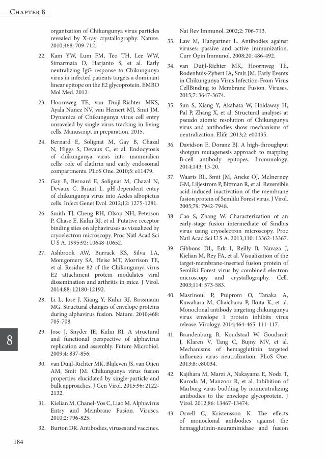
Figure S1 pH dependency of CHIKV-LR 2006 OPY1.Pyrene-labeled virus was mixed with liposomes in HNE buffer and fusion was triggered by addition of a low-pH buffer at t=0. Total fusion extent was determined as the average fusion extent at 50-60 s after acidification. All experiments were performed at least in quadruplicate, error bars depict SEM.
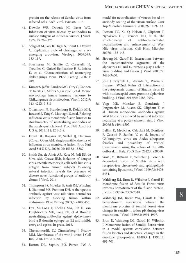
Table S1 Overview of fusion inhibitory capacities of neutralizing MAbs.
Pyrene-labeled CHIKV S27 was pre-opsonized with MAbs and mixed with liposomes in HNE buffer. Fusion was triggered by addition of a low-pH buffer at t=0. Total fusion extent was determined as the average fusion extent at 50-60 s after acidification. For each MAb, % fusion inhibition at the best inhibiting concentration is shown.
Mechanisms of CHIKV neutralization
8
187
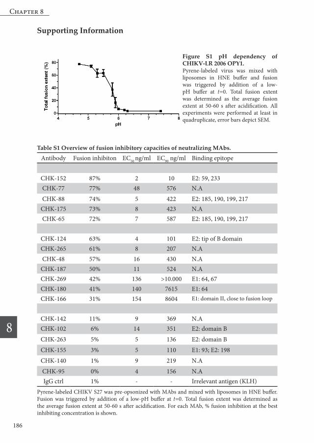
Figure S2 Dissociation of the E2/E1 heterodimer upon low pH exposure[35S]-methionine/L-[35S]-cysteine-labeled CHIKV was mixed with liposomes in HNE buffer or added to HNE buffer alone. Fusion was triggered by the addition of a pre-titrated pH buffer. After 60 s, the mixture was neutralized and solubilized by the addition of Igepal® CA-630 detergent to a final concentration of 1%. Subsequently, dissociation and trimerization of the viral spike proteins was analyzed in a continuous sucrose density gradient. For each condition, at least three independent experiments were performed, one representative curve is shown.
Figure S3 Analysis of the viral fusion kinetics at different temperaturesPyrene-labeled virus was mixed with liposomes in HNE buffer and fusion was triggered by addition of a low-pH buffer at t=0. Total fusion extent was determined as the average fusion extent at 50-60 s after acidification. Fusion rate was determined as the inverse time point at which half of the particles had fused. A series of fusion measurements was performed at temperatures ranging from ~ 19˚C to ~ 37˚C and both temperature and fusion rate were measured. The natural logarithm of the fusion rate k was plotted as a function of the inverse of the temperature T in Kelvin, to compare the data with the logarithm of the Arrhenius equation for a process overcoming a single energy barrier. This equation states k = Ae-Ea/kBT where k is the rate of a process, A a scaling constant, Ea the activation barrier height and kB the Boltzmann constant. Data were fitted linearly (red line; green lines: 95% confidence intervals of fit) with an R² of 0.77, yielding an Ea of 11±2 kcal/mol. No large deviation from Arrhenius behavior was observed.
Figure S4 Docking of individual CHIKV particles at increasing antibody concentrations.R18 labeled CHIKV was incubated with increasing dilutions of MAbs for 15 minutes. Opsonized virus was docked through a flow channel with lipid bilayer for 3 minutes at 50 μL/min through a channel of 0.1 mm² cross-section. Docked particles were observed using TIRF-microscopy and the number of particles in a defined field of view was analyzed.


![Dengue Fever/Severe Dengue Fever/Chikungunya Fever · Dengue fever and severe dengue (dengue hemorrhagic fever [DHF] and dengue shock syndrome [DSS]) are caused by any of four closely](https://static.documents.pub/doc/80x56/5e87bf3e7a86e85d3b149cd7/dengue-feversevere-dengue-feverchikungunya-dengue-fever-and-severe-dengue-dengue.jpg)


