University of Groningen Intricacies of the Molecular Machinery of Catecholamine Biosynthesis and Secretion by Chromaffin Cells of the Normal Adrenal Medulla and in Pheochromocytoma and Paraganglioma Berends, Annika M A; Eisenhofer, Graeme; Fishbein, Lauren; Horst-Schrivers, Anouk N A V D; Kema, Ido P; Links, Thera P; Lenders, Jacques W M; Kerstens, Michiel N Published in: Cancers DOI: 10.3390/cancers11081121 IMPORTANT NOTE: You are advised to consult the publisher's version (publisher's PDF) if you wish to cite from it. Please check the document version below. Document Version Publisher's PDF, also known as Version of record Publication date: 2019 Link to publication in University of Groningen/UMCG research database Citation for published version (APA): Berends, A. M. A., Eisenhofer, G., Fishbein, L., Horst-Schrivers, A. N. A. V. D., Kema, I. P., Links, T. P., Lenders, J. W. M., & Kerstens, M. N. (2019). Intricacies of the Molecular Machinery of Catecholamine Biosynthesis and Secretion by Chromaffin Cells of the Normal Adrenal Medulla and in Pheochromocytoma and Paraganglioma. Cancers, 11(8), [1121]. https://doi.org/10.3390/cancers11081121 Copyright Other than for strictly personal use, it is not permitted to download or to forward/distribute the text or part of it without the consent of the author(s) and/or copyright holder(s), unless the work is under an open content license (like Creative Commons). The publication may also be distributed here under the terms of Article 25fa of the Dutch Copyright Act, indicated by the “Taverne” license. More information can be found on the University of Groningen website: https://www.rug.nl/library/open-access/self-archiving-pure/taverne- amendment. Take-down policy If you believe that this document breaches copyright please contact us providing details, and we will remove access to the work immediately and investigate your claim.
Transcript
University of Groningen
Intricacies of the Molecular Machinery of Catecholamine Biosynthesis and Secretion byChromaffin Cells of the Normal Adrenal Medulla and in Pheochromocytoma andParagangliomaBerends, Annika M A; Eisenhofer, Graeme; Fishbein, Lauren; Horst-Schrivers, Anouk N A VD; Kema, Ido P; Links, Thera P; Lenders, Jacques W M; Kerstens, Michiel NPublished in:Cancers
DOI:10.3390/cancers11081121
IMPORTANT NOTE: You are advised to consult the publisher's version (publisher's PDF) if you wish to cite fromit. Please check the document version below.
Document VersionPublisher's PDF, also known as Version of record
Publication date:2019
Link to publication in University of Groningen/UMCG research database
Citation for published version (APA):Berends, A. M. A., Eisenhofer, G., Fishbein, L., Horst-Schrivers, A. N. A. V. D., Kema, I. P., Links, T. P.,Lenders, J. W. M., & Kerstens, M. N. (2019). Intricacies of the Molecular Machinery of CatecholamineBiosynthesis and Secretion by Chromaffin Cells of the Normal Adrenal Medulla and in Pheochromocytomaand Paraganglioma. Cancers, 11(8), [1121]. https://doi.org/10.3390/cancers11081121
CopyrightOther than for strictly personal use, it is not permitted to download or to forward/distribute the text or part of it without the consent of theauthor(s) and/or copyright holder(s), unless the work is under an open content license (like Creative Commons).
The publication may also be distributed here under the terms of Article 25fa of the Dutch Copyright Act, indicated by the “Taverne” license.More information can be found on the University of Groningen website: https://www.rug.nl/library/open-access/self-archiving-pure/taverne-amendment.
Take-down policyIf you believe that this document breaches copyright please contact us providing details, and we will remove access to the work immediatelyand investigate your claim.
Intricacies of the Molecular Machinery ofCatecholamine Biosynthesis and Secretion byChromaffin Cells of the Normal Adrenal Medullaand in Pheochromocytoma and Paraganglioma
Annika M.A. Berends 1,* , Graeme Eisenhofer 2, Lauren Fishbein 3,Anouk N.A. van der Horst-Schrivers 1, Ido P. Kema 4, Thera P. Links 1,Jacques W.M. Lenders 5,6 and Michiel N. Kerstens 1
1 Department of Endocrinology, University of Groningen, University Medical Center Groningen,9700 RB Groningen, The Netherlands
2 Department of Clinical Chemistry and Laboratory Medicine and Department of Medicine III,University Hospital Carl Gustav Carus, Technical University Dresden, 01069 Dresden, Germany
3 Division of Endocrinology, Metabolism and Diabetes and Division of Biomedical Informatics andPersonalized Medicine, Department of Medicine, University of Colorado School of Medicine,University of Colorado Cancer Center, Aurora, CO 80045, USA
4 Department of Laboratory Medicine, University of Groningen, University Medical Center Groningen,9700 RB Groningen, The Netherlands
5 Department of Medicine III, University Hospital Carl Gustav Carus, Technical University Dresden,01307 Dresden, Germany
6 Department of Internal Medicine, Radboud University Medical Center, 6525 GA Nijmegen, The Netherlands* Correspondence: [email protected]
Received: 22 June 2019; Accepted: 12 July 2019; Published: 6 August 2019�����������������
Abstract: The adrenal medulla is composed predominantly of chromaffin cells producing and secretingthe catecholamines dopamine, norepinephrine, and epinephrine. Catecholamine biosynthesis andsecretion is a complex and tightly controlled physiologic process. The pathways involved havebeen extensively studied, and various elements of the underlying molecular machinery have beenidentified. In this review, we provide a detailed description of the route from stimulus to secretionof catecholamines by the normal adrenal chromaffin cell compared to chromaffin tumor cells inpheochromocytomas. Pheochromocytomas are adrenomedullary tumors that are characterized byuncontrolled synthesis and secretion of catecholamines. This uncontrolled secretion can be partlyexplained by perturbations of the molecular catecholamine secretory machinery in pheochromocytomacells. Chromaffin cell tumors also include sympathetic paragangliomas originating in sympatheticganglia. Pheochromocytomas and paragangliomas are usually locally confined tumors, but about15% do metastasize to distant locations. Histopathological examination currently poorly predictsfuture biologic behavior, thus long term postoperative follow-up is required. Therefore, there is anunmet need for prognostic biomarkers. Clearer understanding of the cellular mechanisms involvedin the secretory characteristics of pheochromocytomas and sympathetic paragangliomas may offerone approach for the discovery of novel prognostic biomarkers for improved therapeutic targetingand monitoring of treatment or disease progression.
Keywords: PPGL; catecholamines; adrenomedullary function
The adrenal medulla occupies the central portion of the adrenal gland and accounts for about 10%of total adrenal gland volume [1]. The adrenal medulla is essentially a specialized sympathetic ganglionreleasing hormones in response to neural input and therefore is an integral part of the autonomic nervoussystem [2,3]. The adrenomedullary chromaffin cells are embryologically derived from migrating neuralcrest cells that develop into sympathoadrenal progenitors [4,5]. These sympathoadrenal progenitorcells also give rise to the chromaffin cells present in the sympathetic chain and prevertebral paraganglia.During adrenal organogenesis, close interactions between its two components, medulla and cortex, arenecessary for differentiation, morphogenesis, and survival of the adrenal gland. This cortical–chromaffincrosstalk remains important for physiological regulation of adrenal hormone biosynthesis in adultlife and also is relevant for the pathogenesis of various adrenal gland disorders [6–9]. One of thehistological infrastructural requirements for this crosstalk is the centripetally directed arterial bloodflow from adrenal cortex to medulla. In addition, cortical cells are diffusely present in the adrenalmedulla and, conversely, chromaffin cells are intermixed with cortical cells within all three zones of theadrenal cortex [10].
The principal function of the adrenal medulla is the biosynthesis and the secretion into thecirculation of the catecholamine epinephrine [6,11]. Epinephrine has a crucial role in the “fight-or-flight”response, which allows an organism to adapt to stressful conditions. The acute rise in epinephrine inresponse to physical or psychological stress stimuli results in hemodynamic and metabolic effects thatmodulate various functions such as blood pressure, cardiac output, and blood glucose by acting oncells expressing α- and β- adrenergic receptors [12,13]. Under basal conditions, however, epinephrinefunctions as a circulating metabolic hormone, and it is the norepinephrine secreted by sympatheticnerves acting immediately in the vicinity of exocytotic secretion that is the catecholamine mainlyregulating cardiovascular function. The norepinephrine that escapes re-uptake processes to enter thecirculation has negligible impact on the cardiovascular system. Nevertheless, both norepinephrine andepinephrine secreted by pheochromocytomas in excessive amounts directly into the circulation canhave profound effects on cardiovascular function, with further impacts of co-secreted peptides. From aclinical perspective, these tumors are the most important disease of the adrenal medulla. Chromaffincell tumors may also arise in extra-adrenal sympathetic paraganglia, in which case they are termedsympathetic paragangliomas [14–16].
A cardinal feature of chromaffin cell tumors is their capacity to produce and secrete excessiveamounts of catecholamines, which may evoke signs and symptoms such as paroxysmal hypertension,sweating, and tachycardia. The hypersecretion of catecholamines may cause acute, life-threateningblood pressure elevations and arrhythmias and is associated with a significantly increased rate ofcardiovascular morbidity and mortality [17–20].
Pheochromocytomas and sympathetic paragangliomas are rare neuroendocrine tumors withrespective reported annual incidences of 0.46 and 0.11 per 100,000 individuals [21]. Detected incidenceof pheochromocytomas has doubled during the past two decades, most likely a result of changesin diagnostic practices leading to earlier detection. The cornerstone of biochemical diagnosis of apheochromocytoma or a sympathetic paraganglioma is the demonstration of elevated plasma or urinaryconcentrations of metanephrine, normetanephrine, or 3-methoxytyramine, i.e., the O-methylatedmetabolites of epinephrine, norepinephrine, or dopamine, respectively [22]. Several anatomical andfunctional imaging studies are available for localization of the tumor, after which curative treatment bysurgical resection can be offered [23].
Pheochromocytomas and paragangliomas are highly heterogeneous neuro-endocrine tumorswith regards to possible anatomic location, genetic context, symptomatology, metastatic potential,and the degree of catecholamine release. Genetic mutations play a critical role in tumorigenesis andaffect various metabolic pathways, which also result in different mutation-dependent biochemicalphenotypes [3,24–26].
Cancers 2019, 11, 1121 3 of 33
In recent years, our knowledge of the genotype–phenotype interrelationship and metabolomicsof these intriguing neuro-endocrine tumors has expanded rapidly. Nevertheless, there are stillseveral areas of uncertainty. For instance, in the absence of metastases, it is difficult to predictwhether a pheochromocytoma or a paraganglioma will demonstrate a benign or a malignant clinicalcourse [16]. There are no clear-cut pathological markers to establish malignancy with certainty atfirst presentation. Also, there is no straightforward relationship between the biochemical phenotypeof a pheochromocytoma or sympathetic paraganglioma and the associated signs or symptoms [3].In the present review, we aim to provide a detailed picture of the pathways involved in catecholamineproduction and secretion in normal adrenomedullary chromaffin cells. We also visit what is knownabout the molecular perturbations in catecholamine biosynthesis and secretion in pheochromocytomaand sympathetic paraganglioma. Improved understanding of these mechanisms at the molecular levelmight provide insight into associated pathological complications, clarify highly variable presentations,and aid in identification of new diagnostic or therapeutic strategies for personalized care.
2. Adrenomedullary Function
2.1. Biosynthesis of Catecholamines
Adrenomedullary catecholamine biosynthesis starts with uptake of the nonessential amino acidL-tyrosine by the chromaffin cell. L-tyrosine is obtained from food sources or is derived from theessential amino acid phenylalanine through the activity of phenylalanine hydroxylase, which is mainlyexpressed in liver, kidney, and pancreas [27,28]. L-tyrosine is transported into the cytoplasm of theadrenal chromaffin cell by the membrane bound L-type amino acid transporter system (LAT1 andLAT2) [29,30]. Catecholamine biosynthesis involves the sequential activity of four enzymes: tyrosinehydroxylase (TH), aromatic L-amino acid decarboxylase (AADC), dopamine β-hydroxylase (DBH),and phenylethanolamine-N-methyltransferase (PNMT). Except for DBH, all these enzymes are localizedin the cytoplasm of the chromaffin cell (Figure 1). The end products of this biosynthetic route aredopamine, norepinephrine, or epinephrine, depending on intracellular enzyme expression. Epinephrineis mainly produced by the adrenomedullary chromaffin cells (>95%) and functions as a hormonereleased directly into the bloodstream. In contrast, circulating norepinephrine is mainly derived fromoverflow of the neurotransmitter from sympathetic nerve endings with adrenomedullary chromaffincell production providing usually a less than 10% contribution [11,13,31].
2.1.1. Tyrosine Hydroxylase
The initial and rate limiting step in catecholamine biosynthesis is the conversion of L-tyrosine toL-3,4-dihydroxyphenylalanine (L-DOPA) by tyrosine hydroxylase (TH, EC 1.14.16.2, molecular massof approximately 240 kDa) [32,33]. Locations of catecholamine biosynthesis are therefore dependenton the expression of TH, which is largely confined to postganglionic sympathetic nerve endingsand adrenal and extra-adrenal chromaffin cells. In the adrenal medulla, this enzyme has a Km of2 × 10−5 mol/L [27]. For this specific hydroxylation step, TH requires tetrahydrobiopterin, molecularoxygen, and Fe2+ as cofactors. Tetrahydrobiopterin is synthesized from guanosine triphosphate(GTP) and serves as a donor for hydrogen atoms to maintain TH in a reduced and active state [27,34].The human TH gene is located at chromosome 11p15.5 and contains 13 exons [35], with four isoformsproduced by alternative mRNA splicing.
Regulation of TH activity is an important way to control catecholamine biosynthesis. This isa complex process encompassing multiple modes of regulation. Short-term post-transcriptionalmechanisms include feedback inhibition by catecholamines, enzyme phosphorylation anddephosphorylation, as well as ubiquitination. Long-term regulation mainly involves transcriptionalmechanisms [36]. The ubiquitin–proteasome pathway is thought to be involved in the degradation ofTH [37]. Catecholamines exert negative feedback control through oxidation of tetrahydrobiopterinto pteridine, thereby preventing the formation of TH in its reduced active state [27]. In addition,
Cancers 2019, 11, 1121 4 of 33
catecholamines act as competitive antagonists of tetrahydrobiopterin at the active site of the catalyticdomain of TH [27]. Short-term regulation of TH activity is also achieved by a mechanism ofphosphorylation and dephosphorylation of one or more of the four serine residues at the regulatorysite of TH. Phosphorylation is catalyzed by multiple kinases (e.g., PKA, PKC, CaMKII, MAPKAP-K2,ERK1, ERK2, MSK1, PRAK) and results in release from the feedback inhibition by catecholamines,thereby stimulating enzyme activity.
Figure 1. The catecholamine biosynthetic pathway in an adrenomedullary chromaffin cell or apheochromocytoma cell. Norepinephrine and epinephrine are stored in separate chromaffin storagevesicles. Abbreviations: LAT: L-type amino acid transporter; TH: tyrosine hydroxylase; L-DOPA:L-3,4-dihydroxyphenylalanine; AADC: aromatic L-amino acid decarboxylase; DBH: dopamineβ-hydroxylase; PNMT: phenylethanolamine-N-methyltransferase; BH4: tetrahydrobiopterin; 02: molecularoxygen; VitB6: pyridoxalphosphate; VitC: ascorbate; VMAT: vesicular monoamine transporters;GR: glucocorticoid receptor.
Dephosphorylation by phosphatase PP2A, and to a lesser extent by PP2C, restorescatecholaminergic inhibition of the TH enzyme [36]. This negative feedback is mediated viaalpha2-adrenergic or D2-dopaminergic receptors, which activate cyclic adenosine monophosphate(cAMP) or Ca2+/calmodulin-dependent protein phosphatases [27]. Prolonged stimulation ofcatecholamine biosynthesis results in induction of TH protein synthesis through several cAMPdependent pathways activating TH gene transcription [27,34,36,38–40].
Given the importance of its activity to catecholamine synthesis and the complexity of its regulation,TH has gained great interest in many fields of biomedical research. Recent studies, for example,have demonstrated the presence of several TH polymorphisms in the general population, some ofwhich appear associated with increased norepinephrine levels and elevated blood pressure [35,41].
2.1.2. Aromatic L-Amino Acid Decarboxylase
The next step in catecholamine biosynthesis is the decarboxylation of L-DOPA to dopamineby cytosolic aromatic L-amino acid decarboxylase (AADC; EC 4.1.1.28). For this conversion,pyridoxalphosphate (vitamin B6) is required as a cofactor [27,42]. AADC is a 100 kDa homodimericprotein encoded by a single gene located at chromosome 7p12.1 with a Km of 4 × 10⁻⁴ mol/L [42,43].The calculated Km greatly exceeds the endogenous concentration of L-DOPA, which means that
Cancers 2019, 11, 1121 5 of 33
the AADC enzyme is not fully saturated, and the rate at which dopamine can be synthesized istherefore limited by the availability of L-DOPA as a substrate [42]. The AADC enzyme has a widetissue distribution and is not specific for chromaffin cells [42,44].
Short-term regulation of AADC enzyme activity by a previously postulated mechanism involvingcAMP or phosphorylation by protein kinases seems to play no significant role in the regulation of thecatecholamine biosynthetic pathway in situ [42,45]. The added value of AADC enzyme upregulationby modulation of gene expression for physiological demands to increase catecholamine production inpostganglionic sympathetic nerve endings remains questionable [45–47].
2.1.3. Dopamine β-hydroxylase
In adrenal chromaffin cells, dopamine is further catalyzed to norepinephrine by dopamineβ-hydroxylase (DBH; EC 1.14.17.1). Because of the intravesicular location of DBH, dopamine first mustbe translocated into norepinephrine storage vesicles by vesicular monoamine transporters (VMATs) [11].The intravesicular conversion of dopamine represents the final step in the biosynthesis of norepinephrine.DBH, a mixed-function oxidase, is a 290 kDa copper protein with a Km of 8.4 × 10⁻⁴ mol/L and utilizesmolecular oxygen, fumarate, and L-ascorbic acid as its main cofactors [27,48,49]. These requirementsfor L-ascorbic acid and fumarate are not specific. Catechol (i.e., pyrocatechol or 1,2 dihydroxybenzene)seems to be a weak substitute for L-ascorbic acid and other activating anions, such as acetate andchloride, which can replicate the effects of fumarate at least partially [27,48,50–52].
In humans, DBH is encoded by a gene located at chromosome 9q34.2. Increased catecholaminebiosynthesis in response to stress is associated with increased levels of mRNAs encoding catecholaminesynthesizing enzymes. In adrenomedullary chromaffin cells, this response to stress is rapid, especiallyfor TH and PNMT [39]. Previous studies also revealed upregulation of adrenal DBH gene expression byvarious transcriptional mechanisms in response to prolonged or repeated stressors [39,53]. However,in contrast to TH, short or intermediate duration of stress does not result in a significant increase ofDBH mRNA [39].
2.1.4. Phenylethanolamine-N-Methyltransferase
Norepinephrine formed in the chromaffin vesicles diffuses passively into the cytosol,where it is converted to epinephrine by the enzyme phenylethanolamine-N-methyl transferase(PNMT; EC 2.1.1.28) [11,24]. PNMT, which has a predicted molecular weight of 30.9 kDa and a Kmof 9.2 × 10⁻⁶ mol/L, requires S-adenosylmethionine as a methyl donor and cosubstrate [27,54,55].PNMT is not substrate specific and also is involved in the biosynthesis of other N-methylated traceamines [11,27]. Expression of PNMT is controlled by glucocorticoid receptor-mediated mechanisms,acting in concert with several other transcription factors such as Egr-1, AP2, Sp1, and MAZ [6,39,56,57].The proximity of adrenocortical cells to the adrenal medulla guarantees high circulating glucocorticoidlevels, which cross the chromaffin cell membrane through passive diffusion. Glucocorticoid binds tothe intracytoplasmatic glucocorticoid receptor, and the receptor–hormone complex migrates to the cellnucleus and binds to the glucocorticoid response element of the promoter region of the PNMT genelocated on chromosome 17q12, activating gene transcription [7,58,59]. This explains why the adrenalgland is the body’s most important source of epinephrine, whereas the expression of extra-adrenalPNMT is limited to a small number of neurons in the central nervous system and to a subset ofcardiomyocytes [6,60,61].
2.1.5. Co-Secreted Products
Chromaffin cells of the adrenal medulla synthesize a large variety of other substances, such asneurotransmitters, enzymes, peptides, and proteins, which are also stored in chromaffin vesicles andco-secreted along with catecholamines [62–64]. Over the past decades, the components of this vesicularcocktail have been studied in great detail (Table 1).
Cancers 2019, 11, 1121 6 of 33
Table 1. Overview of the co-secreted products of chromaffin vesicles with description of their function in normal adrenal medulla and reported alterations inpheochromocytoma and paraganglioma (PPGL).
Component Function in Human Adrenal Medulla Reported Alterations in PPGL
Granins
Chromogranin A–C [65–77]
Role in vesiculogenesis, vesicle protein stability, hormone storage within vesicles.Sorting proteins in the regulated secretory pathway.
Precursor protein for several peptides; chromogranin A (vasostatin I–II, catestatin,cateslytin, chromacin, chromofungin, pancreastatin, parastatin, WE-14, EL35),
chromogranin B (secretolytin), chromogranin C (secretoneurin, EM66, manserin).
Higher plasma levels of chromogranin A and B are reported in PPGL compared tohealthy volunteers.
Chromogranin C mRNA was overexpressed in PPGL compared to non-tumoralchromaffin tissue.
Higher expression of chromogranin A and B in RET associated PPGL compared toVHL associated PPGL at both the mRNA and protein levels.
Downregulation of chromogranin B and chromogranin C observed to be associatedwith malignant behavior.
Secretogranins III–VII [78–87]
Secretogranin III (syn. 1B1075) not found in adrenal medulla.Presence of secretogranin IV (syn. HISL-19), V (syn. 7B2), VI (syn. NESP55) and VII(VGF) reported in human adrenal medulla, exact function still mainly unknown.
Proposed role of secretogranin VII (syn. VGF) in the regulation ofenergy homeostasis.
More pronounced immunoreactivity of secretogranin IV in malignant PPGLcompared to benign PPGL.
Significantly higher plasma levels of secretogranin V in PPGL compared toage-matched normal subjects.
Secretogranin VI immunoreactivity found in PPGL with no differences betweenbenign and malignant tumors. Variable proVGF-immunoreactive fragments are
observed in human PPGL.
Glycoproteins
Glycoprotein I–V [88–90]Glycoprotein I, i.e., DBH, catalyzes the conversion of dopamine into
norepinephrine. Glycoprotein IV, i.e., the H+-ATP-ase subunit M45, provides thedriving force for vesicular uptake of catecholamines by VMAT.
High expression levels of glycoprotein I (DBH) are reported in PPGL.
Prohormone processing enzymes
Aminopeptidase B (Ap-B) [91] Exopeptidase involved in final conversion of proenkephalin to enkephalin. Association of Ap-B with the secretory machinery is suggested in ratpheochromocytoma (PC12) cells.
Aspartic Proteinase [92] Contributes to enkephalin precursor cleaving activity. Unknown
Carboxypeptidase E (CPE) [93,94] Role in peptide processing and sorting of prohormones.
High expression of CPE mRNA are reported. Elevated expression correlated withtumor growth and metastasis in pheochromocytomas.
CPE promotes survival of pheochromocytoma (PC12) cells under nutrientstarvation and hypoxic conditions by upregulation of pro-survival genes possibly
by activation of the ERK1/2 pathway.
Cathepsin L [95,96] Endopeptidase involved in proteolysis of proenkephalin into (Met)enkephalin.Proprotein convertase for biosynthesis of NPY and catestatin. Unknown
Prohormone convertase 1/3and 2 [73,78,97] Conversion of chromogranin C into secretoneurin and EM66.
PC1 and PC2 mRNA expression levels are significantly higher in benign andmalignant PPGL compared to normal adrenal medulla. mRNA expression and
protein levels of PC1 and PC2 is 3–4 times higher in benign tumors compared tomalignant tumors.
Tissue-Type Plasminogen Activator(t-PA) [98,99]
Participation in plasmin-dependent processing of bioactive peptides includingchromogranin A and indirectly modulate chromogranin A release
(negative-feedback loop).Marked expressions of t-PA mRNA are reported in human pheochromocytomas.
Cancers 2019, 11, 1121 7 of 33
Table 1. Cont.
Component Function in Human Adrenal Medulla Reported Alterations in PPGL
Endopin 2 inhibits papain-like cysteine proteases, including cathepsin L, as well asthe serine protease elastase.
Unknown
Transmitter peptides
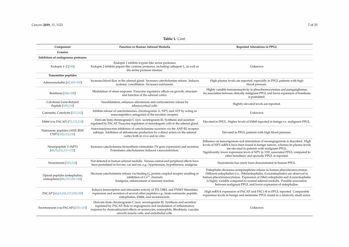
Adrenomedullin [65,101–105] Increases blood flow in the adrenal gland. Increases catecholamine release. Inducessystemic vasodilation. Increases natriuresis.
High plasma levels are reported, especially in PPGL patients with highblood pressure.
Bombesin [106–108] Modulation of stress response. Paracrine regulatory effects on growth, structureand function of the adrenal cortex.
Highly variable immunoreactivity in pheochromocytomas and paragangliomas.An association between clinically malignant PPGL and lower expression of bombesin
is postulated.
Calcitonin Gene-RelatedPeptide [109,110]
Vasodilatation, enhances aldosterone and corticosterone release byadrenocortical cells. Slightly elevated levels are reported.
Catestatin, Cateslytin [111,112] Inhibits release of catecholamines, chromogranin A, NPY, and ATP by acting asnoncompetitive antagonist of the nicotinic receptor. Unknown
EM66 (via PACAP) [73,113,114] Derivate from chromogranin C (syn. secretogranin II). Synthesis and secretionregulated by PACAP. Paracrine regulation of steroidogenic cells in the adrenal gland. Elevated in PPGL. Higher levels of EM66 reported in benign vs. malignant PPGL.
Natriuretic peptides (ANP, BNP,CNP) [102,115,116]
Autocrine/paracrine inhibition of catecholamine secretion via the ANF-R2 receptorsubtype. Inhibition of aldosterone production by a direct action on the adrenal
cortex both in vivo and in vitro.Elevated in PPGL patients with high blood pressure.
Influence on tumorigenesis and stimulation of neoangiogenesis is described. Highlevels of NPY mRNA have been found in benign tumors, whereas its plasma levels
are elevated in patients with malignant PPGL.Significantly lower expression level of NPY in VHL associated PPGL compared to
other hereditary and sporadic PPGL is reported.
Neurotensin [123,124] Not detected in human adrenal medulla. Various central and peripheral effects havebeen postulated in bovine, cat and rat, e.g., hypotension, hypothermia, analgesia. Neurotensin has rarely been demonstrated in human PPGL.
Decrease catecholamine release via binding Gi protein coupled receptor resulting ininhibition of Ca2+ channels
Analgesia, enhancement of immune reaction.
Enkephalin decreases norepinephrine release in human pheochromocytomas.Different enkephalins (i.e., (Met)enkephalin, (Leu)enkephalin) are observed in
human pheochromocytomas. Expression of (Met) enkephalin and (Leu)enkephalinis highly variable compared to normal adrenal medulla. Possible association
between malignant PPGL and lower expression of enkephalins.
PACAP [40,63,101,117,129,130]Induces transcription and stimulates activity of TH, DBH, and PNMT Stimulatesexpression and secretion of several other peptides e.g., brain natriuretic peptide,
enkephalins, EM66, and secretoneurin.
High mRNA expression of PACAP and PAC1-R in PPGL reported. Comparableexpression levels in benign and metastatic PPGL found in a relatively small series.
Secretoneurin (via PACAP) [131–133]
Derivate from chromogranin C (syn. secretogranin II). Synthesis and secretionregulated by PACAP. Role in angiogenesis and modulation of inflammatory
response by chemoattractant effects on monocytes, eosinophils, fibroblasts, vascularsmooth muscle cells, and endothelial cells.
Unknown
Cancers 2019, 11, 1121 8 of 33
Table 1. Cont.
Component Function in Human Adrenal Medulla Reported Alterations in PPGL
Transforming Growth Factorβ [134–136]
Role in regulation of chromaffin cell proliferation and differentiation. Reduction ofTGF β has been shown to increase proliferation of chromaffin cell in vivo. Unknown
Granins
Vasostatins [137–139]The N-terminal fragment of chromogranin A
Inhibition of endothelin-induced vasoconstrictionAntibacterial and antifungal activity.
Unknown
Anti-bacterial/anti-fungal peptides
Chromacin P, G and PG [140] Antibacterial activity against Gram positive bacteria. Unknown
Secretolytin [141] Antibacterial activity against Gram positive bacteria. Unknown
Ubifungin [142] Antifungal activity. Unknown
Other minor components
Ascorbic acid [143] Regulation of DBH activity. Unknown
Coenzyme A glutathionedisulfide [144] Vasoconstriction, modulation of AngII effects. Unknown
Galanin [81,146,147] Stimulation of norepinephrine and glucocorticoid secretion.
Immunoreactivity in human PPGL.Higher levels reported in PPGL compared to normal adrenal medulla.
Variable expression; galanin was predominantly found in noradrenergicpheochromocytoma cells.
Induction of apoptosis in PC12 cells.Inhibition of dopamine secretion in pheochromocytoma cells.
Nucleotides (ATP, ADP, GTP) [148] Formation of intravesicular complex with catecholamines, buffer function,decreases intravesicular osmotic pressure, neuromodulation. Unknown
Substance P [149–153] Inhibition of nicotinic acetylcholine receptor mediatedcatecholamine release. Vasodilation.
Variable immunoreactivity demonstrated in human pheochromocytomas. Elevatedplasma levels in minority of patients.
Vasoactive intestinalpolypeptide [154–156] Stimulation of catecholamine release, stimulation of steroid secretion. Few cases described of human PPGL with concomitant excessive VIP secretion.
Among these substances, chromogranin A and the trophic and secretion stimulating peptidespituitary adenylate cyclase-activating polypeptide (PACAP), neuropeptide Y (NPY), and adrenomedullin(AM) have gained the most attention because of their endocrine, paracrine, and autocrine effects,their importance for vesiculogenesis, and their possible roles in neoplastic chromaffin cell proliferation,differentiation, and survival, as further discussed below [62–65,101,113,129,157].
2.2. Storage and Secretion of Catecholamines
2.2.1. Storage and Vesicular Transmembrane Dynamics
In adrenomedullary chromaffin cells, catecholamines are stored in specialized vesicles.The bidirectional vesicular–cytosolic exchange of catecholamines is a dynamic process of activeuptake into these chromaffin storage vesicles and passive leakage from vesicles into the cytosol [3].After synthesis, dopamine and epinephrine are actively transported from the cytosol into chromaffinstorage vesicles by vesicular monoamine transporters (VMAT1 and VMAT2) [158,159]. The drivingforce for this active transport is provided by an ATP-dependent vesicular membrane proton pump thatmaintains a transvesicular hydrogen ion (H+) electrochemical gradient by acidifying the vesicle matrix.Vesicular uptake for catecholamines via VMAT is accompanied by exchange of an H+ ion from thevesical matrix towards the cytosol [62,160] (Figure 1).
2.2.2. Characteristics of Chromaffin Storage Vesicles
Chromaffin storage vesicles are highly specialized organelles of the chromaffin cells for storageand exocytosis. These membrane-bound electron-dense organelles originate from the Golgi networkand are 150 to 350 nm in diameter. Each adrenomedullary chromaffin cell contains about 12,000 to30,000 of these vesicles, corresponding (on average) to 13.5% of the cytoplasmic cell volume [63,65].
The adrenal medulla of some species harbors two distinct populations of chromaffin cells,which either produce epinephrine or norepinephrine depending on the presence or the absenceof PNMT [3,11,63]. The proportion of epinephrine versus norepinephrine producing chromaffincells in the adrenal medulla varies between species, but the adrenergic phenotype usuallypredominates [3,6,62,63,161]. This is particularly so in humans, where most chromaffin cellsappear to have mixed function [6,11,62,161,162]. Nevertheless, there are clear ultrastructuraldifferences between epinephrine and norepinephrine-containing vesicles when studied by electronmicroscopy. Epinephrine-containing vesicles are round or elongated in shape and demonstratefine granular, medium-density vesicles with a characteristic narrow and uniform peripheral halo,whereas norepinephrine-containing vesicles demonstrate a high density and homogeneous content witha limiting membrane, which may be separated from the matrix constituents by a prominent lucent halo.
The mechanisms initiating and regulating the biogenesis of chromaffin vesicles are largelyunknown. It is believed that structural proteins of the granin family, in particular chromogranin A,have an important role in vesiculogenesis, providing structural domains that drive chromaffin vesicleformation in the Golgi network. Furthermore, the ability of chromogranin A to bind catecholamines isthought to regulate stability of the vesicle by reducing osmotic pressure, thereby preventing vesiclesfrom bursting; it is also thought to protect catecholamines against enzymatic degradation until secretionis warranted [27,63,65].
After the formation of chromaffin vesicles, maturation continues with catecholamine synthesisand storage not occurring until late in vesicle formation [63]. Fully matured chromaffin vesicles remainin the chromaffin cell until stimulation for exocytosis [63,65].
The molecular composition of chromaffin vesicles is complex. Besides catecholamines,intravesicular contents include a diverse mixture of peptides, proteases, enzymes, and granins(chromogranins, secretogranins) with a multiplicity of functions (Table 1). The high concentration ofregulatory and modulating peptides and proteins reflects broad endocrine, paracrine, and autocrinefunctions of adrenomedullary chromaffin vesicles. The physiological processes modulated by these
Cancers 2019, 11, 1121 10 of 33
constituents not only involve the fine-tuning of catecholamine biosynthesis and secretion but alsoencompass various analgesic, immunomodulatory, antimicrobial, and anti-inflammatory responses tocell stress [62,63].
2.2.3. Secretion and Re-Uptake of Catecholamines
After exocytosis of storage vesicles and secretion of catecholamines into the bloodstream,norepinephrine and epinephrine are removed from the circulation by neural and extra-neuronalmonoamine transporters and are inactivated by metabolizing enzymes [163]. The re-uptake mechanismthrough the norepinephrine transporter (NET) by catecholamine synthesizing cells is only relevant insympathetic postganglionic and central nervous system neurons and provides rapid termination of theneurotransmitter signal at the postsynaptic membrane and enables recycling of catecholamines forre-release. NET is not only located presynaptically but also at several extraneuronal sites, includingthe adrenal medulla [31,163,164]. The precise function of the NET in the adrenal gland is, however,not entirely clear. A detailed discussion of reuptake as well as pre- and postsynaptic effects ofcatecholamines is beyond the scope of the current review, and for further reading, we refer to theliterature [163,165,166].
2.3. Regulation of Adrenomedullary Activity
2.3.1. Stimulus-Dependent Exocytosis in Adrenal Chromaffin Cells
Exocytosis of chromaffin storage vesicles is a tightly controlled process. Under basal conditions,only a few secretory vesicles are released into the circulation, resulting in a catecholamine secretion ratein the order of nanograms per minute [117]. Toxic effects of excessive chronic catecholamine release suchas in heart failure, pulmonary edema, and malignant hypertension have been reported from incessantcirculating catecholamine concentrations of 10−6 mol/L or more, corresponding to the release of 5%of all adrenomedullary chromaffin vesicles [167]. In acute stress situations, the amount of releasedvesicles from the total adrenal gland can be temporarily greatly increased, with plasma catecholamineconcentrations reaching up to 60 times more than normal [117,167]. Well-known stimuli that activatethe exocytotic process are hypoglycaemia, hypovolemia, hypotension, hypoxemia, and severe pain oremotional distress [3].
The rather complex mechanisms regulating chromaffin cell exocytotic machinery are executed atneuronal and non-neuronal levels [145,167–169]. It is thought that each adrenomedullary chromaffincell receives its own individual neuronal and non-neuronal input [167].
At a neuronal level, adrenomedullary chromaffin cells are innervated by the cholinergicpreganglionic sympathetic fibers of the splanchnic nerve. One single chromaffin cell can receive inputby up to five synapses [170]. Acetylcholine released by these nerve endings predominantly bindsto nicotinic receptors on the chromaffin cell, resulting in membrane depolarization with subsequentcalcium influx followed by stimulation of exocytosis and catecholamine secretion. Cholinergic receptorsof the muscarinic type are also expressed on the chromaffin cell, but their contribution to catecholaminesecretion is less important. Further enhancement of the stimulation–secretion coupling is provided bypropagation of the secretion signal via gap junctions between chromaffin cells formed by connexins,which are specific proteins involved in cell-to-cell communication. This intercellular communicationcan be upregulated in stressful conditions [170]. Besides acetylcholine, splanchnic nerve terminalsalso contain PACAP as a neurotransmitter. This neuropeptide is not only stored in chromaffin vesiclesand co-secreted with catecholamines but also acts as an important neurotransmitter at the splanchnicmedullary synapse, where it activates the PACAP-preferring receptor (PAC1-R) on the postsynapticmembrane of the chromaffin cell. Laboratory experiments have shown that PACAP is only released athigh frequencies of nerve stimulation, which is the firing rate occurring in stress conditions. In contrastto the acetylcholine evoked catecholamine secretion, adrenomedullary stimulation by PACAP is
Cancers 2019, 11, 1121 11 of 33
not susceptible to desensitization, which ensures robust catecholamine release under conditions ofcontinuous stress (Figure 2) [40,130].
Figure 2. Schematic overview of the stimulation–secretion coupling in the adrenomedullarychromaffin cell with the multiple functionally definable stages and the different secretory pathways.Abbreviations: ER: endoplasmic reticulum; Ach: acetylcholine; VAMP: vesicle-associated membraneprotein; SNAP: synaptosomal-associated protein; NSF: N-ethylmaleimide Soluble Factor proteins;CADPS: Ca2+ dependent secretion activator; CALM: calmodulin; PACAP: pituitary adenylatecyclase-activating polypeptide; PAC1 receptor: PACAP-preferring receptor; GR: glucocorticoid receptor.
Non-neuronal regulation of exocytosis occurs predominantly through autocrine or paracrineroutes. As a result, cellular catecholamine secretion is partially under the influence of the exocytoticactivity of neighboring chromaffin cells [167]. Furthermore, both lipopolysaccharide and cytokinereceptors were recently demonstrated on chromaffin cells, pointing towards a role of the adrenalmedulla in the complex regulation of the inflammatory stress response [40].
In general, the exocytotic secretion of vesicular contents of adrenomedullary chromaffin cells can beachieved via regulated and constitutive secretory pathways. The regulated secretory pathway providesthe principle mechanism responsible for controlled release of catecholamines and is calcium-dependentand responsive to both neuronal and non-neuronal input. In contrast, the constitutive secretorypathway, which is calcium-independent, is mainly unresponsive to neuronal and non-neuronal input.The principal function of the constitutive secretory pathway is thought to be the transport of proteinsand macromolecules to the cell surface for purposes of membrane maintenance and support of theextracellular matrix. In addition, this pathway may also contribute to basal release of catecholamines(Figure 2) [145,169].
2.3.2. Neuronal Regulation of the Calcium-Dependent Catecholamine Secretory Pathway
In recent years, considerable progress has been made in unravelling the complex molecularbackground and the functional elements of the highly regulated exocytotic machinery in
Cancers 2019, 11, 1121 12 of 33
adrenomedullary chromaffin cells (Figure 2) [145,171,172]. Release of acetylcholine by the splanchnicnerve activates the nicotinic receptor on chromaffin cells, resulting in opening of the ionophoric partof the receptor protein, thereby allowing the entry of extracellular sodium (Na+) and calcium (Ca2+).This generates a small membrane depolarization, resulting in the opening of voltage dependent Na+
channels. The subsequent Na+ influx results in a large membrane depolarization, which opens varioustypes of voltage dependent Ca2+ channels [167].
The distribution of the different calcium channel subtypes is species specific. The P/Q-type calciumchannel predominates in human adrenomedullary chromaffin cells [167,171,172]. As a consequence ofelevated intracellular Ca2+ concentrations, the exocytotic machinery and the regulatory componentsfor vesicular exocytosis are activated, which occurs through a pathway consisting of secretory vesiclerecruitment, docking, priming, and fusion with the plasma membrane. Priming is the process in whichsecretory vesicles become fusion competent [167,172]. First, Ca2+ influx leads to dismantling of thecortical actin cytoskeleton of the chromaffin cell, a dynamic network of numerous cytoplasmic proteinslocated on the inner face of the chromaffin cell membrane [173]. Although this can be activated withoutCa2+, this is mainly an ATP and a Ca2+ dependent step, as are recruitment, tethering, and docking ofthe vesicles.
Fusion, release, and retrieval of vesicles can be triggered by Ca2+ in the absence of ATP [145,174].Various soluble and membrane-bound proteins are involved in the complex protein–protein interactionsunderlying membrane trafficking and fusion. Key components of this process are N-ethylmaleimidesoluble factor proteins (soluble cytosolic NSF), soluble NSF attachment proteins (SNAPs), and solubleNSF attachment receptor proteins (SNAREs). The vesicle-associated membrane protein (VAMP orsynaptobrevin) and the calcium binding protein synaptotagmin are SNAREs located at the secretoryvesicle membrane. The synaptosomal-associated protein 25 (SNAP-25) and syntaxin are SNAREsacting on the chromaffin cell plasma membrane [145,167,172]. The complex formed by chromaffinvesicle SNAREs (VAMP, synaptotagmin), the chromaffin cell membrane SNAREs (syntaxin, SNAP-25),and the cytosolic proteins (NSF) is thought to provide the primary molecular machinery responsiblefor the docking and the fusion of synaptic vesicles at the chromaffin cell plasma membrane(Figure 2) [174]. Furthermore, it is believed that NSF and SNAPs have multiple sites of actionson SNARE proteins. Besides pre-docking actions and their function as molecular chaperones in SNAREpriming, they also act on SNAREs post-fusion to facilitate vesicle retrieval and allow recycling of emptyvesicles [145,167,172,174].
Apart from NSF, SNAPs, and SNAREs, several other proteins are involved in calciumtriggered exocytosis. Proposed candidates are the stabilizing protein Munc18-1, the calciumtransducer calmodulin (CALM), the Ca2+ dependent secretion activator (CAPS), rabphilin,and annexins [169,172,174]. Rabphilin3A is a small GTP-ase, which acts as a molecular switch,thereby determining the sensitivity of secretory vesicles for docking and fusion. Overexpression ofrabphilin3A has been found to inhibit exocytosis in adrenomedullary chromaffin cells and probablyprotects against spontaneous exocytosis under basal conditions [175]. Annexins have the ability toform cross-links between secretory vesicles and the plasma membrane by a functional interplay withSNAREs during exocytosis [145,169,176–178].
2.3.3. Non-Neuronal Regulation of Catecholamine Secretion
Along with catecholamines, the adrenal medulla synthesizes and releases numerousenzymes, peptides, and proteins that exert various trophic and neurotransmitter activities,which provide fine-tuning of catecholamine synthesis and secretion in an autocrine anda paracrine manner [62–65,101,111,113,117,118,129,157] (Table 1). We discuss here in more detailneuropeptide Y (NPY), adrenomedullin (AM), PACAP, and the secretion-inhibiting peptidecatestatin [101,111,117,118,129]. These peptides modulate chromaffin cell function through a variety ofmembrane receptors, the vast majority of which belong to the family of G protein-coupled receptors(GPCRs) [117].
Cancers 2019, 11, 1121 13 of 33
Neuropeptide Y is a 36-amino acid neuropeptide, which is widely and abundantly distributed inthe brain and the sympathetic nervous system, including the human adrenal medulla. Several NPYreceptors (i.e., Y1, Y2, Y4, and Y5) are expressed in the adrenal gland, indicating that NPY exerts localautocrine effects. It has been shown that NPY is able to stimulate catecholamine secretion by inducingTH expression and increasing intracellular calcium [63,101,117,118,129,179].
Adrenomedullin (AM) is a 52-amino acid peptide originally isolated from a humanpheochromocytoma and also is present at high concentrations in the normal adrenalmedulla [101,117,129]. AM demonstrates autocrine and paracrine effects through binding to theadrenomedullin receptor (ADMR), the receptor dog cDNA (RDC1), and the calcitonin receptor-likereceptor (CRLR), which results in augmentation of the adrenal blood flow and stimulation ofcatecholamine release. In addition, the endocrine effects of AM include systemic vasodilatationand stimulation of natriuresis [63,101,117,129].
As previously mentioned, PACAP not only acts as a neurotransmitter released by the splanchnicnerve but is also co-secreted with catecholamines, exerting its effects by binding to the PACAP-preferringreceptor (PAC1-R) and VIP/PACAP receptors (VPAC1-R and VPAC2-R), the former representing thepredominant receptor in chromaffin cells (Figure 2) [101,117,129]. PACAP, a neuropeptide of 27 or38 amino acids, enhances catecholamine secretion by induction of transcription as well as stimulatingthe activity of the biosynthetic enzymes TH, DBH, and PNMT [101]. In normal adrenal medullarychromaffin cells, PACAP also stimulates the expression and the secretion of several other peptides,such as brain natriuretic peptide, enkephalins, EM66, and secretoneurin, which in turn exert their ownindividual autocrine/paracrine effects on catecholamine secretion [40,63,101,117,129,130].
Catestatin is a biologically active peptide fragment derived from proteolytic chromograninA cleavage [63]. Catestatin acts mainly as a noncompetitive nicotinic cholinergic antagonist,thus providing a strong negative feedback inhibition of catecholamine secretion [111,112].
3. Pheochromocytoma and Paraganglioma
From a genetic perspective, pheochromocytomas and paragangliomas (PPGL) have one of therichest hereditary backgrounds among all neoplasms. At least 35—perhaps up to 40%—of all PPGLharbor a germline pathogenic variant in one of the several susceptibility genes [180–184]. Initial geneexpression profiling studies by Dahia et al. in 2005 [185] revealed two cluster groups, designatedcluster 1 and cluster 2, that reflected respective activation of pseudohypoxia and kinase signalingpathways. Those different gene expression signatures matched closely to those reported in an earlierstudy for norepinephrine- versus epinephrine-producing sporadic tumors and tumors from patientswith von-Hippel Lindau (VHL) syndrome and multiple endocrine neoplasia type 2 (MEN2) [186].The molecular characterization from The Cancer Genome Atlas (TCGA) project has more recentlyprovided a sophisticated molecular taxonomy of PPGL, which divides these neuroendocrine tumorsinto groups with similar pathogenesis and molecular biology and provides an up-to-date framework(Figure 3) [187–189]. The recent identification of newly recognized somatic mutations in the drivergenes, Cold shock domain-containing E1 (CSDE1) and Mastermind-like transcriptional coactivator 3 (MAML3),could add a third cluster—i.e., the Wnt altered group [187]—to the original classification of Dahia et al.of 2005 [185]. This cluster 3 is associated with an abnormal activation of the Wnt-signaling pathway.
Cluster 1, the pseudohypoxia group, can be subdivided into a tricarboxylic acid (TCA) cycle-and a VHL/EPAS1 related group. The TCA cycle-related subgroup consists of germline pathogenicvariants in genes encoding fumarate hydratase (FH) or one of the succinate dehydrogenase (SDH)subunits A, B, C, D, or the complex assembly factor 2 (AF2). Germline pathogenic variants in MalateDehydrogenase 2 (MDH2) and somatic mutations in Isocitrate Dehydrogenase type 2 (IDH2) genes alsocan be categorized in this subgroup [190,191]. The VHL/EPAS1-related subgroup consists of germlineand somatic pathogenic variants in the genes VHL and EPAS1 [encoding the hypoxia inducible factor2α (HIF2α) protein] [192]. In addition, it was proposed that mutations in genes encoding prolyl
Cancers 2019, 11, 1121 14 of 33
hydroxylase 1 (PHD1, also known as egl nine homolog 2; EGLN2) and iron regulatory protein 1 (IRP1)should also be considered as members of this subgroup [193].
Figure 3. The Pseudohypoxia group (cluster I) divided into two subgroups: tricarboxylic acid (TCA)cycle related, containing germline pathogenic variants in succinate dehydrogenase subunits SDHA,SDHB, SDHC, and SDHD as well as SDHAF2 (SDHx), assembly factor for the succinate dehydrogenase
Cancers 2019, 11, 1121 15 of 33
complex, and FH, a second enzyme in the tricarboxylic acid (TCA) cycle. The second subgroup:VHL/EPAS1—related with somatic and germline pathogenic variants. Pathogenic variants in threeadditional genes encoding for malate dehydrogenase 2 (MDH2), prolyl hydroxylase 1 (PHD1, also knownas egl nine homolog 2; EGLN2), and iron regulatory protein 1 (IRP1) were not included previously in themolecular classification by TCGA but were recently discovered. Based on their signaling pathways, it isbelieved that these new genes should be included as part of the cluster I pseudohypoxia group becauseMDH2 is part of to the TCA cycle and both PDH1 and IRP1 belong to the VHL/EPAS1 related subgroup.Cluster I is characterized by the expression of genes involved in the “hypoxic response”, resulting in a“pseudo-hypoxic” phenotype with uncontrolled expression of HIF1α regulated genes such as VEGF.HIF1α regulates the transcription of genes associated with tumorigenesis and angiogenesis. Wnt alteredsignaling group (cluster III) consists of newly recognized somatic mutations in CSDE1 as well as somaticgene fusions affecting MAML3. This group exclusively consists of somatic mutations that activate theWnt pathway, which is not activated under normal conditions. Wnt signaling and therefore increasedexpression of β-catenin is associated with a poorer prognosis and a higher metastatic potential oftumors. There is still much unknown about this group. Kinase signaling group (Cluster II) consists ofgermline or somatic pathogenic variants in the driver genes RET, NF1, TMEM127, MAX, and HRAS.This cluster is characterized by an increased activation of the MAP kinase and the P13K/AKT pathways,which results in an increased expression of genes involved in protein synthesis, kinase signaling,endocytosis, and preservation of differentiated/mature chromaffin cell catecholamine biosyntheticmachinery. MAX mutated tumors are an exception, since they show an intermediate catecholaminebiochemical phenotype with detectable expression of PNMT and some production of epinephrine.MAX is a distinct sub-cluster of the kinase signaling group and was recently proposed to be possiblyredivided in a new group, the cortical admixture group [187–189,193].
Cluster 2 related mutations are associated with abnormal kinase signaling pathways and includegermline or somatic pathogenic variants in genes encoding for rearranged-during-transfection (RET),neurofibromin (NF1), transmembrane protein 127 (TMEM127), MYC-associated factor X (MAX),and Harvey rat sarcoma proto-oncogene (H-RAS) [193,194].
Each of these clusters is associated with unique downstream signaling pathways, which correspondto certain clinical features and offer potential targets for future diagnostic, therapeutic, and prognosticpurposes (Figure 3).
The hallmark of pheochromocytomas and sympathetic paragangliomas is their ability tosecrete catecholamines in an uncontrolled fashion compared to normal adrenomedullary chromaffincells. Unlike normal adrenomedullary chromaffin cells, chromaffin tumor cells are not innervated,and catecholamine secretion is therefore not stimulated by the previously described neuronalstimuli [195]. Theoretically, and as discussed below, the hypersecretion of catecholamines couldbe explained by various perturbations of the molecular machinery involved in their biosynthesis,secretion, and metabolism.
3.1. Increased Biosynthesis of Catecholamines
The most obvious explanation for the hypersecretion of catecholamines by pheochromocytomasis the increased number of cells able to produce and secrete catecholamines. There are limiteddata on the tissue concentration of catecholamines in normal adrenal medulla as compared withpheochromocytoma, demonstrating either no difference or a higher content of epinephrine in thelatter [88,195]. At the cellular level, there is evidence of an upregulation of expression and activityof the biosynthetic enzymes. In particular, Jarrot et al. [89] described in 1977 that the activity ofTH, AADC, and DBH was enhanced in human pheochromocytoma compared to normal humanadrenal medulla tissue specimens. These early observations were subsequently supported by severalimmunohistochemical studies [196–201].
Isobe et al. demonstrated that pheochromocytoma cells contained increased levels of mRNAencoding TH, AADC, and DBH compared to normal adrenal medulla [88]. They also found
Cancers 2019, 11, 1121 16 of 33
a strong positive correlation between TH mRNA concentration and total catecholamine contentin pheochromocytomas that was absent in normal adrenal medulla tissue. Moreover, they found lowerconcentrations of PNMT mRNA in pheochromocytoma compared to normal adrenal medulla [88],which might be explained, in part, by lower concentrations of cortisol reaching tumor tissues [88,202].Of interest, Kimura et al. [196] demonstrated that PNMT immunoreactivity was limited to the mixedepinephrine and norepinephrine producing pheochromocytomas, and tumor cells with PNMT tendedto be located close to the adrenal cortex.
All above referenced studies involving comparisons of pheochromocytoma tissue with normaladrenal medulla must be interpreted cautiously since it is difficult to isolate normal adrenalmedullary from cortical tissue and thereby establish true differences between normal and tumorcells [203]. What is clear is that catecholamine contents of pheochromocytoma or paraganglioma tumortissue are highly variable, both in terms of total amounts and relative content of norepinephrine andepinephrine [195,199,204]. Most tumors produce relatively low amounts of dopamine, but thereare exceptions, usually isolated paragangliomas presumably lacking significant expression ofDBH [205–207].
3.1.1. Relationship between Genotype and Catecholamine Biochemical Phenotype
Mutation-dependent differentiation of the chromaffin progenitor cells influences the expressionof the biosynthetic enzymes, which leads to distinct phenotypic features of the tumors [206].Three biochemical phenotypes can be distinguished, i.e., noradrenergic, adrenergic, and dopaminergic,based on the main catecholamines that are produced. Typical pheochromocytomas produce bothnorepinephrine and epinephrine in variable proportions. A small subset of PPGL is biochemicallysilent, and almost all of these are paragangliomas [208,209].
The two gene expression clusters (cluster 1 and 2) described earlier are not only characterized bydistinct patterns of gene activation but also distinct catecholamine biochemical phenotypes [186,207,210].Cluster 2 tumors due to somatic and/or germline NF1, RET, TMEM127, MAX, and HRAS pathogenicvariants, which are characterized by activated kinase and protein translation signaling pathways,almost always originate in the adrenals and produce epinephrine due to expression of PNMT. They havemore mature catecholamine secretory pathways and phenotypic features [186,207,210] and also tendto develop later in life than tumors due to cluster 1 mutations.
In contrast, cluster 1 tumors with the pseudohypoxic phenotype due to germline pathogenicvariants of VHL, SDHx, FH, and somatic mutations of EPAS1 (HIF2α) occur with variable frequenciesat extra-adrenal or adrenal locations and show negligible epinephrine production due to near absentexpression of PNMT [186,187,207,210]. This is also independent of their adrenal or extra-adrenallocations due to hypermethylation of the PNMT promoter [211]. The underlying pseudohypoxicphenotype of cluster 1 tumors is due to HIF stabilization, and it appears that it is the stabilizationof HIF2α that is more important than that of HIF1α for the distinct phenotypic features of cluster1 compared to cluster 2 chromaffin cell tumors [186,212–215]. Indeed, HIF2α was identified in 2004 asa key differentially expressed gene, which is more highly expressed in noradrenergic tumors compared toadrenergic tumors, where its expression is essentially absent [186]. Mechanistic studies by Qin et al. [215]showed that expression and the stabilization of HIF2α in chromaffin cells that normally expressedPNMT and produced epinephrine resulted in complete suppression of steroid-induced induction ofPNMT. Since HIF2α is expressed transiently in chromaffin progenitors during embryogenesis [216–218],it is possible that HIF2α stabilization may promote a noradrenergic rather than an adrenergic phenotype.This concept is supported by findings that transgenic mice with mutations that stabilize HIF2α arecharacterized by adrenals that express less PNMT and produce more norepinephrine than epinephrinecompared to adrenals of wild type mice [219].
Thus, it seems that cluster 1 adrenal tumors arise from chromaffin progenitors in which bothtranscriptional expression of HIF2α and stabilization of the translated HIF2α protein blocks the effectsof steroids produced locally by cortical cells, explaining why these tumors in the end do not produce
Cancers 2019, 11, 1121 17 of 33
epinephrine. Such influences occurring during embryogenesis could also be responsible for the youngerage of presentation of patients with cluster 1 compared to cluster 2 tumors as well as their propensityfor a multifocal presentation [211].
The effect of HIF2α to block steroid induced expression of PNMT and other genes is suggestedto involve a mechanism involving the MYC/MAX complex and MYC-mediated control of genetranscription [215]. A role of MAX is indicated by the demonstration that, in rat PC12 pheochromocytomacells, which lack a functional MAX gene, re-expression of MAX facilitates return of steroid-inducedPNMT expression. In contrast, silencing MAX in pheochromocytoma cells that express PNMT resultsin attenuated steroid-induced PNMT [215]. This provides a potential point of intersection for almostall upstream tumor susceptibility genes and also explains the catecholamine biochemical phenotypeof MAX-mutated pheochromocytomas, which express some PNMT and produce some epinephrine,though in amounts that lie in between cluster 1 and cluster 2 tumors [207,220].
3.1.2. Relationship between Genotype and Catecholamine Secretory Pathways
The two distinct genetic clusters seem to be not only associated with differences in biochemicalprofile but also with variations in secretory processes. For example, data derived from microarrays andproteomics have shown reduced expression of various components of the regulated secretory pathway(e.g., SNAP25, syntaxin, rabphilin 3A, annexin) in VHL-related pheochromocytomas compared toRET-related pheochromocytomas [169]. In addition, the rate constant for baseline catecholaminesecretion was found to be 20-fold higher in VHL- than in RET-related pheochromocytoma. Moreover,only in RET-mutated tumors catecholamine, secretion was shown to be responsive to glucagon.These observations suggest that catecholamine secretion in VHL-associated pheochromocytomasexhibits more constitutive-like continuous secretory characteristics, whereas in RET-associatedpheochromocytomas, secretion still is constrained by expression of many components of the regulatedsecretory pathway. Thus, differences in the molecular machinery underlying catecholamine exocytosismay explain the more paroxysmal nature of symptoms and signs in patients with MEN2 as compared tothose with the VHL syndrome. In addition, it was demonstrated that the expression of VMAT1 mRNAwas significantly higher in pheochromocytomas from VHL compared to MEN2 patients [164,189].In addition, higher levels of VMAT1 correlated with lower tumor tissue contents of catecholaminesand lower numbers of catecholamine containing vesicles, which likely reflects the higher turnover ofcatecholamines in noradrenergic VHL-related compared to adrenergic pheochromocytomas [164].
3.2. Alterations in Chromaffin Cell Pathways Associated with Metastatic Pheochromocytomaand Paraganglioma
3.2.1. Clinical Features and Risk Factors
The majority of PPGLs are characterized by a benign clinical course. In 10–15% of cases, however,metastases are present at diagnosis or will develop during follow-up. The most common metastatic sitesfor chromaffin cell tumors are local lymph nodes, bone, liver, and lung [221]. At the moment, there areno discriminative histopathological features by which the biological behavior of a pheochromocytomaor a paraganglioma can be assessed or predicted reliably [222–225]. Therefore, the most recent WHOClassification of Endocrine Organs states that all chromaffin cell tumors are considered to havemetastatic potential for which long-term follow-up of patients is required, even after successfulresection of a pheochromocytoma or a paraganglioma [221].
Several clinical risk factors have been identified that confer an increased risk for development ofmetastatic disease. Large size (in general > 5 cm) and extra-adrenal location of the primary tumor areassociated with metastatic disease [226–230]. Although representing about 20% of the chromaffin celltumors, sympathetic paragangliomas are the primary source for about 60% of cases with metastaticPPGL [229].
Cancers 2019, 11, 1121 18 of 33
Germline pathogenic variants of the SDHB gene are present in a relatively high frequency of 8–10%in patients with a PPGL [184]. Large cohort studies targeted at the genotype–phenotype relationshiphave shown that, in particular carriers of an SDHB germline, pathogenic variants have an increasedrisk of metastatic PPGL [184,230–232]. Typical SDHB related PPGLs occur at a younger age and arisemore frequently in extra-adrenal locations [233]. A possible increased predisposition to metastaticdisease has been suggested for rare germline pathogenic variants in FH [234], SLC25A11 [235], SDHA,and TMEM127 [236], although the number of cases is quite low, making the association difficultto confirm.
In addition to size and location of the PPGL and the presence of certain germline mutations,the risk of metastatic disease is also increased in association with certain alterations in the biochemicalprofile [237]. In particular, plasma concentrations of dopamine and its metabolite 3-methoxytyramineare significantly higher in patients with metastatic PPGL compared to those with non-metastaticdisease [237,238]. Most patients with metastatic PPGLs demonstrate a noradrenergic profile withelevated plasma levels of both norepinephrine and normetanephrine, with concomitant lack of ornegligible relative increases in plasma concentrations of epinephrine and metanephrine compared tosubjects without metastatic disease [237,239]. In a relatively large series of patients with metastaticPPGL, only a minority (11%) of patients had an adrenergic phenotype. Of note, none of these patientshad a SDHB-related pheochromocytoma or paraganglioma [240]. The biochemical profile has alsobeen linked to prognosis, as it was shown that patients with elevated plasma levels of dopamine andnorepinephrine had a faster progression of their disease [239].
3.2.2. Molecular Alterations in SDHx-Related PPGL and Their Effect on Catecholamine Biosynthesis
The SDH complex is a hetero-tetrameric mitochondrial enzyme that consists of two catalyticsubunits (SDHA and SDHB) and two membrane-anchoring subunits (SDHC and SDHD). SDH catalyzesthe oxidation of succinate to fumarate in the TCA cycle and transfers electrons to the ubiquinone(coenzyme Q) pool in the respiratory chain. SDH assembly factor (SDHAF) is required for the flavinationof SDHA, an essential step in formation of the SDH complex.
SDH deficiency leads to the accumulation of succinate, which has structural similarity to2-ketoglutarate. Succinate is therefore able to act as a competitive inhibitor of 2-ketogluarate-dependentdioxygenases, which include prolyl hydroxylase domain proteins (PHDs), ten-eleven translocation(TET) enzymes, and jumonji-domain histone demethylases (JmjC) demethylases [241,242]. This resultsin hypermethylation of CpG (cytosine preceding guanine) islands, regions within the genome that arecommon in promoter sites rich in CpG dinucleotides. Succinate is a typical example of an oncometabolite,a metabolite that abnormally accumulates in cancer cells as a result of a defective gene encoding thecorresponding enzyme, thereby modifying signaling pathways and epigenetic regulation mechanisms.In the study by Letouzé et al. [211], the level of hypermethylation was significantly higher in SDHBcompared to other SDHx mutated tumors. Of interest, it was shown that succinate:fumarate ratios werehigher in tumor tissue derived from patients with a SDHB pathogenic variant as compared from thosewith a SDHC/D pathogenic variant [243]. This suggests that functional activity of the SDH complexis most disrupted in the case of mutations of the SDHB subunit, resulting in higher intracellularconcentrations of oncometabolites and a concurrent higher metastatic risk.
PHDs are involved in the inactivation of the HIF, a heterodimer that consists of two subunits, one αsubunit and one β subunit. There are two different α-subunits (HIF1α and HIF2α) and two different βsubunits (HIF1 β and aryl hydrocarbon receptor nuclear translocator ARNT2). The β subunits areconstitutively expressed, whereas HIF1α and HIF2α are inactivated in the presence of oxygen throughhydroxylation by PHDs and subsequent degradation by the VHL–ubiquitination complex (pVHL).The hydroxylation reaction performed by the PHDs requires oxygen and α-ketoglutarate as substratesas well as iron and ascorbate as cofactors [244].
Thus, PHD is inactive in the presence of hypoxia, resulting in stabilization of HIFα. The unmodifiedHIFα molecule translocates to the nucleus, where it forms a transcriptionally active heterodimer
Cancers 2019, 11, 1121 19 of 33
together with a HIFβ subunit, which is able to stimulate various target genes involved in angiogenesis,energy metabolism, and cell survival.
The epigenetic modifications are thought to play an important role in tumorigenesis by deregulatinggene expression of key genes. Methylome analysis of a large PPGL cohort demonstrated a clearhypermethylator phenotype in the SDHx-related tumors [211].
Besides the PNMT gene, three other genes involved in the catecholamine pathway were alsofound to be hypermethylated, i.e., DRD2, NPY, and SLC6A2 [211]. Transcription of DRD2 results in thesynthesis of the D2-dopamine receptor, and the SLC6A2 gene or solute carrier family 6, member 2 geneencodes the norepinephrine transporter (NET) responsible for reuptake of norepinephrine intopresynaptic nerve terminals.
3.2.3. Relationship between Other Components of the Exocytotic Machinery and Metastatic Disease
The co-secreted neuropeptides neuropeptide Y, adrenomedullin, and PACAP are overexpressedin pheochromocytomas and stimulate catecholamine release [129]. These neuropeptides have alsobeen implicated in influencing cell survival and tumor growth of PPGLs (Table 1) [119,120,129].High expression levels of the adrenomedullin receptor RDC1 were demonstrated in a small seriesof metastatic PPGL. Overexpression of the adrenomedullin receptor RDC1 has been described inseveral cancers and has been found to be associated with invasiveness, survival, proliferation, andneo-angiogenesis [129]. These observations suggest a pathophysiological role of the adrenomedullinreceptor RDC1 in metastatic PPGL.
Recently, overexpression of LAT-1, and to a lesser extent of LAT-2, has been demonstrated inpheochromocytoma. Moreover, LAT-1 overexpression was strongly correlated with higher levels ofurinary catecholamine excretion. It seems plausible that this enhanced expression of LAT is required toensure a sufficient supply of tyrosine as substrate for the increased catecholamine synthesis. Of interest,LAT-1 expression has been described as a poor prognostic marker in various malignancies, includinglung, pancreas, breast, and hepatocellular cancer [245]. It is currently unknown whether LAT expressionhas any prognostic value in PPGL [246–249].
Connexins (Cx) are the specialized proteins of gap junctions, and these structures play animportant role in cell proliferation and differentiation as well as in carcinogenesis. The connexionfamily consists of 21 different proteins, and it has been demonstrated that the expression pattern ofconnexins in pheochromocytomas is different from normal adrenal medulla tissue [170,250]. In addition,the expression of Cx50 was lower in metastatic as compared to benign pheochromocytomas [250].However, data on the association between connexion expression pattern and biological behavior ofpheochromocytomas are very limited.
In summary, our understanding of the intracellular molecular intricacies associated with metastaticPPGL has greatly improved in recent years. Although elucidation of these pathways is still incomplete,research of the genome–metabolome–phenotype relationship has already generated exciting andclinically important information of these rare neuro-endocrine tumors.
4. Conclusions and Future Perspectives
In conclusion, our increased knowledge of catecholamine synthesis, secretion, and regulationhas improved our understanding of chromaffin cell tumorigenesis. Disregulation of these pathwaysis evident in pheochromocytomas and paragangliomas and varies between the different genomicbackgrounds of the tumors. For example, the differences in PNMT, VMAT, RDC1, and LAT1 expressionmay signify a difference in cellular dedifferentiation, making certain tumors more aggressive.In addition, substances such as neuropeptide Y, PACAP, EM66, bombesin, and connexins mightbe differentially expressed by benign and metatstatic PPGL. These potential prognostic biomarkerswill need to be examined in larger and more broad cohorts of PPGLs and in prospective studies todetermine their true potential utility as prognostic markers. Moreover, further studies of chromaffincell products with an unknown role in PPGL (Table 1) might also reveal clinically useful information.
Cancers 2019, 11, 1121 20 of 33
Future research will continue to provide novel and relevant information that will enhance ourcurrent knowledge with respect to both the physiology and the pathophysiology of the chromaffin cell.To this end, an integrative and translational approach is required by combining clinical informationwith (epi)genomics, transcriptomics, and metabolomics. Such new information could, for example,elucidate the precise relationship between the net effect of the mixture of substances that are co-secretedwith catecholamines and the clinical picture. In addition, it would allow better discrimination betweenbenign and potentially metastatic PPGL at the time of initial diagnosis. Moreover, improved insight intothe molecular pathways that drive the transformation of the normal chromaffin cell into the malignantchromaffin cell will also offer novel targets for treatment that hopefully will provide a definitive curefor these rare metastatic neuroendocrine tumors in the future.
Funding: This research received no external funding.
Acknowledgments: We are grateful to Anna Siebers for the artwork.
Conflicts of Interest: The authors declare no conflict of interest.
References
1. Melmed, S.; Polonsky, K.S.; Larsen, P.R.; Kronenberg, H.M. Williams Textbook Endocrinology, 13th ed.; Elsevier:Amsterdam, The Netherlands, 2017; ISBN 978-0-323-29738-7.
2. Eisenhofer, G.; Ehrhart-Bornstein, M.; Bornstein, S. The Adrenal Medulla. Physiology and Pathophysiology.In Handbook of the Autonomic Nervous System in Health and Disease; Bolis, C.L., Govoni, S., Eds.; Marcel Dekker Inc.:New York, NY, USA; Basel, Switzerland, 2003; pp. 185–224.
3. Lenders, J.W.M.; Eisenhofer, G. Pathophysiology and diagnosis of disorder of the adrenal medulla: Focus onpheochromocytoma. Compr. Physiol. 2014, 4, 691–713. [PubMed]
6. Wong, D.L. Why is the adrenal adrenergic (review). Endocr. Pathol. 2003, 14, 25–36. [CrossRef]7. Schinner, S.; Bornstein, S.R. Cortical-chromaffin cell interactions in the adrenal gland. Endocr. Pathol. 2005,
16, 91–98. [CrossRef]8. Merke, D.P.; Chrousos, G.P.; Eisenhofer, G.; Weise, M.; Keil, M.F.; Rogol, A.D.; van Wyk, J.J.; Bornstein, S.R.
Adrenomedullary dysplasia and hypofunction in patients with classic 21-hydroxylase deficiency. N. Engl.J. Med. 2000, 343, 1362–1368. [CrossRef]
9. Haase, M.; Willenberg, H.S.; Bornstein, S.R. Update on the corticomedullary interaction in the adrenal gland.Endocr. Dev. 2011, 20, 28–37.
10. Bornstein, S.R.; Gonzalez-Hernandez, J.A.; Ehrhart-Bornstein, M.; Adler, G.; Scherbaum, W.A. Intimatecontact of chromaffin and cortical cells within the human adrenal gland forms the cellular basis for importantintraadrenal interactions. J. Clin. Endocrinol. Metab. 1994, 78, 225–232.
11. Eisenhofer, G.; Huynh, T.T.; Hiroi, M.; Pacak, K. Understanding catecholamine metabolism as a guide to thebiochemical diagnosis of pheochromocytoma. Rev. Endocr. Metab. Disord. 2001, 2, 297–311. [CrossRef]
12. Tank, A.W.; Lee Wong, D. Peripheral and central effects of circulating catecholamines. Compr. Physiol. 2015.[CrossRef]
13. McCarty, R. Learning about stress: Neuronal, endocrine and behavioural adaptations. Stress 2016, 19, 449–475.[CrossRef] [PubMed]
14. Lenders, J.W.; Eisenhofer, G.; Mannelli, M.; Pacak, K. Phaeochromocytoma. Lancet 2005, 366, 665–675.[CrossRef]
15. McNicol, A.M. Update on tumours of the adrenal cortex, pheochromocytoma and extra-adrenalparaganglioma. Histopathology 2011, 58, 155–168. [CrossRef] [PubMed]
16. Lam, A.K. Update on adrenal tumours in 2017 World Health Organization (WHO) of endocrine tumours.Endocr. Pathol. 2017, 28, 213–227. [CrossRef] [PubMed]
18. Brouwers, F.M.; Lenders, J.W.; Eisenhofer, G.; Pacak, K. Pheochromocytoma as an endocrine emergency.Rev. Endocr. Metab. Disord. 2003, 4, 121–128. [CrossRef] [PubMed]
19. Prejbisz, A.; Lenders, J.W.; Eisenhofer, G.; Januszewicz, A. Mortality associated with phaeochromocytoma.Horm. Metab. Res. 2013, 45, 154–158. [CrossRef] [PubMed]
20. Stolk, R.F.; Bakx, C.; Mulder, J.; Timmers, H.J.; Lenders, J.W. Is the excess cardiovascular morbidity inpheochromocytoma related to blood pressure or to catecholamines? J. Clin. Endocrinol. Metab. 2013, 98,1100–1106. [CrossRef] [PubMed]
21. Berends, A.M.A.; Buitenwerf, E.; de Krijger, R.R.; Veeger, N.J.G.M.; van der Horst-Schrivers, A.N.A.; Links, T.P.;Kerstens, M.N. Incidence of pheochromocytoma and sympathetic paraganglioma in the Netherlands:A nationwide study and systematic review. Eur. J. Intern. Med. 2018, 51, 68–73. [CrossRef]
22. Eisenhofer, G.; Prejbisz, A.; Peitzsch, M.; Pamporaki, C.; Masjkur, J.; Rogowski-Lehmann, N.; Langton, K.;Tsourdi, E.; Peczkowska, M.; Fliedner, S.; et al. Biochemical diagnosis of chromaffin cell tumours in patientsat high and low risk of disease: Plasma versus urinary free or deconjugated O-methylated catecholaminemetabolites. Clin. Chem. 2018, 64, 1646–1656. [CrossRef]
23. Bozkurt, M.F.; Virgolini, I.; Balogova, S.; Beheshti, M.; Rubello, D.; Decristoforo, C.; Ambrosini, V.; Kjaer, A.;Delgado-Bolton, R.; Kunikowska, J.; et al. Guideline for PET/CT imaging of neuroendocrine neoplasmswith 68Ga-DOTA-conjugated somatostatin receptor targeting peptides and 18F-DOPA. Eur. J. Nucl. Med.Mol. Imaging 2017, 44, 1588–1601. [CrossRef] [PubMed]
24. Pacak, K. Pheochromocytoma: A catecholamine and oxidative stress disorder. Endocr. Regul. 2011, 45, 65–90.[CrossRef] [PubMed]
25. Eisenhofer, G.; Klink, B.; Richter, S.; Lenders, J.W.M.; Robledo, M. Metabologenomics of pheochromocytomaand paraganglioma: An integrated approach for personalised biochemical and genetic testing.Clin. Biochem. Rev. 2017, 38, 69–100. [PubMed]
27. Lehnert, H. Regulation of catecholamine synthesizing enzyme gene expression in human pheochromocytoma.Eur. J. Endocrinol. 1998, 138, 363–367. [CrossRef] [PubMed]
28. Garibotto, G.; Tessari, P.; Verzola, D.; Dertenois, L. The metabolic conversion of phenylalanine into tyrosinein the human kidney: Does it have nutritional implications in renal patients? J. Ren. Nutr. 2002, 12, 8–16.[CrossRef] [PubMed]
30. Salisbury, T.B.; Arthur, S. The regulation and function of the L-type amino acid transporter 1 (LAT1) in cancer.Int. J. Mol. Sci. 2018, 19, 2373. [CrossRef]
31. Eisenhofer, G.; Rundquist, B.; Aneman, A.; Friberg, P.; Dakak, N.; Kopin, I.J.; Jacobs, M.C.; Lenders, J.W.Regional release and removal of catecholamines and extraneuronal metabolism to metanephrines. J. Clin.Endocrinol. Metab. 1995, 80, 3009–3017.
32. Nagatsu, T.; Levitt, M.; Udenfriend, S. Tyrosine hydroxylase: The initial step in norepinephrine biosynthesis.J. Biol. Chem. 1964, 239, 2910–2917.
33. Wolf, M.E.; LeWitt, P.A.; Bannon, M.J.; Dragovic, L.J.; Kapatos, G. Effect of ageing on tyrosine hydroxylaseprotein content and the relative number of dopamine nerve terminals in human caudate. J. Neurochem. 1991,56, 1191–1200. [CrossRef] [PubMed]
34. Nagatsu, T. Tyrosine hydroxylase: Human isoforms, structure and regulation in physiology and pathology.Essays Biochem. 1995, 30, 15–35. [PubMed]
36. Daubner, S.C.; Le, T.; Wang, S. Tyrosine hydroxylase and regulation of dopamine synthesis.Arch. Biochem. Biophys. 2011, 508, 1–12. [CrossRef] [PubMed]
37. Doskeland, A.P.; Flatmark, T. Ubiquitination of soluble and membrane bound tyrosine hydroxylase anddegradation of the soluble form. Eur. J. Biochem. 2002, 269, 1561–1569. [CrossRef] [PubMed]
38. Chen, Y.; Best, J.A.; Nagamoto, K.; Tank, A.W. Regulation of tyrosine hydroxylase gene expression by the m1muscarinic acetylcholine receptor in rat pheochromocytoma cells. Mol. Brain Res. 1996, 40, 42–54. [CrossRef]
39. Sabban, E.L.; Kvetnansky, R. Stress-triggered activation of gene expression in catecholaminergic systems:Dynamics of transcriptional events. Trends Neurosci. 2001, 24, 91–98. [CrossRef]
40. Eiden, L.E.; Jiang, S.Z. What’s new in endocrinology: The chromaffin cell. Front. Endocrinol. (Lausanne) 2018,9, 711. [CrossRef]
41. Lee, Y.H.; Gyun, Y.; Moon, J.Y.; Kim, J.S.; Jeong, K.H.; Lee, T.W.; Ihm, C.G.; Lee, S.H. Genetic variations oftyrosine hydroxylase in the pathogenesis of hypertension. Electrolyte Blood Press 2016, 14, 21–26. [CrossRef]
43. Barth, M.; Serre, V.; Hubert, L.; Chaabouni, Y.; Bahi-Buisson, N.; Cadoudal, M.; Rabier, D.; Tich, S.N.;Ribeiro, M.; Ricquier, D.; et al. Kinetic analyses guide the therapeutic decision in a novel form of moderatearomatic acid decarboxylase deficiency. JIMD Rep. 2012, 3, 25–32. [PubMed]
44. Berends, A.M.A.; Kerstens, M.N.; Bolt, J.W.; Links, T.P.; Korpershoek, E.; de Krijger, R.R.;Walenkamp, A.M.E.; Noordzij, W.; van Etten, B.; Kats-Ugurlu, G.; et al. False-positive findings on6-[18F]fluor-L-3,4-dihydroxyphenylalanine PET (18F-FDOPA-PET) performed for imaging of neuroendocrinetumors. Eur. J. Endocrinol. 2018, 179, 127–135. [CrossRef] [PubMed]
45. Waymire, J.C.; Haycock, J.W. Lack of regulation of aromatic L-amino acid decarboxylase in intact bovinechromaffin cells. J. Neurochem. 2002, 81, 589–593. [CrossRef] [PubMed]
46. Li, X.M.; Juorio, A.V.; Boulton, A.A. NSD-1015 alters the gene expression of aromatic L-amino aciddecarboxylase in rat PC12 pheochromocytoma cells. Neurochem. Res. 1993, 18, 915–919. [CrossRef]
47. Lee, J.J.; Jin, C.M.; Kim, Y.K.; Ryu, S.Y.; Lim, S.C.; Lee, M.K. Effects of anonaine on dopamine biosynthesisand L-dopa induced cytotoxicity in PC12 cells. Molecules 2008, 13, 475–487. [CrossRef]
48. Friedman, S.; Kaufman, S. 3,4-Dihydroxyphenylethylamine β-hydroxylase. Physical properties, coppercontent, and role of copper in the catalytic activity. J. Biol. Chem. 1965, 240, 4763–4773.
50. Palatini, P. Fumarate is the cause of the apparent ping-pong kinetics of dopamine beta-hydroxylase.Biochem. Int. 1985, 11, 565–572.
51. Wimalasena, K.; Dharmasena, S.; Wimalasena, D.S.; Hughbanks-Wheaton, D.K. Reduction of dopaminebeta-monooxygenase. A unified model for apparent negative cooperativity and fumarate activation.J. Biol. Chem. 1996, 271, 26032–26043. [CrossRef]
52. Patak, P.; Willenberg, H.S.; Bornstein, S.R. Vitamin C is an important cofactor for both adrenal cortex andadrenal medulla. Endocr. Res. 2004, 30, 871–875. [CrossRef]
53. Trifaro, J. Molecular biology of the chromaffin cell. Ann. N. Y. Acad. Sci. 2002, 971, 11–18. [CrossRef][PubMed]
54. Yu, P.H. Phenylethanolamine N-methyltransferase from the brain and adrenal medulla of the rat:A comparison of their properties. Neurochem. Res. 1978, 3, 755–762. [CrossRef] [PubMed]
56. Wong, D.L.; Lesage, A.; Siddall, B.; Funder, J.W. Glucocorticoid regulation of phenylethanolamineN-methyltransferase in vivo. FASEB J. 1992, 6, 3310–3315. [CrossRef] [PubMed]
57. Wong, D.L.; Siddall, B.J.; Ebert, S.N.; Bell, R.A.; Her, S. Phenylethanolamine N-methyltransferase geneexpression: Synergistic activation by Egr-1, AP-2 and the glucocorticoid receptor. Brain Res. Mol. Brain Res.1998, 61, 154–161. [CrossRef]
58. Ceccatelli, S.; Dagerlind, A.; Schalling, M.; Wikstróm, A.C.; Okret, S.; Gustafsson, J.A.; Goldstein, M.;Hökfelt, T. The glucocorticoid receptor in the adrenal gland is localized in the cytoplasm of adrenaline cells.Acta Physiol. Scand. 1989, 137, 559–560. [CrossRef] [PubMed]
59. Zuckerman-Levin, N.; Tiosano, D.; Eisenhofer, G.; Bornstein, S.; Hochberg, Z. The importance of adrenocorticalglucocorticoids for adrenomedullary and physiological response to stress: A study in isolated glucocorticoiddeficiency. J. Clin. Endocrinol. Metab. 2001, 86, 5920–5924. [CrossRef]
60. Kennedy, B.; Bigby, T.D.; Ziegler, M.G. Nonadrenal epinephrine-forming enzymes in humans. Characteristics,distribution, regulation, and relationship to epinephrine levels. J. Clin. Investig. 1995, 95, 2896–2902.[CrossRef] [PubMed]
61. Osuala, K.; Telusma, K.; Khan, S.M.; Wu, S.; Shah, M.; Baker, C.; Alam, S.; Abukenda, I.; Fuentes, A.;Seifein, H.B.; et al. Distinctive left-sided distribution of adrenergic derived cells in the adult mouse heart.PLoS ONE 2011, 6, e22811. [CrossRef]
62. Winkler, H.; Apps, D.K.; Fischer-Colbrie, R. The molecular function of adrenal chromaffin granules:Established facts and unresolved topics. Neuroscience 1986, 18, 261–290. [CrossRef]
63. Crivellato, E.; Nico, B.; Ribatti, D. The chromaffin vesicle: Advances in understanding the compositionof a versatile, multifunctional secretory organelle. Anat. Rec. (Hoboken) 2008, 291, 1587–1602. [CrossRef][PubMed]
64. Estévez-Herrera, J.; González-Santana, A.; Baz-Dávila, R.; Machado, J.D.; Borges, R. The intravesicularcocktail and its role in the regulation of exocytosis. J. Neurochem. 2016, 137, 897–903. [CrossRef] [PubMed]
65. Estevez-Herrera, J.; Pardo, M.R.; Dominguez, N.; Pereda, D.; Machado, J.D.; Borges, R. The role ofchromogranins in the secretory pathway. Biomol. Concepts 2013, 4, 605–609. [CrossRef] [PubMed]
66. Kim, T.; Tao-Cheng, J.H.; Eiden, L.E.; Loh, Y.P. Chromogranin A, an “on/off” switch controlling dense-coresecretory granule biogenesis. Cell 2001, 106, 499–509. [CrossRef]
67. Stenman, A.; Svahn, F.; Hoijat-Farsangi, M.; Zedenius, J.; Söderkvist, P.; Gimm, O.; Larsson, C.; Juhlin, C.C.Molecular profiling of pheochromocytoma and abdominal paraganglioma stratified by the PASS algorithmreveals chromogranin B as associated with histologic prediction of malignant behavior. Am. J. Surg. Pathol.2019, 43, 409–421. [CrossRef]
68. Zuber, S.; Wesley, R.; Prodanov, T.; Eisenhofer, G.; Pacak, K.; Kantorovich, V. Clinical utility of chromograninA in SDHx-related paragangliomas. Eur. J. Clin. Investig. 2014, 44, 365–371. [CrossRef] [PubMed]
69. O’Connor, D.; Deftos, L. Secretion of chromogranin A by peptide producing endocrine neoplasms. N. Engl.J. Med. 1986, 314, 1145–1151. [CrossRef]
70. Hsiao, R.J.; Parmer, R.J.; Takiyyuddin, M.A.; O’Connor, D.T. Chromogranin A storage and secretion:Sensitivity and specificity for the diagnosis of pheochromocytoma. Medicine (Baltimore) 1991, 70, 33–45.[CrossRef]
71. Stridsberg, M.; Husebye, E.S. Chromogranin A and chromogranin B are sensitive circulating markers forpheochromocytoma. Eur. J. Endocrinol. 1997, 136, 67–73. [CrossRef]
72. Brouwers, F.M.; Gläsker, S.; Nave, A.F.; Vortmeyer, A.O.; Lubensky, I.; Huang, S.; Abu-Asab, M.S.;Eisenhofer, G.; Weil, R.J.; Park, D.M.; et al. Proteomic profiling of von Hippel-Lindau syndrome andmultiple endocrine neoplasia type 2 pheochromocytomas reveals different expression of chromogranin B.Endocr. Relat. Cancer 2007, 14, 463–471. [CrossRef]
73. Guillemot, J.; Thouënnon, E.; Guérin, M.; Vallet-Erdtmann, V.; Ravni, A.; Montéro-Hadjadje, M.; Lefebvre, H.;Klein, M.; Muresan, M.; Seidah, N.G.; et al. Differential expression and processing of secretogranin II inrelation to the status of pheochromocytoma: Implications for the production of the tumoral marker EM66.J. Mol. Endocrinol. 2012, 48, 115–127. [CrossRef] [PubMed]
74. Huh, Y.H.; Jeon, S.H.; Yoo, S.H. Chromogranin B-induced secretory granule biogenesis: Comparison withthe similar role of chromogranin A. J. Biol. Chem. 2003, 278, 40581–40589. [CrossRef] [PubMed]
75. Guillemot, J.; Guerin, M.; Thouënnon, E.; Montero-Hadjadje, M.; Leprince, J.; Lefebvre, H.; Klein, M.;Muresan, M.; Anouar, Y.; Yon, L. Characterization and plasma measurement of the WE-14 peptide in patientswith pheochromocytoma. PLoS ONE 2014, 9, e88698. [CrossRef] [PubMed]
76. Cleary, S.; Phillips, J.K.; Huynh, T.T.; Pacak, K.; Elkahloun, A.G.; Barb, J.; Worrell, R.A.; Goldstein, D.S.;Eisenhofer, G. Neuropeptide Y expression in phaeochromocytomas: Relative absence in tumours frompatients with von Hippel-Lindau syndrome. J. Endocrinol. 2007, 193, 225–233. [CrossRef] [PubMed]
77. Guillemot, J.; Barbier, L.; Thouënnon, E.; Vallet-Erdtmann, V.; Montero-Hadjadje, M.; Lefebvre, H.; Klein, M.;Muresan, M.; Plouin, P.F.; Sei dah, N.; et al. Expression and processing of the neuroendocrine proteinsecretogranin II in benign and malignant pheochromocytomas. Ann. N. Y. Acad. Sci. 2006, 107, 527–532.[CrossRef] [PubMed]
78. Stridsberg, M.; Eriksson, B.; Janson, E.T. Measurement of secretogranins II, III, V and proconvertases 1/3 and2 in plasma from patients with neuroendocrine tumours. Regul. Pept. 2008, 148, 95–98. [CrossRef] [PubMed]
79. Shimizu, K.; Namimatsu, S.; Kitagawa, W.; Akasu, H.; Takatsu, K.; Sugisaki, Y.; Tanaka, S.Immunohistochemical, biochemical and immunoelectron microscopic analysis of antigenic proteins onneuroendocrine cell tumors using monoclonal antibody HISL-19. J. Nippon. Med. Sch. 2002, 69, 365–372.[CrossRef]
80. Marcinkiewicz, M.; Benjannet, S.; Falgueyret, J.P.; Seidah, N.G.; Schurch, W.; Verdy, M.; Cantin, M.;Chretien, M. Identification and localization of 7B2 protein in human, porcine, and rat thyroid gland and inhuman medullary carcinoma. Endocrinology 1988, 123, 866–873. [CrossRef]
81. Hacker, G.W.; Bishop, A.E.; Terenghi, G.; Varndell, I.M.; Aghahowa, J.; Pollard, K.; Thurner, J.;Polak, J.M. Multiple peptide production and presence of general markers detected in 12 cases of humanpheochromocytoma and in mammalian adrenal glands. Virchows Arch. A Pathol. Anat. Histopathol. 1988, 412,399–411. [CrossRef]
82. Natori, S.; Iguchi, H.; Ohashi, M.; Nawata, H. Plasma 7B2 (a novel pituitary protein) immunoreactivityconcentrations in patients with various endocrine disorders. Endocrinol. Jpn. 1988, 35, 651–654. [CrossRef]
83. Srivastava, A.; Padilla, O.; Fischer-Colbrie, R.; Tischler, A.S.; Dayal, Y. Neuroendocrine secretoryprotein-55 (NESP-55) expression discriminates pancreatic endocrine tumors and pheochromocytomasfrom gastrointestinal and pulmonary carcinoids. Am. J. Surg. Pathol. 2004, 28, 1371–1378. [CrossRef][PubMed]
84. Jakobsen, A.M.; Ahlman, H.; Kolby, L.; Abrahamsson, J.; Fischer-Colbrie, R.; Nilsson, O. NESP55, a novelchromogranin-like peptide, is expressed in endocrine tumours of the pancreas and adrenal medulla but notin ileal carcinoids. Br. J. Cancer 2003, 88, 1746–1754. [CrossRef] [PubMed]
85. Rindi, G.; Licini, L.; Necchi, V.; Bottarelli, L.; Campanini, N.; Azzoni, C.; Favret, M.; Giordano, G.;D’Amato, F.; Brancia, C.; et al. Peptide products of the neurotrophin-inducible gene vgf are produced inhuman neuroendocrine cells from early development and increase in hyperplasia and neoplasia. J. Clin.Endocrinol. Metab. 2007, 92, 2811–2815. [CrossRef] [PubMed]
86. Salton, S.R. Neurotrophins, growth-factor-regulated genes and the control of energy balance. Mt. SinaiJ. Med. 2003, 70, 93–100. [PubMed]
87. Salton, S.R.; Ferri, G.L.; Hahm, S.; Snyder, S.E.; Wilson, A.J.; Possenti, R.; Levi, A. VGF: A novel role for thisneuronal and neuroendocrine polypeptide in the regulation of energy balance. Front. Neuroendocr. 2000, 21,199–219. [CrossRef] [PubMed]
88. Isobe, K.; Nakai, T.; Yukimasa, N.; Nanmoku, T.; Takekoshi, K.; Nomura, F. Expression of mRNA coding forfour catecholaminesynthesizing enzymes in human adrenal pheochromocytomas. Eur. J. Endocrinol. 1998,138, 383–387. [CrossRef] [PubMed]
89. Jarrot, B.; Louis, W.J. Abnormalities in enzymes involved in catecholamine synthesis and catabolism inphaeochromocytoma. Clin. Sci. Mol. Med. 1977, 53, 529–535. [CrossRef]
90. Supek, F.; Supekova, L.; Mandiyan, S.; Pan, Y.C.E.; Nelson, H.; Nelson, N. A novel accessory subunit forvacuolar H1-ATPase from chromaffin granules. J. Biol. Chem. 1994, 269, 24102–24106.
91. Balogh, A.; Cadel, S.; Foulon, T.; Picart, R.; der Garabedian, A.; Rousselet, A.; Tougard, C.; Cohen, P.Aminopeptidase B: A processing enzyme secreted and associated with the plasma membrane of ratpheochromocytoma (PC12) cells. J. Cell Sci. 1998, 111, 161–169.
93. Murthy, S.R.; Pacak, K.; Loh, Y.P. Carboxypeptidase E: Elevated expression correlated with tumor growth andmetastasis in pheochromocytomas and other cancers. Cell. Mol. Neurobiol. 2010, 30, 1377–1381. [CrossRef][PubMed]
94. Murthy, S.R.K.; Dupart, E.; Al-Sweel, N.; Chen, A.; Cawley, N.X.; Loh, Y.P. Carboxypeptidase E promotescancer cell survival, but inhibits migration and invasion. Cancer Lett. 2013, 341, 204–213. [CrossRef] [PubMed]
95. Yasothornsrikul, S.; Greenbaum, D.; Medzihradszky, K.F.; Toneff, T.; Bundey, R.; Miller, R.; Schilling, B.;Petermann, I.; Dehnert, J.; Logvinova, A.; et al. Cathepsin L in secretory vesicles functions as aprohormone-processing enzyme for production of the enkephalin peptide neurotransmitter. Proc. Natl. Acad.Sci. USA 2003, 100, 9590–9595. [CrossRef] [PubMed]
96. Funkelstein, L.; Beinfeld, M.; Minokadeh, A.; Zadina, J.; Hook, V. Unique biological function of cathepsin Lin secretory vesicles for biosynthesis of neuropeptides. Neuropeptides 2010, 44, 457–466. [CrossRef] [PubMed]
97. Laslop, A.; Weiss, C.; Savaria, D.; Eiter, C.; Tooze, S.A.; Seidah, N.G.; Winkler, H. Proteolytic processing ofchromogranin B and secretogranin II by prohormone convertases. J. Neurochem. 1998, 70, 374–383. [CrossRef][PubMed]
98. Parmer, R.J.; Mahata, M.; Mahata, S.; Sebald, M.T.; O’Connor, D.T.; Miles, L.A. Tissue plasminogen activator(t-PA) is targeted to the regulated secretory pathway. Catecholamine storage vesicles as a reservoir for therapid release of t-PA. J. Biol. Chem. 1997, 272, 1976–1982. [CrossRef] [PubMed]
99. Parmer, R.J.; Mahata, M.; Gong, Y.; Mahata, S.K.; Jiang, Q.; O’Connor, D.T.; Xi, X.P.; Miles, L.A. Processing ofchromogranin A by plasmin provides a novel mechanism for regulating catecholamine secretion. J. Clin.Investig. 2000, 106, 907–915. [CrossRef] [PubMed]
100. Hwang, S.R.; Bundey, R.; Toneff, T.; Hook, V. Endopin serpin protease inhibitors localize with neuropeptidesin secretory vesicles and neuroendocrine tissues. Neuroendocrinology 2009, 89, 210–216. [CrossRef]
101. Thouënnon, E.; Piere, A.; Yon, L.; Anouar, Y. Expression of trophic peptides and their receptors in chromaffincells and pheochromocytoma. Cell. Mol. Neurobiol. 2010, 30, 1383–1389. [CrossRef]
102. Hu, W.; Shi, L.; Zhou, P.H.; Zhang, X.B. Plasma concentrations of adrenomedullin and atrial and brainnatriuretic peptides in patients with adrenal pheochromocytoma. Oncol. Lett. 2015, 10, 3163–3170. [CrossRef]
103. Shimosawa, T.; Fujita, T. Adrenomedullin and its related peptide. Endocr. J. 2005, 52, 1–10. [CrossRef][PubMed]
et al. Expression of adrenomedullin 2/intermedin in human adrenal tumors and attached non-neoplasticadrenal tissues. J. Endocrinol. 2008, 198, 175–183. [CrossRef] [PubMed]
106. Chejfec, G.; Lee, I.; Warren, W.H.; Gould, V.E. Bombesin in human neuroendocrine (NE) neoplasms.Peptides 1985, 6, 107–112. [CrossRef]
107. Bostwick, D.G.; Bensch, K.G. Gastrin releasing peptide in human neuroendocrine tumours. J. Pathol. 1985,147, 237–244. [CrossRef] [PubMed]
108. Linnoila, R.I.; Lack, E.E.; Steinberg, S.M.; Keiser, H.R. Decreased expression of neuropeptides in malignantparagangliomas: An immunohistochemical study. Hum. Pathol. 1988, 19, 41–50. [CrossRef]
109. Fedorak, I.; Prinz, R.A.; Fiscus, R.R.; Wang, X.; Chaumont, J.; Chejfec, G.; Glisson, S. Plasma calcitoningene-related peptide and atrial natriuretic peptide levels during resection of pheochromocytoma. Surgery 1991,110, 1094–1098. [PubMed]
110. Mazzocchi, G.; Musajo, F.G.; Neri, G.; Gottardo, G.; Nussdorfer, G.G. Adrenomedullin stimulates steroidsecretion by the isolated perfused rat adrenal gland in situ: Comparison with calcitonin gene-relatedpeptide effects. Peptides 1996, 17, 853–857. [CrossRef]
111. Mahata, S.K.; Mahata, M.; Fung, M.; O’Connor, D.T. Catestatin: A multifunctional peptide from chromogranin A.Regul. Pept. 2010, 162, 33–43. [CrossRef] [PubMed]
112. Mahata, S.K.; O’Connor, D.T.; Mahata, M.; Yoo, S.H.; Taupenot, L.; Wu, H.; Gill, B.M.; Parmer, R.J.Novel autocrine feedback control of catecholamine release. A discrete chromogranin a fragment is anoncompetitive nicotinic cholinergic antagonist. J. Clin. Investig. 1997, 100, 123–133. [CrossRef] [PubMed]
113. Guillemot, J.; Ait-Ali, D.; Turquier, V.; Montero-Hadjadje, M.; Fournier, A.; Vaudry, H.; Anouar, Y.; Yon, L.Involvement of multiple signalling pathways in PACAP induced EM66 secretion from chromaffin cells.Regul. Pept. 2006, 137, 79–88. [CrossRef] [PubMed]
114. Yon, L.; Guillemot, J.; Montero-Hadjadje, M.; Grumolato, L.; Leprince, J.; Lefebvre, H.; Contesse, V.; Plouin, P.F.;Vaudry, H.; Anouar, Y. Identification of the secretogranin II-derived peptide EM66 in pheochromocytomas asa potential marker for discriminating benign versus malignant tumors. J. Clin. Endocrinol. Metab. 2003, 88,2579–2585. [CrossRef] [PubMed]
115. Babinski, K.; Haddad, P.; Vallerand, D.; McNicoll, N.; De Lean, A.; Ong, H. Natriuretic peptides inhibitnicotine-induced whole-cell currents and catecholamine secretion in bovine chromaffin cells: Evidence forthe involvement of the atrial natriuretic factor R2 receptors. J. Neurochem. 1995, 64, 1080–1087. [CrossRef][PubMed]
116. Suga, S.; Nakao, K.; Mukoyama, M.; Arai, H.; Hosoda, K.; Ogawa, Y.; Imura, H. Characterization of natriureticpeptide receptors in cultured cells. Hypertension 1992, 19, 762–765. [CrossRef] [PubMed]
117. Lymperopoulos, A.; Brill, A.; McCrink, K.A. GPCRs of adrenal chromaffin cells and catecholamines: The plotthickens. Int. J. Biochem. Cell Biol. 2016, 77, 213–219. [CrossRef]
118. Spinazzi, R.; Andreis, P.G.; Nussdorfer, G. Neuropeptide Y and Y receptors in the autocrine-paracrineregulation of adrenal gland under physiological and pathophysiological conditions (review). Int. J. Mol. Med.2005, 15, 3–13. [CrossRef]
119. Cavadas, C.; Cefai, D.; Rosmaninho-Salgado, J.; Vieira-Coelho, M.A.; Moura, E.; Busso, N.; Pedrazzini, T.;Grand, D.; Rotman, S.; Waeber, B.; et al. Deletion of the neuropeptide Y (NPY) Y1 receptor gene revealsa regulatory role of NPY on catecholamine synthesis and secretion. Proc. Natl. Acad. Sci. USA 2006, 103,10497–10502. [CrossRef]
120. Kitlinska, J.; Abe, K.; Kuo, L.; Pons, J.; Yu, M.; Li, L.; Tilan, J.; Everhart, L.; Lee, E.W.; Zukowska, Z.; et al.Differential effects of neuropeptide Y on the growth and vascularizaton of neural crest-derived tumors.Cancer Res. 2005, 65, 1719–1728. [CrossRef]
121. Helman, L.J.; Cohen, P.S.; Averbuch, S.D.; Cooper, M.J.; Keiser, H.R.; Isreal, M.A. Neuropeptide Y expressiondistinguishes malignant from benign pheochromocytoma. J. Clin. Oncol. 1989, 7, 1720–1725. [CrossRef]
122. Grouzmann, E.; Comoy, E.; Bohuon, C. Plasma neuropeptide Y concentration in patients withneuroendocrine tumors. J. Clin. Endocrinol. Metab. 1989, 68, 808–813. [CrossRef]
123. Tischler, A.S.; Lee, Y.C.; Perlman, R.L.; Costopoulos, D.; Slayton, V.W.; Bloom, S.R. Production of “ectopic”vasoactive intestinal peptide-like and Neurotensin-like immunoreactivity in human pheochromocytoma cellcultures. J. Neurosci. 1984, 4, 1398–1404. [CrossRef] [PubMed]
124. Tischler, A.S.; Lee, Y.C.; Slayton, V.W.; Bloom, S.R. Content and release of neurotensin in PC12pheochromocytoma cell cultures: Modulation by dexamethasone and nerve growth factor. Regul. Pept. 1982,3, 415–421. [CrossRef]
125. Pellizzari, E.H.; Barontini, M.; Figuerola, M.; Cigorraga, S.B.; Levin, G. Possible autocrine enkephalinregulation of catecholamine release in human pheochromocytoma cells. Life Sci. 2008, 83, 413–420. [CrossRef][PubMed]
126. Gupta, N.; Bark, S.J.; Lu, W.D.; Taupenot, L.; O’Connor, D.T.; Pevzner, P.; Hook, V. Mass spectrometry basedneuropeptidomics of secretory vesicles form human adrenal medullary pheochromocytoma reveals novelpeptide products of prohormone processing. J. Proteome Res. 2010, 9, 5065–5075. [CrossRef] [PubMed]
127. Yoshimasa, T.; Nakao, K.; Li, S.; Ikeda, Y.; Suda, M.; Sakamoto, M.; Imura, H. Plasma methionine-enkephalinand leucine-enkephalin in normal subjects and patients with pheochromocytoma. J. Clin. Endocrinol. Metab.1983, 57, 706–712. [CrossRef] [PubMed]
128. Albillos, A.; Gandia, L.; Michelena, P.; Gilabert, J.A.; del Valle, M.; Carbone, E.; Garcia, A.G. The mechanismof calcium channel facilitation in bovine chromaffin cells. J. Physiol. 1996, 494, 687–695. [CrossRef] [PubMed]
129. Thouënnon, E.; Piere, A.; Tanguy, Y.; Guillemot, J.; Manecka, D.L.; Guérin, M.; Ouafik, L.; Muresan, M.;Klein, M.; Bertherat, J.; et al. Expression of trophic amidated peptides and their receptors in benign andmalignant pheochromocytomas: High expression of adrenomedullin RDC1 receptor and implication intumoral cell survival. Endocr. Relat. Cancer 2010, 17, 637–651. [CrossRef] [PubMed]
130. Smith, C.B.; Eiden, L. Is PACAP the major neurotransmitter for stress transduction at the adrenomedullarysynapse? J. Mol. Neurosci. 2012, 48, 403–412. [CrossRef]
131. Turquier, V.; Yon, L.; Grumolato, L.; Alexandre, D.; Fournier, A.; Vaudry, H.; Anouar, Y. Pituitary adenylatecyclase-activating polypeptide stimulates secretoneurin release and secretogranin II gene transcription inbovine adrenochromaffin cells through multiple signalling pathways and increased binding of pre-existingactivator protein-1-like transcription factors. Mol. Pharm. 2001, 60, 42–52.
132. Fischer-Colbrie, R.; Kirchmair, R.; Kahler, C.M.; Wiedermann, C.J.; Saria, A. Secretoneurin: A new player inangiogenesis and chemotaxis linking nerves, blood vessels and the immune system. Curr. Protein Pept. Sci.2005, 6, 373–385. [CrossRef] [PubMed]
133. Wiedermann, C.J. Secretoneurin: A functional neuropeptide in health and disease. Peptides 2000, 21, 1289–1298.[CrossRef]
134. Unsicker, K.; Krieglstein, K. Growth factors in chromaffin cells. Prog. Neurobiol. 1996, 48, 307–324. [CrossRef]135. Combs, S.E.; Ernsberger, U.; Krieglstein, K.; Unsicker, K. Reduction of endogenous TGF-B does not affect
phenotypic development of sympathoadrenal progenitors into adrenal chromaffin cells. Mech. Dev. 2001,109, 295–302. [CrossRef]
139. Aardal, S.; Helle, K.B. The vasoinhibitory activity of bovine chromogranin A fragment (vasostatin) and itsindependence of extracellular calcium in isolated segments of human blood vessels. Regul. Pept. 1992, 41,9–18. [CrossRef]
140. Strub, J.M.; Goumon, Y.; Lugardon, K.; Capon, C.; Lopez, M.; Moniatte, M.; van Dorsselaer, A.; Aunis, D.;Metz-Boutigue, M.H. Antibacterial activity of glycosylated and phosphorylated chromogranin-A derivedpeptide 173–184 from bovine adrenal medullary chromaffin granules. J. Biol. Chem. 1996, 271, 28533–28540.[CrossRef]
141. Strub, J.M.; Garcia-Sablone, P.; Lonning, K.; Taupenot, L.; Hubert, P.; Van Dorsselaer, A.; Aunis, D.;Metz-Boutigue, M.H. Processing of chromogranin B in bovine adrenal medulla. Identification of secretolytin,the endogenous C-terminal fragment of residues 614–626 with antibacterial activity. Eur. J. Biochem. 1995,229, 356–368. [CrossRef]
142. Metz-Boutigue, M.H.; Kieffer, A.E.; Goumon, Y.; Aunis, D. Innate immunity: Involvement of newneuropeptides. Trends Microbiol. 2003, 11, 585–592. [CrossRef]
143. Levine, E.Y.; Levenberg, B.; Kaufman, S. The enzymatic conversion of 3,4-dihydroxyphenylethylamine tonorepinephrine. J. Biol. Chem. 1960, 235, 2080–2086.
144. Schlüter, H.; Meissner, M.; van der Giet, M.; Tepel, M.; Bachmann, J.; Gross, I.; Nordhoff, E.; Karas, M.;Spieker, C.; Witzel, H.; et al. Coenzyme A glutathione disulfide. A potent vasoconstrictor derived from theadrenal gland. Circ. Res. 1995, 76, 675–680. [CrossRef]
146. Tadros, T.S.; Strauss, R.M.; Cohen, C.; Gal, A.A. Galanin immunoreactivity in paragangliomas but not incarcinoid tumors. Appl. Immunohistochem. Mol. Morphol. 2003, 11, 250–252. [CrossRef]
147. Bauer, F.E.; Hacker, G.W.; Terenghi, G.; Adrian, T.E.; Polak, J.M.; Bloom, S.R. Localization and molecularforms of galanin in human adrenals: Elevated levels in pheochromocytomas. J. Clin. Endocrinol. Metab. 1986,63, 1372–1378. [CrossRef] [PubMed]
148. Painter, G.R.; Diliberto, E.J.; Knoth, J. 31P nuclear magnetic resonance study of the metabolic pools ofadenosine triphosphate in cultured bovine adrenal medullary chromaffin cells. Proc. Natl. Acad. Sci. USA1989, 86, 2239–2242. [CrossRef] [PubMed]
149. Sala, F.; Nistri, A.; Criado, M. Nicotinic acetylcholine receptors of adrenal chromaffin cells. Acta Physiol. 2008,192, 203–212. [CrossRef]
150. Simasko, S.M.; Durkin, J.A.; Weiland, G.A. Effects of substance P on nicotinic acetylcholine receptor functionin PC12 cells. J. Neurochem. 1987, 49, 253–260. [CrossRef]
151. Zhou, X.F.; Livett, B.G. Substance P has biphasic effects on catecholamine secretion evoked by electricalstimulation of perfused rat adrenal glands in vitro. J. Auton. Nerv. Syst. 1990, 31, 31–39. [CrossRef]
152. Oehme, P.; Hecht, H.D.; Faulhaber, K.; Nieber, I.; Roske, I.; Rathsack, R. Relationship of substance P tocatecholamines, stress and hypertension. J. Cardiovasc. Pharmacol. 1987, 10, 109–111. [CrossRef]
153. Vinik, A.I.; Shapiro, B.; Thompson, N.W. Plasma gut hormone levels in 37 patients with pheochromocytomas.World J. Surg. 1986, 10, 593–604. [CrossRef] [PubMed]
154. Hu, X.; Cao, W.; Zhao, M. Octreotide reverses shock due to vasoactive intestinal peptide-secreting adrenalpheochromocytoma: A case report and review of literature. World J. Clin. Cases 2018, 6, 862–868. [CrossRef][PubMed]
155. Jiang, J.; Zhang, L.; Wu, Z.; Ai, Z.; Hou, Y.; Lu, Z.; Gao, X. A rare case of watery diarrhea, hypokalemia andachlorhydria syndrome caused by pheochromocytoma. BMC Cancer 2014, 14, 533. [CrossRef] [PubMed]
156. Leibowitz-Amit, R.; Mete, O.; Asa, S.L.; Ezzat, S.; Joshua, A.M. Malignant pheochromocytoma secretingvasoactive intestinal peptide and response to Sunitinib: A case report and literature review. Endocr. Pr. 2014,20, e145–e150. [CrossRef] [PubMed]
157. Ghzili, H.; Grumolato, L.; Thouennon, E.; Tanguy, Y.; Turquier, V.; Vaudry, H.; Anouar, Y. Role of PACAP inthe physiology and pathology of the sympathoadrenal system. Front. Neuroendocrinol. 2008, 29, 128–141.[CrossRef] [PubMed]
158. Henry, J.P.; Sagne, C.; Bedet, C.; Gasnier, B. The vesicular monoamine transporter: From chromaffin granuleto brain. Neurochem. Int. 1998, 32, 227–246. [CrossRef]
159. Wimalasena, K. Vesicular monoamine transporters: Structure-function, pharmacology, and medicinalchemistry. Med. Res. Rev. 2011, 31, 483–519. [CrossRef]
160. Schuldiner, S.; Shirvan, A.; Linial, M. Vesicular neurotransmitter transporters: From bacteria to humans.Physiol. Rev. 1995, 75, 369–392. [CrossRef]
161. Hillarp, N.A.; Hokfelt, B. Evidence of adrenaline and noradrenaline in separate adrenal medullary cells.Acta Physiol. Scand 1953, 30, 55–68. [CrossRef]
162. Carmichael, S.W. The adrenal chromaffin vesicle: An historical perspective. J. Auton. Nerv. Syst. 1983, 7,7–12. [CrossRef]
163. Eisenhofer, G. The role of neuronal and extraneuronal plasma membrane transporters. Pharmacol. Ther. 2001,91, 35–62. [CrossRef]
164. Huynh, T.T.; Pacak, K.; Brouwers, F.M.; Abu-Asab, M.S.; Worrel, R.A.; Walther, M.M.; Elkahloun, A.G.;Goldstein, D.S.; Cleary, S.; Eisenhofer, G. Different expression of catecholamine transporters inphaeochromocytomas from patients with von Hippel-Lindau syndrome and multiple endocrine neoplasiatype 2. Eur. J. Endocrinol. 2005, 153, 551–563. [CrossRef] [PubMed]
165. Motulsky, H.J.; Insel, P.A. Adrenergic receptors in man: Direct identification, physiologic regulation,and clinical alterations. N. Engl. J. Med. 1982, 307, 18–29. [CrossRef] [PubMed]
166. Guimaraes, S.; Moura, D. Vascular Adrenoceptors: An update. Pharm. Rev. 2001, 53, 319–356. [PubMed]167. Aunis, D.; Langley, K. Physiological aspects of exocytosis in chromaffin cells of the adrenal medulla.
Acta Physiol. Scand 1999, 167, 89–97. [CrossRef] [PubMed]168. Wong, D.L.; Anderson, L.J.; Tai, T.C. Cholinergic and peptidergic regulation of phenylethanolamine
N-methyltransferase gene expression. Ann. N. Y. Acad. Sci. 2002, 971, 19–26. [CrossRef] [PubMed]169. Eisenhofer, G.; Huynh, T.T.; Elkahloun, A.; Morris, J.C.; Bratslavsky, G.; Linehan, W.M.; Zhuang, Z.;
Balgley, B.M.; Lee, C.S.; Manelli, M.; et al. Differential expression of the regulated catecholamine secretorypathway in different hereditary forms of pheochromocytoma. Am. J. Physiol. Endocrinol. Metab. 2008, 295,E1223–E1233. [CrossRef] [PubMed]
170. Colomer, C.; Desarmenien, M.G.; Guerineau, N.C. Revisiting the stimulus-secretion coupling in the adrenalmedulla: Role of gap junction-mediated intercellular communication. Mol. Neurobiol. 2009, 40, 87–100.[CrossRef] [PubMed]
178. Gabel, A.G.; Chasserot-Golaz, S. Annexin A2, an essential partner of the exocytotic process in chromaffin cells.J. Neurochem. 2016, 137, 890–896. [CrossRef] [PubMed]
179. Cavadas, C.; Silva, A.L.P.; Mosimann, F.; Cotrim, M.D.; Ribeiro, C.A.; Brunner, H.R.; Grouzmann, E.NPY regulates catecholamine secretion from human adrenal chromaffin cells. J. Clin. Endocrinol. Metab. 2001,86, 5956–5963. [CrossRef]
180. Fishbein, L.; Merrill, S.; Fraker, D.L.; Cohen, D.L.; Nathanson, K.L. Inherited mutations in pheochromocytomaand paraganglioma: Why all patients should be offered genetic testing. Ann. Surg. Oncol. 2013, 20, 1440–1450.[CrossRef]
181. Dahia, P.L. Pheochromocytoma and paraganglioma pathogenesis: Learning from genetic heterogeneity.Nat. Rev. Cancer 2014, 14, 108–119. [CrossRef]
182. Crona, J.; Nordling, M.; Maharjan, R.; Granberg, D.; Stålberg, P.; Hellman, P.; Björklund, P. Integrative geneticcharacterization and phenotype correlations in pheochromocytoma and paraganglioma tumours. PLoS ONE2014, 9, e86756. [CrossRef]
188. Crona, J.; Taïeb, D.; Pacak, K. New perspectives on pheochromocytoma and paraganglioma: Towarda molecular classification. Endocr. Rev. 2017, 38, 489–515. [CrossRef] [PubMed]
189. Fishbein, L.; Wilkerson, M.D. Chromaffin cell biology: Interferences from the Cancer Genome Atlas.Cell Tissue Res. 2018, 372, 339–346. [CrossRef] [PubMed]
190. Calsina, B.; Curras-Freixes, M.; Buffet, A.; Pons, T.; Contreras, L.; Leton, R.; Comino-Mendez, I.;Remacha, L.; Calatayud, M.; Obispo, B.; et al. Role of MDH2 pathogenic variant in pheochromocytoma andparaganglioma patients. Genet. Med. 2018, 20, 1652–1662. [CrossRef] [PubMed]
191. Richter, S.; Gieldon, L.; Pang, Y.; Peitzsch, M.; Huynh, T.; Leton, R.; Viana, B.; Ercolino, T.; Mangelis, A.;Rapizzi, E.; et al. Metabolome-guided genomics to identify pathogenic variants in isocitrate dehydrogenase,fumarate hydratase, and succinate dehydrogenase genes in pheochromocytoma and paraganglioma.Genet. Med. 2019, 21, 705–717. [CrossRef]
192. Yang, C.; Zhuang, Z.; Fliedner, S.M.; Shankavaram, U.; Sun, M.G.; Bullova, P.; Zhu, R.;Elkahloun, A.G.; Kourlas, P.J.; Merino, M. Germ-line PHD1 and PHD2 mutations detected in patientswith pheochromocytoma/paraganglioma-polycythemia. J. Mol. Med. 2015, 93, 93–104. [CrossRef]
193. Alrezk, R.; Suarez, A.; Tena, I.; Pacak, K. Update of pheochromocytoma syndromes: Genetics, biochemicalevaluation and imaging. Front. Endocrinol. (Lausanne) 2018, 9, 515. [CrossRef] [PubMed]
194. Crona, J.; Delgado Verdugo, A.; Maharjan, R.; Stålberg, P.; Granberg, D.; Hellman, P.; Björklund, P. Somaticmutations in H-RAS in sporadic pheochromocytoma and paraganglioma identified by exome sequencing.J. Clin. Endocrinol. Metab. 2013, 98, E1266–E1271. [CrossRef] [PubMed]
195. Nakada, T.; Furuta, H.; Katayama, T. Catecholamine metabolism in pheochromocytoma and normal adrenalmedullae. J. Urol. 1988, 140, 1348–1351. [CrossRef]
203. Fliedner, S.M.; Breza, J.; Kvetnansky, R.; Powers, J.F.; Tischler, A.S.; Wesley, R.; Merino, M.; Lehnert, H.;Pacak, K. Tyrosine hydroxylase, chromogranin A, and steroidogenic acute regulator as markers for successfulseparation of human adrenal medulla. Cell Tissue Res. 2010, 340, 607–612. [CrossRef] [PubMed]
204. Feldman, J.M.; Blalock, J.A.; Zern, R.T.; Wells, S.A., Jr. The relationship between enzyme activity and thecatecholamine content and secretion of pheochromocytomas. J. Clin. Endocrinol. Metab. 1979, 49, 445–451.[CrossRef] [PubMed]
205. Dubois, L.A.; Gray, D.K. Dopamine-secreting pheochromocytomas: In search of a syndrome. World J. Surg.2005, 29, 909–913. [CrossRef] [PubMed]
206. Eisenhofer, G.; Lenders, J.W.; Timmers, H.; Mannelli, M.; Grebe, S.K.; Hofbauer, L.C.; Bornstein, S.R.;Tiebel, O.; Adams, K.; Bratslavsky, G.; et al. Measurements of plasma Methoxytyramine, normetanephrine,and metanephrine as discriminators of different hereditary forms of pheochromocytoma. Clin. Chem. 2011,57, 411–420. [CrossRef] [PubMed]
208. Timmers, H.J.; Pacak, K.; Huynh, T.T.; Abu-Asab, M.; Tsokos, M.; Merino, M.J.; Baysal, B.E.; Adams, K.T.;Eisenhofer, G. Biochemically silent abdominal paragangliomas in patients with mutations in the succinatedehydrogenase subunit B gene. J. Clin. Endocrinol. Metab. 2008, 93, 4826–4832. [CrossRef]
209. Dreijerink, K.M.A.; Rijken, J.A.; Compaijen, C.J.A.C.; Timmers, H.J.L.M.; van der Horst-Schrivers, A.N.A.;van Leeuwaarde, R.S.; van Dam, S.; Leemans, C.R.; van Dam, E.W.C.M.; Dickhoff, C.; et al. Biochemicallysilent sympathetic paraganglioma, pheochromocytoma or metastatic disease in SDHD mutation carriers.J. Clin. Endocrinol. Metab. 2019. [CrossRef]
210. Eisenhofer, G.; Walther, M.; Huynh, T.T.; Li, S.T.; Bornstein, S.R.; Vortmeyer, A.; Mannelli, M.; Goldstein, D.S.;Linehan, W.M.; Lenders, J.W.; et al. Pheochromocytomas in von Hippel-Lindau Syndrome and multipleendocrine neoplasia type 2 display distinct biochemical and clinical phenotypes. J. Clin. Endocrinol. Metab.2001, 86, 1999–2008. [CrossRef]
211. Letouzé, E.; Martinelli, C.; Loriot, C.; Burnichon, N.; Abermil, N.; Ottolenghi, C.; Janin, M.; Menara, M.;Nguyen, A.T.; Benit, P.; et al. SDH mutations establish a hypermethylator phenotype in paraganglioma.Cancer Cell 2013, 23, 739–752. [CrossRef]
212. Favier, J.; Briere, J.J.; Burnichon, N.; Riviere, J.; Vescovo, L.; Benit, P.; Giscos-Douriez, I.; De Reynies, A.;Bertherat, J.; Badoual, C.; et al. The Warburg effect is genetically determined in inherited pheochromocytomas.PLoS ONE 2009, 4, e7094. [CrossRef]
215. Qin, N.; de Cubas, A.A.; Garcia-Martin, R.; Richter, S.; Peitzsch, M.; Menschikowski, M.; Lenders, J.W.;Timmers, H.J.; Mannelli, M.; Opocher, G.; et al. Opposing effects of HIF1a and HIF2a on chromaffin cellphenotypic features and tumor cell proliferation: Insights from MYC associated factor X. Int. J. Cancer 2014,135, 2054–2064. [CrossRef] [PubMed]
216. Tian, H.; Hammer, R.E.; Matsumoto, A.M.; Russell, D.W.; McKnight, S.L. The hypoxia-responsive transcriptionfactor EPAS1 is essential for catecholamine homeostasis and protection against heart failure during embryonicdevelopment. Genes Dev. 1998, 12, 3320–3324. [CrossRef] [PubMed]
217. Favier, J.; Kempf, H.; Corvol, P.; Casc, J.M. Cloning and expression pattern of EPAS1 in the chicken embryo.Colocalization with tyrosine hydroxylase. FEBS Lett. 1999, 462, 19–24. [CrossRef]
218. Nilsson, H.; Jögi, A.; Beckman, S.; Harris, A.L.; Poellinger, L.; Pahlman, S. HIF-2alpha expression in humanfetal paraganglia and neuroblastoma: Relation to sympathetic differentiation, glucose deficiency, and hypoxia.Exp. Cell Res. 2005, 303, 447–456. [CrossRef]
219. Wang, H.; Cui, J.; Yang, C.; Rosenblum, R.S.; Zhang, Q.; Song, Q.; Pang, Y.; Fang, F.; Sun, M.; Dmitriev, P.A transgenic mouse model of Pacak-Zhuang syndrome with an EPAS1 gain of function mutation. Cancers2019, 11, 667. [CrossRef] [PubMed]
220. Burnichon, N.; Cascon, A.; Schiavi, F.; Morales, N.P.; Comino-Mendez, I.; Abermil, N.; Inglada-Perez, L.;de Cubas, A.A.; Amar, L.; Barontini, M.; et al. MAX mutations cause hereditary and sporadicpheochromocytoma and paraganglioma. Clin. Cancer Res. 2012, 18, 2828–2837. [CrossRef] [PubMed]
221. Lloyd, R.V.; Osamura, R.Y.; Kloppel, G.; Rosai, J. WHO Classification of Tumours: Pathology and Genetics ofTumours of Endocrine Organs, 4th ed.; IARC: Lyon, France, 2017.
222. Tischler, A.S.; de Krijger, R.R. Pathology of pheochromocytoma and paraganglioma. Endocr. Relat. Cancer2015, 22, 123–133. [CrossRef]
223. Thompson, L.D. Pheochromocytoma of the adrenal gland scaled score (PASS) to separate benign frommalignant neoplasms: A clinicopathological and immunophenotypic study of 100 cases. Am. J. Surg. Pathol.2002, 26, 551–566. [CrossRef]
224. Wu, D.; Tischler, A.S.; Lloyd, R.V.; DeLellis, R.A.; de Krijger, R.; van Nederveen, F.; Nosé, V. Obserer variationin the application of the pheochromocytoma of the adrenal gland scaled score. Am. J. Surg. Pathol. 2009, 33,599–608. [CrossRef] [PubMed]
225. Kimura, N.; Takayanagi, R.; Takizawa, N.; Itagaki, E.; Katabami, T.; Kakoi, N.; Rakugi, H.; Ikeda, Y.;Tanabe, A.; Nigawara, T.; et al. Pathological grading for predicting metastasis in phaeochromocytoma andparaganglioma. Endocr. Relat. Cancer 2014, 21, 405–414. [CrossRef] [PubMed]
226. Szalat, A.; Fraenkel, M.; Doviner, V.; Salmon, A.; Gross, D.J. Malignant pheochromocytoma: Predictivefactors of malignancy and clinical course in 16 patients at a single tertiary medical center. Endocrine 2010, 39,160–166. [CrossRef] [PubMed]
227. Ayala-Ramirez, M.; Feng, L.; Johnson, M.M.; Ejaz, S.; Habra, M.A.; Rich, T.; Busaidy, N.; Cote, G.J.; Perrier, N.;Phan, A.; et al. Clinical risk factors for malignancy and overall survival in patients with pheochromocytomasand sympathetic paragangliomas: Primary tumor size and primary tumor location as prognostic indicators.J. Clin. Endocrinol. Metab. 2011, 96, 717–725. [CrossRef] [PubMed]
228. Goffredo, P.; Sosa, J.A.; Roman, S.A. Malignant pheochromocytoma and paraganglioma; a population levelanalysis of long-term survival over two decades. J. Surg. Oncol. 2013, 107, 659–664. [CrossRef] [PubMed]
229. Hamidi, O.; Young, W.F., Jr.; Iniguez-Ariza, N.M.; Kittah, N.E.; Gruber, L.; Bancos, C.; Tamhane, S.; Bancos, I.Malignant pheochromocytoma and paraganglioma: 272 patients over 55 years. J. Clin. Endocrinol. Metab.2017, 102, 3296–3305. [CrossRef] [PubMed]
230. Hescot, S.; Curras-Freixes, M.; Deutschbein, T.; van Berkel, A.; Vezzosi, D.; Amar, L.; de la Fouchardiere, C.;Valdes, N.; Riccardi, F.; Do Cao, C.; et al. Prognosis of malignant pheochromocytoma and paraganglioma(MAPP Prono Study): A European Network for the Study of Adrenal Tumors Retrospective Study. J. Clin.Endocrinol. Metab. 2019, 104, 2367–2374. [CrossRef]
232. Amar, L.; Baudin, E.; Burnichon, N.; Peyrard, S.; Silvera, S.; Bertherat, J.; Bertagna, X.; Schlumberger, M.;Jeunemaitre, X.; Gimenez-Roqueplo, A.P.; et al. Succinate dehydrogenase B gene mutations predict survivalin patients with malignant pheochromocytomas or paragangliomas. J. Clin. Endocrinol. Metab. 2007, 92,3822–3828. [CrossRef]
233. Burnichon, N.; Rohmer, V.; Amar, L.; Herman, P.; Leboulleux, S.; Darrouzet, V.; Niccoli, P.; Gaillard, D.;Chabrier, G.; Chabolle, F.; et al. The succinate dehydrogenase genetic testing in a large prospective series ofpatients with paragangliomas. J. Clin. Endocrinol. Metab. 2009, 94, 2817–2827. [CrossRef]
234. Castro-Vega, L.J.; Buffet, A.; De Cubas, A.A.; Cascon, A.; Menara, M.; Khalifa, E.; Amar, L.;Azriel, S.; Bourdeau, I.; Chabre, O.; et al. Germline mutations in FH confer predisposition to malignantpheochromocytomas and paragangliomas. Hum. Mol. Genet. 2014, 23, 2440–2446. [CrossRef] [PubMed]
235. Buffet, A.; Morin, A.; Castro-Vega, L.J.; Habarou, F.; Lussey-Lepoutre, C.; Letouzé, E.; Lefebvre, H.; Guilhem, I.;Haissaguerre, M.; Raingeard, I.; et al. Germline mutations in the mitochrondrial 2-oxoglutarate/malatecarrier SLC25A11 gene confer a predisposition to metastatic paragangliomas. Cancer Res. 2018, 78, 1914–1922.[CrossRef] [PubMed]
236. Bausch, B.; Schiavi, F.; Ni, Y.; Welander, J.; Patocs, A.; Ngeow, J.; Wellner, U.; Malinoc, A.; Taschin, E.;Barbon, G.; et al. Clinical characterization of the pheochromocytoma and paraganglioma susceptibility genesSDHA, TMEM127, MAX, and SDHAF2 for gene-informed prevention. JAMA Oncol. 2017, 3, 1204–1212.[CrossRef] [PubMed]
237. Eisenhofer, G.; Lenders, J.W.; Siegert, G.; Bornstein, S.R.; Friberg, P.; Milosevic, D.; Mannelli, M.;Linehan, W.M.; Adams, K.; Timmers, H.J.; et al. Plasma methoxytyramine; a novel biomarker of metastaticpheochromocytoma and paraganglioma in relation to established risk factors of tumor size, location andSDHB mutation status. Eur. J. Cancer 2012, 48, 1739–1749. [CrossRef] [PubMed]
238. Peitzsch, M.; Prejbisz, A.; Kroiβ, M.; Beuschlein, F.; Arlt, W.; Januszewicz, A.; Siegert, G.; Eisenhofer, G.Analysis of plasma 3-methoxytyramine, normetanephrine and metanephrine by ultraperformance liquidchromatography tandem mass spectrometry: Utility for diagnosis of dopamineproducing metastaticphaeochromocytoma. Ann. Clin. Biochem. 2013, 50, 147–155. [CrossRef]
239. Van der Harst, E.; de Herder, W.W.; de Krijger, R.R.; Bruining, H.A.; Bonjer, H.J.; Lamberts, S.W.;van den Meiracker, A.H.; Stijnen, T.H.; Boomsma, F. The value of plasma markers for the clinical behaviourof phaeochromocytomas. Eur. J. Endocrinol. 2002, 147, 85–94. [CrossRef] [PubMed]
240. Sue, M.; Martucci, V.; Frey, F.; Lenders, J.M.; Timmers, H.J.; Peczkowska, M.; Prejbisz, A.; Swantje, B.;Bornstein, S.R.; Arlt, W.; et al. Lack of utility of SDHB mutation testing in adrenergic metastaticphaeochromocytoma. Eur. J. Endocrinol. 2015, 172, 89–95. [CrossRef]
241. Selak, M.A.; Armour, S.M.; MacKenzie, E.D.; Boulahbel, H.; Watson, D.G.; Mansfield, K.D.; Pan, Y.;Simon, M.C.; Thompson, C.B.; Gottlieb, E. Succinate links TCA cycle dysfunction to oncogenesis by inhibitingHIF-alpha prolyl hydroxylase. Cancer Cell 2005, 7, 77–85. [CrossRef]
242. Xiao, M.; Yang, H.; Xu, W.; Ma, S.; Lin, H.; Zhu, H.; Liu, L.; Liu, Y.; Yang, C.; Xu, Y.; et al. Inhibition ofalpha-KG-dependent histone and DNA demethylases by fumarate and succinate that are accumulated inmutations of FH and SDH tumor suppressors. Genes Dev. 2012, 26, 1326–1338. [CrossRef]
243. Richter, S.; Peitzsch, M.; Rapizzi, E.; Lenders, J.W.; Qin, N.; de Cubas, A.A.; Schiavi, F.; Rao, J.U.;Beuschlein, F.; Quinkler, M.; et al. Krebs cycle metabolite profiling for identification and stratification ofpheochromocytoma/paragangliomas due to succinate dehydrogenase deficiency. J. Clin. Endocrinol. Metab.2014, 99, 3903–3911. [CrossRef]
245. Barollo, S.; Bertazza, L.; Watutantrige-Fernando, S.; Censi, S.; Cavedon, E.; Galuppini, F.; Pennelli, G.;Fassina, A.; Citton, M.; Rubin, B.; et al. Overexpression of L-type amino acid transporter 1 (LAT1) and 2(LAT2): Novel markers of neuroendocrine tumors. PLoS ONE 2016, 11, e0156044. [CrossRef] [PubMed]
246. Yanagida, O.; Kanai, Y.; Chairoungdua, A.; Kim, D.K.; Segawa, H.; Nii, T.; Cha, S.H.; Matsuo, H.; Fukushima, J.;Fukasawa, Y.; et al. Human L-type amino acid transporter 1 (LAT1): Characterization of function andexpression in tumour cell lines. Biochem. Biophys. Acta 2001, 1, 291–302. [CrossRef]
247. Zhao, Y.; Wang, L.; Pan, J. The role of L-type amino acid transporter 1 in human tumours. Intractable RareDis. Res. 2015, 4, 165–169. [CrossRef] [PubMed]
248. Kongpracha, P.; Nagamori, S.; Wiriyasermkul, P.; Tanaka, Y.; Kaneda, K.; Okuda, S.; Ohgaki, R.; Kanai, Y.Structure-activity relationship of novel series of inhibitors for cancer type transporter L-type amino acidtransporter 1 (LAT1). J. Pharmacol. Sci. 2017, 133, 96–102. [CrossRef] [PubMed]
249. Lamberti, G.; Brighi, N.; Maggio, I.; Manuzzi, L.; Peterle, C.; Ambrosini, V.; Ricci, C.; Casadei, R.; Campana, D.The role of the mTOR in neuroendocrine tumors: Future cornerstone of a winning strategy? Int. J. Mol. Sci.2018, 19, 747. [CrossRef]
250. Willenberg, H.S.; Schott, M.; Saeger, W.; Tries, A.; Scherbaum, W.A.; Bornstein, S.R. Expression of connexinsin chromaffin cells of normal human adrenals and in benign and malignant pheochromocytomas. Ann. N. Y.Acad. Sci. 2006, 1073, 578–583. [CrossRef] [PubMed]