University of Groningen Vascular reactivity in cardiopulmonary bypass Samarska, Iryna IMPORTANT NOTE: You are advised to consult the publisher's version (publisher's PDF) if you wish to cite from it. Please check the document version below. Document Version Publisher's PDF, also known as Version of record Publication date: 2011 Link to publication in University of Groningen/UMCG research database Citation for published version (APA): Samarska, I. (2011). Vascular reactivity in cardiopulmonary bypass. Groningen: s.n. Copyright Other than for strictly personal use, it is not permitted to download or to forward/distribute the text or part of it without the consent of the author(s) and/or copyright holder(s), unless the work is under an open content license (like Creative Commons). Take-down policy If you believe that this document breaches copyright please contact us providing details, and we will remove access to the work immediately and investigate your claim. Downloaded from the University of Groningen/UMCG research database (Pure): http://www.rug.nl/research/portal. For technical reasons the number of authors shown on this cover page is limited to 10 maximum. Download date: 11-03-2019
Transcript
University of Groningen
Vascular reactivity in cardiopulmonary bypassSamarska, Iryna
IMPORTANT NOTE: You are advised to consult the publisher's version (publisher's PDF) if you wish to cite fromit. Please check the document version below.
Document VersionPublisher's PDF, also known as Version of record
Publication date:2011
Link to publication in University of Groningen/UMCG research database
Citation for published version (APA):Samarska, I. (2011). Vascular reactivity in cardiopulmonary bypass. Groningen: s.n.
CopyrightOther than for strictly personal use, it is not permitted to download or to forward/distribute the text or part of it without the consent of theauthor(s) and/or copyright holder(s), unless the work is under an open content license (like Creative Commons).
Take-down policyIf you believe that this document breaches copyright please contact us providing details, and we will remove access to the work immediatelyand investigate your claim.
Downloaded from the University of Groningen/UMCG research database (Pure): http://www.rug.nl/research/portal. For technical reasons thenumber of authors shown on this cover page is limited to 10 maximum.
aorta during prolonged recovery following cardiopulmonary bypass
Iryna V. Samarska, Anouk Oldenburger, Hendrik Buikema, Hubert Mungroop Martin C. Houwertjes, Michel M.R.F. Struys,
Anne H. Epema, Robert H. Henning
42 Chapter 3
!
Abstract Background: Changes in vascular reactivity may represent key elements of pathogenesis of cardiopulmonary bypass (CPB). The relationship between endothelial activation and the endothelial dependent vasomotor control following cardiopulmonary bypass has not been addressed. The aims of the present study were to examine vascular reactivity of rat thoracic aorta after CPB within a follow up from 60 min to 5 days and correlate the observed changes in vasoresponsiveness with the level of induction of markers of the endothelial activation. Materials and Methods: Male Wistar rats (n=78) were anesthetized with isoflurane (2,5-3%) and fentanyl/midazolam during CPB. Animals were assigned to sham or CPB with normothermic extracorporeal circulation (ECC) for 60 min at a flow of 100-110 mL kg-1 min-1. After recovery for 60 min to 5 days, constriction to phenylephrine (PE) and potassium chloride (KCl) and relaxation to acetylcholine (Ach) in thoracic aorta were assessed. The expression of E-selectin, VCAM-1, ICAM-1, TNF-!, HO-1 and TGF-! was assessed by PCR and of COX-2, p22phox and TxA2 receptors by western blot. Results: In both Sham and CPB the sensitivity to phenylephrine and potassium chloride were similarly and significantly decreased at day 5 of recovery compared to control (for PE AUC value in au: 322.0±152.7, 435.9±132.8 and 1200±245; Emax KCl in uM: 153.9±15.2, 183.6±10.9 and 331.4±71.3, respectively) At day 5 of recovery, EDHF-mediated relaxation to acetylcholine was significantly decreased in both Sham and CPB as compared to control, while total Ach-mediated relaxation was unchanged. Expressions of all molecular markers were upregulated in CPB group after 1hour recovery period. Conclusions: Extracorporeal circulation induced endothelial cell activation after short-term recovery period (1 hour). Sham-related procedures (surgical intervention and/or prolonged anesthesia) affected vasoresponsiveness by attenuating vascular contractility and elevation of EDHF-mediated reactivity after 5 days of recovery period. However, extracorporeal circulation did not additionally influence vascular reactivity. Hence, endothelial cell activation did not translate/result into impaired endothelium-dependent relaxation in rat aorta. Introduction
Cardiopulmonary bypass (CPB) represents a widely used technique in thoracic surgery enabling various surgical procedures such as coronary artery bypass grafting, valve surgery, heart-lung transplantation and pulmonary surgical interventions. CPB is associated with a systemic inflammatory response syndrome (SIRS), which is triggered by multiple factors such as surgical trauma, contact of blood with artificial surfaces of the extracorporeal circuit, and ischemia-reperfusion injury.1;2 The abovementioned processes may lead to the activation of the endothelium with accompanying up-regulation of the cell adhesions molecules.3-7 Induction of the soluble E-selectin, which mediates adhesion of the neutrophils to the endothelium, has been shown after cardiac surgery previously, and is thought to represent a marker of the neutrophil-evoked endothelial injury.3;8;9 CPB has been documented to change vascular reactivity in
Vascular reactivity and endothelial activation 43
!
mesenteric, cerebral, coronary, and skeletal muscle vessels.10-17 CPB caused endothelial dysfunction of the mesenteric artery after 6 hours of recovery,12 in cerebral artery directly after extracorporeal circulation,13 and reduced myogenic reactivity of the coronary and skeletal muscles arterioles.13;16;17 However, the relationship between endothelial activation and worsening of endothelial dependent vasomotor control following cardiopulmonary bypass has not been addressed.
The aims of the present study were 1) to examine vascular reactivity of rat thoracic aorta after CPB with a follow up from 60 min to 5 days and 2) correlate the observed changes in vasoresponsiveness with the level of induction of markers of the endothelial activation. Material and Methods
The experimental protocol was approved by the Institutional Animal Care and Use Committee of the University of Groningen. This study was performed in n=78 adult male Wistar rats (507.4±31.3 g; Harlan, Zeist, The Netherlands) housed under standard conditions with free access to food (standard rat chow; Hope Farms, Woerden, The Netherlands) and drinking water throughout the study.
Experimental groups
Animals were randomly divided in four experimental groups with different duration of the recovery period after the procedure: (1) an 1hour group where animals were allowed to recover for 60 min after the end of the extracorporeal circulation, (2) a 1day group in which recovery lasted one day, (3) a 2days group that was allowed to recover for 2 days; and (4) a 5days group in which animals were allowed to recover for 5 days. Each group was subdivided in two sub-groups: CPB and Sham. CPB animals were subjected to the full experimental protocol described below, including anesthesia, cannulation, extracorporeal circulation, weaning and the corresponding recovery period. Sham animals followed the same procedure except for the extracorporeal circulation, but maintained cannulated and mechanically ventilated. In order to evaluate the normal vascular reactivity in rats without invasive interventions, an additional untreated Control group was introduced. Animals in Control group were sacrificed under isoflurane anesthesia (2.5-3%) only.
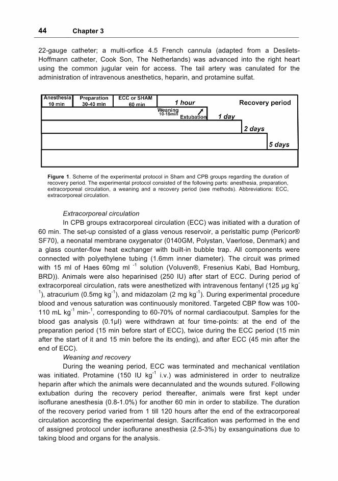
Experimental protocol The experimental protocol consisted of the following parts: anesthesia,
preparation, extracorporeal circulation, weaning with a recovery period (see fig. 1). Anesthesia was induced (2-3% isoflurane in O2/air (1:1)) before the trachea was intubated and the lungs mechanically ventilated (Amsterdam Infant Ventilator; HoekLoos, Amsterdam, The Netherlands). Tidal volume was set to achieve normocapnia (verified by capnography and arterial blood gas analysis), with O2/air (0.5:1) at a respiration frequency of 50 min-1 (0.5 s inspiration time). During the preparation period, the left femoral artery was cannulated (26-gauge catheter) for blood pressure monitoring. Immediately before insertion of the arterial line, 250 IU kg-1 heparin was administered. The carotid artery was canulated for arterial inflow using a
44 Chapter 3
!
22-gauge catheter; a multi-orfice 4.5 French cannula (adapted from a Desilets-Hoffmann catheter, Cook Son, The Netherlands) was advanced into the right heart using the common jugular vein for access. The tail artery was canulated for the administration of intravenous anesthetics, heparin, and protamine sulfat.
Extracorporeal circulation In CPB groups extracorporeal circulation (ECC) was initiated with a duration of
60 min. The set-up consisted of a glass venous reservoir, a peristaltic pump (Pericor® SF70), a neonatal membrane oxygenator (0140GM, Polystan, Vaerlose, Denmark) and a glass counter-flow heat exchanger with built-in bubble trap. All components were connected with polyethylene tubing (1.6mm inner diameter). The circuit was primed with 15 ml of Haes 60mg ml -1 solution (Voluven®, Fresenius Kabi, Bad Homburg, BRD)). Animals were also heparinised (250 IU) after start of ECC. During period of extracorporeal circulation, rats were anesthetized with intravenous fentanyl (125 "g kg-
1), atracurium (0.5mg kg-1), and midazolam (2 mg kg-1). During experimental procedure blood and venous saturation was continuously monitored. Targeted CBP flow was 100-110 mL kg-1 min-1, corresponding to 60-70% of normal cardiacoutput. Samples for the blood gas analysis (0.1"l) were withdrawn at four time-points: at the end of the preparation period (15 min before start of ECC), twice during the ECC period (15 min after the start of it and 15 min before the its ending), and after ECC (45 min after the end of ECC).
Weaning and recovery During the weaning period, ECC was terminated and mechanical ventilation
was initiated. Protamine (150 IU kg-1 i.v.) was administered in order to neutralize heparin after which the animals were decannulated and the wounds sutured. Following extubation during the recovery period thereafter, animals were first kept under isoflurane anesthesia (0.8-1.0%) for another 60 min in order to stabilize. The duration of the recovery period varied from 1 till 120 hours after the end of the extracorporeal circulation according the experimental design. Sacrification was performed in the end of assigned protocol under isoflurane anesthesia (2.5-3%) by exsanguinations due to taking blood and organs for the analysis.
Figure 1. Scheme of the experimental protocol in Sham and CPB groups regarding the duration of recovery period. The experimental protocol consisted of the following parts: anesthesia, preparation, extracorporeal circulation, a weaning and a recovery period (see methods). Abbreviations: ECC, extracorporeal circulation.
!
Vascular reactivity and endothelial activation 45
!
Vascular reactivity Immediately following sacrifice, thoracic aorta was removed and placed in cold
physiological saline solution, cleaned from connective tissue and cut into 8 rings of 2-2.5 mm, which were placed in the individual organ bathes for isotonic tension recording. The baths contained 15ml of Kreb’s solution at 37°C and bubbled continuously with 95%O2/5%CO2 at pH 7.4. Rings were allowed to equilibrate for 40 min to reach a steady baseline. Vascular segments were primed and checked for viability by two consecutive exposures to potassium chloride (60 mM).
The experimental protocol consisted of evaluation of contractile responses to phenylephrine (PE; 10 nM to 100 "M) and endothelium-dependent relaxation to acetylcholine (ACh; 10 nM to 300 "M) in rings precontracted with PE. To assess the contribution of different endothelial mediators, ACh-induced relaxation was additionally studied in the absence and presence of L-NMMA (0.1 mM; an inhibitor of NO-synthase) and/or indomethacine (1 "M; an inhibitor of cyclooxygenase) administered 20 min before application of PE [18]. Prostaglandin-mediated relaxation was determined as a difference between total Ach-mediated relaxation and response to Ach in the presence of indomethacin. NO-mediated contribution was evaluated as the portion of acetylcholine induced relaxation in presence of indomethacin additionally sensitive to eNOS-inhibition by L-NMMA. By exclusion, the rest relaxation was accounted to be EDHF-mediated [19]. To quantify the contribution of the above endothelial mediators, the area between the concentration-response curve in the absence and presence of one or more inhibitors was taken. Following the final concentration of ACh, a maximal concentration of the NO-donor sodium nitroprusside (SNP; 0.1 mM) was applied to assess total endothelium-independent relaxation [20]. Mainly two types of the impacts were evaluated: Sham-related affects (namely, anesthesia and cannulation) and influence of the extracorporeal circulation (ECC) on vascular reactivity.
Western blotting
The methods used were described previously.21 Briefly, after grinding, the frozen abdominal aortas were placed in 300 "l of boiling 2% SDS followed by pounding by a polytron (Kinematica AG Littau, Switzerland). Then, samples were centrifuged (4000 rpm, 1 min) and heated (95 oC) for 5 min. After a second centrifugation (13000 rpm, 3 min), supernatant was collected and used for measurements. Protein concentration was determined by Bio-Rad Protein Assay (Bio-Rad, Hercules, CA). Forty µg of total protein from each sample was separated on 4-20% Precise Protein Gels (Pierce, Rockford, IL) and transferred to nitrocellulose membranes. Anti-cyclooxygenase-2 antibody (BD Bioscience Pharmingen, San Diego, CA), p22 phox (BD Bioscience Pharmingen, San Diego, CA), thromboxane A2 (BD Bioscience Pharmingen, San Diego, CA), and anti-$-actin (Sigma, St. Louis, MO) were used as a primary antibody. Horseradish peroxidase-linked rabbit anti-mouse antibody was applied as a secondary antibody. The blots were analyzed using Super Signal assay (Pierce, Rockford, IL). $-actin served as a housekeeping protein. Experimental levels are expressed as ratios to $-actin protein levels.
46 Chapter 3
!
Real time PCR Total RNA was extracted from abdominal aorta using Trizol Reagent
(Invitrogen, Carlsbad, CA, USA) with subsequent quality control by ethidium bromide staining in 1% agarose gel. DNase treatment of the RNA samples prior to RT-PCR was performed with RQ1 RNase-Free DNase (Promega, Madison, WI, USA). First-Strand cDNA Synthesis from RNA (1"g) was performed by using Reverse Transcription reagents (Promega, Madison, WI, USA). The expressions of E-selectin, VCAM-1, ICAM-1, TNF!, HO-1 and TGF$ 1 (table 1) were analyzed by real-time PCR with Absolute QPCR SYBR Green reagents (Molecular Probes, Leiden, Netherlands) and CFX384 Real-Time PCR Detection System (Bio-Rad Laboratories, Inc., Hercules, CA, USA). The PCR protocol consisted of 15 min at 95ºC, followed by 40 cycles with heating to 95ºC and cooling to 60ºC for 1 minute. The amplification product was evaluated by melting curves analysis and agarose gel (1.5%) electrophoresis. Sequence-specific primers (table 1) were obtained from Biolegio (Nijmegen, the Netherlands). Cycle threshold (CT) values for genes analyzed were normalized to their CT value of GAPDH.
Drugs The ionic millimolar composition of Krebs solution was as follows: 120.4 NaCl,
5.9 KCl, 2.5 CaCl2, 1.2 MgSO4, 25.0 NaHCO3, 1.2 NaH2PO4, 11.5 glucose; these chemicals were obtained from Merck (Darmstadt, Germany). The following drugs were used: acetylcholine chloride (ACh), phenylephrine (PE), indomethacine, SNP, NG-methyl-L-arginine (L-NMMA). Stock solution (10 mmol/L) for indomethacine was prepared in 96% ethanol. All other drugs were dissolved in deionized water. L-NMMA was purchased from MP Biomedicals (Illkirch, France). All other compounds were purchased from Sigma (St. Louis, Missouri, USA). The concentrations presented in the concentration-curve responses are expressed as a final molar concentration in the bath. Data analysis
Data are given as mean±SD and n refers to the number of animals in a corresponding group. Contractile responses to phenylephrine (PE) and potassium chloride (KCl) are presented in "m displacement and relaxations to ACh and SNP were calculated as a percentage of pre-constriction. For each concentration-response curve
the following parameters were determined: 1) the Area Under the Curve (AUC) in arbitrary units (AU) (Sigma, Systat Software, Inc, Germany), 2) the maximal response (Emax) and 3) the concentration at which half-maximal response was reached (EC50). The difference in AUC for concentration-response curves to ACh in the absence or presence of an inhibitor was used as an estimate for the contribution of different endothelial mediators, as described previously [18;19]. Differences were evaluated using one-way ANOVA or repeated measurements ANOVA combined with post-hoc Bonferroni test or with t-test where applicable. Differences were considered significant at P<0.05 (2-tailed). Results
Hemodynamics and blood gas analysis during the protocol During the procedure, hemodynamic measurements (MAP) and blood gas
analysis were performed in order to monitor the animals. At baseline all parameters were in the normal range. During CPB, MAP was significantly lower compared with Sham (65.4 ± 13.9 and 105.3 ± 4.0 mmHg, respectively). During weaning, MAP of CPB animals returned to baseline levels (75.5 ± 25.1 mmHg).
Blood gas analysis was performed before start of ECC, twice during ECC and after weaning (table 2). CPB decreased pH, bicarbonate ion (HCO-
3), and Base excesses and increased levels of pCO2 and pO2, which may represent moderate respiratory acidosis. Hematocrit was significantly decreased in CPB groups as a consequence of hemodilution with priming solution. Intraoperative hyperglycemia was observed in both SHAM and CPB groups during the first 2 hours.
Contractile responses in aorta In order to evaluate contractile properties of rat thoracic aorta, maximal
response to potassium chloride (KCl; fig.2) and full concentration-response curves to phenylephrine (PE; fig.3) were constructed. In both Sham and CPB, maximal contractile responses to KCl were significantly decreased at 5 days of recovery, both compared to controls and to shorter periods of recovery. After 1hour of the recovery period Sham-related procedures (anesthesia and cannulation) decrease contractile response to PE, while ECC counteract those changes in vascular reactivity. Similarly at 5 days of recovery, concentration-response curves for PE showed a right shift and decreased Emax, and hence a decrease in AUC (fig. 3, panel A: IV, table 3).
Thus, the above findings suggest that the Sham-related surgical procedures (extended anesthesia and cannulation) induced the depression in thoracic aorta contractility after prolonged recovery, without significant additional influence of extracorporeal circulation.
48 Chapter 3
!
Tabl
e 2.
Dat
a on
blo
od g
ases
ana
lysi
s, g
luco
se c
once
ntra
tion
and
mea
n ar
teri
al p
ress
ure
(M
AP
, fem
oral
art
ery)
in S
ham
and
CP
B g
roup
s du
ring
the
expe
rim
enta
l pro
toco
l P
aram
eter
s S
HA
M
CP
B
Bef
ore
EC
C
15 m
in E
CC
45
min
EC
C
Aft
er E
CC
B
efor
e E
CC
15
min
EC
C
45 m
in E
CC
A
fter
EC
C
MA
P, m
mH
g 10
2.4±
32.8
10
9.8±
12.9
10
3.5±
22.4
92
.0±1
8.2
84.1
±11.
6 57
.9*±
12.9
81
.4*±
25.6
79
.5±2
5.1
pH
7.43
±0.0
5 7.
45±0
.05
7.46
±0.0
7 7.
41±0
.06
7.42
±0.0
5 7.
38 *
±0.0
7 7.
32 *
±0.0
8 7.
37±0
.05
pCO
2, k
Pa
4.74
±1.0
4.
51±0
.8
4.68
±1.0
5.
15±0
.9
4.77
±0.8
5.
07*±
1.1
5.72
*±1.
0 5.
22±0
.8
pO2,
kP
a 17
.4±4
.7
16.6
±3.9
17
.7±4
.2
16.9
±4.6
19
.6±7
.0
34.1
*±17
.1
35.8
*±18
.3
19.4
±10.
2
HC
O3,
mm
ol/l
22.9
±1.5
23
.3±1
.5
24.7
±1.5
24
.4±2
.2
22.6
±2.3
21
.5±3
.6
21.6
*±2.
4 22
.4*±
1.7
BE
, mm
ol/l
-0
.4±1
.0
0.5±
1.2
1.8±
1.5
0.5±
1.4
-0.6
±1.7
-1
.9*±
2.7
-3.6
*±3.
1 -2
.04±
1.9
Hct
c 0.
4±0.
02
0.
4±0.
03
0.
4±0.
03
0.
4±0.
03
0.4±
0.02
0.3*
±0.0
5
0.3*
±0.0
2
0.3*
±0.0
3
Glu
cose
, mm
ol/l
20.6
±5.4
15.2
±5.4
8.4±
4.3
6.
6±1.
0
22.9
±4.1
20.5
±5.1
11.6
±4.4
6.9±
3.0
Abb
revi
atio
ns: H
ctc,
hem
atoc
rit; C
PB
, car
diop
ulm
onar
y by
pass
. *
deno
tes
P<0
.05,
Sha
m v
s C
PB
, ind
epen
dent
t-te
st
!Ta
ble
3. P
aram
eter
s of
the
Phe
nyle
phri
ne-m
edia
ted
cont
ract
ion
Gro
ups
EC
50, u
Mol
/L
Hill
slo
pe
Max
imal
res
pons
e, u
M
Con
trol
2.
7±0.
5
0.9±
0.1
488.
9±13
.8
Sha
m1h
our
33.8
±4.2
**
0.7±
0.09
42
2.8±
23.5
* C
PB
1 h
our
6.8±
2.0
1.3±
0.6
402.
8±22
.9*
Sha
m 1
day
8.1±
2.7
1.2±
0.6
415.
6±31
.0
CP
B 1
day
6.5±
3.9
1.07
±0.7
39
7.5±
45.5
S
ham
2da
ys
6.4±
1.7
1.4±
0.5
427.
0±19
.7*
CP
B 2
days
5.
1±1.
1 1.
2±0.
3 42
7.3±
14.9
* S
ham
5da
ys
12.6
±3.1
* 1.
03±0
.3
210.
2±13
.0*
CP
B 5
days
18
.1±7
.6
0.76
±0.3
26
4.9±
26.6
* *-
P<0
.05,
vs
Con
trol,
t-tes
t; **
- P<0
.001
, vs
Con
trol,
t-tes
t
Vascular reactivity and endothelial activation 49
!
Relaxant response in aorta Aortic rings were additionally studied for endothelium-dependent responses to
ACh. Data are present as parameters of the concentration-response curves (table 4) and contribution of different relaxant pathways (fig. 4). Relaxant reactivity of thoracic aorta of the control rats were characterized by major contribution of NO in total acetylcholine mediated relaxation counteracted by contractile prostaglandins (fig. 4). Neither extracorporeal circulation, nor the duration of recovery period affected total acetylcholine-mediated relaxation. In contrast, various changes were observed in the contribution of PG-, NO- and EDHF to overall relaxation. Both in Sham and in CPB groups, NO-contribution was non-significantly reduced after 5 days of recovery period. Significantly increased contribution of EDHF-mediated pathways was found at 1day and 5 days time-points of the recovery period. Further, the “amount” of contractile prostaglandins seemed to be augmented during 1-2 days of the recovery period with normalization by the 5th day. Collectively, the findings showed that Sham-related procedures (prolonged anesthetic intervention and/or cannulation) increased the
Figure 2. Vascular contractility to potassium chloride of rat thoracic aorta at different periods of recovery (1 hour, 1day, 2 days, 5 days) in Sham groups and following subjection of the animals to CPB-related procedures. Contractile responses are given as Emax. Data are mean ± SD (n=6-9). * indicates P<0.05 vs Control, one way ANOVA with post-hoc Bonferroni test. Abbreviations: KCl=, potassium chloride; CPB=, cardiopulmonary bypass.
50 Chapter 3
!
contribution of EDHF-pathway in total acetylcholine-mediated relaxation to maintain the overall relaxation response to ACh. However, extracorporeal circulation did not add any influence herein. !
Figure 3. Vascular contractility to phenylephrine of rat thoracic aorta at different periods of recovery (1 hour, 1day, 2 days, 5 days) in Sham groups and following subjection of the animals to CPB-related procedures. Contractile responses are given as full CR-curves (A) and the AUC (B). Data are mean ± SD (n=6-9). * indicates P<0.05 vs Control, one way ANOVA with post-hoc Bonferroni test. # indicates P<0.05 vs Control, repeated measurements ANOVA combined with post-hoc Bonferroni test. Abbreviations: PE=phenylephrine; CPB=cardiopulmonary bypass.
Vascular reactivity and endothelial activation 51
!
Protein expression Because of the elevated contribution of contractile prostaglandins in ACh-induced responses in almost all groups we additionally assessed protein expression of COX-2 - hence, the isoform of inducible COX, might be responsible for the synthesis of contractile prostanoids- and of TxA2 receptors - hence, implied to be involved in cyclooxygenase-dependent vascular contraction [22] – in aortic segments. p22phox protein, a NAD(P)H oxidase subunit associated with increased ROS production and oxidative stress in vascular endothelium [23] , was also assessed. Both COX-2 and TxA2 receptor protein as well as p22phox were similarly expressed in all groups regardless of type of experimental protocol (fig.5, for TxA2 receptors and p22phox – data not shown). However, regression analysis revealed that the degree of relaxation-inhibition by (indomethacine-sensitive) COX-derived prostaglandins was proportionally correlated to COX-2 protein expression (Pearson correlation coefficient=-0.50, n=22, P=0.01), but not the TxA2 receptor or p22phox. These findings suggest the (overall)
Figure 4. Total acetylcholine (ACh-)-induced relaxation (A) - and the contribution of (B) prostaglandins (PG’s), (C) NO, and (D) EDHF hereto, all provided as AUC - in rat thoracic aorta at different periods of recovery (1h, 1day, 2days or 5days) following subjection of the animals to CPB -related procedures. Data are mean ± SD (n=5-8). * indicates P<0.05 vs control, one-way ANOVA with following Bonferroni test. # indicates P<0.05 vs control Mann-Whitney Rank Sum Test. $ indicates P<0.05 vs control; # independent t-test. Abbreviations: CPB= cardiopulmonary bypass.
52 Chapter 3
!
outcome for endothelial function in vascular reactivity in the present study to an important extent was determined by the level of vascular COX-2 protein expression.
Expression of markers of endothelial activation, anti-inflammatory marker, and hypoxia
To investigate changes in endothelial cells related to an inflammatory response, we the evaluated expression of the adhesion factors E-selectin, VCAM-1, ICAM-1 and of TNF-" in abdominal aorta of a subset of animals. HO-1, an inducible enzyme in response oxidative stress and hypoxia and TGF-!, which might modulate the cell adhesion molecules expression, were also evaluated. No expression of these 6 factors was detected in control animals and in Sham and CPB animals beyond 60 min of recovery (fig. 6). In contrast, a consistent upregulation of all factors tested was found in all tested CPB animals at 1 hour recovery period. In contrast, sham animals at 1 h of recovery showed consistently no increase in expression (VCAM-1 and ICAM-1) or only in 1 out of 4 animals (fig. 6). Discussion
The present work studied the changes in vascular responsiveness of thoracic aorta after cardiopulmonary bypass during 5 days of recovery in relation to endothelial cell activation. The main finding of this study is that endothelial cell activation did not result into impaired endothelium-dependent relaxation to ACh in rat aorta. We found that CPB induced endothelial cell activation only after a short-term recovery period (1 hour), as evidenced by a consistently increased expression of 6 markers studied. Secondly, changed vasoresponsiveness, characterized by attenuated vascular contractility and elevation of EDHF-mediated relaxation, was found only after 5 days of recovery. Moreover, these vasomotor changes were to a similar extent present in both CPB and Sham, indicating that they were mainly the result of the accompanying
Figure 5. COX-2 expression in thoracic aorta in all groups (at least 4 per group). Representative blot (A), relative expressions patterns (B) and relation between individual Pr’s-mediated contribution and COX-2 expression; Data were evaluated by regression analysis and Pearson correlation. Abbreviations: CPB= cardiopulmonary bypass.
Vascular reactivity and endothelial activation 53
!
procedures (cannulation procedures and/or prolonged anesthesia), but not of the extracorporeal circulation. Collectively, these data indicate that endothelial cell activation after extracorporeal circulation was not directly related to altered vascular properties of rat thoracic aorta.
Hemodynamics and blood gas analysis during the protocol Hemodynamic data showed that extracorporeal circulation significantly
decreased MAP to around 60-80 mmHg in CBP group, which corresponds to data in previously published studies of CBP in rat.12; 13 Although the phenomenon of CPB-induced hypotension has been addressed previously during last decade, the exact
Figure 6. Relative mRNA expression of E-selectin (A), VCAM-1 (B), ICAM-1 (C), TNF-! (D), HO-1 (E) and TGF-$ (F) in thoracic aorta. Abbreviations: CPB= cardiopulmonary bypass.
!
54 Chapter 3
!
underlying mechanisms are still unknown. In this context, platelet-mediated serotonin release is thought to be one of the most prominent explanations as extracorporeal circulation, contact of blood with artificial surfaces and the roller pump induces platelet activation and triggers release of serotonin. Serotonin acts as vasodilator through activation of the most sensitive and widespread 5-HT2B receptors mediating NO-release.24 Several other changes in parameters (pH, HCO-
3, and BE) indicate moderate metabolic acidosis in CPB animals. Since all levels returned to the normal range by the end of CPB, it is unlikely that this affected vascular reactivity. Previously severe acidosis has been shown to increase circulating TNF, IL-6, and IL-10 in sever septic shock.25 In our experimental setup only mild acidosis occurred, that turned to basal level after the end of extracorporeal circulation, hence it might doubtful modulate to some extend expression of the inflammatory cytokines. Intraoperative hyperglycemia was observed in both Sham and CPB groups during first 2 hours, indicating that this represents a reaction to the surgical intervention.
Vascular reactivity The present work showed an alteration of rat aorta vasoreactivity only after a
prolonged recovery period of 5 days in both Sham and CPB animals, demonstrating these changes to be caused by cannulation and/or prolonged anesthesia. Contractile response to PE and KCl at day 5 were decreased to a similar extent. Its decrease may be related to increased basal release of endothelium-mediated relaxing factors, particularly EDHF, which was found in the same time point of the 5th day of the recovery period. Previously, the role of the endothelium, in phenylephrine- and potassium chloride-mediated contraction has been shown, while mainly the role of COX, 20-HETE and NO have been investigated.25-28 Alternatively, inhibition of the contractile vascular reactivity may be mediated by decreased Ca2+ sensitivity, as both receptor dependent (PE) and independent (KCl) pathways seem affected. Possibly, cannulation and/or prolonged anesthesia inhibit contractile vascular reactivity through modulation of the MLC kinase and/or MLC phosphatase activity.
The main change in relaxant properties constitutes of an increase in the contribution of EDHF to maintain total endothelial dependent relaxation at 1day and 5 days of recovery in both Sham and CPB. Increased EDHF may serve a compensatory role in response to the depletion of the principal relaxant mediator in aorta, NO, as shown in several studies.29-31
Also our data showed that relaxant reactivity of thoracic aorta are characterized by involvement of contractile prostaglandins. Moreover the amount of contractile prostaglandins seemed to be augmented during first 2 days of the recovery period with normalization by the 5th day. Both types of cyclooxygenases (COX-1 and COX-2) can be involved in production of arachidonic acid contracting metabolites. COX-1 is accounted to be constitutively expressed in endothelium, while COX-2 is inducible form. In our study occurrence of contractile prostaglandins correlated with expression of COX-2. That corresponds to previously published data.22;32 Achieved results did not allow us to draw exact conclusion about the changes in COX-2 expression due to large inter-individual variability. However, correlation analysis
Vascular reactivity and endothelial activation 55
!
showed relation between COX-2 expression and the value of the contractile prostaglandin’s participation in the total relaxation.
Endothelial activation and endothelial dysfunction To substantiate endothelial cell activation, we quantified the expression of
adhesion molecules, pro-inflammatory factors and oxidative stress by qPCR and western blot. Substantial upregulation of all these factors was found consistently in CPB and occasionally in Sham only after 60 min of recovery from CPB. Thus, we found ECC activation of E-selectin, which is a main marker of the endothelium activation in response to inflammatory cytokine (TNF-! and IL-1$)33 and the cell adhesions molecules of the immunoglobulin superfamily, ICAM-1 and VCAM-1, that serve as ligands for leukocyte integrins.34 In addition, CPB induced the upregulation of TNF- ! and TGF-$, which might modulate expression of adhesion molecules, provide a chemotactic gradient for leukocytes and other cells participating in an inflammatory response34-36 and HO-1, an enzyme induced in response to oxidative stress and hypoxia, with the same time-course. As a consequence, it is unlikely that endothelial activation, which is mainly present in the CPB group, accounts for vascular changes as these are present in both CPB and Sham to a similar extent. Nevertheless a direct relationship between the both has been suggested before in mouse thoracic aorta in diet-induced obesity model.34;37 Differences in pathophysiology of these two models (acute surgical intervention in our study and chronic disease state in later) do not allow us to make a comparison between two studies. Circulating cell adhesion molecules (E-selectin, VCAM-1, ICAM-1) have been shown to correlate with the development of the nephropathy, retinopathy, and cardiovascular disease in both type 1 and type 2 diabetes and with the degree of atherosclerosis and albuminuria in type-2 diabetic patients.18-21 Impaired acetylcholine-dependent vasodilation was recently associated with increased levels of the plasma soluble E-selectin and P-selectin in essential hypertension.38 However, in abovementioned studies E-selectin was measured in plasma (and therefore has unknown origin), while we evaluated its expression directly in rat abdominal aorta.
In conclusion, our study demonstrates CPB to induce an immediate and short-term endothelial activation in rat aorta. In addition, vasoresponsiveness is altered at the long run due to prolonged cannulation and/or anesthesia, without any additional effect of extracorporeal circulation. Achieved data suppose better understanding of the pathophysiological processes underlying CPB and possible therapeutical approaches of its complications. Reference List 1. Levy JH, Tanaka KA: Inflammatory response to cardiopulmonary bypass. Ann.Thorac.Surg. 2003;
75: S715-S720 2. Rinder C: Cellular inflammatory response and clinical outcome in cardiac surgery.
Curr.Opin.Anaesthesiol. 2006; 19: 65-8
56 Chapter 3
!
3. Galea J, Rebuck N, Finn A, Manche A, Moat N: Expression of soluble endothelial adhesion molecules in clinical cardiopulmonary bypass. Perfusion 1998; 13: 314-21
4. John AE, Galea J, Francis SE, Holt CM, Finn A: Interleukin-8 mRNA expression in circulating leucocytes during cardiopulmonary bypass. Perfusion 1998; 13: 409-17
5. Onorati F, Rubino AS, Nucera S, Foti D, Sica V, Santini F, Gulletta E, Renzulli A: Off-pump coronary artery bypass surgery versus standard linear or pulsatile cardiopulmonary bypass: endothelial activation and inflammatory response. Eur.J.Cardiothorac.Surg. 2009;
7. Paparella D, Yau TM, Young E: Cardiopulmonary bypass induced inflammation: pathophysiology and treatment. An update. Eur.J.Cardiothorac.Surg. 2002; 21: 232-44
8. Kilbridge PM, Mayer JE, Newburger JW, Hickey PR, Walsh AZ, Neufeld EJ: Induction of intercellular adhesion molecule-1 and E-selectin mRNA in heart and skeletal muscle of pediatric patients undergoing cardiopulmonary bypass. J.Thorac.Cardiovasc.Surg. 1994; 107: 1183-92
9. Williams HJ, Rebuck N, Elliott MJ, Finn A: Changes in leucocyte counts and soluble intercellular adhesion molecule-1 and E-selectin during cardiopulmonary bypass in children. Perfusion 1998; 13: 322-7
10. Andrasi TB, Bielik H, Blazovics A, Zima E, Vago H, Szabo G, Juhasz-Nagy A: Mesenteric vascular dysfunction after cardiopulmonary bypass with cardiac arrest is aggravated by coexistent heart failure. Shock 2005; 23: 324-9
12. Doguet F, Litzler PY, Tamion F, Richard V, Hellot MF, Thuillez C, Tabley A, Bouchart F, Bessou JP: Changes in mesenteric vascular reactivity and inflammatory response after cardiopulmonary bypass in a rat model. Ann.Thorac.Surg. 2004; 77: 2130-7
13. Modine T, Azzaoui R, Ouk T, Fayad G, Lacroix D, Warembourg H, Bordet R, Gourlay T: Changes in cerebral vascular reactivity occur early during cardiopulmonary bypass in the rat. Ann.Thorac.Surg. 2006; 82: 672-8
14. Ruel M, Khan TA, Voisine P, Bianchi C, Sellke FW: Vasomotor dysfunction after cardiac surgery. Eur.J.Cardiothorac.Surg. 2004; 26: 1002-14
15. Verrier ED, Morgan EN: Endothelial response to cardiopulmonary bypass surgery. Ann.Thorac.Surg. 1998; 66: S17-S19
16. Wang SY, Friedman M, Franklin A, Sellke FW: Myogenic reactivity of coronary resistance arteries after cardiopulmonary bypass and hyperkalemic cardioplegia. Circulation 1995; 92: 1590-6
17. Wang SY, Stamler A, Li J, Johnson RG, Sellke FW: Decreased myogenic reactivity in skeletal muscle arterioles after hypothermic cardiopulmonary bypass. J.Surg.Res. 1997; 69: 40-4
18. Gschwend S, Pinto-Sietsma SJ, Buikema H, Pinto YM, van Gilst WH, Schulz A, de ZD, Kreutz R: Impaired coronary endothelial function in a rat model of spontaneous albuminuria. Kidney Int. 2002; 62: 181-91
19. Gschwend S, Buikema H, Henning RH, Pinto YM, de ZD, van Gilst WH: Endothelial dysfunction and infarct-size relate to impaired EDHF response in rat experimental chronic heart failure. Eur.J.Heart Fail. 2003; 5: 147-54
20. Westendorp B, Schoemaker RG, van Gilst WH, Buikema H: Improvement of EDHF by chronic ACE inhibition declines rapidly after withdrawal in rats with myocardial infarction. J.Cardiovasc.Pharmacol. 2005; 46: 766-72
21. Darblade B, Pendaries C, Krust A, Dupont S, Fouque MJ, Rami J, Chambon P, Bayard F, Arnal JF: Estradiol alters nitric oxide production in the mouse aorta through the alpha-, but not beta-, estrogen receptor. Circ.Res. 2002; 90: 413-9
22. Feletou M, Verbeuren TJ, Vanhoutte PM: Endothelium-dependent contractions in SHR: a tale of prostanoid TP and IP receptors. Br.J.Pharmacol. 2009; 156: 563-74
23. Siekmeier R, Grammer T, Marz W: Roles of oxidants, nitric oxide, and asymmetric dimethylarginine in endothelial function. J.Cardiovasc.Pharmacol.Ther. 2008; 13: 279-97
25. Kellum JA, Song M, Venkataraman R: Effects of hyperchloremic acidosis on arterial pressure and circulating inflammatory molecules in experimental sepsis. Chest 2004; 125: 243-8
26. Aloamaka CP, Ezimokhai M, Morrison J: The role of endothelium in phenylephrine- and potassium-induced contractions of the rat aorta during pregnancy. Res.Exp.Med.(Berl) 1993; 193: 407-17
27. Chabot F, Mestiri H, Sabry S, l'Ava-Santucci J, Lockhart A, nh-Xuan AT: Role of NO in the pulmonary artery hyporeactivity to phenylephrine in experimental biliary cirrhosis. Eur.Respir.J. 1996; 9: 560-4
28. Guo Z, Su W, Allen S, Pang H, Daugherty A, Smart E, Gong MC: COX-2 up-regulation and vascular smooth muscle contractile hyperreactivity in spontaneous diabetic db/db mice. Cardiovasc.Res. 2005; 67: 723-35
29. Ekse S, Clapp LH, Revhaug A, Ytrebo LM: Endothelium-derived hyperpolarization factor (EDHF) is up-regulated in a pig model of acute liver failure. Scand.J.Gastroenterol. 2007; 42: 356-65
30. Katz SD, Krum H: Acetylcholine-mediated vasodilation in the forearm circulation of patients with heart failure: indirect evidence for the role of endothelium-derived hyperpolarizing factor. Am.J.Cardiol. 2001; 87: 1089-92
31. Luksha L, Agewall S, Kublickiene K: Endothelium-derived hyperpolarizing factor in vascular physiology and cardiovascular disease. Atherosclerosis 2009; 202: 330-44
32. Wong SL, Leung FP, Lau CW, Au CL, Yung LM, Yao X, Chen ZY, Vanhoutte PM, Gollasch M, Huang Y: Cyclooxygenase-2-derived prostaglandin F2alpha mediates endothelium-dependent contractions in the aortae of hamsters with increased impact during aging. Circ.Res. 2009; 104: 228-35
33. Ulbrich H, Eriksson EE, Lindbom L: Leukocyte and endothelial cell adhesion molecules as targets for therapeutic interventions in inflammatory disease. Trends Pharmacol.Sci. 2003; 24: 640-7
34. Asimakopoulos G, Taylor KM: Effects of cardiopulmonary bypass on leukocyte and endothelial adhesion molecules. Ann.Thorac.Surg. 1998; 66: 2135-44
35. Sanjabi S, Zenewicz LA, Kamanaka M, Flavell RA: Anti-inflammatory and pro-inflammatory roles of TGF-beta, IL-10, and IL-22 in immunity and autoimmunity. Curr.Opin.Pharmacol. 2009; 9: 447-53
36. Li MO, Wan YY, Sanjabi S, Robertson AK, Flavell RA: Transforming growth factor-beta regulation of immune responses. Annu.Rev.Immunol. 2006; 24: 99-146
37. Kim F, Pham M, Maloney E, Rizzo NO, Morton GJ, Wisse BE, Kirk EA, Chait A, Schwartz MW: Vascular inflammation, insulin resistance, and reduced nitric oxide production precede the onset of peripheral insulin resistance. Arterioscler.Thromb.Vasc.Biol. 2008; 28: 1982-8
38. de la SA, Larrousse M: Endothelial dysfunction is associated with increased levels of biomarkers in essential hypertension. J.Hum.Hypertens. 2009;