67
University of Jordan 1 Body Fluids & Blood
| Date post: | 27-Dec-2015 |
| Category: |
Documents |
| Upload: | megan-sutton |
| View: | 215 times |
| Download: | 1 times |

University of Jordan 1
Body Fluids & Blood

University of Jordan 2

University of Jordan 3
University of Jordan 4

University of Jordan 5
Movement of water between compartments
Normally, cells neither shrink or swell because intracellular and interstitial fluids have the same osmolarity
Increasing osmolarity of interstitial fluid draws water out of cells and cells shrink
Decreasing osmolarity of interstitial fluid causes cells to swell
Changes in osmolarity most often result from changes in Na+ concentration
Water intoxication – drinking water faster than the kidneys can excrete it
Can lead to convulsions, coma or death
University of Jordan 6
Electrolytes in body fluids Ions form when electrolytes dissolve ad dissociate 4 general functions
Control osmosis of water between body fluid compartments
Help maintain the acid-base balance Carry electrical current Serve as cofactors
University of Jordan 7
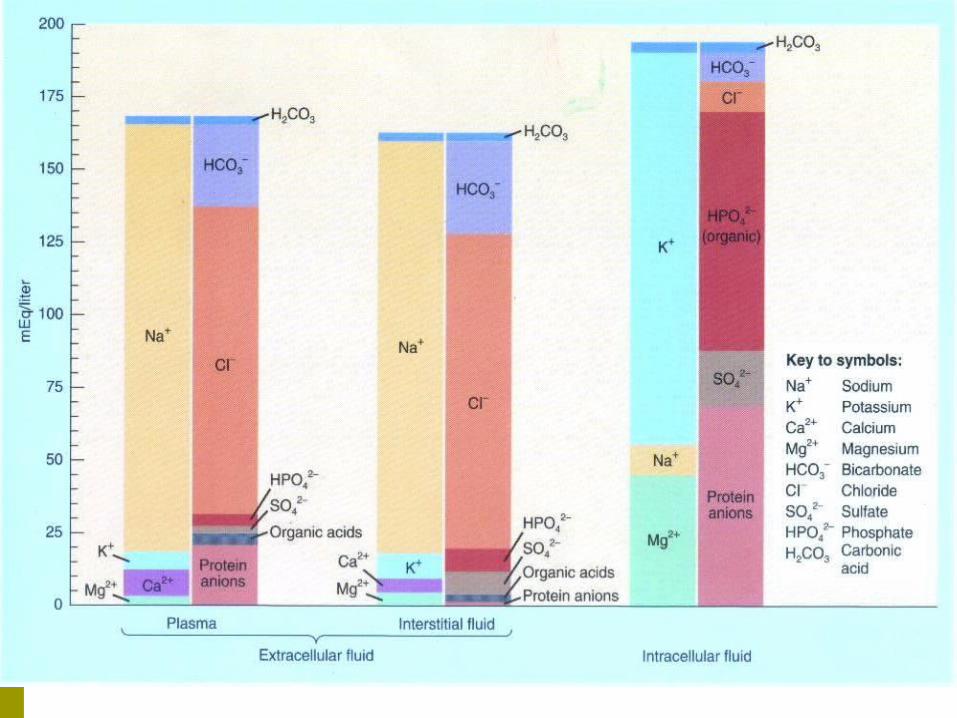
Concentrations in body fluids
Concentration of ions typically expressed in milliequivalents per liter (mEq/liter)
Na+ or Cl- number of mEq/liter = mmol/literCa2+ or HPO4
2- number of mEq/liter = 2 x mmol/liter
Chief difference between 2 ECF compartments (plasma and interstitial fluid) is plasma contains many more protein anions
Largely responsible for blood colloid osmotic pressure

University of Jordan 8
ICF differs considerably from ECF
ECF most abundant cation is Na+, anion is Cl- ICF most abundant cation is K+, anion are
proteins and phosphates (HPO42-)
Na+ /K+ pumps play major role in keeping K+
high inside cells and Na+ high outside cell

University of Jordan 9
Sodium Na+
Most abundant ion in ECF 90% of extracellular cations Plays pivotal role in fluid and electrolyte balance because it
account for almost half of the osmolarity of ECF Level in blood controlled by
Aldosternone – increases renal reabsorption ADH – if sodium too low, ADH release stops Atrial natriuretic peptide – increases renal excretion

University of Jordan 10
Chloride Cl-
Most prevalent anions in ECF Moves relatively easily between ECF and ICF because most
plasma membranes contain Cl- leakage channels and antiporters
Can help balance levels of anions in different fluids Chloride shift in RBCs
Regulated by ADH – governs extent of water loss in urine Processes that increase or decrease renal reabsorption of Na+
also affect reabsorption of Cl-
University of Jordan 11
Potassium K+
Most abundant cations in ICF Key role in establishing resting membrane
potential in neurons and muscle fibers Also helps maintain normal ICF fluid volume Helps regulate pH of body fluids when
exchanged for H+
Controlled by aldosterone – stimulates principal cells in renal collecting ducts to secrete excess K+

University of Jordan 12
Bicarbonate HCO3-
Second most prevalent extracellular anion Concentration increases in blood passing through systemic
capillaries picking up carbon dioxide Carbon dioxide combines with water to form carbonic
acid which dissociates Drops in pulmonary capillaries when carbon dioxide
exhaled Chloride shift helps maintain correct balance of anions in
ECF and ICF Kidneys are main regulators of blood HCO3
-
Can form and release HCO3- when low or excrete excess

University of Jordan 13
Blood Liquid connective tissue 3 general functions1. Transportation
Gases, nutrients, hormones, waste products
2. Regulation pH, body temperature, osmotic pressure
3. Protection Clotting, white blood cells, proteins

University of Jordan 14
Components of Blood Blood plasma – water liquid extracellular
matrix 91.5% water, 8.5% solutes (primarily proteins) Hepatocytes synthesize most plasma proteins
Albumins, fibrinogen, antibodies Other solutes include electrolytes, nutrients,
enzymes, hormones, gases and waste products Formed elements – cells and cell fragments
Red blood cells (RBCs) White blood cells (WBCs) Platelets

University of Jordan 15

University of Jordan 16
Composition of Blood Plasma:
Straw-colored liquid. Consists of H20 and dissolved solutes.
Ions, metabolites, hormones, antibodies. Na+ is the major solute of the plasma.
Plasma proteins: Constitute 7-9% of plasma.
Albumin: Accounts for 60-80% of plasma proteins. Provides the colloid osmotic pressure needed to draw
H20 from interstitial fluid to capillaries. Maintains blood pressure.

University of Jordan 17
Plasma proteins (continued): Globulins:
a globulin: Transport lipids and fat soluble vitamins.
b globulin: Transport lipids and fat soluble vitamins.
g globulin: Antibodies that function in immunity.
Fibrinogen: Constitutes 4% of plasma proteins. Important clotting factor.
Converted into fibrin during the clotting process.
Composition of the Blood (continued)

University of Jordan 18
Serum: Fluid from clotted blood.
Does not contain fibrinogen.
Plasma volume: Number of regulatory mechanisms in the body maintain
homeostasis of plasma volume. Osmoreceptors. ADH. Renin-angiotensin-aldosterone system.
Composition of the Blood (continued)

University of Jordan 19
Formed Elements of Blood
University of Jordan 20
Formation of Blood Cells Negative feedback systems regulate the total number of RBCs
and platelets in circulation Abundance of WBC types based of response to invading
pathogens or foreign antigens Hemopoiesis or hemotopoiesis Red bone marrow primary site Pluripotent stem cells have the ability to develop into many
different types of cells

University of Jordan 21

University of Jordan 22
Formation of Blood Cells Pluripotent stem cells produce
Myeloid stem cells Give rise to red blood cells, platelets, monocytes, neutrophils,
eosinophils and basophils Lymphoid stem cells give rise to
Lymphocytes Hemopoietic growth factors regulate differentiation and proliferation
Erythropoietin – RBCs Thrombopoietin – platelets Colony-stimulating factors (CSFs) and interleukins – WBCs

University of Jordan 23
Leukopoiesis Cytokines stimulate different types and stages of WBC
production. Multipotent growth factor-1, interleukin-1, and interleukin-
3: Stimulate development of different types of WBC cells.
Granulocyte-colony stimulating factor (G-CSF): Stimulates development of neutrophils.
Granulocyte-monocyte colony stimulating factor (GM-CSF): Simulates development of monocytes and eosinophils.

University of Jordan 24
Erythrocytes Flattened biconcave discs. Provide increased surface area through which gas can
diffuse. Lack nuclei and mitochondria.
Half-life ~ 120 days. Each RBC contains 280 million hemoglobin with 4
heme chains (contain iron). Removed from circulation by phagocytic cells in
liver, spleen, and bone marrow.

University of Jordan 25
Leukocytes Contain nuclei and mitochondria. Move in amoeboid fashion.
Can squeeze through capillary walls (diapedesis). Almost invisible, so named after their staining properties.
Granular leukocytes: Help detoxify foreign substances.
Release heparin. Agranular leukocytes:
Phagocytic. Produce antibodies.
University of Jordan 26
Platelets (thrombocytes)
Smallest of formed elements. Are fragments of megakaryocytes. Lack nuclei.
Capable of amoeboid movement. Important in blood clotting:
Constitute most of the mass of the clot. Release serotonin to vasoconstrict and reduce blood flow
to area. Secrete growth factors:
Maintain the integrity of blood vessel wall. Survive 5-9 days.

University of Jordan 27
Blood Cells and Platelets

University of Jordan 28
Hematopoiesis
Undifferentiated cells gradually differentiate to become stem cells, that form blood cells.
Occurs in myeloid tissue (bone marrow of long bones) and lymphoid tissue.
2 types of hematopoiesis: Erythropoiesis:
Formation of RBCs. Leukopoiesis:
Formation of WBCs.

University of Jordan 29
Erythropoiesis
Active process. 2.5 million RBCs are produced every second.
Primary regulator is erythropoietin. Binds to membrane receptors of cells that will become erythroblasts. Erythroblasts transform into normoblasts. Normoblasts lose their nuclei to become reticulocytes. Reticulocytes change into mature RBCs.
Stimulates cell division. Old RBCs are destroyed in spleen and liver.
Iron recycled back to myeloid tissue to be reused in hemoglobin production. Need iron, vitamin B12 and folic acid for synthesis.
University of Jordan 30
Red Blood Cells/ Erythrocytes Contain oxygen-carrying protein hemoglobin Production = destruction with at least 2 million new RBCs per
second Biconcave disc – increases surface area Strong, flexible plasma membrane Glycolipids in plasma membrane responsible for ABO and Rh
blood groups Lack nucleus and other organelles
No mitochondria – doesn’t use oxygen

University of Jordan 31
Hemoglobin
Globin – 4 polypeptide chains Heme in each of 4 chains Iron ion can combine reversibly with one oxygen molecule Also transports 23% of total carbon dioxide
Combines with amino acids of globin Nitric oxide (NO) binds to hemoglobin
Releases NO causing vasodilation to improve blood flow and oxygen delivery

University of Jordan 32
Shapes of RBC and Hemoglobin

University of Jordan 33
Red Blood Cells RBC life cycle
Live only about 120 days Cannot synthesize new components – no nucleus Ruptured red blood cells removed from circulation and
destroyed by fixed phagocytic macrophages in spleen and liver
Breakdown products recycled Globin’s amino acids reused Iron reused Non-iron heme ends as yellow pigment urobilin in urine
or brown pigment stercobilin in feces

University of Jordan 34
Red blood celldeath andphagocytosis
Key:
in blood
in bile
Macrophage inspleen, liver, orred bone marrow
1
Globin
Red blood celldeath andphagocytosis
Key:
in blood
in bile
Macrophage inspleen, liver, orred bone marrow
Heme2
1
Aminoacids
Reused forprotein synthesisGlobin
Red blood celldeath andphagocytosis
Key:
in blood
in bile
Macrophage inspleen, liver, orred bone marrow
Heme
3
2
1
Aminoacids
Reused forprotein synthesisGlobin
Red blood celldeath andphagocytosis
Transferrin
Fe3+
Key:
in blood
in bile
Macrophage inspleen, liver, orred bone marrow
Heme
4
3
2
1
Aminoacids
Reused forprotein synthesisGlobin
Red blood celldeath andphagocytosis
Transferrin
Fe3+
Liver
Key:
in blood
in bile
Macrophage inspleen, liver, orred bone marrow
FerritinHeme
54
3
2
1
Aminoacids
Reused forprotein synthesisGlobin
Red blood celldeath andphagocytosis
Transferrin
Fe3+
Fe3+ Transferrin
Liver
Key:
in blood
in bile
Macrophage inspleen, liver, orred bone marrow
FerritinHeme
654
3
2
1
Aminoacids
Reused forprotein synthesisGlobin
Red blood celldeath andphagocytosis
Transferrin
Fe3+
Fe3+ Transferrin
Liver
+Globin
+Vitamin B12
+Erythopoietin
Key:
in blood
in bile
Macrophage inspleen, liver, orred bone marrow
FerritinHeme Fe3+
7
654
3
2
1
Aminoacids
Reused forprotein synthesisGlobin
Circulation for about120 days
Red blood celldeath andphagocytosis
Transferrin
Fe3+
Fe3+ Transferrin
Liver
+Globin
+Vitamin B12
+Erythopoietin
Key:
in blood
in bile
Erythropoiesis inred bone marrow
Macrophage inspleen, liver, orred bone marrow
FerritinHeme Fe3+
8
7
654
3
2
1
Aminoacids
Reused forprotein synthesisGlobin
Circulation for about120 days
Red blood celldeath andphagocytosis
Transferrin
Fe3+
Fe3+ Transferrin
Liver
+Globin
+Vitamin B12
+Erythopoietin
Key:
in blood
in bile
Erythropoiesis inred bone marrow
Macrophage inspleen, liver, orred bone marrow
FerritinHeme
Biliverdin Bilirubin
Fe3+
9
8
7
654
3
2
1
Aminoacids
Reused forprotein synthesisGlobin
Circulation for about120 days
Bilirubin
Red blood celldeath andphagocytosis
Transferrin
Fe3+
Fe3+ Transferrin
Liver
+Globin
+Vitamin B12
+Erythopoietin
Key:
in blood
in bile
Erythropoiesis inred bone marrow
Macrophage inspleen, liver, orred bone marrow
FerritinHeme
Biliverdin Bilirubin
Fe3+
10
9
8
7
654
3
2
1
Aminoacids
Reused forprotein synthesisGlobin
Stercobilin
Bilirubin
Urobilinogen
Feces
Smallintestine
Circulation for about120 days
Bacteria
Bilirubin
Red blood celldeath andphagocytosis
Transferrin
Fe3+
Fe3+ Transferrin
Liver
+Globin
+Vitamin B12
+Erythopoietin
Key:
in blood
in bile
Erythropoiesis inred bone marrow
Macrophage inspleen, liver, orred bone marrow
FerritinHeme
Biliverdin Bilirubin
Fe3+
12
1110
9
8
7
654
3
2
1
Aminoacids
Reused forprotein synthesisGlobin
Urine
Stercobilin
Bilirubin
Urobilinogen
Feces
Smallintestine
Circulation for about120 days
Bacteria
Bilirubin
Red blood celldeath andphagocytosis
Transferrin
Fe3+
Fe3+ Transferrin
Liver
+Globin
+Vitamin B12
+Erythopoietin
Key:
in blood
in bile
Erythropoiesis inred bone marrow
Kidney
Macrophage inspleen, liver, orred bone marrow
Ferritin
Urobilin
Heme
Biliverdin Bilirubin
Fe3+
13 12
1110
9
8
7
654
3
2
1
Aminoacids
Reused forprotein synthesisGlobin
Urine
Stercobilin
Bilirubin
Urobilinogen
Feces
Largeintestine
Smallintestine
Circulation for about120 days
Bacteria
Bilirubin
Red blood celldeath andphagocytosis
Transferrin
Fe3+
Fe3+ Transferrin
Liver
+Globin
+Vitamin B12
+Erythopoietin
Key:
in blood
in bile
Erythropoiesis inred bone marrow
Kidney
Macrophage inspleen, liver, orred bone marrow
Ferritin
Urobilin
Heme
Biliverdin Bilirubin
Fe3+
14
13 12
1110
9
8
7
654
3
2
1
Formation and Destruction of RBC’s

University of Jordan 35
Erythropoiesis
Starts in red bone marrow with proerythroblast
Cell near the end of development ejects nucleus and becomes a reticulocyte
Develop into mature RBC within 1-2 days
Negative feedback balances production with destruction
Controlled condition is amount of oxygen delivery to tissues
Hypoxia stimulates release of erythropoietin


University of Jordan 37
White Blood Cells/ Leukocytes Have nuclei Do not contain hemoglobin Granular or agranular based on staining highlighting large
conspicuous granules Granular leukocytes
Neutrophils, eosinophils, basophils Agranular leukocytes
Lymphocytes and monocytes

University of Jordan 38
Types of White Blood Cells

University of Jordan 39
Functions of WBCs Usually live a few days Except for lymphocytes – live for months or years Far less numerous than RBCs Leukocytosis is a normal protective response to
invaders, strenuous exercise, anesthesia and surgery
Leukopenia is never beneficial General function to combat invaders by
phagocytosis or immune responses

University of Jordan 40
Emigration of WBCs Many WBCs leave the
bloodstream Emigration (formerly
diapedesis) Roll along endothelium Stick to and then
squeeze between endothelial cells
Precise signals vary for different types of WBCs

University of Jordan 41
WBCs Neutrophils and macrophages are active phagocytes
Attracted by chemotaxis Neutrophils respond most quickly to tissue damage
by bacteria Uses lysozymes, strong oxidants, defensins
Monocytes take longer to arrive but arrive in larger numbers and destroy more microbes Enlarge and differentiate into macrophages

University of Jordan 42
WBCs Basophila leave capillaries and release granules
containing heparin, histamine and serotonin, at sites of inflammation Intensify inflammatory reaction Involved in hypersensitivity reactions (allergies)
Eosinophils leave capillaries and enter tissue fluid Release histaminase, phagocytize antigen-antibody
complexes and effective against certain parasitic worms

University of Jordan 43
Lymphocytes Lymphocytes are the major soldiers of the immune
system B cells – destroying bacteria and inactivating their
toxins T cells – attack viruses, fungi, transplanted cells,
cancer cells and some bacteria Natural Killer (NK) cells – attack a wide variety of
infectious microbes and certain tumor cells

University of Jordan 44
Platelets/ Thrombocytes Myeloid stem cells develop eventually into a
megakaryocyte Splinters into 2000-3000 fragments Each fragment enclosed in a piece of plasma
membrane Disc-shaped with many vesicles but no nucleus Help stop blood loss by forming platelet plug Granules contain blood clot promoting chemicals Short life span – 5-9 days

University of Jordan 45
Stem cell transplants Bone marrow transplant
Recipient's red bone marrow replaced entirely by healthy, noncancerous cells to establish normal blood cell counts
Takes 2-3 weeks to begin producing enough WBCs to fight off infections
Graft-versus-host-disease – transplanted red bone marrow may produce T cells that attack host tissues
Cord-blood transplant Stem cells obtained from umbilical cord shortly before birth Easily collected and can be stored indefinitely Less likely to cause graft-versus-host-disease

University of Jordan 46
Hemostasis Sequence of responses that stops bleeding 3 mechanisms reduce blood loss1. Vascular spasm
Smooth muscle in artery or arteriole walls contracts
2. Platelet plug formation Platelets stick to parts of damaged blood vessel, become
activated and accumulate large numbers
3. Blood clotting (coagulation)

University of Jordan 47
Platelet Plug Formation

University of Jordan 48
Blood Clotting3. Blood clotting
Serum is blood plasma minus clotting proteins
Clotting – series of chemical reactions culminating in formation of fibrin threads
Clotting (coagulation) factors – Ca2+, several inactive enzymes, various molecules associated with platelets or released by damaged tissues

University of Jordan 49
3 Stages of Clotting1. Extrinsic or intrinsic pathways
lead to formation of prothrombinase
2. Prothrombinase converts prothrombin into thrombin
3. Thrombin converts fibrinogen (soluble) into fibrin (insoluble) forming the threads of the clot

University of Jordan 50
Blood Clotting Extrinsic pathway
Fewer steps then intrinsic and occurs rapidly Tissue factor (TF) or thromboplastin leaks into the blood
from cells outside (extrinsic to) blood vessels and initiates formation of prothrombinase
Intrinsic pathway More complex and slower than extrinsic Activators are either in direct contact with blood or
contained within (intrinsic to) the blood Outside tissue damage not needed Also forms prothrombinase

University of Jordan 51
Blood Clotting: Common pathway
Marked by formation of prothrombinase Prothrombinase with Ca2+ catalyzes conversion of
prothrombin to thrombin Thrombin with Ca2+ converts soluble fibrinogen into
insoluble fibrin Thrombin has 2 positive feedback effects
Accelerates formation of prothrombinase Thrombin activates platelets Clot formation remains localized because fibrin absorbs
thrombin and clotting factor concentrations are low
University of Jordan 52
Blood Clotting Function of platelets:
Platelets normally repelled away from endothelial lining by prostacyclin (prostaglandin).
Do not want to clot normal vessels. Damage to the endothelium wall:
Exposes subendothelial tissue to the blood.

University of Jordan 53
Blood Clotting (continued)
Platelet release reaction: Endothelial cells secrete von Willebrand factor to cause
platelets to adhere to collagen. When platelets stick to collagen, they degranulate as
platelet secretory granules: Release ADP, serotonin and thromboxane A2.
Serotonin and thromboxane A2 stimulate vasoconstriction. ADP and thromboxane A2 make other platelets “sticky.”
Platelets adhere to collagen. Stimulates the platelet release reaction.
Produce platelet plug. Strengthened by activation of plasma clotting factors.

University of Jordan 54
Platelet plug strengthened by fibrin. Clot reaction:
Contraction of the platelet mass forms a more compact plug.
Conversion of fibrinogen to fibrin occurs. Conversion of fibrinogen to fibrin:
Intrinsic Pathway: Initiated by exposure of blood to a negatively charged
surface (collagen). This activates factor XII (protease), which activates other
clotting factors. Ca2+ and phospholipids convert prothrombin to thrombin.
Thrombin converts fibrinogen to fibrin. Produces meshwork of insoluble fibrin polymers.
Blood Clotting (continued)

University of Jordan 55
Blood Clotting (continued)
Extrinsic pathway: Thromboplastin is not a part of the blood, so
called extrinsic pathway. Damaged tissue releases thromboplastin.
Thromboplastin initiates a short cut to formation of fibrin.

University of Jordan 56
Blood Clotting (continued)
University of Jordan 57
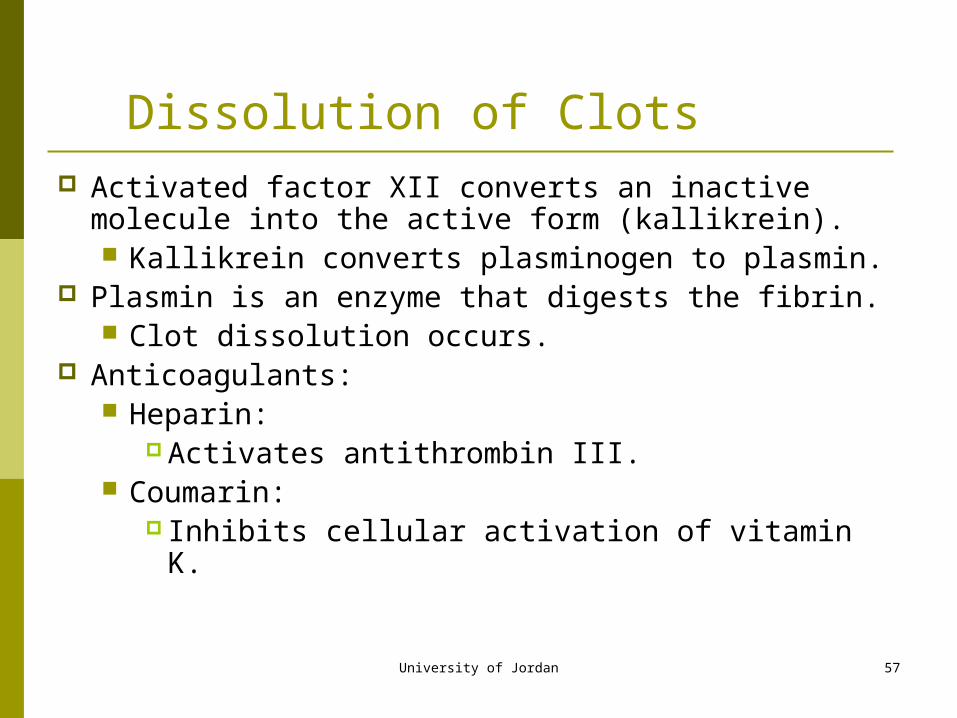
Dissolution of Clots Activated factor XII converts an inactive molecule into the
active form (kallikrein). Kallikrein converts plasminogen to plasmin.
Plasmin is an enzyme that digests the fibrin. Clot dissolution occurs.
Anticoagulants: Heparin:
Activates antithrombin III. Coumarin:
Inhibits cellular activation of vitamin K.

University of Jordan 58
Blood Groups and Blood Types Agglutinogens – surface of RBCs contain genetically
determined assortment of antigens Blood group – based on presence or absence of
various antigens At least 24 blood groups and more than 100 antigens
ABO and Rh

University of Jordan 59
RBC Antigens and Blood Typing
Each person’s blood type determines which antigens are present on their RBC surface.
Major group of antigens of RBCs is the ABO system:
Type AB:Both A and B antigens present.
Type O:Neither A or B antigens present.
Type A:Only A antigens present.
Type B:Only B antigens present.

University of Jordan 60
RBC Antigens and Blood Typing (continued)
Each person inherits 2 genes that control the production of ABO groups.
Type A:May have inherited A gene from each parent.May have inherited A gene from one parent and O gene from the other.
Type B:May have inherited B gene from each parent.May have inherited B gene from one parent and O gene from the other parent.
Type AB:Inherited the A gene from one parent and the B gene from the other parent.
Type O:Inherited O gene from each parent.

University of Jordan 61
ABO Blood Group Based on A and B antigens Type A blood has only antigen A Type B blood has only antigen B Type AB blood has antigens A and B
Universal recipients – neither anti-A or anti-B antibodies
Type O blood has neither antigen Universal donor
Reason for antibodies presence not clear

University of Jordan 62
Antigens and Antibodies of ABO Blood Types

University of Jordan 63
Rh Factor
Another group of antigens found on RBCs. Rh positive:
Has Rho(D) antigens. Rh negative:
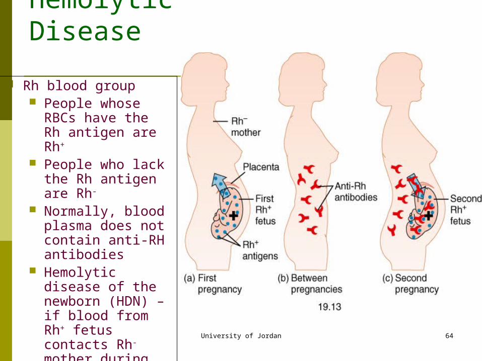
Does not have Rho(D) antigens. Significant when Rh- mother gives birth to Rh+ baby.
At birth, mother may become exposed to Rh+ blood of fetus. Mother at subsequent pregnancies may produce antibodies
against the Rh factor. Erythroblastosis fetalis:
Rh- mother produces antibodies, which cross placenta. Hemolysis of Rh+ RBCs in the fetus.
University of Jordan 64
Rh blood group People whose RBCs
have the Rh antigen are Rh+
People who lack the Rh antigen are Rh-
Normally, blood plasma does not contain anti-RH antibodies
Hemolytic disease of the newborn (HDN) – if blood from Rh+ fetus contacts Rh-mother during birth, anti-Rh antibodies made
Affect is on second Rh+ baby
Hemolytic Disease

University of Jordan 65
Typing Blood Single drops of blood
are mixed with different antisera
Agglutination with an antisera indicates the presence of that antigen on the RBC

University of Jordan 66
Transfusion Reactions
If blood types do not match, the recipient’s antibodies attach to donor’s RBCs and agglutinate.
Type O: Universal donor:
Lack A and B antigens. Recipient’s antibodies
cannot agglutinate the donor’s RBCs.
Type AB: Universal recipient:
Lack the anti-A and anti-B antibodies.
Cannot agglutinate donor’s RBCs.
Insert fig. 13.6

University of Jordan 67


