UNIVERSITY OF NOVA GORICA GRADUATE SCHOOL BIOCHEMICAL CHARACTERISATION OF THE ROLE OF THE HUMAN RECQ1 HELICASE AT THE REPLICATION FORK DISSERTATION Shivasankari Gomathinayagam Mentor: Dr. Alessandro Vindigni Ph.D. Nova Gorica, 2013
Transcript
UNIVERSITY OF NOVA GORICA
GRADUATE SCHOOL
BIOCHEMICAL CHARACTERISATION OF THE ROLE OF
THE HUMAN RECQ1 HELICASE AT THE REPLICATION
FORK
DISSERTATION
Shivasankari Gomathinayagam
Mentor: Dr. Alessandro Vindigni Ph.D.
Nova Gorica, 2013
2
CONTENTS
ABSTRACT 9
TITLE AND ABSTRACT IN SLOVENE 10
1 INTRODUCTION 12
1.1 Helicases 12
1.11 DNA helicases 12
1.12 Classification of helicases 12
1.13 Mechanism of action 14
1.2 RecQ helicases 15
1.21 Domain architecture 16
1.22 Human RecQ helicases and associated diseases 22
1.3 Biochemical properties of RecQ helicases 26
1.31 Helicase activity 26
1.32 Annealing activity 26
1.33 Exonuclease activity 27
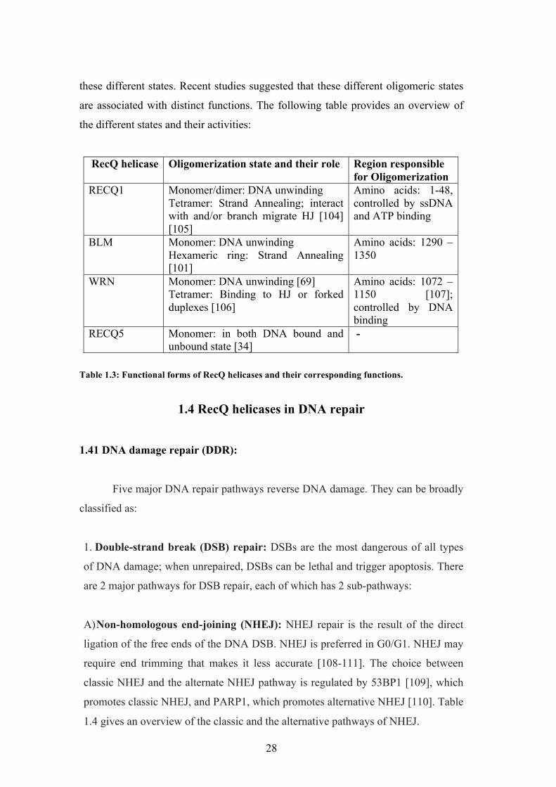
1.34 Functional forms of hRecQ helicases 27
1.4 RecQ helicases in DNA Repair 28
1.41 DNA Damage Repair (DDR) 28
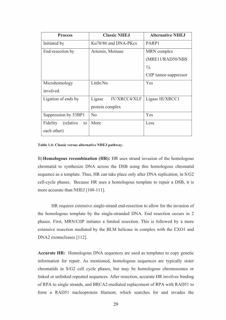
1.42 Defects in DDR 30
1.43 Role of RecQ helicases in DNA repair 31
1.5 RecQ helicases in DNA replication 37
1.51 DNA replication 37
1.52 Replication stress and fork stability 38
1.53 Roles of RecQ helicases in DNA replication 41
1.6 Replication fork regression and restoration 45
1.61 Top1 inhibitors and replication fork reversal 47
2 MATERIALS AND METHODS 50
2.1 Antibodies and chemicals 50
2.2 Cell culture and transfection 50
2.3. Expression and purification of recombinant proteins 50
2.3.1 RECQ1 overexpression and purification 50
2.3.2 Site directed mutagenesis and purification of RECQ1 mutants 51
3
2.3.3 Preparation of the truncated RECQ1 52
2.3.4 Determination of protein concentration 52
2.4 Oligonucleotides 53
2.5 Preparation of DNA substrates 55
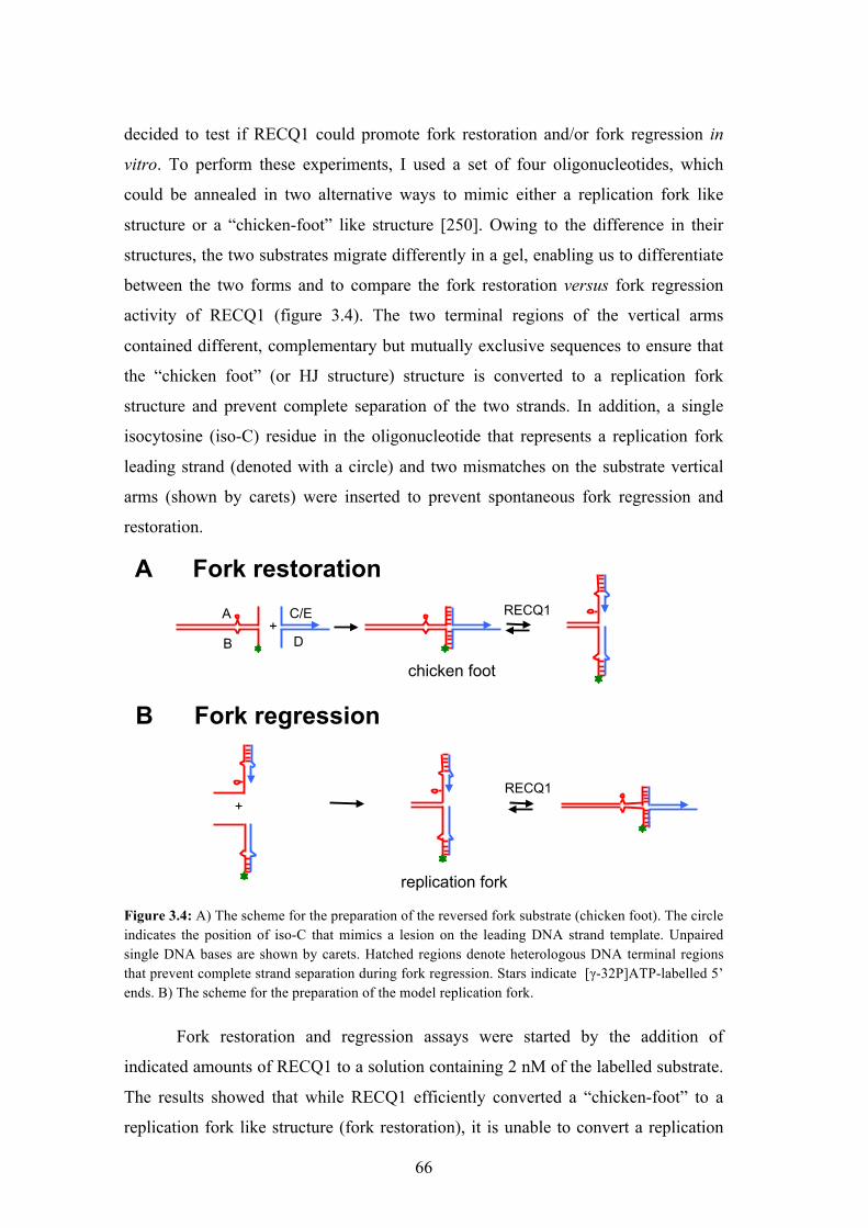
2.5.1 Construction of the replication fork and the chicken-foot like structure 56
2.6. Radiometric biochemical assays 58
2.6.1 Helicase assay 58
2.6.2 DNA strand annealing assay 58
2.6.3 In vitro fork regression and restoration assays 58
2.6.5 Resolving radioactive reactions on native PAGE 59
2.6.6 Quantification and graphs 59
2.7 Purified PAR production 60
2.8 Western blotting 60
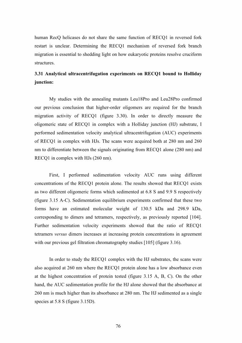
2.9 Analytical ultracentrifugation 60
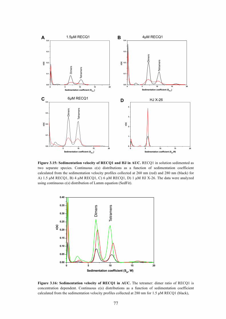
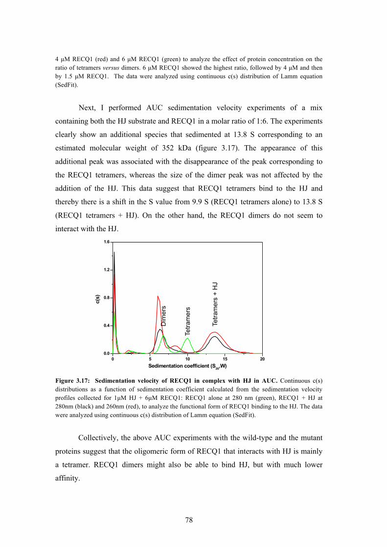
2.10 Gel filtration chromatography 61
2.11 Cryo-EM 61
2.12 In silico analysis 62
3 RESULTS 63
3.1 Biochemical characterization of RECQ1 63
3.11 Expression and purification of hRECQ1 from Sf9 insect cells 63
3.12 Biochemical characterization of the hRECQ1 helicase 64
3.12A Helicase assays using the forked duplex 64
3.12B Strand annealing assays 65
3.2 Role of RECQ1 in replication fork restart 65
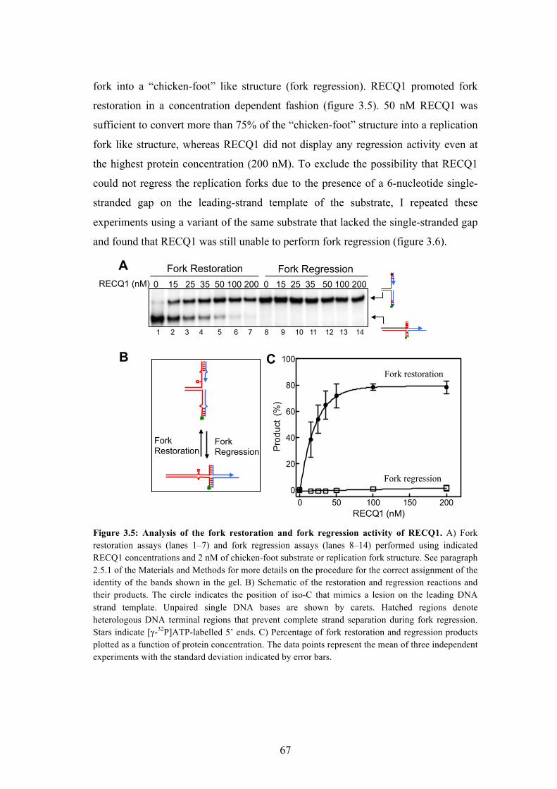
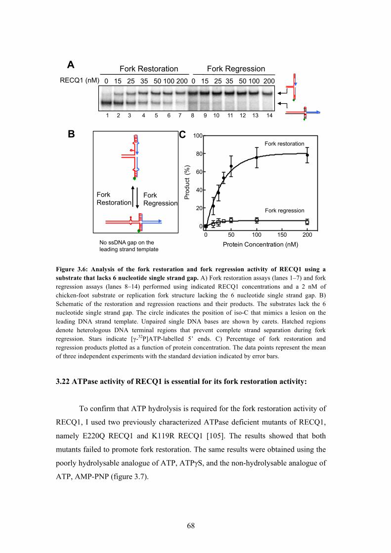
3.21 RECQ1 promotes restart of reversed replication forks in vitro 65
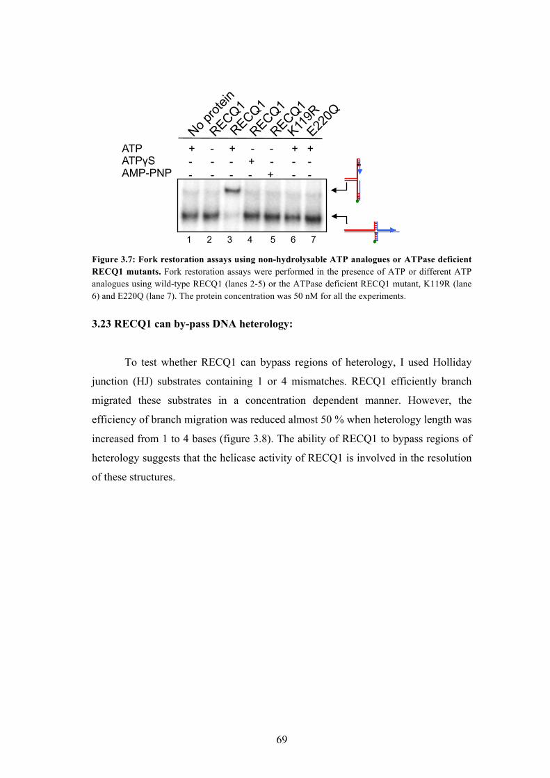
3.22 ATPase activity of RECQ1 is essential for its fork restoration activity 68
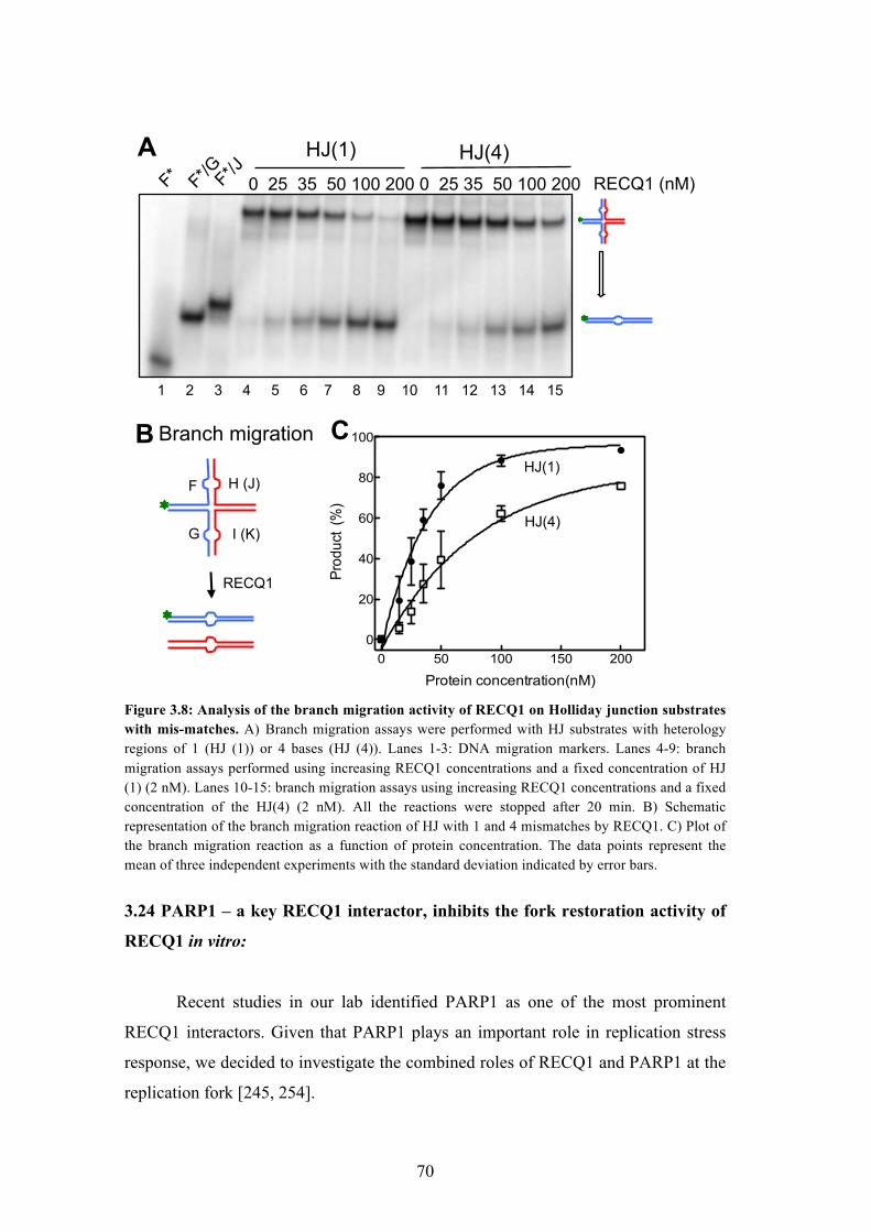
3.23 RECQ1 can bypass DNA heterology 69
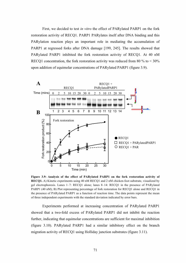
3.24 PARP1 - a key RECQ1 interactor, inhibits the fork restoration activity
of RECQ1 in vitro
70
3.25 PAR polymer is responsible for the inhibitory effect of PARylated
PARP1 on the fork restoration activity of RECQ1
72
3.26 PARylated PARP1 inhibits the DNA unwinding activity of RECQ1 73
4
3.27 PARylated PARP1 specifically inhibits the activity of RECQ1 74
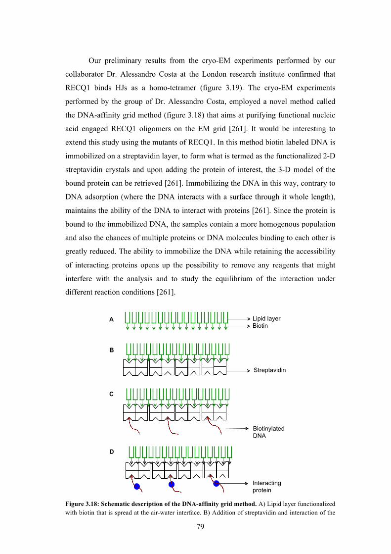
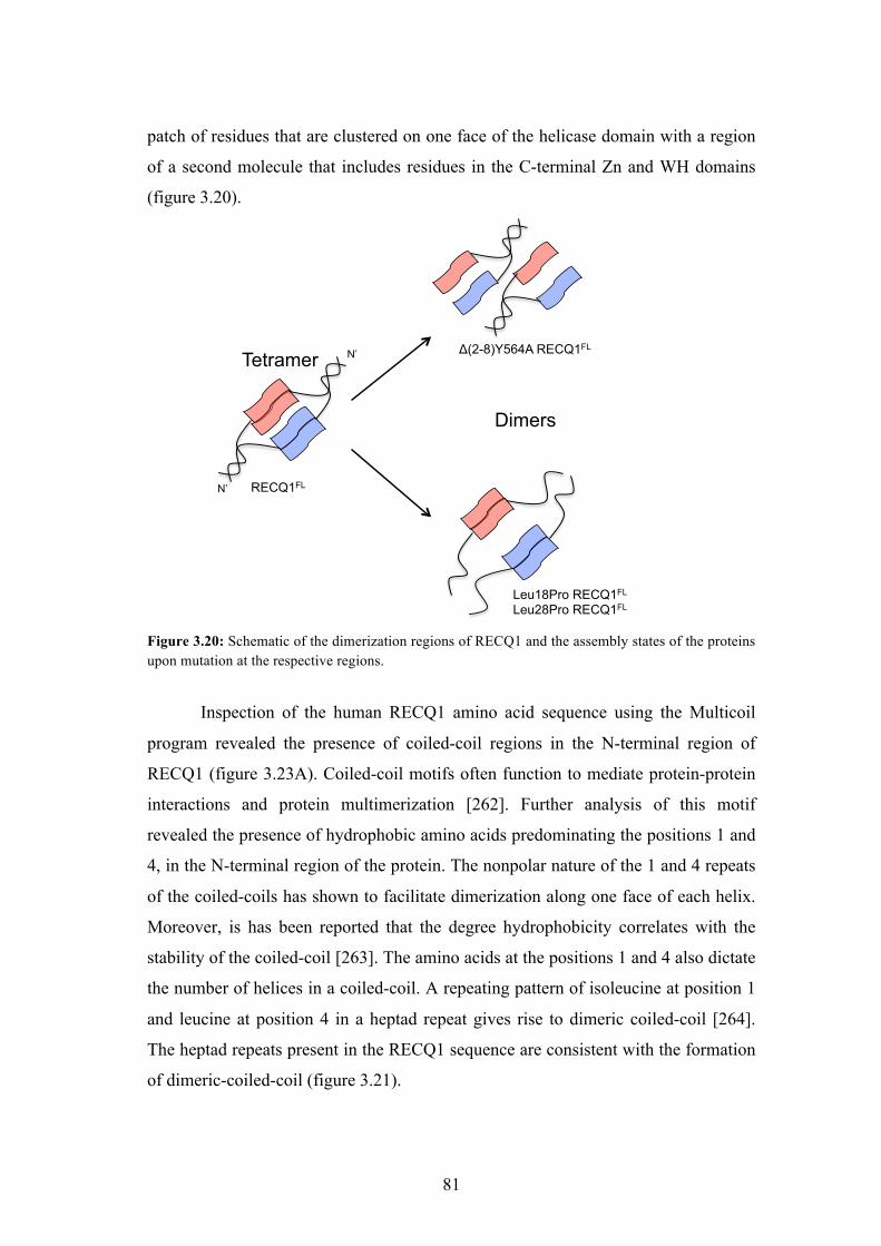
3.3 Architecture of RECQ1 assemblies with the Holliday junction 75
3.31 Analytical ultracentrifugation experiments and cryo-EM on RECQ1
bound to Holliday junction
76
3.4 Identification of coiled-coil in RECQ1 and biochemical
characterization of the coiled-coil mutants
80
3.41 Identification of coiled-coil region in the N-terminus of RECQ1 80
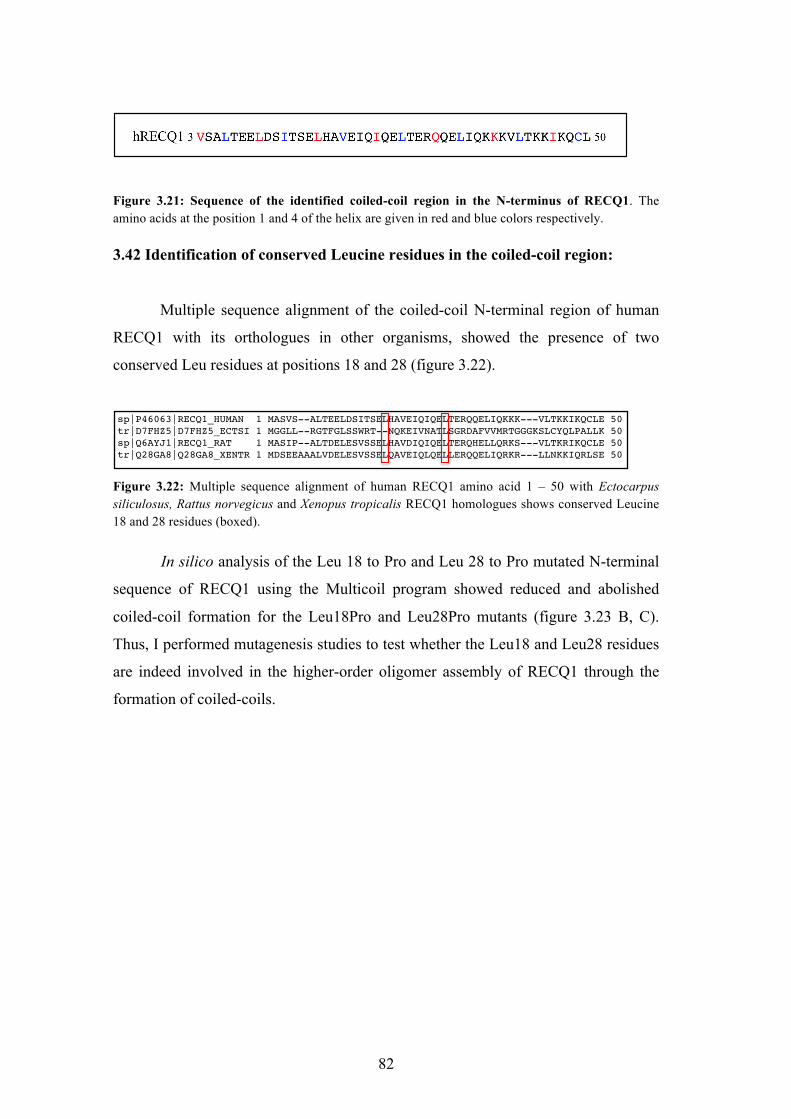
3.42 Identification of conserved Leucine residues in the coiled-coil region 82
3.43 Expression and purification of the RECQ1 mutants 83
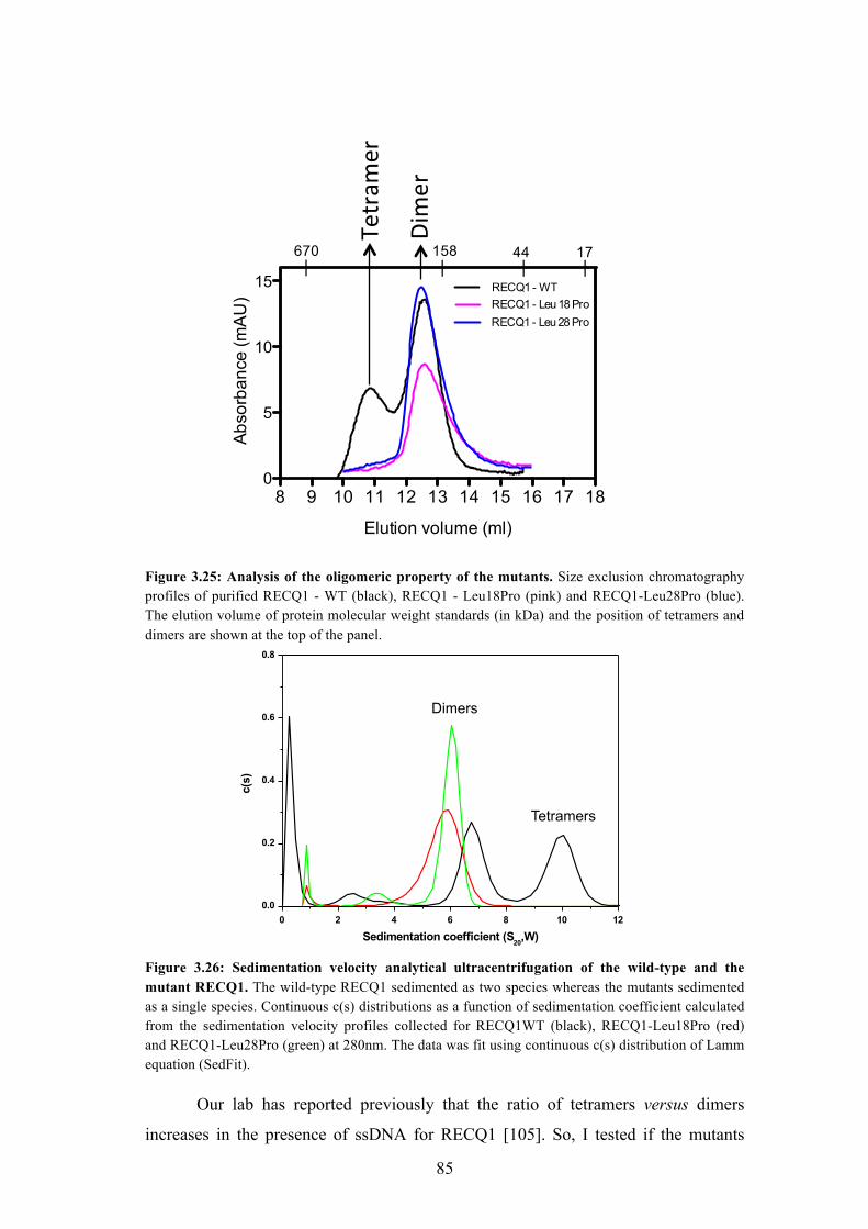
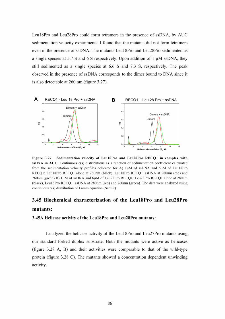
3.44 The Leu to Pro mutation abolishes the formation of tetramers 84
3.45 Biochemical characterization of the Leu18Pro and Leu28Pro mutants 86
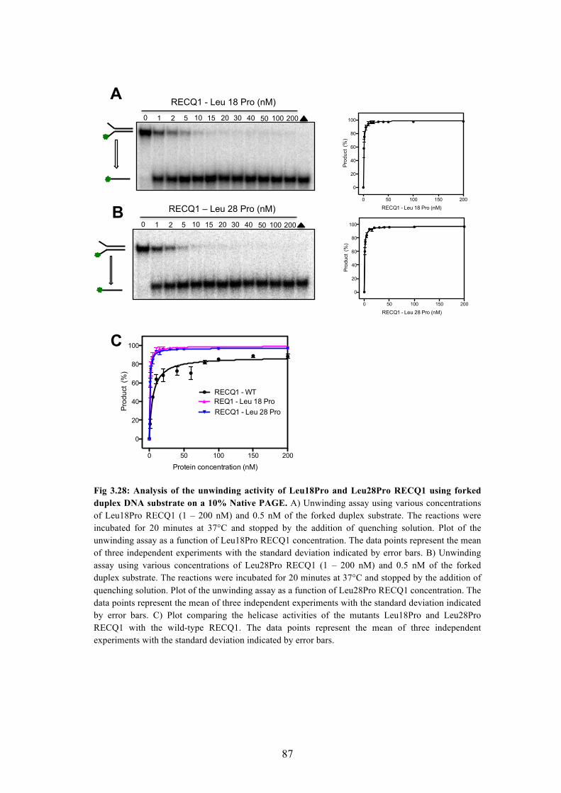
3.45A Helicase activity of the Leu18Pro and Leu28Pro mutants 86
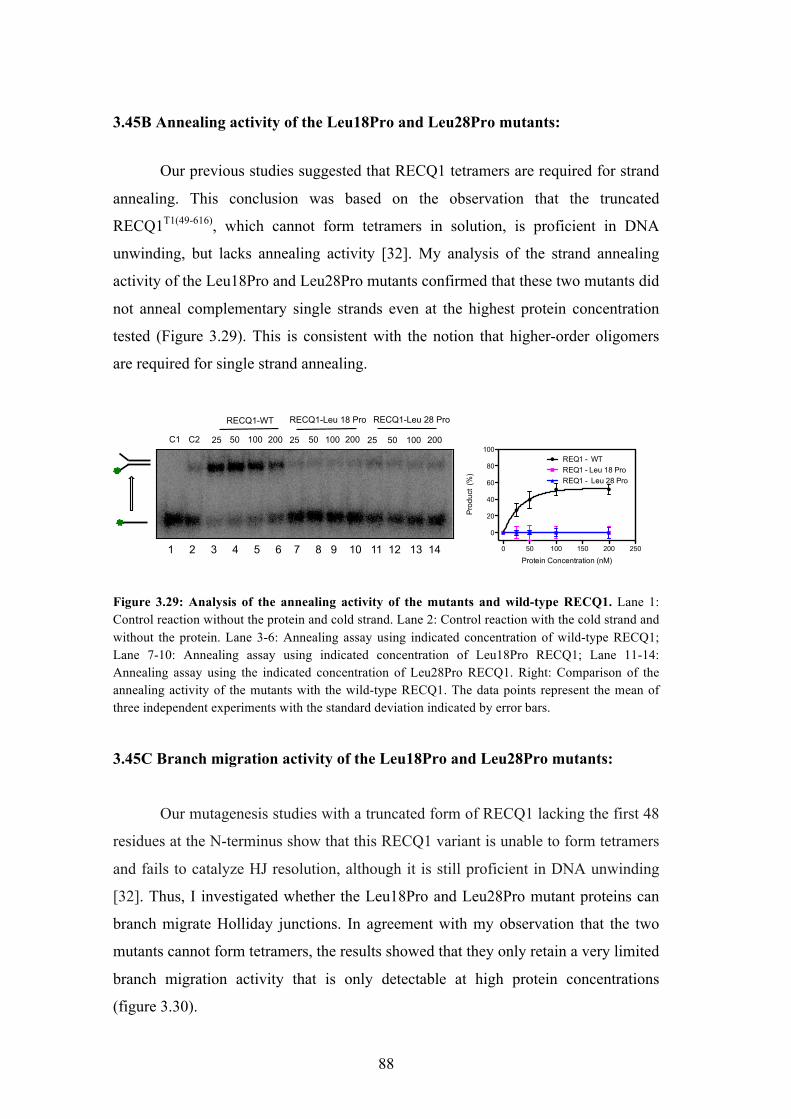
3.45B Annealing activity of the Leu18Pro and Leu28Pro mutants 88
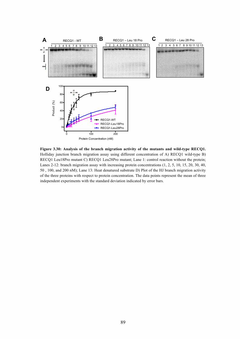
3.45C Branch migration activity of the Leu18Pro and Leu28Pro mutants 88
4 DISCUSSION 90
BIBLIOGRAPHY 101
5
LIST OF FIGURES AND TABLES
FIGURES:
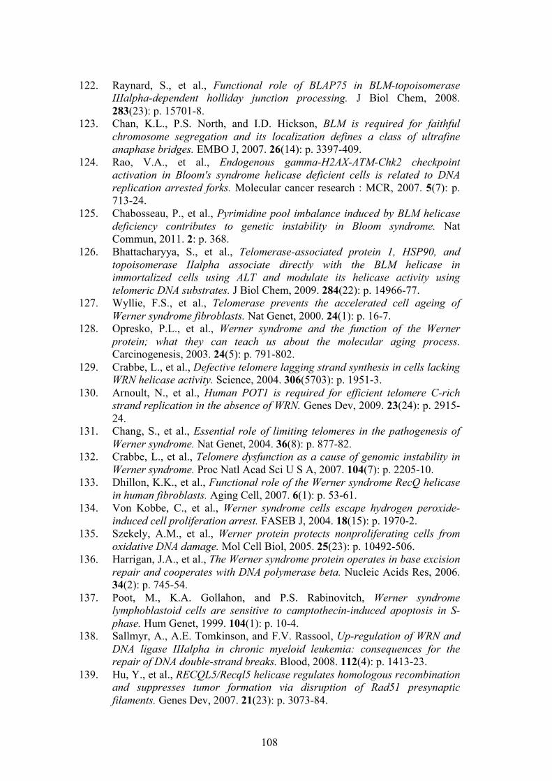
1.1 Classification of DNA helicases based on conserved amino acid
sequences
13
1.2 Models for DNA helicase translocation and unwinding 14
1.3 Domain organization of various RecQ helicases from different organisms 16
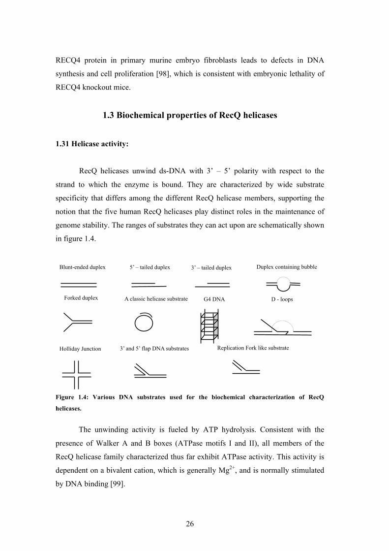
1.4 Various DNA substrates used for the biochemical characterization of
RecQ helicases
26
1.5 Mechanisms of DNA damage tolerance to lesions on the leading strand 46
1.6 Model for replication interference by Top1 poisons and their synergistic
effects with PARP inhibitors
48
2.1 Schematic of the preparation of the chicken-foot like structure 56
2.2 Schematic of the preparation of the replication fork like structure 57
2.3 Preparation of the chicken-foot and the replication fork like structure 57
3.1 SDS-PAGE and western blot analysis of purified hRECQ1 63
3.2 Analysis of the unwinding activity of RECQ1 64
3.3 Analysis of the DNA strand annealing activity of RECQ1 65
3.4 Schematic for the preparation of reversed and replication fork structure 66
3.5 Analysis of the fork restoration and fork regression activity of RECQ1 67
3.6 Analysis of the fork restoration and fork regression activity of RECQ1
using a substrate that lacks 6 nucleotide single strand gap
68
3.7 Fork restoration assays using non-hydrolysable ATP analogues or
ATPase deficient RECQ1 mutants
69
3.8 Analysis of the branch migration activity of RECQ1 on Holliday junction
substrates with mis-matches
70
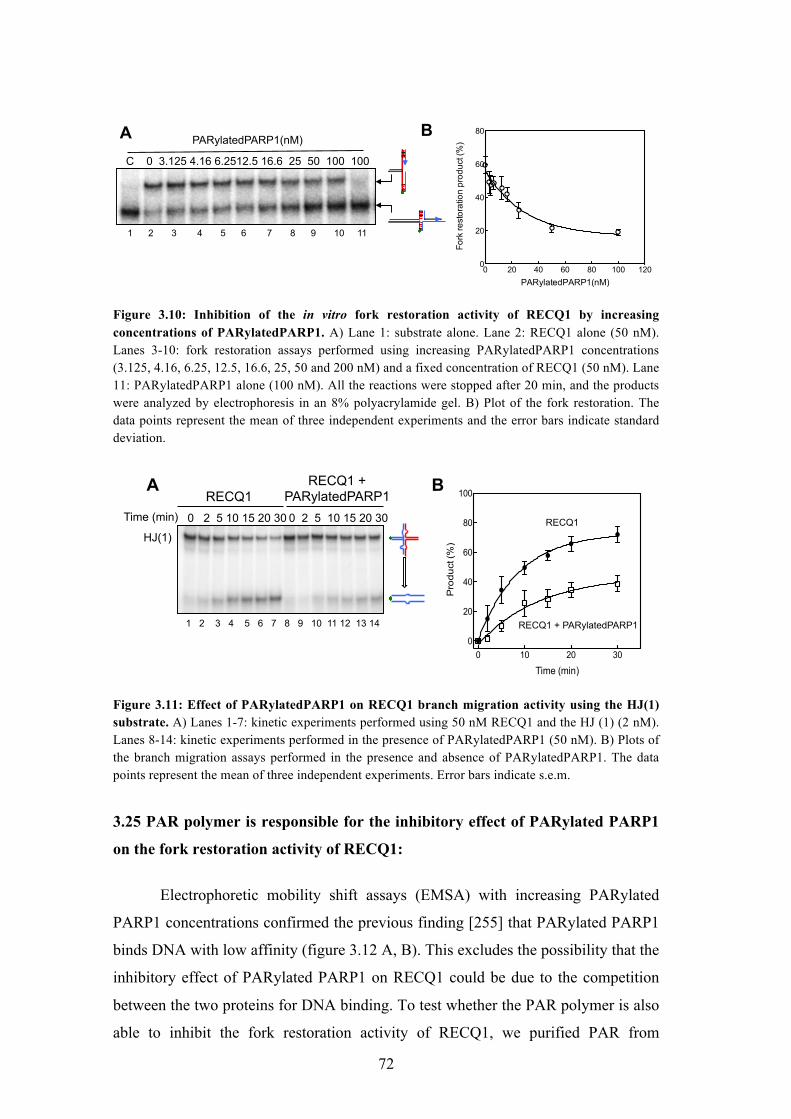
3.9 Analysis of the effect of PARylated PARP1 on the fork restoration
activity of RECQ1
71
3.10 Inhibition of the in vitro fork restoration activity of RECQ1 by
increasing concentrations of PARylatedPARP1
72
3.11 Effect of PARylatedPARP1 on RECQ1 branch migration activity using
the HJ substrate
72
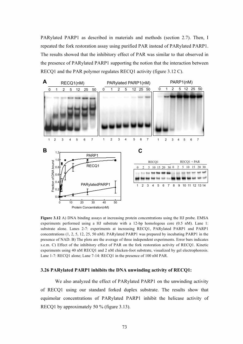
3.12 EMSA experiments performed using a HJ substrate with a 12-bp
homologous core
73
6
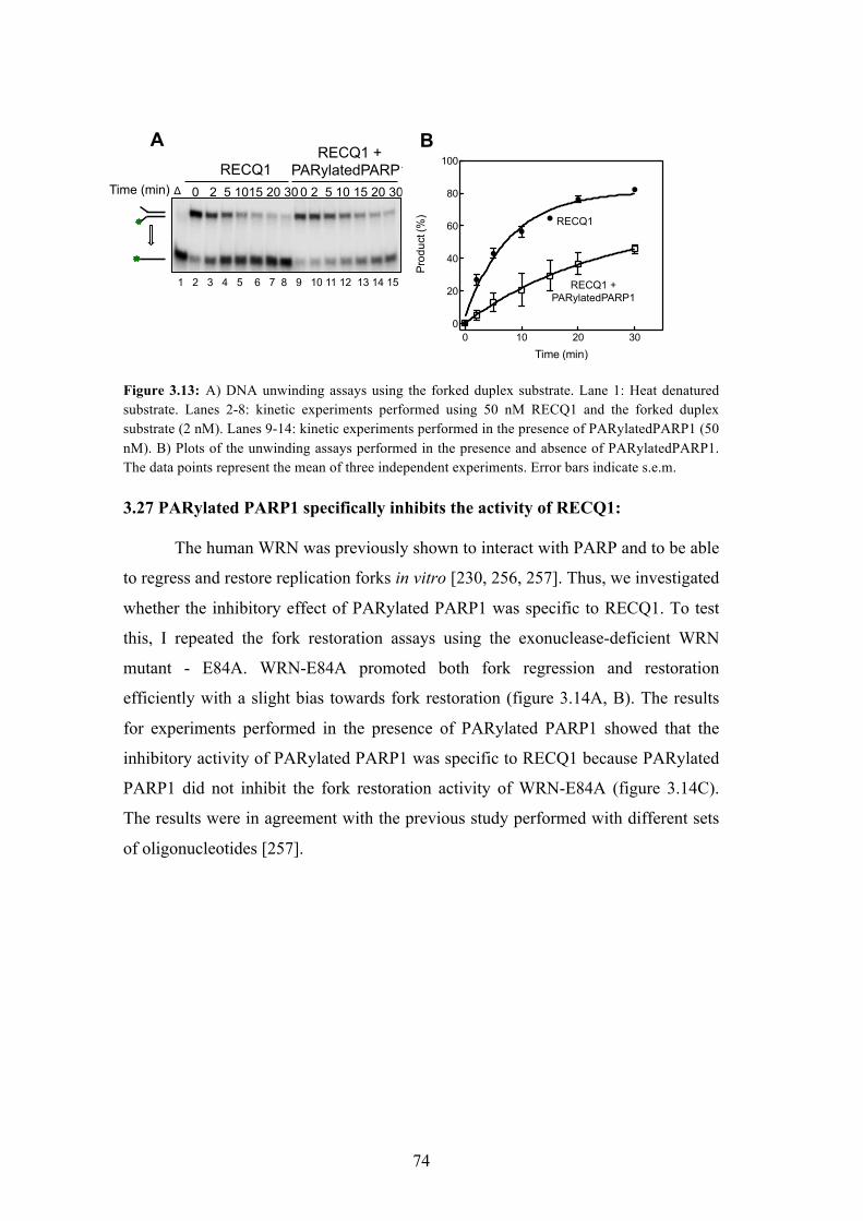
3.13 DNA unwinding assays using the forked duplex substrate 74
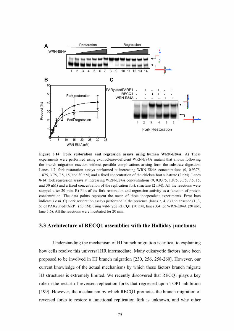
3.14 Fork restoration and regression assays using human WRN-E84A 75
3.15 Sedimentation velocity of RECQ1 and HJ in AUC 77
3.16 Sedimentation velocity of RECQ1 in AUC 77
3.17 Sedimentation velocity of RECQ1 in complex with HJ in AUC 78
3.18 Schematic description of the DNA-affinity grid method 79
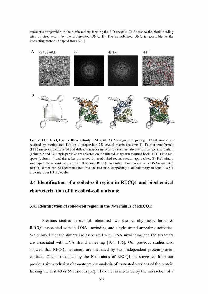
3.19 RecQ1 on a DNA affinity EM grid 80
3.20 Schematic of the dimerization regions of RECQ1 and the assembly
states of the proteins upon mutation at the respective regions
81
3.21 Sequence of the identified coiled-coil region in the N-terminus of
RECQ1
82
3.22 Multiple sequence alignment of human RECQ1 amino acid 1 – 50 with
RECQ1 homologues shows conserved Leucine 18 and 28 residues
82
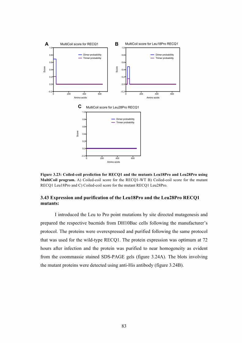
3.23 Coiled-coil prediction for RECQ1 and the mutants Leu18Pro and
Leu28Pro using MultiCoil program
83
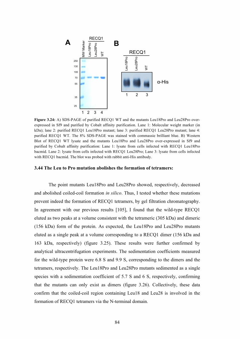
3.24 SDS-PAGE and western blot analysis of purified hRECQ1 WT and the
mutants Leu18Pro and Leu28Pro
84
3.25 Analysis of the oligomeric property of the mutants 85
3.26 Sedimentation velocity analytical ultracentrifugation of the wild-type
and the mutant RECQ1
85
3.27 Sedimentation velocity of Leu18Pro and Leu28Pro RECQ1 in complex
with ssDNA in AUC
86
3.28 Analysis of the unwinding activity of Leu18Pro and Leu28Pro RECQ1 87
3.29 Analysis of the annealing activity of the mutants and wild-type RECQ1 88
3.30 Analysis of the branch migration activity of the mutants and wild-type
RECQ1
89
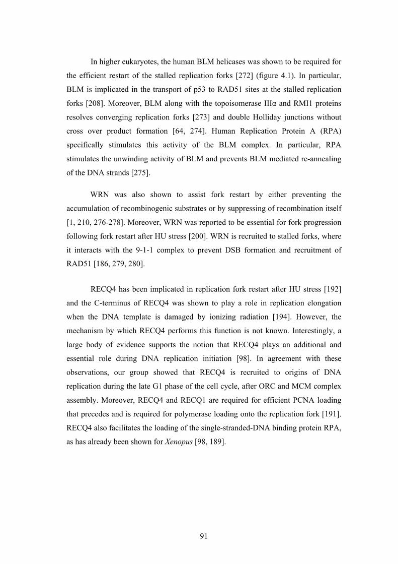
4.1 Pathways of replication fork restart by BLM and WRN 92
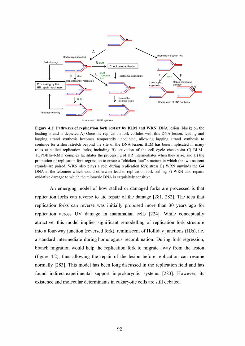
4.2 Schematic of lesion bypass by branch migration 93
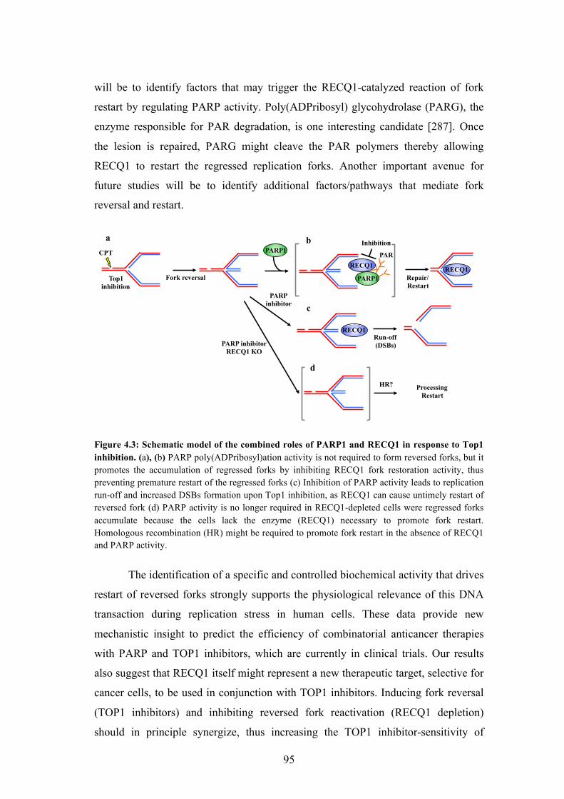
4.3 Schematic model of the combined roles of PARP1 and RECQ1 in
response to Top1 inhibition
95
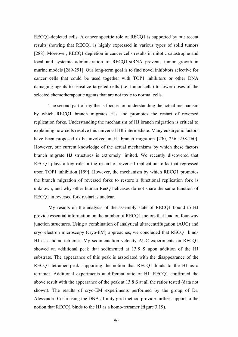
4.4 Schematics showing the extended and stacked conformations of the HJ 97
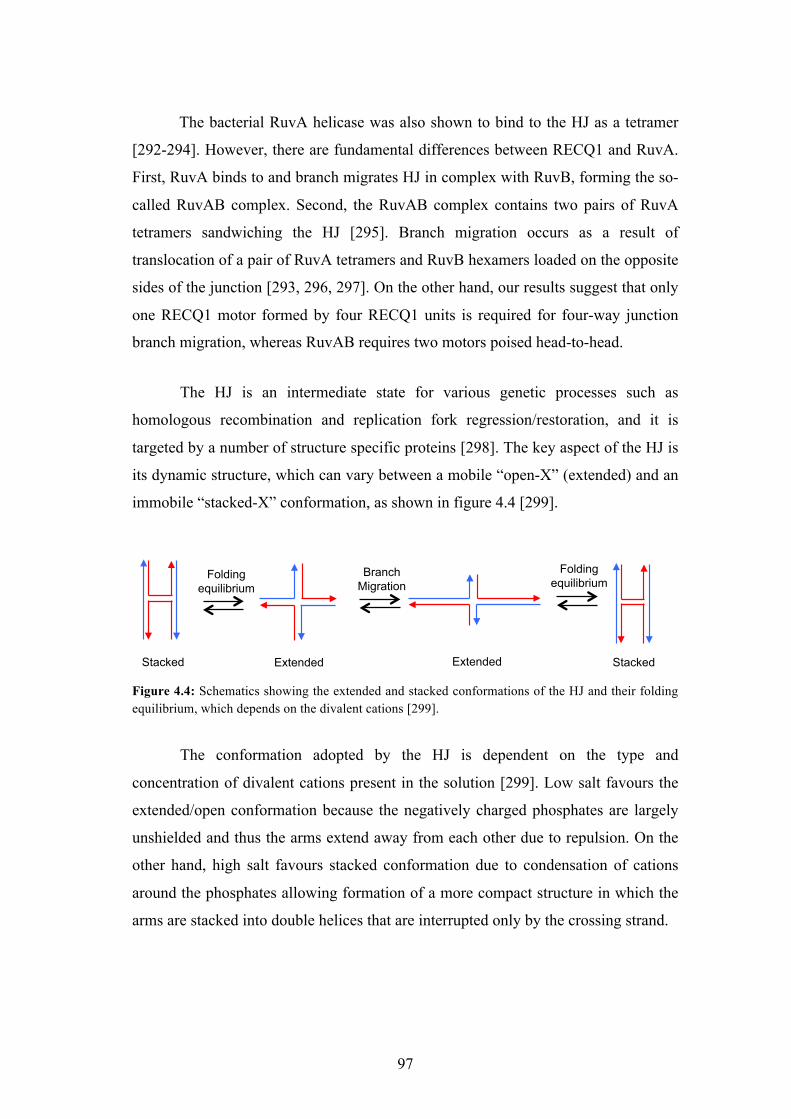
4.5 Structure specific preference of HJ binding proteins
98
7
TABLES:
1.1 Classification of DNA helicases 14
1.2 Characteristic features and functions of HRDC domains of some RecQ
helicases
20
1.3 Functional forms of RecQ helicases and their corresponding functions 28
1.4 Classic versus alternative NHEJ pathway 29
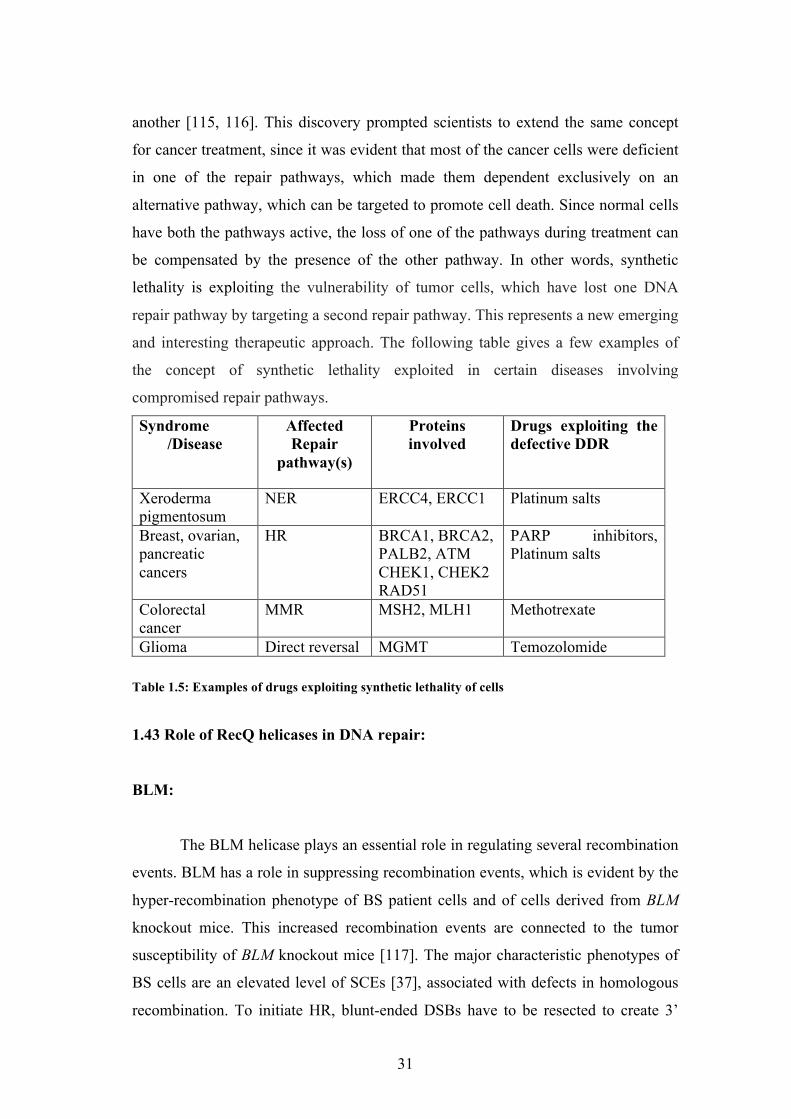
1.5 Examples of drugs exploiting synthetic lethality of cells 31
2.1 Sequences of the oligonucleotides used in the study of fork regression and
restoration
53
8
ACKNOWLEDGMENT
I have realized lately that one of the joys in completion is looking at the
journey I have taken to reach there and remembering fondly the people who helped
me and supported me along this long, but fulfilling road towards my doctoral
degree.
I am immensely pleased to have been a part of Dr. Alessandro Vindigni’s
group. The time spent with Dr. Alessandro has turned out to be enlightening both
academically and personally. I cannot thank him enough for his guidance and
support. I would also like to thank my tutor, Dr. Vittorio Venturi (ICGEB, Trieste).
My thanks to ICGEB and University of Nova Gorica for my stint at St. Louis school
of Medicine, St. Louis, thanks to which I could get a taste of the scientific
atmosphere at ICGEB, Trieste and at St. Louis school of Medicine, St. Louis. I
would like to thank my lab colleagues I have had the pleasure of working with -
Ramiro, Bojana, Gianluca, Francesca and my current lab members Saravana, Matteo
and Sasa for the all the sane discussions and insane fun we had together.
I am happy to acknowledge and profoundly thank the Arturo Falaschi Pre-
Doctoral fellowship for providing financial assistance for my PhD. I thank Dr. Dave
Wood for his valuable suggestions and advice with the biophysical experiments and
Dr. Sergey Korolev (SLU) and Dr. Erik Feldmann (WashU) for helping me with the
ultracentrifugation experiments. I would also like to extend my thanks to the
members of ICGEB and SLU who have helped me during my PhD.
I extend my heartfelt thanks to our collaborator, Dr. Alessandro Costa, and
his group (London Research Institute) for performing the cryo-EM experiments for
us. I would also like to extend my thanks to Dr. Yuna Ayala for all the discussions
we had and the inputs she gave. Special thanks to all my teachers and mentors who
have guided and moulded me into what I am today. I would also like to thank my
mom, dad, extended family and friends for all the amazing support they never failed
to give me. This has been a fulfilling ride indeed, and I take with me brilliant
experiences and memories for a lifetime to come. Thank you!
9
ABSTRACT
RecQ DNA helicases are critical enzymes for the maintenance of genome
integrity. Defects in three of the five human RecQ homologs give rise to distinct
genetic disorders associated with genomic instability, cancer predisposition, and
premature aging. Studies of RecQ helicases in model prokaryotic and eukaryotic
systems have demonstrated their vital roles in DNA replication, recombination and
repair. In particular, different members of RecQ family have been implicated in
various mechanisms that act at the level of stalled or damaged replication forks to
guarantee a faithful replication of our genome.
An emerging model of how stalled or damaged forks are processed is that
replication forks can reverse to aid repair of the damage. In this thesis, I studied the
role of the human RECQ1 helicase in replication fork reversal and restart using a
combination of biochemical and biophysical approaches. I used series of model
replication substrates that mimic either a functional replication fork or a reverse fork
structure to show that RECQ1 specifically promotes the restart of reversed forks, but
not the opposite reaction of fork reversal. I also provided novel insight into the role
of the poly(ADP-ribosyl)ation activity of PARP in fork reversal by showing that
PARylatedPARP1 inhibits the fork restoration activity of RECQ1.
Following these observations, I investigated the molecular mechanism by
which RECQ1 promotes the branch migration of reversed replication forks. My data
show that the functional form of RECQ1 that binds and branch migrates Holliday
junctions is a tetramer in vitro. The formation of the tetramer is mediated by N-
terminal coiled-coil region of RECQ1 involving two key leucine residues (Leu 18
and Leu 28). The point mutation of these leucines impairs the formation of tetramers,
as well as the annealing and Holliday junction branch migration activities of RECQ1,
while it does not affect the helicase activity.
These results together suggest that RECQ1 binds the regressed replication
forks as a tetramer to re-establish a functional replication fork and that the interaction
between RECQ1 and PARylatedPARP1 regulates this activity.
10
Key words: DNA replication stress response; DNA repair; replication fork reversal;
Holliday junctions; RecQ helicases
TITLE AND ABSTRACT IN SLOVENE
Biokemijska karakterizacija vloge !love"ke helikaze RecQ1 pri
replikacijskih vilicah
Helikaze DNA iz dru!ine RecQ so encimi, pomembni za vzdr!evanje
genomske celovitosti. Okvare treh izmed petih "love#kih homologov RecQ
povzro"ajo razli"ne genetske motnje, ki se ka!ejo kot genomska nestabilnost,
pove"ana nagnjenost k razvoju raka in prezgodnje staranje. S preu"evanjem helikaz
RecQ v modelnih prokariontskih in evkariontskih sistemih je bila dokazana njihova
klju"na vloga pri podvojevanju DNA, rekombinaciji in popravljalnih mehanizmih.
Predvsem so encimi iz te dru!ine udele!eni pri razli"nih mehanizmih, ki delujejo na
zaustavljenih ali po#kodovanih replikacijskih vilicah in zagotavljajo zanesljivo
podvojitev genoma.
Pred kratkim je bilo pokazano, da se replikacijske vilice pri okvari ali
zaustavitvi lahko obrnejo in to pripomore k odpravi po#kodbe DNA. V tej nalogi sem
s kombinacijo biokemijskih in biofizikalnih metod preu"evala vlogo "love#ke
helikaze RecQ1 pri obrnitvi replikacijskih vilic ter pri ponovnem za"etku podvajanja.
Uporabila sem vrsto modelnih substratov, ki predstavljajo tako funkcionalne
replikacijske vilice kot strukturo obrnjenih vilic in ugotovila, da encim RecQ1
specifi"no pospe#uje ponoven zagon obrnjenih vilic, ne pa same reakcije obrnitve.
Pridobila sem tudi nove informacije o vlogi poli(ADP-ribozil)acijske aktivnosti
encima PARP pri obrnitvi vilic in sicer, da PARiliran PARP1 inhibira aktivnost
encima RecQ1 pri obnovi replikacijskih vilic.
Da bi bolje razjasnili te ugotovitve, sem preu"evala molekularni mehanizem s
katerim RecQ1 pospe#uje premik razvejitve DNA pri obrnjenih replikacijskih
vilicah. Moji rezultati ka!ejo, da se funkcionalna oblika RecQ1 helikaze in vitro ve!e
na razvejitev Hollidayeve strukture v obliki tetramera. Enote se pove!ejo v tetramer
11
preko N-kon"ne obvite vija"nice, klju"na pa sta aminokislinska ostanka Leu 18 in
Leu 20. To"kovna mutacija teh dveh levcinov prepre"i nastanek tetramera. Tak
encim se ne more vezati na razvejitev Hollidayeve strukture in ne more premakniti
razvejitve, med tem ko helikazna aktivnost encima ni okrnjena.
Skupaj ti rezultati ka!ejo, da se tetramer RecQ1 helikaze ve!e na obrnjene
replikacijske vilice in sodeluje pri ponovni vzpostavitvi funkcionalnih
replikacijskih vilic. Interakcija med proteinoma RecQ1 in PARiliranim PARP1 pa
uravnava to aktivnost.
12
1. INTRODUCTION
1.1 Helicases 1.11 DNA helicases:
Around 1% of the open reading frames (ORF) in the human genome codes
for a class of enzymes called helicases. Helicases use the energy derived from the
hydrolysis of nucleotide triphosphate (NTP) to separate complementary strands of
nucleic acid molecules [1]. The helicases can be broadly classified as DNA or RNA
helicases, based on the substrates they act upon. DNA helicases catalyse the transient
unwinding of duplex DNA in a NTP dependent manner and play important roles in
all aspects of DNA metabolism. They play prominent roles in replication, repair and
recombination and thereby contribute to the maintenance of genome stability of all
living organisms. The importance of DNA helicases for the maintenance of genome
integrity is underlined by the numerous human diseases associated with defects in the
helicase genes [2-4].
1.12 Classification of helicases:
The two most popular methods to classify helicases are based on their
direction of translocation or on presence of particular signature motifs [5]. In vitro
experiments using partial DNA duplex substrates have shown that, helicases can
translocate with either a 3’ ! 5’ or 5’ ! 3’ polarity along single stranded DNA. For
example, helicases such us the minichromosome maintenance proteins (MCM) and
the RecQ helicases translocate on the single-stranded DNA with a 3’ ! 5’ polarity,
while the bacterial DnaB and phage T7 gp4 helicases translocate with a 5’ ! 3’
polarity. However, this classification is only applicable to those helicases that bind a
ssDNA terminus and then translocate along single-stranded before unwinding the
duplex [1].
The second method of classification is based on the analysis of specific
signature motifs. Using this approach, helicases have been classified into 6
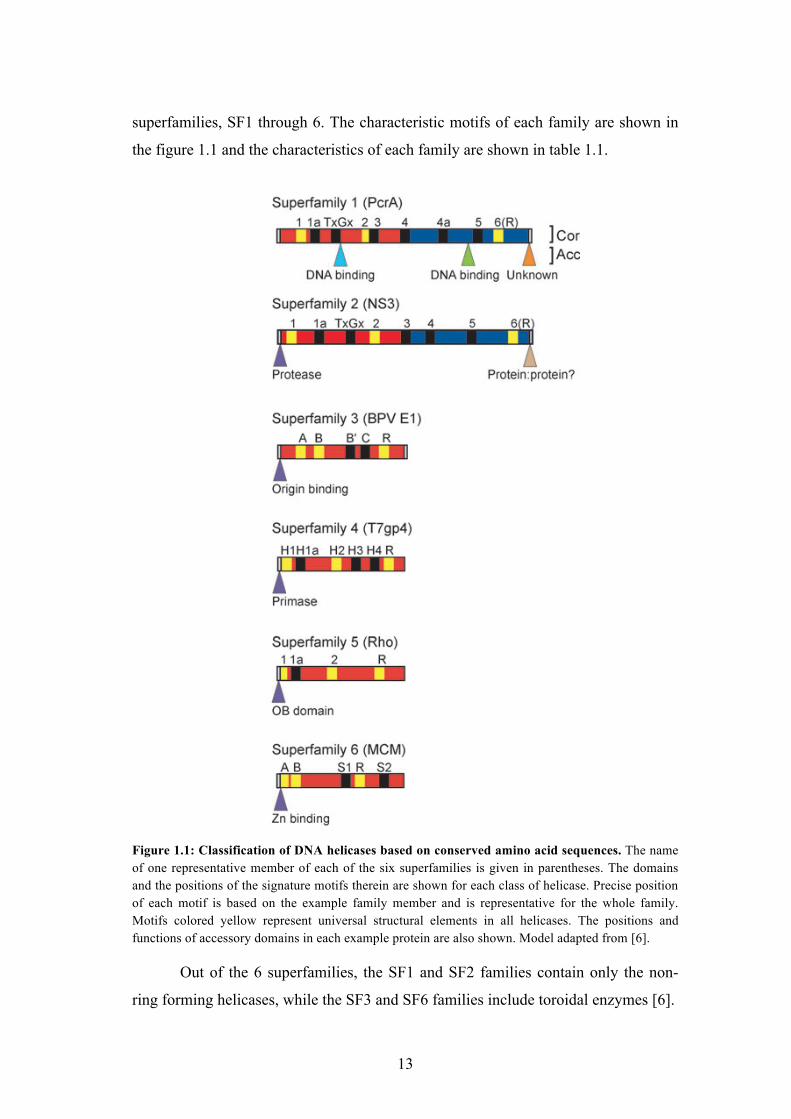
13
superfamilies, SF1 through 6. The characteristic motifs of each family are shown in
the figure 1.1 and the characteristics of each family are shown in table 1.1.
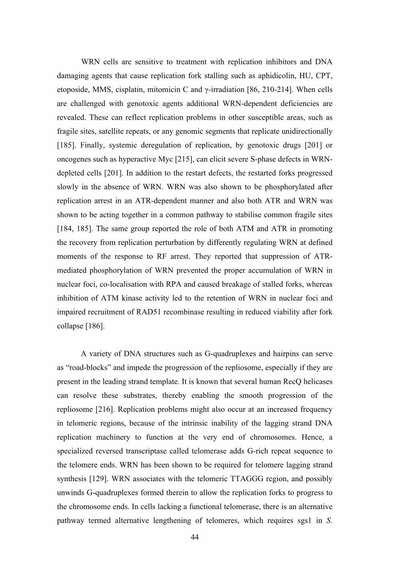
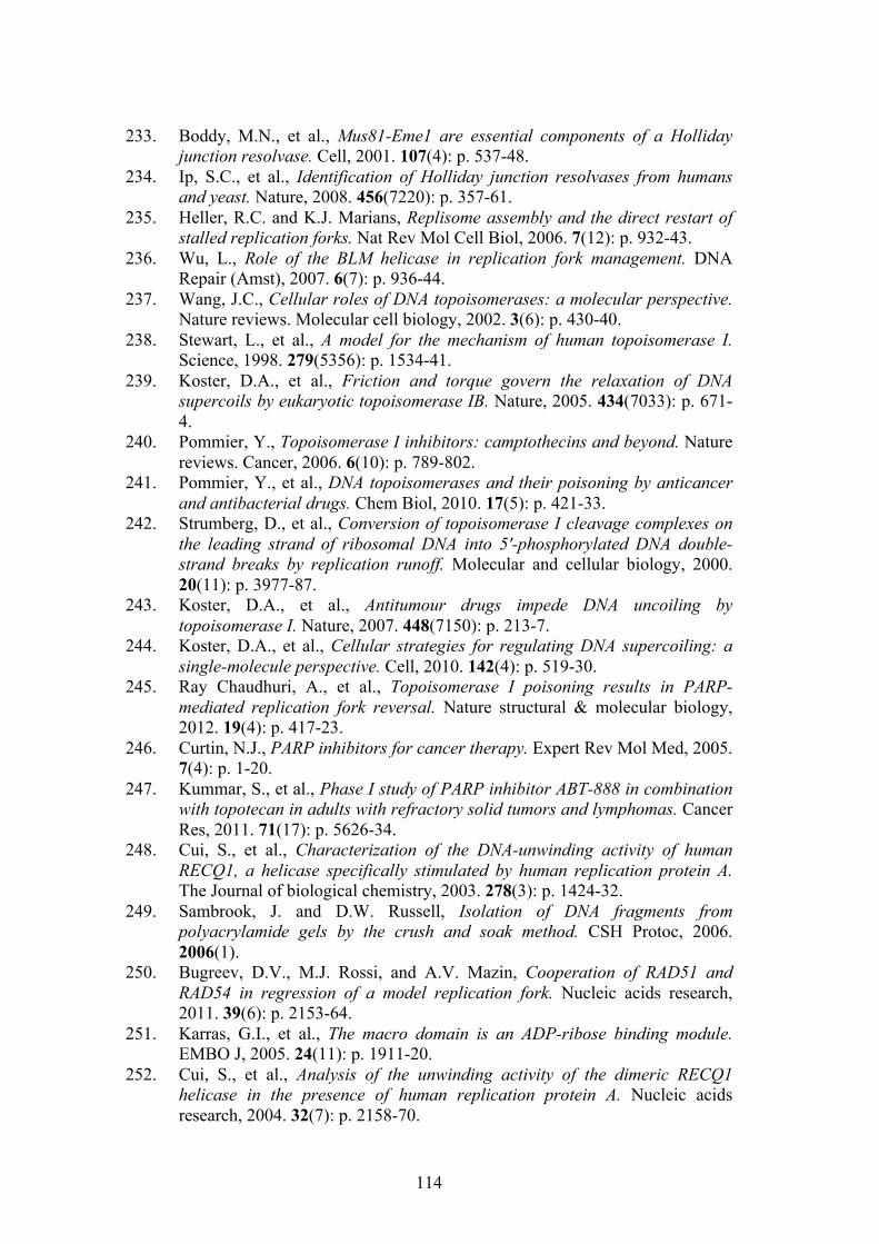
Figure 1.1: Classification of DNA helicases based on conserved amino acid sequences. The name of one representative member of each of the six superfamilies is given in parentheses. The domains and the positions of the signature motifs therein are shown for each class of helicase. Precise position of each motif is based on the example family member and is representative for the whole family. Motifs colored yellow represent universal structural elements in all helicases. The positions and functions of accessory domains in each example protein are also shown. Model adapted from [6].
Out of the 6 superfamilies, the SF1 and SF2 families contain only the non-
ring forming helicases, while the SF3 and SF6 families include toroidal enzymes [6].
14
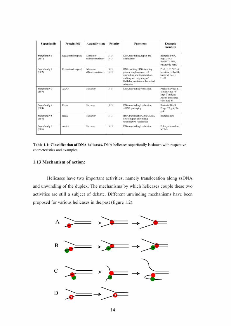
Superfamily Protein fold Assembly state Polarity Functions Example members
Table 1.1: Classification of DNA helicases. DNA helicases superfamily is shown with respective characteristics and examples.
1.13 Mechanism of action:
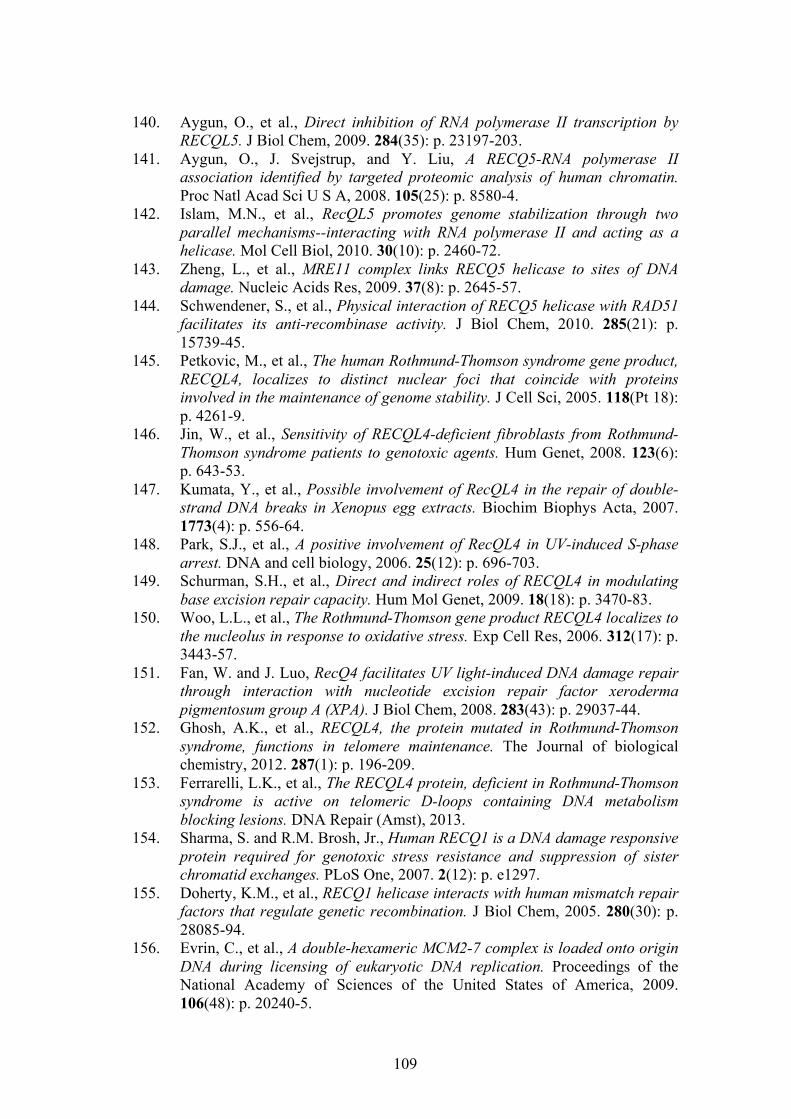
Helicases have two important activities, namely translocation along ssDNA
and unwinding of the duplex. The mechanisms by which helicases couple these two
activities are still a subject of debate. Different unwinding mechanisms have been
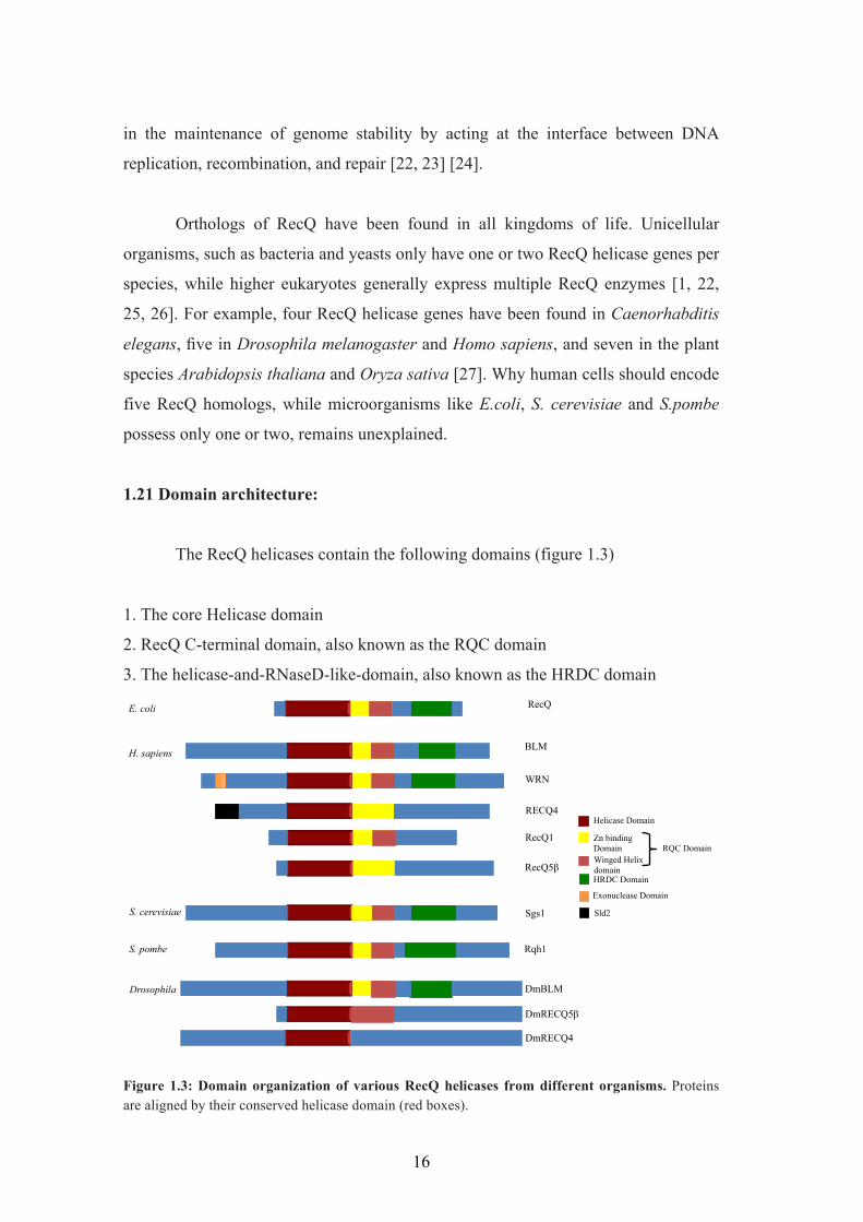
proposed for various helicases in the past (figure 1.2):
A
B
C
D
15
Figure 1.2: Models for DNA helicase translocation and unwinding. A) Inchworm model B) Co-
operative inchworm model, C) Rolling model, D) Hexameric helicase model.
A. Inchworm model: The helicase has two non-identical DNA binding sites that
bind with a defined polarity. The leading site interacts with the duplex region during
successive cycles of unwinding, whereas the tail site interacts with the ssDNA. It is
consistent with any oligomeric state, including monomers [7]. A dimeric inchworm
mechanism has been reported for the bacterial UvrD helicase [8].
B. Co-operative inchworm model: Multiple helicase molecules line up along the
ssDNA lattice to promote DNA unwinding. This mechanism is similar to the
inchworm model, with the exception that it requires multiple helicase molecules [9,
10]. For example, the bacteriophage T4 Dda helicase is functional a monomer, but
becomes more processive when there are multiple molecules acting cooperatively
[10, 11].
C. Rolling model: Each monomer has least two identical DNA-binding sites that can
bind to ssDNA and dsDNA in an alternating fashion. The rolling model requires a
dimeric protein, as previously described for the bacterial Rep helicase [12].
D. Hexameric helicase model: The enzyme forms ring structure that encircles one
strand of the duplex leaving the other strand outside the ring. The formation of a ring
structure in hexameric helicases may be needed to prevent premature dissociation of
the functional helicase molecule from the DNA substrate. Classical examples of
hexameric helicases are the gp4 of bacteriophage T7 [13, 14], Rho of Escherichia
coli [15, 16], and the eukaryotic minichromosomal maintenance (MCM) helicase
[17-19].
1.2 RecQ helicases
The family of RecQ helicases is named after the RecQ gene of Escherichia
coli discovered by Nakayama and his colleagues, more than 20 years ago [20]. RecQ
helicases are highly conserved from bacteria to man. They are part of SF2 family and
they all unwind DNA with 3’ – 5’ polarity [21]. RecQ helicases play an essential role
16
in the maintenance of genome stability by acting at the interface between DNA
replication, recombination, and repair [22, 23] [24].
Orthologs of RecQ have been found in all kingdoms of life. Unicellular
organisms, such as bacteria and yeasts only have one or two RecQ helicase genes per
species, while higher eukaryotes generally express multiple RecQ enzymes [1, 22,
25, 26]. For example, four RecQ helicase genes have been found in Caenorhabditis
elegans, five in Drosophila melanogaster and Homo sapiens, and seven in the plant
species Arabidopsis thaliana and Oryza sativa [27]. Why human cells should encode
five RecQ homologs, while microorganisms like E.coli, S. cerevisiae and S.pombe
possess only one or two, remains unexplained.
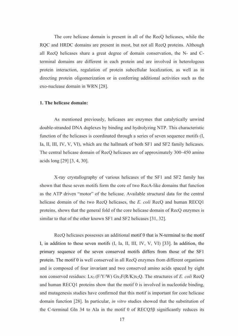
1.21 Domain architecture:
The RecQ helicases contain the following domains (figure 1.3)
1. The core Helicase domain
2. RecQ C-terminal domain, also known as the RQC domain
3. The helicase-and-RNaseD-like-domain, also known as the HRDC domain RecQ
BLM
WRN
RECQ4
RecQ1
RecQ5!
Sgs1
Rqh1
DmBLM
DmRECQ5!
DmRECQ4
E. coli
H. sapiens
S. cerevisiae
S. pombe
Drosophila
Helicase Domain
RQC Domain
HRDC Domain
Sld2
Exonuclease Domain
Winged Helix domain
Zn binding Domain
Figure 1.3: Domain organization of various RecQ helicases from different organisms. Proteins are aligned by their conserved helicase domain (red boxes).
17
The core helicase domain is present in all of the RecQ helicases, while the
RQC and HRDC domains are present in most, but not all RecQ proteins. Although
all RecQ helicases share a great degree of domain conservation, the N- and C-
terminal domains are different in each protein and are involved in heterologous
protein interaction, regulation of protein subcellular localization, as well as in
directing protein oligomerization or in conferring additional activities such as the
exo-nuclease domain in WRN [28].
1. The helicase domain:
As mentioned previously, helicases are enzymes that catalytically unwind
double-stranded DNA duplexes by binding and hydrolyzing NTP. This characteristic
function of the helicases is coordinated through a series of seven sequence motifs (I,
Ia, II, III, IV, V, VI), which are the hallmark of both SF1 and SF2 family helicases.
The central helicase domain of RecQ helicases are of approximately 300–450 amino
acids long [29] [3, 4, 30].
X-ray crystallography of various helicases of the SF1 and SF2 family has
shown that these seven motifs form the core of two RecA-like domains that function
as the ATP driven “motor” of the helicase. Available structural data for the central
helicase domain of the two RecQ helicases, the E. coli RecQ and human RECQ1
proteins, shows that the general fold of the core helicase domain of RecQ enzymes is
similar to that of the other known SF1 and SF2 helicases [31, 32].
RecQ helicases possesses an additional motif 0 that is N-terminal to the motif
I, in addition to these seven motifs (I, Ia, II, III, IV, V, VI) [33]. In addition, the
primary sequence of the seven conserved motifs differs from those of the SF1
protein. The motif 0 is well conserved in all RecQ enzymes from different organisms
and is composed of four invariant and two conserved amino acids spaced by eight
non conserved residues: Lx3 (F/Y/W) Gx3F(R/K)x2Q. The structures of E. coli RecQ
and human RECQ1 proteins show that the motif 0 is involved in nucleotide binding,
and mutagenesis studies have confirmed that this motif is important for core helicase
domain function [28]. In particular, in vitro studies showed that the substitution of
the C-terminal Gln 34 to Ala in the motif 0 of RECQ5$ significantly reduces its
18
ATPase activity [34, 35] and that the same substitution in murine BLM inactivates
the ATPase and the helicase activity of the enzyme [36]. Interestingly, the same
mutation has also been reported in BS patients [37].
The mechanism by which RecQ helicases couple ATP binding/hydrolysis
cycles to unidirectional translocation along ssDNA is poorly understood. Although
the nucleotide bound structures of E. coli RecQ and human RECQ1 show that RecQ
enzymes bind ATP in a conventional way, we currently have little knowledge on
their physical mechanism of DNA unwinding.
E. coli RecQ contains a conserved aromatic-rich loop in its helicase domain
which is located between motifs II and III [38]. A similar conserved aromatic-rich
loop in motif III of SF1 helicases mediates both ATP and single-stranded DNA
binding [39], [7]. Mutational analysis of the RecQ aromatic-rich loop provided
evidence that this region is critical for coupling ATPase and DNA
binding/unwinding activities [38]. The crystal structure of E. coli RecQ [31] has
provided further insight into the functional importance of some of the conserved
helicase motifs. The Motif I helps in making the canonical phosphate and metal
contacts [31]. Mutations in the phosphate binding lysine residue in motif I of WRN
[35], BLM [35], RECQ1 [40] and RECQ5ß [34], and the yeast Sgs1 helicase [41]
seriously impair or abolish their ATPase and DNA-unwinding activities. Motif II
represents the canonical Walker B motif [42] and is therefore implicated in NTP
hydrolysis.
2. The RQC domain:
It is the second most highly conserved domain in RecQ helicases and is
present in almost all of the members of the RecQ family. RECQ4 was the only
human family member that was initially thought to lack this domain, but a recent
bioinformatics analysis performed in our group suggested that the presence of this
domain also in human RECQ4 [43]. The RQC domain is unique to the RecQ family
and it can be divided into two sub-domains:
19
A) Zn2+-binding domain: Characterized by a pair of anti parallel helices and four
Cys residues that coordinates, as the name suggests, a single Zn2+ atom. It is
responsible for the structural integrity and stability of RecQ proteins [44], [45], [46].
It is also believed that the Zn-domain might be involved in DNA and/or protein
interactions, as previously suggested for other proteins that contain a similar domain
[47].
A single amino acid substitution in the Zn-domain of hBLM and bacterial
RecQ showed that the translated variants were insoluble and prone to degradation
[44], [45], [46]. Missense mutations of the Cys residues in hBLM have been reported
in Bloom Syndrome patients [37]. Moreover, the Sgs1 point mutant Cys1047Phe
shows enhanced DNA-damage sensitivity and a hyper-recombination phenotype in
yeast models [48].
B) Winged helix (WH) domain: Unlike the other sub-domains, the WH helix
domains show poor degree of similarity in their primary sequences among various
RecQ helicases. They have the characteristic helix-turn-helix fold that is also present
in variety of DNA binding proteins, such as the transcription factors CAP and
hRFX1, and the human DNA repair protein AGT [49] [50] [51] [52]. Interestingly,
the WH domain of RECQ1 is characterized by the presence of a prominent ß-hairpin
loop, with an aromatic residue (Tyr) at the tip, which is significantly shorter in the
equivalent structures of E. coli RecQ and WRN. This Tyr residue acts as a pin, and
abuts the end of the DNA duplex, thereby promoting strand separation. In agreement
with this conclusion, in vitro studies with purified human RECQ1 show that the
substitution of this Tyr residue with Ala in hRECQ1 abolishes the unwinding activity
of the enzyme. Interestingly, the mutations of the His residue at the tip of the hairpin
loop of E. coli RecQ does not affect its enzymatic activity [32].
The WH domain is also involved in dsDNA recognition. For example, it is
required for G-quadruplex DNA binding in the case of E.coli RecQ and human BLM
while it is needed for the interaction with Holliday junctions and forked substrates in
the case of WRN [53], [54]. Perhaps surprisingly, given its small size and critical
role in mediating DNA binding, studies on WRN have shown that many protein:
20
protein interactions are mediated by the WH domain, suggesting that this helix-turn-
helix motif might also be involved in protein recognition [55].
3) The HRDC Domain:
The third conserved region of RecQ helicases derives its name from its
similarity with the C-terminal region of the RNaseD protein, and hence is called the
helicase-and-RNaseD-like-C-terminal (HRDC) domain [56]. This domain is missing
in several RecQ enzymes. For example, among the five human RecQ helicases, only
BLM and WRN possess a recognizable HRDC domain, which is located at the C-
terminus. Interestingly, the RecQ helicase from Rhodobacter sphaeroides contains
two HRDC domains, while other RecQ helicases from Deinococcus radiodurans,
Neisseria meningitidis, and Neisseria gonorrhea are characterized by three HRDC
repeats. These multiple HRDC domains regulate the enzymatic activity of
Deinococcus radiodurans RecQ and differentially affect the ability of the enzyme to
bind and unwind DNA [57].
Structural and biochemical studies have confirmed that the HRDC domain is
associated with structure-specific recognition of DNA substrates and plays a crucial
role in differentiating the activity and functions of the various RecQ homologs. The
C-terminal fragment of BLM, encompassing the RQC and HRDC domains, is
necessary for the interaction with the telomere-associated protein, TRF2, which
stimulates BLM-mediated unwinding of two telomere substrates in vitro; a 3’-
overhang and a telomere D-loop structure [58]. Collectively, these studies indicate
that the HRDC domain plays an important role both in conferring some specific
enzymatic activities to the individual RecQ enzymes and in DNA structure-specific
recognition. In addition, it may mediate protein-protein interactions.
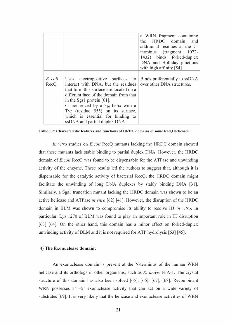
Protein Characteristics of the HRDC domain
Function
Sgs1 Has a lysine- and arginine-rich patch that forms an electropositive surface important for the interaction with ssDNA [59].
Binds both ssDNA and partially ds DNA.
WRN Has a cluster of acidic and hydrophobic residues [60].
Does not appear to interact with DNA in vitro. However,
21
a WRN fragment containing the HRDC domain and additional residues at the C-terminus (fragment 1072-1432) binds forked-duplex DNA and Holliday junctions with high affinity [54].
E. coli RecQ
Uses electropositive surfaces to interact with DNA, but the residues that form this surface are located on a different face of the domain from that in the Sgs1 protein [61]. Characterized by a 310 helix with a Tyr (residue 555) on its surface, which is essential for binding to ssDNA and partial duplex DNA
Binds preferentially to ssDNA over other DNA structures.
Table 1.2: Characteristic features and functions of HRDC domains of some RecQ helicases.
In vitro studies on E.coli RecQ mutants lacking the HRDC domain showed
that these mutants lack stable binding to partial duplex DNA. However, the HRDC
domain of E.coli RecQ was found to be dispensable for the ATPase and unwinding
activity of the enzyme. These results led the authors to suggest that, although it is
dispensable for the catalytic activity of bacterial RecQ, the HRDC domain might
facilitate the unwinding of long DNA duplexes by stably binding DNA [31].
Similarly, a Sgs1 truncation mutant lacking the HRDC domain was shown to be an
active helicase and ATPase in vitro [62] [41]. However, the disruption of the HRDC
domain in BLM was shown to compromise its ability to resolve HJ in vitro. In
particular, Lys 1270 of BLM was found to play an important role in HJ disruption
[63] [64]. On the other hand, this domain has a minor effect on forked-duplex
unwinding activity of BLM and it is not required for ATP hydrolysis [63] [45].
4) The Exonuclease domain:
An exonuclease domain is present at the N-terminus of the human WRN
helicase and its orthologs in other organisms, such as X. laevis FFA-1. The crystal
structure of this domain has also been solved [65], [66], [67], [68]. Recombinant
WRN possesses 3’ –5’ exonuclease activity that can act on a wide variety of
substrates [69]. It is very likely that the helicase and exonuclease activities of WRN
22
are coordinated in vivo, as suggested from the in vitro studies [70]. The exonuclease
domain has a 3’ – 5’ proof reading activity that has been suggested to be required for
DNA non-homologous end joining [68].
1.22 Human RecQ helicases and their associated diseases:
There are five RecQ helicases in humans—RECQ1, BLM, WRN, RECQ4
and RECQ5, with RECQ5 existing in two different forms due to alternative mRNA
splicing [71]. Mutations in the genes of three out of the five human RecQ helicases
are associated with well-defined cancer predisposition and premature aging
disorders. In particular, Bloom syndrome is associated to BLM gene mutations,
Werner syndrome to WRN gene mutations and Rothmund–Thomson syndrome,
RAPADILINO and Baller Gerold syndrome are all caused by mutations in RECQ4
gene [37, 72-74], [75].
1.22.1 Bloom syndrome:
Bloom Syndrome (BS) is an autosomal recessive disorder caused by defects
in the BLM helicase gene. BS is a rare disorder and BS patients are characterized by
proportionate pre- and post-natal growth deficiency, sun-sensitive telangiectatic
hypo- and hyper-pigmented skin, immune deficiency, predisposition to malignancy
and chromosomal instability. BS patients also show a very high incidence of cancers
of various types including leukemias, lymphomas, and carcinomas [76].
Murine model:
Several groups have tried to generate BS mouse models. In one model, ES
cells where a fragment of the BLM gene upstream of the helicase domain was
replaced with a neomycin resistance cassette were transferred into blastocysts and
crossed with WT mice to form the Blm+/- heterozygous mice. These mice are
characterized by growth defects and micronuclei similar to human BS patients [77].
In a second model, ES cells with the exons 10–12 of the BLM gene replaced with
HPRT (hypoxanthine–guanine phosphoribosyltransferase) were injected into the
blastocyst and crossed with WT mice to generate heterozygous BlmCin/-. These mice
23
showed a slight increase in the frequency of micronuclei [78]. In addition, both
mouse models were characterized by a shorter life span. Allan Bradley’s group
generated knockout models using an embryonic stem cell method. They generated
different mutant alleles of the mouse homologue of human BLM: Blmm1 (lacking the
appropriate in frame translation start site), Blmm2 (producing a truncated BLM
polypeptide) and Blmm3 (producing a truncated BLM polypeptide). The heterozygous
mice with Blmm2 or Blmm3, the compound heterozygous Blmm2/m3, and the
homozygous Blmm3/m3 were viable and fertile. However, the homozygous Blmm2/m2
did not develop to term. The Blmm3/m3 mice served as better model for BS and closely
recapitulated the cellular phenotype of BS patients, such as increased SCE in somatic
cells and predisposition to cancer [79].
Cellular phenotype:
Cells from patients with BS and the cells derived from BLM knockout mice
exhibit chromosome instability. BS patients cells are characterized by an increased
numbers of chromatid gaps, breaks, chromosome structural rearrangements and sister
chromatid exchanges (SCE). It has been reported that BS cells as well as BLM-/+
fibroblasts show a tendency for the formation of spontaneously induced micronuclei
[80, 81]. Moreover, these cells display a significantly lower rate of chain elongation
during DNA synthesis [82]. In addition, BS cells are sensitive to UV radiation and
" Drugs used in chemotherapy (e.g., cyclophosphamide, etoposide)
The replication checkpoint stands out as the prime regulator of RF stability
after genotoxic stress. The replication check-point is composed of a network of
sensors and transducers that detect, transmit, and amplify the DNA damage and
replication stress signal, and then promote a DNA damage response that ensures the
stabilization of RFs, DNA repair and cell cycle arrest [159]. The ATM kinase
(Ataxia telangiectasia mutated) with its regulator MRN complex (Mre11-Rad50-
NBS1) and the ATR kinase (ATM and Rad3-related protein) with its regulator
ATRIP (ATR-interacting protein) are the two major players involved in check-point
response.
The ATM pathway is typically activated by double strand breaks. Collapsed
replication forks generate DSBs that also activate ATM [160]. Following DSB
formation, the histone H2AX is phosphorylated at the serine 139 (&-H2AX) by ATM.
This represents as one of the earliest events in DDR [161]. The MRN (MRE11-
RAD50-NBS1) complex, which is one of the first complexes to be recruited to
DSBs, acts as a damage sensor that also physically bridges the ends of the DSB
[162]. MRE11 plays a role in end resection while the NBS1 interacts with ATM, and
40
this interaction is required for the recruitment and retention of ATM to DSBs [163-
167]. ATM phosphorylates and modulates the activity of several protein kinases,
which in turn phosphorylate their own substrates and the most notable among the
kinases is the checkpoint kinase 2 (CHK2) [168]. The components of the MRN
complex are also phosphorylated and contribute to the timely activation of various
DDR branches [169-173].
The ATR checkpoint responds to exposed single-stranded breaks coated with
hRPA (Replication protein A) and promotes their stabilization [174-177]. Stalled
forks generated by helicase-polymerase uncoupling generally presents exposed
ssDNA regions coated by the single-strand binding protein RPA (replication protein
A), which in turn recruits the active ATR-ATRIP (ATR-interacting protein) complex
[178]. In spite of the numerous substrates, the main signal arising from the ATR
cascade is the phosphorylation-activation of Chk1, a serine-threonine kinase.
Globally Chk1 activation leads to cell cycle arrest by phosphorylation-modulation of
CDK regulators CDC25-A, CDC25-B, CDC25-C, and p53 activation. In addition,
the ATR pathway promotes local fork stabilization through phosphorylation of
several targets such as replication components, nucleases, and DNA helicases, which
are required to maintain replisome integrity and prevent fork collapse [178, 179].
The activation ATM and ATR kinases trigger the checkpoint response
through the phosphorylation of targets that are implicated in various downstream
processes [180, 181]. About 25 ATM and ATR substrates have been identified [182],
many as candidates based on known roles in damage signaling.
BLM is a substrate of ATR and is phosphorylated on Thr99 and Thr122 in
HU treated cells [183]. BLM was shown to co-localize with ATR and co-
immunoprecipitate with ATR from cell extracts following DNA replication arrest.
The authors propose that phosphorylation of BLM on Thr-99 and Thr-122 by ATR is
an important component of the response of cells undergoing replication fork arrest
and that it is subsequently essential for adequate cellular recovery from the
replication stress [183]. WRN was shown to be phosphorylated after replication
arrest in an ATR-dependent manner and also both ATR and WRN were shown to be
acting together in a common pathway to stabilise common fragile sites [184, 185].
41
The same group reported the role of both ATM and ATR in promoting the recovery
from replication perturbation by differently regulating WRN at defined moments of
the response to RF arrest. They reported that suppression of ATR-mediated
phosphorylation of WRN prevented the proper accumulation of WRN in nuclear
foci, co-localisation with RPA and caused breakage of stalled forks, whereas
inhibition of ATM kinase activity led to the retention of WRN in nuclear foci and
impaired recruitment of RAD51 recombinase resulting in reduced viability after fork
collapse [186].
1.53 Roles of RecQ helicases in DNA replication:
DNA replication is a complex process that can be divided in different steps
such as assembly of the replication machinery, replication initiation, and replication
fork progression. This process is highly controlled by checkpoint proteins. RecQ
helicases are involved in most DNA replication steps.
Role of RecQ helicases in initiation of DNA replication was first shown for
RECQ4. The first 200 amino acids of human RECQ4 share homology with the yeast
DNA replication initiation factors Drc1 (Schizosaccharomyces pombe) and Sld2 (S.
cerevisiae) [98, 187, 188]. Moreover, Matsuno et al. and Sangrithi et al. showed that
depletion of RECQ4 from Xenopus laevis egg extracts prevents initiation of DNA
replication and suggested that this might be due to a defect in the loading of RPA to
chromatin [98, 189]. Interestingly, the N-terminal Sld2-like domain of human
RECQ4 has been also shown to possess an intrinsic DNA helicase activity [188],
although this result has not been confirmed by future studies [190]. Our group
showed that RECQ4 is recruited to replication origins at late G1 after the origin
recognition complex (ORC) [191]. In agreement with this finding, Yilun Liu’s group
showed that human RECQ4 interacts with the MCM [192], while J.K.Lee’s group
showed that RECQ4 is essential for replisome assembly [193]. Our group also
showed that RECQ4 is required for the loading of PCNA and the ssDNA binding
protein RPA onto the replication fork, supporting the notion that RECQ4 is an
essential factor for replication initiation [191]. The notion that RECQ4 plays an
essential role during DNA replication is supported by the embryonic lethality of
RecQ4 knockout mice [95]. Moreover, a recent study suggested that the helicase and
42
the C-terminal domain of RECQ4 facilitate DNA replication elongation in cells that
have been exposed to IR [194]. Given that all the RECQ4 mutations identified in
RTS patients are located within or after the exons encoding the central helicase
domain, it is possible that these patients still express a truncated RECQ4 protein with
an intact Sld2-like domain required for replication initiation. Therefore, all of the
RECQ4 mutations associated with disease might be hypomorphic [92, 96].
While RECQ4 is important for origin firing, BLM can suppress origin firing.
In eukaryotic cells, there are multiple DNA replication origins with a different, but
regulated order of activation. Some of the origins are dormant and are normally
suppressed [195]. Upon replication inhibition, these dormant origins can fire,
probably to compensate for the slower overall DNA replication rate and/or to permit
replication of loci to be rescued by a converging fork. Davies et al showed that BS
cells have high origin firing frequency after release from HU stress. They showed
that BLM is required for the efficient restart of the stalled replication forks and for
the suppression of dormant origin firing [196]. These activities require the helicase
activity of BLM and phosphorylation of the Thr 99 residue targeted by stress-
activated kinases (ATR in the context of replication blockade) [183].
Several evidence support the notion that the human RECQ1 helicase might
also play a role during DNA replication. Wang et al. employed a DNA affinity
purification and mass spectrometry procedure to show that RECQ1 is physically
associated with KSHV ori-Lyt through the viral proteins K8 and RTA; they
speculated that RECQ1 is not only an integral component of the pre-replication
complex, but also the so long-sought helicase that unwinds origin DNA in the
initiation of KSHV lytic DNA replication [197]. Later it was found that RECQ1 is
also associated with the ori-Lyt and Zta of another virus, Epstein–Barr virus (EBV).
Depletion of RECQ1 by shRNA resulted in reduced lytic DNA replication [198].
Successively, our group showed by chromatin imunoprecipitation approaches that
RECQ1 physically associates with replication origins in a cell cycle-regulated
fashion in unperturbed cells [191]. However, the exact role of RECQ1 during DNA
replication remained to be determined and my thesis and our recent paper provide
new clues on the role of RECQ1 in this process [199].
43
Various studies suggested a possible role of RecQ helicases during
replication fork progression. For example, depletion of BLM or RECQ1 was shown
to reduce the replication fork speed [191, 200, 201], while BS cells are known to
accumulate abnormal replication intermediates [202]. Conversely, depletion of WRN
or RECQ4 does not seem to influence the fork progression rate [191, 201]. RecQ
helicases seem to be particularly important for replication fork progression when
there is a replication blockage. In particular, the acute depletion of several human
RecQ family members is associated with an increased sensitivity to several DNA
damaging agents and replication inhibitors [203]. For example, RECQ1, BLM,
WRN, RECQ4 and RECQ5 depleted cells are sensitive to CPT [151, 154, 199, 203-
205] and HU treatment (except RECQ5) [192, 196, 201, 203, 206]. RECQ4 depleted
cells are sensitive to UV damage [151]. In addition, RecQ helicases localize at
damaged replication sites upon replication perturbation. For example, BLM
translocate from PML bodies to damaged replication forks upon DNA damage
induction, while WRN translocate from the nucleolus to nucleoplasmic foci upon UV
exposure [207].
BLM and WRN have been shown to be phosphorylated during replication
stress to fine-tune their function at the replication fork. BLM depleted cells or BS
cells show reduced efficiency of replication fork restart after HU or aphidicolin
treatment, demonstrating a key role for BLM helicase through its helicase activity for
replication restart. Mechanistically, BLM was suggested to restart replication forks
by transporting p53 to RAD51 sites at the stalled replication forks [208]. BLM forms
a complex with topoisomerase III alpha, RMI1, RMI2 and replication protein A,
called the 'BLMcx' [209]. This complex has been shown to be recruited to the
replicating chromatin during normal S-phase and members of the complex are
phsophorylated in response to DNA damage. During replication stress, BLM from
the BLMcx and FANCD2 cooperate to promote restart of stalled replication forks
while suppressing firing of new replication origins [209]. BLM was also shown to
co-localize with ATR and co-immunoprecipitate with ATR from cell extracts
following DNA replication arrest. The authors propose that phosphorylation of BLM
on Thr-99 and Thr-122 by ATR is an important component of the response of cells
undergoing replication fork arrest and that it is subsequently essential for adequate
cellular recovery from the replication stress [183].
44
WRN cells are sensitive to treatment with replication inhibitors and DNA
damaging agents that cause replication fork stalling such as aphidicolin, HU, CPT,
etoposide, MMS, cisplatin, mitomicin C and &-irradiation [86, 210-214]. When cells
are challenged with genotoxic agents additional WRN-dependent deficiencies are
revealed. These can reflect replication problems in other susceptible areas, such as
fragile sites, satellite repeats, or any genomic segments that replicate unidirectionally
[185]. Finally, systemic deregulation of replication, by genotoxic drugs [201] or
oncogenes such as hyperactive Myc [215], can elicit severe S-phase defects in WRN-
depleted cells [201]. In addition to the restart defects, the restarted forks progressed
slowly in the absence of WRN. WRN was also shown to be phosphorylated after
replication arrest in an ATR-dependent manner and also both ATR and WRN was
shown to be acting together in a common pathway to stabilise common fragile sites
[184, 185]. The same group reported the role of both ATM and ATR in promoting
the recovery from replication perturbation by differently regulating WRN at defined
moments of the response to RF arrest. They reported that suppression of ATR-
mediated phosphorylation of WRN prevented the proper accumulation of WRN in
nuclear foci, co-localisation with RPA and caused breakage of stalled forks, whereas
inhibition of ATM kinase activity led to the retention of WRN in nuclear foci and
impaired recruitment of RAD51 recombinase resulting in reduced viability after fork
collapse [186].
A variety of DNA structures such as G-quadruplexes and hairpins can serve
as “road-blocks” and impede the progression of the repliosome, especially if they are
present in the leading strand template. It is known that several human RecQ helicases
can resolve these substrates, thereby enabling the smooth progression of the
repliosome [216]. Replication problems might also occur at an increased frequency
in telomeric regions, because of the intrinsic inability of the lagging strand DNA
replication machinery to function at the very end of chromosomes. Hence, a
specialized reversed transcriptase called telomerase adds G-rich repeat sequence to
the telomere ends. WRN has been shown to be required for telomere lagging strand
synthesis [129]. WRN associates with the telomeric TTAGGG region, and possibly
unwinds G-quadruplexes formed therein to allow the replication forks to progress to
the chromosome ends. In cells lacking a functional telomerase, there is an alternative
pathway termed alternative lengthening of telomeres, which requires sgs1 in S.
45
cerevisiae [217] [218] [219]. The alternative lengthening of telomeres pathway is
dependent on the HR factors Rad50 and Rad52, but not on RAD51. WRN is able,
through the combined use of its helicase and exonuclease activities, to resolve the D-
loop HR intermediate to release the 3) invading tail. This activity might be used to
disrupt recombination events at telomeres or to disrupt the natural telomeric T-loop
(a specialized D-loop) in order to permit replication of the telomeric end.
1.6 Replication fork regression and restoration
The effects of DNA lesions on replication fork progression vary based on the
strand in which the lesions are. For example, the lesions on the discontinuously
synthesized lagging strands can be skipped over and a new Okazaki fragment can be
re-initiated downstream of the lesion to be repaired after completion of DNA
replication [220, 221]. On the other hand, lesions on the leading strand are more
likely to block fork progression. In response to this threat, DNA damage tolerance
pathways have evolved to enable DNA polymerases to bypass lesions on the leading
DNA strand template.
Stalling of the replication fork at a lesion on the leading strand template can
result in the uncoupling of leading and lagging strand synthesis; DNA synthesis
continues only on the undamaged lagging template strand generating ssDNA gaps
that may activate HR [222]. Alternatively, DNA lesion bypass can occur through the
template strand switch mechanism [223]. This mechanism includes conversion of the
fork into a reversed fork structure reminisce of a Holliday junction (known as the
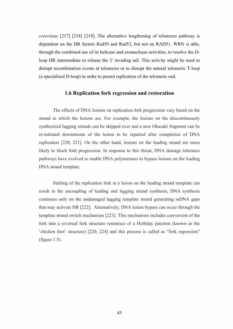
‘chicken foot’ structure) [220, 224] and this process is called as “fork regression”
(figure 1.5).
46
Stalled RF
Translesion synthesis
Fork regression
Template switch
Lesion
Figure 1.5: Mechanisms of DNA damage tolerance to lesions on the leading strand. The lesion in the leading strand could be either by-passed by translesion synthesis or by template switch mechanism, or alternatively by re-modelling the fork into a chicken-foot like structure by a process called fork regression.
Following fork regression, the DNA lesion is repaired and a reverse branch
migration reaction takes place to re-establish a functional fork structure.
Alternatively, the extended lagging daughter strand is used as a template for the
synthesis of the leading strand followed by reverse branch migration to bypass the
blocking lesion and re-establish the functional replication fork. Part of this thesis
focuses on investigating the roles of human RecQ helicases in fork regression and
restoration.
RECQ1, WRN, and BLM are all able to bind and branch migrate Holliday
junctions in vitro [225-228]. In addition, BLM and WRN also able to promote fork
regression and restoration in vitro [225, 227, 229-232] suggesting that RecQ
helicases could play an essential role in the formation and resolution of these
replication structures in cells. Alternatively, the regressed forks could be resolved by
must be repaired by HR through DNA strand invasion to re-establish a functional
replication fork [235] [236].
47
This reaction is promoted in vitro by RecQ helicases, including BLM and
WRN, which might allow the bypass of a DNA lesion blocking the fork and the
restart of DNA replication. Once the regressed fork is formed, a four-way Holliday
junction is generated, which could potentially be migrated back by RECQ1, BLM or
WRN to restore a functional replication fork.
1.61 Top1 inhibitors and replication fork reversal:
DNA topoisomerases are enzymes that relax DNA torsional strain generated
during replication, transcription, recombination, repair, and chromosome
condensation [237], and hence are vital to all cells undergoing division. The
relaxation of DNA supercoiling by topoisomerase I (Top1) is enabled by a
mechanism of controlled rotation around a transient DNA single-strand break [238,
239]. During this process, the enzyme forms an intermediate covalent complex with
the DNA, mediated by a bond between the active site tyrosine (Tyr723 in human
Top1) and the cleaved phosphate group [237]. Top1 and Top2 inhibitors rely on the
transient trapping of these specialized nucleases on their 3’-single-strand and 5’-
double-strand DNA substrate, respectively, thus preventing the religation step [237].
Because of the high proliferation of cancer cells, drugs that target Top1 such as
camptothecin, or Top2 such as etoposide, are potential chemotherapeutics and some
of them have been already clinically approved for cancer treatment [240, 241]. In
particular, the S-phase dependent cytotoxicity of Top1 inhibitors was thought to arise
from replication run-off at Top1-DNA frozen complexes located on the leading
strand, triggering to the accumulation of lethal one-side DSBs [242]. This model was
recently challenged by the work of Koster et al. where using a combination of single-
molecule and in vivo experiments, the authors demonstrated that Top1 inhibitors
hinder the uncoiling activity of Top1, thus promoting positive supercoiling formation
and replication fork slowing. They also proposed that the resulting accumulation of
positive supercoils ahead of the replication machinery is the major mechanism of
fork collapse and cell death upon camptothecin exposure [243, 244].
Recently, the group of Massimo Lopes in Zurich has extended this
observation by demonstrating that clinically relevant doses of Top1 inhibitors are
associated with replication fork slowing, without DSBs formation. By exploiting a
combination of in vivo psoralen cross-linking and EM analysis to directly visualize
48
replication intermediates, they were able to detect a high frequency of regressed
forks upon Top1 poisoning in yeast, Xenopus laevis egg extract, and mammalian
cells. In contrast to previous findings, they found that the replication fork slowing
and reversal associated with Top1 inhibitors are not checkpoint or recombination
dependent. Moreover, they discovered that poly(ADP-ribose) polymerases 1
(PARP1) activity, at least in X. laevis egg extract and mammalian cells, is essential
to slow the replication forks on CPT-damaged templates by promoting the
accumulation of regressed forks. PARP1 depletion or inhibition revert this effect of
Top1 poisoning, leading to the formation of DSBs likely by replication run-off at
Top1-DNA covalent complexes (Top1cc) (figure 1.6) [245]. PARP1 plays a critical
role in mediating the cellular sensitivity to camptothecin derivatives and several
clinical trials are currently investigating the potential advantages of combined
therapies with PARP and Top1 inhibitors [246, 247].
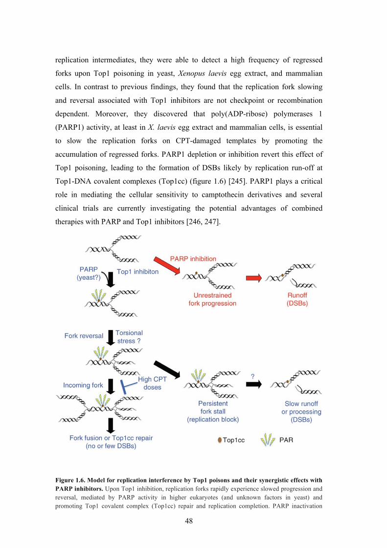
!
Figure 1.6. Model for replication interference by Top1 poisons and their synergistic effects with PARP inhibitors. Upon Top1 inhibition, replication forks rapidly experience slowed progression and reversal, mediated by PARP activity in higher eukaryotes (and unknown factors in yeast) and promoting Top1 covalent complex (Top1cc) repair and replication completion. PARP inactivation
49
leads to increased DSBs, owing to unrestrained fork runoff at Top1cc. High CPT doses lead to incomplete replication and persistent fork stalling, causing DSBs by eventual fork collapse and/or processing; PAR, poly(ADP-ribose) Adapted from Ray Chaudhuri A., 2012 [245].
The results of Dr. Lopes’ group provide a new rationale for the synergic
effect of these inhibitors on actively proliferating cancer cells. They also point to
fork reversal as a general strategy that allows the repair enzymes to fix the lesion
before the replication resumes, thus avoiding replication fork collision with single,
and possibly, double strand breaks. Nevertheless, this work opens several relevant
biological questions: 1) since PARP1 is just a signaling molecule without motor
activity, what other factors are involved in PARP-mediated fork reversal? 2) Which
are the cellular factor responsible for the restart of the reversed forks? My thesis
provides answers to these questions by pointing to an essential role of the human
RECQ1 helicase in this process.
50
2. MATERIALS AND METHODS
2.1 Antibodies and chemicals:
The antibodies used were rabbit anti-His antibody (sc-803) from Santa Cruz,
rabbit anti-RECQ1 polyconal antibody was custom made against the full length
sequence of the protein.
NAD+, ATP, ATP&S, TCEP, imidazole were all from Sigma. Protease
inhibitor cocktail tablets were from Roche. The radioactive [32&P]-ATP was from
PerkinElmer. The antibiotics were from Sigma. Bluo-gal was from Invitrogen. The
restriction enzymes and polymerases were from NEB and Agilent technologies
respectively. For protein purification, the TALON Cobalt resin was from Clontech
and the gel filtration columns were from GE healthcare. The BioSpin columns for the
removal of extra nucleotides were from Bio-Rad. All other chemicals were of
reagent grade or higher from various vendors.
2.2 Cell culture and transfection:
The Sf9 cells were grown at 27°C in SFM II media (Invitrogen). Sf9 cells
were transfected using cellfectin II (invitrogen) following the supplier’s protocol.
Briefly 8 X 10⁵ cells were transfected with 1 µg of bacmid using cellfectin II and
incubated at 27ºC. The virus was collected 72 hours post transfection and stored
protected from light at 4°C (short term storage) or -80°C (long term storage).
2.3. Expression and purification of recombinant proteins:
2.3.1. RECQ1 overexpression and purification:
RECQ1 was overexpressed and purified from Sf9 cells as already described
[248]. RECQ1 bacmid was isolated from DH10Bac (Invitrogen) cells after
transposition, confirmed by PCR and was transfected into Sf9 cells using cellfectin II
reagent. After 72 hours, the medium containing the baculovirus was collected,
51
centrifuged to remove the cell debris and stored protected from light. To increase the
titre, the baculovirus was amplified 3 - 4 times by infecting log-phase Sf9 cells,
followed by collecting the media containing the viral particles. For RECQ1
overexpression, a litre of the Sf9 cell suspension culture at a density of 150 X 104
cells /ml was infected with the appropriate amount of the baculovirus and the
infected cells were collected 72 hours post infection. The pellet was washed with
phosphate buffered saline (PBS) and stored at - 80°C until use.
For protein purification, the infected cells were re-suspended in lysis buffer
(20 mM Tris–HCl, pH 7.4, 400 mM KCl, EDTA free protease inhibitor, 5 mM $-
mercaptoethanol) and sonicated on ice five times at maximum amplitude for 30 s
with a 30 s gap. The lysate was cleared by centrifugation at 15,000 rpm for 45
minutes. Meanwhile, the TALON cobalt resin (Clontech) was washed twice with
Milli Q water and equilibrated in the lysis buffer. The cleared lysate was allowed to
bind to the resin (1 ml resin / 5 mg protein) for 2 hours at 4°C in a nutator. After
binding, the resin was washed thrice with high salt buffer (20 mM Tris–HCl, pH 7.4,
500 mM KCl, 12.5 mM imidazole, 5 mM !-mercaptoethanol) and twice with low
salt buffer (20 mM Tris–HCl, pH 7.4, 150 mM KCl, 12.5 mM imidazole, 5 mM !-
mercaptoethanol). The resin was loaded into a column and the polyhistidine tagged
RECQ1 was eluted using 120 mM imidazole in the elution buffer (20 mM Tris-HCl,
pH 7.4, 100 mM KCl, 5 mM !-mercaptoethanol,). The eluted fractions were run on a
SDS-PAGE gel and the fractions containing the protein were pooled and dialyzed at
4°C against 150 mM KCl, 1 mM DTT and 20 mM Tris-HCl pH: 7.4 three times for
two hours each. The dialyzed protein was quantified, aliquoted and stored at -80°C
after flash freezing in liquid nitrogen.
2.3.2 Site directed mutagenesis and purification of RECQ1 mutants:
The ATPase mutants K119R-RECQ1, E220Q-RECQ1 and the annealing
mutants L18P-RECQ1 and L28P-RECQ1 were prepared using the wild-type
pFASTBAC1-RECQ1 recombinant plasmid as template. The mutations were done
using the QuickChange XL site-directed mutagenesis kit (Stratagene) following the
manufacturer’s protocol. The respective bacmids were produced in DH10Bac cells,
52
verified by PCR and sequencing. The bacmids were isolated and transfected into Sf9
cells for protein overexpression and purification following the protocol used for the
wild-type RECQ1 (section 2.3.1).
2.3.3. Preparation of the truncated RECQ1:
The RECQ1T1 (49-616) cloned into the pNIC-CTHF vector was transformed and
expressed in BL21(DE3)-R3-pRARE E.coli expression strain. The protein was
overexpressed and purified as already described [32]. The expressed protein has an
additional Methionine at the N-terminus and an
AENLYF*SHHHHHHDYKDDDDK C-terminal extension containing the TEV
protease cleavage site and a His tag followed by a FLAG tag. The protein was
overexpressed in Terrific Broth modified medium (sigma) by IPTG induction. 2-3
OD600 cells were induced with 0.2 mM IPTG and then grown at 18°C overnight. The
cells were collected by centrifugation and lysed in lysis buffer (50 mM HEPES pH:
7.5, 500 mM NaCl, 10 mM imidazole, 1 mM TCEP, 5 % glycerol and protease
inhibitors) by sonication (4 times; 30 s pulse with 30 s gap). The lysate was cleared
by centrifugation and incubated with Ni-NTA resin (Qiagen) for 2 hours at 4°C in a
nutator. The protein bound resin was washed with the lysis buffer supplemented with
30 mM imidazole and eluted with the lysis buffer supplemented with 500 mM
imidazole. The eluted protein was confirmed on a SDS-PAGE gel and the protein
containing fractions were pooled and dialyzed against 20mM Tris pH: 7.4, 150 mM
KCl, 1 mM TCEP.
2.3.4 Determination of protein concentration:
The absorbance of the protein was recorded at 280 nm in a nanodrop. The
theoretical extinction coefficient of the protein was determined from Expasy
employing the Protparam tool (http://ca.expasy.org/tools/protparam.html). Protein
concentrations were determined using Beer-Lamberts law:
Protein concentration (M) = Abs280 (cm-1) / Extinction coefficient (M-1cm-1)
53
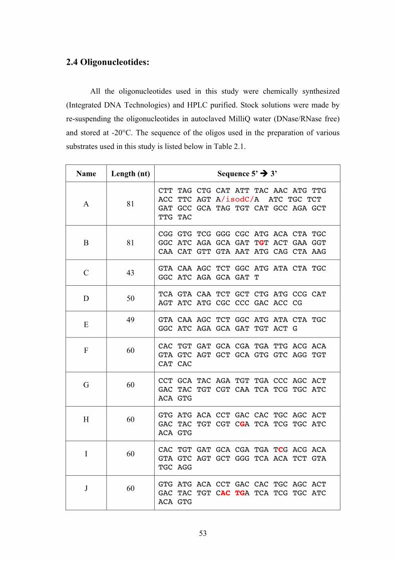
2.4 Oligonucleotides:
All the oligonucleotides used in this study were chemically synthesized
(Integrated DNA Technologies) and HPLC purified. Stock solutions were made by
re-suspending the oligonucleotides in autoclaved MilliQ water (DNase/RNase free)
and stored at -20°C. The sequence of the oligos used in the preparation of various
substrates used in this study is listed below in Table 2.1.
Name Length (nt) Sequence 5’ ! 3’
A 81
CTT TAG CTG CAT ATT TAC AAC ATG TTG ACC TTC AGT A/isodC/A ATC TGC TCT GAT GCC GCA TAG TGT CAT GCC AGA GCT TTG TAC
B 81 CGG GTG TCG GGG CGC ATG ACA CTA TGC GGC ATC AGA GCA GAT TGT ACT GAA GGT CAA CAT GTT GTA AAT ATG CAG CTA AAG
C 43 GTA CAA AGC TCT GGC ATG ATA CTA TGC GGC ATC AGA GCA GAT T
X26-4 61 TGC CGA TAT TGA CAA GAC GGC AAA GAT GTC CTA GCA ATC CAT TGG TGA TCA CTG GTA GCG G
Table 2.1. Sequences of the oligonucleotides used in the study of fork regression and restoration. Bold red letters indicate the nucleotides that form mismatched pairs in the branch migration products.
55
2.5 Preparation of DNA substrates:
One of the strands in each substrate was 5’-end labelled with [&32P]ATP
(3000 Ci/mmol) for 45 minutes using T4 polynucleotidyl kinase (NEB) and the
reaction was terminated by heat inactivating the enzyme at 95°C for 6 minutes. The
unincorporated [&32P]ATP was removed using the Bio-spin 30 columns (Bio-Rad).
For the preparation of the helicase assay substrate (forked duplex), the labelled
strand P (table 2.1) was annealed with 1.4 of excess of the unlabelled complementary
strand Q (table 2.1) in annealing buffer (10 mM Tris-HCl pH 8.3, 50 mM NaCl)
followed by heating at 95°C for 6 minutes and then slowly cooling to room
temperature. For the annealing assay the labelled strand P (table 2.1) was used as a
substrate.
For the preparation of Holliday junction substrate for branch migration
(X12), the labelled strand X12-1 (table 2.1) was annealed with 1.5 fold excess of the
three complementary unlabelled strands X12-2, X12-3, X12-4 (table 2.1) in
annealing buffer (supplemented with 5 mM MgCl2) followed by heating at 95°C for
6 minutes and then slowly cooling to room temperature. The Holliday junction was
purified in a sepharose 4B (5ml) column and the collected fractions were analysed on
a 10 % PAGE gel. The fractions were selected based on their purity. A similar
protocol with oligos F, G, H, I/J (table 2.1) was followed for the preparation of HJ
(1) and HJ (4) with mis-matches. To prepare the Holliday junction substrates for
EMSA, labelled oligonucleotide L (table 2.1) and 1.5-fold excess of unlabelled
oligonucleotides M, N, O (table 2.1) were incubated in annealing buffer
supplemented with 5 mM MgCl2 for 30 min at 37°C and for a further 30 min at room
temperature.
For analytical ultracentrifugation experiments, the X26 Holliday junction
substrates were prepared by annealing the four strands X26-1, X26-2, X26-3 and
X26-4 (table 2.1) in annealing buffer, followed by heating at 95°C for 6 minutes and
then slowly cooling to room temperature. The annealed products were run on a
PAGE gel and visualised by staining with Stains all (Sigma). The band
corresponding to the Holliday junction was excised and the Holliday junction was
56
purified from the gel by “crush and soak” method [249]. Briefly, the gel slices were
frozen and crushed into small pieces and buffer was added to the tubes. The tubes
were incubated at 37°C with gentle shaking and after a few hours, the buffer
containing the HJ was pipetted out, quantified and stored for further use.
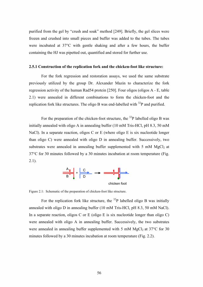
2.5.1 Construction of the replication fork and the chicken-foot like structure:
For the fork regression and restoration assays, we used the same substrate
previously utilized by the group Dr. Alexander Mazin to characterize the fork
regression activity of the human Rad54 protein [250]. Four oligos (oligos A - E, table
2.1) were annealed in different combinations to form the chicken-foot and the
replication fork like structures. The oligo B was end-labelled with 32P and purified.
For the preparation of the chicken-foot structure, the 32P labelled oligo B was
initially annealed with oligo A in annealing buffer (10 mM Tris-HCl, pH 8.3, 50 mM
NaCl). In a separate reaction, oligos C or E (where oligo E is six nucleotide longer
than oligo C) were annealed with oligo D in annealing buffer. Successively, two
substrates were annealed in annealing buffer supplemented with 5 mM MgCl2 at
37°C for 30 minutes followed by a 30 minutes incubation at room temperature (Fig.
2.1).
+ A
chicken foot
B
C
D
Figure 2.1: Schematic of the preparation of chicken-foot like structure.
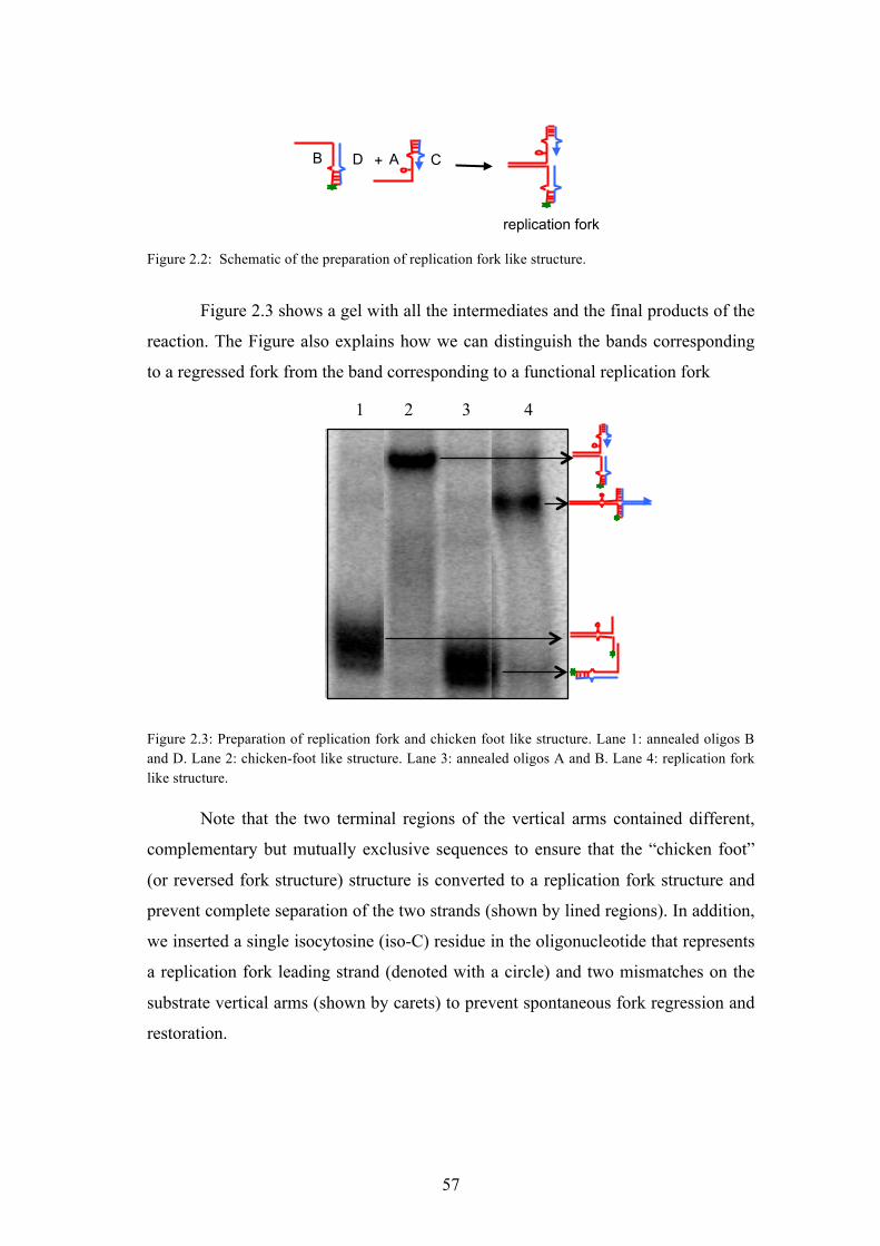
For the replication fork like structure, the 32P labelled oligo B was initially
annealed with oligo D in annealing buffer (10 mM Tris-HCl, pH 8.3, 50 mM NaCl).
In a separate reaction, oligos C or E (oligo E is six nucleotide longer than oligo C)
were annealed with oligo A in annealing buffer. Successively, the two substrates
were annealed in annealing buffer supplemented with 5 mM MgCl2 at 37°C for 30
minutes followed by a 30 minutes incubation at room temperature (Fig. 2.2).
57
replication fork
+ A B D C
Figure 2.2: Schematic of the preparation of replication fork like structure.
Figure 2.3 shows a gel with all the intermediates and the final products of the
reaction. The Figure also explains how we can distinguish the bands corresponding
to a regressed fork from the band corresponding to a functional replication fork
1 2 3 4
Figure 2.3: Preparation of replication fork and chicken foot like structure. Lane 1: annealed oligos B and D. Lane 2: chicken-foot like structure. Lane 3: annealed oligos A and B. Lane 4: replication fork like structure.
Note that the two terminal regions of the vertical arms contained different,
complementary but mutually exclusive sequences to ensure that the “chicken foot”
(or reversed fork structure) structure is converted to a replication fork structure and
prevent complete separation of the two strands (shown by lined regions). In addition,
we inserted a single isocytosine (iso-C) residue in the oligonucleotide that represents
a replication fork leading strand (denoted with a circle) and two mismatches on the
substrate vertical arms (shown by carets) to prevent spontaneous fork regression and
restoration.
58
2.6. Radiometric biochemical assays:
2.6.1 Helicase assay:
In the helicase assay we measure the release of a 32P-labeled ss-DNA
fragment from the partial duplex DNA substrate by the RECQ1 helicase. The 20 *l
reaction mix typically contained the 32P-labeled DNA substrate (0.5 nM) in the
helicase buffer (20 mM Tris- HCl (pH 7.5), 8 mM DTT, 5 mM MgCl2, 10 mM KCl,
80 mg/ml BSA, 10% Glycerol) with 5 mM ATP. The reaction was initiated by the
addition of the required amounts of RECQ1, and the mixture was incubated at 37°C
for 20 min. In case of time dependent assays, 5 nM of RECQ1 was used and at the
indicated time points 20 *l of the reaction mix was withdrawn. The reactions were
terminated by the addition of 20 *l of (0.4 M EDTA pH 8.0, 10% glycerol, 10%
SDS). The reaction products were resolved on a native 10 % PAGE and visualised
by autoradiography.
2.6.2 DNA strand annealing assay:
The strand-annealing activity of RECQ1 was measured using partially
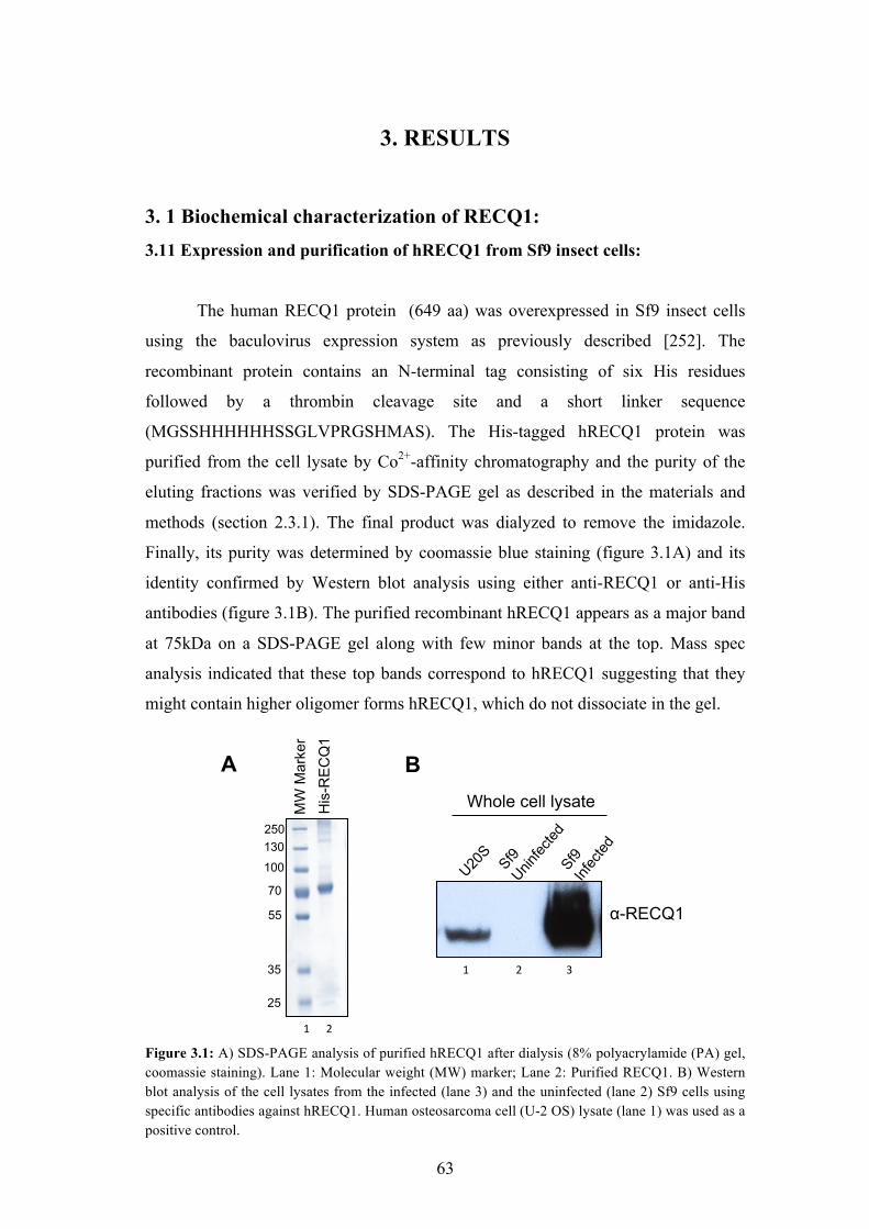
3. 1 Biochemical characterization of RECQ1: 3.11 Expression and purification of hRECQ1 from Sf9 insect cells:
The human RECQ1 protein (649 aa) was overexpressed in Sf9 insect cells
using the baculovirus expression system as previously described [252]. The
recombinant protein contains an N-terminal tag consisting of six His residues
followed by a thrombin cleavage site and a short linker sequence
(MGSSHHHHHHSSGLVPRGSHMAS). The His-tagged hRECQ1 protein was
purified from the cell lysate by Co2+-affinity chromatography and the purity of the
eluting fractions was verified by SDS-PAGE gel as described in the materials and
methods (section 2.3.1). The final product was dialyzed to remove the imidazole.
Finally, its purity was determined by coomassie blue staining (figure 3.1A) and its
identity confirmed by Western blot analysis using either anti-RECQ1 or anti-His
antibodies (figure 3.1B). The purified recombinant hRECQ1 appears as a major band
at 75kDa on a SDS-PAGE gel along with few minor bands at the top. Mass spec
analysis indicated that these top bands correspond to hRECQ1 suggesting that they
might contain higher oligomer forms hRECQ1, which do not dissociate in the gel.
His
-RE
CQ
1 M
W M
arke
r
250 130
100
70
55
35
25
A B
Sf9 Inf
ected
Sf9 Unin
fected
U20S
!-RECQ1
Whole cell lysate
!""""""#""
"""!"""""""""""""""""#""""""""""""""""$""
Figure 3.1: A) SDS-PAGE analysis of purified hRECQ1 after dialysis (8% polyacrylamide (PA) gel, coomassie staining). Lane 1: Molecular weight (MW) marker; Lane 2: Purified RECQ1. B) Western blot analysis of the cell lysates from the infected (lane 3) and the uninfected (lane 2) Sf9 cells using specific antibodies against hRECQ1. Human osteosarcoma cell (U-2 OS) lysate (lane 1) was used as a positive control.
64
3.12 Biochemical characterization of the hRECQ1 helicase:
All purified RECQ1 samples were initially tested to confirm that they retain
normal unwinding and annealing activity, and that there is no exonuclease
contamination. The enzyme activity was tested using our standard helicase and DNA
annealing assays using [32&P]-ATP labelled model DNA substrates (Table 2.1). The
exonucleases are a common contaminant of His-tagged proteins purified from insect
cells and it is important to make sure that they are absent because they would
otherwise interfere with all the activity assays involving DNA [253]. Normally,
binding of the exonuclease to the resin is avoided by saturating the resin with the
recombinant protein.
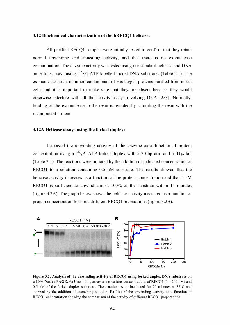
3.12A Helicase assays using the forked duplex:
I assayed the unwinding activity of the enzyme as a function of protein
concentration using a [32&P]-ATP forked duplex with a 20 bp arm and a dT30 tail
(Table 2.1). The reactions were initiated by the addition of indicated concentration of
RECQ1 to a solution containing 0.5 nM substrate. The results showed that the
helicase activity increases as a function of the protein concentration and that 5 nM
RECQ1 is sufficient to unwind almost 100% of the substrate within 15 minutes
(figure 3.2A). The graph below shows the helicase activity measured as a function of
protein concentration for three different RECQ1 preparations (figure 3.2B).
C 1 2 5 10 15 20 30 40 50 100 200 !
RECQ1 (nM)
0 50 100 150 200 250
0
20
40
60
80
100
RECQ1(nM)
Prod
uct
(%)
Batch 1Batch 2Batch 3
A B
Figure 3.2: Analysis of the unwinding activity of RECQ1 using forked duplex DNA substrate on a 10% Native PAGE. A) Unwinding assay using various concentrations of RECQ1 (1 – 200 nM) and 0.5 nM of the forked duplex substrate. The reactions were incubated for 20 minutes at 37°C and stopped by the addition of quenching solution. B) Plot of the unwinding activity as a function of RECQ1 concentration showing the comparison of the activity of different RECQ1 preparations.
65
3.12B Strand annealing assays:
The ability of RECQ1 to anneal two complementary strands (strand
annealing) was analysed as a function of increasing protein concentrations. The
reactions were carried out in the absence of ATP. The results showed that the strand
annealing activity of RECQ1 increased with increasing protein concentrations and
reached a plateau at 15 nM RECQ1 (figure 3.3A). Different RECQ1 preparations
displayed very similar annealing activity, as shown in the graph below (figure 3.3B).
C 1 2 5 10 15 20 30 40 50 100 200
RECQ1 (nM)
0 50 100 150 200 250
0
20
40
60
80
RECQ1(nM)
Prod
uct
(%)
Batch 1Batch 2Batch 3
A B
Figure 3.3: Analysis of the DNA strand annealing activity of RECQ1. A) DNA strand annealing assay using various concentration of RECQ1 (1 – 200 nM) and 0.5 nM labelled single strand DNA. The reactions were initiated by the addition of 1.2 nM complementary unlabelled strand in the absence of ATP and incubated for 30 minutes at 37°C. B) Plot of the annealing activity as a function of RECQ1 concentration showing the comparison of the activity of different RECQ1 preparations.
3.2 Role of RECQ1 in replication fork restart: 3.21 RECQ1 promotes restart of reversed replication forks in vitro:
Our group demonstrated that RECQ1 interacts with the DNA replication
origins and that the amount of RECQ1 loaded on the origins increases during the
early S-phase of the cell cycle when replication origins begin firing and the
replication forks are formed [191]. We also showed that RECQ1 deficient cells are
characterized by a slower replication fork progression and are sensitive to certain
DNA damaging agents that interfere with DNA replication [191]. These data support
the notion that RECQ1 plays an important role at the replication fork.
Replication fork restoration/regression is an emerging mechanism to explain
how replication forks are processed following DNA damage induction. Thus, we
66
decided to test if RECQ1 could promote fork restoration and/or fork regression in
vitro. To perform these experiments, I used a set of four oligonucleotides, which
could be annealed in two alternative ways to mimic either a replication fork like
structure or a “chicken-foot” like structure [250]. Owing to the difference in their
structures, the two substrates migrate differently in a gel, enabling us to differentiate
between the two forms and to compare the fork restoration versus fork regression
activity of RECQ1 (figure 3.4). The two terminal regions of the vertical arms
contained different, complementary but mutually exclusive sequences to ensure that
the “chicken foot” (or HJ structure) structure is converted to a replication fork
structure and prevent complete separation of the two strands. In addition, a single
isocytosine (iso-C) residue in the oligonucleotide that represents a replication fork
leading strand (denoted with a circle) and two mismatches on the substrate vertical
arms (shown by carets) were inserted to prevent spontaneous fork regression and
restoration.
+ RECQ1
A Fork restoration A
B
C/E
D
chicken foot
RECQ1
B Fork regression
replication fork
+
Figure 3.4: A) The scheme for the preparation of the reversed fork substrate (chicken foot). The circle indicates the position of iso-C that mimics a lesion on the leading DNA strand template. Unpaired single DNA bases are shown by carets. Hatched regions denote heterologous DNA terminal regions that prevent complete strand separation during fork regression. Stars indicate [&-32P]ATP-labelled 5’ ends. B) The scheme for the preparation of the model replication fork.
Fork restoration and regression assays were started by the addition of
indicated amounts of RECQ1 to a solution containing 2 nM of the labelled substrate.
The results showed that while RECQ1 efficiently converted a “chicken-foot” to a
replication fork like structure (fork restoration), it is unable to convert a replication
67
fork into a “chicken-foot” like structure (fork regression). RECQ1 promoted fork
restoration in a concentration dependent fashion (figure 3.5). 50 nM RECQ1 was
sufficient to convert more than 75% of the “chicken-foot” structure into a replication
fork like structure, whereas RECQ1 did not display any regression activity even at
the highest protein concentration (200 nM). To exclude the possibility that RECQ1
could not regress the replication forks due to the presence of a 6-nucleotide single-
stranded gap on the leading-strand template of the substrate, I repeated these
experiments using a variant of the same substrate that lacked the single-stranded gap
and found that RECQ1 was still unable to perform fork regression (figure 3.6).
b
0 50 100 150 2000
20
40
60
80
100
RECQ1 (nM)
Prod
uct
(%)
Fork regression
Restoration
Regression
RECQ1
Restoration Regression
1 2 3 4 5 6 7 8 9 10 11 12 13 14
c Fork restoration
+!RECQ1!
Fork restoration!
+!RECQ1!
Fork regression!
A!
B!
C (E)!
A!
D!
B! D!
chicken foot!
replication fork!
a
A!
B!
C !
D!
C (E)!
!
0 15 25 35 50 100 200 0 15 25 35 50 100 200
Fork Restoration Fork Regression RECQ1 (nM)
Fork Restoration
Fork Regression
A
B C
1 2 3 4 5 6 7 8 9 10 11 12 13 14
Figure 3.5: Analysis of the fork restoration and fork regression activity of RECQ1. A) Fork restoration assays (lanes 1–7) and fork regression assays (lanes 8–14) performed using indicated RECQ1 concentrations and 2 nM of chicken-foot substrate or replication fork structure. See paragraph 2.5.1 of the Materials and Methods for more details on the procedure for the correct assignment of the identity of the bands shown in the gel. B) Schematic of the restoration and regression reactions and their products. The circle indicates the position of iso-C that mimics a lesion on the leading DNA strand template. Unpaired single DNA bases are shown by carets. Hatched regions denote heterologous DNA terminal regions that prevent complete strand separation during fork regression. Stars indicate [&-32P]ATP-labelled 5’ ends. C) Percentage of fork restoration and regression products plotted as a function of protein concentration. The data points represent the mean of three independent experiments with the standard deviation indicated by error bars.
68
0 50 100 150 2000
20
40
60
80
100
Protein Concentration (nM)
Prod
uct
(%)
Fork restoration
Fork regression
No ssDNA gap on the leading strand template
A 0 15 25 35 50 100 200 0 15 25 35 50 100 200
Fork Restoration Fork Regression RECQ1 (nM)
Fork Restoration
Fork Regression
B C
1 2 3 4 5 6 7 8 9 10 11 12 13 14
Figure 3.6: Analysis of the fork restoration and fork regression activity of RECQ1 using a substrate that lacks 6 nucleotide single strand gap. A) Fork restoration assays (lanes 1–7) and fork regression assays (lanes 8–14) performed using indicated RECQ1 concentrations and a 2 nM of chicken-foot substrate or replication fork structure lacking the 6 nucleotide single strand gap. B) Schematic of the restoration and regression reactions and their products. The substrates lack the 6 nucleotide single strand gap. The circle indicates the position of iso-C that mimics a lesion on the leading DNA strand template. Unpaired single DNA bases are shown by carets. Hatched regions denote heterologous DNA terminal regions that prevent complete strand separation during fork regression. Stars indicate [&-32P]ATP-labelled 5’ ends. C) Percentage of fork restoration and regression products plotted as a function of protein concentration. The data points represent the mean of three independent experiments with the standard deviation indicated by error bars.
3.22 ATPase activity of RECQ1 is essential for its fork restoration activity:
To confirm that ATP hydrolysis is required for the fork restoration activity of
RECQ1, I used two previously characterized ATPase deficient mutants of RECQ1,
namely E220Q RECQ1 and K119R RECQ1 [105]. The results showed that both
mutants failed to promote fork restoration. The same results were obtained using the
poorly hydrolysable analogue of ATP, ATP&S, and the non-hydrolysable analogue of
ATP, AMP-PNP (figure 3.7).
69
+ - + - - + + - - - + - - - - - - - + - -
ATP ATP!S
RECQ1
RECQ1
AMP-PNP
RECQ1
K119R
E220Q
No prot
ein
RECQ1
1 2 3 4 5 6 7 Figure 3.7: Fork restoration assays using non-hydrolysable ATP analogues or ATPase deficient RECQ1 mutants. Fork restoration assays were performed in the presence of ATP or different ATP analogues using wild-type RECQ1 (lanes 2-5) or the ATPase deficient RECQ1 mutant, K119R (lane 6) and E220Q (lane 7). The protein concentration was 50 nM for all the experiments. 3.23 RECQ1 can by-pass DNA heterology:
To test whether RECQ1 can bypass regions of heterology, I used Holliday
migrated these substrates in a concentration dependent manner. However, the
efficiency of branch migration was reduced almost 50 % when heterology length was
increased from 1 to 4 bases (figure 3.8). The ability of RECQ1 to bypass regions of
heterology suggests that the helicase activity of RECQ1 is involved in the resolution
of these structures.
70
A F*/G
HJ(1) HJ(4) F* F*/J
0 25 35 50 100 200 0 25 35 50 100 200
0 50 100 150 2000
20
40
60
80
100
Protein concentration(nM)
Prod
uct (
%)
HJ(1)
HJ(4)
B Branch migration
RECQ1
F
G
H (J)
I (K)
C
RECQ1 (nM)
1 2 3 4 5 6 7 8 9 10 11 12 13 14 15
Figure 3.8: Analysis of the branch migration activity of RECQ1 on Holliday junction substrates with mis-matches. A) Branch migration assays were performed with HJ substrates with heterology regions of 1 (HJ (1)) or 4 bases (HJ (4)). Lanes 1-3: DNA migration markers. Lanes 4-9: branch migration assays performed using increasing RECQ1 concentrations and a fixed concentration of HJ (1) (2 nM). Lanes 10-15: branch migration assays using increasing RECQ1 concentrations and a fixed concentration of the HJ(4) (2 nM). All the reactions were stopped after 20 min. B) Schematic representation of the branch migration reaction of HJ with 1 and 4 mismatches by RECQ1. C) Plot of the branch migration reaction as a function of protein concentration. The data points represent the mean of three independent experiments with the standard deviation indicated by error bars.
3.24 PARP1 – a key RECQ1 interactor, inhibits the fork restoration activity of
RECQ1 in vitro:
Recent studies in our lab identified PARP1 as one of the most prominent
RECQ1 interactors. Given that PARP1 plays an important role in replication stress
response, we decided to investigate the combined roles of RECQ1 and PARP1 at the
replication fork [245, 254].
71
First, we decided to test in vitro the effect of PARylated PARP1 on the fork
restoration activity of RECQ1. PARP1 PARylates itself after DNA binding and this
PARylation reaction plays an important role in mediating the accumulation of
PARP1 at regressed forks after DNA damage [199, 245]. The results showed that
PARylated PARP1 inhibited the fork restoration activity of RECQ1. At 40 nM
RECQ1 concentration, the fork restoration activity was reduced from 80 % to < 30%
upon addition of equimolar concentrations of PARylated PARP1 (figure 3.9).
0 5 10 15 20 25 300
20
40
60
80
100
Time (min)
Fork
rest
orat
ion
prod
uct (
%)
a
RECQ1 RECQ1 + PARylatedPARP1
0 2 5 10 15 20 30
RECQ1 RECQ1 +
PARylatedPARP1 T (min) 0 2 5 10 15 20 30
Restoration
1 2 3 4 5 6 7 8 9 10 11 12 13 14 b
RECQ1 + PAR 0 2 5 10 15 20 30
RECQ1 + PAR
Fork restoration
15 16 17 18 19 20 21
!
Time (mins)
0 5 10 15 20 25 300
20
40
60
80
100
Time (min)
Fork
rest
orat
ion
prod
uct (
%)
a
RECQ1 RECQ1 + PARylatedPARP1
0 2 5 10 15 20 30
RECQ1 RECQ1 +
PARylatedPARP1 T (min) 0 2 5 10 15 20 30
Restoration
1 2 3 4 5 6 7 8 9 10 11 12 13 14 b
RECQ1 + PAR 0 2 5 10 15 20 30
RECQ1 + PAR
Fork restoration
15 16 17 18 19 20 21
!
0 5 10 15 20 25 300
20
40
60
80
100
Time (min)Fo
rk re
stor
atio
n pr
oduc
t (%
)
a
RECQ1 RECQ1 + PARylatedPARP1
0 2 5 10 15 20 30
RECQ1 RECQ1 +
PARylatedPARP1 T (min) 0 2 5 10 15 20 30
Restoration
1 2 3 4 5 6 7 8 9 10 11 12 13 14 b
RECQ1 + PAR 0 2 5 10 15 20 30
RECQ1 + PAR
Fork restoration
15 16 17 18 19 20 21
!
A
B 1 2 3 4 5 6 7 8 9 10 11 12 13 14
Figure 3.9: Analysis of the effect of PARylated PARP1 on the fork restoration activity of RECQ1. A) Kinetic experiments using 40 nM RECQ1 and 2 nM chicken-foot substrate, visualized by gel electrophoresis. Lanes 1–7: RECQ1 alone; lanes 8–14: RECQ1 in the presence of PARylated PARP1 (40 nM); B) Plot representing percentage of fork restoration for RECQ1 alone and RECQ1 in the presence of PARylated PARP1 as a function of reaction time. The data points represent the mean of three independent experiments with the standard deviation indicated by error bars.
Experiments performed at increasing concentration of PARylated PARP1
showed that a two-fold excess of PARylated PARP1 did not inhibit the reaction
further, indicating that equimolar concentrations are sufficient for maximal inhibition
(figure 3.10). PARylated PARP1 had a similar inhibitory effect on the branch
migration activity of RECQ1 using Holliday junction substrates (figure 3.11).
72
0 20 40 60 80 100 1200
20
40
60
80
PARylatedPARP1(nM)
Fork
rest
orat
ion
prod
uct (
%)PARylatedPARP1(nM)
C 0 3.125 4.16 6.2512.5 16.6 25 50 100 100
A B
1 2 3 4 5 6 7 8 9 10 11
Figure 3.10: Inhibition of the in vitro fork restoration activity of RECQ1 by increasing concentrations of PARylatedPARP1. A) Lane 1: substrate alone. Lane 2: RECQ1 alone (50 nM). Lanes 3-10: fork restoration assays performed using increasing PARylatedPARP1 concentrations (3.125, 4.16, 6.25, 12.5, 16.6, 25, 50 and 200 nM) and a fixed concentration of RECQ1 (50 nM). Lane 11: PARylatedPARP1 alone (100 nM). All the reactions were stopped after 20 min, and the products were analyzed by electrophoresis in an 8% polyacrylamide gel. B) Plot of the fork restoration. The data points represent the mean of three independent experiments and the error bars indicate standard deviation.
Figure 3.11: Effect of PARylatedPARP1 on RECQ1 branch migration activity using the HJ(1) substrate. A) Lanes 1-7: kinetic experiments performed using 50 nM RECQ1 and the HJ (1) (2 nM). Lanes 8-14: kinetic experiments performed in the presence of PARylatedPARP1 (50 nM). B) Plots of the branch migration assays performed in the presence and absence of PARylatedPARP1. The data points represent the mean of three independent experiments. Error bars indicate s.e.m.
3.25 PAR polymer is responsible for the inhibitory effect of PARylated PARP1
on the fork restoration activity of RECQ1:
Electrophoretic mobility shift assays (EMSA) with increasing PARylated
PARP1 concentrations confirmed the previous finding [255] that PARylated PARP1
binds DNA with low affinity (figure 3.12 A, B). This excludes the possibility that the
inhibitory effect of PARylated PARP1 on RECQ1 could be due to the competition
between the two proteins for DNA binding. To test whether the PAR polymer is also
able to inhibit the fork restoration activity of RECQ1, we purified PAR from
73
PARylated PARP1 as described in materials and methods (section 2.7). Then, I
repeated the fork restoration assay using purified PAR instead of PARylated PARP1.
The results showed that the inhibitory effect of PAR was similar to that observed in
the presence of PARylated PARP1 supporting the notion that the interaction between
RECQ1 and the PAR polymer regulates RECQ1 activity (figure 3.12 C).
Figure 3.12 A) DNA binding assays at increasing protein concentrations using the HJ probe. EMSA experiments performed using a HJ substrate with a 12-bp homologous core (0.5 nM). Lane 1: substrate alone. Lanes 2-7: experiments at increasing RECQ1, PARylated PARP1 and PARP1 concentrations (1, 2, 5, 12, 25, 50 nM). PARylated PARP1 was prepared by incubating PARP1 in the presence of NAD. B) The plots are the average of three independent experiments. Error bars indicates s.e.m. C) Effect of the inhibitory effect of PAR on the fork restoration activity of RECQ1. Kinetic experiments using 40 nM RECQ1 and 2 nM chicken-foot substrate, visualized by gel electrophoresis. Lane 1-7: RECQ1 alone; Lane 7-14: RECQ1 in the presence of 100 nM PAR.
3.26 PARylated PARP1 inhibits the DNA unwinding activity of RECQ1: We also analyzed the effect of PARylated PARP1 on the unwinding activity
of RECQ1 using our standard forked duplex substrate. The results show that
equimolar concentrations of PARylated PARP1 inhibit the helicase activity of
RECQ1 by approximately 50 % (figure 3.13).
74
0 2 5 1015 20 30
RECQ1 0 2 5 10 15 20 30
RECQ1 + PARylatedPARP1
! Time (min)
0 10 20 300
20
40
60
80
100
Time (min)
Prod
uct (
%) RECQ1
RECQ1 + PARylatedPARP1
B A
1 2 3 4 5 6 7 8 9 10 11 12 13 14 15
Figure 3.13: A) DNA unwinding assays using the forked duplex substrate. Lane 1: Heat denatured substrate. Lanes 2-8: kinetic experiments performed using 50 nM RECQ1 and the forked duplex substrate (2 nM). Lanes 9-14: kinetic experiments performed in the presence of PARylatedPARP1 (50 nM). B) Plots of the unwinding assays performed in the presence and absence of PARylatedPARP1. The data points represent the mean of three independent experiments. Error bars indicate s.e.m. 3.27 PARylated PARP1 specifically inhibits the activity of RECQ1:
The human WRN was previously shown to interact with PARP and to be able
to regress and restore replication forks in vitro [230, 256, 257]. Thus, we investigated
whether the inhibitory effect of PARylated PARP1 was specific to RECQ1. To test
this, I repeated the fork restoration assays using the exonuclease-deficient WRN
mutant - E84A. WRN-E84A promoted both fork regression and restoration
efficiently with a slight bias towards fork restoration (figure 3.14A, B). The results
for experiments performed in the presence of PARylated PARP1 showed that the
inhibitory activity of PARylated PARP1 was specific to RECQ1 because PARylated
PARP1 did not inhibit the fork restoration activity of WRN-E84A (figure 3.14C).
The results were in agreement with the previous study performed with different sets
of oligonucleotides [257].
75
0 5 10 15 20 25 30 350
10
20
30
40
50
60
WRN-E84A (nM)
Pro
duct
(%)
Fork restoration
Fork regression
A
C
WRN-E84A
Restoration Regression
Fork Restoration
PARylatedPARP1 RECQ1 WRN-E84A
- + - + - + - - + + - - - - - - + +
1 2 3 4 5 6
B
1 2 3 4 5 6 7 8 9 10 11 12 13 14
Figure 3.14: Fork restoration and regression assays using human WRN-E84A. A) These experiments were performed using exonuclease-deficient WRN-E84A mutant that allows following the branch migration reaction without possible complications arising form the substrate digestion. Lanes 1-7: fork restoration assays performed at increasing WRN-E84A concentrations (0, 0.9375, 1.875, 3.75, 7.5, 15, and 30 nM) and a fixed concentration of the chicken foot substrate (2 nM). Lanes 8-14: fork regression assays at increasing WRN-E84A concentrations (0, 0.9375, 1.875, 3.75, 7.5, 15, and 30 nM) and a fixed concentration of the replication fork structure (2 nM). All the reactions were stopped after 20 min. B) Plot of the fork restoration and regression activity as a function of protein concentration. The data points represent the mean of three independent experiments. Error bars indicate s.e.m. C) Fork restoration assays performed in the presence (lanes 2, 4, 6) and absence (1, 3, 5) of PARylatedPARP1 (50 nM) using wild-type RECQ1 (50 nM, lanes 3,4) or WRN-E84A (20 nM, lane 5,6). All the reactions were incubated for 20 min.
3.3 Architecture of RECQ1 assemblies with the Holliday junctions:
Understanding the mechanism of HJ branch migration is critical to explaining
how cells resolve this universal HR intermediate. Many eukaryotic factors have been
proposed to be involved in HJ branch migration [230, 256, 258-260]. However, our
current knowledge of the actual mechanisms by which these factors branch migrate
HJ structures is extremely limited. We recently discovered that RECQ1 plays a key
role in the restart of reversed replication forks that regressed upon TOP1 inhibition
[199]. However, the mechanism by which RECQ1 promotes the branch migration of
reversed forks to restore a functional replication fork is unknown, and why other
76
human RecQ helicases do not share the same function of RECQ1 in reversed fork
restart is unclear. Determining the RECQ1 mechanism of reversed fork branch
migration is essential to shedding light on how eukaryotic proteins resolve cruciform
structures.
3.31 Analytical ultracentrifugation experiments on RECQ1 bound to Holliday
junction:
My studies with the annealing mutants Leu18Pro and Leu28Pro confirmed
our previous conclusion that higher-order oligomers are required for the branch
migration activity of RECQ1 (figure 3.30). In order to directly measure the
oligomeric state of RECQ1 in complex with a Holliday junction (HJ) substrate, I
performed sedimentation velocity analytical ultracentrifugation (AUC) experiments
of RECQ1 in complex with HJs. The scans were acquired both at 280 nm and 260
nm to differentiate between the signals originating from RECQ1 alone (280 nm) and
RECQ1 in complex with HJs (260 nm).
First, I performed sedimentation velocity AUC runs using different
concentrations of the RECQ1 protein alone. The results showed that RECQ1 exists
as two different oligomeric forms which sedimented at 6.8 S and 9.9 S respectively
(figure 3.15 A-C). Sedimentation equilibrium experiments confirmed that these two
forms have an estimated molecular weight of 130.5 kDa and 298.9 kDa,
corresponding to dimers and tetramers, respectively, as previously reported [104].
Further sedimentation velocity experiments showed that the ratio of RECQ1
tetramers versus dimers increases at increasing protein concentrations in agreement
with our previous gel filtration chromatography studies [105] (figure 3.16).
In order to study the RECQ1 complex with the HJ substrates, the scans were
also acquired at 260 nm where the RECQ1 protein alone has a low absorbance even
at the highest concentration of protein tested (figure 3.15 A, B, C). On the other
hand, the AUC sedimentation profile for the HJ alone showed that the absorbance at
260 nm is much higher than its absorbance at 280 nm. The HJ sedimented as a single
species at 5.8 S (figure 3.15D).
77
Dim
ers
0 5 10 15 200.0
0.1
0.2
0.3
0.4
c(s)
Sedimentation coefficient (S20,w)
5 10 15 200.0
0.1
0.2
0.3
0.4
c(s)
Sedimentation coefficient (S20,w)
0 5 10 15 200.0
0.1
0.2
0.3
0.4
c(s)
Sedimentation coefficient (S20,w)
Tetra
mer
s
Tetra
mer
s
Tetra
mer
s
Dim
ers
Dim
ers
6µM RECQ1
4µM RECQ1 1.5µM RECQ1
HJ X-26
A B
C D
0 5 10 15 200
1
2
3
4
c(s)
Sedimentation coefficient (S20,W)
Figure 3.15: Sedimentation velocity of RECQ1 and HJ in AUC. RECQ1 in solution sedimented as two separate species. Continuous c(s) distributions as a function of sedimentation coefficient calculated from the sedimentation velocity profiles collected at 260 nm (red) and 280 nm (black) for A) 1.5 µM RECQ1, B) 4 µM RECQ1, C) 6 µM RECQ1, D) 1 µM HJ X-26. The data were analyzed using continuous c(s) distribution of Lamm equation (SedFit).
0 5 10 15 200.00
0.05
0.10
0.15
0.20
0.25
0.30
0.35
0.40
c(s)
Sedimentation coefficient (S20, W)
Dim
ers
Tetra
mer
s
Figure 3.16: Sedimentation velocity of RECQ1 in AUC. The tetramer: dimer ratio of RECQ1 is concentration dependent. Continuous c(s) distributions as a function of sedimentation coefficient calculated from the sedimentation velocity profiles collected at 280 nm for 1.5 µM RECQ1 (black),
78
4 µM RECQ1 (red) and 6 µM RECQ1 (green) to analyze the effect of protein concentration on the ratio of tetramers versus dimers. 6 µM RECQ1 showed the highest ratio, followed by 4 µM and then by 1.5 µM RECQ1. The data were analyzed using continuous c(s) distribution of Lamm equation (SedFit).
Next, I performed AUC sedimentation velocity experiments of a mix
containing both the HJ substrate and RECQ1 in a molar ratio of 1:6. The experiments
clearly show an additional species that sedimented at 13.8 S corresponding to an
estimated molecular weight of 352 kDa (figure 3.17). The appearance of this
additional peak was associated with the disappearance of the peak corresponding to
the RECQ1 tetramers, whereas the size of the dimer peak was not affected by the
addition of the HJ. This data suggest that RECQ1 tetramers bind to the HJ and
thereby there is a shift in the S value from 9.9 S (RECQ1 tetramers alone) to 13.8 S
(RECQ1 tetramers + HJ). On the other hand, the RECQ1 dimers do not seem to
interact with the HJ.
0 5 10 15 20
0.0
0.4
0.8
1.2
1.6
c(s)
Sedimentation coefficient (S20,W)
Dim
ers
Tetra
mer
s
Tetra
mer
s +
HJ
Figure 3.17: Sedimentation velocity of RECQ1 in complex with HJ in AUC. Continuous c(s) distributions as a function of sedimentation coefficient calculated from the sedimentation velocity profiles collected for 1µM HJ + 6µM RECQ1: RECQ1 alone at 280 nm (green), RECQ1 + HJ at 280nm (black) and 260nm (red), to analyze the functional form of RECQ1 binding to the HJ. The data were analyzed using continuous c(s) distribution of Lamm equation (SedFit).
Collectively, the above AUC experiments with the wild-type and the mutant
proteins suggest that the oligomeric form of RECQ1 that interacts with HJ is mainly
a tetramer. RECQ1 dimers might also be able to bind HJ, but with much lower
affinity.
79
Our preliminary results from the cryo-EM experiments performed by our
collaborator Dr. Alessandro Costa at the London research institute confirmed that
RECQ1 binds HJs as a homo-tetramer (figure 3.19). The cryo-EM experiments
performed by the group of Dr. Alessandro Costa, employed a novel method called
the DNA-affinity grid method (figure 3.18) that aims at purifying functional nucleic
acid engaged RECQ1 oligomers on the EM grid [261]. It would be interesting to
extend this study using the mutants of RECQ1. In this method biotin labeled DNA is
immobilized on a streptavidin layer, to form what is termed as the functionalized 2-D
streptavidin crystals and upon adding the protein of interest, the 3-D model of the
bound protein can be retrieved [261]. Immobilizing the DNA in this way, contrary to
DNA adsorption (where the DNA interacts with a surface through it whole length),
maintains the ability of the DNA to interact with proteins [261]. Since the protein is
bound to the immobilized DNA, the samples contain a more homogenous population
and also the chances of multiple proteins or DNA molecules binding to each other is
greatly reduced. The ability to immobilize the DNA while retaining the accessibility
of interacting proteins opens up the possibility to remove any reagents that might
interfere with the analysis and to study the equilibrium of the interaction under
different reaction conditions [261].
Lipid layer Biotin
Streptavidin
Biotinylated DNA
Interacting protein
A
B
C
D
Figure 3.18: Schematic description of the DNA-affinity grid method. A) Lipid layer functionalized with biotin that is spread at the air-water interface. B) Addition of streptavidin and interaction of the
80
tetrameric streptavidin to the biotin moiety forming the 2-D crystals. C) Access to the biotin binding sites of streptavidin by the biotinylated DNA. D) The immobilized DNA is accessible to the interacting protein. Adapted from [261].
A
B
Figure 3.19: RecQ1 on a DNA affinity EM grid. A) Micrograph depicting RECQ1 molecules retained by biotinylated HJs on a streptavidin 2D crystal matrix (column 1). Fourier-transformed (FFT) images are computed and diffraction spots masked to erase any streptavidin lattice information (column 2 and 3). Single particles are selected on the filtered image transformed back (FFT-1) into real space (column 4) and thereafter processed by established reconstruction approaches. B) Preliminary single-particle reconstruction of an HJ-bound RECQ1 assembly. Two copies of a DNA-associated RECQ1 dimer can be accommodated into the EM map, supporting a stoichiometry of four RECQ1 protomers per HJ molecule.
3.4 Identification of a coiled-coil region in RECQ1 and biochemical
characterization of the coiled-coil mutants:
3.41 Identification of coiled-coil region in the N-terminus of RECQ1:
Previous studies in our lab identified two distinct oligomeric forms of
RECQ1 associated with its DNA unwinding and single strand annealing activities.
We showed that the dimers are associated with DNA unwinding and the tetramers
are associated with DNA strand annealing [104, 105]. Our previous studies also
showed that RECQ1 tetramers are mediated by two independent protein-protein
contacts. One is mediated by the N-terminus of RECQ1, as suggested from our
previous size exclusion chromatography analysis of truncated versions of the protein
lacking the first 48 or 56 residues [32]. The other is mediated by the interaction of a
81
patch of residues that are clustered on one face of the helicase domain with a region
of a second molecule that includes residues in the C-terminal Zn and WH domains
(figure 3.20).
Dimers
Tetramer !(2-8)Y564A RECQ1FL
Leu18Pro RECQ1FL
Leu28Pro RECQ1FL
RECQ1FL
N’
N’
Figure 3.20: Schematic of the dimerization regions of RECQ1 and the assembly states of the proteins upon mutation at the respective regions.
Inspection of the human RECQ1 amino acid sequence using the Multicoil
program revealed the presence of coiled-coil regions in the N-terminal region of
RECQ1 (figure 3.23A). Coiled-coil motifs often function to mediate protein-protein
interactions and protein multimerization [262]. Further analysis of this motif
revealed the presence of hydrophobic amino acids predominating the positions 1 and
4, in the N-terminal region of the protein. The nonpolar nature of the 1 and 4 repeats
of the coiled-coils has shown to facilitate dimerization along one face of each helix.
Moreover, is has been reported that the degree hydrophobicity correlates with the
stability of the coiled-coil [263]. The amino acids at the positions 1 and 4 also dictate
the number of helices in a coiled-coil. A repeating pattern of isoleucine at position 1
and leucine at position 4 in a heptad repeat gives rise to dimeric coiled-coil [264].
The heptad repeats present in the RECQ1 sequence are consistent with the formation
of dimeric-coiled-coil (figure 3.21).
82
Figure 3.21: Sequence of the identified coiled-coil region in the N-terminus of RECQ1. The amino acids at the position 1 and 4 of the helix are given in red and blue colors respectively. 3.42 Identification of conserved Leucine residues in the coiled-coil region:
Multiple sequence alignment of the coiled-coil N-terminal region of human
RECQ1 with its orthologues in other organisms, showed the presence of two
conserved Leu residues at positions 18 and 28 (figure 3.22).
! Figure 3.22: Multiple sequence alignment of human RECQ1 amino acid 1 – 50 with Ectocarpus siliculosus, Rattus norvegicus and Xenopus tropicalis RECQ1 homologues shows conserved Leucine 18 and 28 residues (boxed).
In silico analysis of the Leu 18 to Pro and Leu 28 to Pro mutated N-terminal
sequence of RECQ1 using the Multicoil program showed reduced and abolished
coiled-coil formation for the Leu18Pro and Leu28Pro mutants (figure 3.23 B, C).
Thus, I performed mutagenesis studies to test whether the Leu18 and Leu28 residues
are indeed involved in the higher-order oligomer assembly of RECQ1 through the
formation of coiled-coils.
83
0 200 400 600-0.2
0.0
0.2
0.4
0.6
0.8
1.0
Amino acids
Scor
e
Dimer probablilityTrimer probability
MultiCoil score for Leu18Pro RECQ1
0 200 400 600-0.2
0.0
0.2
0.4
0.6
0.8
1.0
Amino acids
Scor
e
Dimer probablilityTrimer probability
0 200 400 600-0.2
0.0
0.2
0.4
0.6
0.8
1.0
Amino acids
Scor
e
Dimer probablilityTrimer probability
MultiCoil score for RECQ1
MultiCoil score for Leu28Pro RECQ1
A B
C
Figure 3.23: Coiled-coil prediction for RECQ1 and the mutants Leu18Pro and Leu28Pro using MultiCoil program. A) Coiled-coil score for the RECQ1-WT B) Coiled-coil score for the mutant RECQ1 Leu18Pro and C) Coiled-coil score for the mutant RECQ1 Leu28Pro. 3.43 Expression and purification of the Leu18Pro and the Leu28Pro RECQ1 mutants:
I introduced the Leu to Pro point mutations by site directed mutagenesis and
prepared the respective bacmids from DH10Bac cells following the manufacturer’s
protocol. The proteins were overexpressed and purified following the same protocol
that was used for the wild-type RECQ1. The protein expression was optimum at 72
hours after infection and the protein was purified to near homogeneity as evident
from the coommassie stained SDS-PAGE gels (figure 3.24A). The blots involving
the mutant proteins were detected using anti-His antibody (figure 3.24B).
84
Leu2
8Pro
WT
MW
Mar
ker
70
100
Leu1
8Pro
55
35
25
130
250
A B
Leu2
8Pro
WT
Leu1
8Pro
!-His
RECQ1
RECQ1
1 2 3 4
1 2 3
Figure 3.24: A) SDS-PAGE of purified RECQ1 WT and the mutants Leu18Pro and Leu28Pro over-expressed in Sf9 and purified by Cobalt affinity purification. Lane 1: Molecular weight marker (in kDa); lane 2: purified RECQ1 Leu18Pro mutant; lane 3: purified RECQ1 Leu28Pro mutant; lane 4: purified RECQ1 WT. The 8% SDS-PAGE was stained with commassie brilliant blue. B) Western Blot of RECQ1 WT lysate and the mutants Leu18Pro and Leu28Pro over-expressed in Sf9 and purified by Cobalt affinity purification: Lane 1: lysate from cells infected with RECQ1 Leu18Pro bacmid. Lane 2: lysate from cells infected with RECQ1 Leu28Pro; Lane 3: lysate from cells infected with RECQ1 bacmid. The blot was probed with rabbit anti-His antibody. 3.44 The Leu to Pro mutation abolishes the formation of tetramers:
The point mutants Leu18Pro and Leu28Pro showed, respectively, decreased
and abolished coiled-coil formation in silico. Thus, I tested whether these mutations
prevent indeed the formation of RECQ1 tetramers, by gel filtration chromatography.
In agreement with our previous results [105], I found that the wild-type RECQ1
eluted as two peaks at a volume consistent with the tetrameric (305 kDa) and dimeric
(156 kDa) form of the protein. As expected, the Leu18Pro and Leu28Pro mutants
eluted as a single peak at a volume corresponding to a RECQ1 dimer (156 kDa and
163 kDa, respectively) (figure 3.25). These results were further confirmed by
analytical ultracentrifugation experiments. The sedimentation coefficients measured
for the wild-type protein were 6.8 S and 9.9 S, corresponding to the dimers and the
tetramers, respectively. The Leu18Pro and Leu28Pro mutants sedimented as a single
species with a sedimentation coefficient of 5.7 S and 6 S, respectively, confirming
that the mutants can only exist as dimers (figure 3.26). Collectively, these data
confirm that the coiled-coil region containing Leu18 and Leu28 is involved in the
formation of RECQ1 tetramers via the N-terminal domain.
85
8 9 10 11 12 13 14 15 16 17 180
5
10
15
Elution volume (ml)
Abso
rban
ce (m
AU)
| | ||670 158 44 17
RECQ1 - WTRECQ1 - Leu 18 ProRECQ1 - Leu 28 Pro
!"#$%&&
'$(%)#
$%&&
Figure 3.25: Analysis of the oligomeric property of the mutants. Size exclusion chromatography profiles of purified RECQ1 - WT (black), RECQ1 - Leu18Pro (pink) and RECQ1-Leu28Pro (blue). The elution volume of protein molecular weight standards (in kDa) and the position of tetramers and dimers are shown at the top of the panel.
0 2 4 6 8 10 120.0
0.2
0.4
0.6
0.8
c(s)
Sedimentation coefficient (S20,W)
Dimers
Tetramers
Figure 3.26: Sedimentation velocity analytical ultracentrifugation of the wild-type and the mutant RECQ1. The wild-type RECQ1 sedimented as two species whereas the mutants sedimented as a single species. Continuous c(s) distributions as a function of sedimentation coefficient calculated from the sedimentation velocity profiles collected for RECQ1WT (black), RECQ1-Leu18Pro (red) and RECQ1-Leu28Pro (green) at 280nm. The data was fit using continuous c(s) distribution of Lamm equation (SedFit).
Our lab has reported previously that the ratio of tetramers versus dimers
increases in the presence of ssDNA for RECQ1 [105]. So, I tested if the mutants
86
Leu18Pro and Leu28Pro could form tetramers in the presence of ssDNA, by AUC
sedimentation velocity experiments. I found that the mutants did not form tetramers
even in the presence of ssDNA. The mutants Leu18Pro and Leu28Pro sedimented as
a single species at 5.7 S and 6 S respectively. Upon addition of 1 µM ssDNA, they
still sedimented as a single species at 6.6 S and 7.3 S, respectively. The peak
observed in the presence of ssDNA corresponds to the dimer bound to DNA since it
is also detectable at 260 nm (figure 3.27).
0 2 4 6 8 10 120.0
0.2
0.4
0.6
0.8
1.0
c(s)
Sedimentation coefficient (S20, W)
RECQ1 - Leu 18 Pro + ssDNA RECQ1 – Leu 28 Pro + ssDNA
0 2 4 6 8 10 120.0
0.1
0.2
0.3
0.4
0.5
c(s)
Sedimentation coefficient (S20, W)
A B
Dimers
Dimers + ssDNA
Dimers
Dimers + ssDNA
Figure 3.27: Sedimentation velocity of Leu18Pro and Leu28Pro RECQ1 in complex with ssDNA in AUC. Continuous c(s) distributions as a function of sedimentation coefficient calculated from the sedimentation velocity profiles collected for A) 1µM of ssDNA and 6µM of Leu18Pro RECQ1: Leu18Pro RECQ1 alone at 280nm (black), Leu18Pro RECQ1+ssDNA at 280nm (red) and 260nm (green) B) 1µM of ssDNA and 6µM of Leu28Pro RECQ1: Leu28Pro RECQ1 alone at 280nm (black), Leu18Pro RECQ1+ssDNA at 280nm (red) and 260nm (green). The data were analyzed using continuous c(s) distribution of Lamm equation (SedFit). 3.45 Biochemical characterization of the Leu18Pro and Leu28Pro
mutants: 3.45A Helicase activity of the Leu18Pro and Leu28Pro mutants:
I analyzed the helicase activity of the Leu18Pro and Leu27Pro mutants using
our standard forked duplex substrate. Both the mutants were active as helicases
(figure 3.28 A, B) and their activities were comparable to that of the wild-type
protein (figure 3.28 C). The mutants showed a concentration dependent unwinding
activity.
87
0 50 100 150 200
0
20
40
60
80
100
Protein concentration (nM)
Prod
uct
(%)
RECQ1 - WTREQ1 - Leu 18 ProRECQ1 - Leu 28 Pro
0 50 100 150 200
0
20
40
60
80
100
RECQ1 - Leu 28 Pro (nM)
Prod
uct
(%)
0 1 2 5 10 15 20 30 200 50 100
40
0 1 2 5 10 15 20 30 200 50 100
40
RECQ1 - Leu 18 Pro (nM) A
B
C
RECQ1 – Leu 28 Pro (nM) 0 50 100 150 200
0
20
40
60
80
100
RECQ1 - Leu 18 Pro (nM)
Prod
uct
(%)
Fig 3.28: Analysis of the unwinding activity of Leu18Pro and Leu28Pro RECQ1 using forked duplex DNA substrate on a 10% Native PAGE. A) Unwinding assay using various concentrations of Leu18Pro RECQ1 (1 – 200 nM) and 0.5 nM of the forked duplex substrate. The reactions were incubated for 20 minutes at 37°C and stopped by the addition of quenching solution. Plot of the unwinding assay as a function of Leu18Pro RECQ1 concentration. The data points represent the mean of three independent experiments with the standard deviation indicated by error bars. B) Unwinding assay using various concentrations of Leu28Pro RECQ1 (1 – 200 nM) and 0.5 nM of the forked duplex substrate. The reactions were incubated for 20 minutes at 37°C and stopped by the addition of quenching solution. Plot of the unwinding assay as a function of Leu28Pro RECQ1 concentration. The data points represent the mean of three independent experiments with the standard deviation indicated by error bars. C) Plot comparing the helicase activities of the mutants Leu18Pro and Leu28Pro RECQ1 with the wild-type RECQ1. The data points represent the mean of three independent experiments with the standard deviation indicated by error bars.
88
3.45B Annealing activity of the Leu18Pro and Leu28Pro mutants:
Our previous studies suggested that RECQ1 tetramers are required for strand
annealing. This conclusion was based on the observation that the truncated
RECQ1T1(49-616), which cannot form tetramers in solution, is proficient in DNA
unwinding, but lacks annealing activity [32]. My analysis of the strand annealing
activity of the Leu18Pro and Leu28Pro mutants confirmed that these two mutants did
not anneal complementary single strands even at the highest protein concentration
tested (Figure 3.29). This is consistent with the notion that higher-order oligomers
are required for single strand annealing.
C1 C2
RECQ1-WT RECQ1-Leu 18 Pro
1 2 3 4 5 6 7 8 9 10 11 12 13 14
25 50 100 200 25 50 100 200 25 50 100 200
0 50 100 150 200 250
0
20
40
60
80
100
Protein Concentration (nM)
Prod
uct
(%)
REQ1 - WTREQ1 - Leu 18 ProREQ1 - Leu 28 Pro
RECQ1-Leu 28 Pro
Figure 3.29: Analysis of the annealing activity of the mutants and wild-type RECQ1. Lane 1: Control reaction without the protein and cold strand. Lane 2: Control reaction with the cold strand and without the protein. Lane 3-6: Annealing assay using indicated concentration of wild-type RECQ1; Lane 7-10: Annealing assay using indicated concentration of Leu18Pro RECQ1; Lane 11-14: Annealing assay using the indicated concentration of Leu28Pro RECQ1. Right: Comparison of the annealing activity of the mutants with the wild-type RECQ1. The data points represent the mean of three independent experiments with the standard deviation indicated by error bars.
3.45C Branch migration activity of the Leu18Pro and Leu28Pro mutants:
Our mutagenesis studies with a truncated form of RECQ1 lacking the first 48
residues at the N-terminus show that this RECQ1 variant is unable to form tetramers
and fails to catalyze HJ resolution, although it is still proficient in DNA unwinding
[32]. Thus, I investigated whether the Leu18Pro and Leu28Pro mutant proteins can
branch migrate Holliday junctions. In agreement with my observation that the two
mutants cannot form tetramers, the results showed that they only retain a very limited
branch migration activity that is only detectable at high protein concentrations
Figure 3.30: Analysis of the branch migration activity of the mutants and wild-type RECQ1. Holliday junction branch migration assay using different concentration of A) RECQ1 wild-type B) RECQ1 Leu18Pro mutant C) RECQ1 Leu28Pro mutant; Lane 1: control reaction without the protein; Lanes 2-12: branch migration assay with increasing protein concentrations (1, 2, 5, 10, 15, 20, 30, 40, 50 , 100, and 200 nM); Lane 13: Heat denatured substrate D) Plot of the HJ branch migration activity of the three proteins with respect to protein concentration. The data points represent the mean of three independent experiments with the standard deviation indicated by error bars.
90
4. DISCUSSION
RecQ helicases are a ubiquitous family of DNA unwinding enzymes that play
an important role in DNA replication, repair and recombination [1, 23, 24] and hence
they have been rightly called as multifunctional genome caretakers [118]. The
interest around this highly conserved family of helicases increased significantly upon
the discovery that three out of the five human RecQ helicase genes (BLM, WRN and
RECQ4) are linked to specific genetic disorders characterized by premature aging,
genomic instability and cancer predisposition [37, 73-75, 92]. The faithful replication
of our genome is constantly threatened by endogenous or exogenous agents that can
induce DNA damage and cause replication forks to stall or collapse [265]. Several
proteins that are not part of the core replication machinery are required to mediate
the efficient restart of these stalled replication forks to guarantee a faithful
duplication of our genome [265].
RecQ helicases are among the key factors that assist replication forks in
dealing with replication stress. For example, E.coli RecQ has at least two important
roles at stalled replication forks. Along with RECJ, E.coli RecQ selectively promotes
the degradation of the nascent lagging strand at blocked replication forks [266] and
plays a role in suppressing illegitimate recombination [266-268]. Moreover, it has
been reported that E.coli RecQ helps to eliminate structures that impede fork
movement (e.g., G4 DNA structures) by binding to the ssDNA gap on the leading
strand and unwinding the template dsDNA ahead of the fork. Subsequently, E.coli
RecQ switches to the lagging strand and create an ssDNA region by unwinding the
lagging strand, which may serve as an initiating signal for RecA-dependent SOS
induction and recombination repair [269]. S.cerevisiae Sgs1 has also at least two
roles during replication perturbation. At double strand breaks, Sgs1 unwinds the
DNA to produce an intermediate that could be 5’-end resected by yeast Dna2. The
resulting the 3’ ssDNA overhang becomes the substrate for the DNA strand
exchange protein RAD51 [270, 271]. Moreover, Sgs1 along with the ATM-related
kinase Mec1 was shown to stabilize DNA polymerases ' and , at the stalled forks
following HU treatment [174].
91
In higher eukaryotes, the human BLM helicases was shown to be required for
the efficient restart of the stalled replication forks [272] (figure 4.1). In particular,
BLM is implicated in the transport of p53 to RAD51 sites at the stalled replication
forks [208]. Moreover, BLM along with the topoisomerase III' and RMI1 proteins
resolves converging replication forks [273] and double Holliday junctions without
cross over product formation [64, 274]. Human Replication Protein A (RPA)
specifically stimulates this activity of the BLM complex. In particular, RPA
stimulates the unwinding activity of BLM and prevents BLM mediated re-annealing
of the DNA strands [275].
WRN was also shown to assist fork restart by either preventing the
accumulation of recombinogenic substrates or by suppressing of recombination itself
[1, 210, 276-278]. Moreover, WRN was reported to be essential for fork progression
following fork restart after HU stress [200]. WRN is recruited to stalled forks, where
it interacts with the 9-1-1 complex to prevent DSB formation and recruitment of
RAD51 [186, 279, 280].
RECQ4 has been implicated in replication fork restart after HU stress [192]
and the C-terminus of RECQ4 was shown to play a role in replication elongation
when the DNA template is damaged by ionizing radiation [194]. However, the
mechanism by which RECQ4 performs this function is not known. Interestingly, a
large body of evidence supports the notion that RECQ4 plays an additional and
essential role during DNA replication initiation [98]. In agreement with these
observations, our group showed that RECQ4 is recruited to origins of DNA
replication during the late G1 phase of the cell cycle, after ORC and MCM complex
assembly. Moreover, RECQ4 and RECQ1 are required for efficient PCNA loading
that precedes and is required for polymerase loading onto the replication fork [191].
RECQ4 also facilitates the loading of the single-stranded-DNA binding protein RPA,
as has already been shown for Xenopus [98, 189].
92
Processing by the HR repair machinery
Template switching
Stalled replication fork
Continuation of DNA synthesis
BLM BLM TOPOIII! RMI1
BLM Checkpoint activation
Replisome stabilization
Removal of blocking lesion
Fork cleavage
Replication fork regression
BLM
WRN
ROS
G quadruplex suppression
Repair of oxidative damage
Continuation of DNA synthesis
Telomeric replication fork !"
#"
$"%" &" '"
Figure 4.1: Pathways of replication fork restart by BLM and WRN. DNA lesion (black) on the leading strand is depicted A) Once the replication fork collides with this DNA lesion, leading and lagging strand synthesis becomes temporarily uncoupled, allowing lagging strand synthesis to continue for a short stretch beyond the site of the DNA lesion. BLM has been implicated in many roles at stalled replication forks, including B) activation of the cell cycle checkpoint C) BLM–TOPOIII'–RMI1 complex facilitates the processing of HR intermediates when they arise, and D) the promotion of replication fork regression to create a “chicken-foot” structure in which the two nascent strands are paired. WRN also plays a role during replication fork stress E) WRN unwinds the G4 DNA at the telomere which would otherwise lead to replication fork stalling F) WRN also repairs oxidative damage to which the telomeric DNA is exquisitely sensitive.
An emerging model of how stalled or damaged forks are processed is that
replication forks can reverse to aid repair of the damage [281, 282]. The idea that
replication forks can reverse was initially proposed more than 30 years ago for
replication across UV damage in mammalian cells [224]. While conceptually
attractive, this model implies significant remodelling of replication fork structure
into a four-way junction (reversed fork), reminiscent of Holliday junctions (HJs), i.e.
a standard intermediate during homologous recombination. During fork regression,
branch migration would help the replication fork to migrate away from the lesion
(figure 4.2), thus allowing the repair of the lesion before replication can resume
normally [283]. This model has been long discussed in the replication field and has
found indirect experimental support in prokaryotic systems [283]. However, its
existence and molecular determinants in eukaryotic cells are still debated.
93
Lesion
Lesion by-pass
Holliday junction intermediate
Replication fork
Figure 4.2: Schematic of lesion bypass by branch migration. A lesion in the leading strand template could result in the formation of a blocked fork with a gap. Branch migration would migrate the strands away from the lesion, preventing a fork collapse.
The first evidence that replication forks regress in human cells came from a
recent study with topoisomerase I (TOP1) inhibitors, an important class of anticancer
drugs currently in clinical use [245]. Their cytotoxicity, and thus their efficacy, has
been generally linked to their ability to cause the accumulation of DNA nicks, which
are later converted into double-stranded breaks (DSBs) by the collision of the DNA
replication fork with the primary lesion [240, 284]. An alternative mechanism
whereby DSBs originate from the Mus81 endonuclease-dependent cleavage of
replication forks stalled by TOP1 inhibition has also been proposed [285]. The
discovery that replication forks can regress upon TOP1 inhibition provided new
insight into the molecular basis of TOP1 cytotoxicity by showing that clinically
relevant, nanomolar doses of TOP1 poisons induce replication fork slowing and
reversal in a process that can be uncoupled from DSB formation [245]. The same
authors also showed that fork reversal requires poly(ADP-ribose) polymerase
(PARP1) activity. However, the cellular factors required to restart replication forks
after the lesion is repaired were not identified. Moreover, the role of PARP in
promoting fork reversal remained unexplained.
RECQ helicases are likely candidates to be involved in fork reversal and
restart because they possess a number of catalytic activities that might be relevant to
restart reversed replication forks, such as their strong ATPase- and Mg2+-dependent
branch migration activity (BLM, WRN, RECQ1) [225, 232, 260], and their well-
94
characterized strand annealing activity (RECQ1, BLM, WRN, RECQ4 and
RECQ5$) [34, 40, 101, 102].
My thesis sheds light on the role of the human RECQ1 helicase in replication
fork restart. The notion that RECQ1 might be involved in replication stress response
is supported by the previous observations that RECQ1 loads on DNA replication
origins in a cell cycle dependent fashion and that the amount of RECQ1 loaded on
replication origins increases upon DNA damage induction [191]. Moreover, RECQ1-
deficient cells are sensitive to treatment with a range of replication inhibitors. In
addition, RECQ1 also possesses a strong ATP-dependent branch migration activity
similar to other proteins suggested to play a role in fork reversal and restart [286].
My experiments with purified RECQ1 on the substrates that mimic stalled fork or a
regressed fork showed that RECQ1 is able to efficiently restore reversed replication
forks in a concentration dependent fashion (figures 3.5 and 3.6). The human BLM
and WRN helicases are able to catalyze the regression of model replication forks
[230, 260] and can also convert reversed forks reminiscent of a HJ structure into
functional replication forks [256]. On the other hand, my biochemical data show that
RECQ1 differs from BLM and WRN beacause it can only promote fork restoration,
but not the opposite reaction of fork reversal. This data is also supported in vivo by
DNA fiber and electron microscopy experiments [199].
My results also provide a new understanding of the molecular role of PARP
in fork reversal by showing that the PARylation activity of PARP is important to
regulate the activity of RECQ1 on replication forks. Our lab has recently found that
PARP1 interacts with RECQ1 in vivo and in vitro [199]. Following this finding, I
tested the effect of PARP1 on RECQ1 activity and found that PARylated PARP1
inhibits the fork restoration activity of RECQ1 in vitro. I also demonstrated that the
inhibitory effect of PARylated PARP1 on RECQ1 fork restoration activity is not due
to a competition for DNA binding. The model that emerged from our studies (figure
4.3) proposes that PARP1 binds to the regressed replication forks and PARylates
itself. RECQ1 is recruited and interacts with the PARylated PARP1 at the regressed
forks. This interaction inhibits the fork restoration activity of RECQ1 until the lesion
is repaired and thereby prevents the premature restart of the replication forks, which
would otherwise lead to DSB accumulation. Studies performed by other members of
our group validated this model in a cellular context [199]. An important next step
95
will be to identify factors that may trigger the RECQ1-catalyzed reaction of fork
restart by regulating PARP activity. Poly(ADPribosyl) glycohydrolase (PARG), the
enzyme responsible for PAR degradation, is one interesting candidate [287]. Once
the lesion is repaired, PARG might cleave the PAR polymers thereby allowing
RECQ1 to restart the regressed replication forks. Another important avenue for
future studies will be to identify additional factors/pathways that mediate fork
reversal and restart.
Top1 inhibition
Repair/ Restart
PARP inhibitor
Processing Restart
HR?
Fork reversal
CPT PARP1 PAR
RECQ1
Inhibition
PARP1 RECQ1
RECQ1 Run-off (DSBs) PARP inhibitor
RECQ1 KO
a b
c
d
Figure 4.3: Schematic model of the combined roles of PARP1 and RECQ1 in response to Top1 inhibition. (a), (b) PARP poly(ADPribosyl)ation activity is not required to form reversed forks, but it promotes the accumulation of regressed forks by inhibiting RECQ1 fork restoration activity, thus preventing premature restart of the regressed forks (c) Inhibition of PARP activity leads to replication run-off and increased DSBs formation upon Top1 inhibition, as RECQ1 can cause untimely restart of reversed fork (d) PARP activity is no longer required in RECQ1-depleted cells were regressed forks accumulate because the cells lack the enzyme (RECQ1) necessary to promote fork restart. Homologous recombination (HR) might be required to promote fork restart in the absence of RECQ1 and PARP activity.
The identification of a specific and controlled biochemical activity that drives
restart of reversed forks strongly supports the physiological relevance of this DNA
transaction during replication stress in human cells. These data provide new
mechanistic insight to predict the efficiency of combinatorial anticancer therapies
with PARP and TOP1 inhibitors, which are currently in clinical trials. Our results
also suggest that RECQ1 itself might represent a new therapeutic target, selective for
cancer cells, to be used in conjunction with TOP1 inhibitors. Inducing fork reversal
(TOP1 inhibitors) and inhibiting reversed fork reactivation (RECQ1 depletion)
should in principle synergize, thus increasing the TOP1 inhibitor-sensitivity of
96
RECQ1-depleted cells. A cancer specific role of RECQ1 is supported by our recent
results showing that RECQ1 is highly expressed in various types of solid tumors
[288]. Moreover, RECQ1 depletion in cancer cells results in mitotic catastrophe and
local and systemic administration of RECQ1-siRNA prevents tumor growth in
murine models [289-291]. Our long-term goal is to find novel inhibitors selective for
cancer cells that could be used together with TOP1 inhibitors or other DNA
damaging agents to sensitize targeted cells (i.e. tumor cells) to lower doses of the
selected chemotherapeutic agents that are not toxic to normal cells.
The second part of my thesis focuses on understanding the actual mechanism
by which RECQ1 branch migrates HJs and promotes the restart of reversed
replication forks. Understanding the mechanism of HJ branch migration is critical to
explaining how cells resolve this universal HR intermediate. Many eukaryotic factors
have been proposed to be involved in HJ branch migration [230, 256, 258-260].
However, our current knowledge of the actual mechanisms by which these factors
branch migrate HJ structures is extremely limited. We recently discovered that
RECQ1 plays a key role in the restart of reversed replication forks that regressed
upon TOP1 inhibition [199]. However, the mechanism by which RECQ1 promotes
the branch migration of reversed forks to restore a functional replication fork is
unknown, and why other human RecQ helicases do not share the same function of
RECQ1 in reversed fork restart is unclear.
My results on the analysis of the assembly state of RECQ1 bound to HJ
provide essential information on the number of RECQ1 motors that load on four-way
junction structures. Using a combination of analytical ultracentrifugation (AUC) and
cryo electron microscopy (cryo-EM) approaches, we concluded that RECQ1 binds
HJ as a homo-tetramer. My sedimentation velocity AUC experiments on RECQ1
showed an additional peak that sedimented at 13.8 S upon addition of the HJ
substrate. The appearance of this peak is associated with the disappearance of the
RECQ1 tetramer peak supporting the notion that RECQ1 binds to the HJ as a
tetramer. Additional experiments at different ratio of HJ: RECQ1 confirmed the
above result with the appearance of the peak at 13.8 S at all the ratios tested (data not
shown). The results of cryo-EM experiments performed by the group of Dr.
Alessandro Costa using the DNA-affinity grid method provide further support to the
notion that RECQ1 binds to the HJ as a homo-tetramer (figure 3.19).
97
The bacterial RuvA helicase was also shown to bind to the HJ as a tetramer
[292-294]. However, there are fundamental differences between RECQ1 and RuvA.
First, RuvA binds to and branch migrates HJ in complex with RuvB, forming the so-
called RuvAB complex. Second, the RuvAB complex contains two pairs of RuvA
tetramers sandwiching the HJ [295]. Branch migration occurs as a result of
translocation of a pair of RuvA tetramers and RuvB hexamers loaded on the opposite
sides of the junction [293, 296, 297]. On the other hand, our results suggest that only
one RECQ1 motor formed by four RECQ1 units is required for four-way junction
branch migration, whereas RuvAB requires two motors poised head-to-head.
The HJ is an intermediate state for various genetic processes such as
homologous recombination and replication fork regression/restoration, and it is
targeted by a number of structure specific proteins [298]. The key aspect of the HJ is
its dynamic structure, which can vary between a mobile “open-X” (extended) and an
immobile “stacked-X” conformation, as shown in figure 4.4 [299].
Folding equilibrium
Branch Migration
Folding equilibrium
Stacked Extended Extended Stacked Figure 4.4: Schematics showing the extended and stacked conformations of the HJ and their folding equilibrium, which depends on the divalent cations [299].
The conformation adopted by the HJ is dependent on the type and
concentration of divalent cations present in the solution [299]. Low salt favours the
extended/open conformation because the negatively charged phosphates are largely
unshielded and thus the arms extend away from each other due to repulsion. On the
other hand, high salt favours stacked conformation due to condensation of cations
around the phosphates allowing formation of a more compact structure in which the
arms are stacked into double helices that are interrupted only by the crossing strand.
98
A BRuvA
hMSH4
hMSH5
Stacked conformation
Extended conformation
Figure 4.5: Structure specific preference of HJ binding proteins. A) The hMSH4-hMSH5 preferentially binds the stacked conformation of the HJ and embraces the arms of the HJ to form the meiosis-specific sliding camp [300]. B) The E.coli RuvA of the RuvAB complex binds to the extended conformation of HJ as a tetramer and stabilizes the HJ for subsequent branch migration by the RuvB protein [301].
Though there is no studies showing the conformation of the HJ structures in
vivo, various in vitro studies have showed that the enzymes that bind to or branch
migrate HJs are structure specific. Moreover, there are examples of proteins that
prefer the open conformation versus the stacked conformation and vice versa (figure
4.5). The fact that these proteins exist is indirect evidence that these two different HJ
conformations might indeed exist in vivo. For example, the E.coli RuvA protein
binds to the extended/open conformation of HJ and promotes branch migration
[301], whereas the hMSH4–hMSH5 complex prefers folded conformations of the HJ
and acts as a clamp for branch migration and also cross-over during meiosis [300]. In
cases where branch migration is required, keeping the junction in an unfolded
conformation might be sufficient for providing a long-range spontaneous migration
of the junction. At the same time, folding the junction is an effective brake that
immediately blocks its movement. Therefore, various structural proteins competing
for binding to the HJ can play roles as molecular switches turning on or off branch
migration. Our cryo-EM data support the notion that RECQ1 binds the
extended/open conformation of the HJ similar to the bacterial RuvA protein,
although further biochemical studies at different salt concentrations will be needed to
confirm this model.
I designed new RECQ1 mutants that cannot oligomerize to confirm that
RECQ1 tetramers are indeed required for HJ branch migration. In particular, I
identified a coiled-coil region in the N-terminus of RECQ1 that contains two Leucine
residues well conserved among the RECQ1 homologs. Coiled-coil regions were
99
previously shown to promote protein oligomerization [263]. Previous studies
performed in our laboratory have shown that the N-terminal region of human
RECQ1 is involved in higher-order oligomer formation [231] and it is also essential
for the annealing activity of the protein [32]. To gain more insight into the role of
these Leucine residues in coiled-coil mediated oligomerization, I made two point
mutants of RECQ1 - Leu18Pro RECQ1 and Leu28Pro RECQ1, and I biochemically
characterized their assembly states and catalytic activities. First of all, I confirmed by
analytical ultracentrifugation and size exclusion chromatography experiments that
these two mutants do not form tetramers, even if ssDNA is added to the solution.
However, they are still able to form RECQ1 dimers. The helicase assays with these
two mutants confirmed that both mutants are still able to unwind DNA, in agreement
with our previous observation that RECQ1 dimers are required for DNA unwinding.
On the other hand, our previous studies suggest that RECQ1 tetramers are required
for more specialized activities such as DNA strand annealing and HJ branch
migration. In agreement with this conclusion, I found the Leu18Pro and Leu28Pro
mutants were not able to anneal complementary single stranded DNA. Moreover,
these mutants have a severely reduced branch migration activity (figure 3.30)
suggesting that the active form of RECQ1 that branch migrates HJ is an indeed a
tetramer [32]. Similar results were obtained using a truncated RECQ1 variant lacking
the first 48 aa at the N-terminus and the last 33 aa in the C-terminus (hRECQ1 (49-
616)) [32]. Collectively, these findings support the notion that RECQ1 tetramers are
required for HJ branch migration.
All the five human RECQ helicases are known to promote the annealing of
complementary single-stranded DNA fragments. In addition, several other DNA
helicases such as human DNA2 [302], PIF1 [303] and TWINKLE [304] possess both
DNA unwinding and annealing activities. The exact cellular function of the
annealing activity of all these proteins has however yet to be determined. The
Leu18Pro and Lue28Pro RECQ1 mutants represent two new “separation” of function
mutants that retain the ability to unwind DNA, but lack annealing activity. These
mutants might be useful to determine the biological pathways that require the strand
annealing versus unwinding activity of RECQ1.
100
One possibility is that RecQ helicases might combine their unwinding and
annealing activity to branch migrate HJ. The fact that the N-terminally deleted
mutants of RECQ1 lack annealing activity and fail to branch migrate HJs would
support this model. In agreement with this conclusion, the Leu18Pro and Leu28Pro
mutants are also characterized by a weak branch migration activity that is detectable
only at higher protein concentration.
Processing of HJs by various enzymes is a critical event for the maintenance
of genome stability, and defects in HJ resolution are associated with many cancer
related pathologies. Understanding how different proteins branch migrate HJ
structures will provide the groundwork necessary to understand the link between
deficiencies in these proteins, increased genomic instability and occurrence of well-
characterized cancer-related pathologies. Moreover, the knowledge on the specific
functions of these proteins in fork reversal and restart will offer new molecular
perspectives for future chemotherapeutic regimens based on genotoxic agents that
inhibit DNA replication and induce replication fork reversal.
101
BIBLIOGRAPHY
1. Bachrati, C.Z. and I.D. Hickson, RecQ helicases: guardian angels of the DNA replication fork. Chromosoma, 2008. 117(3): p. 219-33.
2. Ellis, N.A., DNA helicases in inherited human disorders. Current opinion in genetics & development, 1997. 7(3): p. 354-63.
3. Matson, S.W., D.W. Bean, and J.W. George, DNA helicases: enzymes with essential roles in all aspects of DNA metabolism. BioEssays : news and reviews in molecular, cellular and developmental biology, 1994. 16(1): p. 13-22.
4. von Hippel, P.H., Helicases become mechanistically simpler and functionally more complex. Nature structural & molecular biology, 2004. 11(6): p. 494-6.
5. West, S.C., DNA helicases: new breeds of translocating motors and molecular pumps. Cell, 1996. 86(2): p. 177-80.
6. Singleton, M.R., M.S. Dillingham, and D.B. Wigley, Structure and mechanism of helicases and nucleic acid translocases. Annual review of biochemistry, 2007. 76: p. 23-50.
7. Velankar, S.S., et al., Crystal structures of complexes of PcrA DNA helicase with a DNA substrate indicate an inchworm mechanism. Cell, 1999. 97(1): p. 75-84.
8. Maluf, N.K., C.J. Fischer, and T.M. Lohman, A Dimer of Escherichia coli UvrD is the active form of the helicase in vitro. J Mol Biol, 2003. 325(5): p. 913-35.
9. Byrd, A.K. and K.D. Raney, Protein displacement by an assembly of helicase molecules aligned along single-stranded DNA. Nature structural & molecular biology, 2004. 11(6): p. 531-8.
10. Byrd, A.K. and K.D. Raney, Increasing the length of the single-stranded overhang enhances unwinding of duplex DNA by bacteriophage T4 Dda helicase. Biochemistry, 2005. 44(39): p. 12990-7.
11. Byrd, A.K. and K.D. Raney, Protein displacement by an assembly of helicase molecules aligned along single-stranded DNA. Nat Struct Mol Biol, 2004. 11(6): p. 531-8.
12. Wong, I. and T.M. Lohman, Allosteric effects of nucleotide cofactors on Escherichia coli Rep helicase-DNA binding. Science, 1992. 256(5055): p. 350-5.
13. Egelman, E.H., et al., Bacteriophage T7 helicase/primase proteins form rings around single-stranded DNA that suggest a general structure for hexameric helicases. Proc Natl Acad Sci U S A, 1995. 92(9): p. 3869-73.
14. Patel, S.S. and M.M. Hingorani, Oligomeric structure of bacteriophage T7 DNA primase/helicase proteins. J Biol Chem, 1993. 268(14): p. 10668-75.
15. Finger, L.R. and J.P. Richardson, Stabilization of the hexameric form of Escherichia coli protein rho under ATP hydrolysis conditions. J Mol Biol, 1982. 156(1): p. 203-19.
16. Geiselmann, J., et al., Physical properties of the Escherichia coli transcription termination factor rho. 1. Association states and geometry of the rho hexamer. Biochemistry, 1992. 31(1): p. 111-21.
17. Adachi, Y., J. Usukura, and M. Yanagida, A globular complex formation by Nda1 and the other five members of the MCM protein family in fission yeast. Genes Cells, 1997. 2(7): p. 467-79.
102
18. Ishimi, Y., A DNA helicase activity is associated with an MCM4, -6, and -7 protein complex. J Biol Chem, 1997. 272(39): p. 24508-13.
19. Lei, M., Y. Kawasaki, and B.K. Tye, Physical interactions among Mcm proteins and effects of Mcm dosage on DNA replication in Saccharomyces cerevisiae. Mol Cell Biol, 1996. 16(9): p. 5081-90.
20. Nakayama, H., et al., Isolation and genetic characterization of a thymineless death-resistant mutant of Escherichia coli K12: identification of a new mutation (recQ1) that blocks the RecF recombination pathway. Molecular & general genetics : MGG, 1984. 195(3): p. 474-80.
21. Cobb, J.A. and L. Bjergbaek, RecQ helicases: lessons from model organisms. Nucleic acids research, 2006. 34(15): p. 4106-14.
22. Bachrati, C.Z. and I.D. Hickson, RecQ helicases: suppressors of tumorigenesis and premature aging. The Biochemical journal, 2003. 374(Pt 3): p. 577-606.
23. Bohr, V.A., Rising from the RecQ-age: the role of human RecQ helicases in genome maintenance. Trends in biochemical sciences, 2008. 33(12): p. 609-20.
24. Hickson, I.D., RecQ helicases: caretakers of the genome. Nature reviews. Cancer, 2003. 3(3): p. 169-78.
25. Hanada, K. and I.D. Hickson, Molecular genetics of RecQ helicase disorders. Cellular and molecular life sciences : CMLS, 2007. 64(17): p. 2306-22.
26. Mandell, J.G., et al., Expression of a RecQ helicase homolog affects progression through crisis in fission yeast lacking telomerase. The Journal of biological chemistry, 2005. 280(7): p. 5249-57.
27. Hartung, F. and H. Puchta, The RecQ gene family in plants. Journal of plant physiology, 2006. 163(3): p. 287-96.
28. Vindigni, A. and I.D. Hickson, RecQ helicases: multiple structures for multiple functions? HFSP journal, 2009. 3(3): p. 153-64.
29. Lohman, T.M., E.J. Tomko, and C.G. Wu, Non-hexameric DNA helicases and translocases: mechanisms and regulation. Nature reviews. Molecular cell biology, 2008. 9(5): p. 391-401.
30. Singleton, M.R. and D.B. Wigley, Modularity and specialization in superfamily 1 and 2 helicases. Journal of bacteriology, 2002. 184(7): p. 1819-26.
31. Bernstein, D.A., M.C. Zittel, and J.L. Keck, High-resolution structure of the E.coli RecQ helicase catalytic core. The EMBO journal, 2003. 22(19): p. 4910-21.
32. Pike, A.C., et al., Structure of the human RECQ1 helicase reveals a putative strand-separation pin. Proceedings of the National Academy of Sciences of the United States of America, 2009. 106(4): p. 1039-44.
33. Bernstein, D.A. and J.L. Keck, Domain mapping of Escherichia coli RecQ defines the roles of conserved N- and C-terminal regions in the RecQ family. Nucleic acids research, 2003. 31(11): p. 2778-85.
34. Garcia, P.L., et al., Human RECQ5beta, a protein with DNA helicase and strand-annealing activities in a single polypeptide. The EMBO journal, 2004. 23(14): p. 2882-91.
35. Neff, N.F., et al., The DNA helicase activity of BLM is necessary for the correction of the genomic instability of bloom syndrome cells. Molecular biology of the cell, 1999. 10(3): p. 665-76.
103
36. Bahr, A., et al., Point mutations causing Bloom's syndrome abolish ATPase and DNA helicase activities of the BLM protein. Oncogene, 1998. 17(20): p. 2565-71.
37. Ellis, N.A., et al., The Bloom's syndrome gene product is homologous to RecQ helicases. Cell, 1995. 83(4): p. 655-66.
38. Zittel, M.C. and J.L. Keck, Coupling DNA-binding and ATP hydrolysis in Escherichia coli RecQ: role of a highly conserved aromatic-rich sequence. Nucleic acids research, 2005. 33(22): p. 6982-91.
39. Korolev, S., et al., Major domain swiveling revealed by the crystal structures of complexes of E. coli Rep helicase bound to single-stranded DNA and ADP. Cell, 1997. 90(4): p. 635-47.
40. Sharma, S., et al., Biochemical analysis of the DNA unwinding and strand annealing activities catalyzed by human RECQ1. The Journal of biological chemistry, 2005. 280(30): p. 28072-84.
41. Lu, J., et al., Human homologues of yeast helicase. Nature, 1996. 383(6602): p. 678-9.
42. Bennett, R.J. and J.L. Keck, Structure and function of RecQ DNA helicases. Critical reviews in biochemistry and molecular biology, 2004. 39(2): p. 79-97.
43. Marino, F., A. Vindigni, and S. Onesti, Bioinformatic analysis of RecQ4 helicases reveals the presence of a RQC domain and a Zn knuckle. Biophys Chem, 2013. 177-178: p. 34-9.
44. Guo, R.B., et al., Structural and functional characterizations reveal the importance of a zinc binding domain in Bloom's syndrome helicase. Nucleic acids research, 2005. 33(10): p. 3109-24.
45. Janscak, P., et al., Characterization and mutational analysis of the RecQ core of the bloom syndrome protein. Journal of molecular biology, 2003. 330(1): p. 29-42.
46. Liu, J.L., et al., The zinc finger motif of Escherichia coli RecQ is implicated in both DNA binding and protein folding. The Journal of biological chemistry, 2004. 279(41): p. 42794-802.
47. Berg, J.M. and Y. Shi, The galvanization of biology: a growing appreciation for the roles of zinc. Science, 1996. 271(5252): p. 1081-5.
48. Onoda, F., et al., Elevation of sister chromatid exchange in Saccharomyces cerevisiae sgs1 disruptants and the relevance of the disruptants as a system to evaluate mutations in Bloom's syndrome gene. Mutation research, 2000. 459(3): p. 203-9.
49. Daniels, D.S., et al., DNA binding and nucleotide flipping by the human DNA repair protein AGT. Nature structural & molecular biology, 2004. 11(8): p. 714-20.
50. Gajiwala, K.S. and S.K. Burley, Winged helix proteins. Current opinion in structural biology, 2000. 10(1): p. 110-6.
51. Gajiwala, K.S., et al., Structure of the winged-helix protein hRFX1 reveals a new mode of DNA binding. Nature, 2000. 403(6772): p. 916-21.
52. Schultz, S.C., G.C. Shields, and T.A. Steitz, Crystal structure of a CAP-DNA complex: the DNA is bent by 90 degrees. Science, 1991. 253(5023): p. 1001-7.
53. Huber, M.D., et al., A conserved G4 DNA binding domain in RecQ family helicases. Journal of molecular biology, 2006. 358(4): p. 1071-80.
104
54. von Kobbe, C., et al., Werner syndrome protein contains three structure-specific DNA binding domains. The Journal of biological chemistry, 2003. 278(52): p. 52997-3006.
55. Lee, J.W., et al., Pathways and functions of the Werner syndrome protein. Mechanisms of ageing and development, 2005. 126(1): p. 79-86.
56. Morozov, V., et al., A putative nucleic acid-binding domain in Bloom's and Werner's syndrome helicases. Trends in biochemical sciences, 1997. 22(11): p. 417-8.
57. Killoran, M.P. and J.L. Keck, Three HRDC domains differentially modulate Deinococcus radiodurans RecQ DNA helicase biochemical activity. The Journal of biological chemistry, 2006. 281(18): p. 12849-57.
58. Lillard-Wetherell, K., et al., Association and regulation of the BLM helicase by the telomere proteins TRF1 and TRF2. Human molecular genetics, 2004. 13(17): p. 1919-32.
59. Liu, Z., et al., The three-dimensional structure of the HRDC domain and implications for the Werner and Bloom syndrome proteins. Structure, 1999. 7(12): p. 1557-66.
60. Kitano, K., N. Yoshihara, and T. Hakoshima, Crystal structure of the HRDC domain of human Werner syndrome protein, WRN. The Journal of biological chemistry, 2007. 282(4): p. 2717-28.
61. Bernstein, D.A. and J.L. Keck, Conferring substrate specificity to DNA helicases: role of the RecQ HRDC domain. Structure, 2005. 13(8): p. 1173-82.
62. Bennett, R.J., J.A. Sharp, and J.C. Wang, Purification and characterization of the Sgs1 DNA helicase activity of Saccharomyces cerevisiae. The Journal of biological chemistry, 1998. 273(16): p. 9644-50.
63. Wu, L., et al., The HRDC domain of BLM is required for the dissolution of double Holliday junctions. The EMBO journal, 2005. 24(14): p. 2679-87.
64. Wu, L. and I.D. Hickson, The Bloom's syndrome helicase suppresses crossing over during homologous recombination. Nature, 2003. 426(6968): p. 870-4.
65. Huang, S., et al., The premature ageing syndrome protein, WRN, is a 3'-->5' exonuclease. Nature genetics, 1998. 20(2): p. 114-6.
66. Mushegian, A.R., et al., Positionally cloned human disease genes: patterns of evolutionary conservation and functional motifs. Proceedings of the National Academy of Sciences of the United States of America, 1997. 94(11): p. 5831-6.
67. Shen, J.C., et al., Werner syndrome protein. I. DNA helicase and dna exonuclease reside on the same polypeptide. The Journal of biological chemistry, 1998. 273(51): p. 34139-44.
68. Perry, J.J., et al., WRN exonuclease structure and molecular mechanism imply an editing role in DNA end processing. Nature structural & molecular biology, 2006. 13(5): p. 414-22.
69. Choudhary, S., J.A. Sommers, and R.M. Brosh, Jr., Biochemical and kinetic characterization of the DNA helicase and exonuclease activities of werner syndrome protein. The Journal of biological chemistry, 2004. 279(33): p. 34603-13.
70. Opresko, P.L., et al., Coordinate action of the helicase and 3' to 5' exonuclease of Werner syndrome protein. The Journal of biological chemistry, 2001. 276(48): p. 44677-87.
105
71. Shimamoto, A., et al., Human RecQ5beta, a large isomer of RecQ5 DNA helicase, localizes in the nucleoplasm and interacts with topoisomerases 3alpha and 3beta. Nucleic acids research, 2000. 28(7): p. 1647-55.
72. Kitao, S., et al., Rothmund-thomson syndrome responsible gene, RECQL4: genomic structure and products. Genomics, 1999. 61(3): p. 268-76.
73. Siitonen, H.A., et al., Molecular defect of RAPADILINO syndrome expands the phenotype spectrum of RECQL diseases. Human molecular genetics, 2003. 12(21): p. 2837-44.
74. Van Maldergem, L., et al., Revisiting the craniosynostosis-radial ray hypoplasia association: Baller-Gerold syndrome caused by mutations in the RECQL4 gene. Journal of medical genetics, 2006. 43(2): p. 148-52.
75. Yu, C.E., et al., Positional cloning of the Werner's syndrome gene. Science, 1996. 272(5259): p. 258-62.
76. Ellis, N.A. and J. German, Molecular genetics of Bloom's syndrome. Human molecular genetics, 1996. 5 Spec No: p. 1457-63.
77. Chester, N., et al., Stage-specific apoptosis, developmental delay, and embryonic lethality in mice homozygous for a targeted disruption in the murine Bloom's syndrome gene. Genes & development, 1998. 12(21): p. 3382-93.
78. Goss, K.H., et al., Enhanced tumor formation in mice heterozygous for Blm mutation. Science, 2002. 297(5589): p. 2051-3.
79. Luo, G., et al., Cancer predisposition caused by elevated mitotic recombination in Bloom mice. Nature genetics, 2000. 26(4): p. 424-9.
80. Frorath, B., et al., Heterozygous carriers for Bloom syndrome exhibit a spontaneously increased micronucleus formation in cultured fibroblasts. Human genetics, 1984. 67(1): p. 52-5.
81. Rosin, M.P. and J. German, Evidence for chromosome instability in vivo in Bloom syndrome: increased numbers of micronuclei in exfoliated cells. Human genetics, 1985. 71(3): p. 187-91.
82. Hand, R. and J. German, A retarded rate of DNA chain growth in Bloom's syndrome. Proceedings of the National Academy of Sciences of the United States of America, 1975. 72(2): p. 758-62.
83. Ababou, M., et al., Bloom's syndrome protein response to ultraviolet-C radiation and hydroxyurea-mediated DNA synthesis inhibition. Oncogene, 2002. 21(13): p. 2079-88.
84. Epstein, C.J., et al., Werner's syndrome a review of its symptomatology, natural history, pathologic features, genetics and relationship to the natural aging process. Medicine (Baltimore), 1966. 45(3): p. 177-221.
85. Lauper, J.M., et al., Spectrum and risk of neoplasia in Werner syndrome: a systematic review. PLoS One, 2013. 8(4): p. e59709.
86. Lebel, M. and P. Leder, A deletion within the murine Werner syndrome helicase induces sensitivity to inhibitors of topoisomerase and loss of cellular proliferative capacity. Proceedings of the National Academy of Sciences of the United States of America, 1998. 95(22): p. 13097-102.
87. Lebel, M., R.D. Cardiff, and P. Leder, Tumorigenic effect of nonfunctional p53 or p21 in mice mutant in the Werner syndrome helicase. Cancer research, 2001. 61(5): p. 1816-9.
88. Lombard, D.B., et al., Mutations in the WRN gene in mice accelerate mortality in a p53-null background. Molecular and cellular biology, 2000. 20(9): p. 3286-91.
106
89. Salk, D., et al., Systematic growth studies, cocultivation, and cell hybridization studies of Werner syndrome cultured skin fibroblasts. Human genetics, 1981. 58(3): p. 310-6.
90. Choi, D., et al., Telomerase expression prevents replicative senescence but does not fully reset mRNA expression patterns in Werner syndrome cell strains. FASEB journal : official publication of the Federation of American Societies for Experimental Biology, 2001. 15(6): p. 1014-20.
91. Balraj, P., et al., An unusual mutation in RECQ4 gene leading to Rothmund-Thomson syndrome. Mutation research, 2002. 508(1-2): p. 99-105.
92. Kitao, S., et al., Mutations in RECQL4 cause a subset of cases of Rothmund-Thomson syndrome. Nature genetics, 1999. 22(1): p. 82-4.
93. Lindor, N.M., et al., Rothmund-Thomson syndrome due to RECQ4 helicase mutations: report and clinical and molecular comparisons with Bloom syndrome and Werner syndrome. American journal of medical genetics, 2000. 90(3): p. 223-8.
94. Lype, M., et al., Baller-Gerold syndrome: Further evidence for association with prenatal exposure to valproate. Annals of Indian Academy of Neurology, 2008. 11(1): p. 52-5.
95. Ichikawa, K., T. Noda, and Y. Furuichi, [Preparation of the gene targeted knockout mice for human premature aging diseases, Werner syndrome, and Rothmund-Thomson syndrome caused by the mutation of DNA helicases]. Nihon yakurigaku zasshi. Folia pharmacologica Japonica, 2002. 119(4): p. 219-26.
96. Hoki, Y., et al., Growth retardation and skin abnormalities of the Recql4-deficient mouse. Human molecular genetics, 2003. 12(18): p. 2293-9.
97. Mann, M.B., et al., Defective sister-chromatid cohesion, aneuploidy and cancer predisposition in a mouse model of type II Rothmund-Thomson syndrome. Human molecular genetics, 2005. 14(6): p. 813-25.
98. Sangrithi, M.N., et al., Initiation of DNA replication requires the RECQL4 protein mutated in Rothmund-Thomson syndrome. Cell, 2005. 121(6): p. 887-98.
99. Sharma, S., K.M. Doherty, and R.M. Brosh, Jr., Mechanisms of RecQ helicases in pathways of DNA metabolism and maintenance of genomic stability. The Biochemical journal, 2006. 398(3): p. 319-37.
100. Machwe, A., et al., RecQ family members combine strand pairing and unwinding activities to catalyze strand exchange. The Journal of biological chemistry, 2005. 280(24): p. 23397-407.
101. Cheok, C.F., et al., The Bloom's syndrome helicase promotes the annealing of complementary single-stranded DNA. Nucleic acids research, 2005. 33(12): p. 3932-41.
102. Macris, M.A., et al., Biochemical characterization of the RECQ4 protein, mutated in Rothmund-Thomson syndrome. DNA repair, 2006. 5(2): p. 172-80.
103. Sugiyama, T., J.H. New, and S.C. Kowalczykowski, DNA annealing by RAD52 protein is stimulated by specific interaction with the complex of replication protein A and single-stranded DNA. Proceedings of the National Academy of Sciences of the United States of America, 1998. 95(11): p. 6049-54.
107
104. Lucic, B., et al., A prominent beta-hairpin structure in the winged-helix domain of RECQ1 is required for DNA unwinding and oligomer formation. Nucleic acids research, 2011. 39(5): p. 1703-17.
105. Muzzolini, L., et al., Different quaternary structures of human RECQ1 are associated with its dual enzymatic activity. PLoS biology, 2007. 5(2): p. e20.
106. Compton, S.A., et al., The Werner syndrome protein binds replication fork and holliday junction DNAs as an oligomer. The Journal of biological chemistry, 2008. 283(36): p. 24478-83.
107. Muftuoglu, M., et al., Acetylation regulates WRN catalytic activities and affects base excision DNA repair. PloS one, 2008. 3(4): p. e1918.
108. Ciccia, A. and S.J. Elledge, The DNA damage response: making it safe to play with knives. Molecular cell, 2010. 40(2): p. 179-204.
109. Ward, I., et al., The tandem BRCT domain of 53BP1 is not required for its repair function. The Journal of biological chemistry, 2006. 281(50): p. 38472-7.
110. Wang, M., et al., PARP-1 and Ku compete for repair of DNA double strand breaks by distinct NHEJ pathways. Nucleic acids research, 2006. 34(21): p. 6170-82.
111. Lieber, M.R., The mechanism of human nonhomologous DNA end joining. The Journal of biological chemistry, 2008. 283(1): p. 1-5.
112. Nimonkar, A.V., et al., BLM-DNA2-RPA-MRN and EXO1-BLM-RPA-MRN constitute two DNA end resection machineries for human DNA break repair. Genes Dev, 2011. 25(4): p. 350-62.
113. San Filippo, J., P. Sung, and H. Klein, Mechanism of eukaryotic homologous recombination. Annual review of biochemistry, 2008. 77: p. 229-57.
114. Nimonkar, A.V., R.A. Sica, and S.C. Kowalczykowski, Rad52 promotes second-end DNA capture in double-stranded break repair to form complement-stabilized joint molecules. Proceedings of the National Academy of Sciences of the United States of America, 2009. 106(9): p. 3077-82.
115. Dobzhansky, T., Genetics of Natural Populations. Xiii. Recombination and Variability in Populations of Drosophila Pseudoobscura. Genetics, 1946. 31(3): p. 269-90.
116. Lucchesi, J.C., Synthetic lethality and semi-lethality among functionally related mutants of Drosophila melanfgaster. Genetics, 1968. 59(1): p. 37-44.
117. Stenner-Liewen, F., et al., Definition of tumor-associated antigens in hepatocellular carcinoma. Cancer Epidemiol Biomarkers Prev, 2000. 9(3): p. 285-90.
118. Chu, W.K. and I.D. Hickson, RecQ helicases: multifunctional genome caretakers. Nat Rev Cancer, 2009. 9(9): p. 644-54.
119. Branzei, D. and M. Foiani, RecQ helicases queuing with Srs2 to disrupt Rad51 filaments and suppress recombination. Genes Dev, 2007. 21(23): p. 3019-26.
120. Raynard, S., W. Bussen, and P. Sung, A double Holliday junction dissolvasome comprising BLM, topoisomerase IIIalpha, and BLAP75. J Biol Chem, 2006. 281(20): p. 13861-4.
121. Wu, L., et al., BLAP75/RMI1 promotes the BLM-dependent dissolution of homologous recombination intermediates. Proc Natl Acad Sci U S A, 2006. 103(11): p. 4068-73.
108
122. Raynard, S., et al., Functional role of BLAP75 in BLM-topoisomerase IIIalpha-dependent holliday junction processing. J Biol Chem, 2008. 283(23): p. 15701-8.
123. Chan, K.L., P.S. North, and I.D. Hickson, BLM is required for faithful chromosome segregation and its localization defines a class of ultrafine anaphase bridges. EMBO J, 2007. 26(14): p. 3397-409.
124. Rao, V.A., et al., Endogenous gamma-H2AX-ATM-Chk2 checkpoint activation in Bloom's syndrome helicase deficient cells is related to DNA replication arrested forks. Molecular cancer research : MCR, 2007. 5(7): p. 713-24.
125. Chabosseau, P., et al., Pyrimidine pool imbalance induced by BLM helicase deficiency contributes to genetic instability in Bloom syndrome. Nat Commun, 2011. 2: p. 368.
126. Bhattacharyya, S., et al., Telomerase-associated protein 1, HSP90, and topoisomerase IIalpha associate directly with the BLM helicase in immortalized cells using ALT and modulate its helicase activity using telomeric DNA substrates. J Biol Chem, 2009. 284(22): p. 14966-77.
127. Wyllie, F.S., et al., Telomerase prevents the accelerated cell ageing of Werner syndrome fibroblasts. Nat Genet, 2000. 24(1): p. 16-7.
128. Opresko, P.L., et al., Werner syndrome and the function of the Werner protein; what they can teach us about the molecular aging process. Carcinogenesis, 2003. 24(5): p. 791-802.
129. Crabbe, L., et al., Defective telomere lagging strand synthesis in cells lacking WRN helicase activity. Science, 2004. 306(5703): p. 1951-3.
130. Arnoult, N., et al., Human POT1 is required for efficient telomere C-rich strand replication in the absence of WRN. Genes Dev, 2009. 23(24): p. 2915-24.
131. Chang, S., et al., Essential role of limiting telomeres in the pathogenesis of Werner syndrome. Nat Genet, 2004. 36(8): p. 877-82.
132. Crabbe, L., et al., Telomere dysfunction as a cause of genomic instability in Werner syndrome. Proc Natl Acad Sci U S A, 2007. 104(7): p. 2205-10.
133. Dhillon, K.K., et al., Functional role of the Werner syndrome RecQ helicase in human fibroblasts. Aging Cell, 2007. 6(1): p. 53-61.
134. Von Kobbe, C., et al., Werner syndrome cells escape hydrogen peroxide-induced cell proliferation arrest. FASEB J, 2004. 18(15): p. 1970-2.
135. Szekely, A.M., et al., Werner protein protects nonproliferating cells from oxidative DNA damage. Mol Cell Biol, 2005. 25(23): p. 10492-506.
136. Harrigan, J.A., et al., The Werner syndrome protein operates in base excision repair and cooperates with DNA polymerase beta. Nucleic Acids Res, 2006. 34(2): p. 745-54.
137. Poot, M., K.A. Gollahon, and P.S. Rabinovitch, Werner syndrome lymphoblastoid cells are sensitive to camptothecin-induced apoptosis in S-phase. Hum Genet, 1999. 104(1): p. 10-4.
138. Sallmyr, A., A.E. Tomkinson, and F.V. Rassool, Up-regulation of WRN and DNA ligase IIIalpha in chronic myeloid leukemia: consequences for the repair of DNA double-strand breaks. Blood, 2008. 112(4): p. 1413-23.
139. Hu, Y., et al., RECQL5/Recql5 helicase regulates homologous recombination and suppresses tumor formation via disruption of Rad51 presynaptic filaments. Genes Dev, 2007. 21(23): p. 3073-84.
109
140. Aygun, O., et al., Direct inhibition of RNA polymerase II transcription by RECQL5. J Biol Chem, 2009. 284(35): p. 23197-203.
141. Aygun, O., J. Svejstrup, and Y. Liu, A RECQ5-RNA polymerase II association identified by targeted proteomic analysis of human chromatin. Proc Natl Acad Sci U S A, 2008. 105(25): p. 8580-4.
142. Islam, M.N., et al., RecQL5 promotes genome stabilization through two parallel mechanisms--interacting with RNA polymerase II and acting as a helicase. Mol Cell Biol, 2010. 30(10): p. 2460-72.
143. Zheng, L., et al., MRE11 complex links RECQ5 helicase to sites of DNA damage. Nucleic Acids Res, 2009. 37(8): p. 2645-57.
144. Schwendener, S., et al., Physical interaction of RECQ5 helicase with RAD51 facilitates its anti-recombinase activity. J Biol Chem, 2010. 285(21): p. 15739-45.
145. Petkovic, M., et al., The human Rothmund-Thomson syndrome gene product, RECQL4, localizes to distinct nuclear foci that coincide with proteins involved in the maintenance of genome stability. J Cell Sci, 2005. 118(Pt 18): p. 4261-9.
146. Jin, W., et al., Sensitivity of RECQL4-deficient fibroblasts from Rothmund-Thomson syndrome patients to genotoxic agents. Hum Genet, 2008. 123(6): p. 643-53.
147. Kumata, Y., et al., Possible involvement of RecQL4 in the repair of double-strand DNA breaks in Xenopus egg extracts. Biochim Biophys Acta, 2007. 1773(4): p. 556-64.
148. Park, S.J., et al., A positive involvement of RecQL4 in UV-induced S-phase arrest. DNA and cell biology, 2006. 25(12): p. 696-703.
149. Schurman, S.H., et al., Direct and indirect roles of RECQL4 in modulating base excision repair capacity. Hum Mol Genet, 2009. 18(18): p. 3470-83.
150. Woo, L.L., et al., The Rothmund-Thomson gene product RECQL4 localizes to the nucleolus in response to oxidative stress. Exp Cell Res, 2006. 312(17): p. 3443-57.
151. Fan, W. and J. Luo, RecQ4 facilitates UV light-induced DNA damage repair through interaction with nucleotide excision repair factor xeroderma pigmentosum group A (XPA). J Biol Chem, 2008. 283(43): p. 29037-44.
152. Ghosh, A.K., et al., RECQL4, the protein mutated in Rothmund-Thomson syndrome, functions in telomere maintenance. The Journal of biological chemistry, 2012. 287(1): p. 196-209.
153. Ferrarelli, L.K., et al., The RECQL4 protein, deficient in Rothmund-Thomson syndrome is active on telomeric D-loops containing DNA metabolism blocking lesions. DNA Repair (Amst), 2013.
154. Sharma, S. and R.M. Brosh, Jr., Human RECQ1 is a DNA damage responsive protein required for genotoxic stress resistance and suppression of sister chromatid exchanges. PLoS One, 2007. 2(12): p. e1297.
155. Doherty, K.M., et al., RECQ1 helicase interacts with human mismatch repair factors that regulate genetic recombination. J Biol Chem, 2005. 280(30): p. 28085-94.
156. Evrin, C., et al., A double-hexameric MCM2-7 complex is loaded onto origin DNA during licensing of eukaryotic DNA replication. Proceedings of the National Academy of Sciences of the United States of America, 2009. 106(48): p. 20240-5.
110
157. Brown, T.A., Genomes. 2nd ed. 2002, Oxford New York: Wiley-Liss. xxvii, 572 p.
158. Sclafani, R.A. and T.M. Holzen, Cell cycle regulation of DNA replication. Annual review of genetics, 2007. 41: p. 237-80.
159. Branzei, D. and M. Foiani, Maintaining genome stability at the replication fork. Nat Rev Mol Cell Biol, 2010. 11(3): p. 208-19.
160. Errico, A. and V. Costanzo, Mechanisms of replication fork protection: a safeguard for genome stability. Crit Rev Biochem Mol Biol, 2012. 47(3): p. 222-35.
161. Burma, S., et al., ATM phosphorylates histone H2AX in response to DNA double-strand breaks. J Biol Chem, 2001. 276(45): p. 42462-7.
162. Stracker, T.H. and J.H. Petrini, The MRE11 complex: starting from the ends. Nat Rev Mol Cell Biol, 2011. 12(2): p. 90-103.
163. Difilippantonio, S., et al., Distinct domains in Nbs1 regulate irradiation-induced checkpoints and apoptosis. J Exp Med, 2007. 204(5): p. 1003-11.
164. Difilippantonio, S. and A. Nussenzweig, The NBS1-ATM connection revisited. Cell Cycle, 2007. 6(19): p. 2366-70.
165. Falck, J., J. Coates, and S.P. Jackson, Conserved modes of recruitment of ATM, ATR and DNA-PKcs to sites of DNA damage. Nature, 2005. 434(7033): p. 605-11.
166. Lee, J.H. and T.T. Paull, ATM activation by DNA double-strand breaks through the Mre11-Rad50-Nbs1 complex. Science, 2005. 308(5721): p. 551-4.
167. Stracker, T.H., et al., The carboxy terminus of NBS1 is required for induction of apoptosis by the MRE11 complex. Nature, 2007. 447(7141): p. 218-21.
168. Smith, J., et al., The ATM-Chk2 and ATR-Chk1 pathways in DNA damage signaling and cancer. Adv Cancer Res, 2010. 108: p. 73-112.
169. Di Virgilio, M., C.Y. Ying, and J. Gautier, PIKK-dependent phosphorylation of Mre11 induces MRN complex inactivation by disassembly from chromatin. DNA Repair (Amst), 2009. 8(11): p. 1311-20.
170. Gatei, M., et al., ATM protein-dependent phosphorylation of Rad50 protein regulates DNA repair and cell cycle control. J Biol Chem, 2011. 286(36): p. 31542-56.
171. Gatei, M., et al., ATM-dependent phosphorylation of nibrin in response to radiation exposure. Nat Genet, 2000. 25(1): p. 115-9.
172. Lim, D.S., et al., ATM phosphorylates p95/nbs1 in an S-phase checkpoint pathway. Nature, 2000. 404(6778): p. 613-7.
173. Zhao, S., et al., Functional link between ataxia-telangiectasia and Nijmegen breakage syndrome gene products. Nature, 2000. 405(6785): p. 473-7.
174. Cobb, J.A., et al., DNA polymerase stabilization at stalled replication forks requires Mec1 and the RecQ helicase Sgs1. The EMBO journal, 2003. 22(16): p. 4325-36.
175. Lucca, C., et al., Checkpoint-mediated control of replisome-fork association and signalling in response to replication pausing. Oncogene, 2004. 23(6): p. 1206-13.
176. Lopes, M., et al., The DNA replication checkpoint response stabilizes stalled replication forks. Nature, 2001. 412(6846): p. 557-61.
177. Sogo, J.M., M. Lopes, and M. Foiani, Fork reversal and ssDNA accumulation at stalled replication forks owing to checkpoint defects. Science, 2002. 297(5581): p. 599-602.
111
178. Zou, L. and S.J. Elledge, Sensing DNA damage through ATRIP recognition of RPA-ssDNA complexes. Science, 2003. 300(5625): p. 1542-8.
179. Lopez-Contreras, A.J. and O. Fernandez-Capetillo, The ATR barrier to replication-born DNA damage. DNA Repair (Amst), 2010. 9(12): p. 1249-55.
180. Bartek, J. and J. Lukas, DNA damage checkpoints: from initiation to recovery or adaptation. Current opinion in cell biology, 2007. 19(2): p. 238-45.
181. Branzei, D. and M. Foiani, Regulation of DNA repair throughout the cell cycle. Nature reviews. Molecular cell biology, 2008. 9(4): p. 297-308.
182. Shiloh, Y., The ATM-mediated DNA-damage response: taking shape. Trends Biochem Sci, 2006. 31(7): p. 402-10.
183. Davies, S.L., et al., Phosphorylation of the Bloom's syndrome helicase and its role in recovery from S-phase arrest. Molecular and cellular biology, 2004. 24(3): p. 1279-91.
184. Pichierri, P., F. Rosselli, and A. Franchitto, Werner's syndrome protein is phosphorylated in an ATR/ATM-dependent manner following replication arrest and DNA damage induced during the S phase of the cell cycle. Oncogene, 2003. 22(10): p. 1491-500.
185. Pirzio, L.M., et al., Werner syndrome helicase activity is essential in maintaining fragile site stability. J Cell Biol, 2008. 180(2): p. 305-14.
186. Ammazzalorso, F., et al., ATR and ATM differently regulate WRN to prevent DSBs at stalled replication forks and promote replication fork recovery. EMBO J, 2010. 29(18): p. 3156-69.
187. Masumoto, H., et al., S-Cdk-dependent phosphorylation of Sld2 essential for chromosomal DNA replication in budding yeast. Nature, 2002. 415(6872): p. 651-5.
188. Xu, X. and Y. Liu, Dual DNA unwinding activities of the Rothmund-Thomson syndrome protein, RECQ4. The EMBO journal, 2009. 28(5): p. 568-77.
189. Matsuno, K., et al., The N-terminal noncatalytic region of Xenopus RecQ4 is required for chromatin binding of DNA polymerase alpha in the initiation of DNA replication. Mol Cell Biol, 2006. 26(13): p. 4843-52.
190. Rossi, M.L., et al., Conserved helicase domain of human RecQ4 is required for strand annealing-independent DNA unwinding. DNA Repair (Amst), 2010. 9(7): p. 796-804.
191. Thangavel, S., et al., Human RECQ1 and RECQ4 helicases play distinct roles in DNA replication initiation. Molecular and cellular biology, 2010. 30(6): p. 1382-96.
192. Xu, X., et al., MCM10 mediates RECQ4 association with MCM2-7 helicase complex during DNA replication. EMBO J, 2009. 28(19): p. 3005-14.
193. Im, J.S., et al., Assembly of the Cdc45-Mcm2-7-GINS complex in human cells requires the Ctf4/And-1, RecQL4, and Mcm10 proteins. Proc Natl Acad Sci U S A, 2009. 106(37): p. 15628-32.
194. Kohzaki, M., et al., The helicase domain and C-terminus of human RecQL4 facilitate replication elongation on DNA templates damaged by ionizing radiation. Carcinogenesis, 2012. 33(6): p. 1203-10.
195. Ge, X.Q., D.A. Jackson, and J.J. Blow, Dormant origins licensed by excess Mcm2-7 are required for human cells to survive replicative stress. Genes Dev, 2007. 21(24): p. 3331-41.
196. Davies, S.L., P.S. North, and I.D. Hickson, Role for BLM in replication-fork restart and suppression of origin firing after replicative stress. Nature structural & molecular biology, 2007. 14(7): p. 677-9.
112
197. Wang, Y., et al., Kaposi's sarcoma-associated herpesvirus ori-Lyt-dependent DNA replication: involvement of host cellular factors. J Virol, 2008. 82(6): p. 2867-82.
198. Wang, P., et al., Topoisomerase I and RecQL1 function in Epstein-Barr virus lytic reactivation. J Virol, 2009. 83(16): p. 8090-8.
199. Berti, M., et al., Human RECQ1 promotes restart of replication forks reversed by DNA topoisomerase I inhibition. Nat Struct Mol Biol, 2013. 20(3): p. 347-54.
200. Sidorova, J.M., et al., Distinct functions of human RECQ helicases WRN and BLM in replication fork recovery and progression after hydroxyurea-induced stalling. DNA Repair (Amst), 2013. 12(2): p. 128-39.
201. Sidorova, J.M., et al., The RecQ helicase WRN is required for normal replication fork progression after DNA damage or replication fork arrest. Cell cycle, 2008. 7(6): p. 796-807.
202. Lonn, U., et al., An abnormal profile of DNA replication intermediates in Bloom's syndrome. Cancer Res, 1990. 50(11): p. 3141-5.
203. Rao, V.A., et al., Phosphorylation of BLM, dissociation from topoisomerase IIIalpha, and colocalization with gamma-H2AX after topoisomerase I-induced replication damage. Mol Cell Biol, 2005. 25(20): p. 8925-37.
204. Pichierri, P., et al., Werner's syndrome protein is required for correct recovery after replication arrest and DNA damage induced in S-phase of cell cycle. Molecular biology of the cell, 2001. 12(8): p. 2412-21.
205. Hu, Y., et al., Recql5 plays an important role in DNA replication and cell survival after camptothecin treatment. Mol Biol Cell, 2009. 20(1): p. 114-23.
206. Popuri, V., et al., RECQ1 is required for cellular resistance to replication stress and catalyzes strand exchange on stalled replication fork structures. Cell Cycle, 2012. 11(22): p. 4252-65.
207. Davalos, A.R., et al., ATR and ATM-dependent movement of BLM helicase during replication stress ensures optimal ATM activation and 53BP1 focus formation. Cell Cycle, 2004. 3(12): p. 1579-86.
208. Sengupta, S., et al., BLM helicase-dependent transport of p53 to sites of stalled DNA replication forks modulates homologous recombination. EMBO J, 2003. 22(5): p. 1210-22.
209. Chaudhury, I., et al., FANCD2 regulates BLM complex functions independently of FANCI to promote replication fork recovery. Nucleic Acids Res, 2013.
210. Pichierri, P., et al., Werner's syndrome protein is required for correct recovery after replication arrest and DNA damage induced in S-phase of cell cycle. Mol Biol Cell, 2001. 12(8): p. 2412-21.
211. Poot, M., et al., Werner syndrome diploid fibroblasts are sensitive to 4-nitroquinoline-N-oxide and 8-methoxypsoralen: implications for the disease phenotype. FASEB J, 2002. 16(7): p. 757-8.
212. Prince, P.R., et al., Cell fusion corrects the 4-nitroquinoline 1-oxide sensitivity of Werner syndrome fibroblast cell lines. Hum Genet, 1999. 105(1-2): p. 132-8.
213. Saintigny, Y., et al., Homologous recombination resolution defect in werner syndrome. Mol Cell Biol, 2002. 22(20): p. 6971-8.
214. Yannone, S.M., et al., Werner syndrome protein is regulated and phosphorylated by DNA-dependent protein kinase. J Biol Chem, 2001. 276(41): p. 38242-8.
113
215. Robinson, K., et al., c-Myc accelerates S-phase and requires WRN to avoid replication stress. PLoS One, 2009. 4(6): p. e5951.
216. Chakraverty, R.K. and I.D. Hickson, Defending genome integrity during DNA replication: a proposed role for RecQ family helicases. Bioessays, 1999. 21(4): p. 286-94.
217. Huang, P., et al., SGS1 is required for telomere elongation in the absence of telomerase. Curr Biol, 2001. 11(2): p. 125-9.
218. Cohen, H. and D.A. Sinclair, Recombination-mediated lengthening of terminal telomeric repeats requires the Sgs1 DNA helicase. Proc Natl Acad Sci U S A, 2001. 98(6): p. 3174-9.
219. Johnson, F.B., et al., The Saccharomyces cerevisiae WRN homolog Sgs1p participates in telomere maintenance in cells lacking telomerase. EMBO J, 2001. 20(4): p. 905-13.
220. Michel, B., et al., Recombination proteins and rescue of arrested replication forks. DNA repair, 2007. 6(7): p. 967-80.
221. Wu, L. and I.D. Hickson, DNA helicases required for homologous recombination and repair of damaged replication forks. Annual review of genetics, 2006. 40: p. 279-306.
222. Pages, V. and R.P. Fuchs, Uncoupling of leading- and lagging-strand DNA replication during lesion bypass in vivo. Science, 2003. 300(5623): p. 1300-3.
223. Mazloum, N. and W.K. Holloman, Brh2 promotes a template-switching reaction enabling recombinational bypass of lesions during DNA synthesis. Molecular cell, 2009. 36(4): p. 620-30.
224. Higgins, N.P., K. Kato, and B. Strauss, A model for replication repair in mammalian cells. Journal of molecular biology, 1976. 101(3): p. 417-25.
225. Constantinou, A., et al., Werner's syndrome protein (WRN) migrates Holliday junctions and co-localizes with RPA upon replication arrest. EMBO reports, 2000. 1(1): p. 80-4.
226. Karow, J.K., et al., The Bloom's syndrome gene product promotes branch migration of holliday junctions. Proceedings of the National Academy of Sciences of the United States of America, 2000. 97(12): p. 6504-8.
227. Mohaghegh, P., et al., The Bloom's and Werner's syndrome proteins are DNA structure-specific helicases. Nucleic acids research, 2001. 29(13): p. 2843-9.
228. Sharma, S., et al., WRN helicase and FEN-1 form a complex upon replication arrest and together process branchmigrating DNA structures associated with the replication fork. Molecular biology of the cell, 2004. 15(2): p. 734-50.
229. Ralf, C., I.D. Hickson, and L. Wu, The Bloom's syndrome helicase can promote the regression of a model replication fork. J Biol Chem, 2006. 281(32): p. 22839-46.
230. Machwe, A., et al., The Werner and Bloom syndrome proteins catalyze regression of a model replication fork. Biochemistry, 2006. 45(47): p. 13939-46.
231. Popuri, V., et al., The Human RecQ helicases, BLM and RECQ1, display distinct DNA substrate specificities. J Biol Chem, 2008. 283(26): p. 17766-76.
232. Karow, J.K., et al., The Bloom's syndrome gene product promotes branch migration of holliday junctions. Proc Natl Acad Sci U S A, 2000. 97(12): p. 6504-8.
114
233. Boddy, M.N., et al., Mus81-Eme1 are essential components of a Holliday junction resolvase. Cell, 2001. 107(4): p. 537-48.
234. Ip, S.C., et al., Identification of Holliday junction resolvases from humans and yeast. Nature, 2008. 456(7220): p. 357-61.
235. Heller, R.C. and K.J. Marians, Replisome assembly and the direct restart of stalled replication forks. Nat Rev Mol Cell Biol, 2006. 7(12): p. 932-43.
236. Wu, L., Role of the BLM helicase in replication fork management. DNA Repair (Amst), 2007. 6(7): p. 936-44.
237. Wang, J.C., Cellular roles of DNA topoisomerases: a molecular perspective. Nature reviews. Molecular cell biology, 2002. 3(6): p. 430-40.
238. Stewart, L., et al., A model for the mechanism of human topoisomerase I. Science, 1998. 279(5356): p. 1534-41.
239. Koster, D.A., et al., Friction and torque govern the relaxation of DNA supercoils by eukaryotic topoisomerase IB. Nature, 2005. 434(7033): p. 671-4.
240. Pommier, Y., Topoisomerase I inhibitors: camptothecins and beyond. Nature reviews. Cancer, 2006. 6(10): p. 789-802.
241. Pommier, Y., et al., DNA topoisomerases and their poisoning by anticancer and antibacterial drugs. Chem Biol, 2010. 17(5): p. 421-33.
242. Strumberg, D., et al., Conversion of topoisomerase I cleavage complexes on the leading strand of ribosomal DNA into 5'-phosphorylated DNA double-strand breaks by replication runoff. Molecular and cellular biology, 2000. 20(11): p. 3977-87.
243. Koster, D.A., et al., Antitumour drugs impede DNA uncoiling by topoisomerase I. Nature, 2007. 448(7150): p. 213-7.
244. Koster, D.A., et al., Cellular strategies for regulating DNA supercoiling: a single-molecule perspective. Cell, 2010. 142(4): p. 519-30.
245. Ray Chaudhuri, A., et al., Topoisomerase I poisoning results in PARP-mediated replication fork reversal. Nature structural & molecular biology, 2012. 19(4): p. 417-23.
246. Curtin, N.J., PARP inhibitors for cancer therapy. Expert Rev Mol Med, 2005. 7(4): p. 1-20.
247. Kummar, S., et al., Phase I study of PARP inhibitor ABT-888 in combination with topotecan in adults with refractory solid tumors and lymphomas. Cancer Res, 2011. 71(17): p. 5626-34.
248. Cui, S., et al., Characterization of the DNA-unwinding activity of human RECQ1, a helicase specifically stimulated by human replication protein A. The Journal of biological chemistry, 2003. 278(3): p. 1424-32.
249. Sambrook, J. and D.W. Russell, Isolation of DNA fragments from polyacrylamide gels by the crush and soak method. CSH Protoc, 2006. 2006(1).
250. Bugreev, D.V., M.J. Rossi, and A.V. Mazin, Cooperation of RAD51 and RAD54 in regression of a model replication fork. Nucleic acids research, 2011. 39(6): p. 2153-64.
251. Karras, G.I., et al., The macro domain is an ADP-ribose binding module. EMBO J, 2005. 24(11): p. 1911-20.
252. Cui, S., et al., Analysis of the unwinding activity of the dimeric RECQ1 helicase in the presence of human replication protein A. Nucleic acids research, 2004. 32(7): p. 2158-70.
115
253. Pawelczak, K.S. and J.J. Turchi, Purification and characterization of exonuclease-free Artemis: Implications for DNA-PK-dependent processing of DNA termini in NHEJ-catalyzed DSB repair. DNA repair, 2010. 9(6): p. 670-7.
254. Ying, S., F.C. Hamdy, and T. Helleday, Mre11-dependent degradation of stalled DNA replication forks is prevented by BRCA2 and PARP1. Cancer research, 2012. 72(11): p. 2814-21.
255. Ferro, A.M. and B.M. Olivera, Poly(ADP-ribosylation) in vitro. Reaction parameters and enzyme mechanism. J Biol Chem, 1982. 257(13): p. 7808-13.
256. Machwe, A., et al., The Werner and Bloom syndrome proteins help resolve replication blockage by converting (regressed) holliday junctions to functional replication forks. Biochemistry, 2011. 50(32): p. 6774-88.
257. von Kobbe, C., et al., Poly(ADP-ribose) polymerase 1 regulates both the exonuclease and helicase activities of the Werner syndrome protein. Nucleic Acids Res, 2004. 32(13): p. 4003-14.
258. Bugreev, D.V., O.M. Mazina, and A.V. Mazin, Rad54 protein promotes branch migration of Holliday junctions. Nature, 2006. 442(7102): p. 590-3.
259. Gari, K., et al., Remodeling of DNA replication structures by the branch point translocase FANCM. Proc Natl Acad Sci U S A, 2008. 105(42): p. 16107-12.
260. Mazina, O.M., et al., Polarity and bypass of DNA heterology during branch migration of Holliday junctions by human RAD54, BLM, and RECQ1 proteins. J Biol Chem, 2012. 287(15): p. 11820-32.
261. Crucifix, C., M. Uhring, and P. Schultz, Immobilization of biotinylated DNA on 2-D streptavidin crystals. J Struct Biol, 2004. 146(3): p. 441-51.
262. Rose, A. and I. Meier, Scaffolds, levers, rods and springs: diverse cellular functions of long coiled-coil proteins. Cell Mol Life Sci, 2004. 61(16): p. 1996-2009.
263. Perry, J.J., et al., Identification of a coiled coil in werner syndrome protein that facilitates multimerization and promotes exonuclease processivity. J Biol Chem, 2010. 285(33): p. 25699-707.
264. Woolfson, D.N., The design of coiled-coil structures and assemblies. Adv Protein Chem, 2005. 70: p. 79-112.
265. Petermann, E. and T. Helleday, Pathways of mammalian replication fork restart. Nature reviews. Molecular cell biology, 2010. 11(10): p. 683-7.
266. Courcelle, J. and P.C. Hanawalt, RecQ and RecJ process blocked replication forks prior to the resumption of replication in UV-irradiated Escherichia coli. Mol Gen Genet, 1999. 262(3): p. 543-51.
267. Hanada, K., et al., RecQ DNA helicase is a suppressor of illegitimate recombination in Escherichia coli. Proc Natl Acad Sci U S A, 1997. 94(8): p. 3860-5.
268. Karow, J.K., L. Wu, and I.D. Hickson, RecQ family helicases: roles in cancer and aging. Curr Opin Genet Dev, 2000. 10(1): p. 32-8.
269. Hishida, T., et al., Role of the Escherichia coli RecQ DNA helicase in SOS signaling and genome stabilization at stalled replication forks. Genes Dev, 2004. 18(15): p. 1886-97.
270. Cejka, P., et al., DNA end resection by Dna2-Sgs1-RPA and its stimulation by Top3-Rmi1 and Mre11-Rad50-Xrs2. Nature, 2010. 467(7311): p. 112-6.
271. Zhu, Z., et al., Sgs1 helicase and two nucleases Dna2 and Exo1 resect DNA double-strand break ends. Cell, 2008. 134(6): p. 981-94.
116
272. Davies, S.L., P.S. North, and I.D. Hickson, Role for BLM in replication-fork restart and suppression of origin firing after replicative stress. Nat Struct Mol Biol, 2007. 14(7): p. 677-9.
273. Suski, C. and K.J. Marians, Resolution of converging replication forks by RecQ and topoisomerase III. Mol Cell, 2008. 30(6): p. 779-89.
274. Plank, J.L., J. Wu, and T.S. Hsieh, Topoisomerase IIIalpha and Bloom's helicase can resolve a mobile double Holliday junction substrate through convergent branch migration. Proc Natl Acad Sci U S A, 2006. 103(30): p. 11118-23.
275. Yang, J., et al., BLM and RMI1 alleviate RPA inhibition of TopoIIIalpha decatenase activity. PLoS One, 2012. 7(7): p. e41208.
276. Cheng, R.Z., et al., Homologous recombination is elevated in some Werner-like syndromes but not during normal in vitro or in vivo senescence of mammalian cells. Mutat Res, 1990. 237(5-6): p. 259-69.
277. Fukuchi, K., G.M. Martin, and R.J. Monnat, Jr., Mutator phenotype of Werner syndrome is characterized by extensive deletions. Proc Natl Acad Sci U S A, 1989. 86(15): p. 5893-7.
278. Yamagata, K., et al., Bloom's and Werner's syndrome genes suppress hyperrecombination in yeast sgs1 mutant: implication for genomic instability in human diseases. Proc Natl Acad Sci U S A, 1998. 95(15): p. 8733-8.
279. Franchitto, A., et al., Replication fork stalling in WRN-deficient cells is overcome by prompt activation of a MUS81-dependent pathway. J Cell Biol, 2008. 183(2): p. 241-52.
280. Pichierri, P., et al., The RAD9-RAD1-HUS1 (9.1.1) complex interacts with WRN and is crucial to regulate its response to replication fork stalling. Oncogene, 2012. 31(23): p. 2809-23.
281. Michel, B., et al., Rescue of arrested replication forks by homologous recombination. Proc Natl Acad Sci U S A, 2001. 98(15): p. 8181-8.
282. Seigneur, M., et al., RuvAB acts at arrested replication forks. Cell, 1998. 95(3): p. 419-30.
283. Atkinson, J. and P. McGlynn, Replication fork reversal and the maintenance of genome stability. Nucleic Acids Res, 2009. 37(11): p. 3475-92.
284. Hsiang, Y.H., M.G. Lihou, and L.F. Liu, Arrest of replication forks by drug-stabilized topoisomerase I-DNA cleavable complexes as a mechanism of cell killing by camptothecin. Cancer Res, 1989. 49(18): p. 5077-82.
285. Regairaz, M., et al., Mus81-mediated DNA cleavage resolves replication forks stalled by topoisomerase I-DNA complexes. J Cell Biol, 2011. 195(5): p. 739-49.
286. Bugreev, D.V., R.M. Brosh, Jr., and A.V. Mazin, RECQ1 possesses DNA branch migration activity. J Biol Chem, 2008. 283(29): p. 20231-42.
287. Bonicalzi, M.E., et al., Regulation of poly(ADP-ribose) metabolism by poly(ADP-ribose) glycohydrolase: where and when? Cell Mol Life Sci, 2005. 62(7-8): p. 739-50.
288. Mendoza-Maldonado, R., et al., The human RECQ1 helicase is highly expressed in glioblastoma and plays an important role in tumor cell proliferation. Mol Cancer, 2011. 10: p. 83.
289. Arai, A., et al., RECQL1 and WRN proteins are potential therapeutic targets in head and neck squamous cell carcinoma. Cancer Res, 2011. 71(13): p. 4598-607.
117
290. Futami, K., et al., Induction of mitotic cell death in cancer cells by small interference RNA suppressing the expression of RecQL1 helicase. Cancer Sci, 2008. 99(1): p. 71-80.
291. Futami, K., et al., Anticancer activity of RecQL1 helicase siRNA in mouse xenograft models. Cancer Sci, 2008. 99(6): p. 1227-36.
292. Ariyoshi, M., et al., Crystal structure of the holliday junction DNA in complex with a single RuvA tetramer. Proc Natl Acad Sci U S A, 2000. 97(15): p. 8257-62.
293. Hargreaves, D., et al., Crystal structure of E.coli RuvA with bound DNA Holliday junction at 6 A resolution. Nat Struct Biol, 1998. 5(6): p. 441-6.
294. Roe, S.M., et al., Crystal structure of an octameric RuvA-Holliday junction complex. Mol Cell, 1998. 2(3): p. 361-72.
295. West, S.C., Processing of recombination intermediates by the RuvABC proteins. Annu Rev Genet, 1997. 31: p. 213-44.
296. Hiom, K., I.R. Tsaneva, and S.C. West, The directionality of RuvAB-mediated branch migration: in vitro studies with three-armed junctions. Genes Cells, 1996. 1(5): p. 443-51.
297. Yamada, K., et al., Crystal structure of the RuvA-RuvB complex: a structural basis for the Holliday junction migrating motor machinery. Mol Cell, 2002. 10(3): p. 671-81.
298. Karymov, M., et al., Holliday junction dynamics and branch migration: single-molecule analysis. Proc Natl Acad Sci U S A, 2005. 102(23): p. 8186-91.
299. Lilley, D.M., Structures of helical junctions in nucleic acids. Q Rev Biophys, 2000. 33(2): p. 109-59.
300. Snowden, T., et al., hMSH4-hMSH5 recognizes Holliday Junctions and forms a meiosis-specific sliding clamp that embraces homologous chromosomes. Mol Cell, 2004. 15(3): p. 437-51.
301. McGlynn, P. and R.G. Lloyd, Action of RuvAB at replication fork structures. J Biol Chem, 2001. 276(45): p. 41938-44.
302. Masuda-Sasa, T., P. Polaczek, and J.L. Campbell, Single strand annealing and ATP-independent strand exchange activities of yeast and human DNA2: possible role in Okazaki fragment maturation. J Biol Chem, 2006. 281(50): p. 38555-64.
303. Gu, Y., Y. Masuda, and K. Kamiya, Biochemical analysis of human PIF1 helicase and functions of its N-terminal domain. Nucleic Acids Res, 2008. 36(19): p. 6295-308.
304. Sen, D., et al., Human mitochondrial DNA helicase TWINKLE is both an unwinding and annealing helicase. J Biol Chem, 2012. 287(18): p. 14545-56.