+ All Categories
Home > Documents > Unknown: Biopsy of a 5-mm cystic lesion on the right heel of a 48-year-old woman

Unknown: Biopsy of a 5-mm cystic lesion on the right heel of a 48-year-old woman

Date post: 07-Apr-2018
Category:
Upload: deba-p-sarma
View: 220 times
Download: 0 times
Share this document with a friend
18
Unknown: Biopsy of a 5-mm cystic lesion on the right heel of a 48-year-old woman Todd Stevens MD, Deba P Sarma MD Department of Pathology, Creighton University Medical Center, Omaha, Nebraska.
Transcript

8/6/2019 Unknown: Biopsy of a 5-mm cystic lesion on the right heel of a 48-year-old woman

http://slidepdf.com/reader/full/unknown-biopsy-of-a-5-mm-cystic-lesion-on-the-right-heel-of-a-48-year-old 1/18

Unknown: Biopsy of a 5-mm cystic lesion onthe right heel of a 48-year-old woman

Todd Stevens MD, Deba P Sarma MD

Department of Pathology, Creighton University

Medical Center, Omaha, Nebraska.

8/6/2019 Unknown: Biopsy of a 5-mm cystic lesion on the right heel of a 48-year-old woman

http://slidepdf.com/reader/full/unknown-biopsy-of-a-5-mm-cystic-lesion-on-the-right-heel-of-a-48-year-old 2/18

  These are the microscopic images from an excisional

biopsy of an asymptomatic cystic lesion that had

been present for several years on the right heel of a

48-year-old woman.

8/6/2019 Unknown: Biopsy of a 5-mm cystic lesion on the right heel of a 48-year-old woman

http://slidepdf.com/reader/full/unknown-biopsy-of-a-5-mm-cystic-lesion-on-the-right-heel-of-a-48-year-old 3/18

Figure 1. Epithelium-lined cyst in the subcutaneous tissue. Notethe lack of skin appendages in tissue surrounding cyst.

8/6/2019 Unknown: Biopsy of a 5-mm cystic lesion on the right heel of a 48-year-old woman

http://slidepdf.com/reader/full/unknown-biopsy-of-a-5-mm-cystic-lesion-on-the-right-heel-of-a-48-year-old 4/18

Figure 2. Note the cilia on the luminal border of the columnar epithelium.

8/6/2019 Unknown: Biopsy of a 5-mm cystic lesion on the right heel of a 48-year-old woman

http://slidepdf.com/reader/full/unknown-biopsy-of-a-5-mm-cystic-lesion-on-the-right-heel-of-a-48-year-old 5/18

Figure 3. Pan-CK (CK AE 1/3): Positive cytoplasmic cytokeratinimmunostain.

8/6/2019 Unknown: Biopsy of a 5-mm cystic lesion on the right heel of a 48-year-old woman

http://slidepdf.com/reader/full/unknown-biopsy-of-a-5-mm-cystic-lesion-on-the-right-heel-of-a-48-year-old 6/18

Figure 4. CEA immunostaining: Negative.

8/6/2019 Unknown: Biopsy of a 5-mm cystic lesion on the right heel of a 48-year-old woman

http://slidepdf.com/reader/full/unknown-biopsy-of-a-5-mm-cystic-lesion-on-the-right-heel-of-a-48-year-old 7/18

Figure 5. ER (Estrogen Receptor) immunostain: Positive nuclear stain.

8/6/2019 Unknown: Biopsy of a 5-mm cystic lesion on the right heel of a 48-year-old woman

http://slidepdf.com/reader/full/unknown-biopsy-of-a-5-mm-cystic-lesion-on-the-right-heel-of-a-48-year-old 8/18

Figure 6. PR (Progesterone Receptor) immunostain: Positvenuclear stain.

8/6/2019 Unknown: Biopsy of a 5-mm cystic lesion on the right heel of a 48-year-old woman

http://slidepdf.com/reader/full/unknown-biopsy-of-a-5-mm-cystic-lesion-on-the-right-heel-of-a-48-year-old 9/18

What Is Your Diagnosis?

8/6/2019 Unknown: Biopsy of a 5-mm cystic lesion on the right heel of a 48-year-old woman

http://slidepdf.com/reader/full/unknown-biopsy-of-a-5-mm-cystic-lesion-on-the-right-heel-of-a-48-year-old 10/18

Answer:Cutaneous ciliated cyst (Cutaneous Mullerian cyst)

8/6/2019 Unknown: Biopsy of a 5-mm cystic lesion on the right heel of a 48-year-old woman

http://slidepdf.com/reader/full/unknown-biopsy-of-a-5-mm-cystic-lesion-on-the-right-heel-of-a-48-year-old 11/18

Cutaneous ciliated cyst (CCC) is a rare solitary cystic lesion occurringon the lower limb of women, typically in the younger, pre-menopausal

years of life [1-6]. Histologically, CCC is an unencapsulated dermal

and/or subcutaneous unilocular or multilocular cyst, with variable

surrounding fibroadipose tissue, resembling fallopian tube (minus the

muscular wall). Cyst walls are lined by cuboidal to columnar ciliatedepithelium, occasionally pseudostratified, and often arranged in

papillary projections. Areas of squamous metaplasia can be seen, butthere is an absence of skin appendages [1]. Lack of mucin producing

cells is the rule [1].

8/6/2019 Unknown: Biopsy of a 5-mm cystic lesion on the right heel of a 48-year-old woman

http://slidepdf.com/reader/full/unknown-biopsy-of-a-5-mm-cystic-lesion-on-the-right-heel-of-a-48-year-old 12/18

Most favor a Mullerian origin for CCC, presumably from sequesteredportions of the paramesonephros in the developing lower limb buds [3,

4, 5, 6]. Evidence for a Mullerian origin for CCC include the strong

association with the female gender, location on lower limbs, frequent

growth during reproductive years, and the Estrogen Receptor (ER) andProgesterone Receptor (PR) positivity [3, 6]. Others have suggested

ciliated metaplasia of sweat glands as a possible alternative etiology of

CCC [1, 5]. CCC, however, lacks connection with adnexal structures

[1, 6] and has an immunohistochemical profile more in keeping with

fallopian tube, including positivity for ER, PR, and negativity with CEAand GCDFP-15, whereas the reverse pattern is true for eccrine glands

[3].

8/6/2019 Unknown: Biopsy of a 5-mm cystic lesion on the right heel of a 48-year-old woman

http://slidepdf.com/reader/full/unknown-biopsy-of-a-5-mm-cystic-lesion-on-the-right-heel-of-a-48-year-old 13/18

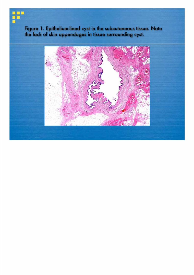
In addition, eccrine glands in human skin show a much broader distribution

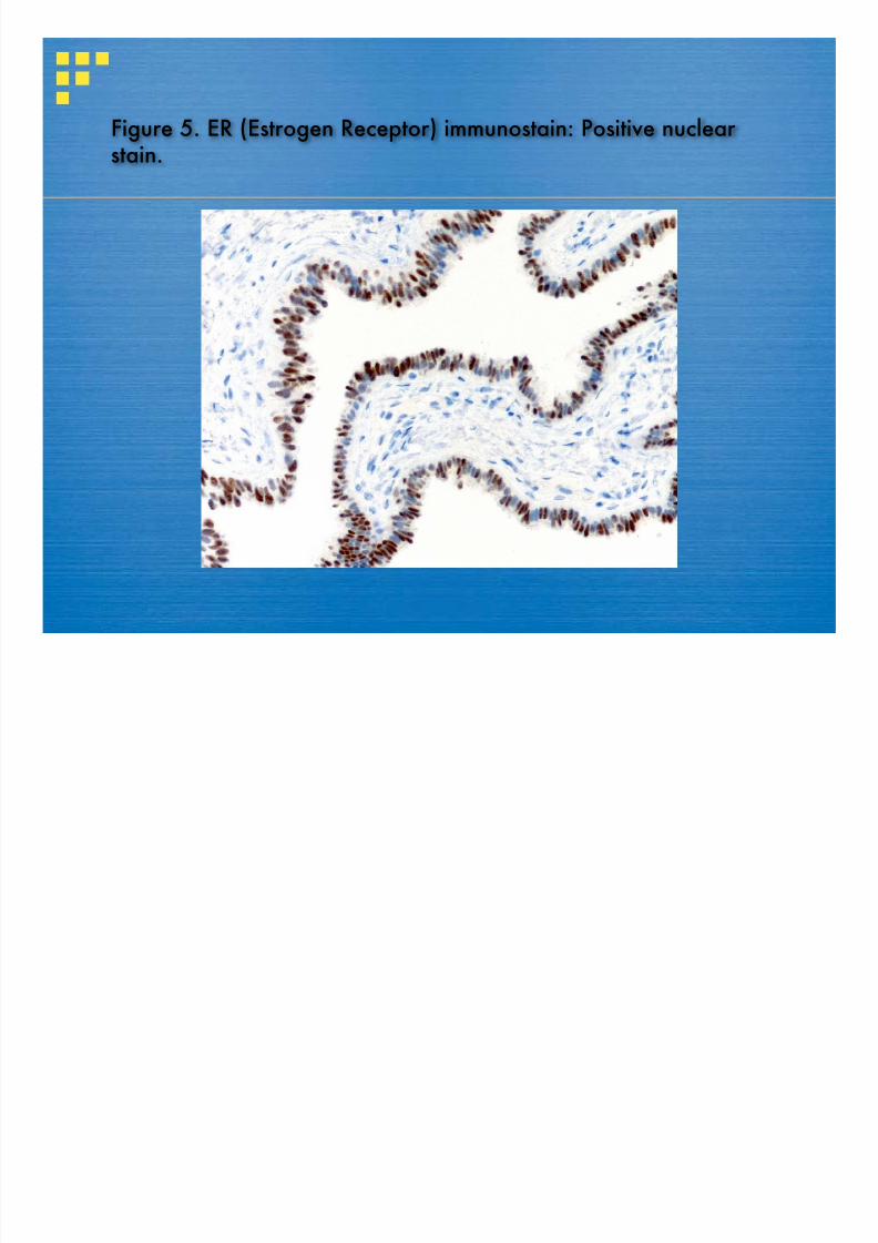
than just the lower limb [3], by far the most predominant site for CCC[1].Futhermore, the myoepithelial layer typical of sweat glands is lacking in

CCC [3, 6]. Lastly, cilia in CCC, like those of the fallopian tube, are of theultrastructural 9+2 type, whereas the cilia seen in fetal eccrine ducts and

eccrine tumors are of 9+0 type [1, 3, 4, 6]. Indeed, cutaneous Mullerian cyst

[3, 5, 6] has been offered as a preferred name over CCC to reflect that

ciliated epithelium can line other cutaneous cysts such as bronchogenic

cyst, thyroglossal duct cyst, branchial cleft cyst, thymic cyst, perianalcaudal gut cyst, and vulvar cyst [2, 3, 4]. Lee et al concluded that the rare

cases of CCC occurring in males and in unusual locations such as scalp

are thought to arise from ciliated metaplasia of eccrine or apocrine glands,

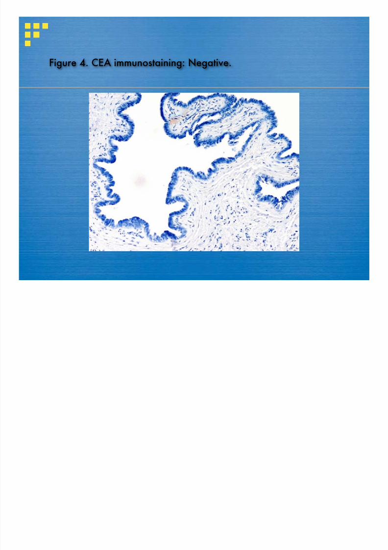
based on the fact that many of these cases are CEA positive and showevidence of a myoepithelial layer [5].

8/6/2019 Unknown: Biopsy of a 5-mm cystic lesion on the right heel of a 48-year-old woman

http://slidepdf.com/reader/full/unknown-biopsy-of-a-5-mm-cystic-lesion-on-the-right-heel-of-a-48-year-old 14/18

8/6/2019 Unknown: Biopsy of a 5-mm cystic lesion on the right heel of a 48-year-old woman

http://slidepdf.com/reader/full/unknown-biopsy-of-a-5-mm-cystic-lesion-on-the-right-heel-of-a-48-year-old 15/18

Thyroglossal duct cysts are located in the midline of neck and may be

associated with a sinus tract and thyroid follicles. The characteristic

location of vulvar cyst along with the mucin-producing, endocervical-like

epithelium typical of this entity easily distinguishes it from CCC/CMC. Infact, vulvar cyst may be a variant of Mullerian cyst, because vulvar cysts

are also presumably of Mullerian origin [2]. Perianal (caudal gut) cysts are

located in the perineum and they contain goblet cells and mucous glands,

unlike CCC/CMC [2]. Cutaneous endosalpingiosis usually occurs in thecontext of an abdominal scar [2]. The presence of cilia, the absence of

keratin in the cyst cavity, and the lack of appendageal structures helps

distinguish CCC/CMC from other more common cutaneous cysts such as

epidermal cyst, pilar cyst, steatocystoma, and hidrocystoma.

8/6/2019 Unknown: Biopsy of a 5-mm cystic lesion on the right heel of a 48-year-old woman

http://slidepdf.com/reader/full/unknown-biopsy-of-a-5-mm-cystic-lesion-on-the-right-heel-of-a-48-year-old 16/18

  Cutaneous ciliated cyst is a benign lesion, first described by Hess in

1890 [1] and later named CCC by Farmer and Helwig [1]. Cutaneous

Mullerian cyst (CMC) is a term preferred by some over CCC [3, 5, 6]

because of its presumed Mullerian origin and to prevent confusion

with other skin cysts showing cilia [2, 3, 4]. CCC/CMC often enlarges

and becomes clinically apparent during hormonally active years andis cured by excision [3, 5]. Careful attention to clinicopathologicfeatures help distinguish it from other ciliated cysts of the skin.

8/6/2019 Unknown: Biopsy of a 5-mm cystic lesion on the right heel of a 48-year-old woman

http://slidepdf.com/reader/full/unknown-biopsy-of-a-5-mm-cystic-lesion-on-the-right-heel-of-a-48-year-old 17/18

References

1. Farmer ER, Helwig EB. Cutaneous ciliated cysts. Arch Dermatol. 1978; 114:70-73. [PubMed]

2. Kurban RS, Bhawan J. Cutaneous cysts lined by nonsquamous epithelium. Am JDermatopathol. 1991; 13:509-17. [PubMed]

3. Bivin WW Jr, Heath JE, Drachenberg CB, Strauch ED, Papadimitriou JC. Cutaneous ciliatedcyst: a case report with focus on mullerian heterotopia and comparison with eccrine sweatglands. Am J Dermatopathol. 2010; 32:731-4. [PubMed]

4. Torisu-Itakura H, Itakura E, Horiuchi R, Matsumura M, Kiryu H, Takeshita T,Ohjimi Y, FurueM. Cutaneous ciliated cyst on the leg in a woman of menopausal age. Acta Derm Venereol.2009;89:323-4. [PubMed]

5. Lee JS, Kim YC, Lee ES. Cutaneous ciliated cyst of the inguinal area in a man. J Dermatol.2006; 33:146-9. [PubMed]

6. Dini M, Lo Russo G, Baroni G, Colafranceschi M. Cutaneous ciliated cyst: a case report withimmunohistochemical evidence for dynein in ciliated cells. Am J Dermatopathol. 2000;22:519-23. [PubMed]

8/6/2019 Unknown: Biopsy of a 5-mm cystic lesion on the right heel of a 48-year-old woman

http://slidepdf.com/reader/full/unknown-biopsy-of-a-5-mm-cystic-lesion-on-the-right-heel-of-a-48-year-old 18/18

REF:

Todd Stevens, Deba P Sarma.(2011), Unknown: Biopsy of a 5-

 mm cystic lesion on the right heel of a 48-year-old woman.

 Dermatology Online Journal 17 (6): 6, June 2011.


Recommended