Unmasking Proteolytic Activity for Adult Visual CortexPlasticity by the Removal of Lynx1
Noreen Bukhari,1,2,3,4,5 Poromendro N. Burman,1,2,3,4,5 Ayan Hussein,1,2,3,4,5 Michael P. Demars,1,2,3,4,5
Masato Sadahiro,1,2,3,4,5 X Daniel M. Brady,6 Stella E. Tsirka,7 Scott J. Russo,2,5 and Hirofumi Morishita1,2,3,4,5
Departments of 1Psychiatry, 2Neuroscience, and 3Ophthalmology, 4Mindich Child Health and Development Institute, and 5Friedman Brain Institute, IcahnSchool of Medicine at Mount Sinai, New York, New York 10029, 6Department of Physiology, University of California at San Francisco, San Francisco,California 94143, and 7Department of Pharmacological Sciences, Stony Brook University, Stony Brook, New York 11794
Experience-dependent cortical plasticity declines with age. At the molecular level, experience-dependent proteolytic activity of tissueplasminogen activator (tPA) becomes restricted in the adult brain if mice are raised in standard cages. Understanding the mechanism forthe loss of permissive proteolytic activity is therefore a key link for improving function in adult brains. Using the mouse primary visualcortex (V1) as a model, we demonstrate that tPA activity in V1 can be unmasked following 4 d of monocular deprivation when the miceolder than 2 months are raised in standard cages by the genetic removal of Lynx1, a negative regulator of adult plasticity. This was alsoassociated with the reduction of stubby and thin spine density and enhancement of ocular dominance shift in adult V1 of Lynx1 knock-out(KO) mice. These structural and functional changes were tPA-dependent because genetic removal of tPA in Lynx1 KO mice can block themonocular deprivation-dependent reduction of dendritic spine density, whereas both genetic and adult specific inhibition of tPA activitycan ablate the ocular dominance shift in Lynx1 KO mice. Our work demonstrates that the adult brain has an intrinsic potential forexperience-dependent elevation of proteolytic activity to express juvenile-like structural and functional changes but is effectively limitedby Lynx1 if mice are raised in standard cages. Insights into the Lynx1-tPA plasticity mechanism may provide novel therapeutic targets foradult brain disorders.
tral issue is that this limits recovery of function for brain disorderslater in life. Successful therapies are therefore contingent on un-derstanding the mechanism limiting adult brain plasticity(Mitchell and Sengpiel, 2009; Bavelier et al., 2010).
The rodent visual system is a well-defined model to study themechanism restricting experience-dependent plasticity in the
Received Oct. 17, 2014; revised July 30, 2015; accepted Aug. 4, 2015.Author contributions: N.B., S.E.T., S.J.R., and H.M. designed research; N.B., P.N.B., A.H., M.P.D., M.S., and H.M.
performed research; D.M.B. contributed unpublished reagents/analytic tools; N.B., P.N.B., A.H., M.P.D., M.S., andH.M. analyzed data; N.B. and H.M. wrote the paper.
This work was supported by National Eye Institute R01EY024918 to H.M., National Institute of NeurologicalDisorders and Stroke 5T32NS551147–5 to N.B., National Institute on Drug Abuse T32 DA007135 to M.P.D., NationalInstitute of Mental Health T32MH096678 to M.S., National Institute of General Medical Sciences R25GM064118 toA.H., Knights Templar Eye Foundation to H.M., March of Dimes to H.M., Whitehall Foundation to H.M., and Brain andBehavior Research Foundation to H.M. We thank Drs. Dan Christoffel and Sam Golden (Icahn School of Medicine atMount Sinai) for providing technical expertise on spine analysis; Dr. Rachel Neve (Massachusetts Institute of Tech-
nology) for supplying HSV-GFP; and Dr. Nathaniel Heintz (Rockefeller University) for providing Lynx1 knock-outmice.
The authors declare no competing financial interests.Correspondence should be addressed to Dr. Hirofumi Morishita, Icahn School of Medicine at Mount Sinai, One
Gustave L. Levy Place, Box 1230, New York, NY 10029. E-mail: [email protected]:10.1523/JNEUROSCI.4315-14.2015
Experience-dependent proteolytic activity of tissue plasminogen activator (tPA) becomes restricted in the adult brain in correla-tion with the decline in cortical plasticity when mice are raised in standard cages. We demonstrated that removal of Lynx1, one ofnegative regulators of plasticity, unmasks experience-dependent tPA elevation in visual cortex of adult mice reared in standardcages. This proteolytic elevation facilitated dendritic spine reduction and ocular dominance plasticity in adult visual cortex. Thisis the first demonstration of adult brain to retain the intrinsic capacity to elevate tPA in an experience-dependent manner but iseffectively limited by Lynx1. tPA-Lynx1 may potentially be a new candidate mechanism for interventions that were shown toactivate plasticity in adult brain.
The Journal of Neuroscience, September 16, 2015 • 35(37):12693–12702 • 12693
adult (Morishita and Hensch, 2008; Espinosa and Stryker, 2012;Levelt and Hubener, 2012; Hubener and Bonhoeffer, 2014). Dur-ing the juvenile critical period, short-term monocular depriva-tion (MD) produces robust functional and structural plasticity; ashift in ocular dominance (OD) of V1 neurons in favor of theopen eye measured electrophysiologically (Fagiolini et al., 1994;Gordon and Stryker, 1996), and the elevated motility and reduc-tion of dendritic spines (Mataga et al., 2004; Oray et al., 2004).Importantly, such juvenile forms of plasticity become limited inadult V1 under standard housing conditions.
However, OD plasticity can be either reactivated or preservedeven in the adult with additional environmental, such as darkexposure (He et al., 2006, 2007; Duffy and Mitchell, 2013;Stodieck et al., 2014), environmental enrichment (Sale et al.,2007; Greifzu et al., 2014), visual stimulation (Matthies et al.,2013), sensory-motor interaction (Kaneko and Stryker, 2014; Fuet al., 2015), voluntary physical exercise (Kalogeraki et al., 2014),and social experience (Balog et al., 2014) in addition to pharmaco-logical interventions (Putignano et al., 2007; Maya Vetencourt et al.,2008; Morishita et al., 2010; Nabel and Morishita, 2013), suggestingthe intrinsic potential of the adult brain to unmask plasticity. Al-though there is significant progress in determining the underlyingneural mechanisms of interventions, such as reduced intracorticalinhibition (Greifzu et al., 2014; Stodieck et al., 2014), disinhibitorycortical circuit (Fu et al., 2015), and serotonin (Matthies et al., 2013),it is essential to keep uncovering molecular bases of such intrinsicpotential of adult V1 to promote plasticity and understand how suchpotentials can be unmasked in adult V1.
One of the few permissive molecular pathways known to re-flect the decline of experience-dependent plasticity with age is oftissue plasminogen activator (tPA), a major serine protease in thebrain. MD during the critical period results in elevated tPA activ-ity, whereas MD in adult mice shows no change in tPA activity ifmice are raised in standard cages (Mataga et al., 2002, 2004).Genetic removal of tPA results in a loss of OD and spine plasticityduring critical period (Mataga et al., 2004; Oray et al., 2004),suggesting that tPA is one of the major molecular factors corre-sponding to structural and functional plasticity during juvenileperiod but not adulthood. The aim of our study was to under-stand why experience-dependent elevation of tPA and associatedstructural and functional plasticity becomes limited in the adultbrain �2 months raised in mice raised in standard cages and howit can be activated in the adult mice brain under these conditions.
We previously showed that Lynx1, an inhibitor of nicotinicacetylcholine receptors (Miwa et al., 1999) is a molecular inhibi-tor of OD plasticity that increases into adulthood whenexperience-dependent elevation of tPA is reciprocally lost. Ge-netic removal of this molecular inhibitor unmasks OD plasticityin the adult brain (Morishita et al., 2010), but the molecular andstructural underpinnings remain unclear. We thus hypothesizedthat the adult brain has an intrinsic potential for experience-dependent tPA elevation but is effectively masked by Lynx1 ifmice are raised in standard cages. We found that the removal ofthis molecular brake unlocks the proteolytic activity, which isnecessary for the juvenile form of structural and functional plas-ticity in the adult brain. Our study demonstrates the central roleof experience-dependent elevation of tPA activity in restoringadult brain plasticity.
Materials and MethodsAnimals. A total of 77 males and 49 females C57BL/6 (Charles RiverLaboratory, The Jackson Laboratory), Lynx1 KO (gifted by Dr. NathanielHeintz: Rockefeller University) (Miwa et al., 2006), and Lynx1-tPA Dou-
ble Knock-out (DKO) mice (tPA KO mice were originally purchasedfrom The Jackson Laboratory #002508, backcrossed to C57BL/6 for 12generations by S.T. and transferred to H.M.) (Carmeliet et al., 1994) wereused at postnatal age 53–392 d (1.7–13.1 months) (averages and ranges ofages of mice for each experimental groups are described in figure leg-ends). Both male and female were used (details of each experimentalgroups are described in figure legends). All mice were housed in groupsof 2–5 together with the sibling groups of the same sex in standard anduniform cage sizes (199 mm � 391 mm � 160 mm: width � depth �height, GM500, Tecniplast) and maintained on a 12 h light/dark cyclewith ad libitum access to food and water. Mouse procedures were per-formed in accordance with the Institutional Animal Care and Use Com-mittee guidelines of the Icahn School of Medicine at Mount Sinai.
MD. Adult mice (�p60) were anesthetized with isoflurane. Eyelidmargins were trimmed by iris scissor and eyes sutured shut for 4 d. Afterpreforming MD, mice were returned to their home cages until the time ofdeath.
Stereotaxic surgery and viral gene transfer. Mice were anesthetized withisoflurane and positioned in a small-animal stereotaxic instrument (Na-rishige). For the dendritic spine analysis, a 30 gauge syringe needle(Hamilton) was used to infuse 0.5 �l of Herpes simplex virus expressinggreen fluorescent protein (HSV-GFP provided by Dr. Rachel Neve, Mas-sachusetts Institute of Technology: 1.5 Å 108 infectious units/ml) intothe binocular zone of right V1 (lamda coordinates: anteroposterior 0mm; mediolateral 3.0 mm; dorsoventral 0.2 mm) at a rate of 0.1 �l/min3 d before perfusion. For tPA serpin injections, 21 d before MD, Lynx1KO mice were infused with either (1) adeno-associated virus (AAV)-GFP(a mixture of AAV8-hSyn-cre-mcherry, UNC core: 3 � 10 12 IU/ml; plusAAV8-Ef1a-DIO-EGFP, UNC core: 8 � 10 12 IU/ml) or (2) AAV-tPAserpin (a mixture of AAV8-hSyn-cre-mcherry plus AAV8-Ef1a-DIO-EGFP-2A-tPA serpini1, UNC core: 2.9 � 10 12 IU/ml) in right V1 at 600nl/injection � 3 injections (lamda coordinates: (1) anteroposterior: 0.0mm; mediolateral: 3.1 mm; dorsoventral: 0.4 mm; (2) anteroposterior:0.0 mm; mediolateral: 2.85 mm; dorsoventral: 0.4 mm; (3) anteroposte-rior: 0.3 mm; mediolateral: 3.0 mm; dorsoventral: 0.4 mm) for total 1.8�l injection/mouse.
Perfusion and tissue processing. After 4 d of MD, mice were anesthetizedwith pentobarbital and transcardially perfused with PFA, postfixed withPFA, and cryoprotected with sucrose/PBS. Brains were coronally sec-tioned (7 �m in situ hybridization (ISH), 35 �m for immunohistochemis-try, 150 �m for spine imaging). Brains injected with AAVs were directlymounted on slides using Vectashield (Vector Laboratories) mounting me-dium and coverslipped before imaging. For amidolytic assay, mouse V1 (2mm � 2 mm) was isolated, flash frozen, and stored at �80C.
Generation and validation of AAV-tPA serpin. cDNA of tPA serpin(Serpini1) was amplified from cDNA library generated from mouse WTV1, placed in vector pcDNA3.1(�) via Gibson assembly (New EnglandBiolabs), and Escherichia coli transformed. Positive colonies were con-firmed by sequencing (Genewiz). Vectors were then isolated by Midiprep(QIAGEN) and plasmid expression checked by transfection of N2A cells.To create pAAV vector, an inverted bicistronic 2A sequence was insertedinto pAAV-Ef1a-DIO-EGFP-WPRE-pA (Addgene, #37084) upstream ofEGFP by PCR linearization and overhang production on pAAV vector.TPA serpin pcDNA3.1(�) vector was used as a template to produce tPAserpin, which was inserted into pAAV vector using Gibson assembly,transformation, restriction digest, and gene sequence as above. Follow-ing colony verification, Maxiprep was performed (QIAGEN, Maxi kit)and the amplified pAAV-Ef1a-DIO-EGFP-2A-tPA serpin-WPRE-pAplasmid was sent to the UNC viral core for viral packaging. To confirmviral efficiency, tPA serpin and EGFP RNA probes were synthesized usingT3/T7 RNA polymerase (Roche) labeled with fluorescein. To performISH, sections were fixed with 4% PFA and tPA serpin-fluorescein andEGFP-digoxigenin probes were hybridized to frozen sections overnightat 72°C. To amplify the signal, probes were detected using anti-digoxigenin or fluorescein antibody conjugated to alkaline phosphatase(Roche), or the TSA-Plus DNP System (PerkinElmer Life Sciences) incombination with fast red staining. Images were acquired using a fluo-rescent microscope and quantified with ImageJ (National Institutes ofHealth).
12694 • J. Neurosci., September 16, 2015 • 35(37):12693–12702 Bukhari et al. • Lynx1 Masks tPA Activity to Limit Adult Plasticity
Amidolytic assay. Isolated mouse V1 that includes the binocular zone(2 mm � 2 mm) was homogenized in Tris-buffered saline, pH 7.0. Sam-ples were centrifuged at 20,800 � g for 15 min at 4°C and supernatantisolated. Brain homogenates were incubated with 0.19 �M plasminogen,10 nM fibrinogen in a 50 mM Tris buffer, pH 7.4, with 2% BSA and 0.3 mM
chromogenic substrate-2251 (DiaPharma) at 37°C. Cleavage of the chro-mogenic substrate S-2251 by serine protease-generated plasmin and thesubsequent color change was quantified at 405 nm. A dose–response forpure recombinant tPA activity was run in parallel alongside the test sam-ples and the time point when R 2 value for the dose–response �1 was usedto measure the concentration of serine protease in the samples. All sam-ples were run in triplicates. Recombinant tPA Serpin protein (90 nM
Serpini1–2485M: creative biomart) was used to block tPA in the indi-cated samples.
Dendritic spine imaging and analysis. HSV-GFP-injected brain sectionswere blocked with 10% goat serum/PBS for 1 h at room temperature(RT), then incubated overnight in rabbit anti-GFP (Invitrogen, 1:1000)at RT, washed in PBS multiple times, followed by secondary antibody inrabbit anti-Alexa-488 (Invitrogen, 1/5000) for 4 h at RT. Sections werethen washed multiple times in PBS and dehydrated in successively in-creased ethanol concentrations (75%, 95%, 100%), and then Citrasol-vent. Sections were mounted on Superfrost Plus slides using Permount(Sigma) mounting medium and coverslipped. Images were acquired on aconfocal LSM 710 (Carl Zeiss). Apical dendrites from V1 neurons withsoma in layer II/III were selected. To qualify for spine analysis, dendriticsegments had to satisfy the following requirements: (1) the segment hadto be completely filled (all endings were excluded); (2) the dendriticsegment had to be �200 �m from the surface of V1; and (3) the segmentcould not be overlapping with other dendritic branches (Christoffel et al.,2011; Golden and Russo, 2012). Dendritic segments were imaged using a100� lens (numerical aperture 1.4; Carl Zeiss) and a zoom of 3.0. Pixelsize was 0.03 �m in the x–y plane and 0.01 �m in the z plane. Images weretaken with a resolution of 1024 Å � 300, pixel dwell time was 1.58 � m/s,and the line average was set to 8. An average of 75 dendritic spines pergroup (n � 31 mice, n � 4 – 6 mice per group) totaling �460 dendriticspines were analyzed. Images were deconvolved using a two step resolu-tion enhancement: first with AutoDeblur software (Media Cybernetics)and then by z-smear correction in Neuron-Studio (Rodriguez et al.,2006; Dumitriu et al., 2011). For quantification, Neuron-Studio was usedto classify spines as thin, mushroom, or stubby based on the followingvalues: (1) aspect ratio, (2) head to neck ratio, and (3) head diameter.Spines with a neck can be classified as either thin or mushroom, and thosewithout a significant neck are classified as stubby. Spines with a neck arelabeled as thin or mushroom based on head diameter (Rodriguez et al.,2006, 2008). Spine density � total spine count/dendritic length. All mea-surements were made with an observer blinded to the genotype of themice.
In vivo extracellular recording. Recording was performed under Nem-butal/chlorprothixene anesthesia. Briefly, visually evoked single-unit re-sponses were recorded with 16 channel silicone probes (Neuronexus) inresponse to high-contrast single bar generated by visage system (Cam-bridge Research Systems). The signal was amplified and thresholded(OmniPlex, Plexon). To ensure single-unit isolation, the waveforms ofrecorded units were further examined offline (Offline Sorter, Plexon).For each animal, 3–10 single units were recorded in each of the 4 – 6vertical penetrations spaced evenly (250 �m intervals) across mediolat-eral extent of V1 to map the monocular and binocular zones and avoidsampling bias. To analyze the electrophysiology data, normalized ODindex of single neurons is computed by custom-made MATLAB program(The MathWorks) by peristimulus time histogram analysis of peak tobaseline spiking activity in response to each eye: {[Peak (ipsi) � baseline(ipsi)] � [Peak (contra) � baseline (contra)]}/{[Peak (ipsi) � baseline(ipsi)] � [Peak(contra) � baseline (contra)]}. OD scores were convertedfrom OD index using a 7 point classification scheme as follows: �1 to�0.5 � 1; �0.5 to �0.3 � 2; �0.3 to �0.1 � 3; �0.1 to 0.1 � 4; 0.1 to0.3 � 5; 0.3 to 0.5 � 6; and 0.5 to 1 � 7. For each binocular zone,contralateral bias index (CBI) is calculated according to the followingformula: [(n1 � n7) � 2/3(n2 � n6) � 1/3(n3 � n5) � N]/2 N, whereN � total number of cells and nx � number of cells corresponding to OD
score of x. The changes in magnitude of each eye’s responses by MD wererepresented as percentage change ((MD � no MD)/no MD � 100) usingnormalized firing rate of each neuron after MD, and averaged normal-ized firing rates of no MD control. The firing rate of each unit wasnormalized as [Peak(contra) � baseline (contra)]/[Peak (contra) �baseline (contra)] for contralateral response, and [Peak (ipsi) � baseline(ipsi)]/[Peak (ipsi) � baseline (ipsi)] for ipsilateral response (Kravitz andPeoples, 2008). All AAV-injected mice were perfused after recordingsand sectioned to examine the extent of AAV transduction by the presenceof GFP signal in V1. Only mice that exhibited GFP signal in the recordedV1 area were included for the analysis of OD plasticity. A quantitativemeasurement of GFP intensity to correlate GFP expression with the mag-nitude of OD plasticity was hampered by the combinatorial damage ofpostrecorded V1 tissues due to multiple penetrations by the electrodesused for recording as well as the injection sites created by syringe needlesused for AAV injections.
Statistics. All data are expressed as mean SEM. Mean differencesbetween two groups were determined using Student’s t test or Mann–Whitney U test. For multiple-group comparisons, mean differencesbetween groups were determined using a one-way ANOVA or Kruskal–Wallis test, followed by Tukey or Dunn’s post hoc tests when the maineffect or interaction was significant at p � 0.05. For electrophysiologyexperiments, � 2 test was used to measure OD shift, Student’s t test wasused to compare CBI between two groups, and Kolmogorov–Smirnov(K-S) test was used to compare cumulative percentage of OD indexacross two groups. A minimum p value of 0.05 was accepted as statisti-cally significant throughout.
ResultstPA activity elevates in adult V1 following MD in Lynx1KO miceTo examine the potential of adult V1 to elevate experience-dependent proteolytic activity when mice are raised in standardcages, we first measured tPA activity by an amidolytic assay frommouse V1 homogenates. Average age range of each experimentalgroups were 4.7– 8.1 months (Fig. 1). We found no difference intPA activity in adult WT V1 with and without 4 d of MD (WTadult no MD, n � 12; 0.52 0.034 vs WT adult MD, n � 12;0.46 0.024, p � 0.154 Student’s t test; Figure 1A) consistentwith previously reported findings (Mataga et al., 2004).
However, in adult Lynx1KO mice, we found a significant in-crease in tPA activity in V1 compared with its genotypic no MD(Lynx1 KO adult no MD, n � 18; 0.46 0.019 vs Lynx1 KO adultMD, n � 16; 0.66 0.082, p � 0.05, Mann–Whitney U test) andcompared with its corresponding WT adult MD group (WT adultMD vs Lynx1 KO adult MD, p � 0.05, Kruskal–Wallis test andpost hoc Dunn’s test; Fig. 1A).
To confirm the specificity of the proteolytic response, we thenused the pharmacologic inhibitor tPA serpin (Serpini1) (Galli-ciotti and Sonderegger, 2006). The elevated tPA activity in adultLynx1 KO mice with MD was significantly decreased with tPAserpin in ex vivo V1 homogenates (Lynx1 KO MD, n � 4; 0.61 0.079 vs Lynx1 KO � tPA Serpin, n � 4; 0.49 0.076, p � 0.05,paired Student’s t test; Figure 1B). Collectively, these findingssuggest that the adult V1 has potential for elevating tPA activityafter MD but is effectively limited by Lynx1 if mice are raised instandard cages.
Stubby and thin spines decrease in a tPA-dependent mannerin adult V1 of Lynx1 KO mice after MDBecause MD-dependent elevation of tPA activity in juvenile V1was shown to facilitate structural spine plasticity (Mataga et al.,2004; Oray et al., 2004), we asked whether tPA activity may affectdendritic spine plasticity in adult Lynx1 KO mice raised in stan-dard cages. We visualized distal apical dendritic spines from layer
Bukhari et al. • Lynx1 Masks tPA Activity to Limit Adult Plasticity J. Neurosci., September 16, 2015 • 35(37):12693–12702 • 12695
II/III neurons of adult V1 by injecting HSV-expressing GFP(Christoffel et al., 2011). Following 4 d of MD, the adult V1 wasisolated and spine density was quantified using Neuron-Studio(see Materials and Methods) (Rodriguez et al., 2006, 2008;Christoffel et al., 2011; Dumitriu et al., 2011). Spines were sub-categorized into three subtypes called mushroom, stubby, andthin spines based on previously established Neuron-Studio pa-rameters (Table 1) (Rodriguez et al., 2006, 2008; Golden andRusso, 2012). As our dendritic spine labeling technique could notreliably measure spines in dendritic segments at set distancesfrom the neuronal soma (Mataga et al., 2004), we focused on themost distal segments of apical dendrites to provide a uniform andunbiased analysis. Average age range of each experimental groupsfor spine analysis was 2.6 – 4.2 months (Fig. 2).
In adult V1 without visual manipulation, there was no statis-tical difference in total spine density between WT and Lynx1 KOmice (spine counts/�m: WT no MD, n � 5; 1.48 0.040 vsLynx1 KO no MD, n � 5; 1.35 0.032, p � 0.05, one-wayANOVA) or the spine subtypes: stubby (WT no MD, n � 5;0.30 0.010 vs Lynx1 KO no MD, n � 5; 0.35 0.019, p � 0.05,
one-way ANOVA), thin (WT no MD, n � 5; 0.68 0.046 vsLynx1 KO no MD, n � 5; 0.72 0.036, p � 0.05, one-wayANOVA) or mushroom (WT no MD, n � 5; 0.35 0.024 vsLynx1 KO no MD, n � 5; 0.27 0.028, p � 0.05, one-wayANOVA; Table 1).
Next, to analyze to what extent Lynx1 removal impacts MD-dependent dendritic spine plasticity, the spine density data ofmice with MD (Table 1) was normalized to its no MD control ofthe same genotype and compared between genotypes (Fig. 2C).We found in V1 of adult Lynx1 KO mice a slight, albeit notstatistically significant, MD-dependent decrease in total spinedensity (WT, n � 6; �9.71 4.654% vs Lynx1, n � 6; �24.91 2.352%, p � 0.33, one-way ANOVA and post hoc Tukey; Figure2B,C). When we further subcategorized these spines into threesubtypes, we found that removal of Lynx1 led to significant MD-dependent density reduction of the stubby spines (WT ��0.62 2.444% vs Lynx1 KO � �30.90 7.361%, p � 0.005,one-way ANOVA post hoc Tukey) and thin spines (WT � 9.72 6.025% vs Lynx1 KO � �26.87 7.696%, p � 0.004, one-wayANOVA post hoc Tukey) in adult V1 following MD comparedwith its WT counterparts (Fig. 2B,C). In contrast, WT and Lynx1KO mice show no statistically significant difference in MD-dependent changes in mushroom spine density (WT � �16.4 7.27% vs Lynx1 � 5.51 5.102%, p � 0.05, one-way ANOVA:Fig. 2B,C). These results are consistent with previously publishedresults on the role of stubby and thin spine subtypes in brainplasticity (Dumitriu et al., 2010; Christoffel et al., 2011).
To test the contribution of tPA activity on spine densitychanges in adult Lynx1KO mice, we next analyzed dendriticspines in mice lacking both tPA and Lynx1 genes (Lynx1-tPADKO). First, without visual deprivation, the Lynx1-tPA DKOmice have significantly less baseline density of total, thin, andstubby spine compared with their WT and Lynx1 KO counter-parts (p � 0.0001, p � 0.01, and p � 0.0033, one-way ANOVApost hoc Tukey), and less density of the mushroom spine subtypescompared with the WT group only (p � 0.0453, one-wayANOVA post hoc Tukey) (Table 1), suggesting some contributionof tPA to spine density at the baseline level independent of visualdeprivation.
Next, to analyze the contribution of tPA activity on MD-dependent spine density changes in adult Lynx1KO mice, thespine density data of mice with MD (Table 1) was normalized toits no MD control of the same genotype and compared betweengenotypes (Fig. 2C). Strikingly, the genetic removal of tPA inLynx1 KO mice ablated the MD-dependent reduction of thinspines and stubby spines observed in Lynx1KO mice (thin spines:Lynx1-tPA DKO, n � 6; 11.46 2.319% vs Lynx1 KO, p � 0.003,one-way ANOVA post hoc Tukey, stubby spines: Lynx1-tPADKO, n � 6; 9.97 5.516%, vs Lynx1KO, p � 0.0005, one-wayANOVA, post hoc Tukey; Figure 2B,C). When we compared thechanges of mushroom spine density of Lynx1-tPA DKO miceafter MD with that of Lynx1 KO mice after MD, we find that,unlike stubby and thin spine subtypes, there was no significantdifference in the change of mushroom spine density (Lynx1-tPADKO, n � 6; 18.11 1.356% vs Lynx1KO, p � 0.37, one-wayANOVA and post hoc Tukey; Fig. 2B,C). However, it should benoted that, in DKO mice, we also observed a significant MD-dependent density increase only in mushroom spines but not inthin and stubby spines compared between MD and no MDgroups (DKO no MD, n � 4; 0.25 0.015 vs DKO MD, n � 6;0.30 0.003, p � 0.03, Student’s t test; Table 1), or comparedwith MD-dependent change in WT mice (p � 0.01, one-wayANOVA and post hoc Tukey), suggesting an additional complex
Figure 1. MD-dependent elevation of tPA proteolytic activity in adult V1 of Lynx1KO mice. A,Measurement of tPA activity using amidolytic assay in right V1 homogenates from adult WT andLynx1 KO mice with (dark gray) and without (white) 4 d of monocular deprivation of left eye(MD). n � 12–18 mice. *p � 0.05 (Kruskal–Wallis test with post hoc Dunn’s test, and Mann–Whitney U test). B, tPA serpin, a known molecular inhibitor of tPA, was applied in ex vivo V1homogenates from adult Lynx1 KO mice after 4 d (4D) of MD and tPA activity measured usingamidolytic assay as above. n � 4 mice *p � 0.05 (paired Student’s t test). The range and theaverage age of mice for each subgroup were as follows: WT no MD: 2.6 – 6.8 months, average6.1 months, WT MD: 2.6 – 6.8 months, average 4.7 months, Lynx1 KO no MD: 2.4 –13.1 months,average 8.1 months, and Lynx1 KO MD: 2.3–11.0 months, average 5.9 months. As a recent studyreported, no significant OD plasticity in WT mice �P72 (age range P57–P80) (Stodieck et al.,2014), we also compared the tPA activity only with the mice older than P72 (excluding 2 miceeach from KO noMD group and KO MD group who were younger than P72). In these sets of miceover P72, Lynx1KO MD groups (14 mice) maintained a statistically significant increase in tPAactivity compared with age-matched controls ( p � 0.031 vs WT MD group, 12 mice; p � 0.020vs Lynx1 KO no MD group, 16 mice, t test). When only older adults (�P110) were compared,Lynx1KO MD groups (7 mice) also maintained statistically significant increase in tPA activitycompared with age-matched controls ( p � 0.046 vs WT MD group, 6 mice; p � 0.003 vs Lynx1KO no MD group, 12 mice, t test). Only male mice were used in WT groups. In the Lynx1 KOgroups, a comparison between the two genders revealed no statistical difference in tPA activitywithin the Lynx1 KO no MD group (9 male, 6 female, excluding 3 mice with unidentified gender,p � 0.098, t test) or MD group (7 male and 9 female, p � 0.180, t test).
Table 1. Absolute spine density in adult V1 with and without MDa
nTotal(sc/�m)
Stubby(sc/�m)
Thin(sc/�m)
Mushroom(sc/�m)
WT no MD 5 1.48 0.040 0.30 0.010 0.68 0.046 0.35 0.024WT MD 6 1.34 0.069 0.30 0.007 0.75 0.041 0.30 0.026Lynx1 KO no MD 5 1.35 0.032 0.35 0.019 0.72 0.036 0.27 0.028Lynx1 KO MD 6 0.97 0.047 0.24 0.026 0.53 0.056 0.28 0.010Lynx1-tPA DKO no MD 4 0.89 0.049 0.23 0.002 0.42 0.057 0.25 0.015Lynx1-tPA DKO MD 6 1.04 0.071 0.26 0.013 0.47 0.010 0.30 0.003aAbsolute spine density of total spines and three spine subtypes (stubby, thin, mushroom) in apical dendrites fromV1 neurons with soma in layer II/III of WT, Lynx1 KO, and Lynx1-tPA DKO mice with and without MD. Data are meanspine density (spine counts sc�/�m) SEM (%) (n � 4 – 6).
12696 • J. Neurosci., September 16, 2015 • 35(37):12693–12702 Bukhari et al. • Lynx1 Masks tPA Activity to Limit Adult Plasticity
mechanism affecting the Lynx1-tPA DKO mushroom spine den-sity that is distinct from the experience-dependent mechanismaffecting the stubby and thin spine densities. This additionalmechanism suggests a complex interaction between tPA, Lynx1,and experience, which requires exploration that lies beyond thescope of the current study. Collectively, our data demonstratethat removal of Lynx1 significantly decreases stubby and thinspines following MD in a tPA-dependent manner.
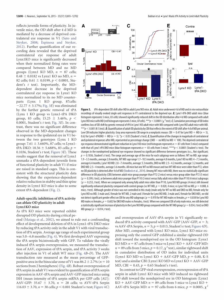
tPA is required for the elevated OD plasticity in adult Lynx1KO miceTo examine the role of tPA activity on OD plasticity in adultLynx1 KO mice when mice are raised in standard cages, we ex-amined the neuronal spiking response of each eye in the V1 using
in vivo extracellular recordings following4 d of MD in adult Lynx1-tPA DKO mice.Average age range of each experimentalgroup for OD analysis was 2.8 – 6.4months (Fig. 3).
We first confirmed our previous find-ings of extended OD plasticity in adultLynx1KO mice with the upgraded single-cell sorting system (see Materials andMethods). Consistent with the previousstudy (Morishita et al., 2010), adult Lynx1KO mice without MD (n � 6 mice, 82cells, CBI � 0.6829) showed similar base-line OD to WT mice without MD (n � 11mice, 193 cells, CBI � 0.6883) (OD distri-bution: p � 0.73, � 2 test, cumulative dis-tribution of OD index: p � 0.81, K-S test,CBI: p � 0.922, Student’s t test). After 4 dof MD in adult Lynx1KO mice, there is asignificant rightward OD shift toward thenondeprived eye (Lynx1 KO MD: n � 5mice, 87 cells, CBI � 0.4674) comparedwith both WT MD (n � 7 mice, 86 cells,CBI � 0.6744. OD distribution: p �0.0001, � 2 test, cumulative distribution ofOD index: p � 0.0001, K-S test, CBI: p �0.0025 Student’s t test) and Lynx1 KO noMD (n � 6 mice, 82 cells, CBI � 0.6829;OD distribution: p � 0.0001, � 2 test, cu-mulative distribution of OD index: p �0.0001, K-S test, CBI: p � 0.0031, Stu-dent’s t test), confirming the extended ODplasticity in adult Lynx1KO mice (Mor-ishita et al., 2010).
Genetic deletion of tPA itself did notresult in a significant change in baselineOD distribution in Lynx1 KO mice with-out MD (Lynx1-tPA DKO mice with noMD: n � 5 mice, 76 cells, CBI � 0.6425)compared with Lynx1 KO no MD (p �0.26, � 2 test), no shift in cumulative dis-tribution of OD index for individual units(p � 0.63, K-S test), and no difference inCBI (p � 0.30, Student’s t test). However,in contrast to adult Lynx1KO mice withMD, 4 d of MD in adult Lynx1-tPA DKOmice (n � 5 mice, 83 cells) showed norightward shift toward the nondeprivedeye in OD distribution (p � 0.0001, � 2
test; Fig. 3B), loss of shift in cumulative distribution of OD indexfor individual units (p � 0.0001, K-S test; Fig. 3C), and signifi-cant increase in CBI (CBI � 0.6205, p � 0.032, Student’s t test;Fig. 3D), suggesting that the deletion of tPA led to the loss of ODplasticity in adult Lynx1KO mice. Along the same lines, after 4 dof MD, adult Lynx1-tPA DKO mice did not reveal any significantchange in OD compared with its no MD genotypic counterpart(OD distribution, p � 0.29, � 2 test, cumulative distribution ofOD index: p � 0.7842, K-S test, CBI: p � 0.86, Student’s t test).Collectively, our results support a role for tPA in the OD shift inadult Lynx1 KO mice after MD.
Finally, as tPA is implicated in OD plasticity during the juve-nile critical period (Mataga et al., 2002, 2004), we further exam-ined to what extent elevated OD plasticity in adult Lynx1KO mice
Figure 2. MD-dependent reduction of thin and stubby spine density in adult V1 of Lynx1KO mice is dependent on tPA. A, Adultmice (�postnatal day 60) underwent HSV-GFP injection 1 d after MD. Four days after MD, adult V1 was isolated and processed forspine analysis. B, Representative images of GFP-labeled dendritic spines (green) and corresponding gray schema below labeledwith stubby (pink), thin (yellow), mushroom (nonlabeled white) spines in WT, Lynx1 KO, and Lynx1-tPA DKO adult mice V1 afterMD. C, Graphs showing MD-dependent changes of total spine density and densities of three subsets (stubby, thin, and mushroom)spines in adult V1 of WT, Lynx1 KO, and Lynx1-tPA DKO mice. Each genotype was initially normalized to its no MD control of thesame genotype, and all spine density graphs are represented as percentage change ((MD � no MD)/no MD � 100); n � 4 – 6.*p�0.05 (one-way ANOVA and post hoc Tukey test). The age range and the average of the mice for each subgroup were as follows:WT no MD: 3.8 – 4.3 months, average 4.2 months; WT MD: 3.6 – 4.1 months, average 3.9 months; Lynx1 KO no MD: 2.8 –5.0months, average 3.3 months; Lynx1 KO MD: 2.5–3.1 months, average 2.6 months; DKO no MD 2.1– 4.3 months, average 3.3months; and DKO MD 2.5– 4.9 months, average 3.6 months. There was no statistically significant correlation between age and totalspine density within each subset groups (Pearson correlation coefficient analysis: R 2 � 0.426, p � 0.160 for WT no MD, R 2 �0.036, p � 0.719 for WT no MD, R 2 � 0.815, p � 0.097 for KO no MD, R 2 � 0.0572, p � 0.761 for KO MD, R 2 � 0.985, p � 0.078for DKO no MD, R 2 � 0.110, p � 0.586 for DKO MD). Although the gender of mice was not controlled in this study (5 male for WTno MD, 3 male and 3 female for WT MD, 3 male and 2 female for KO no MD, 1 male and 5 female for KO MD, 3 male and 1 female forDKO no MD, 3 male and 3 female for DKO MD), the groups that had necessary number of mice for quantification (�3 mice for eachgender) did not show gender differences ( p � 0.719 WT MD male vs female mice, p � 0.736 for DKO MD male vs female group,t test).
Bukhari et al. • Lynx1 Masks tPA Activity to Limit Adult Plasticity J. Neurosci., September 16, 2015 • 35(37):12693–12702 • 12697
reflects juvenile forms of plasticity. In ju-venile mice, the OD shift after 4 d MD ismediated by a decrease of deprived con-tralateral eye response in V1 (Sato andStryker, 2008; Espinosa and Stryker,2012). Further quantification of our re-cording data revealed that the deprivedcontralateral eye response of adultLynx1KO mice is significantly decreasedwhen their normalized firing rates werecompared between MD and no MDgroups (Lynx1 KO MD, n � 87 cells;0.48 0.0182 vs Lynx1 KO no MD, n �82 cells; 0.61 0.0199, p � 0.0001, Stu-dent’s t test). Importantly, the MD-dependent decrease in the deprivedcontralateral eye response in Lynx1 KOmice normalized to its no MD counter-parts (Lynx 1 KO group, 87cells:�22.77 3.17%; Fig. 3E) was eliminatedby the further genetic removal of tPA(Lynx 1 KO group vs Lynx1-tPA DKOgroup, 83 cells; 15.25 3.46%, p �0.0001, Student’s t test; Fig. 3E). In con-trast, there was no significant differenceobserved in the MD-dependent changesin response to the ipsilateral eye in V1 be-tween the two genotypes (Lynx 1 KOgroup: 7.61 3.660%, 87 cells; vs Lynx1-tPA DKO: 18.56 5.688%, 83 cells; p �0.1036, Student’s t test; Figure 3E). Theseresults suggest that the removal of Lynx1unmasks a tPA-dependent juvenile formof functional plasticity in adult V1 if miceare raised in standard cages. This is con-sistent with the structural plasticity datashowing that the experience-dependentrelative reduction in stubby and thin spinedensity in Lynx1 KO mice is also to someextent tPA-dependent (Fig. 2).
Adult-specific inhibition of tPA activitycan ablate OD plasticity in adultLynx1KO miceAs tPA KO mice were reported exhibitdisrupted OD plasticity during critical pe-riod (Mataga et al., 2002), we aimed to rule out a confoundingeffect of developmental deletion of tPA in Lynx1-tPA DKO miceby reducing tPA activity only in the adult V1 with viral transduc-tion of tPA serpin. Average age range of each experimental groupwas 5.0 – 6.8 months (Fig. 4). We first developed AAV expressingthe tPA serpin bicistronically with GFP. To validate the virallyinduced tPA serpin overexpression, we measured the transduc-tion of AAV, expression of tPA serpin, and tPA activity 3 weeksafter injection in V1 of adult WT mice (Fig. 4B–D). The AAVtransduction rate measured as the mean percentage of GFP-positive area in the binocular zone of V1 was 86.2 2.7% (n � 10sections from 2 hemispheres) (Fig. 4B). Robust overexpression oftPA serpin in adult V1 was evident by quantification of tPA serpinexpression in AAV-tPA serpin and AAV-GFP injected mice usingISH (mean intensity of tPA serpin mRNA per GFP cells [AU]:AAV-GFP: 35.67 3.70, n � 28 cells; vs AVV-tPA Serpin116.03 3.70, n � 30 cells; p � 0.001 Student’s t test; Figure 4C)
and overexpression of AAV-tPA-serpin in V1 significantly re-duced tPA activity compared with AAV-GFP (AAV-GFP, n � 3;vs AAV-tPA Serpin, n � 5; p � 0.013, Student’s t test; Figure 4D).After MD, compared with Lynx1 KO mice, Lynx1 KO mice ex-pressing only the control GFP exhibited a similar rightward ODshift toward the nondeprived eye in the OD histogram (Lynx1KO MD: n � 87 cells from 5 mice vs Lynx1 KO � AAV-GFP MD:n � 89 cells from 5 mice, p � 0.17, � 2 test), similar rightward shiftin cumulative distribution of OD index for individual units(Lynx1 KO MD vs Lynx1 KO � AAV-GFP MD, p � 0.80, K-Stest) and a similar CBI (Lynx1 KO MD vs Lynx1 KO � AAV-GFPMD, CBI � 0.43, p � 0.65 Student’s t test).
In contrast to GFP viral overexpression, overexpression of tPAserpin in adult Lynx1 KO mice with MD induced no rightwardOD shift toward the nondeprived eye in OD histogram (Lynx1KO � AAV-GFP MD: n � 89 cells from 5 mice vs Lynx1 KO �AAV-tPA Serpin MD: n � 97 cells from 6 mice, p � 0.0005, � 2
Figure 3. tPA-dependent OD shift after MD in adult Lynx1KO mice. A, Adult mice underwent 4 d of MD and in vivo extracellularrecordings of visually evoked single unit responses in V1 contralateral to the deprived eye. B, Lynx1-tPA-DKO adult mice (bluehistogram represents 5 mice, 83 cells) showed significantly reduced shift in the OD distribution after 4 d MD compared with adultLynx1KO mice with MD (red histogram represents 5 mice, 87cells). ***p � 0.0001 (� 2 test). C, Cumulative percentage of OD indexconfirms loss of OD shift by genetic removal of tPA in Lynx1 KO adult mice with MD compared with Lynx1KO adult mice with MD.***p � 0.001 (K-S test). D, Quantification of adult OD plasticity by CBI that reflects the extent of OD shift after 4 d of MD per animal.Low CBI indicates higher plasticity. Gray area represents CBI range in a nonplastic mouse. CBI � 0.47 for Lynx1KO � MD (n � 5),0.62 for Lynx1-tPADKO � MD (n � 5). *p � 0.05 (Student’s t test). E, Quantification of the changes in magnitude of contralateraland ipsilateral responses after MD, represented as percentage change ((MD � no MD)/no MD � 100). The deprived contralateraleye response demonstrated significant reduction in Lynx1 KO mice (red histogram represents n � 87 cells from 5 mice) comparedwith that of Lynx1-tPA DKO mice (blue histogram represents n � 83 cells from 5 mice). ****p � 0.0001 (Student’s t test). Thechanges in the nondeprived ipsilateral eye response showed no significant difference between genotypes (n.s., Not significant;p � 0.1036, Student’s t test). The range and average age of the mice for each subgroup were as follows: WT no MD: age range:1.9 –3.5 months, average 2.8 months, WT MD: age range 1.7–10.5 months, average 4.8 months, Lynx1 KO no MD: 4 –7.5 months,average 6.4 months, Lynx1 KO MD: 2.8 –7.4 months, average: 5.3 months, DKO no MD: 2.5– 4.2 months, average: 3.2 months, andDKO MD: 2.5– 4 months, average 3.6 months. All mice but one WT no MD mouse and two WT MD mice were older than P72, whenno OD plasticity is detected after 4 d of MD (Stodieck et al., 2014). Among WT mice with MD, there was no statistically significantdifference in OD plasticity (CBI) between adult mice group younger than P72 (2 mice) versus mice group older than P72 (5 mice)( p � 0.599, t test), or between adult mice younger than P110 (3 mice) versus fully adult mice older than P110 (4 mice) subgroups( p � 0.253, t test). When we compared only among fully adult mice older than P110, Lynx1KO MD groups (4 mice) still showedsignificantly enhanced plasticity compared with control groups (vs WT MD, p � 0.020, 4 mice; vs Lynx1 KO no MD, p � 0.008, 6mice, t test). Although gender of mice was not controlled in this study (male only for WT no MD and DKO no MD, female only forLynx1KO no MD, 5 male and 2 female for WT MD, 2 male and 3 female for Lynx1KO MD, 3 male and 2 female for DKO MD), we didnot detect significant gender effect on CBI for any MD subgroups ( p � 0.970 for WT MD males vs females, p � 0.838 for Lynx1KOMD males vs females, p � 0.602 for DKO MD males vs females, t test). When we compared CBI of only male mice, we still detecteda statistically significant increase of plasticity in the Lynx1KO MD group compared with the WT MD group ( p � 0.034, t test) or DKOMD group ( p � 0.014, t test).
12698 • J. Neurosci., September 16, 2015 • 35(37):12693–12702 Bukhari et al. • Lynx1 Masks tPA Activity to Limit Adult Plasticity
test; Fig. 4E), loss of shift in cumulative distribution of OD indexfor individual units (Lynx1 KO � AAV-GFP vs Lynx1 KO AAV-tPA Serpin, p � 0.0004, K-S test; Fig. 4F), and significant increasein CBI (Lynx1 KO � AAV-GFP, n � 5; vs Lynx1 KO � AAV-tPASerpin, n � 6; CBI � 0.57, p � 0.0005, Student’s t test; Figure 4G).Along the same lines, after 4 d of MD, adult Lynx1 KO mice withAAV-tPA Serpin overexpression in V1 did not reveal any signif-icant change in OD compared with its no MD genotypic coun-terpart (n � 3 mice, 46 cells, CBI � 0.59: OD distribution, p �0.75, � 2 test, cumulative distribution of OD index: p � 0.98, K-Stest, CBI: p � 0.60, Student’s t test). Although these experimentsusing Lynx1KO mice cannot fully rule out the confounding effectof developmental deletion of Lynx1, we expect such an effect isminimal because: (1) the OD plasticity of Lynx1KO mice duringcritical period is normal in contrast to tPA KO mice that havedisrupted OD plasticity during critical period; and (2) an elevatedOD plasticity in adult Lynx1KO mice can be blocked by an acuteapplication of nAChR antagonists (Morishita et al., 2010). Ourseparate parallel study is expected to draw a conclusion by testingif the adult specific knockdown of Lynx1 expression in WT V1 issufficient to phenocopy an elevated OD plasticity in adultLynx1KO mice (M.S., P.B., M.P.D., H.M.). Collectively, our datasupport an adult specific role of tPA activity in OD plasticity inLynx1 KO mice after MD when mice are raised in standard cages.
DiscussionThe aim of our study was to understand why experience-dependent tPA elevation does not occur in the adult mice brain
older than 2 months if raised in standard cage. We demonstratedthat removal of Lynx1, one of the negative regulators of plasticity,can unmask experience-dependent tPA elevation in adult brain.This sensory-dependent proteolytic elevation facilitated den-dritic spine reduction and OD plasticity in adult V1. Our workdemonstrates, for the first time, that adult brain retains the in-trinsic capacity to elevate tPA in an experience-dependent man-ner even after closure of the classical critical period for ODplasticity. However, unlike juvenile brain, the adult brain of stan-dard cage-raised mice requires a two hit approach (sensory de-privation and removal of negative regulators such as Lynx1) tounmask plasticity.
tPA and Lynx1 then may become promising molecular targetsfor restoring adult plasticity. Recently, multiple interventionswere reported to activate and/or preserve plasticity in adult V1,including dark rearing (He et al., 2006, 2007; Duffy and Mitchell,2013; Stodieck et al., 2014), environmental enrichment (Sale etal., 2007; Greifzu et al., 2014), visual stimulation (Matthies et al.,2013), sensory-motor interaction (Kaneko and Stryker, 2014),voluntary physical exercise (Kalogeraki et al., 2014), and socialexperience (Balog et al., 2014) in addition to pharmacologicalinterventions (Putignano et al., 2007; Maya Vetencourt et al.,2008; Morishita et al., 2010; Nabel and Morishita, 2013). Impor-tantly, these interventions seem to have differential specificityand magnitude (Hubener and Bonhoeffer, 2014). Although someunderlying molecular mechanisms have been identified, such asreduced intracortical inhibition (Greifzu et al., 2014; Stodieck et
Figure 4. Adult specific inhibition of tPA ablates MD-dependent OD shift in Lynx1KO mice. A, AAV-GFP or AAV-tPA serpin was injected in Lynx1 KO adult mice. MD was performed 21 d after braininjection, and in vivo electrophysiology 4 d after MD. B, Representative images of the binocular zone of V1 from three different distances from bregma in anterior–posterior axis (�3.1 mm, �3.5mm, �4.0 mm) 21 d after AAV-tPA serpin injection showing bicistronically expressed GFP fluorophore. Binocular zone of V1 is outlined by dashed white-line based on Paxinos and Franklin (1997).Scale bar, 250 �m. C, Quantification of tPA serpin expression in AAV-tPA serpin and AAV-GFP-injected mice using ISH, data are represented as mean intensity of tPA serpin mRNA signal/GFP cell (n�30 cells for AAV-tPA seprin overexpression and n � 28 cells for AAV-GFP overexpression). ***p � 0.001 (Student’s t test). D, Quantification of tPA activity after AAV-tPA Serpin or AAV-GFP injectionin V1 using amidolytic assay n � 3–5. *p � 0.05 (Student’s t test). E, Lynx1 KO � AAV-tPA serpin mice (blue histogram represents n � 6 mice, 97 cells) showed no rightward shift in the ODdistribution after 4 d MD in contrast to Lynx1KO � AAV-GFP mice with MD (red histogram represents n � 5 mice, 89 cells). ***p � 0.0005 (� 2 test). F, Cumulative percentage of OD index confirmsloss of OD shift after injection of tPA serpin compared with control AAV-GFP injection in Lynx1 KO mice with MD. ***p � 0.001 (K-S test). G, Quantification of adult OD plasticity by CBI. CBI � 0.43in Lynx1 KO � AAV-GFP and CBI � 0.57 in Lynx1 KO � tPA serpin. *p � 0.05 (Student’s t test). Gray area represents CBI range in a nonplastic mouse. All mice were older than P110. The averageage and age range of the mice for each subgroup are as follows: Lynx1 KO AAV-GFP MD: 4.9 –5.1 months, average 5.0 months, Lynx1 KO AAV-NS MD: 5.3–7.9 months, average 6.8 months. Onlyfemales were used for recordings with AAV injections.
Bukhari et al. • Lynx1 Masks tPA Activity to Limit Adult Plasticity J. Neurosci., September 16, 2015 • 35(37):12693–12702 • 12699
al., 2014), disinhibitory cortical circuit (Fu et al., 2015), and se-rotonin (Matthies et al., 2013), the contribution of tPA or Lynx1has never been examined. Our study suggests that tPA and Lynx1may potentially be a new candidate mechanism for interventionsthat may activate plasticity in adult brain. As the removal ofLynx1 unmasks a juvenile form of plasticity (Figs. 2, 3), tPA-Lynx1 may be particularly relevant to the interventions that wereshown to restore juvenile forms of plasticity, such as environ-mental enrichment (Greifzu et al., 2014) and voluntary physicalexercise (Kalogeraki et al., 2014). Thus, it would be interesting toexamine whether the expression of tPA proteolytic activity canremain elevated upon MD in these rearing conditions in futurestudies. More conceptually, the fact that some environment canlead to juvenile level of plasticity in the adult raises a possibilitythat limited plasticity in the adult mice raised in standard labo-ratory cages may not necessarily reflect the normal physiologicalchange, but rather a special case in an impoverished environ-ment. In future, it is important to conduct the experiments inan environment as close as possible to a natural environmentto assess the “true” contribution of Lynx1-tPA system foradult plasticity.
Age is another important factor that can impact the OD plas-ticity in adult mice. A recent study showed that young adults(�P110) but not full adults (�P110) exhibited an adult form ofOD plasticity after 7 d of MD (Lehmann and Lowel, 2008). Itshould be noted, though, that such an age-dependent effect crit-ically depends on the length of MD. Importantly, shorter 4 d ofMD does not induce OD plasticity in young adult mice well underthe age of P110 to P72 (age range P58-P80) (Stodieck et al., 2014),P80 (Huang et al., 2015), and P84-P91 (Sato and Stryker, 2008).In our study, the average age of each experimental group wasolder than P72, and we similarly did not detect significant differ-ence in OD plasticity by 4 d of MD between adult WT micesubgroups younger versus older than P72 or P110 (Fig. 3), sug-gesting that our study is likely not sensitive enough to detectage-dependent changes of OD plasticity, if any, among adults.Indeed, for both the tPA activity and OD plasticity, when datawere compared only with the mice older than P72 or P110,Lynx1KO MD groups maintained statistically significant increasein tPA activity (Fig. 1) and OD plasticity (Fig. 3) compared withage-matched controls. We also did not detect any statisticallysignificant correlation between age and total spine density withineach subset groups (Fig. 2). Gender can also potentially impactOD plasticity in the adults. A recent imaging study reported thatfully adult female mice can exhibit OD plasticity when pair-housed in standard cage during 4 d of MD (Balog et al., 2014),raising a possibility that the female mice can be more plastic incertain rearing conditions. Although our study did not controlfor gender across groups, no difference in gender-dependent ef-fect on plasticity was detected for any subgroup that had enoughmice number for quantification (detail in figure legends). In ad-dition, when we compared only male mice, we still detected sta-tistically significant increase of OD plasticity in Lynx1KO MDgroup compared with WT MD group or DKO MD group (Fig. 3).Overall, our study did not detect significant age and gender effectin any of our measurements, and is least likely confounded bythese factors. Nonetheless, to fully examine the age effect andgender effect that can confound the results, an extensive studyusing both 4 and 7 d of MD is required for all measurements infuture studies as it was done for the hemodynamic imaging (Le-hmann and Lowel, 2008).
Our study also provides the novel mechanisms of plasticitylimited by Lynx1 in adult brain. Lynx1 acts as an inhibitor of the
nicotinic Acetylcholine receptor (Miwa et al., 1999, 2006, 2011;Ibanez-Tallon et al., 2002; Picciotto et al., 2012). A previous studyshowed that its removal allows for increased nicotinic signalingand functional adult brain plasticity (Morishita et al., 2010). Butthe role of Lynx1 in structural plasticity remained unclear. Thecurrent study indicates that dendritic spine changes are impor-tant contributors to experience-dependent adult brain plasticityin the absence of Lynx1. Specifically, we found that MD results inreduction of distal apical dendritic spines from V1 neurons inlayer II/III in adult Lynx1 KO mice, a pattern very similar to theMD-dependent spine plasticity during the juvenile period(Mataga et al., 2004). Consistent with the structural mechanisms,our in vivo electrophysiological recordings also identified that thefunctional OD plasticity in adult Lynx1 KO mice is mediated by adecrease in the response of the deprived contralateral eye in V1, asignature of juvenile-like plasticity. Our structural and functionalstudies collectively suggest that the removal of Lynx1 can un-mask juvenile form of plasticity, consistent with the elevationof tPA activity. Interestingly, the deletion of postsynaptic densityprotein-95 (PSD-95) also induces juvenile form of OD plasticityin adult mouse V1 (Huang et al., 2015). As the PSD-95 KO miceaccompany failed maturation of silent synapses, it would be in-teresting to test whether Lynx1-tPA system similarly regulates thematuration of silent synapses as well as to examine whetherPSD-95 regulates dendritic spine plasticity as Lynx1 does.
Elucidating the cellular and molecular basis of the Lynx1-tPAinteraction will be an important area of future exploration. Al-though our study focused on the role of Lynx1 to regulate tPAactivity, it is possible that other negative regulators, such as chon-droitin sulfate proteoglycans (CSPGs), the myelin inhibitor pro-tein, Nogo66 receptor 1 (NgR1), Paired-immunoglobulin-likereceptor B (PirB), and Cartilage link protein (Crtl1) (Pizzorussoet al., 2002; McGee et al., 2005; Syken et al., 2006; Carulli et al.,2010) may also play a role in limiting experience-dependent tPAactivity. Future studies should help to distinguish whether mul-tiple negative regulators act on multiple experience-dependentpermissive factors (Maya-Vetencourt et al., 2012; Spolidoro et al.,2012) or whether tPA acts as a molecular hub gated by multiplenegative regulators. Determination of the targets of tPA in theplasticity pathway will be also important to draw conclusionsabout the neural mechanisms. Mataga and colleagues (Mataga etal., 2002, 2004) previously discussed BDNF, NCAM, laminin,and CSPGs as potential molecular and structural targets of tPA injuvenile brain. Our study focused on the role of tPA in the adultbrain. Unlike juvenile brain, the extracellular matrix of the adultbrain is rich in CSPGs (Pizzorusso et al., 2002; Cabungcal et al.,2013; de Vivo et al., 2013). We previously showed that tPA candegrade CSPGs and allow for enhanced recovery in adult CNSinjury models (Wu et al., 2000; Nolin et al., 2008; Bukhari et al.,2011). Our work demonstrated that tPA and its substrate plas-minogen bind to the core proteins of CSPGs, which function as ascaffold to accelerate the tPA-driven conversion of plasminogento plasmin. Once generated, plasmin then degrades the CSPGscaffold protein. Whether similar or distinct mechanisms are in-volved in V1 plasticity model is an important future question.
Our work indicates that the adult brain retains intrinsic capac-ity to elevate permissive tPA activity but is effectively masked byLynx1 to limit cortical plasticity. Removal of Lynx1 can allow forelevation of experience-dependent proteolytic activity and sub-sequent structural and functional plasticity. These mechanisticinsights will provide novel therapeutic targets for adult braindisorders.
12700 • J. Neurosci., September 16, 2015 • 35(37):12693–12702 Bukhari et al. • Lynx1 Masks tPA Activity to Limit Adult Plasticity
ReferencesBalog J, Matthies U, Naumann L, Voget M, Winter C, Lehmann K (2014) Social
Bavelier D, Levi DM, Li RW, Dan Y, Hensch TK (2010) Removing brakes onadult brain plasticity: from molecular to behavioral interventions. J Neu-rosci 30:14964 –14971. CrossRef Medline
Bukhari N, Torres L, Robinson JK, Tsirka SE (2011) Axonal regrowth afterspinal cord injury via chondroitinase and the tissue plasminogen activator(tPA)/plasmin system. J Neurosci 31:14931–14943. CrossRef Medline
Cabungcal JH, Steullet P, Morishita H, Kraftsik R, Cuenod M, Hensch TK, DoKQ (2013) Perineuronal nets protect fast-spiking interneurons againstoxidative stress. Proc Natl Acad Sci U S A 110:9130 –9135. CrossRefMedline
Carmeliet P, Schoonjans L, Kieckens L, Ream B, Degen J, Bronson R, De VosR, van den Oord JJ, Collen D, Mulligan RC (1994) Physiological conse-quences of loss of plasminogen activator gene function in mice. Nature368:419 – 424. CrossRef Medline
Carulli D, Pizzorusso T, Kwok JC, Putignano E, Poli A, Forostyak S, AndrewsMR, Deepa SS, Glant TT, Fawcett JW (2010) Animals lacking link pro-tein have attenuated perineuronal nets and persistent plasticity. Brain133:2331–2347. CrossRef Medline
Christoffel DJ, Golden SA, Dumitriu D, Robison AJ, Janssen WG, Ahn HF,Krishnan V, Reyes CM, Han MH, Ables JL, Eisch AJ, Dietz DM, FergusonD, Neve RL, Greengard P, Kim Y, Morrison JH, Russo SJ (2011) Ikap-paB kinase regulates social defeat stress-induced synaptic and behavioralplasticity. J Neurosci 31:314 –321. CrossRef Medline
de Vivo L, Landi S, Panniello M, Baroncelli L, Chierzi S, Mariotti L, SpolidoroM, Pizzorusso T, Maffei L, Ratto GM (2013) Extracellular matrix inhib-its structural and functional plasticity of dendritic spines in the adultvisual cortex. Nat Commun 4:1484. CrossRef Medline
Duffy KR, Mitchell DE (2013) Darkness alters maturation of visual cortexand promotes fast recovery from monocular deprivation. Curr Biol 23:382–386. CrossRef Medline
Dumitriu D, Hao J, Hara Y, Kaufmann J, Janssen WG, Lou W, Rapp PR,Morrison JH (2010) Selective changes in thin spine density and mor-phology in monkey prefrontal cortex correlate with aging-related cogni-tive impairment. J Neurosci 30:7507–7515. CrossRef Medline
Dumitriu D, Rodriguez A, Morrison JH (2011) High-throughput, detailed,cell-specific neuroanatomy of dendritic spines using microinjection andconfocal microscopy. Nat Protoc 6:1391–1411. CrossRef Medline
Espinosa JS, Stryker MP (2012) Development and plasticity of the primaryvisual cortex. Neuron 75:230 –249. CrossRef Medline
Fagiolini M, Pizzorusso T, Berardi N, Domenici L, Maffei L (1994) Func-tional postnatal development of the rat primary visual cortex and the roleof visual experience: dark rearing and monocular deprivation. Vision Res34:709 –720. CrossRef Medline
Fu Y, Kaneko M, Tang Y, Alvarez-Buylla A, Stryker MP (2015) A corticaldisinhibitory circuit for enhancing adult plasticity. eLife 4:e05558.CrossRef Medline
Galliciotti G, Sonderegger P (2006) Neuroserpin. Front Biosci 11:33– 45.CrossRef Medline
Golden SA, Russo SJ (2012) Mechanisms of psychostimulant-inducedstructural plasticity. Cold Spring Harb Perspect Med 2:a011957. CrossRefMedline
Gordon JA, Stryker MP (1996) Experience-dependent plasticity of binocu-lar responses in the primary visual cortex of the mouse. J Neurosci 16:3274 –3286. Medline
Greifzu F, Pielecka-Fortuna J, Kalogeraki E, Krempler K, Favaro PD, SchluterOM, Lowel S (2014) Environmental enrichment extends ocular domi-nance plasticity into adulthood and protects from stroke-induced impair-ments of plasticity. Proc Natl Acad Sci U S A 111:1150 –1155. CrossRefMedline
He HY, Hodos W, Quinlan EM (2006) Visual deprivation reactivates rapidocular dominance plasticity in adult visual cortex. J Neurosci 26:2951–2955. CrossRef Medline
He HY, Ray B, Dennis K, Quinlan EM (2007) Experience-dependent recov-ery of vision following chronic deprivation amblyopia. Nat Neurosci 10:1134 –1136. CrossRef Medline
Hensch TK (2004) Critical period regulation. Annu Rev Neurosci 27:549 –579. CrossRef Medline
Huang X, Stodieck SK, Goetze B, Cui L, Wong MH, Wenzel C, Hosang L,
Dong Y, Lowel S, Schluter OM (2015) Progressive maturation of silentsynapses governs the duration of a critical period. Proc Natl Acad SciU S A 112:E3131–3140. CrossRef Medline
Hubener M, Bonhoeffer T (2014) Neuronal plasticity: beyond the criticalperiod. Cell 159:727–737. CrossRef Medline
Ibanez-Tallon I, Miwa JM, Wang HL, Adams NC, Crabtree GW, Sine SM,Heintz N (2002) Novel modulation of neuronal nicotinic acetylcholinereceptors by association with the endogenous prototoxin lynx1. Neuron33:893–903. CrossRef Medline
Kalogeraki E, Greifzu F, Haack F, Lowel S (2014) Voluntary physical exer-cise promotes ocular dominance plasticity in adult mouse primary visualcortex. J Neurosci 34:15476 –15481. CrossRef Medline
Kaneko M, Stryker MP (2014) Sensory experience during locomotion pro-motes recovery of function in adult visual cortex. eLife 3:e02798. CrossRefMedline
Knudsen EI (2004) Sensitive periods in the development of the brain andbehavior. J Cogn Neurosci 16:1412–1425. CrossRef Medline
Kravitz AV, Peoples LL (2008) Background firing rates of orbitofrontal neu-rons reflect specific characteristics of operant sessions and modulate pha-sic responses to reward-associated cues and behavior. J Neurosci 28:1009 –1018. CrossRef Medline
Lehmann K, Lowel S (2008) Age-dependent ocular dominance plasticity inadult mice. PLoS One 3:e3120. CrossRef Medline
Levelt CN, Hubener M (2012) Critical-period plasticity in the visual cortex.Annu Rev Neurosci 35:309 –330. CrossRef Medline
Mataga N, Nagai N, Hensch TK (2002) Permissive proteolytic activity forvisual cortical plasticity. Proc Natl Acad Sci U S A 99:7717–7721.CrossRef Medline
Mataga N, Mizuguchi Y, Hensch TK (2004) Experience-dependent pruningof dendritic spines in visual cortex by tissue plasminogen activator. Neu-ron 44:1031–1041. CrossRef Medline
Matthies U, Balog J, Lehmann K (2013) Temporally coherent visual stimuliboost ocular dominance plasticity. J Neurosci 33:11774 –11778. CrossRefMedline
Maya Vetencourt JF, Sale A, Viegi A, Baroncelli L, De Pasquale R, O’Leary OF,Castren E, Maffei L (2008) The antidepressant fluoxetine restores plas-ticity in the adult visual cortex. Science 320:385–388. CrossRef Medline
Maya-Vetencourt JF, Tiraboschi E, Greco D, Restani L, Cerri C, Auvinen P,Maffei L, Castren E (2012) Experience-dependent expression of NPAS4regulates plasticity in adult visual cortex. J Physiol 590:4777– 4787.CrossRef Medline
McGee AW, Yang Y, Fischer QS, Daw NW, Strittmatter SM (2005)Experience-driven plasticity of visual cortex limited by myelin and Nogoreceptor. Science 309:2222–2226. CrossRef Medline
Mitchell DE, Sengpiel F (2009) Neural mechanisms of recovery followingearly visual deprivation. Philos Trans R Soc Lond B Biol Sci 364:383–398.CrossRef Medline
Miwa JM, Ibanez-Tallon I, Crabtree GW, Sanchez R, Sali A, Role LW, HeintzN (1999) lynx1, an endogenous toxin-like modulator of nicotinic ace-tylcholine receptors in the mammalian CNS. Neuron 23:105–114.CrossRef Medline
Miwa JM, Stevens TR, King SL, Caldarone BJ, Ibanez-Tallon I, Xiao C, Fitz-simonds RM, Pavlides C, Lester HA, Picciotto MR, Heintz N (2006) Theprototoxin lynx1 acts on nicotinic acetylcholine receptors to balance neu-ronal activity and survival in vivo. Neuron 51:587– 600. CrossRef Medline
Miwa JM, Freedman R, Lester HA (2011) Neural systems governed by nic-otinic acetylcholine receptors: emerging hypotheses. Neuron 70:20 –33.CrossRef Medline
Morishita H, Hensch TK (2008) Critical period revisited: impact on vision.Curr Opin Neurobiol 18:101–107. CrossRef Medline
Morishita H, Miwa JM, Heintz N, Hensch TK (2010) Lynx1, a cholinergicbrake, limits plasticity in adult visual cortex. Science 330:1238 –1240.CrossRef Medline
Nabel EM, Morishita H (2013) Regulating critical period plasticity: insightfrom the visual system to fear circuitry for therapeutic interventions.Front Psychiatry 4:146. CrossRef Medline
Nolin WB, Emmetsberger J, Bukhari N, Zhang Y, Levine JM, Tsirka SE(2008) tPA-mediated generation of plasmin is catalyzed by the pro-teoglycan NG2. Glia 56:177–189. CrossRef Medline
Oray S, Majewska A, Sur M (2004) Dendritic spine dynamics are regulatedby monocular deprivation and extracellular matrix degradation. Neuron44:1021–1030. CrossRef Medline
Bukhari et al. • Lynx1 Masks tPA Activity to Limit Adult Plasticity J. Neurosci., September 16, 2015 • 35(37):12693–12702 • 12701
Paxinos G, Franklin KBJ (1997) The mouse brain in stereotaxic coordinates.San Diego: Academic.
Picciotto MR, Higley MJ, Mineur YS (2012) Acetylcholine as a neuromodu-lator: cholinergic signaling shapes nervous system function and behavior.Neuron 76:116 –129. CrossRef Medline
Pizzorusso T, Medini P, Berardi N, Chierzi S, Fawcett JW, Maffei L (2002)Reactivation of ocular dominance plasticity in the adult visual cortex.Science 298:1248 –1251. CrossRef Medline
Putignano E, Lonetti G, Cancedda L, Ratto G, Costa M, Maffei L, PizzorussoT (2007) Developmental downregulation of histone posttranslationalmodifications regulates visual cortical plasticity. Neuron 53:747–759.CrossRef Medline
Rodriguez A, Ehlenberger DB, Hof PR, Wearne SL (2006) Rayburst sam-pling, an algorithm for automated three-dimensional shape analysis fromlaser scanning microscopy images. Nat Protoc 1:2152–2161. CrossRefMedline
Rodriguez A, Ehlenberger DB, Dickstein DL, Hof PR, Wearne SL (2008)Automated three-dimensional detection and shape classification of den-dritic spines from fluorescence microscopy images. PLoS One 3:e1997.CrossRef Medline
Sale A, Maya Vetencourt JF, Medini P, Cenni MC, Baroncelli L, De PasqualeR, Maffei L (2007) Environmental enrichment in adulthood promotes
amblyopia recovery through a reduction of intracortical inhibition. NatNeurosci 10:679 – 681. CrossRef Medline
Sato M, Stryker MP (2008) Distinctive features of adult ocular dominanceplasticity. J Neurosci 28:10278 –10286. CrossRef Medline
Spolidoro M, Putignano E, Munafo C, Maffei L, Pizzorusso T (2012) Inhi-bition of matrix metalloproteinases prevents the potentiation ofnondeprived-eye responses after monocular deprivation in juvenile rats.Cereb Cortex 22:725–734. CrossRef Medline
Stodieck SK, Greifzu F, Goetze B, Schmidt KF, Lowel S (2014) Brief darkexposure restored ocular dominance plasticity in aging mice and after acortical stroke. Exp Gerontol 60:1–11. CrossRef Medline
Syken J, Grandpre T, Kanold PO, Shatz CJ (2006) PirB restricts ocular-dominance plasticity in visual cortex. Science 313:1795–1800. CrossRefMedline
Wiesel TN (1982) Postnatal development of the visual cortex and the influ-ence of environment. Nature 299:583–591. CrossRef Medline
Wu YP, Siao CJ, Lu W, Sung TC, Frohman MA, Milev P, Bugge TH, Degen JL,Levine JM, Margolis RU, Tsirka SE (2000) The tissue plasminogen acti-vator (tPA)/plasmin extracellular proteolytic system regulates seizure-induced hippocampal mossy fiber outgrowth through a proteoglycansubstrate. J Cell Biol 148:1295–1304. CrossRef Medline
12702 • J. Neurosci., September 16, 2015 • 35(37):12693–12702 Bukhari et al. • Lynx1 Masks tPA Activity to Limit Adult Plasticity


![ELEN0445-1 - Microgrids [2mm] Introduction to mathematical ...](https://static.documents.pub/doc/80x56/62569807f86f3f149d4937e3/elen0445-1-microgrids-2mm-introduction-to-mathematical-.jpg)


