Page 1
ORIGINAL ARTICLE
Unnatural amino acids increase activity and specificityof synthetic substrates for human and malarial cathepsin C
Marcin Poreba • Marko Mihelic • Priscilla Krai • Jelena Rajkovic •
Artur Krezel • Malgorzata Pawelczak • Michael Klemba •
Dusan Turk • Boris Turk • Rafal Latajka • Marcin Drag
Received: 3 September 2013 / Accepted: 15 December 2013
� The Author(s) 2013. This article is published with open access at Springerlink.com
Abstract Mammalian cathepsin C is primarily responsi-
ble for the removal of N-terminal dipeptides and activation
of several serine proteases in inflammatory or immune
cells, while its malarial parasite ortholog dipeptidyl ami-
nopeptidase 1 plays a crucial role in catabolizing the
hemoglobin of its host erythrocyte. In this report, we
describe the systematic substrate specificity analysis of
three cathepsin C orthologs from Homo sapiens (human),
Bos taurus (bovine) and Plasmodium falciparum (malaria
parasite). Here, we present a new approach with a tailored
fluorogenic substrate library designed and synthesized to
probe the S1 and S2 pocket preferences of these enzymes
with both natural and a broad range of unnatural amino
acids. Our approach identified very efficiently hydrolyzed
substrates containing unnatural amino acids, which resulted
in the design of significantly better substrates than those
previously known. Additionally, in this study significant
differences in terms of the structures of optimal substrates
for human and malarial orthologs are important from the
therapeutic point of view. These data can be also used for
the design of specific inhibitors or activity-based probes.
Keywords Cysteine protease � Non-proteinogenic �Unnatural amino acid � Substrate library � Fluorogenic
substrate
Introduction
The specificity ratio between enzyme orthologs or homo-
logs is one of the most important factors in terms of drug
design or specific enzyme activity monitoring using
chemical probes. For example, there are several families of
proteases, such as caspases, cathepsins or aminopeptidases,
which are able to cleave the same peptide sequences, which
significantly complicate their targeting with specific
chemical tools, such as substrates or activity-based probes.
This goal is even more complicated when trying to design
specific molecules for monitoring enzyme orthologs, for
example, in the case of parasite infection in humans. An
excellent example of this difficulty is human cathepsin C
and its malarial ortholog dipeptidyl aminopeptidase 1
(DPAP1).
Cathepsin C (DPPI, EC 3.4.14.1, dipeptidyl peptidase I)
is a lysosomal cysteine protease expressed in the majority
of mammalian tissues (Tallan et al. 1952). It is considered a
major coordinator for the activation of several serine
Electronic supplementary material The online version of thisarticle (doi:10.1007/s00726-013-1654-2) contains supplementarymaterial, which is available to authorized users.
M. Poreba � R. Latajka � M. Drag (&)
Division of Bioorganic Chemistry, Faculty of Chemistry,
Wroclaw University of Technology, Wybrzeze Wyspianskiego
27, 50-370 Wrocław, Poland
e-mail: [email protected]
M. Mihelic � J. Rajkovic � D. Turk � B. Turk
Department of Biochemistry and Molecular and Structural
Biology, Jozef Stefan Institute, Ljubljana, Slovenia
P. Krai � M. Klemba
Department of Biochemistry, Virginia Tech, Blacksburg,
VA 24061, USA
A. Krezel
Laboratory of Chemical Biology, Faculty of Biotechnology,
University of Wrocław, ul. Joliot-Curie 14a, 50-383 Wrocław,
Poland
M. Pawelczak
Faculty of Chemistry, University of Opole, ul. Oleska 48,
45-052 Opole, Poland
123
Amino Acids
DOI 10.1007/s00726-013-1654-2
Page 2
proteases (cathepsin G, cytotoxic lymphocyte derived
granzymes A and B, neutrophil elastase) in immune/
inflammatory cells (Turk et al. 2001; Pham and Ley 1999;
McGuire et al. 1993). Defects in cathepsin C expression
lead to several disorders, including Haim–Munk and
Papillon–Lefevre syndromes (Hart et al. 1999, 2000).
Other studies report the involvement of cathepsin C in
cytotoxic lymphocyte-mediated apoptosis, angiogenesis or
host immune defense (Gocheva and Joyce 2007; Adkison
et al. 2002). Structurally, human cathepsin C is a homo-
tetramer (*200 kDa) comprising four identical catalyti-
cally active subunits (Dahl et al. 2001; Turk et al. 2001).
Each subunit contains a light chain, a heavy chain and an
exclusion domain. Mechanistically, cathepsin C is a classic
exopeptidase, which trims dipeptides from the N-terminus
of peptide substrates.
The malarial ortholog of mammalian cathepsin C,
dipeptidyl aminopeptidase 1 (DPAP1), is a cysteine pro-
tease, which most efficiently hydrolyzes amide bonds at
acidic pH (Wang et al. 2011). DPAP1 is located in food
vacuoles and plays the role of an intermediate protease
between the endopeptidase and aminopeptidase activities
(Kudo et al. 2012). Specifically, according to current
hypotheses, DPAP1 hydrolyzes the peptide sequences
generated by three classes of endopeptidases: aspartic
proteases (plasmepsins), cysteine proteases (falcipains) and
metalloproteases (falcilysin). This leads to short peptide
fragments, which are further hydrolyzed into single amino
acids in the vacuole by the metal-dependent M1-family
aminopeptidase, PfA-M1 (Ragheb et al. 2011). These
amino acids are either used by parasites for protein syn-
thesis or are released by the parasite into the surrounding
media (Krugliak et al. 2002). In contrast to humans, par-
asite genomes contain another two DPAP1-related
enzymes, DPAP2 and DPAP3. DPAP3 inhibition leads to
the blockage of parasite egress (Arastu-Kapur et al. 2008).
A recent work by Tanaka et al. demonstrates that DPAP2 is
active in gametocyte. It also showed that the DPAP2 KO in
P. falciparum or P. berghei has no effect on parasite
development, thus indicating that DPAP2 is not essential
(Tanaka et al. 2013).
The implication of cathepsin C and DPAP1 in patho-
logical disorders makes both enzymes very interesting
medicinal targets. To date, human cathepsin C has espe-
cially been investigated, with several chemical approaches
leading to potent substrates, inhibitors and activity-based
probes (Yuan et al. 2006; Guay et al. 2010). DPAP1 has
been less extensively investigated. The substrate specificity
of both enzymes, interrogated with a combinatorial library
of fluorogenic dipeptides containing natural amino acids,
revealed some differences in the recognition of the S1 and
S2 subsites, but few significant differences between the
enzymes have been found (Wang et al. 2011).
In this report, we have designed and synthesized a
fluorogenic dipeptide substrate library containing all natu-
ral amino acids (except cysteine, which is prone to oxi-
dation) and several structurally different unnatural amino
acids. We hypothesized that the application of such a broad
range of different amino acid structures would help to
identify more significant differences in the optimal sub-
strates recognized by human and malarial cathepsin C and
to design more active substrates in terms of kinetic
parameters. To obtain better insight into cathepsin C
orthologs, in addition to human and malarial cathepsin C,
we also analyzed the substrate specificity of the bovine
(Bos Taurus) ortholog.
The approach presented here can be generally used for
the substrate specificity screening of other diaminopeptid-
ases. When the structure of an enzyme is not available,
information from the library can be used to predict the size
of and preferences for the S1 and S2 pockets and can be
further applied to the design of specific substrates or
inhibitors. We also demonstrate that this approach allows
the differentiation of enzyme orthologs from different
species, yielding information about evolutionary changes.
Materials and methods
General
Fmoc-Rink-amide AM polystyrene resin (mesh 100–200,
0.64 mmol/g), piperidine, O-(benzotriazol-1-yl)-N,N,N0,N0-tetramethyluronium hexafluorophosphate (HBTU) and tri-
fluoroacetic acid (TFA) were purchased from Iris Biotech
GmbH. Anhydrous N,N-dimethylformamide (DMF) was
purchased from J. T. Baker. Dichloromethane (DCM),
methanol (MeOH) and diethyl ether (Et2O) were purchased
from POCH S.A. (Poland). Fmoc-protected amino acids
were purchased from Sigma Aldrich, Iris Biotech GmbH,
Fluka and Novabiochem. O-(7-azabenzotriazol-1-yl)-
N,N,N0N0-tetramethyluronium hexafluorophosphonate
(HATU) was purchased from Novabiochem. 2,4,6-tri-
methylpyridine (collidine), N,N-diisopropylethylamine
(DIPEA), triisopropylsilane (TIPS), DEAE-Sepharose,
Sephadex G-200, 2-mercaptoethanol, diisopropyl phosp-
horofluoridate (DFP), bovine serum albumin and EDTA-
Na2 were purchased from Sigma Aldrich (USA). Molecular
weight calibration markers for gel filtration and protein
markers for SDS-PAGE were purchased from Bio-Rad
(USA). Gly-Phe-pNA was a gift from Dr. Maciej Ma-
kowski, Department of Chemistry, University of Opole
(Opole, Poland). All chemicals and solvents were used
without further purification. 7-Fmoc-aminocoumarin-4-
acetic acid was synthesized in our laboratory according to
the procedure described previously (Maly et al. 2002).
M. Poreba et al.
123
Page 3
Human, bovine and malarial cathepsin C expression
and purification
Human cathepsin C was expressed according to the proce-
dure described elsewhere (Dahl et al. 2001). Cathepsin C was
activated by cathepsin L (molar ratio 20:1, respectively) in
the following buffer: 20 mM citric acid, 150 mM NaCl,
1 mM EDTA and 5 mM DTT, pH 4.5. Cathepsin C was
activated for 4 h. The protein mixture was loaded onto a
preparative HiLoad Superdex 200 size-exclusion column
using fast protein liquid chromatography (AKTA Purifier,
1.6 cm 9 60 cm, GE Healthcare, Sweden) equilibrated with
50 mM sodium acetate, 1 mM EDTA and 300 mM NaCl,
pH 5.5, at a flow rate of 1.2 mL/min and 5 �C. The protein
was analyzed by SDS-PAGE, and fractions containing
cathepsin C were collected and concentrated to 5 lM. Active
site titration was carried out as described in Online Resource
2. The enzyme was 65 % active. Cathepsin C concentrations
and kcat values are given per enzyme complex.
Bovine cathepsin C (DPP I; EC 3.4.14.1) was purified
from bovine spleen after acid extraction, heat treatment,
ammonium sulfate fractionation, gel filtration chromatog-
raphy and ion-exchange chromatography. Purification was
carried out according to the method developed by
McDonald et al. and supplemented with an ion-exchange
chromatography step on a column of DEAE-Sepharose
(Ken McDonald et al. 1972). The purified enzyme showed
a native molecular mass of *200 kDa by Sephadex G-200
column chromatography. Enzyme concentration was cal-
culated based on total protein.
Purified recombinant P. falciparum dipeptidyl amino-
peptidase 1 (rDPAP1) was generated as described in Wang
et al. (2011). Enzyme concentration was calculated based
on total protein. DPAP1 concentrations, and kcat values are
given per enzyme complex.
Synthesis of the substrate library
NH2-ACC resin was prepared by the reaction of Amide-
Rink resin with the 7-Fmoc-aminocoumarin-4-acetic acid.
The resin was first swollen in anhydrous dichloromethane
for an hour and washed with DMF, and the Fmoc-protecting
group was removed by 20 % piperidine/80 % DMF (25, 5,
5 min). This prepared resin was washed three times with
DMF. Next, deprotected resin was dissolved in DMF, and
Fmoc-ACC-OH (2.0 eq.), HBTU (2.0 eq.) and DIPEA
(2.0 eq.) were added. The mixture was agitated for 24 h,
filtered and washed (3 times with DMF). The resin was then
redissolved in DMF, and the second coupling was performed
with Fmoc-ACC-OH (1.0 eq.), HBTU (1.0 eq.) and DIPEA
(1.0 eq.). The mixture was agitated for the next 24 h, filtered
and washed (3 times with DMF). The substitution level after
the second coupling was [98 %. The Fmoc-ACC resin was
deprotected with 20 % piperidine/80 % DMF (25, 5, 5 min),
filtered, washed (3 times with DMF, 3 times with DCM and
3 times with MeOH) and dried over P2O5. The NH2-ACC
resin was used to construct both the P1 and the P2 fluoro-
genic substrate libraries.
P1 library
Dried NH2-ACC resin was split into 36 portions (100 mg
each) and placed into the wells of a 96-well semiautomatic
FlexChem synthesizer. The resin was then swollen in
anhydrous DCM for an hour. Next, the NH2-ACC resin
was filtered, washed (3 times with DMF) and solvated in
DMF. An individual Fmoc-amino acid-OH (2.5 eq.),
HATU (2.5 eq.) and collidine (2.5 eq.) were sequentially
added to the wells, and the reaction was agitated for 24 h,
filtered and washed (3 times with DMF). The second
coupling was as follows: individual Fmoc-amino acid-OH
(1.0 eq.), HATU (1.0 eq.) and collidine (1.0 eq.). The
mixture was shaken for the next 24 h, filtered and washed
(3 times with DMF). The Fmoc-protecting group was
removed by 20 % piperidine/80 % DMF (25, 5, 5 min),
and the resin was washed three times with DMF. In the P2
position, L-methionine was fixed. The resin was swollen in
anhydrous DMF, and Fmoc-L-Met-OH (2.5 eq.), HBTU
(2.5 eq.) and DIPEA (2.5 eq.) were added. The mixture
was shaken for 3 h, filtered and washed (3 times with
DMF). Next, the Fmoc-protecting group was removed as
previously described. The resin was filtered, washed three
times with DMF, three times with DCM, three times with
MeOH and dried over P2O5. Finally, the dry NH2-L-Met-X-
ACC resin was obtained. The cold mixture of TFA (95 %),
water (2.5 %) and TIPS (2.5 %) was used to remove the P1
library from the resin (agitating for 2 h). Each single
substrate was precipitated in cold ether and centrifuged.
After decantation, each substrate was dissolved in DMSO
and purified by HPLC on a Waters M600 solvent delivery
module with a Waters M2489 detector system using a
semi-preparative Waters Spherisorb S10ODS2 column.
The solvent composition was as follows: phase A (water/
0.1 % TFA) and phase B (acetonitrile/water 80 %/20 % (v/
v) with 0.1 % of TFA). The purity of each substrate was
confirmed by analytical HPLC using a Waters Spherisorb
S5ODS2 column. All compounds were at least 95 % pure.
Each of the 36 dipeptidyl fluorogenic substrates was
lyophilized, weighed and dissolved in DMSO to a final
concentration of 20 mM. Finally, the molecular weight of
each substrate was confirmed by ESI-MS analysis.
P2 library
Dry NH2-ACC resin was split into 57 portions (100 mg
each) and placed into the wells of a 96-well
Substrate specificity of mammalian and malarial cathepsin C orthologs
123
Page 4
semiautomatic FlexChem synthesizer. Then, the resin was
swollen in anhydrous DCM for an hour. Next, the NH2-
ACC resin was filtered, washed (3 times with DMF) and
solvated with DMF. The P1 position was fixed with L-
homophenylalanine as follows: Fmoc-L-hPhe-OH
(2.5 eq.), HATU (2.5 eq.) and collidine (2.5 eq.) were
added to each well, and the mixtures were shaken for
24 h, filtered, washed (3 times with DMF) and redis-
solved in DMF. In the second coupling, Fmoc-L-hPhe-OH
(1.0 eq.), HATU (1.0 eq.) and collidine (1.0 eq.) were
added to each well, and the mixtures were agitated for the
next 24 h, filtered and washed as previously described.
The substitution level after the second coupling was
[95 % (HPLC analysis). Next, Fmoc-L-hPhe-ACC resin
was deprotected with 20 % piperidine/80 % DMF (25, 5,
5 min), and the resin was washed three times with DMF.
The P2 position was substituted with individual Fmoc-
amino acid-OH as follows: Fmoc-amino acid-OH
(2.5 eq.), HBTU (2.5 eq.) and DIPEA (2.5 eq.) were
added to individual wells containing solvated (DMF)
NH2-L-hPhe-ACC resin. The mixtures were agitated for
3 h, filtered and washed (3 times with DMF). The Fmoc-
protecting group was removed with 20 % piperidine/80 %
DMF (25, 5, 5 min), and the resin was washed three times
with DMF, three times with DCM and three times with
MeOH and dried over P2O5. The cold mixture of TFA
(95 %), water (2.5 %) and TIPS (2.5 %) was used to
remove the P2 library from the resin. Each single sub-
strate was precipitated in cold ether and centrifuged. After
the decantation and lyophilization, each substrate was
dissolved in DMSO to a final concentration of 20 mM.
There was no need to further purify the substrates because
the substitution level after coupling L-hPhe was at least
95 %, and the P2 coupling reaction occurs with a 100 %
yield. The purity of each single substrate was confirmed
by analytical HPLC using a Waters Spherisorb S5ODS2
column. The solvent composition was as follows: phase A
(water/0.1 % TFA) and phase B (acetonitrile/water 80 %/
20 % (v/v) with 0.1 % of TFA).
Assay of the substrate library
The P1 and P2 fluorogenic substrate libraries were
screened against cathepsin C from two mammals (Homo
sapiens and Bos taurus) and one protozoan parasite
(Plasmodium falciparum). The P1 library (NH2-L-Met-X-
ACC) consists of 36 individual compounds, and the P2
library (NH2-X-L-hPhe-ACC) consists of 57 individual
compounds. Human cathepsin C was assayed in the fol-
lowing buffer: 100 mM sodium acetate, 100 mM NaCl,
1 mM EDTA, and 5 mM DTT, pH 5.5. Bovine spleen
cathepsin C was activated in 170 mM NaCl solution
containing 1 mM EDTA and 1 mM DTT at 37 �C for
0.5 h. The enzyme was then assayed in 0.1 M acetate
buffer containing 30 mM NaCl, 1 mM EDTA and 1 mM
DTT, pH 5.0. Buffers for the screening of mammalian
cathepsin C orthologs were prepared at 23 �C, and assays
were conducted at 37 �C.
Assays of DPAP1 were conducted in 50 mM Na-MES,
pH 6, 30 mM NaCl, 1 mM EDTA, 2 mM DTT and 0.1 %
Triton X-100 at 25 �C. Each library compound was
assayed at a concentration of 1 lM. The library was
screened using a DPAP1 concentration of 2 nM. To obtain
reliable rates with the most efficiently cleaved substrates,
the enzyme concentration needed to be reduced to 0.5 nM.
Rates from these reactions were multiplied by a factor of
four to enable comparison with rates obtained with 2 nM
enzyme. The background rate was determined in an
enzyme blank containing 1.0 lM Val-Arg-ACC (Wang
et al. 2011) and was subtracted from all enzymatic rates.
Before being added to the substrate, cathepsins were
preincubated at 37 �C for 30 min. The final library con-
centration was 1 lM. Enzyme concentrations were
between 1 and 5 nM.
The hydrolysis of ACC substrates was monitored con-
tinuously with an excitation wavelength of 355 nm and an
emission wavelength of 460 nm using a Spectra MAX
Gemini EM fluorimeter (Molecular Devices). The total
time of each assay was between 5 and 15 min. From each
single experiment, the linear portion of the progress curve
was used to calculate the final substrate rate of hydrolysis
and reported as the relative fluorescence unit per second
(RFU/s). Each experiment was repeated at least three
times, and the standard error of measurements was calcu-
lated. The average value for each substrate was compared
with the best-cleaved substrate of a given library. All data
were presented on a two-dimensional graph, where the x-
axis represents individual fluorogenic substrates and the y-
axis represents the production of relative fluorescence units
set to 100 % for Met-Arg-ACC in P1 and Arg-hPhe in P2
library.
Determination of kinetic parameters
(kcat, Km, and kcat/Km) for individual substrates
Selected substrates were analyzed against human
cathepsin C with the above assay buffers. Before being
added to the substrate, all enzymes were preincubated at
37 �C for 30 min. The ACC final concentration was
calculated by a total digestion assay for human cathepsin
C. In each measurement, ten independent substrates with
known concentrations were chosen, and the average value
was calculated. To measure the Km value, eight different
concentrations of the given substrates and constant
enzyme concentrations were used. The reaction volume
was 100 lL, and the enzyme concentration was 1.0 nM
M. Poreba et al.
123
Page 5
for human cathepsin C. All experimental conditions were
as above. The hydrolysis of ACC substrates was moni-
tored as in the previous section. The total time of each
assay was between 10 and 30 min. All experiments were
repeated at least three times, and the average value with
standard deviation was calculated. The concentration of
DMSO in each experiment was \1 % (v/v). For DPAP1,
kinetic analyses were conducted in triplicate as previ-
ously described using 2 nM DPAP1 (Wang et al. 2011).
We have also calculated the kinetic parameters for
human cathepsin C using optimal cathepsin C (NH2-Abu-
Nle(OBzl)-ACC) and DPAP1 (Pip-Lys-ACC) substrates
at different pH, DTT and NaCl concentrations as well as
in optimal condition for each enzyme buffer. They are
attached in Online Resource.
Results
Design of the P1 and P2 dipeptide libraries
To use unnatural amino acids in our approach, we have not
used the classic positional scanning substrate combinatorial
library (PS-SCL) methodology due to its use of only nat-
ural amino acids. In our approach, we have synthesized the
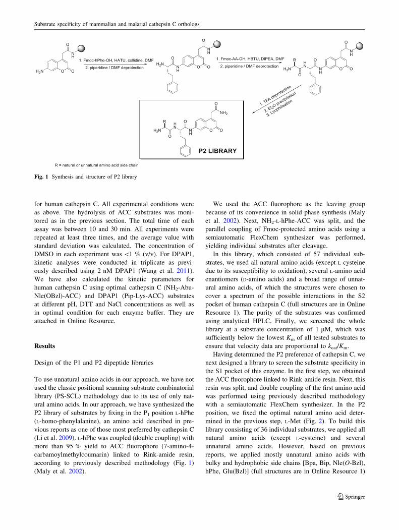
P2 library of substrates by fixing in the P1 position L-hPhe
(L-homo-phenylalanine), an amino acid described in pre-
vious reports as one of those most preferred by cathepsin C
(Li et al. 2009). L-hPhe was coupled (double coupling) with
more than 95 % yield to ACC fluorophore (7-amino-4-
carbamoylmethylcoumarin) linked to Rink-amide resin,
according to previously described methodology (Fig. 1)
(Maly et al. 2002).
We used the ACC fluorophore as the leaving group
because of its convenience in solid phase synthesis (Maly
et al. 2002). Next, NH2-L-hPhe-ACC was split, and the
parallel coupling of Fmoc-protected amino acids using a
semiautomatic FlexChem synthesizer was performed,
yielding individual substrates after cleavage.
In this library, which consisted of 57 individual sub-
strates, we used all natural amino acids (except L-cysteine
due to its susceptibility to oxidation), several L-amino acid
enantiomers (D-amino acids) and a broad range of unnat-
ural amino acids, of which the structures were chosen to
cover a spectrum of the possible interactions in the S2
pocket of human cathepsin C (full structures are in Online
Resource 1). The purity of the substrates was confirmed
using analytical HPLC. Finally, we screened the whole
library at a substrate concentration of 1 lM, which was
sufficiently below the lowest Km of all tested substrates to
ensure that velocity data are proportional to kcat/Km.
Having determined the P2 preference of cathepsin C, we
next designed a library to screen the substrate specificity in
the S1 pocket of this enzyme. In the first step, we obtained
the ACC fluorophore linked to Rink-amide resin. Next, this
resin was split, and double coupling of the first amino acid
was performed using previously described methodology
with a semiautomatic FlexChem synthesizer. In the P2
position, we fixed the optimal natural amino acid deter-
mined in the previous step, L-Met (Fig. 2). To build this
library consisting of 36 individual substrates, we applied all
natural amino acids (except L-cysteine) and several
unnatural amino acids. However, based on previous
reports, we applied mostly unnatural amino acids with
bulky and hydrophobic side chains [Bpa, Bip, Nle(O-Bzl),
hPhe, Glu(Bzl)] (full structures are in Online Resource 1)
Fig. 1 Synthesis and structure of P2 library
Substrate specificity of mammalian and malarial cathepsin C orthologs
123
Page 6
(Li et al. 2009). Finally, all the substrates were cleaved
from the resin and purified using preparative HPLC and
analyzed using analytical HPLC.
Exactly as described above, we determined the pre-
liminary conditions (Km) of all cleaved substrates and
performed the parallel screening of the library at a final
substrate concentration of 1.0 lM.
Substrate specificity analysis of human and bovine
cathepsin C and DPAP1
Bovine cathepsin C is often used to mimic human cathepsin
C. We applied our library to compare both enzymes directly
in terms of substrate specificity. The analysis of the S1 pocket
preferences of mammalian cathepsin C demonstrates that
these enzymes recognize exactly the same residues at almost
the same level (Fig. 3a, b). The most preferred amino acids
can be assigned to one of the following groups: hydrophobic
[Nle(O-Bzl), Bpa, Bip, Tyr(Bzl), Glu(Bzl), hPhe], basic (Arg,
Lys) or aliphatic (Nva, Met, Leu). This finding demonstrates
that the S1 pocket size is much larger than the natural amino
acids and can very easily accommodate more bulky residues,
clearly confirming a conserved level of structure organization
of these enzymes.
This observation is also in agreement with data pub-
lished by Li et al. (2009), which demonstrated that some
unnatural amino acids (L-hPhe or L-Bpa) bind much better
than L-Phe (Fig. 3). Among natural amino acids, the ones
best tolerated by human and bovine cathepsin C were
Arg, Lys, Gln and Met. These data are in quite good
agreement with previously published data by Wang et al.
(2011), who applied a combinatorial library approach.
However, it needs to be underlined that the best natural
amino acid (Arg) was recognized only at *20 % com-
pared to the best unnatural derivative from our library
(L-Nle(O-Bzl)).
Malarial DPAP1 substrate specificity in the S1 pocket is
much more restricted compared to the mammalian ortho-
logs tested here. DPAP1 preferentially recognizes and
hydrolyzes such amino acids like Nle(O-Bzl), Lys,
Glu(Bzl), Arg, Met, Gln, Thr, hPhe and Nva (Fig. 3c).
These data are in quite good agreement with the substrate
specificity of mammalian orthologs. The most striking
difference can be observed in the case of large and bulky
unnatural amino acids (Bip, Bpa, Cha). DPAP1 very min-
imally hydrolyzes these derivatives, which clearly dem-
onstrates the difference in S1 pocket preferences between
mammalian and parasite orthologs.
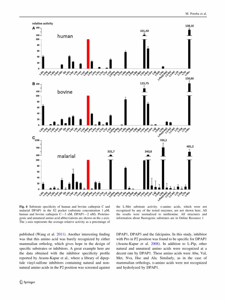
The analysis of the S2 pocket of both mammalian
orthologs demonstrates, similar to the case of the S1
pocket, a very high level of agreement in the activity and
tolerance of amino acids (Fig. 4a, b).
The amino acids best recognized by both enzymes were
Abu, Hse, Met, Nva, Nle and Ala. All these amino acids are
rather small and have aliphatic chains. Bulky and hydro-
phobic side chains were practically unrecognized by either
enzyme in the S2 pocket. No activity of human cathepsin C
toward amino acids with basic side chains (Arg, Lys, Orn,
Dap and Dab) was observed, in agreement with previously
published data (Wang et al. 2011). Additionally, none of
the D-amino acids was recognized and hydrolyzed by
mammalian orthologs, which indicates the high stereo-
specificity of both enzymes around the S2 binding pocket
(structures in Online Resource 1). Although we found
slightly different overall ratios from the report by Wang
et al. (2011) with regard to natural amino acids, our values
are in quite good agreement in terms of overall amino acid
preferences. The differences observed can result from
various screening conditions or the composition of the
library.
The substrate specificity analysis of malarial DPAP1
demonstrates a very striking difference from mammalian
Fig. 2 Synthesis and structure of the P1 library
M. Poreba et al.
123
Page 7
orthologs. L-Pip, a six-membered cyclic unnatural homo-
log of proline with one extra methylene residue, was the
best tolerated in the S2 pocket. Substrates with this residue
were recognized at least twice as well by DPAP1 than the
second best-recognized amino acid, Abu, and almost two-
and-a-half times better than the best-recognized natural
amino acid, Val. Data obtained for natural amino acids are
also in quite good agreement with these previously
Fig. 3 Substrate specificity of human and bovine cathepsin C and
malarial DPAP1 in the S1 pocket (substrate concentration 1 lM,
human and bovine cathepsin C—3 nM, rDPAP1—2 nM). Proteino-
genic and unnatural amino acid abbreviations are shown on the x-axis.
The y-axis represents the average relative activity as a percentage of
the L-Arg substrate activity. All the results were normalized to L-Arg.
All structures and information about fluorogenic substrates are in
Online Resource 1
Substrate specificity of mammalian and malarial cathepsin C orthologs
123
Page 8
published (Wang et al. 2011). Another interesting finding
was that this amino acid was barely recognized by either
mammalian ortholog, which gives hope in the design of
specific substrates or inhibitors. A great example here are
the data obtained with the inhibitor specificity profile
reported by Arastu-Kapur et al., where a library of dipep-
tide vinyl-sulfone inhibitors containing natural and non-
natural amino acids in the P2 position was screened against
DPAP1, DPAP3 and the falcipains. In this study, inhibitor
with Pro in P2 position was found to be specific for DPAP1
(Arastu-Kapur et al. 2008). In addition to L-Pip, other
natural and unnatural amino acids were recognized at a
decent rate by DPAP1. These amino acids were Abu, Val,
Met, Nva, Hse and Ala. Similarly, as in the case of
mammalian orthologs, D-amino acids were not recognized
and hydrolyzed by DPAP1.
Fig. 4 Substrate specificity of human and bovine cathepsin C and
malarial DPAP1 in the S2 pocket (substrate concentration 1 lM,
human and bovine cathepsin C—3 nM, DPAP1—2 nM). Proteino-
genic and unnatural amino acid abbreviations are shown on the x-axis.
The y-axis represents the average relative activity as a percentage of
the L-Met substrate activity. D-amino acids, which were not
recognized by any of the tested enzymes, are not shown here. All
the results were normalized to methionine. All structures and
information about fluorogenic substrates are in Online Resource 1
M. Poreba et al.
123
Page 9
Detailed kinetic analysis of fluorogenic substrates
of human cathepsin C and DPAP1
In the first step of the analysis, we have focused on the
kinetic parameters (Km, kcat, kcat/Km) of human cathepsin C
for several substrates selected from the P1 and P2 libraries.
In the P1 library, we found that the highest enzyme effi-
ciency (kcat/Km) for human cathepsin C (5.3 9
106 s-1 M-1) was observed with Met-Nle(O-Bzl)-ACC,
which is in good agreement with the library screening data.
Very good kinetic values were also observed for the other
dipeptide substrates with bulky and hydrophobic unnatural
amino acids in the P1 position, such as Met-Glu(Bzl), Met-
Bip and Met-Bpa (Table 1).
Analysis of the kinetic parameters for human cathepsin
C in the P2 position demonstrates a high preference for
small aliphatic side chains. The highest enzyme efficiency
(kcat/Km), 1.8 9 106 s-1 M-1 (human) was found with an
unnatural amino acid derivative, Abu (Table 2 ). Other
preferred derivatives in the S2 pocket were Hse and Met.
All these values are in very good agreement with the
library screening data (Fig. 4).
To validate the substrate preferences in the P1 and P2
pockets of human cathepsin C, we designed and synthe-
sized fluorogenic dipeptide substrates that contain the best-
recognized amino acids, Nle(O-Bzl) in the P1 position and
Abu in the P2 position. In parallel, we synthesized and
routinely used a commercial substrate for mammalian
cathepsin C, which has phenylalanine in the P1 position
and glycine in the P2 position. Next, we directly compared
all of the kinetic parameters of both substrates and found
that the substrate we designed with unnatural amino acids
Table 1 Kinetic parameters (Km, kcat, kcat/Km) of selected substrates for human cathepsin C from the P1 library (NH2-L-Met-X-ACC). Each
measurement was repeated at least three times
NH2-Met-X-ACC Human cathepsin C
X: code/name X: structure Km lM kcat s-1 kcat/Km 9 105 s-1 M-1
Arg (arginine)
NH
NHH2N
7.49 ± 0.61 12.80 ± 0.08 17.2 ± 0.15
hPhe (homophenylalanine) 3.46 ± 0.09 5.63 ± 0.27 16.1 ± 0.15
Bip (biphenylalanine) 4.11 ± 0.13 6.28 ± 0.61 14.7 ± 0.33
Bpa (4-benzoyl-phenylalanine)
O
5.43 ± 0.25 15.56 ± 0.87 28.3 ± 0.58
Nle(O-Bzl) (6-benzyloxynorleucine)
O
1.68 ± 0.11 9.27 ± 0.35 53.2 ± 2.55
Glu(Bzl) (glutamic acid benzyl ester)
OO
2.18 ± 0.05 6.05 ± 0.26 27.8 ± 0.18
Substrate specificity of mammalian and malarial cathepsin C orthologs
123
Page 10
is more than 400 times better than Gly-Phe in terms of kcat/
Km values. A further analysis of the kinetic parameters
demonstrates that the difference between both substrates is
primarily seen in the Km value, which is significantly
higher for Gly-Phe (Table 3). Interestingly, the turnover
number (kcat) did not differ as greatly between these sub-
strates (approximately, five times greater for our substrate
than for Gly-Phe).
Finally, we compared the kinetic parameters between
human and parasite orthologs with the hope of finding
significant differences that would allow us to differentiate
the enzymes. Our primary aim was to find a sequence that
would be very efficiently recognized by DPAP1, but sig-
nificantly less so by the human ortholog. Sequence align-
ment analysis reveals that parasitic DPAP1 shares 24 and
26 % identity with human and bovine orthologs, respec-
tively. This was reflected in significant differences in the
library screening data, where the malarial parasite ortholog
had substrate specificity distinct from mammalian ortho-
logs. The most striking difference was observed in the P2
position, where the unnatural Pip derivative was recog-
nized very efficiently by DPAP1, but minimally by
Table 2 Kinetic parameters (Km, kcat, kcat/Km) of selected substrates for human cathepsin C from the P2 library (NH2-X-L-hPhe-ACC)
NH2-X-hPhe-ACC Human cathepsin C
X: code/name X: structure Km lM kcat s-1 kcat/Km 9 105 s-1 M-1
Ala (alanine) CH324.8 ± 1.78 13.3 ± 0.88 5.44 ± 0.29
Leu (leucine) CH3
CH3
19.6 ± 1.46 5.00 ± 0.11 2.44 ± 0.04
Met (methionine)S
CH33.6 ± 0.12 5.59 ± 0.14 15.2 ± 0.75
Nle (norleucine) CH39.9 ± 0.78 8.35 ± 0.31 8.16 ± 0.16
Hse (homoserine) OH 8.8 ± 0.66 11.4 ± 0.45 12.5 ± 0.12
Abu (homoalanine) CH36.3 ± 0.58 10.4 ± 0.68 17.7 ± 0.82
Each experiment was repeated at least three times
Table 3 Kinetic parameters (Km, kcat, kcat/Km) of the best substrate identified and a commercial substrate for human cathepsin C. Each
experiment was repeated at least three times
ACC substrate NH2-Abu-Nle(O-Bzl)-ACC NH2-Gly-Phe-ACC
Structure
ACCNH
HN
H2N
O
O
H3C
O
ACCNH
HN
H2N
O
O
Km lM 1.88 ± 0.11 167.2 ± 5.3
kcat s-1 17.8 ± 0.56 3.66 ± 0.11
kcat/Km 9 105 s-1 M-1 94.5 ± 0.34 0.22 ± 0.013
M. Poreba et al.
123
Page 11
mammalian orthologs. The comparison of kinetic param-
eters for the P2 library sequence Pip-hPhe confirms the
library-based data. DPAP1 hydrolyzed this sequence three
times more efficiently (kcat/Km—3.22 9 105 s-1 M-1)
than the human ortholog (kcat/Km—1.16 9 105 s-1 M-1)
(Table 4). To further improve the efficiency of the sub-
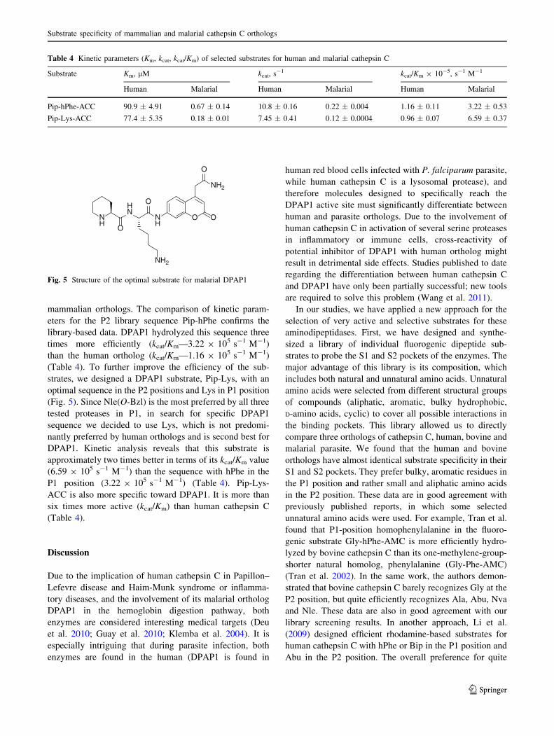
strates, we designed a DPAP1 substrate, Pip-Lys, with an
optimal sequence in the P2 positions and Lys in P1 position
(Fig. 5). Since Nle(O-Bzl) is the most preferred by all three
tested proteases in P1, in search for specific DPAP1
sequence we decided to use Lys, which is not predomi-
nantly preferred by human orthologs and is second best for
DPAP1. Kinetic analysis reveals that this substrate is
approximately two times better in terms of its kcat/Km value
(6.59 9 105 s-1 M-1) than the sequence with hPhe in the
P1 position (3.22 9 105 s-1 M-1) (Table 4). Pip-Lys-
ACC is also more specific toward DPAP1. It is more than
six times more active (kcat/Km) than human cathepsin C
(Table 4).
Discussion
Due to the implication of human cathepsin C in Papillon–
Lefevre disease and Haim-Munk syndrome or inflamma-
tory diseases, and the involvement of its malarial ortholog
DPAP1 in the hemoglobin digestion pathway, both
enzymes are considered interesting medical targets (Deu
et al. 2010; Guay et al. 2010; Klemba et al. 2004). It is
especially intriguing that during parasite infection, both
enzymes are found in the human (DPAP1 is found in
human red blood cells infected with P. falciparum parasite,
while human cathepsin C is a lysosomal protease), and
therefore molecules designed to specifically reach the
DPAP1 active site must significantly differentiate between
human and parasite orthologs. Due to the involvement of
human cathepsin C in activation of several serine proteases
in inflammatory or immune cells, cross-reactivity of
potential inhibitor of DPAP1 with human ortholog might
result in detrimental side effects. Studies published to date
regarding the differentiation between human cathepsin C
and DPAP1 have only been partially successful; new tools
are required to solve this problem (Wang et al. 2011).
In our studies, we have applied a new approach for the
selection of very active and selective substrates for these
aminodipeptidases. First, we have designed and synthe-
sized a library of individual fluorogenic dipeptide sub-
strates to probe the S1 and S2 pockets of the enzymes. The
major advantage of this library is its composition, which
includes both natural and unnatural amino acids. Unnatural
amino acids were selected from different structural groups
of compounds (aliphatic, aromatic, bulky hydrophobic,
D-amino acids, cyclic) to cover all possible interactions in
the binding pockets. This library allowed us to directly
compare three orthologs of cathepsin C, human, bovine and
malarial parasite. We found that the human and bovine
orthologs have almost identical substrate specificity in their
S1 and S2 pockets. They prefer bulky, aromatic residues in
the P1 position and rather small and aliphatic amino acids
in the P2 position. These data are in good agreement with
previously published reports, in which some selected
unnatural amino acids were used. For example, Tran et al.
found that P1-position homophenylalanine in the fluoro-
genic substrate Gly-hPhe-AMC is more efficiently hydro-
lyzed by bovine cathepsin C than its one-methylene-group-
shorter natural homolog, phenylalanine (Gly-Phe-AMC)
(Tran et al. 2002). In the same work, the authors demon-
strated that bovine cathepsin C barely recognizes Gly at the
P2 position, but quite efficiently recognizes Ala, Abu, Nva
and Nle. These data are also in good agreement with our
library screening results. In another approach, Li et al.
(2009) designed efficient rhodamine-based substrates for
human cathepsin C with hPhe or Bip in the P1 position and
Abu in the P2 position. The overall preference for quite
Table 4 Kinetic parameters (Km, kcat, kcat/Km) of selected substrates for human and malarial cathepsin C
Substrate Km, lM kcat, s-1 kcat/Km 9 10-5, s-1 M-1
Human Malarial Human Malarial Human Malarial
Pip-hPhe-ACC 90.9 ± 4.91 0.67 ± 0.14 10.8 ± 0.16 0.22 ± 0.004 1.16 ± 0.11 3.22 ± 0.53
Pip-Lys-ACC 77.4 ± 5.35 0.18 ± 0.01 7.45 ± 0.41 0.12 ± 0.0004 0.96 ± 0.07 6.59 ± 0.37
O O
NH2
O
NH
HN
NH
O
O
NH2
Fig. 5 Structure of the optimal substrate for malarial DPAP1
Substrate specificity of mammalian and malarial cathepsin C orthologs
123
Page 12
broad substrate specificity in the P1 position of mammalian
orthologs and rather narrow substrate specificity in the P2
position can be explained by the analysis of available
crystal structures (Turk et al. 2001; Dahl et al. 2001;
Molgaard et al. 2007). The S1 pocket is located on the
surface of the enzyme and is exposed to the solvent. Its
large size demonstrates that it preferentially accommodates
very bulky and hydrophobic residues, such as these found
in our studies. We assume that the large size of the residues
results in more interactions with the surface of the enzyme
and thus increases its affinity to the substrate. This
hypothesis can by confirmed by the kinetic data we
obtained for NH2-Abu-Nle(O-Bzl)-ACC and NH2-Gly-
Phe-ACC, which differ very significantly in terms of the
kcat/Km value, but not as significantly in the kcat value
(Table 3). The determining factor here is the Km value,
which reflects, to a significant extent, the binding affinity of
the substrate. Further analysis of the human cathepsin C
crystal structure demonstrates that the S2 pocket is rather
long and narrow. This explains why rather small and ali-
phatic amino acids, such as Ala, Abu, Met, Hse, Nle or
Nva, are preferred in the P2 position. The structure of the
optimal substrate NH2-Abu-Nle(O-Bzl)-ACC can also be
used in further studies for the design of inhibitors or
activity-based probes for cathepsin C. The observed sub-
strate specificity also demonstrates that bovine cathepsin C,
which is much more readily available and less expensive,
can substitute for the human ortholog in routine studies on
new chemical tools.
In the studies with malarial DPAP1, we provide new
evidence that this enzyme quite significantly differs in
substrate specificity from mammalian orthologs. In contrast
to human and bovine cathepsin C, DPAP1 demonstrates
very little preference in the P1 position toward bulky and
hydrophobic amino acids of phenylalanine like structure
and efficiently hydrolyzes amino acids with long aliphatic
side chains with aromatic group at the end (Nle(O-Bzl),
Glu(Bzl)) or with basic side chains, Arg and Lys. As a
result of library screening studies, we have found that
DPAP1 preferentially cleaves the unnatural cyclic amino
acid Pip in the P2 position, which is barely tolerated by
human orthologs. These results allowed us to design an
optimal DPAP1 substrate, Pip-Lys-ACC, for which the kcat/
Km values were calculated. With kcat/Km values equal to
6.59 9 105 s-1 M-1 (DPAP1) and 0.96 9 105 s-1 M-1
(cathepsin C), we have demonstrated that this substrate is
quite selective toward the malarial ortholog. On the other
hand, this might seem quite low given the huge difference
in the initial screen. The reason is that DPAP1 is 30–100
times less efficient than cathepsin C using natural amino
acid dipeptide substrates, as demonstrated by Wang et al.
(2011). It is then apparent that the specificity has been
shifted *100-fold in favor of DPAP1.
The analyses presented here focused on the S1 and S2
pockets, but it is also likely that enhanced interactions with
natural peptide and protein substrates utilize interactions at
the C-terminal side (S0 side) of the scissile bond. Unfor-
tunately, there is no currently available technology to probe
the S0 site of aminopeptidases with synthetic substrates.
In conclusion, we have designed and tested for the first
time a tailored fluorogenic substrate library containing nat-
ural and unnatural amino acids to define the specificity of the
active site of human, bovine and malarial cathepsin C. Our
results clearly demonstrate very significant differences in the
preference for binding of both natural and unnatural amino
acids to the S1 and S2 pockets between mammalian and
malarial orthologs. The catalytic rates of hydrolysis for
substrates with unnatural amino acids designed based on
library screening were significantly improved, proving the
utility of this approach. For example, for human cathepsin C,
we have obtained a substrate that is more than 400 times
better in terms of kcat/Km than the commonly used com-
mercial substrate. The observed significant differences in
terms of the structure of the substrates between human and
malarial orthologs can be used for the design of specific
inhibitors or activity-based probes (ABPs). Finally, the
methodology used here provides the proof of concept for the
application of this library in screening other types of dia-
minopeptidases or for the direct comparison of enzyme
orthologs from different organisms.
Acknowledgments This work was supported by grant number NN
302 276437. Marcin Drag and Marcin Poreba are grateful to the
Foundation for Polish Science for support. The research was sup-
ported by Wroclaw Research Center EIT? under the project Bio-
technologies and Advanced Medical Technologies (‘‘BioMed’’)
(POIG 01.01.02-02-003/08-00) financed by the European Regional
Development Fund (Operational Programme Innovative Economy,
1.1.2).
Conflict of interest The authors declare that they have no conflict
of interest.
Open Access This article is distributed under the terms of the
Creative Commons Attribution License which permits any use, dis-
tribution, and reproduction in any medium, provided the original
author(s) and the source are credited.
References
Adkison AM, Raptis SZ, Kelley DG, Pham CT (2002) Dipeptidyl
peptidase I activates neutrophil-derived serine proteases and
regulates the development of acute experimental arthritis. J Clin
Invest 109(3):363–371. doi:10.1172/JCI13462
Arastu-Kapur S, Ponder EL, Fonovic UP, Yeoh S, Yuan F, Fonovic
M, Grainger M, Phillips CI, Powers JC, Bogyo M (2008)
Identification of proteases that regulate erythrocyte rupture by
the malaria parasite Plasmodium falciparum. Nat Chem Biol
4(3):203–213. doi:10.1038/nchembio.70
M. Poreba et al.
123
Page 13
Dahl SW, Halkier T, Lauritzen C, Dolenc I, Pedersen J, Turk V, Turk
B (2001) Human recombinant pro-dipeptidyl peptidase I
(cathepsin C) can be activated by cathepsins L and S but not
by autocatalytic processing. Biochemistry 40(6):1671–1678 pii:
bi001693z
Deu E, Leyva MJ, Albrow VE, Rice MJ, Ellman JA, Bogyo M (2010)
Functional studies of Plasmodium falciparum dipeptidyl amino-
peptidase I using small molecule inhibitors and active site
probes. Chem Biol 17(8):808–819. doi:10.1016/j.chembiol.2010.
06.007
Gocheva V, Joyce JA (2007) Cysteine cathepsins and the cutting edge
of cancer invasion. Cell Cycle 6(1):60–64
Guay D, Beaulieu C, Percival MD (2010) Therapeutic utility and
medicinal chemistry of cathepsin C inhibitors. Curr Top Med
Chem 10(7):708–716
Hart TC, Hart PS, Bowden DW, Michalec MD, Callison SA, Walker
SJ, Zhang Y, Firatli E (1999) Mutations of the cathepsin C gene
are responsible for Papillon–Lefevre syndrome. J Med Genet
36(12):881–887
Hart TC, Hart PS, Michalec MD, Zhang Y, Firatli E, Van Dyke TE,
Stabholz A, Zlotogorski A, Shapira L, Soskolne WA (2000)
Haim–Munk syndrome and Papillon–Lefevre syndrome are
allelic mutations in cathepsin C. J Med Genet 37(2):88–94
Ken McDonald J, Callahan PX, Ellis S (1972) [22] Preparation and
specificity of dipeptidyl aminopeptidase I. Methods Enzymol
25C:272–281. doi:10.1016/S0076-6879(72)25024-8
Klemba M, Gluzman I, Goldberg DE (2004) A Plasmodium
falciparum dipeptidyl aminopeptidase I participates in vacuolar
hemoglobin degradation. J Biol Chem 279(41):43000–43007.
doi:10.1074/jbc.M408123200
Krugliak M, Zhang J, Ginsburg H (2002) Intraerythrocytic Plasmo-
dium falciparum utilizes only a fraction of the amino acids
derived from the digestion of host cell cytosol for the biosyn-
thesis of its proteins. Mol Biochem Parasitol 119(2):249–256
Kudo M, Melton AC, Chen C, Engler MB, Huang KE, Ren X, Wang
Y, Bernstein X, Li JT, Atabai K, Huang X, Sheppard D (2012)
IL-17A produced by alphabeta T cells drives airway hyper-
responsiveness in mice and enhances mouse and human airway
smooth muscle contraction. Nat Med 18(4):547–554. doi:10.
1038/nm.2684
Li J, Petrassi HM, Tumanut C, Masick BT, Trussell C, Harris JL
(2009) Substrate optimization for monitoring cathepsin C
activity in live cells. Bioorg Med Chem 17(3):1064–1070.
doi:10.1016/j.bmc.2008.02.002
Maly DJ, Leonetti F, Backes BJ, Dauber DS, Harris JL, Craik CS,
Ellman JA (2002) Expedient solid-phase synthesis of fluorogenic
protease substrates using the 7-amino-4-carbamoylmethylcou-
marin (ACC) fluorophore. J Org Chem 67(3):910–915 jo016140o
McGuire MJ, Lipsky PE, Thiele DL (1993) Generation of active
myeloid and lymphoid granule serine proteases requires pro-
cessing by the granule thiol protease dipeptidyl peptidase I.
J Biol Chem 268(4):2458–2467
Molgaard A, Arnau J, Lauritzen C, Larsen S, Petersen G, Pedersen J
(2007) The crystal structure of human dipeptidyl peptidase I
(cathepsin C) in complex with the inhibitor Gly-Phe-CHN2.
Biochem J 401(3):645–650. doi:10.1042/BJ20061389
Pham CT, Ley TJ (1999) Dipeptidyl peptidase I is required for the
processing and activation of granzymes A and B in vivo. Proc
Natl Acad Sci USA 96(15):8627–8632
Ragheb D, Dalal S, Bompiani KM, Ray WK, Klemba M (2011)
Distribution and biochemical properties of an M1-family amino-
peptidase in Plasmodium falciparum indicate a role in vacuolar
hemoglobin catabolism. J Biol Chem 286(31):27255–27265.
doi:10.1074/jbc.M111.225318
Tallan HH, Jones ME, Fruton JS (1952) On the proteolytic enzymes
of animal tissues. X. Beef spleen cathepsin C. J Biol Chem
194(2):793–805
Tanaka TQ, Deu E, Molina-Cruz A, Ashburne MJ, Ali O, Suri A,
Kortagere S, Bogyo M, Williamson KC (2013) Plasmodium
dipeptidyl aminopeptidases as malaria transmission-blocking
drug targets. Antimicrob Agents Chemother 57(10):4645–4652.
doi:10.1128/AAC.02495-12
Tran TV, Ellis KA, Kam CM, Hudig D, Powers JC (2002) Dipeptidyl
peptidase I: importance of progranzyme activation sequences,
other dipeptide sequences, and the N-terminal amino group of
synthetic substrates for enzyme activity. Arch Biochem Biophys
403(2):160–170
Turk D, Janjic V, Stern I, Podobnik M, Lamba D, Dahl SW, Lauritzen
C, Pedersen J, Turk V, Turk B (2001) Structure of human
dipeptidyl peptidase I (cathepsin C): exclusion domain added to
an endopeptidase framework creates the machine for activation
of granular serine proteases. EMBO J 20(23):6570–6582. doi:10.
1093/emboj/20.23.6570
Wang F, Krai P, Deu E, Bibb B, Lauritzen C, Pedersen J, Bogyo M,
Klemba M (2011) Biochemical characterization of Plasmodium
falciparum dipeptidyl aminopeptidase 1. Mol Biochem Parasitol
175(1):10–20. doi:10.1016/j.molbiopara.2010.08.004
Yuan F, Verhelst SH, Blum G, Coussens LM, Bogyo M (2006) A
selective activity-based probe for the papain family cysteine
protease dipeptidyl peptidase I/cathepsin C. J Am Chem Soc
128(17):5616–5617. doi:10.1021/ja060835v
Substrate specificity of mammalian and malarial cathepsin C orthologs
123