Journal of Medical Genetics (1970). 7, 417. An Unusual Chromosomal Aberration in a Case of Chediak-Higashi Syndrome BURHAN SAY, ERGUL TUNCBILEK, BEHICE YAMAK, and SEVIM BALCI From the Department of Pediatrics and the Clinical Genetics Laboratory, Hacettepe University, Hacettepe Children's Hospital, Ankara, Turkey Chediak-Higashi syndrome which appears to be a lysosomal disease (Douglas and Fudenberg, 1969; White, 1966) is characterized by partial albinism, photophobia, recurrent infections, hepatospleno- megaly, and a distinctive leucocyte anomaly (Chediak, 1952; Higashi, 1954). Available genetic and fine structural studies suggest that the condition is inherited as an autosomal recessive trait (Sadan et al., 1965; Douglas, Blume, and Wolff, 1969). Chromosome studies have been done in a few cases, but as expected have not shown any significant numerical chromosomal abnormalities (Kritzler et al., 1964; Rosenszajn et al., 1969). The condition does not seem to be confined to a particular popula- tion group, since patients with this syndrome have been reported from various parts of the world. The purpose of this communication is to report a case of Chediak-Higashi syndrome from Turkey, in which chromosome studies revealed an unexpected and unusual abnormality in the form of partial monosomy for a G group chromosome (45,XY,G - 46,XY/46,XY,Gr). Case Report A 3-year-old boy was referred to us by his local physician with a history of recurrent infections of un- known aetiology (Fig. 1). His past history revealed that he had had an infection associated with a rash which was diagnosed as scarlet fever approximately one year previously, since when he had been treated for multiple episodes of diarrhoea and tonsillitis. Two weeks before his referral he was found to have enlarged submandibular and cervical glands associated with high fever and weight loss. The mother was 29 and the father 36 years of age, and there was no consanguinity between them. His 3- month-old brother was reported to be in good health. A detailed family history revealed the presence of other members (with and without fair hair) who had photo- phobia. Physical examination. The patient's height was 92 cm. (10th centile), weight 14-5 kg. (50th centile), and head circumference 49 cm. (75th centile). He was pale, and acutely ill. His hair was light-coloured, in contrast to his parents' dark hair. The skin was dry, with slight desquamation, and there was horizontal nystagmus and photophobia in both eyes, which also had an anti- mongoloid slant. Other pertinent clinical findings in- cluded an enlarged left submandibular lymph node (2 x 1 cm. in size), and multiple slightly enlarged lymph nodes over the axillary and inguinal areas. The liver was palpable 2-5 and the spleen 3 cm. below the costal margins. Chest x-ray revealed pneumonia. Laboratory findings. Routine haematological studies showed the patient's haemoglobin to be 7-8 g./ 100 ml., haematocrit 26%, WBC 8600/cu. mm., and reticulocyte count, 10-2%. Peripheral smears revealed hypochromia, anisocytosis, polychromasia, and poikilo- cytosis, and there were abundant thrombocytes. The most interesting finding was the presence of discrete Received 14 January 1970. 8+ FIG. 1. The patient. 417 on 18 July 2018 by guest. Protected by copyright. http://jmg.bmj.com/ J Med Genet: first published as 10.1136/jmg.7.4.417 on 1 December 1970. Downloaded from
Transcript
Journal of Medical Genetics (1970). 7, 417.
An Unusual Chromosomal Aberration in a Case ofChediak-Higashi Syndrome
BURHAN SAY, ERGUL TUNCBILEK, BEHICE YAMAK, and SEVIM BALCIFrom the Department of Pediatrics and the Clinical Genetics Laboratory, Hacettepe University,
Hacettepe Children's Hospital, Ankara, Turkey
Chediak-Higashi syndrome which appears to be alysosomal disease (Douglas and Fudenberg, 1969;White, 1966) is characterized by partial albinism,photophobia, recurrent infections, hepatospleno-megaly, and a distinctive leucocyte anomaly(Chediak, 1952; Higashi, 1954). Available geneticand fine structural studies suggest that the conditionis inherited as an autosomal recessive trait (Sadan etal., 1965; Douglas, Blume, and Wolff, 1969).Chromosome studies have been done in a few cases,but as expected have not shown any significantnumerical chromosomal abnormalities (Kritzler etal., 1964; Rosenszajn et al., 1969). The conditiondoes not seem to be confined to a particular popula-tion group, since patients with this syndrome havebeen reported from various parts of the world.The purpose of this communication is to report a
case of Chediak-Higashi syndrome from Turkey, inwhich chromosome studies revealed an unexpectedand unusual abnormality in the form of partialmonosomy for a G group chromosome (45,XY,G -46,XY/46,XY,Gr).
Case ReportA 3-year-old boy was referred to us by his local
physician with a history of recurrent infections of un-known aetiology (Fig. 1). His past history revealedthat he had had an infection associated with a rash whichwas diagnosed as scarlet fever approximately one yearpreviously, since when he had been treated for multipleepisodes of diarrhoea and tonsillitis. Two weeks beforehis referral he was found to have enlarged submandibularand cervical glands associated with high fever and weightloss. The mother was 29 and the father 36 years of age,and there was no consanguinity between them. His 3-month-old brother was reported to be in good health. Adetailed family history revealed the presence of othermembers (with and without fair hair) who had photo-phobia.
Physical examination. The patient's height was92 cm. (10th centile), weight 14-5 kg. (50th centile), andhead circumference 49 cm. (75th centile). He was pale,and acutely ill. His hair was light-coloured, in contrastto his parents' dark hair. The skin was dry, with slightdesquamation, and there was horizontal nystagmus andphotophobia in both eyes, which also had an anti-mongoloid slant. Other pertinent clinical findings in-cluded an enlarged left submandibular lymph node(2 x 1 cm. in size), and multiple slightly enlarged lymphnodes over the axillary and inguinal areas. The liverwas palpable 2-5 and the spleen 3 cm. below the costalmargins. Chest x-ray revealed pneumonia.
Laboratory findings. Routine haematologicalstudies showed the patient's haemoglobin to be 7-8 g./100 ml., haematocrit 26%, WBC 8600/cu. mm., andreticulocyte count, 10-2%. Peripheral smears revealedhypochromia, anisocytosis, polychromasia, and poikilo-cytosis, and there were abundant thrombocytes. Themost interesting finding was the presence of discrete
Received 14 January 1970.
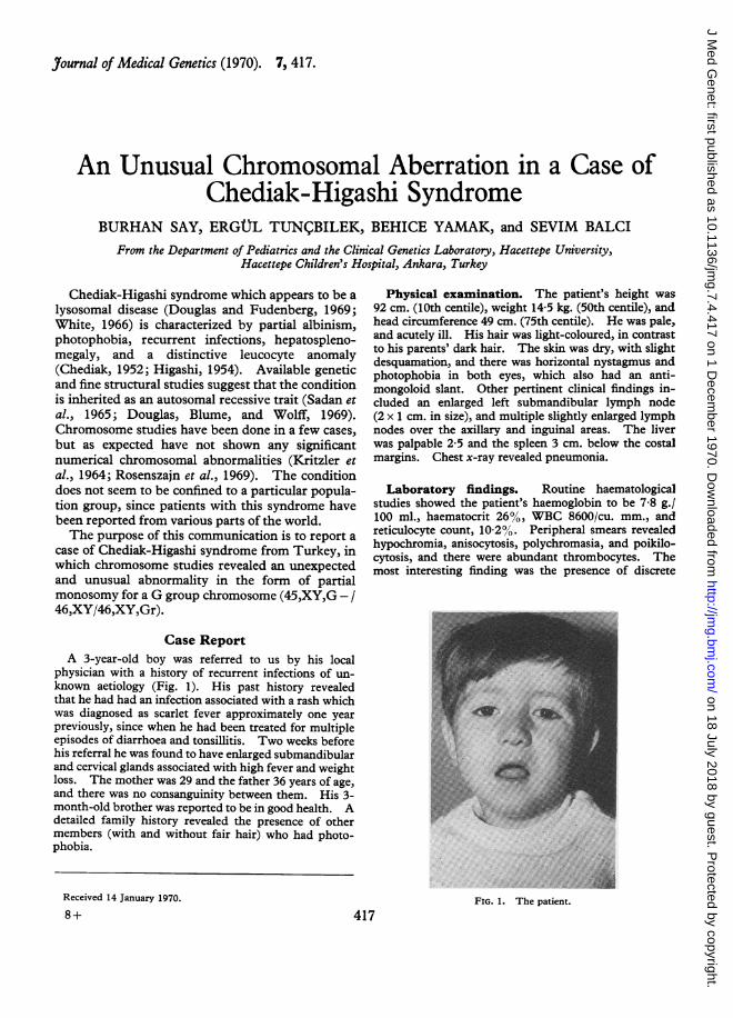
8+FIG. 1. The patient.
417
on 18 July 2018 by guest. Protected by copyright.
http://jmg.bm
j.com/
J Med G
enet: first published as 10.1136/jmg.7.4.417 on 1 D
inclusions could be seen in the blood smears obtainedfrom the parents, two grandparents, or a maternal aunt.
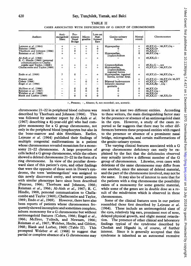
Cytogenetic investigations. Chromosome studiesof the patient and his parents were made using culturesof peripheral blood, for which a modification of Moor-head et al.'s method (1960) was used. The chromo-somes in 102 metaphase plates were counted andanalysed, and in 19 of them a small acrocentric chromo-some from the G group was missing. In all these latterplates, the Y chromosome could be clearly identified byits well-known morphological characteristics.
In a further 17 metaphase plates a minute ring chro-mosome, replacing a G chromosome, was present (Fig.4). The remaining 62 plates showed 46 chromosomes,none of which had any structural abnormalities. Anadditional 4 plates showed inconsistent abnormalities.Chromosome studies of the parents revealed no abnor-malities (Table I).
in man was considered to be incompatible withlife. The first case of complete monosomy for
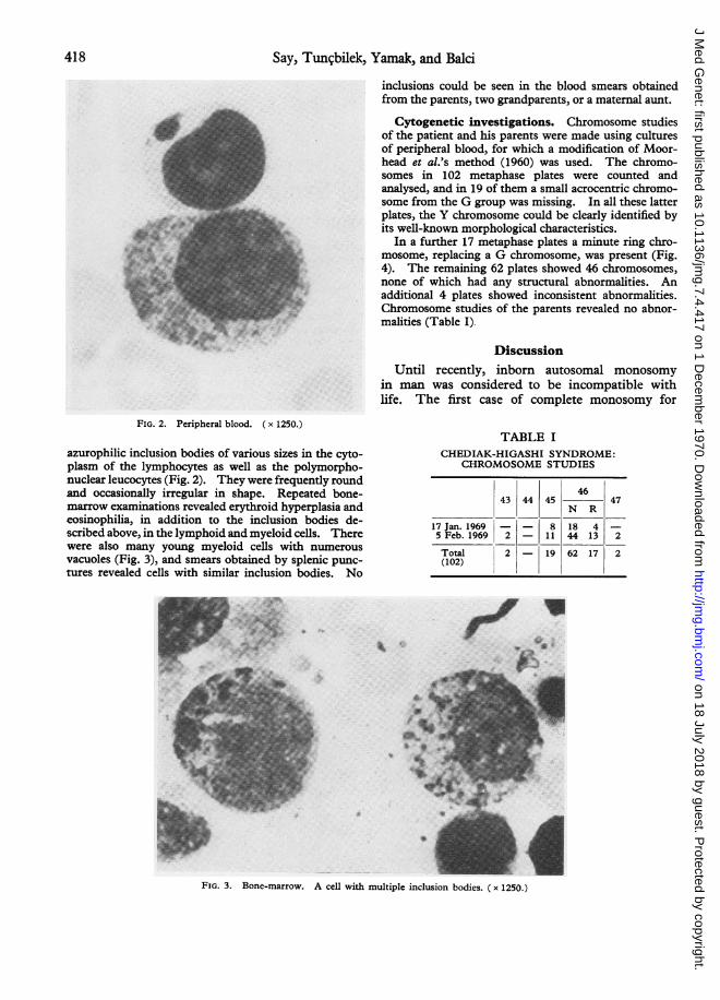
FIG. 2. Peripheral blood. ( x 1250.)
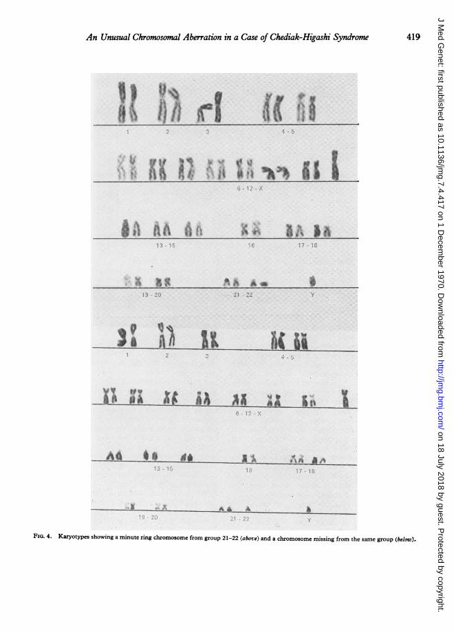
azurophilic inclusion bodies of various sizes in the cyto-plasm of the lymphocytes as well as the polymorpho-nuclear leucocytes (Fig. 2). They were frequently roundand occasionally irregular in shape. Repeated bone-marrow examinations revealed erythroid hyperplasia andeosinophilia, in addition to the inclusion bodies de-scribed above, in the lymphoid and myeloid cells. Therewere also many young myeloid cells with numerousvacuoles (Fig. 3), and smears obtained by splenic punc-tures revealed cells with similar inclusion bodies. No
±, Present; -, Absent; 0, not recorded; ace, acentric.
chromosome 21-22 in peripheral blood cultures wasdescribed by Thorburn and Johnson (1966). Thiswas followed by another report by Al-Aish et al.(1967) describing a 4j-year-old girl who had com-plete monosomy for a G group chromosome, notonly in the peripheral blood lymphocytes but also inthe bone-marrow and skin fibroblasts. Earlier,Lejeune et al. (1964) published their findings ofmultiple congenital malformations in a patientwhose chromosomes revealed mosaicism for a mono-somy 21-22 chromosome. A large proportion ofcells lacked a G group chromosome, while the othersshowed a deleted chromosome 21-22 in the form of aring chromosome. In view of the peculiar down-ward slant of this patient's eyes, and other findingsthat were the opposite of those seen in Down's syn-drome, the term 'antimongolism' was assigned tothis newly discovered entity, and several patientswith similar phenotype have since been described(Penrose, 1966; Thorburn and Johnson, 1966;Reisman et al., 1966; Al-Aish et al., 1967; R. C.Hindle, 1969, personal communication to Challa-combe and Taylor (1969); Challacombe and Taylor,1969; Endo et al., 1969). However, there have alsobeen reports of patients whose chromosomes fre-quently showed incomplete and sometimes complete,inborn monosomy for a G chromosome but withoutantimongoloid features (Cohen, 1966; Engel et al.,1966; McIlree, Tulloch, and Newsam, 1966;Reisman et al., 1967; Weleber, Hecht, and Giblett,1968; Blank and Lorber, 1969) (Table II). Thisprompted Weleber et al. (1968) to suggest thatpartial or complete absence of a G chromosome may
result in at least two different entities. Accordingto these writers, the main distinguishing factor maybe the presence or absence of an antimongoloid slantin the eyes. However, a study of the cases re-ported so far suggests that there may be other dif-ferences between these proposed entities with regardto the presence or absence of a prominent nasalbridge, micrognathia, and certain malformations ofthe genito-urinary system.The varying clinical features associated with a G
group chromosome deficiency can easily be ex-plained by the fact that the deficiencies observedmay actually involve a different member of the Ggroup of chromosomes. Likewise, even cases withdeletions of the same chromosome may differ fromone another, since the amount of deleted material,and the part of the chromosome involved, may not bethe same. It may also be of interest to note that forthe patients with a ring chromosome the possibilityexists of a monosomy for some genetic material,while some of the genes are in double dose as a re-sult of the misdivision which is observed in thesechromosomes.Some of the clinical features seen in our patient
resembled those first described by Lejeune et al.(1964). These include an antimongoloid slant ofthe eyes, relatively big ears, prominent root of nose,delayed physical growth, and slight mental retarda-tion. The presence of other clinical and laboratoryfindings typical of the syndrome described byChediak and Higashi is, of course, of furtherinterest. Since it is generally accepted that thissyndrome is inherited as an autosomal recessive
420
on 18 July 2018 by guest. Protected by copyright.
http://jmg.bm
j.com/
J Med G
enet: first published as 10.1136/jmg.7.4.417 on 1 D
An Unusual Chromosomal Aberration in a Case of Chediak-Higashi Syndrome
trait, and also that the chromosome studies of thesepatients have so far revealed no similar abnor-malities, it is quite likely that the unusual chromo-somal abnormality seen in our case is coincidental,though in a recent study chromatid and chromoso-mal breakages, elongated chromosomes, and hetero-pycnotic gaps were found (Rosenzajn et al., 1969).On the other hand, it should be emphasized thatonly a few cases of Chediak-Higashi syndrome havebeen investigated for chromosome abnormalities,and detailed reports on leucocyte morphology inpatients with antimongolism are lacking. For thisreason it is hoped that our paper will prompt otherworkers to report whether or not any relation existsbetween the loss of genetic material from a G groupchromosome and the clinical and laboratory findingsseen in patients with Chediak-Higashi syndrome.
Finally it should be remembered that patientswith this condition have a high incidence of lympho-reticular malignancy, and acquired monosomy Ghas been reported in acute myeloblastic leukaemia(Sandberg et al., 1964), in the terminal stage of theDi Guglielmo syndrome (Castoldi et al., 1968), andin a case of refractory sideroblastic anaemia withleukaemic transformation (Silberman and Krmpotic,1969). This indicates that follow-up of our casewould be of interest to see whether or not malignanttransformation occurs at an early age.
SummaryA 3-year-old boy with Chediak-Higashi syndrome
is described. Chromosomal analyses interestinglyrevealed a partial monosomy of the chromosome 21.The possible aetiological significance of this unusualchromosome aberration is discussed.
REFEENCES
Al-Aish, M. S., de la Cruz, F., Goldsmith, L. A., Volpe, J., Mella,G., and Robinson, J. C. (1967). Autosomal monosomy in man.Complete monosomy G (21-22) in a four-and-one-half-year-oldmentally retarded girl. New England Journal of Medicine, 277,777-784.
Blank, C. E., and Lorber, J. (1969). A patient with 45,XX,G -/46,XX,Gr mosaism. Journal of Medical Genetics, 6, 220-223.
Castoldi, G., Yam, L. T., Mitus, W. J.. and Crosby, W. H. (1968).Chromosomal studies in erythroleukemia and chronic erythremicmyelosis. Blood, 31, 202-215.
Challacombe, D. N., and Taylor, A. (1969). Monosomy for a Gautosome. Archives of Disease in Childhood, 44, 113-119.
Chediak, M. (1952). Nouvelle anomalie leucocytaire de caractereconstitutional et familial. Revue d'Hematologie, 7, 362-371.
Cohen, M. M. (1966). Chromosomal mosaicism associated with acase of cyclopia. journal of Pediatrics, 69, 793-798.
Douglas, S. D., Blume, R. S., and Wolff, S. M. (1969). Fine struc-tural studies of leukocytes from patients and heterozygotes withthe Chediak-Higashi syndrome. Blood, 33, 527-540.-, and Fudenberg, H. H. (1969). Chediak-Higashi syndrome.
Medical Clinics of North America, 53, 914-922.Endo, A., Yarnamoto, M., Watanabe, G.-I., Suziki, Y., and Sakai, K.
(1969). 'Antimongolism' syndrome. British Medical Journal, 4,148-149.
Engel, E., Hastings, C. P., Merrill, R. E., McFarland, B. S., andNance, W. E. (1966). Apparent cri-du-chat and 'antimongol-ism' in one patient. Lancet, 1, 1130-1132.
Higashi, 0. (1954). Congenital gigantism of peroxidase granules.The first case ever reported of qualitative abnormity of peroxi-dase. Tohoku_Journal of Experimental Medicine, 59, 315-332.
Kritzler, R. A., Temer, J. Y., Lindenbaum, J., Magidson, J.,Williams, R., Preisig, R., and Phillips, G. B. (1964). Chediak-Higashi syndrome. Cytologic and serum lipid observations in acase and family. American_Journal of Medicine, 36, 583-594.
Lejeune, J., Berger, R., Rethore, M. O., Archambault, L., Jer6me,H., Thieffry, S., Aicardi, J., Broyer, M., Lafourcade, J., Cruveiller,J., and Turpin, R. (1964). Monosomie partielle pour un petitacrocentrique. Comptes Rendus Hebdomadaires des Seances del'Acadlmie des Sciences, 259, 4187-4190.
McIlree, M. E., Tulloch, W. S., and Newsam, J. E. (1966). Studieson human meiotic chromosomes from testicular tissue. Lancet, 1,679-682.
Moorhead, P. S., Nowell, P. C., Mellman, W. J., Battips, D. M., andHungerford, D. A. (1960). Chromosome preparations of leuko-cytes from human peripheral blood. Experimental Cell Research,20, 613-616.
Penrose, L. S. (1966). Anti-mongolism. Lancet, 1, 497.Reisman, L. E., Darnell, A., Murphy, J. W., Hall, B., and Kasahara,
S. (1967). A child with partial deletion of a G-group autosome.American Journal of Diseases of Children, 114, 336-339.-, Kasahara, S., Chung, C. Y., Darnell, A., and Hall, B. (1966).
Anti-mongolism, studies in an infant with a partial monosomy ofthe 21 chromosome. Lancet, 1, 394-397.
Rosenszajn, A. L., Radnai, J., Tatarski, A., and Benderlei, A. (1969).Blood cell culture and chromosomal findings in Chediak-Higashisyndrome (Abst.). Israel_Journal of Medical Sciences, 5, 1087.
Sadan, N., Yaffe, D., Rozenszajn, L., Adar, H., Soroker, B., andEfrati, P. (1965). Cytochemical and genetic studies in four casesof Chediak-Higashi-Steinbrinck syndrome. Acta Haematologica,34, 20-29.
Sandberg, A. A., Ishihara, T., Kikuchi, Y., and Crosswhite, L. H.(1964). Chromosomal differences among the acute leukemias.Annals of the New York Academy of Sciences, 113, 663-716.
Silberman, S., and Krmpotic, E. (1969). Refractory anemia withleukemic transformation and chromosomal change. A case report.Acta Haematologica, 41, 186-192.
Thorburn, M. J., and Johnson, B. E. (1966). Apparent monosomyof a G autosome in a Jamaican infant. journal of Medical Genetics,3, 290-292.
Weleber, R. G., Hecht, F., and Giblett, E. R. (1968). Ring-Gchromosome, a new G-deletion syndrome ? American Journal ofDiseases of Children, 115, 489-493.
White, J. G. (1966). The Chediak-Higashi syndrome: a possiblelysosomal disease. Blood, 28, 143-156.
421
on 18 July 2018 by guest. Protected by copyright.
http://jmg.bm
j.com/
J Med G
enet: first published as 10.1136/jmg.7.4.417 on 1 D


![[Songbook] Djavan Vol. II [Almir Chediak]](https://static.documents.pub/doc/80x56/55cf8e1a550346703b8e8990/songbook-djavan-vol-ii-almir-chediak-5640e69478127.jpg)


