33
Update on Surgical Treatment of Pituitary Tumors Kristen Riley, MD, FACS Associate Professor, Division of Neurosurgery, Department of Surgery

Update on Surgical Treatment of Pituitary Tumors
Kristen Riley, MD, FACS
Associate Professor, Division of Neurosurgery, Department of Surgery

Pituitary Tumors • Pituitary adenoma: common intracranial
neoplasm
• Prevalence: 1 per 1000 individuals worldwide, incidence of newly diagnosed clinically active PA 1-4 per population of 100,000/yr.
• Most commonly benign, WHO Grade I, reported rate of 15% atypical, rarely see pituitary carcinoma

Classification of Pituitary tumors
• Size
• Functionality

Size
• Microadenoma vs Macroadenoma
– Less than 10mm=microadenoma
– Radiographic diagnosis
– Microadenomas only surgical if functional
– Microadenomas are not large enough to compress optic pathways

Macroadenoma Microadenoma
Functionality • Functional vs Nonfunctional
– Laboratory diagnosis
– Functional tumors- secrete hormone
– prolactin = Prolactinoma
– ACTH = Cushing’s disease
– Growth Hormone = Acromegaly
– Nonfunctional- no excess hormone secretion, but endocrine implications from secondary hypopituitarism

Indications for Surgery • Functional tumor- (not prolactinoma) ACTH or
GH secreting. Surgery provides best chance for biochemical cure. If tumor can be completely resected.
• Nonfunctional tumor causing visual loss or demonstrating growth over time
• Pituitary apoplexy- Emergency

Prolactinoma response to medical therapy
• Prolactinomas (prolactin greater than 100) are treated medically not surgically
• Indications for surgery:
– apoplexyhemorrhagic or nonhemorrhagic
– True unresponsiveness to medical therapy
• Large tumor with gradually occurring visual deficit– NOT an indication for surgery

12/29/04
Prolactin-
8,350

5/3/05
Prolactin- 10.7
Dostinex-0.75mg 2 x wk


30 yo HM • Presented with 3 yrs of headaches • Progressive visual loss over years • Noted bitemporal heminopsia more than one year ago • Blind for 6 wks • Prolactin 4600 at diagnosis. Started Cabergoline 0.5mg BID, able to
see some within 1-2 wks, • Prolactin 19 after 3 months • Post-tx imaging at 6 months with involution approx 50%, VA 20/20
OD, with field cut, LP OD • Post-tx imaging at 1 yr, greater than 80% involution • On steroid, thyroid and testosterone replacement

presentation 3 months of medical tx 1 yr of medical tx
Surgical options • Transsphenoidal
–Options: sublabial (outdated), transseptal, transnasal
–Visualization tools: Microscope, endoscope (“minimally invasive”)
• Supratentorial approach- craniotomy

Benefits of endoscopic approach
• Minimally invasive
• No nasal packing for majority of cases
• More panoramic visualization: flashlight versus snowcone

Microscope utilization: Light source a set distance away from the nose, additional distance from nare/septum to sella Light source begins wide, cones down
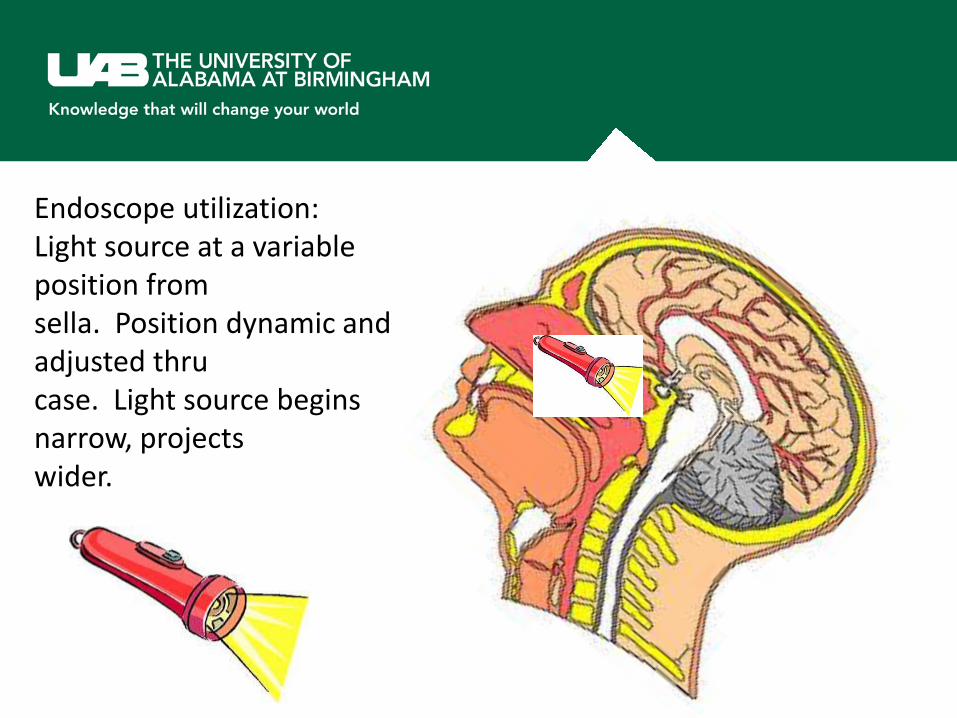
Endoscope utilization: Light source at a variable position from sella. Position dynamic and adjusted thru case. Light source begins narrow, projects wider.

UAB Endoscopic Cases • 1st use of endoscope in transsphenoidal case Oct 2005-
to “look around” • Since November 2005 utilized endoscopic techniques.
For the first 10 cases, had both microscope and endoscope available while in transition. Since 2006, all transsphenoidal pituituary cases have been performed endoscopically (Riley)
• Currently, approximately 60 pituitary surgery cases/yr • Additional 15-20 endoscopic skull base cases for other
pathology

Tumor versus normal gland • Pituitary tumors arise from the
adenohypophysis- typically expand anterior pituitary, compress any normal pituitary gland, deviate stalk
• Microadenoma resection: easy to differentiate tumor from normal gland typically
• Macroadenoma resection: depending on size of tumor, may not see any normal gland
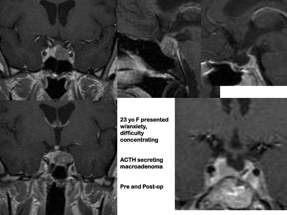
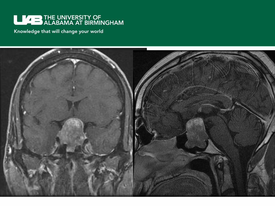
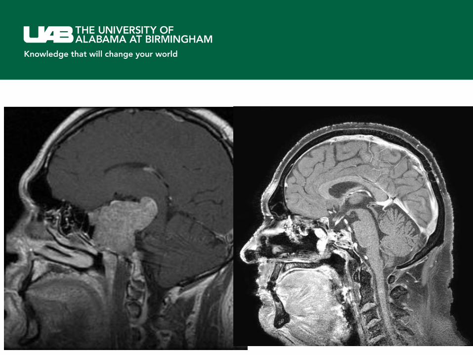
23 yo F presented
w/anxiety,
difficulty
concentrating
ACTH secreting
macroadenoma
Pre and Post-op


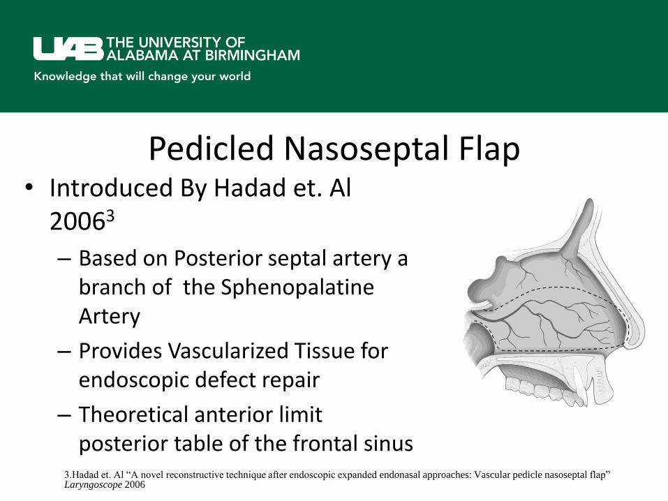
Pedicled Nasoseptal Flap • Introduced By Hadad et. Al
20063
– Based on Posterior septal artery a branch of the Sphenopalatine Artery
– Provides Vascularized Tissue for endoscopic defect repair
– Theoretical anterior limit posterior table of the frontal sinus
3.Hadad et. Al “A novel reconstructive technique after endoscopic expanded endonasal approaches: Vascular pedicle nasoseptal flap” Laryngoscope 2006

Nasoseptal flap postoperative considerations
• Nasal packing for 2 weeks
• IV antibiotics immediately postop, then oral antibiotics until packing removed
• No valsalva
• More activity restrictions
• Olfaction risks increased
• More discomfort


Surgical risks • CSF leak • Pituitary dysfunction (hypocortisolemia,
hypothyroidism, diabetes insipidus, hypogonadism)
• Post operative hemorrhage- visual decline • Anesthetic risks • Sinusitis/ Nasal complications • Cranial nerve deficits: firm tumors, craniotomy
approaches into cavernous sinus


