M. Jackson URT August 29, 2001 Page 1 of 21 UPPER RESPIRATORY TRACT INFECTIONS Introduction I. Variety of organisms colonize oropharynx & upper respiratory tract A. Many commensals colonize upper respiratory tract B. Respiratory tract is continuum from sinuses to alveoli
Transcript
M. JacksonURT
August 29, 2001Page 1 of 21
UPPER RESPIRATORY TRACT INFECTIONS
IntroductionI. Variety of organisms colonize oropharynx & upper respiratory tract
A. Many commensals colonize upper respiratory tractB. Respiratory tract is continuum from sinuses to alveoli
M. JacksonURT
August 29, 2001Page 2 of 21
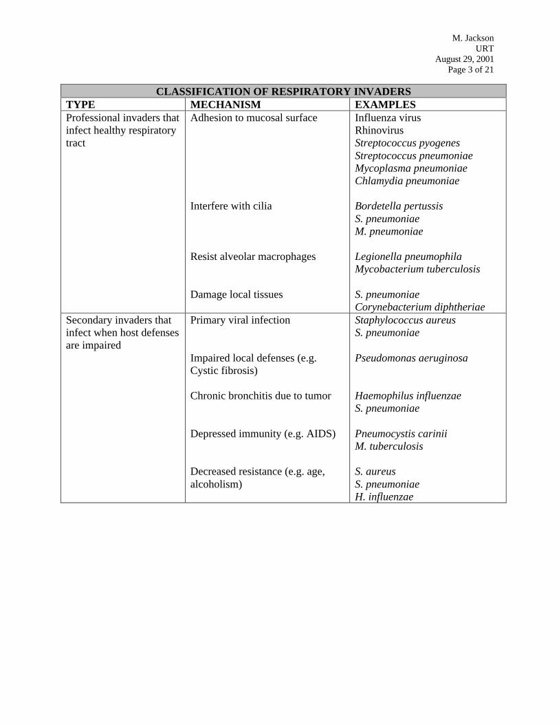
II. Distinction between primary and opportunistic respiratory tract pathogensA. Many pathogens gains entry via upper respiratory tract and become systemicB. “Professional invaders” uniquely adapted to upper respiratory tractC. Secondary pathogens cause infection following initial insult by primary pathogen
NORMAL RESPIRATORY TRACT FLORARESIDENCY STATE (INCIDENCE RATE) MICROBECommon (>50%) Oral streptococci
I. Virulence factors relevant to Oral InfectionsA. Oral bacteria produce lymphocyte activators that induce host inflammatory responseB. Release of PMN contents & complement activation exacerbates tissue damage
II. Etiology / PathogenesisA. Chronic marginal gingivitis (between teeth and gums)
1. Inflammatory infiltrate: PMNs & lymphocytes in connective tissue attached to tooth2. Gingivitis can occur in 2 weeks without proper tooth care
B. Periodontitis (teeth and supporting tissue)1. Progressive gingivitis results in periodontitis2. Resorption of bone around neck of tooth, loss of periodontal ligament and the tooth3. Agents of gingivitis not known; anaerobes responsible for chronic marginal periodontitis4. Oral anaerobes live in dental plaque next to gingival tissues
C. General features of anaerobic infections1. Average of 1011 microbes/g in gingival crevices, predominantly anaerobes2. Autoinfections caused by normal flora, usually mixed (polymicrobic)3. Anaerobes typically form localized abscesses4. Bacteria do not invade in gingivitis; remain part of plaque outside host defenses5. Bacterial invasion may occur with periodontitis
D. Acute necrotizing ulcerative gingivitis (trench mouth)1. Prevotella, Fusobacterium associated with ulceration of gingiva2. Invasion of oral epithelium and pharynx; can lead to bone resorption and tooth loss
III. Clinical identification of organismA. Diagnosis of periodontal disease by symptomsB. Mixed anaerobic infection not differentiated
1. Generally, no specific designation of Gram reaction or morphology2. Abscess can be sampled
a. Culture must be maintained under anaerobic conditionsb. Predominantly Gram neg. rods, some Gram pos. (Peptostreptococcus) & PMNs
M. JacksonURT
August 29, 2001Page 5 of 21
Actinomyces israeliiI. Etiology / Pathogenesis
A. Actinomyces israelii is normal flora anaerobe of humans1. Colonizes mucosal surfaces, from oropharynx to lower intestine2. Endogenous infection only upon penetration of epithelial barrier (low O2 tension)
B. Actinomycosis in cervicofacial area follows mouth trauma, e.g. tooth extraction1. Slowly progressing disease2. Inflammatory sinuses filled with pus and bacteria from initial site of infection3. Sinus extension or aspiration may lead to thoracic actinomycosis
M. JacksonURT
August 29, 2001Page 6 of 21
II. Clinical identification of organismA. Isolation, staining and culturing of pus
1. Actinomyces are Gram-positive filamentous rods resembling fungi2. “Sulfur granules” seen in pus, diagnostic for Actinomyces infection
a. Yellow granules resembling sulfurb. Composed of intertwined Actinomyces elements with tissue exudates
3. Infection is polymicrobic⇒ sinuses also contain Gram-negative rodsB. Culture conditions
1. Slow (4-10 day) growth under anaerobic or microaerophilic conditions2. Contaminating bacteria may overwhelm slow-growing Actinomyces
M. JacksonURT
August 29, 2001Page 7 of 21
Viridans StreptococciI. Virulence factors relevant to Oral Infections
Glucans (complex polysaccharides) that permit attachment to teeth
II. Etiology / PathogenesisA. Normal flora of oral and nasopharyngeal cavity; S. mutans associated with dental cariesB. Subacute bacterial endocarditis
1. Tooth extraction⇒ transient bacteremia2. May lead to colonization of damaged heart valves
III. Clinical identification of organismA. Gram positive cocci, catalase negativeB. Not Lancefield groupedC. Many different species, all classified as viridans Streptococci
UPPER RESPIRATORY TRACT INFECTIONSFUNGAL INFECTIONS OF THE ORAL CAVITY
Candida albicansI. Virulence factors relevant to Oral Infections
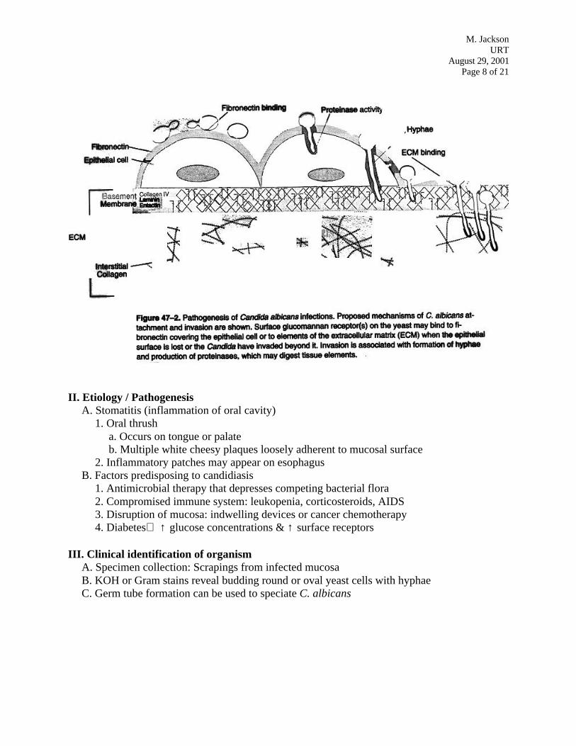
A. Adhesion through mannoprotein binding to fibronectin receptorsB. Invasion
1. Invasive hyphae bind to fibronectin, collagen, laminin→ transverse mucosal barriers2. Proteases and elastases may have role in the invasion process
M. JacksonURT
August 29, 2001Page 8 of 21
II. Etiology / PathogenesisA. Stomatitis (inflammation of oral cavity)
1. Oral thrusha. Occurs on tongue or palateb. Multiple white cheesy plaques loosely adherent to mucosal surface
2. Inflammatory patches may appear on esophagusB. Factors predisposing to candidiasis
1. Antimicrobial therapy that depresses competing bacterial flora2. Compromised immune system: leukopenia, corticosteroids, AIDS3. Disruption of mucosa: indwelling devices or cancer chemotherapy4. Diabetes⇒ ↑ glucose concentrations & ↑ surface receptors
III. Clinical identification of organismA. Specimen collection: Scrapings from infected mucosaB. KOH or Gram stains reveal budding round or oval yeast cells with hyphaeC. Germ tube formation can be used to speciate C. albicans
Streptococcus pneumoniaeI. Virulence factors relevant to Ear & Sinus Infections
A. Polysaccharide capsule (84 capsular serotypes)1. Primary virulence factor of S. pneumoniae2. Interferes with classical and alternate complement pathways3. Anti-capsule antibodies confer immunity
B. Cell wall teichoic acid and peptidoglycan contribute to inflammatory response
II. Etiology / PathogenesisA. High nasopharynx carriage rate (10-30%) predisposes for upper respiratory tract infectionsB. Acute Otitis Media (middle ear infection)
1. S. pneumoniae single most common cause after 3 months of life (35-40%)2. Viral infection or allergies are predisposing factors3. Eustachian tube inflammation⇒ bacterial enter middle ear from nasopharynx4. Shortness & pliancy of infants’ eustachian tubes contributes to susceptibility
C. Sinus infection1. S. pneumoniae is major cause of acute and chronic sinusitis in all age groups2. Predisposing factors: viral infection, allergy, or anatomical blockage
III. Clinical identification of organismA. Diagnosis generally based on clinical examination
1. Tympanic membrane swells due to pus formation with otitis media2. Symptoms and radiography used for diagnosis of sinusitis
B. Needle aspiration1. Pus behind tympanic membrane can be collected in difficult cases of otitis media2. Sinus wall puncture or catheterization for sinusitis3. Stain of aspirate shows Gram-positive lancet-shaped diplococci
C. Biochemical assays1. S. pneumoniae (a.k.a. pneumococcus) not part of Lancefield grouping2. Serotyping; Optochin (P disk) susceptibility
BIOCHEMICAL REACTIONS OF STREPTOCOCCI & ENTEROCOCCIBacitracinsusceptible
Optochinsusceptible
Bilesoluble
Bile/EsculinReaction
-
Group A Streptococci -Group B Streptococci -S. pneumoniae -Viridans Streptococci -Enterococci -
M. JacksonURT
August 29, 2001Page 10 of 21
Haemophilus influenzaeI. Virulence factors relevant to Ear & Sinus Infections
A. Polysaccharide capsule most important virulence factor of H. influenzae1. Capsule is antiphagocytic and subject to antigenic variation2. Capsule of polyribitol phosphate (PRP)⇒ 6 different serotypes, a-f3. H. influenzae serotype b (Hib) most virulent
B. IgA protease may facilitate colonization of nasopharynx
C. Non-pilus adhesins that direct tissue tropism to mucosal surfaces
M. JacksonURT
August 29, 2001Page 11 of 21
II. Etiology / PathogenesisA. H. influenzae 2nd most common cause of otitis media and major cause of sinusitis
1. Common cause of otitis media in children less than 5 years old2. Viral infection is predisposing, displacement of H. influenzae (flora) into sterile sites
B. High carriage rate (50-80%) of H. influenzae in upper respiratory tract1. Normal flora strains usually lack capsule2. Most otitis media isolates non-typable and may not be influenced by Hib vaccine
C. Otitis media or sinusitis caused by Hib may lead to meningitis
III. Clinical identification of organismA. Diagnosis based on clinical examination; needle aspirate in refractory cases
B. H. influenzae is small, Gram-negative coccobacillus
C. H. influenzae require Hematin (X factor) and/or NAD (V factor) for growth
D. Capsule serotyping
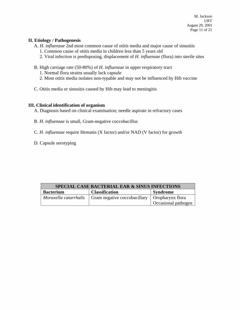
SPECIAL CASE BACTERIAL EAR & SINUS INFECTIONSBacterium Classification SyndromeMoraxella catarrhalis Gram negative coccobacillary Oropharynx flora
Occasional pathogen
M. JacksonURT
August 29, 2001Page 12 of 21
UPPER RESPIRATORY TRACT:BACTERIAL INFECTIONS OF THE PHARYNX
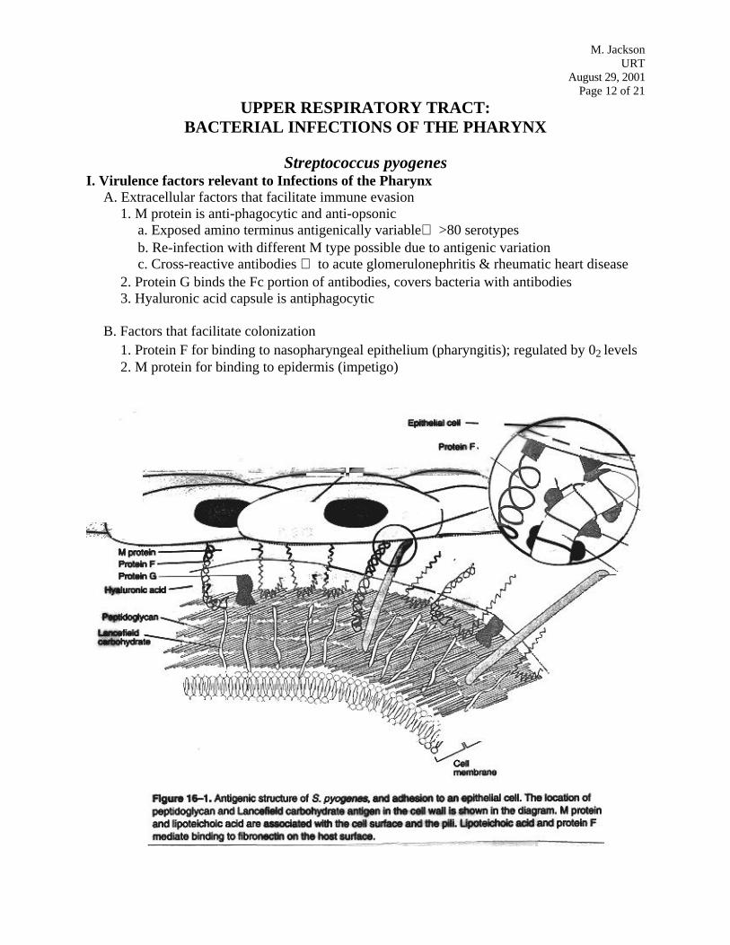
Streptococcus pyogenesI. Virulence factors relevant to Infections of the Pharynx
A. Extracellular factors that facilitate immune evasion1. M protein is anti-phagocytic and anti-opsonic
a. Exposed amino terminus antigenically variable⇒ >80 serotypesb. Re-infection with different M type possible due to antigenic variationc. Cross-reactive antibodies ⇒ to acute glomerulonephritis & rheumatic heart disease
2. Protein G binds the Fc portion of antibodies, covers bacteria with antibodies3. Hyaluronic acid capsule is antiphagocytic
B. Factors that facilitate colonization1. Protein F for binding to nasopharyngeal epithelium (pharyngitis); regulated by 02 levels2. M protein for binding to epidermis (impetigo)
M. JacksonURT
August 29, 2001Page 13 of 21
M. JacksonURT
August 29, 2001Page 14 of 21
C. Exotoxins1. SLO & SLS
a. Cause 02-labile (SLO) or 02 stable (SLS) β-hemolysis on blood agar platesb. Form large pores in cell membranes⇒ lysis of leukocytes
2. Streptococcal Pyrogenic Exotoxins (Spe A-C)a. Erythrogenic or Scarlet Fever Toxinsb. SpeA produced only by minority of lysogenized Group A Streptococcusc. Superantigens with sequence homology to staphylococcal exotoxinsd. Induce cytokine release
i. Fever & rash (scarlet fever)ii. T-cell stimulation and B cell suppressioniii. Enhanced sensitivity to endotoxic shock
e. Responsible for toxic shock like syndrome in S. pyogenes bacteremia
M. JacksonURT
August 29, 2001Page 15 of 21
II. Etiology / PathogenesisA. Pharyngitis
1. Viruses predominate; most frequent bacterial cause is group A S. pyogenes2. Common in 5-15 yr. age group; spread person-person by droplet3. Prompt antimicrobial therapy required
a. Prevents poststreptococcal sequelaeb. Circumvents natural development of type-specific immunity
B. Scarlet Fever can occur simultaneously with pharyngitis1. Caused by pyrogenic exotoxins (Spe)2. Scarlet rash spreads from mouth & face to trunk & extremities; strawberry tongue3. Occurrence and severity reduced in comparison to early 1900s
C. Poststreptococcal Sequelae: Acute Rheumatic Fever1. Formerly on decline, rheumatic heart disease has reappeared in some parts of the world2. Begins ~3 wk. after pharyngitis3. Syndrome
a. Symptoms: fever, subcutaneous nodules, chorea (neurologic), migratory polyarthritisb. Cardiac: carditis, cardiac enlargement, murmurs, heart failurec. Aschoff body seen with rheumatic carditis
i. Lesion of lymphocytes and macrophages aggregated around fibrinoid depositsii. Caused by cell-mediated response
d. Subacute bacterial endocarditisi. Acute rheumatic fever may damage heart valves ⇒ formation of vegetationsii. Provides site for colonization during transient bacteremia by viridans streptococci
4. Heart damage caused by anti-streptococcal antibodies that cross-react with cardiac tissuea. Anti-streptococcal antibodies are to cell wall, cell membrane, and M proteinb. Epitopes shared with cardiac sarcolemma membranes, smooth muscle cells, valvesc. Recurrent attacks with new M types leads to progressive heart damaged. SLO, Spe, streptokinase may contribute directly to cardiac damage
cont
M. JacksonURT
August 29, 2001Page 16 of 21
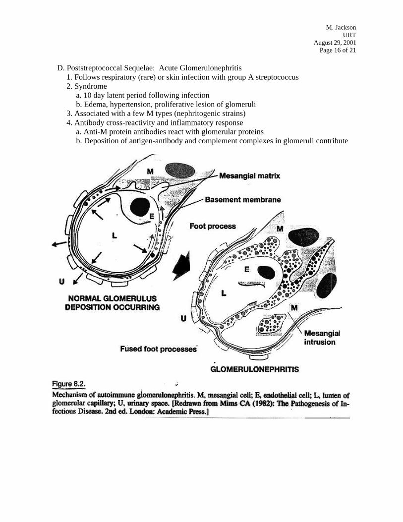
D. Poststreptococcal Sequelae: Acute Glomerulonephritis1. Follows respiratory (rare) or skin infection with group A streptococcus2. Syndrome
a. 10 day latent period following infectionb. Edema, hypertension, proliferative lesion of glomeruli
3. Associated with a few M types (nephritogenic strains)4. Antibody cross-reactivity and inflammatory response
a. Anti-M protein antibodies react with glomerular proteinsb. Deposition of antigen-antibody and complement complexes in glomeruli contribute
M. JacksonURT
August 29, 2001Page 17 of 21
III. Clinical identification of organismA. Throat swab of tonsils and pharynx→ culture on blood agar for β-hemolysis
1. Gram positive cocci in chains2. Clear zone around colony; SLO (CO2 incubation) and SLS responsible for β-hemolysis
B. Rapid agglutination tests to identify Lancefield Group AC. Throat culture contaminants: S. pneumoniae, S. aureus, N. meningitidis, H. influenzaeD. Biochemical tests
1. Catalase test negative (differentiation from Staphylococcus)2. Bacitracin sensitivity assay on agar plate
E. High titers of anti-SLO antibodies (ASO) seen in patients with rheumatic fever
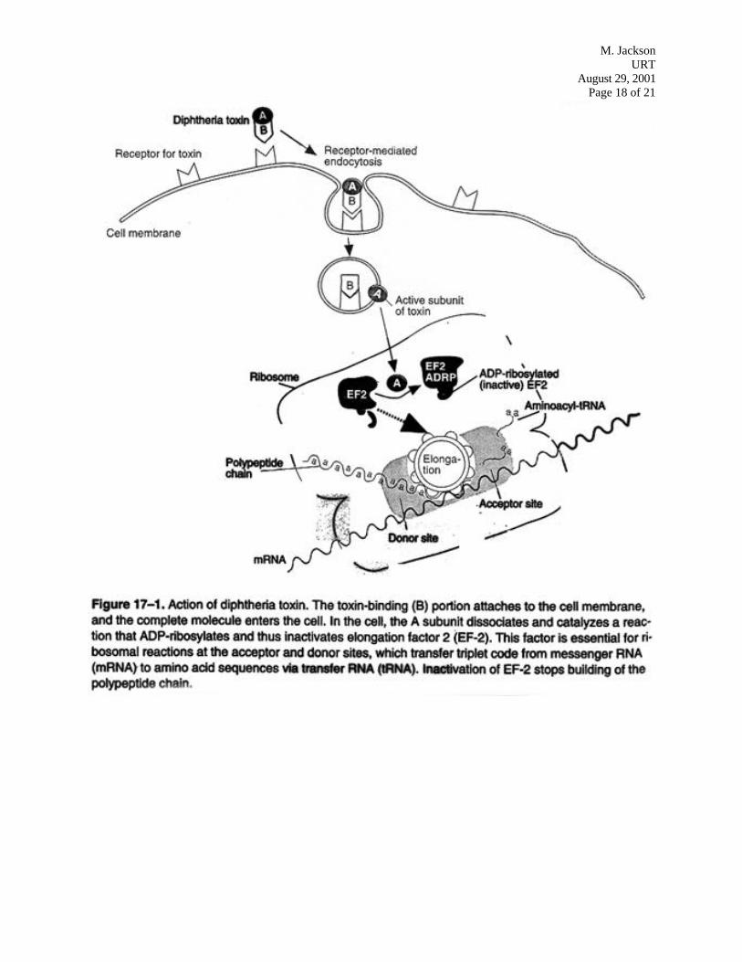
Corynebacterium diphtheriaeI. Virulence factors relevant to Infections of the Pharynx
A. Diphtheria toxin (DT) sole virulence factor of Diphtheria1. Best and earliest studied bacterial cytotoxin2. Basis for concept of “virulence factor”
B. Structure and function of DT1. AB toxin with enzymatic (A) and binding (B) subunits2. Synthesized as single polypeptide chain nicked between A and B subunits3. Receptor binding and entry
a. B subunit binds epidermal growth factor precursor in mammalian cell membranesb. Holotoxin uptake by receptor mediated endocytosisc. Reduction in endocytotic vesicle releases A subunit
4. A subunit enzymatic mechanism of actiona. A subunit ADP-ribosylates elongation factor 2 (ADPR-EF2) of any eucaryotic cellb. Reaction: NAD + EF2 ↔ ADPR-EF2 + nicotinamide + H+
c. EF2 for translocation of ribosome along mRNA; ADPR-EF2⇒ translation ceasesd. DT A subunit has same mechanism as Pseudomonas exotoxin A
M. JacksonURT
August 29, 2001Page 18 of 21
M. JacksonURT
August 29, 2001Page 19 of 21
C. Genetics of DT synthesis1. tox gene carried by bacteriophages β and ϖ2. DT synthesis negatively regulated by iron
M. JacksonURT
August 29, 2001Page 20 of 21
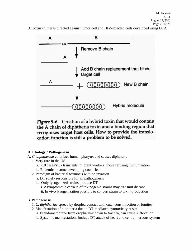
D. Toxin chimeras directed against tumor cell and HIV-infected cells developed using DTA
II. Etiology / PathogenesisA. C. diphtheriae colonizes human pharynx and causes diphtheria
1. Very rare in the USa. ~10 cases/yr. - transients, migrant workers, those refusing immunizationb. Endemic in some developing countries
2. Paradigm of bacterial toxinosis with no invasiona. DT solely responsible for all pathogenesisb. Only lysogenized strains produce DT
i. Asymptomatic carriers of toxinogenic strains may transmit diseaseii. In vivo lysogenization possible to convert strain to toxin-production
B. Pathogenesis1. C. diphtheriae spread by droplet, contact with cutaneous infection or fomites
2. Manifestation of diphtheria due to DT-mediated cytotoxicity at sitea. Pseudomembrane from oropharynx down to trachea, can cause suffocationb. Systemic manifestations include DT attack of heart and central nervous system
M. JacksonURT
August 29, 2001Page 21 of 21
III. Clinical identification of organismA. Diagnosis principally based on clinical symptoms
B. Isolation from throat swab difficult - normal resident of many individuals1. Gram positive, club-shaped rods, cells remain attached after division⇒ “Chinese letters”2. Culture of organism with demonstration of toxin production
CASE STUDYFOR
UPPER RESPIRATORY TRACT INFECTIONS
An 18-month-old girl developed a runny nose, watery eyes, and a cough. As her symptoms ofrhinitis abated, the girl became irritable and developed a slight fever (38°C). She alsocomplained of a sore throat and an earache. Her pediatrician observed a bulging tympanicmembrane and prescribed a 7-day course of antibiotics.
Questions:What are the most likely etiologic agents causing the ear infection?Name a key virulence factor that the common etiologic agents possess.What predisposing feature of this case contributed to the otitis media?
SPECIAL CASE BACTERIAL INFECTIONS OF THE PHARYNXBacterium Classification SyndromeNeisseria gonorrhoeae Gram negative diplococcus Oral-genital contact