PREGULATED EXPRESSION OF ONCOMODULIN, THE BETASOFORM OF PARVALBUMIN, IN PERIKARYA AND AXONS IN THE
IENCEPHALON OF PARVALBUMIN KNOCKOUT MICE
pol
Kp
AcepeseatctswdaInCtdoerR
n(r2Rwrhscap(c2s
. CSILLIK,a* B. SCHWALLER,b1 A. MIHALY,a2
. HENZI,b1 E. LOSONCZIc3 AND E. KNYIHAR-CSILLIKc2
Department of Anatomy, Albert Szent-Györgyi Medical School, Uni-ersity of Szeged, Hungary
Unit of Anatomy, Department of Medicine, University of Fribourg,witzerland
Department of Neurology, Albert Szent-Györgyi Medical School, Uni-ersity of Szeged, Hungary
bstract—The calcium-binding proteins parvalbumin, calbi-din D-28k, calretinin and calcineurin are present in subsetsf GABAergic gigantic calyciform presynaptic terminals ofhe reticular thalamic nucleus (RTN). Previously it was hy-othesized that GABA and calcium-binding proteins includ-
ng parvalbumin are not only colocalized in the same neuronubpopulation, but that GABA synthesis and parvalbuminxpression could be also genetically regulated by a commonechanism. Moreover, parvalbumin expression levels could
nfluence GABA synthesis. For this, we analyzed GABA im-unoreactivity in RTN gigantic calyciform presynaptic termi-
als of parvalbumin–deficient (PV�/�) mice. With respect toABA immunoreactivity we found no differences compared
o wild–type animals. However, using a polyclonal parvalbu-in antibody raised against full-length rat muscle parvalbu-in on brain sections of PV�/� mice, we observed paradox-
cal parvalbumin immunoreactivity in partly varicose axons inhe diencephalon, mainly in the lamina medullaris externaurrounding the thalamus. A detailed immunohistochemical,iochemical and molecular biological analysis revealed this
mmunoreactivity to be the result of an upregulation of onco-odulin (OM), the mammalian beta isoform of parvalbumin inV�/� mice. In addition, OM was present in a sparse sub-opulation of neurons in the thalamus and in the dentateyrus. OM expression has not been observed before in neu-ons of the mammalian brain; its expression was restricted touter hair cells in the organ of Corti. Our results indicate thathe absence of parvalbumin has no major effect on theABA-synthesizing system in RTN presynaptic terminals ex-luding a direct effect of parvalbumin on this regulation.owever, a likely homeostatic mechanism is induced result-
ng in the upregulation of OM in selected axons and neuronal
Present address: Abteilung Anatomie, Department Medizin, Univer-ität Fribourg, Route Albert-Gockel 1, CH-1700 Fribourg, Switzerland.Present address: Department of Anatomy, University Medicalchool, 40 Kossuth Lajos sgt, H-6701 Szeged, Hungary.Present address: Richter Gedeon NYRT, Gyomroi ut 19-21, Buda-est 1103, Hungary.Correspondence to: B. Csillik, Department of Anatomy, Universityedical School, 40 Kossuth Lajos sgt, H-6701 Szeged, Hungary. Tel:36-62-544-918; fax: �36-62-545-707.-mail address: [email protected] (B. Csillik).bbreviations: CaBP, calcium-binding proteins; OM, oncomodulin;AGE, polyacrylamide gel electrophoresis; PBS, phosphate-buffered
ey words: reticular thalamic nucleus, thalamus, GABA,arvalbumin, oncomodulin, regeneration.
t the level of gross anatomy, the reticular thalamic nu-leus (RTN) is a crescent-like structure, similar to a half-ggshell, surrounding the lateral, superior and inferior as-ects of the thalamus (Paxinos and Watson, 1982; Sidmant al., 1971). The GABAergic RTN is a heterogeneoustructure, from cytoarchitectural, immunocytochemical,lectrophysiological and neuropharmacological viewpointslike (Steriade, 2001). It occupies a strategic position be-ween specific nuclei of the thalamus and the cerebralortex (Steriade, 2001; Pinault, 2004), as a preferentialarget of corticothalamic projections, located at the inter-ection of thalamo–cortical and cortico–thalamic path-ays. The RTN is implied in almost all the functional mo-alities represented by its motor, somatosensory, visceral,uditory, gustatory and limbic sectors (Shosaku et al., 1989).
n addition, the RTN plays an important part in transformingociception into pain (Gauriau and Bernard, 2002; Knyihar-sillik and Csillik, 2006), by regulating attention and distrac-
ion to potentially painful stimuli, functioning as a coincidenceetector (Kilmer, 2001). It has been shown by electrophysi-logical and immunohistochemical studies by Knyihar-Csillikt al. (2005) that a two-way traffic between RTN and theetrosplenial cortex is involved in the communication betweenTN and cerebral cortex.
In the large GABAergic calyciform presynaptic termi-als of RTN of the rat, several calcium-binding proteinsCaBP) including parvalbumin (PV), calbindin D-28k, cal-etinin and calcineurin have been observed (Csillik et al.,002a,b, 2004, 2005, 2006). At the light microscopic level,TN is characterized by an intense PV immunoreaction,hich originally was exclusively attributed to PV-immuno-
eactive neurons (Celio, 1990). Recent light- and electronistochemical studies revealed however that some of thetructures, erroneously identified as nuclei, are in realityross-sections of large dendrites, while the PV-immunore-ctive structures thought to correspond to nerve cellerikarya, are in reality presynaptic dendraxonic terminalsCsillik et al., 2002a, 2004). The PV-immunoreactive caly-iform presynaptic terminals contain GABA (Csillik et al.,005, 2006). This raised the question whether the GABA-ynthesizing enzyme GAD and calcium-binding proteins,
n particular the one most often present in these terminals,s reserved.
B. Csillik et al. / Neuroscience 165 (2010) 749–757750
.e. PV, are not only co-expressed in the same neuronubpopulation, but whether they are also genetically reg-lated by a common mechanism. Furthermore, the obser-ation that PV is almost exclusively expressed in GABAer-ic neurons suggested that PV directly or indirectly couldffect the type or amount of neurotransmitter in the PV-
mmunoreactive neuron population. In the cortex of PV�/�ice, differences in the firing properties of pyramidal cells
uggested that PV plays a key role in the regulation of localnhibitory effects exerted by GABAergic interneurons onyramidal neurons (Schwaller et al., 2004, MCN). Further-ore, analysis of non-linear coupling of local field potentials
ndicated that absence of PV not only affects local neuronaletworks within the cortex, but might also affect interactingites that are distant from the recording electrodes but closeo each other, e.g. within the thalamus (Villa et al., 2000).hus, we decided to study by immunohistochemistry theistribution of CaBPs in the RTN of PV knockout (PV�/�)nimals. Unexpectedly, in brain sections from PV�/� micetained with one particular polyclonal anti-PV antiserum pro-uced against full-length rat PV, “PV-immunoreactive” struc-ures were detected. Both, structural details of these immu-opositive entities and the identity of the molecule giving riseo this paradoxical PV staining, oncomodulin (OM), the betasoform of PV, are described in this report.
EXPERIMENTAL PROCEDURES
n these studies, the number of animals (rats/mice) was kept to ainimum and efforts were taken to minimize the suffering; youngdult Wistar rats, young adult male mice of the C57Bl/6J strain andomozygous parvalbumin-knockout (PV�/�) mice (Schwaller et al.,999; Vecellio et al., 2000) were analyzed. As compared to the
nitial strain reported before (Schwaller et al., 1999) with a mixed29Ola Hsd�C57Bl/6J genetic background, the animals used inhis study had been backcrossed to C57Bl/6J animals for at least0 generations and are thus considered to be congenic with57Bl/6J mice (new name C57PV�/�). For the genotyping,enomic DNA was isolated from tail biopsies that was followed byCR using primer pairs that were either specific for exon 3 (de-
eted in PV�/� mice) or for part of the neomycin resistanceassette (absent in PV�/� mice). All mice were housed in groupsefore use; they were adult (25–30 g) when used for experiments.are of the animals complied with the guidelines of the Hungarianinistry of Welfare and was in accordance with the Europeanommunities Council Directive (November 24, 1986; 86/609/EC), the NIH Guide for the Care and Use of Laboratory Animals
NIH Publications No. 85–23, revised 1985) and the Guidelines forthics in Animal Experiments, University of Szeged, Albert Szent-yörgyi Medical School. After an i.p. injection with a lethal dose ofhloral hydrate, the animals were subjected to transcardial fixationith 4% formaldehyde, 0.5% glutaraldehyde in phosphate-buff-red saline (PBS). Brains were removed and processed in anscending series of sucrose, containing 4% formaldehyde.
V immunohistochemistry
erial cryostat sections, 20 �m thick, were obtained in the para-edial plane, comprising a thickness of 1–3 mm as measured
rom the midsagittal plane. RTN was localized in mice, using thearameters of the Sidman-Angevine-Pierce stereotactic atlas971, and in rats, using the Paxinos–Watson atlas (1982). On
ree-floating sections or in sections adhered to Superfrost Ultra-lus specimen holders (Menzel, Braunschweig, Germany), PV
as detected immunohistochemically in rats and mice, by using a K
abbit polyclonal anti-mouse PV antibody raised against full-lengthV from rat (sc-7449, Santa Cruz, CA, USA) Preliminary experi-ents showed best staining results with antibody dilutions of:2000–1:3500. Endogenous peroxidase activity was blocked by.3% hydrogen peroxide diluted in methanol, for 10 min, followedy three successive rinses in 0.1 M phosphate buffer, pH 7.4.ree-floating or adherent sections were pre-treated with blockingerum (0.1–1.0 M PBS, 10% normal goat serum, 1% bovineerum albumin (BSA) and 0.3% Triton X-100) on a shaker plate atoom temperature for 1 h, and then transferred into a solutionontaining the primary antibody. Incubation was carried out at 4 °Cn a shaker for 36 h, followed by three rinses in 0.1 M phosphateuffer. To detect the bound primary antibody, we used the avidin-iotin-peroxidase method. Kits were obtained from Vector Labo-atories (Burlingame, CA, USA). The secondary antibody, biotin-lated anti-rabbit immunoglobulin was applied for 90 min at roomemperature. Three more rinses in 0.1 M phosphate buffer wereollowed by incubation with the avidin-biotinylated peroxidaseomplex for 60 min at room temperature. After three rinses in 0.1
phosphate buffer, peroxidase activity was visualized by theistochemical reaction involving diamino-benzidine-tetrahydro-hloride (DAB) and hydrogen peroxide (3 �l of 30% H2O2 in 10 ml% DAB). After three rinses in 0.1 M phosphate buffer, the sec-ions were dehydrated in a graded series of alcohol, cleared inylene and coverslipped with Permount.
M immunohistochemistry
n identical protocol was used for the detection of OM, the mam-alian beta isoform of parvalbumin (MacManus, 1979), but as the
rst antibody the affinity-purified polyclonal goat anti-OM antibodyPV-beta, N-19, Santa Cruz Biotechnology, Inc., Santa Cruz, CA,SA) was used. This peptide antibody was raised against a region
n the N-terminus of human OM showing very low homology touman PV. For control experiments, the blocking peptide sc-446P (Santa Cruz) was applied. Alternatively, the polyclonalabbit anti-OM antiserum OM3 from Swant (Bellinzona, Switzer-and) raised against recombinant full-length rat OM was used.taining of sections with both sera yielded identical results. For allntibodies, a series of experiments were carried out to demon-trate the specificity of the antibody and included: (1) omission ofhe first specific antiserum; (2) incubation with normal rabbit orouse serum instead of incubation with the anti-PV or anti-OMntibodies; (3) treatment according to the avidin-biotin complexethod, from which one of the steps had been omitted and (4)readsorption of the specific antibody with blocking peptides.one of the specimens treated by one of the above methodshowed any specific immunoreactivity.
ABA immunoreactivity
ABA immunoreactivity was detected, applying the same protocolut using a rabbit anti-GABA antiserum (Chemicon, Temecula,A, USA) at a dilution of 1:500.
tereology
he number of calyciform terminals was determined in consecutiveoronal section series according to the method of West et al. (1991),sing a 100� oil immersion lens. Analysis was performed with theid of an Olympus microscope (Olympus, Tokyo, Japan) equippedith a video camera and stepping motors. Counting frames wereuperimposed on video images of the microscopic fields.
estern blot analysis
estern blot analysis was performed on tissue from the diencepha-ons of brains of adult PV�/� mice, according to the protocol of
amps and Sefton (1988), modified by Rogers et al. (1991) as
f(clS4ttA(rbPwwbetnuTaltpptw
R
Bwtfaf
apwmwpitOh�icmntrfTolcffS
D
De
Irt
Frdidi
B. Csillik et al. / Neuroscience 165 (2010) 749–757 751
ollows. The tissue was homogenized with a Disperser T10 BasicIKA-Werke, Oberkochen, Germany) in 50 mM Tris buffer (pH 7.5)ontaining 150 mM NaCl, 0.1% Igepal, 0.1% colic acid, 2 �g/ml
eupeptin, 2 mM PMSF, 1 �g/ml pepstatin, 2 mM EDTA and 0.1%DS. The homogenates were centrifuged at 12,000�g for 10 min at°C (Micro 200R, Hettich GmbH). The supernatants were used for
he detection of OM. The protein concentration was determined usinghe bicinchoninic acid (BCA) Protein Assay Kit (Novagen®, 71285-3).
sodium dodecyl sulphate polyacrylamide gel electrophoresisSDS-PAGE) was carried out using a 13% gel (according to theelative molecular size of OM: �12 kDa). The proteins were electro-lotted onto a nitrocellulose membrane by using the BioRad Mini-rotein II System at 300 mA for 1 h. The membrane was incubatedith 5% non-fat dried milk dissolved in TBS-Tween for 1 h. Afterashing the samples in TBS-Tween (3�5 min), the blots were incu-ated with a rabbit antibody to OM (Swant, Switzerland). Preliminaryxperiments showed the best results at a 1:1000 working dilu-ion. Incubation with the primary antibody was carried out in 1%on-fat dried milk in 0.1% NaN3 overnight, at room temperaturender continuous shaking. After washing the samples in TBS-ween (3�5 min), the blots were incubated with the secondaryntibody (alkaline-phosphatase-conjugated anti-rabbit IgG) fol-
owing the manufacturer’s instructions (1:2000). After washinghe samples in TBS-Tween (4x) and in TBS, the detection waserformed with BCIP/NBT (5-bromo-4-chloro-3-indolyl phos-hate/nitro blue tetrazolium) tablets, obtained from Sigma, inhe dark room. After washing in sterile water, the membranesere dried and scanned.
NA isolation and RT-PCR
rains derived from mice transcardially perfused with 0.9% NaClere homogenized in 1 ml of TRIZOL® reagent per 50–100 mg of
issue using a homogenizer (Polytron) and were then incubatedor 5 min at RT. Chloroform (0.2 ml/ml of TRIZOL® reagent) wasdded, tubes were vigorously shaken for 15 s and incubated at RT
or 2–3 min. After centrifugation (12,000�g,15 min, 4 °C), the
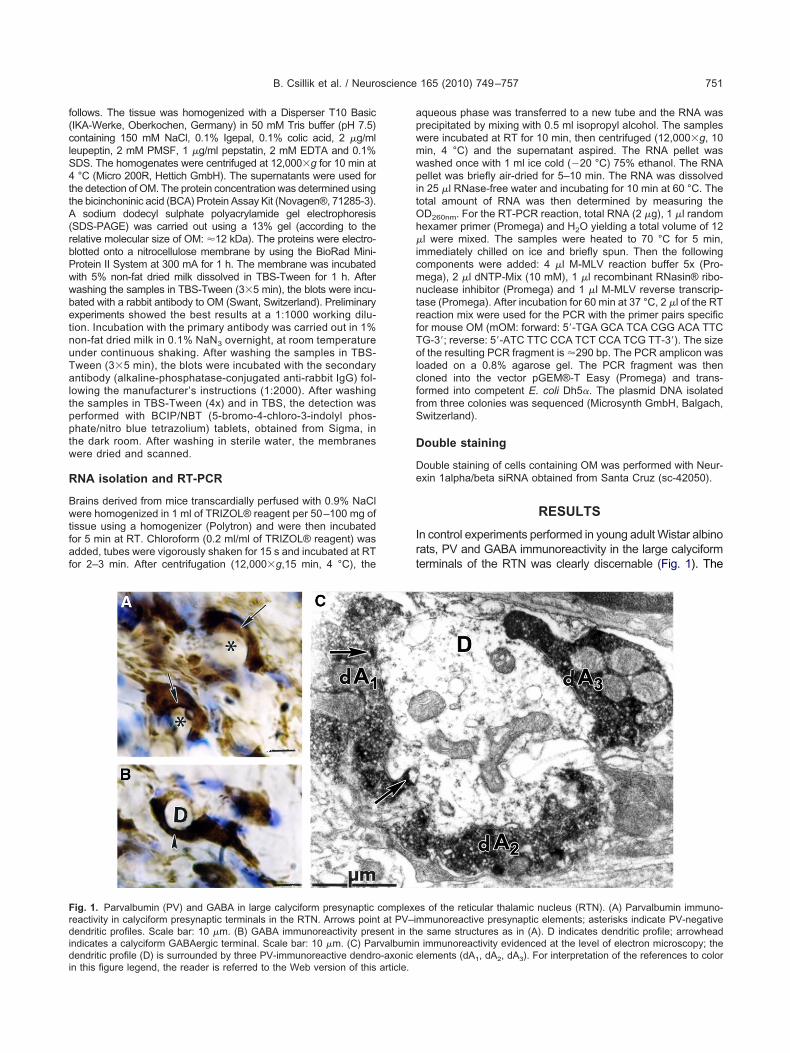
ig. 1. Parvalbumin (PV) and GABA in large calyciform presynapticeactivity in calyciform presynaptic terminals in the RTN. Arrows poinendritic profiles. Scale bar: 10 �m. (B) GABA immunoreactivity pres
ndicates a calyciform GABAergic terminal. Scale bar: 10 �m. (C) Par
endritic profile (D) is surrounded by three PV-immunoreactive dendro-axonic e
n this figure legend, the reader is referred to the Web version of this article.
queous phase was transferred to a new tube and the RNA wasrecipitated by mixing with 0.5 ml isopropyl alcohol. The samplesere incubated at RT for 10 min, then centrifuged (12,000�g, 10in, 4 °C) and the supernatant aspired. The RNA pellet wasashed once with 1 ml ice cold (�20 °C) 75% ethanol. The RNAellet was briefly air-dried for 5–10 min. The RNA was dissolved
n 25 �l RNase-free water and incubating for 10 min at 60 °C. Theotal amount of RNA was then determined by measuring theD260nm. For the RT-PCR reaction, total RNA (2 �g), 1 �l randomexamer primer (Promega) and H2O yielding a total volume of 12l were mixed. The samples were heated to 70 °C for 5 min,
mmediately chilled on ice and briefly spun. Then the followingomponents were added: 4 �l M-MLV reaction buffer 5x (Pro-ega), 2 �l dNTP-Mix (10 mM), 1 �l recombinant RNasin® ribo-uclease inhibitor (Promega) and 1 �l M-MLV reverse transcrip-
ase (Promega). After incubation for 60 min at 37 °C, 2 �l of the RTeaction mix were used for the PCR with the primer pairs specificor mouse OM (mOM: forward: 5=-TGA GCA TCA CGG ACA TTCG-3=; reverse: 5=-ATC TTC CCA TCT CCA TCG TT-3=). The sizef the resulting PCR fragment is �290 bp. The PCR amplicon was
oaded on a 0.8% agarose gel. The PCR fragment was thenloned into the vector pGEM®-T Easy (Promega) and trans-ormed into competent E. coli Dh5�. The plasmid DNA isolatedrom three colonies was sequenced (Microsynth GmbH, Balgach,witzerland).
ouble staining
ouble staining of cells containing OM was performed with Neur-xin 1alpha/beta siRNA obtained from Santa Cruz (sc-42050).
RESULTS
n control experiments performed in young adult Wistar albinoats, PV and GABA immunoreactivity in the large calyciformerminals of the RTN was clearly discernable (Fig. 1). The
s of the reticular thalamic nucleus (RTN). (A) Parvalbumin immuno-munoreactive presynaptic elements; asterisks indicate PV-negative
e same structures as in (A). D indicates dendritic profile; arrowheadimmunoreactivity evidenced at the level of electron microscopy; the
complexet at PV–iment in thvalbumin
lements (dA1, dA2, dA3). For interpretation of the references to color
nndcm2nt
fPP(toi(
Fts he asterist the Web
Ftopclr
B. Csillik et al. / Neuroscience 165 (2010) 749–757752
umber of PV and GABA immunoreactive calyciform termi-als was 2010�17 in each hemisphere of the rat, as evi-enced by stereological methods. Much lower numbers ofalyciform terminals were observed in the RTN of C57Bl/6Jice (Fig. 2); quantitative analysis revealed the presence of62�12 PV� and GABA-immunoreactive calyciform termi-als. While immunoreactivity for GABA appeared to be unal-
ered in these animals (Fig. 3A), a specific immunoreaction
ig. 2. Presynaptic calyciform terminals in the RTN of a C57Bl/6J merminal (arrow); the asterisk indicates a PV-negative dendritic profile. Strong immunoreactivity in a presynaptic calyciform terminal (arrow); the references to color in this figure legend, the reader is referred to
ig. 3. Presynaptic calyciform terminals in the RTN of a PV�/� mousehalamic nucleus of a PV�/� mouse is not different when compared tf the GABA-immunoreactive calyciform terminals; asterisk indicatesresynaptic calyciform terminals in the reticular thalamic nucleus of aalyciform terminal; the faint contrast seen in this microphotograph is
owered condenser) and is not the result of unspecific staining. The asterisk ineferences to color in this figure legend, the reader is referred to the Web vers
or PV was absent in the gigantic calyciform terminals ofV�/� mice (Fig. 3B), in line with a complete absence ofV-ir structures reported in the cortex of PV�/� mice
Schwaller et al., 2004). Totally unexpected, numerous, par-ially varicose “PV-immunoreactive” axons appeared in vari-us parts of the diencephalon; some of them were localized
n the lamina medullaris externa, surrounding the thalamusFig. 4A). The most likely explanation for these results was
) Strong PV immunoreactivity is present in a presynaptic calyciform10 �m. (B) A consecutive section was stained against GABA showingk indicates a dendritic profile. Scale bar: 10 �m. For interpretation ofversion of this article.
A immunoreactivity of presynaptic calyciform terminals in the reticularir in a wild–type mouse (compare to Fig. 2B). An arrow points at oneprofile. Scale bar: 10 �m. (B) No specific PV signal was detected inouse. The arrowhead points at the outlines of a PV-immunonegativee selected parameters of microscopy (nearly closed diaphragm and
ouse. (Acale bar:
. (A) GABo GABA-dendriticPV�/� mdue to th
dicates a dendritic profile. Scale bar: 10 �m. For interpretation of theion of this article.
tsbpCOpei
tpt(
Per
Fm1at the Web
B. Csillik et al. / Neuroscience 165 (2010) 749–757 753
he presence of a molecule cross-reacting with the PV anti-erum. On the basis of the large similarity between parval-umin (an alpha isoform) and OM (a beta isoform of thearvalbumin subfamily of EF-hand CaBPs, for details seeelio et al., 1996) that is 48% identity at the amino acid level,M was considered as the prime candidate leading to thearadoxical PV staining. Thus we carried out a series ofxperiments to verify that the observed immunoreactiv-
ig. 4. (A) Bundles of oncomodulin (OM)-expressing axons (arrowagnification in the brain of a PV�/� mouse. For the immunostaining00 �m. Individual OM-expressing axons, some of them varicose (B), oxonal structures can be seen in a specimen stained with the OM-spehe references to color in this figure legend, the reader is referred to
ty was due to OM expression. Axonal structures, similar w
o those previously observed in PV�/� mice with theolyclonal PV antiserum were stained with the OM an-
ibody raised against the N-terminal OM-specific peptideFig. 4B, C).
To ascertain that the observed immunoreactivity inV�/� brains was due to expression of OM, a series ofxperiments were carried out and the results are summa-ized here. The immunoreactive structures were stained
lamina medullaris external surrounding the thalamus (th) at low) the OM antiserum N-19 sc-7446 (Santa Cruz) was used. Scale bar:ing varicosities (C) are depicted at higher magnification. In (D), similar
serum OM3 (Swant). Scale bar for B–D: 10 �m. For interpretation ofversion of this article.
s) of thein (A�C
thers lackcific anti
ith OM antibodies derived from two different sources; the
apOa(aO
p7atO
md(unctrtf
tlwpOn1iimntoc
fCrOtbwbaw
Ticdphobe(no2s
Mwio1soTidtbfrt
Fssitbot
B. Csillik et al. / Neuroscience 165 (2010) 749–757754
bove-mentioned OM peptide antibody (Santa Cruz) and aolyclonal rabbit antiserum raised against full-length ratM (Swant; for details, see Material and Methods). Thexonal structures stained with the antiserum OM3 (Swant)Fig. 4D) were asbsolutely identical to those seen with thentibody N-19 sc-7446 (Santa Cruz) for the visualization ofM (Fig. 4B, C).
Furthermore, while immunoreactivity persisted afterreincubation with the PV-specific blocking peptide sc-449P (Fig. 5A), specific staining with the OM peptidentiserum N-19 sc-7446 completely disappeared, whenhe primary antibody solution was preincubated with theM-specific blocking peptide sc-7446 P (Fig. 5B).
Specific OM expression was not restricted to OM-im-unopositive processes or fiber bundles, but was alsoetected in perikarya of cells scattered in the diencephalonFig. 6A–D), the cytological identity of which could not benambiguously ascertained; accordingly, these may beeuroblasts, macrophages or possibly cells of neuroendo-rine origin as well. OM appeared in the processes ofhese cells in a vesicular or granular form (Fig. 6A, B)esembling those reported by Yin et al. (2006). Apparently,he vesicles/granules are sometimes confined to or trans-
ig. 5. (A). Bundle of OM-ir axons in specimen from a PV�/� mousetained with the OM specific peptide antibody sc-7446 (B). In a serialection pretreated with the OM blocking peptide sc-7446P, no specificmmunoreaction was observed in nerve fibers. The OM immunoreac-ivity persisted when the serial section was incubated with the parval-umin blocking peptide sc-7449P. Scale bar: 10 �m. For interpretationf the references to color in this figure legend, the reader is referred tohe Web version of this article.
ormed into varicosities of nerve-fibers (Fig. 6C). OM con- l
aining vesicles and/or granules were present in the cellu-ar cytoplasm of certain cells, where the immunoreactivityas accumulated in clusters of vesicles/granules within theerikaryon (Fig. 6D). In order to ascertain whether or not,M-expressing cells belong to the cell line of neuroblasts/eurons, double staining with the neuron-specific Neurexinalpha/beta siRNA was performed. In some of the OM-
mmunopositive cells, the OM immunoreactivity colocal-zed with the Neurexin staining (Fig. 6E), whereas in the
ajority of OM immunopositive cells Neurexin staining wasot observed. Thus, this indicates that OM immunoreac-ivity was present in at least two different cell populations,ne likely of neuronal origin (Neurexin 1alpha/beta-positiveells).
Western blot analysis of diencephalon homogenatesrom PV�/� mice with the OM-specific antibody (Santaruz) yielded a weak band of approximately 12 kDa cor-
esponding to the calculated Mr of 12,260 Da for mouseM (Fig. 7A). Furthermore, an OM-specific PCR using
otal RNA isolated from PV�/� brains resulted in a specificand of the expected size of 290 bp (Fig. 7B). A mucheaker signal was also seen with RNA isolated from therains of C57Bl/6J wild–type mice suggesting minutemounts of OM mRNA may be also present in brains ofild–type mice.
DISCUSSION
he unchanged GABA staining in the RTN of PV�/� mices a strong indication that PV does not exert a significantontrol on GABA expression in the diencephalon. Evi-ently, subtle differences in GABA distribution and/or ex-ression could not be determined by the methods usedere and further electrophysiological analysis of the RTNf PV�/� mice needs to be carried out. Whether a linketween GABA and calcium handling systems (e.g. PV)xists also in other parts of the central nervous systemCNS) (e.g. PMCA2 in GABAergic terminals of PV inter-eurons in the cortex, as reported by Burette et al., 2009,r in the amygdala, as shown by McDonald and Mascagni,001), where GABA colocalizes with PV, remains to behown.
OM, the beta isoform of parvalbumin, discovered byacManus (1979) and by Brewer and MacManus (1987)as known until now to be present in adult mammals only
n the organ of Corti of the inner ear, in particular in theuter hair cells (Thalmann et al., 1995; Sakaguchi et al.,998, Hackney et al., 2005). According to our presenttudies, an extensive system of OM-immunoreactive ax-ns is present in the diencephalon of PV-deficient animals.hese OM-immunoreactive axons were present, inter alia,
n the lamina medullaris externa of the thalamus of PV-eficient animals. Remarkably, in wild–type animals onlyhe presence of met-enkephalin-immunopositive nerve-fi-ers has been reported in the lamina medullaris externa soar (Conrath et al., 1986). The fact that the OM-immuno-eactive fibers had not been previously observed in wild–ype mice may be due to either extremely low expression
evels or the absence of any OM expression at the protein
lCb
tmP
Fpaaa lpha/betaS re legend
Fcpd((a
B. Csillik et al. / Neuroscience 165 (2010) 749–757 755
evel. However, our RT-PCR results indicate that also in57Bl/6J mice, low levels of OM mRNA are present in therain. In contrast to our initial hypothesis that the neuro-
ig. 6. (A, B): Expression of OM in cellular processes in the thalamus ofrocesses of the depicted cells originating from the thalamus. In (C), vesicmongst immunonegative cell bodies (D): Cell body giving raise to OMccumulation of OM-immunoreactive vesicles/granules in the perikaryon.t double staining for OM (brown) and the neuronal marker Neurexin 1acale bar: 10 �m. For interpretation of the references to color in this figu
ig. 7. (A) Western blot analysis of OM in a tissue homogenate isoorresponding to OM (left lane) has a relative molecular mass of �12rotein ladder (right lane). The upper weaker bands in the left lane areerived from reverse-transcribed RNA isolated from the diencephaloPromega); the position of the 300 bp band is marked by arrow. (2) pos
3) negative control (H2O only, no RT product added). (4) RT-PCR product fromC57Bl/6J wild–type mouse. Note that a very faint band of the correct size (2
ransmitter (GABA) and PV could be regulated in a similaranner, we found no evidence that the absence of PV inV�/� mice entailed a change in GABA, since GABA
mouse. Expression is limited to vesicles/granules (arrows) outlining thees are localized to varicosities of nerve-fibers (arrows heads), proceedingositive axons in the thalamus of a PV�/� mouse. Arrowhead points ats nucleus. (E): A cell close to the hilus of the dentate gyrus. Arrow points(violet) suggesting that the cell may belong to the neuroblastic cell line., the reader is referred to the Web version of this article.
m the diencephalon of a PV�/� mouse. The specific band (arrow)imated from the sizes of marker proteins contained in the prestainedunts of the ladder proteins. (B) Agarose gel (0.8%) of PCR amplicons
7Bl/6J wild–type and PV�/� mice. Lanes: (1) 100 bp DNA Ladderrol (290 bp) using mouse OM cDNA in vector pGEM as PCR template.
a PV�/�les/granulimmunopN indicate
lated frokDa est
trace amons of C5itive cont
the diencephalon of a C57-PV�/� mouse. (5) RT-PCR product from90 bp) is present in samples of the diencephalon of wild–type mice.
iw
nrmptIadudfi(p7rhaoCTsptbspSktb
eitiibpTwlericiSpDacGs
Oo
rmaMHmbwgtoOmcpnp(sSs2wtPeOif
AH13fM
B
B
B
B
C
C
C
C
C
B. Csillik et al. / Neuroscience 165 (2010) 749–757756
mmunoreaction of large calyciform presynaptic terminalsas essentially unaffected.
Since our initial observation of a paradoxical PV immu-oreactivity in the PV�/� mice was based on the cross-eactivity of the applied PV antibody with its closest familyember of the EF-hand family of CaBPs, namely OM, weerformed several experiments to ascertain the identity ofhe antigen detected by the antibodies used in this study.n the report by Fu et al. (2004), the specificity of thentibody sc-7449 for alpha-PV (C-19, Santa Cruz) wasemonstrated. The specificity of the antibody sc-7446sed for the detection of OM (OCM N-19, Santa Cruz) isescribed in the paper of Devarajan et al. (2005). Andnally, the specificity of the polyclonal OM antibody OM3distributed by Swant, Bellinzona, Switzerland) was re-orted by Rentsch et al. (2006). The PV immunogen sc-449P shares 56% identity with protein phosphatase 3egulatory subunit, beta isoform: for a short peptide thisomology is considered to be too low to cross-react withny known protein, since cross-reactivity is predicted toccur at 85% or greater sequence identity (L. Hatt, Santaruz Biotechnology, CA, USA, personal communication).he OM peptide to produce the antibody sc-7446 does nothare any significant sequence identity with any knownrotein. Thus, the blocking peptides sc-7449P to neutralize
he PV antibody and sc-7446P to neutralize the OM anti-ody were designed specifically for each antibody andhare the same low sequence identity towards the othereptide. Also for the OM specific antibody OM3 fromwant, no crossreactivity for PV has been reported to ournowledge. In contrast, the polyclonal PV antiserum ini-ially used in our study, cross-reacts with the beta parval-umin, OM.
While PV is a mobile cytosolic CaBP und thus consid-red to be distributed homogenously throughout the cells
n which it is expressed, the staining for OM observed inhe brain of PV�/� mice appeared not homogenous. OM-mmunoreactivity was seen in granular/vesicular structuresn cell processes (Fig. 6A, B) or in the periphery of cellodies. Also OM-immunoreactivity in fiber bundles ap-eared to be relatively non-homogenous (e.g. Fig. 5A).his suggests that OM could be enclosed in compartmentsithin these cells (vesicles/granules), which would be in
ine with the idea that OM may be secreted and havextracellular functions (see below). On the other hand,ecent structural data on OM, most importantly calcium-nduced conformational changes, indicate that this proteinould also have calcium sensor functions, possibly alsonside cells including nerve-fibers (for details, seechwaller, 2009 and below). Also in situ hybridization ex-eriments, which are now in progress using the plasmidNA mentioned in the “Experimental procedure” chapter,nd to be published in a forthcoming paper (personalommunication of Prof. Karoly Gulya et al., Albert Szent-yörgyi Medical School, Szeged, Hungary) seem totrenghten our case.
Besides the nerve fibers, we also detected severalM-immunoreactive cellular elements scattered through-
ut the thalamus and the dentate gyrus. According to
ecent results, extracellular OM produced by macrophagesay promote axonal elongation and regeneration (Yin etl., 2006); this theory was supported by the studies ofüller et al. (2007, 2009) and Benowitz and Yin (2008).owever, more recent studies raise questions about theacrophage origin of OM (Hauk et al., 2008; Charalam-ous et al., 2008) and it remains to be demonstratedhether extracellular OM is able to promote axonal out-rowth or axonal regeneration also under normal condi-ions (Taylor et al., 2009). According to our studies, basedn double staining with the marker Neurexin, some of theM-expressing cells close to the gyrus dentatus of PV�/�ice belong to the neuroblastic cell line or could even
orrespond to neurons. The dentate gyrus of the hip-ocampus is one of the brain regions, where prominenteurogenesis is reported to occur in adult mammals (Kem-ermann et al., 2004) although, in the view of other authorsRakic, 2006; Breunig et al., 2007) adult neuro-neogene-is, in particular in the human neocortex, is controversial.ince changes in neurogenesis have been reported ineveral genetically-modified mouse strains (Li and Ames,008; Kuhn et al., 2005), it will be challenging to seehether the selective upregulation of OM in specific cells in
he dentate gyrus is connected to altered neurogenesis inV�/� mice. Assuming a putative neurotrophic role ofxtracellular OM derived from either macrophages or fromM-expressing neurons, i.e. to induce axonal regeneration
n the adult CNS, this presents an unsurpassed challengeor clinical neurologists and neurosurgeons alike.
cknowledgments—These studies have been supported by theungarian Medical Research Council (ETT), grants 007/2003 and90/2006 and the Swiss National Science Foundation (grant10000-113518/1 to B. S.). We are indebted to Mrs. Valeria Széllor technical assistance in immunohistochemical experiments andr. Mihály Dezsõ for computerized photomicrography.
REFERENCES
enowitz L, Yin Y (2008) Rewiring the injured CNS: lessons from theoptic nerve. Exp Neurol 209:389–398.
reunig JJ, Arellano JI, Macklis JD, Rakic P (2007) Everything thatglitters isn’t gold: a critical review of postnatal neural precursoranalyses. Cell Stem Cell 1:612–627.
rewer LM, MacManus JP (1987) Detection of oncomodulin, an on-codevelopment protein in human placenta and choriocarcinomacell lines. Placenta 8:351–363.
urette AC, Strehler EE, Weinberg RJ (2009) “Fast” plasma mem-brane calcium pump PMCA2a concentrates in GABAergic termi-nals in the adult rat brain. J Comp Neurol 512:500–513.
elio MR (1990) Calbindin D-28k and parvalbumin in the rat nervoussystem. Neuroscience 35:375–475.
elio M, Pauls T, Schwaller B, eds. (1996) Guidebook to the calcium-binding proteins. Oxford, UK: Oxford University Press.
haralambous P, Hurst LA, Thanos S (2008) Engrafted chicken neuraltube-derived stem cells support the innate propensity for axonalregeneration within the rat optic nerve. Invest Ophthalmol Vis Sci49:3513–3524.
onrath M, Covenas R, Romo R, Cheramy A, Bourgoin S, Hamon M(1986) Distribution of Met-enkephalin immunoreactive fibres in thethalamus of the cat. Neurosci Lett 65:299–303.
sillik B, Palfi A, Gulya K, Samsam M, Mihaly A, Vecsei L, Knyihar-
Csillik E (2002a) Parvalbumin immunoreactive large presynaptic
C
C
C
C
D
F
G
H
H
K
K
K
K
K
K
L
M
M
M
M
P
P
R
R
R
S
S
S
S
S
S
S
T
T
V
V
W
Y
B. Csillik et al. / Neuroscience 165 (2010) 749–757 757
complexes in the reticular thalamic nucleus of the rat. Acta PhysiolHung 89:12.
sillik B, Palfi A, Gulya K, Mihaly A, Knyihar-Csillik E (2002b) Somato-dendritic synapses in the nucleus reticularis thalami of the rat. ActaBiol Hung 53:33–41.
sillik B, Mihaly A, Gulya K, Samsam M, Knyihar-Csillik E (2004)Parvalbumin immunoreactive calyciform presynaptic complexes inthe thalamic reticular nucleus. Ann Anat 186S:40.
sillik B, Mihaly A, Krisztin-Peva B, Chadaide Z, Samsam M, Knyihar-Csillik E (2005) GABAergic parvalbumin immunoreactive largecalyciform presynaptic complexes in the reticular nucleus of the ratthalamus. J Chem Neuroanat 30:17–26.
sillik B, Mihaly A, Krisztin-Peva B, Fenyo R, Knyihar-Csillik E (2006)Calcium-binding proteins in GABAergic calyciform synapses of thereticular thalamic nucleus. Neuroreport 17:575–578.
evarajan K, Marchant EG, Rusak B (2005) Circadian and light reg-ulation of oxytocin and parvalbumin protein levels in the ciliatedependymal layer of the third ventricle in the C57 mouse. Neuro-science 134:539–547.
u YS, Shih YT, Cheng YC, Min MY (2004) Transformation of humanumbilical mesenchymal cells into neurons in vitro. J Biomed Sci11:652–660.
auriau C, Bernard J-F (2002) Pain pathways and parabrachial cir-cuits in the rat. Exp Physiol 87:251–258.
ackney CM, Mahendrasingam S, Penn A, Fettiplace R (2005) Theconcentrations of calcium buffering proteins in mammalian co-chlear hair cells. J Neurosci 25:7867–7875.
auk TG, Müller A, Lee J, Schwendener R, Fischer D (2008) Neuro-protective and axon growth promoting effects of intraocular inflam-mation do not depend on oncomodulin or the presence of largenumbers of activated macrophages. Exp Neurol 209:469–482.
empermann G, Jessberger S, Steiner B, Kronenberg G (2004) Mile-stones of neuronal development in the adult hippocampus. TrendsNeurosci 27:447–452.
ilmer W (2001) A thalamo-cortical model of the executive attentionsystem. Biol Cybern 84:279–289.
nyihar-Csillik E, Csillik B (2006) Plasticity of nociception: recentadvances in function-oriented structural pain research. Clin Neu-rosci/Ideggyogy Sz 59:87–97.
nyihar-Csillik E, Chadaide Z, Mihaly A, Krisztin-Peva B, Csillik B(2005) Effect of electrical stimulation of the reticular nucleus of thethalamus upon c-fos immunoreactivity in the retrosplenial cortex.Ann Anat 187:245–249.
uhn M, Shah S, Natasha T, Rittling SR (2005) A mouse model ofbreast cancer metastasis to the choroid of the eye. Clin ExpMetastasis 22:685–690.
i C, Ames JB (2008) 1H, 15N and 13C chemical shift assigments ofcalcium-bound calcium-binding protein 1 (CaBP’). Biomol NMRAssign 2:61–63.
acManus JP (1979) Occurrence of a low-molecular-weight calcium-binding protein in neoplastic liver. Cancer Res 39:3000–3005.
cDonald AJ, Mascagni F (2001) Colocalzation of calcium-bindingproteins and GABA in neurons of the rat basolateral amygdale.Neuroscience 105:681–693.
üller A, Hauk TG, Fischer D (2007) Astrocyte-derived CNTFswitches mature RGCs to a regenerative state following inflamma-tory stimulation. Brain 130:3308–3320.
üller A, Hauk TG, Leibinger M, Marienfeld R, Fischer D (2009)
Exogenous CNTF stimulates axon regeneration of retinal gan-
glion cells partially via endogenous CNTF. Mol Cell Neurosci41:233–246.
axinos G, Watson C (1982) The Rat Brain in stereotaxis coordinates,pp 1–82. Sydney, New York, London, Paris, San Diego, SanFrancisco, San Paulo, Tokyo, Toronto: Academic Press.
inault D (2004) The thalamic reticular nucleus. Brain Res Brain ResRev 46:1–31.
akic P (2006) Neuroscience: no more cortical neurons for you. Sci-ence 313:928–929.
entsch JM, Hergersberg M, Banville D, Berchtold MW (2006) TheLTR promoter of the rat oncomodulin gene is regulated by thecell-line specific accessibility in the LTR U3 region. Arch BiochemBiophys 447:68–79.
ogers SW, Hughes TE, Hollmann M, Gasic GP, Deneris ES, Heine-mann S (1991) The characterization and localization of the gluta-mate receptor subunit GluR1 in the rat brain. J Neurosci 11:2713–2724.
akaguchi N, Henzl MT, Thalmann I, Thalmann R, Schulte BA (1998)Oncomodulin is expressed exclusively by outer hair cells in theorgan of Corti. J Histochem Cytochem 46:29–39.
chwaller B (2009) The continuing disappearance of “pure” Ca2�buffers. Cell Mol Life Sci 66:275–300.
chwaller B, Dick J, Dhoor G, Carroll S, Vrbova G, Nicotera P, PetteD, Wyss A, Bluethmann H, Hunziker W, Celio MR (1999) Pro-longed contraction-relaxation cycle of fast-twitch muscles in parv-albumin knockout mice. Am J Physiol Cell Physiol 276:C395–C403.
chwaller B, Tetko IV, Tandon P, Silveira DC, Vreugdenhil M, Henzi T,Potier MC, Celio MR, Villa AE (2004) Parvalbumin deficiency af-fects network properties resulting in increased susceptibility toepileptic seizures. Mol Cell Neurosci 25:650–663.
hosaku A, Kayama Y, Sumitomo I, Sugitani M, Iwama K (1989)Analysis of recurrent inhibitory circuits in the thalamus: neurophys-iology of the thalamic reticular nucleus. Prog Neurobiol 32:77–102.
idman RL, Angevine JB, Pierce ET (1971) Atlas of the mouse brainand spinal cord, pp 1–302. Cambridge, MA: Harvard UniversityPress.
teriade M (2001) The GABAergic reticular nucleus: a preferentialtarget of corticothalamic projections. Proc Natl Acad Sci U S A98:3625–3627.
aylor AM, Berchtold NC, Perreau VM, Tu CH, Li Jeon N, Cotman CW(2009) Axonal regeneration in uninjured and regenerating corticalmammalian axons. J Neurosci 29:4697–4707.
halmann I, Shibasaki O, Comegys TH, Heinzl MT, Senarita M, Thal-mann R (1995) Detection of a beta-parvalbumin isoform in themammalian inner ear. Biochem Biophys Res Commun 215:142–147.
ecellio M, Schwaller B, Meyer M, Hunziker W, Celio MR (2000)Alterations in Purkinje cell spines of calbindin D-28 k and parval-bumin knock-out mice. Eur J Neurosci 12:945–954.
illa AEP, Dutoit P, Tetko IV, Hunziker W, Celio M, Schwaller B (2000)Non-linear coupling of local field potentials across cortical sites inparvalbumin-deficient mice. Singapore: World Scientific Publish-ing, Pte. Ltd.
est MJ, Slomianka L, Gungersen HJ (1991) Unbiased stereologicalestimation of the total number of neurons in the subdivisions of therat hippocampus using the optical fractionator. Anat Rec231:482–497.
in Y, Henzl MT, Lorber B, Nakazawa T, Thomas TT, Jiang F, LangerR, Benowitz LI (2006) Oncomodulin is a macrophage-derived sig-nal for axon regeneration in retinal ganglion cells. Nat Neurosci
9:843–852.
(Accepted 22 October 2009)(Available online 27 October 2009)


![INDEX [] · Cadwald er Cahall Callaway Callihan Caly Cameron , Campbell Canaday Cannell Cannon Cant rell Carlisle 37 Carlock 55, 88, 94-5, 171; 192~~198, 200](https://static.documents.pub/doc/80x56/5f15acf84efa420dac3f557b/index-cadwald-er-cahall-callaway-callihan-caly-cameron-campbell-canaday-cannell.jpg)


