+ All Categories
Home > Documents > Urinary Tract Infections in Children: EAU/ESPU Guidelines · Urinary Tract Infections in Children:...

Urinary Tract Infections in Children: EAU/ESPU Guidelines · Urinary Tract Infections in Children:...

Date post: 14-Mar-2020
Category:
Upload: others
View: 11 times
Download: 0 times
Share this document with a friend
13
Guidelines Urinary Tract Infections in Children: EAU/ESPU Guidelines Raimund Stein a, *, Hasan S. Dogan b , Piet Hoebeke c , Radim Kocˇvara d , Rien J.M. Nijman e , Christian Radmayr f , Serdar Tekgu ¨l b a Division of Paediatric Urology, Department of Urology, Mainz University Medical Centre, Johannes Gutenberg University, Mainz, Germany; b Hacettepe University, Faculty of Medicine, Department of Urology, Division of Paediatric Urology, Ankara, Turkey; c Department of Urology, Ghent University Hospital, Gent, Belgium; d Department of Urology, General Teaching Hospital in Praha, and Charles University 1st Faculty of Medicine, Praha, Czech Republic; e Department of Urology, Division of Pediatric Urology, University of Groningen, Groningen, The Netherlands; f Department of Urology, Medical University of Innsbruck, Innsbruck, Austria EUROPEAN UROLOGY 67 (2015) 546–558 available at www.sciencedirect.com journal homepage: www.europeanurology.com Article info Article history: Accepted November 5, 2014 Keywords: Urinary tract infection Children Urine sampling Diagnosis Treatment Antibacterial treatment Ultrasound Follow-up imaging Renal scar guidelines EAU ESPU Please visit www.eu-acme.org/ europeanurology to read and answer questions on-line. The EU-ACME credits will then be attributed automatically. Abstract Context: In 30% of children with urinary tract anomalies, urinary tract infection (UTI) can be the first sign. Failure to identify patients at risk can result in damage to the upper urinary tract. Objective: To provide recommendations for the diagnosis, treatment, and imaging of children presenting with UTI. Evidence acquisition: The recommendations were developed after a review of the literature and a search of PubMed and Embase. A consensus decision was adopted when evidence was low. Evidence synthesis: UTIs are classified according to site, episode, symptoms, and com- plicating factors. For acute treatment, site and severity are the most important. Urine sampling by suprapubic aspiration or catheterisation has a low contamination rate and confirms UTI. Using a plastic bag to collect urine, a UTI can only be excluded if the dipstick is negative for both leukocyte esterase and nitrite or microscopic analysis is negative for both pyuria and bacteriuria. A clean voided midstream urine sample after cleaning the external genitalia has good diagnostic accuracy in toilet-trained children. In children with febrile UTI, antibiotic treatment should be initiated as soon as possible to eradicate infection, prevent bacteraemia, improve outcome, and reduce the likelihood of renal involvement. Ultrasound of the urinary tract is advised to exclude obstructive uropathy. Depending on sex, age, and clinical presentation, vesicoureteral reflux should be excluded. Antibacterial prophylaxis is beneficial. In toilet-trained children, bladder and bowel dysfunction needs to be excluded. Conclusions: The level of evidence is high for the diagnosis of UTI and treatment in children but not for imaging to identify patients at risk for upper urinary tract damage. Patient summary: In these guidelines, we looked at the diagnosis, treatment, and imaging of children with urinary tract infection. There are strong recommendations on diagnosis and treatment; we also advise exclusion of obstructive uropathy within 24 h and later vesicoureteral reflux, if indicated. # 2014 European Association of Urology. Published by Elsevier B.V. All rights reserved. * Corresponding author. Division of Paediatric Urology, Department of Urology, Mainz University Medical Centre, Johannes Gutenberg University, Langenbeckstr. 1, 55131 Mainz, Germany. Tel. +49 6131 171; Fax: +49 (0)613 117 7690. E-mail address: [email protected] (R. Stein). http://dx.doi.org/10.1016/j.eururo.2014.11.007 0302-2838/# 2014 European Association of Urology. Published by Elsevier B.V. All rights reserved.
Transcript

E U R O P E A N U R O L O G Y 6 7 ( 2 0 1 5 ) 5 4 6 – 5 5 8

avai lable at www.sciencedirect .com

journal homepage: www.europeanurology.com

Guidelines

Urinary Tract Infections in Children: EAU/ESPU Guidelines

Raimund Stein a,*, Hasan S. Dogan b, Piet Hoebeke c, Radim Kocvara d,Rien J.M. Nijman e, Christian Radmayr f, Serdar Tekgul b

a Division of Paediatric Urology, Department of Urology, Mainz University Medical Centre, Johannes Gutenberg University, Mainz, Germany; b Hacettepe

University, Faculty of Medicine, Department of Urology, Division of Paediatric Urology, Ankara, Turkey; c Department of Urology, Ghent University Hospital,

Gent, Belgium; d Department of Urology, General Teaching Hospital in Praha, and Charles University 1st Faculty of Medicine, Praha, Czech Republic;e Department of Urology, Division of Pediatric Urology, University of Groningen, Groningen, The Netherlands; f Department of Urology, Medical University of

Innsbruck, Innsbruck, Austria

Article info

Article history:Accepted November 5, 2014

Keywords:

Urinary tract infection

Children

Urine sampling

Diagnosis

Treatment

Antibacterial treatment

Ultrasound

Follow-up imaging

Renal scar

guidelines

EAU

ESPU

Please visit

www.eu-acme.org/

europeanurology to read and

Abstract

Context: In 30% of children with urinary tract anomalies, urinary tract infection (UTI)can be the first sign. Failure to identify patients at risk can result in damage to the upperurinary tract.Objective: To provide recommendations for the diagnosis, treatment, and imaging ofchildren presenting with UTI.Evidence acquisition: The recommendations were developed after a review of theliterature and a search of PubMed and Embase. A consensus decision was adoptedwhen evidence was low.Evidence synthesis: UTIs are classified according to site, episode, symptoms, and com-plicating factors. For acute treatment, site and severity are the most important. Urinesampling by suprapubic aspiration or catheterisation has a low contamination rate andconfirms UTI. Using a plastic bag to collect urine, a UTI can only be excluded if thedipstick is negative for both leukocyte esterase and nitrite or microscopic analysis isnegative for both pyuria and bacteriuria. A clean voided midstream urine sample aftercleaning the external genitalia has good diagnostic accuracy in toilet-trained children. Inchildren with febrile UTI, antibiotic treatment should be initiated as soon as possible toeradicate infection, prevent bacteraemia, improve outcome, and reduce the likelihood ofrenal involvement. Ultrasound of the urinary tract is advised to exclude obstructiveuropathy. Depending on sex, age, and clinical presentation, vesicoureteral reflux shouldbe excluded. Antibacterial prophylaxis is beneficial. In toilet-trained children, bladderand bowel dysfunction needs to be excluded.Conclusions: The level of evidence is high for the diagnosis of UTI and treatment inchildren but not for imaging to identify patients at risk for upper urinary tract damage.Patient summary: In these guidelines, we looked at the diagnosis, treatment, andimaging of children with urinary tract infection. There are strong recommendationson diagnosis and treatment; we also advise exclusion of obstructive uropathy within24 h and later vesicoureteral reflux, if indicated.

# 2014 European Association of Urology. Published by Elsevier B.V. All rights reserved.

answer questions on-line.

The EU-ACME credits will

then be attributed* Corresponding author. Division of Paediatric Urology, Department of Urology, Mainz UniversityMedical Centre, Johannes Gutenberg University, Langenbeckstr. 1, 55131 Mainz, Germany.

+49 (0)613 117 [email protected] (R. Stein).

automatically. Tel. +49 6131 171; Fax:E-mail address: steinrai

http://dx.doi.org/10.1016/j.eururo.2014.11.0070302-2838/# 2014 European Association of Urology. Published by Elsevier B.V. All rights reserved.

E U R O P E A N U R O L O G Y 6 7 ( 2 0 1 5 ) 5 4 6 – 5 5 8 547

1. Introduction

In 30% of children with urinary tract anomalies, urinary

tract infection (UTI) can be the first sign [1]. If we fail to

identify patients at risk, damage to the upper urinary tract

may occur. Up to 85% of infants and children with febrile UTI

have visible photon defects on technetium Tc 99–labelled

dimercaptosuccinic acid (DMSA) scanning, and 10–40% of

these children have permanent renal scarring [2–4] that

may lead to poor renal growth, recurrent pyelonephritis,

impaired glomerular function, early hypertension, end-

stage renal disease, and preeclampsia [5–10].

Identifying children at risk of renal parenchymal damage

and follow-up imaging after UTI is controversial. In these

guidelines, we provide recommendations for the diagnosis,

treatment, and imaging of children presenting with UTI

based on evidence, and when this is lacking, based on expert

consensus.

2. Background

UTI is the most common bacterial infection in childhood

[11–14], and up to 30% of infants and children experience

recurrent infections during the first 6–12 mo after initial

UTI [15,16]. In very young infants, symptoms of UTI differ in

many ways from those in older infants and children. The

prevalence is higher in the first age group, with a male

predominance. Most infections are caused by Escherichia

coli, although in the first year of life Klebsiella pneumoniae,

Enterobacter spp, Enterococcus spp, and Pseudomonas are

more frequent than later in life, and there is a higher risk of

urosepsis compared with adulthood [17–19].

The incidence of UTIs depends on age and sex. In the first

year of life, UTIs are more common in boys (3.7%) than in

girls (2%). This is even more pronounced in febrile infants in

the first 2 mo of life, with an incidence of 5% in girls and

20.3% in uncircumcised boys, as demonstrated in one

prospective study of >1000 patients using urine specimens

obtained by catheterisation [18]. Later, the incidence

changes, and about 3% of prepubertal girls and 1% of

prepubertal boys are diagnosed with a UTI [17–19].

3. Methodology

Several guidelines on dealing with specific subgroups of UTI

are currently available, some of which are driven by

economic and health care issues [20–22]. The recommen-

dations in these guidelines were developed by the European

Association of Urology (EAU)/European Society for Paediat-

ric Urology (ESPU) Paediatric Guidelines Committee after a

review of the literature and a search of PubMed and Embase

for UTI and newborn, infants, preschool, school, child, and

adolescent. A consensus decision was adopted when

evidence was low. In these cases, all relevant papers and

statements were discussed by all the authors until a

consensus was achieved. The same criteria for the levels of

evidence and grades of recommendation as in the EAU

guidelines were used [23].

4. Classification

The four widely used infection classification systems

depend on the site, episode, symptoms, and complicating

factors. For acute treatment, the site and severity are the

most important.

4.1. Classification according to site

Cystitis (lower urinary tract) is inflammation of the urinary

bladder mucosa with symptoms including dysuria, stran-

guria, frequency, urgency, malodorous urine, incontinence,

haematuria, and suprapubic pain. However, in newborns and

infants, these symptoms are rarely diagnosed accurately.

Pyelonephritis (upper urinary tract) is diffuse pyogenic

infection of the renal pelvis and parenchyma with

symptoms including fever (�38 8C). But unlike adults,

infants and young children may have nonspecific signs such

as poor appetite, failure to thrive, lethargy, irritability,

vomiting, or diarrhoea.

4.2. Classification according to episode

Classifications are first infection and recurrent infection,

which is subdivided into unresolved or persistent and

reinfection [24].

4.3. Classification according to symptoms

Asymptomatic bacteriuria (ABU) indicates attenuation of

uropathogenic bacteria by the host or colonisation of the

bladder by nonvirulent bacteria that are incapable of

activating a symptomatic response (no leucocyturia or

symptoms). In patients with significant bacteriuria, leuco-

cyturia can be present without any symptoms.

Symptomatic UTI includes irritative voiding symptoms,

suprapubic pain (cystitis), fever, and malaise (pyelonephri-

tis). In patients with a neurogenic bladder and malodorous

urine, it is difficult to distinguish between ABU and

symptomatic UTI.

4.4. Classification according to complicating factors

Uncomplicated UTI is an infection in a patient with a

morphologic and functional normal upper and lower

urinary tract, normal renal function, and a competent

immune system.

Complicated UTI occurs in newborns, in most patients

with clinical evidence of pyelonephritis, and in children

with known mechanical or functional obstructions or

problems of the upper or lower urinary tract [25].

5. Diagnostic work-up

5.1. Medical history

The site, episode, symptoms, and complicating factors are

identified by taking the patient’s history. This includes

questions on primary (first) or secondary (recurring)

E U R O P E A N U R O L O G Y 6 7 ( 2 0 1 5 ) 5 4 6 – 5 5 8548

infection, febrile or nonfebrile UTIs; malformations of the

urinary tract (eg, pre- or postnatal ultrasound [US]

screening), previous operations, drinking, and voiding

habits; family history; whether there is constipation or

the presence of lower urinary tract symptoms; and sexual

history in adolescents.

5.2. Clinical signs and symptoms

Fever may be the only symptom of UTI, especially in young

children [14,26–30]. Newborns with pyelonephritis or

urosepsis can present with nonspecific symptoms (failure

to thrive, jaundice, vomiting, hyperexcitability, lethargy,

hypothermia, and sometimes without fever) [31,32]. Septic

shock is unusual, even with high fever [24], unless

obstruction is present or the child is otherwise compro-

mised. In older children, lower urinary tract symptoms

include dysuria, stranguria, frequency, urgency, malodor-

ous urine, incontinence, haematuria, and suprapubic pain,

and for the upper urinary tract, fever and flank pain.

UTI in infancy may also be accompanied by a transient

pseudohypoaldosteronism with profound hyponatraemia

with or without hyperkalaemia [33,34].

5.3. Physical examination

A complete paediatric physical examination is required to

exclude any other source of fever, and especially if the fever

has no apparent cause, UTI should be ruled out. Physical

examination should search for signs of constipation,

palpable and painful kidney, palpable bladder (stigmata

of spina bifida or sacral agenesis spine and feet), for genital

disorders (phimosis, labial adhesion, postcircumcision

meatal stenosis, abnormal urogenital confluence, cloacal

malformations, vulvitis, epididymoorchitis), and measure

temperature.

5.4. Urine sampling, analysis, and culture

Before any antimicrobial agent is given, urine sampling

must be performed. The technique used to obtain urine for

urinalysis or culture affects the rate of contamination that in

turn influences interpretation of the results, especially in

early infancy [29,35].

5.4.1. Urine sampling

5.4.1.1. Newborns, infants, and non–toilet-trained children. In new-

borns, infants, and non–toilet-trained children, there are

four main methods for obtaining urine with varying

contamination rates and invasiveness.

A plastic bag attached to the cleaned genitalia is the

technique used most often in daily practice. It is helpful

when the culture result is negative. UTI can be excluded

without the need for confirmatory culture if the dipstick

is negative for both leukocyte esterase and nitrite, or

microscopic analysis is negative for both pyuria and

bacteriuria [36]. As a result of the high contamination rate

and high incidence of false-positive results, urine bag

culture alone is not sufficiently reliable for diagnosing UTI.

For clean-catch urine collection, the infant is placed

in the lap of a parent or nurse holding a sterile foil

bowl underneath the infant’s genitalia [37]. This is time

consuming and requires careful instructing of the parents.

There seems to be a good correlation between the results of

a urine culture obtained by this method and by suprapubic

bladder aspiration (SPA) [20,37]. However, the contamina-

tion rates were 26% in clean-catch urine compared with 1%

in the SPA group in a 2012 study [38].

Bladder catheterisation may be an alternative to SPA,

although the rates of contamination are higher [39]. The risk

factors for a high contamination rate using this technique

are patients <6 mo of age, difficult catheterisation, and

uncircumcised boys [40].

Therefore, in children �6 mo of age and uncircumcised

boys, use of a new sterile catheter with each repeated

attempt at catheterisation may reduce contamination

[40]. Otherwise, SPA should be the method of choice.

Catheterisation is preferable in children with urosepsis

when a permanent catheter may be considered in the acute

phase.

SPA is the most sensitive method for obtaining an

uncontaminated urine sample. Using US to assess bladder

filling simplifies the aspiration [41,42]. Bladder puncture

causes more pain than catheterisation in infants <2 mo of

age [43]. The Eutectic Mixture of Local Anesthetics, an

emulsion containing a 1:1 mixture of lidocaine and

prilocaine, can be used topically to reduce pain [44].

5.4.1.2. Toilet-trained children. In toilet-trained children, a clean

voided midstream urine sample has a good rate of accuracy

[45]. It is important to clean the genitalia beforehand to

reduce the contamination rate [46]. In this age group, clean-

catch voided urine, preferably midstream, has a sensitivity

of 75–100% and a specificity of 57–100%, as shown in five

studies using an SPA urine sample as the reference standard

[45].

If there is strong suspicion of upper UTI and for the

differential diagnosis of sepsis, it is appropriate to obtain an

adequate urine sample by catheterisation or SPA [20]. In

infants, the use of a bag is reliable only if the dipstick is

negative; otherwise, the urine should be obtained through

catheterisation or SPA. This is also recommended for

exclusion or confirmation of UTI in older children who

are severely ill.

5.4.2. Urine analysis

Dipsticks and microscopy are commonly used for urinalysis.

Some centres use flow imaging analysis technology.

Most dipsticks test for nitrite, leukocyte esterase,

protein, glucose, and blood. A dipstick test that is positive

for leucocyte esterase and nitrite is highly sensitive for UTI

[20,45,47]. A test that is negative for leukocyte esterase

and nitrite is highly specific for ruling out UTI [45]. A few

studies have suggested that glucose is also a useful marker

[45]. Only one study has looked at the diagnostic accuracy

of a dipstick test for blood. It found that blood demon-

strated poor sensitivity (25%) and high specificity (85%)

[48].

Table 1 – Criteria for urinary tract infections in children from the EAU guidelines on urological infections

Urine specimen from suprapubicbladder puncture

Urine specimen frombladder catheterisation

Urine specimen frommidstream void

Any number of CFU per millilitre

(at least 10 identical colonies)

�1000–50 000 CFU/ml �104 CFU/ml with symptoms

�105 CFU/ml without symptoms

CFU = colony-forming units.

Modified with permission from the European Association of Urology [75].

E U R O P E A N U R O L O G Y 6 7 ( 2 0 1 5 ) 5 4 6 – 5 5 8 549

Microscopy is used to detect pyuria and bacteriuria.

Bacteriuria alone has a higher sensitivity than pyuria alone,

although if both are positive, there is a high likelihood of UTI

[45].

Flow imaging analysis technology is increasingly used to

classify particles in uncentrifuged urine specimens [49]. The

numbers of white blood cells, squamous epithelial cells, and

red cells correlate well with those found by manual

methods [20].

5.4.3. Urine culture

In patients with negative results on dipstick, microscopic, or

automated urinalysis, urine culture is unnecessary if there

is an alternative cause of the fever or inflammatory signs.

However, if the dipstick and/or urinalysis are positive,

confirmation of UTI by urine culture is mandatory.

The classical definition of>105 CFU/ml of voided urine is

still used to define significant UTI in adult women

[50,51]. However, the count can vary and be related to

the method of specimen collection, diuresis, and the

duration and temperature of storage between collection

and cultivation [52]. The recent American Academy of

Pediatrics (AAP) Guidelines on UTI suggest that the

diagnosis should be based on the presence of both pyuria

and at least 50 000 CFU/ml in an SPA sample. However,

some studies have shown that in voided specimens,

�10 000 organisms may indicate significant UTI [53,54].

If urine is obtained by catheterisation, 1000–50 000 CFU/

ml is considered positive, and any counts obtained after SPA

should be considered significant. Mixed cultures indicate

contamination (Table 1).

5.5. Blood test

Serum electrolytes and blood cell counts should be obtained

for monitoring ill patients with febrile UTI. C-reactive

protein has a lower specificity for identifying patients with

renal parenchymal involvement [55], whereas serum

procalcitonin (>0.5 ng/ml) can be used as a reliable serum

marker [55–58]. In a severely ill child, blood cultures should

be taken as well as US imaging of the urinary tract.

5.6. Ultrasound

Early US examination is indicated in children with febrile

UTI and urosepsis to discriminate initially between

complicated and uncomplicated UTI. It is also indicated if

UTI is associated with pain or haematuria, or according to

the preference of the treating physician/surgeon.

6. Therapy

Before any antibiotic therapy is started, a urine specimen

should be obtained for urinalysis and urine culture. In

febrile children with signs of UTI (clinical signs, positive

dipstick and/or positive microscopy), antibiotic treatment

should be initiated as soon as possible to eradicate the

infection, prevent bacteraemia, improve clinical outcome,

diminish the likelihood of renal involvement during the

acute phase of infection, and reduce the risk of renal

scarring [31,59–61]. In children with febrile UTI and no

previous normal US examination, US of the urinary tract

within 24 h is advised to exclude obstructive uropathy,

depending on the clinical situation.

6.1. Asymptomatic bacteriuria

In ABU without leucocyturia, antibiotic treatment should be

avoided unless UTI causes problems or an operative

procedure is planned. In a screening study from Sweden,

2.5% of the boys and 0.9% of the girls <1 yr of age had ABU

verified by SPA. Among those infants, one girl and one boy

developed symptoms of pyelonephritis close to the time of

detection; the others remained asymptomatic. The median

persistence of bacteriuria was 2 mo in girls and 1.5 mo in

boys [62]. Therefore screening for and treatment of ABU

should be discouraged, irrespective of the method of urine

sampling.

6.2. Cystitis in children >3 mo of age

There are conflicting data concerning the duration of

antibiotic therapy in this scenario, although there seems

to be an advantage in treating these children for >1–2 d

[63–65]. Therefore, in patients with uncomplicated cystitis,

oral treatment should be given for at least 3–4 d.

6.3. Febrile children: administration route

When choosing between oral and parenteral therapy, these

factors should be considered: patient age; clinical suspicion

of urosepsis; severity of illness; refusal of fluids, food,

and/or oral medication; vomiting; diarrhoea; noncompli-

ance; and complicated febrile UTI (eg, upper tract dilatation).

As a result of the increased incidence of urosepsis and

severe pyelonephritis in newborns and infants <2 mo of

age, parenteral antibiotic therapy is recommended. Elec-

trolyte disorders with life-threatening hyponatraemia and

hyperkalaemia based on pseudohypoaldosteronism can

E U R O P E A N U R O L O G Y 6 7 ( 2 0 1 5 ) 5 4 6 – 5 5 8550

occur in such cases [33,34,66]. Combination treatment with

ampicillin and an aminoglycoside (eg, tobramycin or

gentamicin) or a third-generation cephalosporin achieves

excellent therapeutic results. A daily single dose of

aminoglycosides is safer and equally effective as twice

daily [66–68].

The prevalence of antibiotic resistance in uropathogenic

E coli differs markedly among countries, with high

resistance in Iran and Vietnam [69]. There are upcoming

reports of UTIs caused by extended-spectrum b-lactamase

(ESBL)–producing Enterobacteriaceae in children. In one

study from Turkey, 49% of the children <1 yr of age and

38% of those >1 yr of age had ESBL-producing bacteria.

Within these groups 83% were resistant to trimethoprim/

sulfamethoxazole, 18% to nitrofurantoin, 47% to quinolones,

and 40% to aminoglycosides [70]. Fortunately, the outcome

appears to be the same as for children with non–ESBL-

producing bacteria, despite the fact that initial intravenous

empirical antibiotic therapy was inappropriate in one study

[71].

The choice of agent is also based on local antimicrobial

sensitivity patterns and should be adjusted later according

to sensitivity testing of the isolated uropathogen [20]. Not

all available antibiotics are approved by national health

authorities for use in paediatric populations, especially in

infants.

6.4. Duration of therapy in febrile urinary tract infection

The duration of parenteral application is still controversial

[20,66,72,73]. The consensus of the guideline panellists,

as well as the AAP recommendations, is that parenteral

antibiotic therapy should be continued until the child is

afebrile, after which oral antibiotics should be given for

7–14 d [20].

If ambulatory (outpatient) therapy is chosen in late

infancy, adequate surveillance, medical supervision, and, if

necessary, adjustment of therapy must be guaranteed. In

the initial phase of therapy, close contact with the family is

advised [74].

In complicated UTI with uropathogens other than E coli,

parenteral treatment with broad-spectrum antibiotics is

preferred [66]. Temporary urinary diversion may be

required in obstructive uropathy, depending on clinical

status and/or response to antibiotic therapy.

6.5. Antimicrobial agents

Tables 2–4 list the recommended antibacterial therapies for

different urogenital infections [75].

6.6. Prophylaxis

Some prospective randomised studies have challenged the

efficacy of antibacterial prophylaxis [76–80]. However, a

subgroup of patients, missed by the large randomised

studies, benefits from prophylaxis (Table 5). The Swedish

reflux study [81] clearly demonstrated that chemoprophy-

laxis is effective in preventing new renal scars in infant girls

with reflux III and IV. No patients in the prophylaxis group

developed new renal scars, whereas 8 of 43 girls in the

surveillance group and 5 of 42 in the endoscopically treated

group had new renal scars at DMSA scanning after 2 yr.

None of the 75 boys developed a new renal scar [81].

A recent study compared children with infantile

vesicoureteral reflux (VUR) with recurrent UTI (33 male,

11 female; mean age: 3.2 mo) and without recurrent

UTI (40 male, 7 female; mean age: 4.8 mo) [82]. They

demonstrated that during the first year of life, the earlier the

first UTI occurs, the higher the chance of recurrence. Higher

grades of reflux, bilateral VUR, and the first infection not

caused by E coli significantly increase the risk of recurrent

UTIs [82]. Clearly, there is a benefit for girls with dilating

reflux, and long-term antibacterial prophylaxis should be

considered in those cases of high susceptibility to UTI and

risk of acquired renal damage.

The recently published Randomized Intervention for

Children with Vesicoureteral Reflux (RIVUR) trial including

607 children (280 with a reflux I or II and 322 with a reflux

III or IV) demonstrated that antimicrobial prophylaxis with

trimethoprim/sulfamethoxazole reduced the risk of recur-

rence by 50%. In particular, children with a febrile index

infection, bladder and bowel dysfunction (BBD), or dilating

reflux benefitted from prophylaxis. The number of new

renal scars was not different in this study [83].

The indication for using cephalosporins for chemopro-

phylaxis should be reconsidered in regions with a high

incidence of ESBL-producing bacteria in children [70,71].

Cranberry juice is increasingly used to prevent UTI. In

one randomised Finnish trial, cranberry juice did not

significantly reduce the number of children who experi-

enced recurrence of UTI, but it was effective in reducing the

actual number of recurrences and related antimicrobial use

[84]. In another study of only 40 children, cranberry juice

with high concentrations of proanthocyanidin (37%) re-

duced the average incidence of UTI over a 12-mo period to

0.4 patient/year with 1.15 in the placebo group [85].

Compliance with prophylaxis is important. In some

studies, between 17% and 69% of the patients were

compliant [86–88]. Compliance depends greatly on parent

and patient education [89].

In boys with phimosis, early treatment should be

discussed (local corticosteroid or surgery).

7. Monitoring of urinary tract infection

With successful treatment, urine usually becomes sterile

after 24 h, and leucocyturia normally disappears within

3–4 d. Normalisation of body temperature can be expected

within 24–48 h after the start of therapy in 90% of cases. In

patients with prolonged fever and failing recovery, treat-

ment-resistant uropathogens or the presence of congenital

uropathy or acute urinary obstruction should be considered.

Immediate US examination is necessary, if not performed

initially as recommended.

Procalcitonin (among other laboratory inflammatory

parameters such as C-reactive protein and leukocyte count)

can be used as a reliable serum marker for early prediction

Table 2 – Frequently used antibacterial agents for treatment of paediatric urinary tract infections

Chemotherapeutics Daily dosage Application Comments

0–12 yr Adolescents,if different

Parenteral cephalosporins

Group 3a (eg, cefotaxime)

Group 3b (eg, ceftazidime)

Ceftriaxone

100–200 mg/kg

100–150 mg/kg

75 mg/kg

3–6 g

2–6 g

IV in 2–3 D

IV in 2–3 D

IV in 1 D

Oral cephalosporins

Group 3 (eg, ceftibuten)

Group 3 (eg, cefixime)

Group 2 (eg, cefpodoxime proxetil)

Group 2 (eg, cefuroxime axetil)

Group 1 (eg, cefaclor)

9 mg/kg

8–12 mg/kg

8–10 mg/kg

20–30 mg/kg

50–100 mg/kg

0.4 g

0.4 g

0.4 g

0.5–1.0 g

1.5–4.0 g

PO in 1–2 D

PO in 1–2 D

PO in 2 D

PO in 3 D

PO in 2–3 D

TMP

or

TMP/Sulfamethoxazole

5–6 mg/kg

5–6 mg/kg (TMP fraction)

320 mg

PO in 2 D

PO in 2 D

Ampicillin

Amoxicillin

Amoxicillin/clavulanic acid (parenteral)

Amoxicillin/clavulanic acid (oral)

Piperacillin

100–200 mg/kg

50–100 mg/kg

60–100 mg/kg

45 mg/kg (amoxicillin fraction);

maximum: 500 mg clavulanic

acid per day

300 mg/kg per day

3–6 g

1.5–6.0 g

3.6–6.6 g

1500 and 375 mg

IV in 3–4 D

PO in 2–3 D*

IV in 3 D

IV in 3 D

PO in 3 D

PO in 3 D;

IV in 3–4 D

Ampicillin and amoxicillin

are not eligible for

calculated therapy

Tobramycin

Gentamicin

5 mg/kg

5 mg/kg

3–5 mg/kg;

maximum: 0.4 g

3–5 mg/kg;

maximum: 0.4 g

IV in 1 D

IV in 1 D

Drug monitoring

Ciprofloxacin Children and adolescents (1–17 yr): 20–30 mg/kg

(maximum dose: 400 mg) (parenterally)

Children and adolescents (1–17 yr): 20–40 mg/kg

(maximum dose: 750 mg) (PO)

IV in 3 D

PO in 2 D

Approved in most European

countries as second- or third-line

medication for complicated UTIs;

antibiotic of last resort

Nitrofurantoin 3–5 mg – PO in 2 D Contraindicated in the case

of renal insufficiency

D = doses per day; IV = intravenous; PO = oral; TMP = trimethoprim; UTI = urinary tract infection.* Infants: 2 D; children 1–12 yr: 3 D; adolescents: 2–3 D.

Modified with permission from the European Association of Urology [75].

E U R O P E A N U R O L O G Y 6 7 ( 2 0 1 5 ) 5 4 6 – 5 5 8 551

of renal parenchymal inflammation with a first febrile UTI

[56]. In patients with febrile UTI, serum electrolytes and

blood cell counts should be obtained.

7.1. Patients at risk

Patients at risk are those with antenatally diagnosed

uropathy, photopaenia on DMSA scanning after UTI, abnor-

mal US examination (eg, upper urinary tract dilatation, small

duplex kidney [or even small/dysplastic kidney], thick

bladder wall, postvoid residual urine [if possible, US should

always be performed with a full and empty bladder]),

ureterocele, posterior urethral valves, urogenital abnormali-

ties, intestinal connections to the perineum, previous UTI,

dysfunctional voiding, enlarged bladder, poor urine flow,

constipation, abdominal mass, spinal anomaly, family history

of VUR, and those with poor family compliance.

If no other cause is found, additional imaging is

recommended for those with recurrent fever, poor growth,

failure to thrive, or high blood pressure. If the parents refuse

further imaging (voiding cystourethrography [VCUG] or

DMSA scanning), they must be informed that there is at

least a 30% chance of reflux and that renal scarring can

develop.

8. Imaging

8.1. Ultrasound

Renal and bladder US is advised in all children with febrile

UTI to exclude dilatation or anomalies of the upper and

lower urinary tract if no improvement is seen within 24 h

because some conditions are life threatening. It can be

delayed in those with a previous normal US examination,

depending on the clinical situation. Abnormal results are

found in approximately 15% of cases, and 1–2% have

abnormalities that require prompt action (eg, additional

evaluation, referral, diversion, or surgery) [20].

In other studies, renal US has revealed abnormalities in

up to 37% of cases, whereas VCUG showed VUR in 27% of

cases [1]. Dilating VUR (with [intermittent] dilatation of the

renal pelvis and calices) was missed by US in 24–33% of

cases; in two published series [90,91], 14 of 23 patients with

normal US had recurrent pyelonephritis [90], with another

study finding the figure to be approximately two of three

patients <2 yr of age who presented with febrile UTI [92].

Postvoid residual urine should be measured in toilet-

trained children to exclude voiding abnormalities. If pelvic

US shows filling of the rectum >30 mm, constipation must

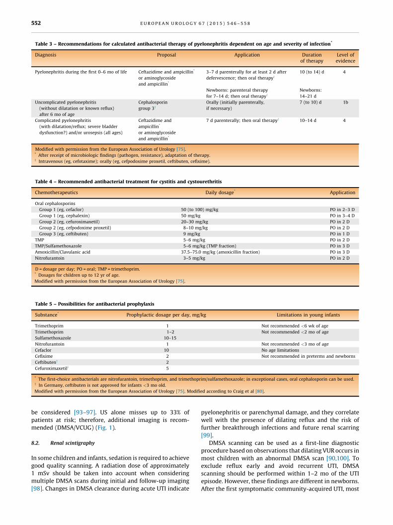
Table 3 – Recommendations for calculated antibacterial therapy of pyelonephritis dependent on age and severity of infection*

Diagnosis Proposal Application Durationof therapy

Level ofevidence

Pyelonephritis during the first 0–6 mo of life Ceftazidime and ampicillin*

or aminoglycoside

and ampicillin*

3–7 d parenterally for at least 2 d after

defervescence; then oral therapyy

Newborns: parenteral therapy

for 7–14 d; then oral therapyy

10 (to 14) d

Newborns:

14–21 d

4

Uncomplicated pyelonephritis

(without dilatation or known reflux)

after 6 mo of age

Cephalosporin

group 3yOrally (initially parenterally,

if necessary)

7 (to 10) d 1b

Complicated pyelonephritis

(with dilatation/reflux; severe bladder

dysfunction?) and/or urosepsis (all ages)

Ceftazidime and

ampicillin*

or aminoglycoside

and ampicillin*

7 d parenterally; then oral therapyy 10–14 d 4

Modified with permission from the European Association of Urology [75].* After receipt of microbiologic findings (pathogen, resistance), adaptation of therapy.y Intravenous (eg, cefotaxime); orally (eg, cefpodoxime proxetil, ceftibuten, cefixime).

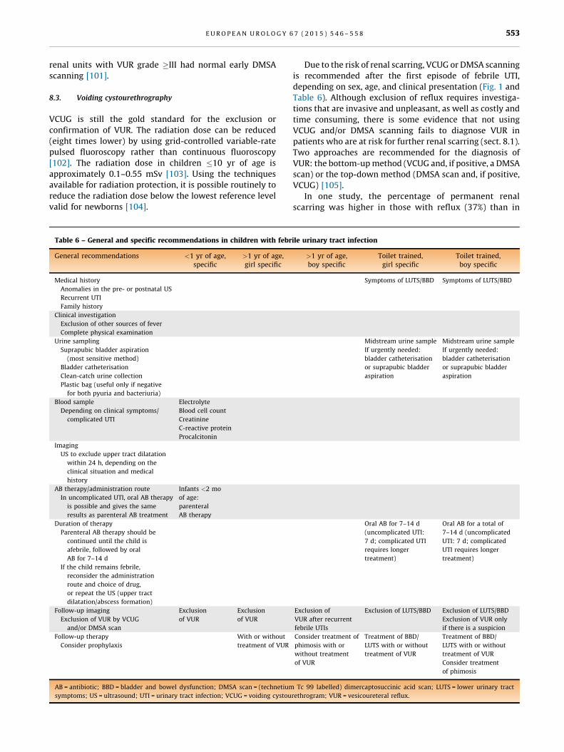
Table 4 – Recommended antibacterial treatment for cystitis and cystourethritis

Chemotherapeutics Daily dosage* Application

Oral cephalosporins

Group 1 (eg, cefaclor) 50 (to 100) mg/kg PO in 2–3 D

Group 1 (eg, cephalexin) 50 mg/kg PO in 3–4 D

Group 2 (eg, cefuroximaxetil) 20–30 mg/kg PO in 2 D

Group 2 (eg, cefpodoxime proxetil) 8–10 mg/kg PO in 2 D

Group 3 (eg, ceftibuten) 9 mg/kg PO in 1 D

TMP 5–6 mg/kg PO in 2 D

TMP/Sulfamethoxazole 5–6 mg/kg (TMP fraction) PO in 3 D

Amoxicillin/Clavulanic acid 37.5–75.0 mg/kg (amoxicillin fraction) PO in 3 D

Nitrofurantoin 3–5 mg/kg PO in 2 D

D = dosage per day; PO = oral; TMP = trimethoprim.* Dosages for children up to 12 yr of age.

Modified with permission from the European Association of Urology [75].

Table 5 – Possibilities for antibacterial prophylaxis

Substance* Prophylactic dosage per day, mg/kg Limitations in young infants

Trimethoprim 1 Not recommended <6 wk of age

Trimethoprim

Sulfamethoxazole

1–2

10–15

Not recommended <2 mo of age

Nitrofurantoin 1 Not recommended <3 mo of age

Cefaclor 10 No age limitations

Cefixime 2 Not recommended in preterms and newborns

Ceftibuteny 2

Cefuroximaxetily 5

* The first-choice antibacterials are nitrofurantoin, trimethoprim, and trimethoprim/sulfamethoxazole; in exceptional cases, oral cephalosporin can be used.y In Germany, ceftibuten is not approved for infants <3 mo old.

Modified with permission from the European Association of Urology [75]. Modified according to Craig et al [80].

E U R O P E A N U R O L O G Y 6 7 ( 2 0 1 5 ) 5 4 6 – 5 5 8552

be considered [93–97]. US alone misses up to 33% of

patients at risk; therefore, additional imaging is recom-

mended (DMSA/VCUG) (Fig. 1).

8.2. Renal scintigraphy

In some children and infants, sedation is required to achieve

good quality scanning. A radiation dose of approximately

1 mSv should be taken into account when considering

multiple DMSA scans during initial and follow-up imaging

[98]. Changes in DMSA clearance during acute UTI indicate

pyelonephritis or parenchymal damage, and they correlate

well with the presence of dilating reflux and the risk of

further breakthrough infections and future renal scarring

[99].

DMSA scanning can be used as a first-line diagnostic

procedure based on observations that dilating VUR occurs in

most children with an abnormal DMSA scan [90,100]. To

exclude reflux early and avoid recurrent UTI, DMSA

scanning should be performed within 1–2 mo of the UTI

episode. However, these findings are different in newborns.

After the first symptomatic community-acquired UTI, most

E U R O P E A N U R O L O G Y 6 7 ( 2 0 1 5 ) 5 4 6 – 5 5 8 553

renal units with VUR grade �III had normal early DMSA

scanning [101].

8.3. Voiding cystourethrography

VCUG is still the gold standard for the exclusion or

confirmation of VUR. The radiation dose can be reduced

(eight times lower) by using grid-controlled variable-rate

pulsed fluoroscopy rather than continuous fluoroscopy

[102]. The radiation dose in children �10 yr of age is

approximately 0.1–0.55 mSv [103]. Using the techniques

available for radiation protection, it is possible routinely to

reduce the radiation dose below the lowest reference level

valid for newborns [104].

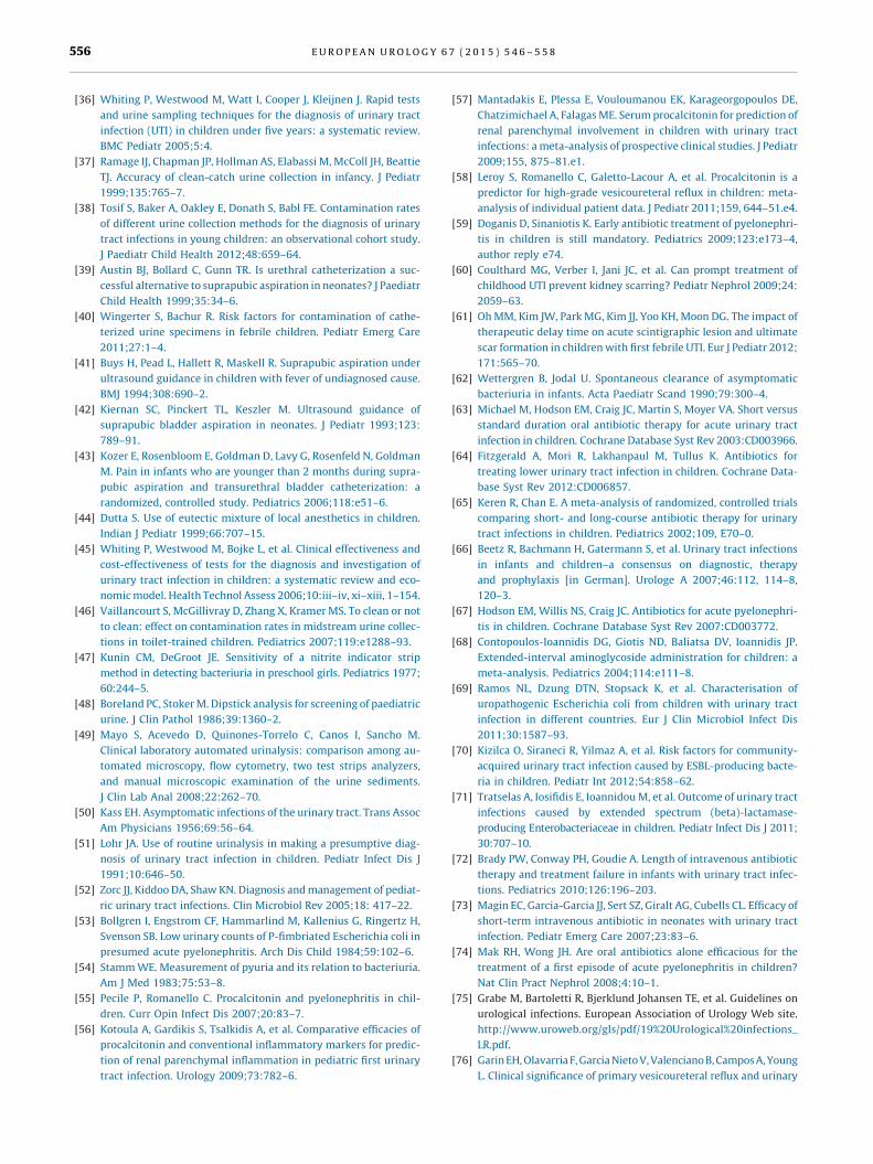
Table 6 – General and specific recommendations in children with febr

General recommendations <1 yr of age,specific

>1 yr of age,girl specific

Medical history

Anomalies in the pre- or postnatal US

Recurrent UTI

Family history

Clinical investigation

Exclusion of other sources of fever

Complete physical examination

Urine sampling

Suprapubic bladder aspiration

(most sensitive method)

Bladder catheterisation

Clean-catch urine collection

Plastic bag (useful only if negative

for both pyuria and bacteriuria)

Blood sample

Depending on clinical symptoms/

complicated UTI

Electrolyte

Blood cell count

Creatinine

C-reactive protein

Procalcitonin

Imaging

US to exclude upper tract dilatation

within 24 h, depending on the

clinical situation and medical

history

AB therapy/administration route

In uncomplicated UTI, oral AB therapy

is possible and gives the same

results as parenteral AB treatment

Infants <2 mo

of age:

parenteral

AB therapy

Duration of therapy

Parenteral AB therapy should be

continued until the child is

afebrile, followed by oral

AB for 7–14 d

If the child remains febrile,

reconsider the administration

route and choice of drug,

or repeat the US (upper tract

dilatation/abscess formation)

Follow-up imaging

Exclusion of VUR by VCUG

and/or DMSA scan

Exclusion

of VUR

Exclusion

of VUR

Follow-up therapy

Consider prophylaxis

With or without

treatment of VUR

AB = antibiotic; BBD = bladder and bowel dysfunction; DMSA scan = (technetium

symptoms; US = ultrasound; UTI = urinary tract infection; VCUG = voiding cystou

Due to the risk of renal scarring, VCUG or DMSA scanning

is recommended after the first episode of febrile UTI,

depending on sex, age, and clinical presentation (Fig. 1 and

Table 6). Although exclusion of reflux requires investiga-

tions that are invasive and unpleasant, as well as costly and

time consuming, there is some evidence that not using

VCUG and/or DMSA scanning fails to diagnose VUR in

patients who are at risk for further renal scarring (sect. 8.1).

Two approaches are recommended for the diagnosis of

VUR: the bottom-up method (VCUG and, if positive, a DMSA

scan) or the top-down method (DMSA scan and, if positive,

VCUG) [105].

In one study, the percentage of permanent renal

scarring was higher in those with reflux (37%) than in

ile urinary tract infection

>1 yr of age,boy specific

Toilet trained,girl specific

Toilet trained,boy specific

Symptoms of LUTS/BBD Symptoms of LUTS/BBD

Midstream urine sample

If urgently needed:

bladder catheterisation

or suprapubic bladder

aspiration

Midstream urine sample

If urgently needed:

bladder catheterisation

or suprapubic bladder

aspiration

Oral AB for 7–14 d

(uncomplicated UTI:

7 d; complicated UTI

requires longer

treatment)

Oral AB for a total of

7–14 d (uncomplicated

UTI: 7 d; complicated

UTI requires longer

treatment)

Exclusion of

VUR after recurrent

febrile UTIs

Exclusion of LUTS/BBD Exclusion of LUTS/BBD

Exclusion of VUR only

if there is a suspicion

Consider treatment of

phimosis with or

without treatment

of VUR

Treatment of BBD/

LUTS with or without

treatment of VUR

Treatment of BBD/

LUTS with or without

treatment of VUR

Consider treatment

of phimosis

Tc 99 labelled) dimercaptosuccinic acid scan; LUTS = lower urinary tract

rethrogram; VUR = vesicoureteral reflux.

[(Fig._1)TD$FIG]

Fig. 1 – Algorithm for assessment and treatment of first febrile urinary tract infection.BBD = Bladder Bowel Dysfunction; DMSA = dimercaptosuccinic acid; IV = intravenous; MRI = magnetic resonance imaging; UTI = urinary tract infection;VCUG = voiding cystourethrography; VUR = vesicoureteral reflux.

E U R O P E A N U R O L O G Y 6 7 ( 2 0 1 5 ) 5 4 6 – 5 5 8554

those without reflux (12%), even if the delay between the

onset of symptoms and treatment was shorter for those

with reflux (4.3 � 1.8 d) than for those without reflux

(4.9 � 2.4 d) [106].

The timing of VCUG does not influence the presence or

severity of VUR [107,108]. Performance of early VCUG in

patients with proven sterile urine does not cause any

significant morbidity [109,110]. VCUG should be performed

after UTI has been treated. To date, no randomised study

has demonstrated that it is safe to perform VCUG during

ongoing UTI and that the results of VCUG change the

treatment.

9. Bladder and bowel dysfunction

BBD is a risk factor for which every child with UTI should be

screened at presentation. Correction of lower urinary tract

dysfunction is important to decrease the rate of UTI

recurrence. If there are signs of BBD during infection-free

intervals, further diagnosis and effective treatment are

strongly recommended [111–114]. Treatment of constipa-

tion leads to a decrease in UTI recurrence [115–117]. Exclu-

sion of BBD is therefore strongly recommended in any child

with febrile and/or recurrent UTI, and, if present, treatment

of BBD is necessary [118].

10. Conclusions

Figure 1 and Table 6 summarise the general recommenda-

tions:

� C

lassification of a UTI is made according to the site,

episode, symptoms, and complicating factors. For acute

treatment, the site and severity are of the most

importance.

� Im

mediate US of the kidney and bladder are necessary in

patients with febrile UTI to exclude underlying uropathy.

� T

reatment of patients with febrile UTIs should be

initiated after urine analysis and culture to confirm the

diagnosis.

� S

PA and catheterisation have the lowest contamination

rate for urine sampling. Using a plastic bag (most

commonly used in daily practice), UTI can be excluded

if the dipstick is negative for both leukocyte esterase and

nitrite or microscopic analysis is negative for both pyuria

and bacteriuria.

� P

rophylaxis has been shown to be beneficial in prevent-

ing new renal scars in infant girls with dilating reflux III

and IV. Reflux should be excluded in patients with febrile

UTIs.

� In

toilet-trained children, BBD should be excluded.

Author contributions: Raimund Stein had full access to all the data in the

study and takes responsibility for the integrity of the data and the

accuracy of the data analysis.

Study concept and design: Stein, Dogan, Hoebeke, Kocvara, Nijman,

Radmayr, Tekgul.

Acquisition of data: Stein, Dogan, Hoebeke, Kocvara, Nijman, Radmayr,

Tekgul.

Analysis and interpretation of data: Stein, Dogan, Hoebeke, Kocvara,

Nijman, Radmayr, Tekgul.

Drafting of the manuscript: Stein, Dogan, Hoebeke, Kocvara, Nijman,

Radmayr, Tekgul.

Critical revision of the manuscript for important intellectual content: Stein,

Dogan, Hoebeke, Kocvara, Nijman, Radmayr, Tekgul.

Statistical analysis: No statistical analysis was carried out.

E U R O P E A N U R O L O G Y 6 7 ( 2 0 1 5 ) 5 4 6 – 5 5 8 555

Obtaining funding: Tekgul.

Administrative, technical, or material support: Stein, Nijman, Tekgul.

Supervision: Stein, Nijman, Tekgul.

Other (specify): None.

Financial disclosures: Raimund Stein certifies that all conflicts of interest,

including specific financial interests and relationships and affiliations

relevant to the subject matter or materials discussed in the manuscript

(eg, employment/affiliation, grants or funding, consultancies, honoraria,

stock ownership or options, expert testimony, royalties, or patents filed,

received, or pending), are the following: Raimund Stein is a company

consultant for Grachtenhaus Apotheke, and participates in trials for

Bayer Health Care on gonadal function, sexual function, and quality of

life in patients with spina bifida. Radim Kocvara participates in trials for

Ferring Pharmaceuticals CZ and receives fellowships and travel grants

from B. Braun Medical and Ferring Pharmaceuticals CZ. Serdar Tekgul

participates in trials for Sanofi Aventis, Astellas, Sanofi, and Pfizer. The

other authors have nothing to disclose.

Funding/Support and role of the sponsor: None.

References

[1] Sastre JB, Aparicio AR, Cotallo GD, Colomer BF, Hernandez MC.

Urinary tract infection in the newborn: clinical and radio imaging

studies. Pediatr Nephrol 2007;22:1735–41.

[2] Jakobsson B, Svensson L. Transient pyelonephritic changes on

99mTechnetium-dimercaptosuccinic acid scan for at least five

months after infection. Acta Paediatr 1997;86:803–7.

[3] Benador D, Benador N, Slosman D, Mermillod B, Girardin E. Are

younger children at highest risk of renal sequelae after pyelone-

phritis? Lancet 1997;349:17–9.

[4] Hewitt IK, Zucchetta P, Rigon L, et al. Early treatment of acute

pyelonephritis in children fails to reduce renal scarring: data from

the Italian Renal Infection Study Trials. Pediatrics 2008;122:486–90.

[5] Fotter R. Paediatric uroradiology. In: Baert AL, Sartor K, editors.

Medical radiology: diagnostic imaging. Berlin, Germany: Springer;

2001.

[6] Sacks SH, Verrier Jones K, Roberts R, Asscher AW, Ledingham JG.

Effect of symptomless bacteriuria in childhood on subsequent

pregnancy. Lancet 1987;2:991–4.

[7] Jacobson SH, Eklof O, Eriksson CG, Lins LE, Tidgren B, Winberg J.

Development of hypertension and uraemia after pyelonephritis in

childhood: 27 year follow up. BMJ 1989;299:703–6.

[8] Smellie JM, Prescod NP, Shaw PJ, Risdon RA, Bryant TN. Childhood

reflux and urinary infection: a follow-up of 10–41 years in

226 adults. Pediatr Nephrol 1998;12:727–36.

[9] Larcombe J. Urinary tract infection. Clin Evid 2002;7:377–85.

[10] Round J, Fitzgerald AC, Hulme C, Lakhanpaul M, Tullus K. Urinary

tract infections in children and the risk of ESRF. Acta Paediatr

2012;101:278–82.

[11] Stull TL, LiPuma JJ. Epidemiology and natural history of urinary

tract infections in children. Med Clin North Am 1991;75:

287–97.

[12] Marild S, Jodal U. Incidence rate of first-time symptomatic urinary

tract infection in children under 6 years of age. Acta Paediatr

1998;87:549–52.

[13] O’Brien K, Stanton N, Edwards A, Hood K, Butler CC. Prevalence of

urinary tract infection (UTI) in sequential acutely unwell children

presenting in primary care: exploratory study. Scand J Prim Health

Care 2011;29:19–22.

[14] Hoberman A, Chao HP, Keller DM, Hickey R, Davis HW, Ellis D.

Prevalence of urinary tract infection in febrile infants. J Pediatr

1993;123:17–23.

[15] Mangiarotti P, Pizzini C, Fanos V. Antibiotic prophylaxis in chil-

dren with relapsing urinary tract infections: review. J Chemother

2000;12:115–23.

[16] Nuutinen M, Uhari M. Recurrence and follow-up after urinary tract

infection under the age of 1 year. Pediatr Nephrol 2001;16:69–72.

[17] Shaikh N, Morone NE, Bost JE, Farrell MH. Prevalence of urinary

tract infection in childhood: a meta-analysis. Pediatr Infect Dis J

2008;27:302–8.

[18] Zorc JJ, Levine DA, Platt SL, et al. Clinical and demographic factors

associated with urinary tract infection in young febrile infants.

Pediatrics 2005;116:644–8.

[19] Kanellopoulos TA, Salakos C, Spiliopoulou I, Ellina A, Nikolako-

poulou NM, Papanastasiou DA. First urinary tract infection in

neonates, infants and young children: a comparative study.

Pediatr Nephrol 2006;21:1131–7.

[20] Subcommittee on Urinary Tract Infection, Steering Committee on

Quality Improvement and Management, Roberts KB. Urinary tract

infection: clinical practice guideline for the diagnosis and man-

agement of the initial UTI in febrile infants and children 2 to

24 months. Pediatrics 201;128:595–610.

[21] Ammenti A, Cataldi L, Chimenz R, et al. Febrile urinary tract

infections in young children: recommendations for the diagnosis,

treatment and follow-up. Acta Paediatr 2012;101:451–7.

[22] National Institute for Health and Care Excellence (NICE). Urinary

tract infection in children: diagnosis, treatment and long-term

management. NICE Web site. http://www.nice.org.uk/guidance/

cg054.

[23] Shortliffe LMD. Infection and inflammation of the pediatric geni-

tourinary tract. In: Wein AJ, Kavoussi LR, Novick AC, et al., editors.

Campbell-Walsh urology. ed. 9. Philadelphia, PA: Saunders; 2007.

p. 3232–68.

[24] Craig JC, Irwig LM, Knight JF, Sureshkumar P, Roy LP. Symptomatic

urinary tract infection in preschool Australian children. J Paediatr

Child Health 1998;34:154–9.

[25] Burns MW, Burns JL, Krieger JN. Pediatric urinary tract infection.

Diagnosis, classification, and significance. Pediatr Clin North Am

1987;34:1111–20.

[26] Bonadio WA. Urine culturing technique in febrile infants. Pediatr

Emerg Care 1987;3:75–8.

[27] Bauchner H, Philipp B, Dashefsky B, Klein JO. Prevalence of bacte-

riuria in febrile children. Pediatr Infect Dis J 1987;6:239–42.

[28] Roberts KB, Charney E, Sweren RJ, et al. Urinary tract infection in

infants with unexplained fever: a collaborative study. J Pediatr

1983;103:864–7.

[29] Craig JC, Williams GJ, Jones M, et al. The accuracy of clinical symp-

toms and signs for the diagnosis of serious bacterial infection in

young febrile children: prospective cohort study of 15 781 febrile

illnesses. BMJ 2010;340:c1594.

[30] Slater M, Krug SE. Evaluation of the infant with fever without

source: an evidence based approach. Emerg Med Clin North Am

1999;17:97–126, viii–ix.

[31] Beetz R. Evaluation and management of urinary tract infections in

the neonate. Curr Opin Pediatr 2012;24:205–11.

[32] Biyikli NK, Alpay H, Ozek E, Akman I, Bilgen H. Neonatal urinary

tract infections: analysis of the patients and recurrences. Pediatr

Int 2004;46:21–5.

[33] Tutunculer F, Gunoz H, Bas F, Bundak R, Saka N, Neyzi O. Transient

Pseudohypoaldosteronism in an infant with urinary tract anoma-

ly. Pediatr Int 2004;46:618–20.

[34] Nandagopal R, Vaidyanathan P, Kaplowitz P. Transient pseudohy-

poaldosteronism due to urinary tract infection in infancy: a report

of 4 cases. Int J Pediatr Endocrinol 2009;2009:195728.

[35] Tullus K. Difficulties in diagnosing urinary tract infections in small

children. Pediatr Nephrol 2011;26:1923–6.

E U R O P E A N U R O L O G Y 6 7 ( 2 0 1 5 ) 5 4 6 – 5 5 8556

[36] Whiting P, Westwood M, Watt I, Cooper J, Kleijnen J. Rapid tests

and urine sampling techniques for the diagnosis of urinary tract

infection (UTI) in children under five years: a systematic review.

BMC Pediatr 2005;5:4.

[37] Ramage IJ, Chapman JP, Hollman AS, Elabassi M, McColl JH, Beattie

TJ. Accuracy of clean-catch urine collection in infancy. J Pediatr

1999;135:765–7.

[38] Tosif S, Baker A, Oakley E, Donath S, Babl FE. Contamination rates

of different urine collection methods for the diagnosis of urinary

tract infections in young children: an observational cohort study.

J Paediatr Child Health 2012;48:659–64.

[39] Austin BJ, Bollard C, Gunn TR. Is urethral catheterization a suc-

cessful alternative to suprapubic aspiration in neonates? J Paediatr

Child Health 1999;35:34–6.

[40] Wingerter S, Bachur R. Risk factors for contamination of cathe-

terized urine specimens in febrile children. Pediatr Emerg Care

2011;27:1–4.

[41] Buys H, Pead L, Hallett R, Maskell R. Suprapubic aspiration under

ultrasound guidance in children with fever of undiagnosed cause.

BMJ 1994;308:690–2.

[42] Kiernan SC, Pinckert TL, Keszler M. Ultrasound guidance of

suprapubic bladder aspiration in neonates. J Pediatr 1993;123:

789–91.

[43] Kozer E, Rosenbloom E, Goldman D, Lavy G, Rosenfeld N, Goldman

M. Pain in infants who are younger than 2 months during supra-

pubic aspiration and transurethral bladder catheterization: a

randomized, controlled study. Pediatrics 2006;118:e51–6.

[44] Dutta S. Use of eutectic mixture of local anesthetics in children.

Indian J Pediatr 1999;66:707–15.

[45] Whiting P, Westwood M, Bojke L, et al. Clinical effectiveness and

cost-effectiveness of tests for the diagnosis and investigation of

urinary tract infection in children: a systematic review and eco-

nomic model. Health Technol Assess 2006;10:iii–iv, xi–xiii, 1–154.

[46] Vaillancourt S, McGillivray D, Zhang X, Kramer MS. To clean or not

to clean: effect on contamination rates in midstream urine collec-

tions in toilet-trained children. Pediatrics 2007;119:e1288–93.

[47] Kunin CM, DeGroot JE. Sensitivity of a nitrite indicator strip

method in detecting bacteriuria in preschool girls. Pediatrics 1977;

60:244–5.

[48] Boreland PC, Stoker M. Dipstick analysis for screening of paediatric

urine. J Clin Pathol 1986;39:1360–2.

[49] Mayo S, Acevedo D, Quinones-Torrelo C, Canos I, Sancho M.

Clinical laboratory automated urinalysis: comparison among au-

tomated microscopy, flow cytometry, two test strips analyzers,

and manual microscopic examination of the urine sediments.

J Clin Lab Anal 2008;22:262–70.

[50] Kass EH. Asymptomatic infections of the urinary tract. Trans Assoc

Am Physicians 1956;69:56–64.

[51] Lohr JA. Use of routine urinalysis in making a presumptive diag-

nosis of urinary tract infection in children. Pediatr Infect Dis J

1991;10:646–50.

[52] Zorc JJ, Kiddoo DA, Shaw KN. Diagnosis and management of pediat-

ric urinary tract infections. Clin Microbiol Rev 2005;18: 417–22.

[53] Bollgren I, Engstrom CF, Hammarlind M, Kallenius G, Ringertz H,

Svenson SB. Low urinary counts of P-fimbriated Escherichia coli in

presumed acute pyelonephritis. Arch Dis Child 1984;59:102–6.

[54] Stamm WE. Measurement of pyuria and its relation to bacteriuria.

Am J Med 1983;75:53–8.

[55] Pecile P, Romanello C. Procalcitonin and pyelonephritis in chil-

dren. Curr Opin Infect Dis 2007;20:83–7.

[56] Kotoula A, Gardikis S, Tsalkidis A, et al. Comparative efficacies of

procalcitonin and conventional inflammatory markers for predic-

tion of renal parenchymal inflammation in pediatric first urinary

tract infection. Urology 2009;73:782–6.

[57] Mantadakis E, Plessa E, Vouloumanou EK, Karageorgopoulos DE,

Chatzimichael A, Falagas ME. Serum procalcitonin for prediction of

renal parenchymal involvement in children with urinary tract

infections: a meta-analysis of prospective clinical studies. J Pediatr

2009;155, 875–81.e1.

[58] Leroy S, Romanello C, Galetto-Lacour A, et al. Procalcitonin is a

predictor for high-grade vesicoureteral reflux in children: meta-

analysis of individual patient data. J Pediatr 2011;159, 644–51.e4.

[59] Doganis D, Sinaniotis K. Early antibiotic treatment of pyelonephri-

tis in children is still mandatory. Pediatrics 2009;123:e173–4,

author reply e74.

[60] Coulthard MG, Verber I, Jani JC, et al. Can prompt treatment of

childhood UTI prevent kidney scarring? Pediatr Nephrol 2009;24:

2059–63.

[61] Oh MM, Kim JW, Park MG, Kim JJ, Yoo KH, Moon DG. The impact of

therapeutic delay time on acute scintigraphic lesion and ultimate

scar formation in children with first febrile UTI. Eur J Pediatr 2012;

171:565–70.

[62] Wettergren B, Jodal U. Spontaneous clearance of asymptomatic

bacteriuria in infants. Acta Paediatr Scand 1990;79:300–4.

[63] Michael M, Hodson EM, Craig JC, Martin S, Moyer VA. Short versus

standard duration oral antibiotic therapy for acute urinary tract

infection in children. Cochrane Database Syst Rev 2003:CD003966.

[64] Fitzgerald A, Mori R, Lakhanpaul M, Tullus K. Antibiotics for

treating lower urinary tract infection in children. Cochrane Data-

base Syst Rev 2012:CD006857.

[65] Keren R, Chan E. A meta-analysis of randomized, controlled trials

comparing short- and long-course antibiotic therapy for urinary

tract infections in children. Pediatrics 2002;109, E70–0.

[66] Beetz R, Bachmann H, Gatermann S, et al. Urinary tract infections

in infants and children–a consensus on diagnostic, therapy

and prophylaxis [in German]. Urologe A 2007;46:112, 114–8,

120–3.

[67] Hodson EM, Willis NS, Craig JC. Antibiotics for acute pyelonephri-

tis in children. Cochrane Database Syst Rev 2007:CD003772.

[68] Contopoulos-Ioannidis DG, Giotis ND, Baliatsa DV, Ioannidis JP.

Extended-interval aminoglycoside administration for children: a

meta-analysis. Pediatrics 2004;114:e111–8.

[69] Ramos NL, Dzung DTN, Stopsack K, et al. Characterisation of

uropathogenic Escherichia coli from children with urinary tract

infection in different countries. Eur J Clin Microbiol Infect Dis

2011;30:1587–93.

[70] Kizilca O, Siraneci R, Yilmaz A, et al. Risk factors for community-

acquired urinary tract infection caused by ESBL-producing bacte-

ria in children. Pediatr Int 2012;54:858–62.

[71] Tratselas A, Iosifidis E, Ioannidou M, et al. Outcome of urinary tract

infections caused by extended spectrum (beta)-lactamase-

producing Enterobacteriaceae in children. Pediatr Infect Dis J 2011;

30:707–10.

[72] Brady PW, Conway PH, Goudie A. Length of intravenous antibiotic

therapy and treatment failure in infants with urinary tract infec-

tions. Pediatrics 2010;126:196–203.

[73] Magin EC, Garcia-Garcia JJ, Sert SZ, Giralt AG, Cubells CL. Efficacy of

short-term intravenous antibiotic in neonates with urinary tract

infection. Pediatr Emerg Care 2007;23:83–6.

[74] Mak RH, Wong JH. Are oral antibiotics alone efficacious for the

treatment of a first episode of acute pyelonephritis in children?

Nat Clin Pract Nephrol 2008;4:10–1.

[75] Grabe M, Bartoletti R, Bjerklund Johansen TE, et al. Guidelines on

urological infections. European Association of Urology Web site.

http://www.uroweb.org/gls/pdf/19%20Urological%20infections_

LR.pdf.

[76] Garin EH, Olavarria F, Garcia Nieto V, Valenciano B, Campos A, Young

L. Clinical significance of primary vesicoureteral reflux and urinary

E U R O P E A N U R O L O G Y 6 7 ( 2 0 1 5 ) 5 4 6 – 5 5 8 557

antibiotic prophylaxis after acute pyelonephritis: a multicenter,

randomized, controlled study. Pediatrics 2006;117:626–32.

[77] Montini G, Rigon L, Zucchetta P, et al. Prophylaxis after first febrile

urinary tract infection in children? A multicenter, randomized,

controlled, noninferiority trial. Pediatrics 2008;122:1064–71.

[78] Pennesi M, Travan L, Peratoner L, et al. Is antibiotic prophylaxis in

children with vesicoureteral reflux effective in preventing pyelo-

nephritis and renal scars? A randomized, controlled trial. Pediat-

rics 2008;121:e1489–94.

[79] Roussey-Kesler G, Gadjos V, Idres N, et al. Antibiotic prophylaxis

for the prevention of recurrent urinary tract infection in children

with low grade vesicoureteral reflux: results from a prospective

randomized study. J Urol 2008;179:674–9, discussion 679.

[80] Craig JC, Simpson JM, Williams GJ, et al. Antibiotic prophylaxis and

recurrent urinary tract infection in children. N Engl J Med 2009;

361:1748–59.

[81] Brandstrom P, Jodal U, Sillen U, Hansson S. The Swedish reflux

trial: review of a randomized, controlled trial in children with

dilating vesicoureteral reflux. J Pediatr Urol 2011;7:594–600.

[82] Park S, Song SH, Lee C, Kim JW, Kim KS. Bacterial pathogens in first

febrile urinary tract infection affect breakthrough infections in

infants with vesicoureteral reflux treated with prophylactic anti-

biotics. Urology 2013;81:1342–5.

[83] RIVUR Trial Investigators. Hoberman A, Greenfield SP, Mattoo TK,

et al. Antimicrobial prophylaxis for children with vesicoureteral

reflux. N Engl J Med 2014;370:2367–76.

[84] Salo J, Uhari M, Helminen M, et al. Cranberry juice for the prevention

of recurrences of urinary tract infections in children: a randomized

placebo-controlled trial. Clin Infect Dis 2012;54:340–6.

[85] Afshar K, Stothers L, Scott H, MacNeily AE. Cranberry juice for the

prevention of pediatric urinary tract infection: a randomized

controlled trial. J Urol 2012;188(Suppl):1584–7.

[86] Smyth AR, Judd BA. Compliance with antibiotic prophylaxis in

urinary tract infection. Arch Dis Child 1993;68:235–6.

[87] Copp HL, Nelson CP, Shortliffe LD, Lai J, Saigal CS, Kennedy WA.

Compliance with antibiotic prophylaxis in children with vesicour-

eteral reflux: results from a national pharmacy claims database.

J Urol 2010;183:1994–9.

[88] Hensle TW, Hyun G, Grogg AL, Eaddy M. Part 2: Examining pediatric

vesicoureteral reflux: a real-world evaluation of treatment patterns

and outcomes. Curr Med Res Opin 2007;23(Suppl 4):S7–13.

[89] Yiee JH, Baskin LS, Patel N, Yang J, Disandro M. Prospective blinded

laboratory assessment of prophylactic antibiotic compliance in a

pediatric outpatient setting. J Urol 2012;187:2176–81.

[90] Preda I, Jodal U, Sixt R, Stokland E, Hansson S. Value of ultrasound

in evaluation of infants with first urinary tract infection. J Urol

2010;183:1984–8.

[91] Juliano TM, Stephany HA, Clayton DB, et al. Incidence of abnormal

imaging and recurrent pyelonephritis after first febrile urinary tract

infection in children 2 to 24 months old. J Urol 2013;190(Suppl):

1501–10.

[92] Massanyi EZ, Preece J, Gupta A, Lin SM, Wang MH. Utility of

screening ultrasound after first febrile UTI among patients with

clinically significant vesicoureteral reflux. Urology 2013;82:905–9.

[93] Burgers R, de Jong TP, Benninga MA. Rectal examination in children:

digital versus transabdominal ultrasound. J Urol 2013;190:

667–72.

[94] Berger MY, Tabbers MM, Kurver MJ, Boluyt N, Benninga MA. Value

of abdominal radiography, colonic transit time, and rectal ultra-

sound scanning in the diagnosis of idiopathic constipation in

children: a systematic review. J Pediatr 2012;161:44–50.e1–2.

[95] Joensson IM, Siggaard C, Rittig S, Hagstroem S, Djurhuus JC.

Transabdominal ultrasound of rectum as a diagnostic tool in

childhood constipation. J Urol 2008;179:1997–2002.

[96] Singh SJ, Gibbons NJ, Vincent MV, Sithole J, Nwokoma NJ, Alagars-

wami KV. Use of pelvic ultrasound in the diagnosis of megarectum

in children with constipation. J Pediatr Surg 2005;40:1941–4.

[97] Klijn AJ, Asselman M, Vijverberg MA, Dik P, de Jong TP. The

diameter of the rectum on ultrasonography as a diagnostic tool

for constipation in children with dysfunctional voiding. J Urol

2004;172:1986–8.

[98] Smith T, Evans K, Lythgoe MF, Anderson PJ, Gordon I. Radiation

dosimetry of technetium-99m-DMSA in children. J Nucl Med 1996;

37:1336–42.

[99] Shiraishi K, Yoshino K, Watanabe M, Matsuyama H, Tanikaze S.

Risk factors for breakthrough infection in children with primary

vesicoureteral reflux. J Urol 2010;183:1527–31.

[100] Quirino IG, Silva JM, Diniz JS, et al. Combined use of late phase

dimercapto-succinic acid renal scintigraphy and ultrasound as

first line screening after urinary tract infection in children. J Urol

2011;185:258–63.

[101] Siomou E, Giapros V, Fotopoulos A, et al. Implications of 99mTc-

DMSA scintigraphy performed during urinary tract infection in

neonates. Pediatrics 2009;124:881–7.

[102] Ward VL, Strauss KJ, Barnewolt CE, et al. Pediatric radiation

exposure and effective dose reduction during voiding cystoure-

thrography. Radiology 2008;249:1002–9.

[103] Lee R, Thomas KE, Connolly BL, Falkiner M, Gordon CL. Effective

dose estimation for pediatric voiding cystourethrography using an

anthropomorphic phantom set and metal oxide semiconductor

field-effect transistor (MOSFET) technology. Pediatr Radiol 2009;39:

608–15.

[104] Born M, Spiller L, Bachour H, Heydweiller A, Franke I. Dose area

product of pediatric VCUG with regard to the strongly lowered

German diagnostic reference levels [in German]. Rofo 2013;185:

262–7.

[105] Tekgul S, Riedmiller H, Hoebeke P, et al. EAU guidelines on

vesicoureteral reflux in children. Eur Urol 2012;62:534–42.

[106] Oh MM, Kim JW, Park MG, Kim JJ, Yoo KH, Moon du G. The impact

of therapeutic delay time on acute scintigraphic lesion and ulti-

mate scar formation in children with first febrile UTI. Eur J Pediatr

2012;171:565–70.

[107] Doganis D, Mavrikou M, Delis D, Stamoyannou L, Siafas K,

Sinaniotis K. Timing of voiding cystourethrography in infants with

first time urinary infection. Pediatr Nephrol 2009;24:319–22.

[108] Sathapornwajana P, Dissaneewate P, McNeil E, Vachvanichsanong

P. Timing of voiding cystourethrogram after urinary tract infec-

tion. Arch Dis Child 2008;93:229–31.

[109] Spencer JD, Bates CM, Mahan JD, et al. The accuracy and health

risks of a voiding cystourethrogram after a febrile urinary tract

infection. J Pediatr Urol 2012;8:72–6.

[110] Mahyar A, Ayazi P, Tarlan S, Moshiri A, Hamidfar M, Barikani A.

When is the best time for voiding cystourethrogram in urinary

tract infection of children? Acta Med Iran 2012;50:468–72.

[111] Koff SA, Wagner TT, Jayanthi VR. The relationship among dysfunc-

tional elimination syndromes, primary vesicoureteral reflux and

urinary tract infections in children. J Urol 1998; 160:1019–22.

[112] Hoebeke P, Vande Walle J, Everaert K, Van Laecke E, Van Gool JD.

Assessment of lower urinary tract dysfunction in children with

non-neuropathic bladder sphincter dysfunction. Eur Urol 1999;

35:57–69.

[113] van Gool JD. Dysfunctional voiding: a complex of bladder/sphincter

dysfunction, urinary tract infections and vesicoureteral reflux. Acta

Urol Belg 1995;63:27–33.

[114] van Gool JD, Kuitjen RH, Donckerwolcke RA, Messer AP, Vijverberg

M. Bladder-sphincter dysfunction, urinary infection and vesico-

ureteral reflux with special reference to cognitive bladder train-

ing. Contrib Nephrol 1984;39:190–210.

E U R O P E A N U R O L O G Y 6 7 ( 2 0 1 5 ) 5 4 6 – 5 5 8558

[115] De Paepe H, Renson C, Van Laecke E, Raes A, Vande Walle J,

Hoebeke P. Pelvic-floor therapy and toilet training in young chil-

dren with dysfunctional voiding and obstipation. BJU Int 2000;

85:889–93.

[116] Loening-Baucke V. Urinary incontinence and urinary tract infec-

tion and their resolution with treatment of chronic constipation of

childhood. Pediatrics 1997;100:228–32.

[117] O’Regan S, Yazbeck S, Schick E. Constipation, bladder instabili-

ty, urinary tract infection syndrome. Clin Nephrol 1985;23:

152–4.

[118] Tekgul S, Dogan HS, Hoebeke P, et al. Guidelines on paediatric

urology. European Association of Urology Web site. http://www.

uroweb.org/gls/pdf/23%20Paediatric%20Urology_LR%20March%

2025th.pdf.


Recommended