Evidence for both a regulatory mutation and a structural mutation in a family with maple syrup urine disease. B Zhang, … , D W Crabb, R A Harris J Clin Invest. 1989; 83(4):1425-1429. https://doi.org/10.1172/JCI114033. Maple syrup urine disease (MSUD) results from a deficiency of branched chain alpha- ketoacid dehydrogenase (BCKDH). We have studied the etiology of MSUD by determining the enzyme activity, protein, and mRNA levels of BCKDH in fibroblasts from a classic MSUD patient and his parents. By enzymatic amplification of the patient's mRNA followed by cloning and DNA sequencing, we have identified a T to A transversion that alters a tyrosine to an asparagine at residue 394 of the E1 alpha subunit. Amplification of both mRNA and genomic DNA, in combination with allele-specific oligonucleotide hybridization, demonstrated that the father was heterozygous for this mutant allele. The mother was homozygous for the allele encoding the normal Tyr394, but expressed only about half of the normal level of mRNA and protein. The patient was genetically heterozygous for this altered allele, although only the abnormal allele was expressed as mRNA. We conclude that the patient was a compound heterozygote, inheriting an allele encoding an abnormal E1 alpha from the father, and an allele from the mother containing a cis-acting defect in regulation which abolished the expression of one of the E1 alpha alleles. Our results revealed for the first time that a case of MSUD was caused by structural and regulatory mutations involving the E1 alpha subunit. Research Article Find the latest version: http://jci.me/114033-pdf
Transcript
Evidence for both a regulatory mutation and astructural mutation in a family with maple syrupurine disease.
Maple syrup urine disease (MSUD) results from a deficiency of branched chain alpha-ketoacid dehydrogenase (BCKDH). We have studied the etiology of MSUD by determiningthe enzyme activity, protein, and mRNA levels of BCKDH in fibroblasts from a classicMSUD patient and his parents. By enzymatic amplification of the patient's mRNA followedby cloning and DNA sequencing, we have identified a T to A transversion that alters atyrosine to an asparagine at residue 394 of the E1 alpha subunit. Amplification of bothmRNA and genomic DNA, in combination with allele-specific oligonucleotide hybridization,demonstrated that the father was heterozygous for this mutant allele. The mother washomozygous for the allele encoding the normal Tyr394, but expressed only about half of thenormal level of mRNA and protein. The patient was genetically heterozygous for this alteredallele, although only the abnormal allele was expressed as mRNA. We conclude that thepatient was a compound heterozygote, inheriting an allele encoding an abnormal E1 alphafrom the father, and an allele from the mother containing a cis-acting defect in regulationwhich abolished the expression of one of the E1 alpha alleles. Our results revealed for thefirst time that a case of MSUD was caused by structural and regulatory mutations involvingthe E1 alpha subunit.
Evidence for Both a Regulatory Mutation and a StructuralMutation in a Family with Maple Syrup Urine DiseaseBei Zhang,* Howard J. Edenberg,** David W. Crabb,*1 and Robert A. Harris*Departments of Biochemistry,* Medicine) and Medical Genetics,t Indiana University School of Medicine, Indianapolis, Indiana 46223
Abstract
Maple syrup urine disease (MSUD) results from a deficiencyof branched chain a-ketoacid dehydrogenase (BCKDH). Wehave studied the etiology of MSUDby determining the enzymeactivity, protein, and mRNAlevels of BCKDHin fibroblastsfrom a classic MSUDpatient and his parents. By enzymaticamplification of the patient's mRNAfollowed by cloning andDNAsequencing, we have identified a T to A transversion thatalters a tyrosine to an asparagine at residue 394 of the Elasubunit. Amplification of both mRNAand genomic DNA, incombination with allele-specific oligonucleotide hybridization,demonstrated that the father was heterozygous for this mutantallele. The mother was homozygous for the allele encoding thenormal Tyr394, but expressed only about half of the normallevel of mRNAand protein. The patient was genetically het-erozygous for this altered allele, although only the abnormalallele was expressed as mRNA.Weconclude that the patientwas a compound heterozygote, inheriting an allele encoding anabnormal Ela from the father, and an allele from the mothercontaining a cis-acting defect in regulation which abolished theexpression of one of the Ela alleles. Our results revealed forthe first time that a case of MSUDwas caused by structuraland regulatory mutations involving the Ela subunit.
Introduction
Branched chain a-ketoacid dehydrogenase (BCKDH)' cata-lyzes the rate-limiting step in the catabolism of the branchedchain amino acids (1, 2). Covalent modification via phosphor-ylation by a specific kinase and dephosphorylation by a spe-cific phosphatase has been established as an important mecha-nism for regulation of BCKDHactivity (3-5). The enzyme
Address reprint requests to Dr. Harris, Department of Biochemistry,Indiana University School of Medicine, 635 Barnhill Drive, Indiana-polis, IN 46223.
Receivedfor publication 16 December 1988.
1. Abbreviations used in this paper: BCKDH, branched chain a-keto-acid dehydrogenase; E 1, decarboxylase component of BCKDH;Ela, asubunit of E1; EIfl, fi subunit of E1; E2, acyl-transferase component ofBCKDH; E3, dihydrolipoyl dehydrogenase; KIV, a-keto-isovalericacid; MSUD,maple syrup urine disease; PCR, polymerase chain reac-tion.
complex consists of 2-oxoisovalerate dehydrogenase (E 1, com-posed of Ela and Elfl, EC 1.2.4.4.), dihydrolipoamide acyl-transferase (E2, no ECnumber), and dihydrolipoamide dehy-drogenase (E3, EC 1.8.1.4.). Ela is probably the catalytic sub-unit, and it contains the phosphorylation sites (two serineresidues) responsible for regulation of the enzyme by covalentmodification (6).
Maple syrup urine disease (MSUD), inherited in an auto-somal recessive fashion, is caused by a deficiency of BCKDH(7). The disease is characterized by ketoacidosis and mentalretardation, with the molecular defects most often (8) but notexclusively (9, 10) in the El component. Four clinically differ-ent forms of this disease have been described: classic, thiamin-responsive, intermediate, and intermittent (7). To gain insightinto the structure and mechanism of regulation of BCKDHcomplex and to define the genetic defects of MSUD,we clonedcDNAs encoding BCKDHElCa subunit from both rat andhuman liver (1 1, 12). Wereport here the first determination atthe DNAand RNAlevel of the mutations causing a case ofclassic MSUD: two different mutant alleles of the EI a gene.
Methods
Radioisotopes and chemicals. a-Keto[ 1-'4C]isovaleric acid([l-'4C]KIV) was produced from [1-'4C]valine obtained from Re-search Products International Corp. (Mount Prospect, IL) (13). Mostmolecular cloning reagents and enzymes were from Bethesda ResearchLaboratories (Gaithersburg, MD). Taq DNApolymerase was fromPerkin Elmer Cetus (Norwalk, CT). The nick-translation kit was fromAmersham Corp. (Arlington Heights, IL), and radiolabeled nucleo-tides were from Du Pont-New England Research Products (Bos-ton, MA).
Cell strains and culture. Fibroblasts derived from a patient withclassic MSUD(GM649) and his father (GM650) and mother (GM65 1)were obtained from NIGMSHumanGenetic Mutant Cell Repository.Normal human fibroblast cell line was obtained from American TypeCulture Collection (Rockville, MD). Fibroblasts were grown in mono-layer culture in DME(Gibco, Grand Island, NY) containing 10%(vol/vol) fetal calf serum (FCS, Sigma Chemical Co., St. Louis, MO)and antibiotics (penicillin, 100 U/ml; streptomycin, 100 gg/ml) at37°C in 5%CO2atmosphere. The cells were grown to confluency andharvested by trypsinization 3-6 d after the last medium change.
Enzyme assay. Cells were collected by trypsinization, washed withPBS containing 10% FCS, and incubated with 1 mMa-chloroisoca-prate in the culture medium at 37°C for 15 min to activate BCKDHcomplex by dephosphorylation. Extracts of the cells were prepared byfreeze-thawing in complete Krebs-Ringer buffer. BCKDHtotal activ-ity was measured radiochemically at 37°C with [1-'4C]KIV as sub-strate (14).
Western blot. Crude mitochondrial pellets from the fibroblasts wereprepared by treating cells with 1% digitonin (15) in buffer containing0.25 M sucrose, 1 mMEDTA, 10 mMTris-HCl (pH 7.5), and a
Molecular Basis for Maple Syrup Urine Disease 1425
spectrum of protease inhibitors (1 mMN-a-tosyl-L-lysylchlorometh-ane, 0.1 mg/ml trypsin inhibitor from egg white, 0.5 MMeach of leu-peptin, aprotinin, and pepstatin A) and stored at -70'C. The frozenpellets were then resuspended in ice-cold buffer containing 30 mMKP1(pH 7.5), 0.1 mMEDTA, 0.1 mMEGTA, 1 mMdithiothreitol (DTT),1%Triton, and the protease inhibitors as described above. The sampleswere sonicated three times for 10 s each with 30-s intervals on ice. Asoluble fraction was obtained by centrifugation for 5 min at 14,000 gand protein concentration of the samples was determined by the bi-cinchoninic acid method (16). 100 Mg of mitochondrial protein wasthen subjected to electrophoretic analysis on 10% polyacrylamide gelswith 5%stacking gels (17). The protein was transferred to a polyvinyli-dene difluoride membrane (Millipore Corp., Bedford, MA) by elec-troblotting. The membrane was incubated with polyclonal antibodiesagainst El and E2 subunits of the BCKDHcomplex. The bound pri-mary antibodies were then detected with the AuroProbe BLplus kit(Janssen Life Science Products, Olen, Belgium) following the manufac-turer's instructions. The amounts of immunoreactive Ela, El#, andE2 proteins were quantitated with a GS300 Transmittance/Reflec-tance Scanning Densitometer (Hoefer Scientific Instruments, SanFrancisco, CA) and analyzed with an Apple IIe computer.
Northern blot. Total cellular RNA(20 Mg) prepared from fibroblastsusing the guanidinium isothiocyanate method (18) was electropho-resed in a 1.5% agarose gel containing 2.2 Mformaldehyde, transferredto a Hybond membrane (Amersham Corp.), and probed with nick-translated human liver E lIa cDNA. The hybridization was carried outin 50% formamide, 50% Thomas solution A (19), and 0.5% SDS at42°C overnight. The blot was washed three times in 2 X SSC/0. 1%SDSat room temperature (15 min each) and 2 times in 0.2 X SSC/0.1%SDSat 60°C (30 min each) followed by autoradiography with KodakXAR-5 film and two intensifying screens at -70°C for 48 h. The blotwas scanned with an AMBIS #-scanner (Applied Microbiological Sup-plies, Inc., San Diego) to quantitate radioactivities associated withsignals.
Polymerase chain reaction (PCR). First-strand cDNAs were gener-ated from 30 Mg of total RNAusing MMLVreverse transcriptase(Bethesda Research Laboratories) in 50 mMTris-HCl (pH 8.3), 75mMKCl, 10 mMDTT, 3 mMMgC12, and 0.5 mMdNTPs (dATP,dCTP, dGTP, and dTTP) with specific antisense oligonucleotides oroligo (dT) as primers. The cDNAs were then subjected to 30-40 cyclesof enzymatic amplification (20). The five sets of sense/antisense oligo-nucleotides designed based on the normal human and rat cDNA se-quences were:
5'-CCCCAGCAGGCAGCAGCAACAG-3'/
TCTTGTAGAGCTTCAGCACCTT;
ATCCAGCCCAACGTCATCTCTG/
TTGCCATAGCACTGGGCCATGA;
AGGTGTGCTGATGTATCGGGAC/
ATACCCGGGGCCTCGTGCTGCA;
CTTCTGCCGGAACAATGGCTAC/
GACTGCTTCCTCCAGGCCTTCT;
GCACTATCTGCTGAGCCAAGGC/
CCTTAGAGTGGGGCTACCTCTC.
The reaction was carried out in IOO-Mul mixtures containing 10 Ml of theabove reverse transcription mixture and 50 mMTris-CI (pH 8.7), 6mMMgCl2, 16 mM(NH4)2SO4, 10%DMSO,20.0 tg/ml BSA, 10 mMP-mercaptoethanol, 100 pmol'of each primer, 250 MMeach of the fourdNTPs, and 2.5 U of Taq DNApolymerase. Each cycle consisted of 1min at 93°C, 1 min at 50°C, and 1-3 min at 63°C. The amplifiedcDNAswere subcloned into M13, and four independent clones of eachamplified cDNA segment were sequenced using Sequenase (United
States Biochemical Corp., Cleveland, OH). Genomic DNAwas puri-fied from the fibroblast cells (21) and amplified by PCRas describedabove. The sense/antisense primers for amplification of genomic DNAwere:
Y-TCTCTGGCCCGCCACCTGC-3'/
TGGGCTGAGCAGGTCTCACT.
Allele-specific oligonucleotide hybridization. The amplified cDNAand genomic DNAwere slot blotted onto ZetaProbe membranes (Bio-Rad Laboratories, Richmond, CA). The allele-specific probes were:GAGCACTACCCACTG(normal), and GAGCACAACCCACTG(mutant). The probes were end-labeled using T4 polynucleotide kipaseand ['y-32P]ATP. The hybridization and washing were carried out at 45and 50°C, respectively, in the solutions described (22). The filters werefirst probed with the normal allele-specific oligonucleotide. The samefilters were boiled to remove the probe and then hybridized to themutant oligonucleotide. The filters were autoradiographed at -70°Cfor 1-12 h.
Results
BCKDHactivity and protein level in culturedfibroblastsfrom anormal human and the MSUDfamily. As indicated in Table I,total BCKDHactivity (4) in fibroblast extracts from the pa-tient (GM649) is < 10% of normal; the father (GM650) andmother (GM65 1) each have about half of the normal activity.Earlier kinetic studies showed that El was the component defi-cient in GM649(8). Western blots (Fig. 1) were performed tomeasure the protein level of BCKDHcomplex in the family.The results demonstrated that the patient, mother, and fatherhad 12%, 55%, and 59%, respectively, of the normal amount ofimmunoreactive Eta protein. The reduction in the amount ofE Ca was closely paralleled by the reduction of El, in eachfamily membser when compared to the normal control. Allsamples contained nearly identical amounts of E2.
Measurement of Ela mRNA-leveL Northern blots of totalRNAprobed with human liver Ela cDNA(12) demonstrateda single mRNAband of 1.8 kb (Fig. 2). The radioactivity ofthis band was quantitated and a ratio of 2.1:1.1:2.2:1.0 wasobtained for normal/patient/father/mother. The higher levelof BCKDHEl ca mRNAin the normal control and the fatherthan in the patient and the mother was confirmed by reprob-ing the filters for fl-actin mRNAto normalize the amount oftotal RNAloaded on the gels (data not shown).
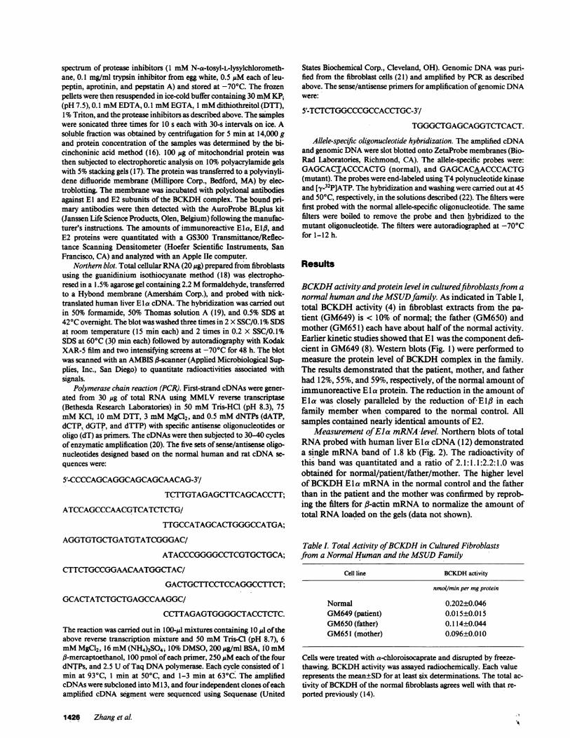
Table I. Total Activity of BCKDHin Cultured Fibroblastsfrom a Normal Humanand the MSUDFamily
Cell line BCKDHactivity
nmol/min per mgprotein
Normal 0.202±0.046GM649(patient) 0.015±0.015GM650(father) 0.1 14±0.044GM651 (mother) 0.096±0.010
Cells were treated with a-chloroisocaprate and disrupted by freeze-thawing. BCKDHactivity was assayed radiochemically. Each valuerepresents the mean±SD for at least six determinations. The total ac-tivity of BCKDHof the normal fibroblasts agrees well with that re-ported previously (14).
1426 Zhang et al
M) 0 -
E E
zoo
E2
Eia o%0El g Figure 1. Western blot of BCKDHex-
tracted from cultured fibroblasts. Mito-chondrial protein was subjected toSDS-PAGEfollowed by immunoblot-ting analysis. The positions of El a,Elf, and E2 subunits are indicated bythe labels.
Identification of Tyr to Asn mutation. To define the muta-tions at the DNAand RNAlevel, we cloned and sequenced thecDNA from the patient by reverse transcription of RNAfol-lowed by PCR to amplify specifically BCKDHEla cDNA.Five sets of sense-antisense primers (each defining a 300-400-bp region of the mRNA)were designed based on the normalhuman Eta cDNA sequence (12). To obtain sequence infor-mation for the NH2-terminal 20 residues of the mature humanprotein, which had not been previously reported, PCRwasperformed using a sense primer based on the leader peptidesequence of the rat El a cDNA (1 1) (Fig. 3). Thus, the com-plete coding region of the mature El a was amplified in over-lapping segments and subcloned into M13 for DNAsequenc-ing. Only one single base substitution was found: TACencod-ing tyrosine at residue 394 was changed to AAC encodingasparagine. To demonstrate that this change was not an am-
0EL..0z
0)
(0
CD
0LO(0
CD
LA(0
CI)
5' CAG TTT TCA TCT CTG GAT GAC AAG CCC CAG TTC CCAPhe Ser Ser Leu Asp Asp Lys Pro Gln Phe Pro
GGG GCC TCG GCG GAG TTT ATA GAT AAG TTG GAA TTC 3'Gly Ala Ser Ala Glu Phe Ile Asp Lys Leu Glu Phe
Figure 3. Nucleotide and deduced NH2-terminal amino acid se-quence of the mature human BCKDHE I a protein.
plification or cloning artifact, four independent amplifiedfragments containing residue 394 were sequenced from bothdirections; the Tyr (TAG) to Asn (AAC) substitution wasfound in all cases.
Allele-specific oligonucleotide hybridization. To confirmthe point mutation, sequences flanking codon 394 were am-plified from the RNAof the patient and his parents. Twoallele-specific oligonucleotides centered at codon 394 (onecontaining TAC and one with AAC) were synthesized andused to probe slot blots of the amplified RNAsegments. Theamplified RNAfrom the patient hybridized only to the mu-tant allele-specific probe and that from the father hybridized toboth probes (Fig. 4 A). The mother's RNA, however, hybrid-ized only to the normal allele-specific probe, indicating thatthe mother expressed only the normal allele.
These allele-specific oligonucleotides were also used toprobe the amplified genomic DNAsegments. The DNAfromthe father and the mother followed the same pattern as that ofthe mRNA: the father was heterozygous and the mother ho-mozygous for the normal allele (Fig. 4 B). However, the DNAfrom the patient hybridized to both probes, indicating that hewas heterozygous at the gene level with regard to the pointmutation, although he expressed only the'mutant mRNA.These experiments have been replicated with three indepen-dent amplifications of RNAand genomic DNA, to rule outartifacts.
Discussion
Wehave studied the molecular basis for MSUDin a familywith the classic form of the disease by analyzing the enzyme
AI
Normal CS -
GM649
2
ewGM650
GM651 0.",..* #I, ALA&-,
B1
Normal Sm
GM649
Figure 2. mRNAlevel of BCKDHE I a in fibroblast cell lines. TotalRNAfrom the fibroblasts was fractionated by electrophoresis andblotted to Hybond membrane. The membrane was then probed withlabeled human ElIa cDNA, washed, and subjected to autoradiography.
GM650
GM651
0A_amo
Figure 4. Allele-specific oli-gonucleotide hybridizationanalysis of the (A) ampli-fied cDNAand (B) geno-mic DNA. Reverse tran-
2 scription and PCRwerecarried out as described inMethods. The slot blotswere first hybridized with
Dow normal allele-specific oligo-nucleotide probe (lane 1).After removal of the nor-mal probe, the blots werehybridized with mutant al-lele-specific probe (lane 2).
Molecular Basis for Maple Syrup Urine Disease 1427
E1 asb .6
activity, protein, and mRNAlevels. The patient had a quitelow level of BCKDHtotal activity. The parents each haveabout half of the normal enzyme activity, as expected for theobligatory carriers of this autosomal recessive genetic disease.
As shown by Western and Northern blot analysis, themRNAlevel and the amount of immunoreactive El proteinwere each half of the normal values in-the mother, suggestingthe existence of a regulatory mutation which reduced thesteady state level of EIa mRNA. In contrast, the father had anormal level of Ela mRNA,but only 59% of the normal levelof Ela protein, suggesting a structural mutation which re-sulted in either destabilization of the El protein or inefficienttranslation of the E la mRNA. The patient had half of thenormal ElIa mRNAlevel, and only 12% of the normal immu-noreactive Ela protein, consistent with the hypothesis thatthere were different mutations in the two parents: a cis-actingregulatory mutation in the mother'that greatly reduced theexpression of one of the Ela alleles, and a structural mutationin the father that produced an altered and unstable protein.
Since the reduction of the level of Ela mRNAcorrelateswith the reduction in the amount of E l a protein in the patientand his mother, the concomitant reduced level of E I# shownin the Western blot (Fig. 1) is best explained by proteolysis ofEl,3 subunits which cannot be incorporated into the stable(a[i2A) complex form. Studies of the pyruvate dehydrogenasecomplex, which has structural similarities to BCKDHcomplexincluding an El component composed of Ela and EI fl sub-units, suggest that the presence of the Ela subunit is necessaryfor the stability of the E I# subunit (23).
In order to define the mutations causing the disease in thisfamily, we sequenced the cDNAfrom the patient encoding theentire mature protein of the El a subunit by reverse transcrip-tion of RNAfollowed by PCRand DNAsequencing. Only onesingle-base mutation (T to A) that caused a Tyr to Asn substi-tution at codon 394 was identified. The mutation was con-firmed by allele-specific oligonucleotide hybridization of thepatient's amplified RNAunder the conditions which differen-tiate single-base mismatch (Fig. 4). The allele-specific oligonu-cleotide hybridization experiment was also carried out usingamplified RNAand genomic DNAfrom the patient's fatherand mother. The father's RNAand genomic DNAhybridizedto both normal and mutant probes, indicating that he washeterozygous for this mutation, and expressed both the normaland mutant alleles. The mother's RNAand DNA, however,hybridized only to the normal allele-specific probe, indicatingthat she was homozygous normal with regard to the pointmutation.
These data indicate that the affected child was a compoundheterozygote. He inherited a mutant allele from his fatherwhich contained a Tyr(394) to Asn substitution. Since thefather is heterozygous for this mutation and has the normalE aa mRNAlevel but reduced amount of El protein, we pro-pose that this mutation affects either the translation efficiencyof the mRNAor, more likely, the stability of the protein. Themother is homozygous for the normal codon 394, but ex-presses only half as much EIa mRNA; she must carry a cis-acting mutation in one of her Ela alleles that essentially abol-ishes its expression as mRNA.The child inherited this nonex-pressed allele (which had the normal Tyr 394) from his motherand the mutant allele (which contained Asn 394) from hisfather. This explains how the patient was genetically heterozy-gous for the Tyr to Asn substitution, yet expressed only the
allele which contained the point mutation. Southern blots ofgenomic DNAfrom the members of this family probed withhuman E la cDNAwere identical to those of the normal indi-viduals, suggesting that there was no major gene deletion orrearrangement in either mother or patient. MSUDin this pa-tient is thus due to two different mutant El a alleles: a cis-act-ing mutation which nearly eliminates expression of one alleleand a point mutation which results in reduced translation effi-ciency and/or in an unstable El a protein.
In conclusion, our results delineate the molecular basis ofMSUDin a family with the classic form of the disease at DNAand RNAlevel. The application of the methods established inthis study should greatly facilitate the understanding of themolecular defects of this clinically heterogeneous disease.
Acknowledgments
Wethank Dr. Peter J. Roach for synthesizing oligonucleotides andBrett Robbins for assisting with the PCRand subcloning.
This work was supported in part by grants from the Riley MemorialAssociation, Showalter Foundation, U.S. Public Health Service grantsDK-19259, AA-06460, AA-06434, AA-00081, and a predoctoral fel-lowship to Ms. Zhang from the March of Dimes Birth Defects Foun-dation.
References
1. Pettit, F. H., S. J. Yeaman, and L. J. Reed. 1978. Purificationand characterization of branched chain a-ketoacid dehydrogenasecomplex of bovine kidney. Proc. NatL. Acad. Sci. USA. 75:4881-4885.
2. Paxton, R., and R. A. Harris. 1982. Isolation of rabbit liverbranched chain a-ketoacid dehydrogenase and regulation by phos-phorylation. J. Biol. Chem. 257:14433-14439.
3. Damuni, Z., M. L. Merryfield, J. S. Humphreys, and L. J. Reed.1984. Purification and properties of branched chain a-ketoacid dehy-drogenase phosphatase from bovine kidney. Proc. Nat!. Acad. Sci.USA. 81:4335-4338.
4. Fatania, H. R., K. S. Lau, and P. J. Randle. 1981. Inactivation ofpurified ox kidney branched chain 2-oxoacid dehydrogenase complexby phosphorylation. FEBS (Fed. Eur. Biochem. Soc.) Lett. 132:285-288.
5. Paxton, R., M. J. Kuntz, and R. A. Harris. 1986. Phosphoryla-tion sites and inactivation of branched chain a-ketoacid dehydroge-nase isolated from rat heart, bovine kidney, and rabbit liver, kidney,heart, brain and skeletal muscle. Arch. Biochem. Biophys. 244:187-201.
6. Yeaman, S. J. 1986. The mammalian 2-oxoacid dehydrogenases:a complex family. Trends Biochem. Sci. 11:293-296.
7. Tanaka, K., and L. E. Rosenberg. 1983. Disorders of branchedchain amino acid and organic acid metabolism. In The MetabolicBasis of Inherited Disease. J. B. Stanbury, J. B. Wyngaarden, D. S.Fredrickson, J. L. Goldstein, and M. S. Brown, editors. McGraw-HillBook Co., Inc., NewYork. 440-473.
8. Chuang, D. T., L. S. Ku, D. S. Kerr, and R. P. Cox. 1982.Detection -of heterozygotes in maple-syrup-urine disease: measure-ments of branched-chain a-ketoacid dehydrogenase and its compo-nents in cell cultures. Am. J. Hum. Genet. 34:416-424.
9. Danner, D. J., N. Armstrong, S. C. Heffelfinger, E. T. Sewell,J. H. Priest, and L. J. Elsas. 1985. Absence of branched-chain a-ketoa-cid acyltransferase as a cause of maple syrup urine disease. J. Clin.Invest. 75:858-860.
10. Indo, Y., A. Kitano, F. Endo, I. Akaboshi, and I. Matsuda.1987. Altered kinetic properties of the branched-chain a-ketoacid de-hydrogenase complex due to mutation of the ,-subunit of the
1428 Zhang et al.
branched chain a-ketoacid decarboxylase (El) component in lympho-blastoid cells derived from patients with maple syrup urine disease. J.Clin. Invest. 80:63-70.
11. Zhang, B., M. J. Kuntz, G. W. Goodwin, R. A. Harris, andD. W. Crabb. 1987. Molecular cloning of a cDNAfor the E I a subunitof rat liver branched chain a-ketoacid dehydrogenase. J. Biol. Chem.262:15220-15224.
12. Zhang, B., D. W. Crabb, and R. A. Harris. 1988. Nucleotideand deduced amino acid sequence of the El a subunit of human liverbranched-chain a-ketoacid dehydrogenase. Gene 69:159-164.
13. Rudiger, H. W., U. Langenbeck, and H. W. Goedde. 1972. Asimple method for the preparation of '4C-labelled branched-chaina-oxo acids. Biochem. J. 126:445-446.
14. Chuang, D. T., and R. P. Cox. 1988. Enzyme assays withmutant cell lines of maple syrup urine disease. Methods Enzymol.166:135-145.
15. Mackall, J., M. Meredith, and M. D. Lane. 1979. A mild pro-cedure for the rapid release of cytoplasmic enzymes from culturedanimal cells. Anal. Biochem. 95:270-274.
16. Smith, P. K., R. I. Krohn, G. T. Hermanson, A. K. Mallia,F. H. Gartner, M. D. Provenzano, E. K. Fujmoto, N. M. Goeke, B. J.Olson, and D. C. Klenk. 1985. Measurement of protein using bicin-choninic acid. Anal. Biochem. 150:76-85.
17. Laemmli, U. K. 1970. Cleavage of structural proteins during
the assembly of the head of bacteriophage T4. Nature (Lond.).227:680-685.
18. Chirgwin, J. M., A. E. Praybyla, R. J. MacDonald, and W. J.Rutter. 1979. Isolation of biologically active ribonucleic acid fromsources enriched in ribonuclease. Biochemistry 18:5294-5299.
19. Thomas, P. S. 1980. Hybridization of denatured RNAandsmall DNAfragments transferred to nitrocellulose. Proc. NatL. Acad.Sci. USA. 77:5201-5205.
20. Saiki, R. K., D. H. Gelfand, S. Stofeel, S. J. Scharf, R. Highchi,G. T. Horn, K. B. Mullis, and H. A. Erlich. 1988. Primer-directedenzymatic amplification of DNAwith a thermostable DNApolymer-ase. Science (Wash. DC). 239:487-491.
21. Maniatis, T., E. F. Fritsch, and J. Sambrook. 1982. Molecularcloning: A Laboratory Manual. Cold Spring Harbor Laboratory, ColdSpring Harbor, NY. 280-281.
22. Farr, C. J., R. K. Saiki, H. A. Erlich, F. McCormick, and C. J.Marshall. 1988. Analysis of RAS gene mutations in acute myeloidleukemia by polymerase chain reaction and oligonucleotide probes.Proc. Natl. Acad. Sci. USA. 85:1629-1633.
23. Wexler, I. D., D. S. Kerr, L. Ho, M. M. Lusk, R. A. Pepin, A. A.Javed, J. E. Mole, B. W. Jesse, T. J. Thekkumkara, G. Pons, and M. S.Patel. 1988. Heterogeneous expression of protein and mRNAin pyru-vate dehydrogenase deficiency. Proc. Natl. Acad. Sci. USA. 85:7336-7340.
Molecular Basis for Maple Syrup Urine Disease 1429