Urolithin A, a Novel Natural Compound to TargetPI3K/AKT/mTOR Pathway in Pancreatic CancerTulasigeri M. Totiger1, Supriya Srinivasan1, Venkatakrishna R. Jala2,Purushottam Lamichhane1, Austin R. Dosch1, Alexander A. Gaidarski III1,Chandrashekhar Joshi1, Shobith Rangappa3, Jason Castellanos4,Praveen Kumar Vemula5, Xi Chen6, Deukwoo Kwon6, Nilesh Kashikar7,Michael VanSaun1, Nipun B. Merchant1, and Nagaraj S. Nagathihalli1
Abstract
Pancreatic ductal adenocarcinoma (PDAC) is an aggressivemalignancy and is highly resistant to standard treatment regi-mens. Targeted therapies against KRAS, a mutation present inan overwhelming majority of PDAC cases, have been largelyineffective. However, inhibition of downstream componentsin the KRAS signaling cascade provides promising therapeutictargets in the management of PDAC and warrants furtherexploration. Here, we investigated Urolithin A (Uro A), anovel natural compound derived from pomegranates, whichtargets numerous kinases downstream of KRAS, in particularthe PI3K/AKT/mTOR signaling pathways. We showed thattreatment of PDAC cells with Uro A blocked the phosphory-lation of AKT and p70S6K in vitro, successfully inhibited thegrowth of tumor xenografts, and increased overall survival of
Ptf1aCre/þ;LSL-KrasG12D/þ;Tgfbr2flox/flox (PKT)mice comparedwith vehicle or gemcitabine therapy alone. Histologic eval-uation of these Uro A–treated tumor samples confirmedmechanistic actions of Uro A via decreased phosphorylationof AKT and p70S6K, reduced proliferation, and increasedcellular apoptosis in both xenograft and PKT mouse models.In addition, Uro A treatment reprogrammed the tumormicroenvironment, as evidenced by reduced levels ofinfiltrating immunosuppressive cell populations such asmyeloid-derived suppressor cells, tumor-associated macro-phages, and regulatory T cells. Overall, this work providesconvincing preclinical evidence for the utility of Uro A as atherapeutic agent in PDAC through suppression of thePI3K/AKT/mTOR pathway.
IntroductionPancreatic ductal adenocarcinoma (PDAC) is the third leading
cause of cancer-related death in the United States with a 5-yearsurvival rate below 9%. The prevalence of distant metastases andchemotherapeutic resistance are hallmarks of the disease whichlargely account for its dismal prognosis (1, 2). Current standard-of-care drug regimens including gemcitabine, FOLFIRINOX, andnab-paclitaxel/gemcitabine have shown limited clinical efficacyand are often poorly tolerated in patients due to toxic side effects
(3). Hence, there is a desperate need to develop novel therapeuticapproaches which reduce PDAC tumor burden without produc-ing significant off-target effects.
Natural compounds have garnered increasing attentionamong the scientific community for their low cost, highbioavailability, and limited toxicity compared with syntheticpharmaceutical agents (4). Mounting evidence suggests thatmany of these compounds possess intrinsic antioxidant, anti-inflammatory, and antitumor activities (5). In fact, a growingnumber of FDA-approved anticancer agents are derived fromeither naturally occurring compounds or their derivatives(5, 6). Interestingly, population-based epidemiologic studieshave shown a strong inverse correlation between consumptionof berries, such as black raspberries, pomegranates, and straw-berries, and incidence of PDAC (6, 7). Although the mechanismis poorly understood, it has been postulated that this effect isdue to the high concentration of ellagitannins present in thesefoods. Ellagitannins are hydrolyzed in the gut to release ellagicacid (EA), a compound which inhibits multiple oncogenicpathways which are activated in PDAC such as COX-2, NF-kB,Notch, and Wnt signaling. Preclinical studies have shown thattargeted blockade of these pathways by EA is successful inreducing epithelial–mesenchymal transition, angiogenesis,fibrosis, and pancreatic stellate cell activation in PDAC (8,9). Despite these promising developments, EA is unfortunatelypoorly absorbed within the human gut, limiting its efficacy as atherapeutic agent. However, EA is metabolized into a numberof downstream compounds through microbial processing,including the urolithin A, B, and C (10, 11). Of these, Urolithin
1Department of Surgery, University of Miami Miller School of Medicine, SylvesterComprehensiveCancer Center, Miami, Florida. 2Department ofMicrobiology andImmunology, University of Louisville, Louisville, Kentucky. 3AdichunchanagiriInstitute for Molecular Medicine, AIMS, Karnataka, India. 4Department ofSurgery, Vanderbilt University School of Medicine, Nashville, Tennessee.5Institute for Stem Cell Biology and Regenerative Medicine (inStem), Bangalore,Karnataka, India. 6Department of Public Health, University of MiamiMiller Schoolof Medicine, Miami, Florida. 7Department of Pathology, University of Colorado,Denver, Colorado.
Note: Supplementary data for this article are available at Molecular CancerTherapeutics Online (http://mct.aacrjournals.org/).
T.M. Totiger and S. Srinivasan contributed equally to this article.
Corresponding Author: Nagaraj S. Nagathihalli, University of Miami, MillerSchool of Medicine, Sylvester Comprehensive Cancer Center, 1550 NW 10thAve, FOX140, Miami, FL 33136. Phone: 361-720-9347; Fax: 305-243-2810; E-mail:[email protected]
A (Uro A) exhibits potent antioxidant and anti-inflammatoryproperties, suggesting it may be the dominant compoundwhich is responsible for the intrinsic antitumor activity of EA(12, 13). These data suggest that although dietary ingestion ofEA is unable to produce therapeutic levels, direct oral admin-istration of Uro A may be a promising target in the treatment ofPDAC due to its improved bioavailability and potent antitumoreffects. Furthermore, preclinical models have demonstratedthat Uro A is well tolerated and does not elicit any adversetoxic effects at clinically relevant doses (11).
Despite their limited efficacy, gemcitabine- and 5-FU–basedadjuvant chemoradiation has been considered the standard ofcare for PDAC. To date, the addition of targeted moleculartherapies to these cytotoxic compounds has failed to show anysignificant improvement in improving overall survival (OS) inPDAC (14, 15), presumably due to redundant signaling pathwaysand feedback loops within tumor cells (12). Furthermore, over-coming these resistance mechanisms by combining multipleinhibitors is not feasible due to cumulative toxicity. As such,single chemotherapeutic agents that are both well tolerated andable to target multiple kinase pathways simultaneously presentnovel targets in the treatment of PDAC. Uro A has been shown todownregulate multiple tumor pathways in colon, prostate, andbladder cancer through downregulation of several oncogenessuch as Kras and c-myc, upregulation of tumor-suppressor genessuch as FGFR2 and EGFR, and modulation of enzyme activity,such as CYP1 (13). Despite the promising effects seen in treatingother malignancies, the anticancer effect of Uro A in PDAC iscurrently unknown (8, 9).
We hypothesized thatUroA exerts its antitumor effects throughinhibition of multiple protumorigenic pathways in PDAC. Ourresults clearly demonstrated that Uro A downregulated theoncogenic PI3K/AKT/mTOR signaling pathway, induced cell-cycle arrest, and increased apoptosis. Uro A successfully attenu-ated tumor growth in both tumor xenografts and geneticallyengineered mouse models in vivo by not only disrupting PI3K/AKT signaling, but also inducing significant changes within theimmunosuppressive microenvironment of PDAC.
Materials and MethodsCell lines and drugs
The human PDAC cell lines MiaPaCa2, PANC1, AsPC1,CFPAC1, Capan1, Capan2, SW1990, HPAC, and BxPC3 wereobtained from the American Type Culture Collection (ATCC).The K8484 (Pdx1aCre/þ; LSL-KRASG12D/þ; p53R172H/þ) cell linewas obtained fromDr. Tuveson (Cold SpringHarbor Laboratory).All tumor cells were maintained according to the ATCCguidelines. ATCC cell lines were characterized and verified freeof Mycoplasma contamination, tested by Hoechst DNA stain(indirect) and agar culture (direct) methods. Cell authenticationwas performedbyusing short tandem repeatDNAprofiling (latestdate: June 16, 2016, and July 21, 2017) and cell lines testednegative for Mycoplasma via Genetica cell line testing usingeMYCO plus kit (iNtRON Biotechnology). Cells with relativelylow-passage numbers (< 20) were used in the study.
Uro A synthesis and the structure of the compound were asdetailed previously (16), and gemcitabinewas purchased fromEliLilly and Company. AKT activators SC79 and IGF-1 were pur-chased from Sigma Aldrich and R&D systems, respectively. AKTinhibitor MK2206 was purchased from Selleckem.
Western blottingCell lysis and Western blotting were done as previously
described (17). Briefly, cells were washed, lysed, and removedfrom culture dishes by scraping after treatment. Cell lysis wasperformed using RIPA buffer (0.1% SDS, 50 mmol/L Tris�HCl,150 mmol/L NaCl, 1% NP-40, and 0.5% Na deoxycholate) withprotease inhibitor cocktail (Sigma) and PhosSTOP phosphataseinhibitor (Roche). Lysates were sonicated and centrifuged at10,000 g for 15minutes at 4�C to collect supernatant. The proteinconcentration of the lysate was determined by Bio-Rad proteinassay kit (Bio-Rad). Per lane, 35 mg of whole-cell lysate wasseparated on NuPAGENovex 4%–12% Bis-Tris Gels and trans-ferred on iBlot transfer stack andPVDFmembranes using iBlot dryblotting transfer system (Life Technologies). For immunodetec-tion, membranes were incubated with antibodies listed in Sup-plementary Table S1. The membranes were subsequently incu-bated with corresponding secondary anti-mouse or anti-rabbitsecondary antibodies conjugated with horseradish peroxidase(Jackson ImmunoResearch). Finally, the immunoreactive bandswere developed with Pierce ECL Western Blotting Substrate(Thermo Scientific) and recorded on blue basic autoradiographyfilm (Bioexpress).
The human tyrosine kinase array was purchased from R&DSystems (Cat#: ARY003B) and used according to the manufac-turer's recommended conditions. Both immunoblots and arrayintensity were then quantified using Image J image analysissoftware. Statistical analysis was performed using Prism software(Graphpad Software Inc.).
Cell viability assay (MTT)PDAC cells were seeded at a concentration of 1 � 104 cells per
well in 96-well plates. Twenty-four hours after seeding, theattached cells were treated with DMSO or Uro A (0–100 mmol/L)for 48 hours, and cell viability was determined by MTT assay(Sigma) according to the manufacturer's direction. IC50 wascalculated using Prism software (Graphpad Software Inc.). Eachcondition was assayed in triplicate.
Apoptosis assayApoptosis was assessed by flow cytometric detection of phos-
photidyl serine externalization using the FITC Annexin V Apo-ptosis Detection Kit II (BD Biosciences). MiaPaCa2 cells weretreated with Uro A for 24 hours. The cells were then trypsinizedgently, washed twice with cold PBS, resuspended in 1X bindingbuffer, and then incubated with 5 mL of FITC Annexin V and 5 mLof propidium iodide (PI). After incubation for 15minutes at roomtemperature (25�C) in the dark, 400 mL of 1X binding buffer wasadded, and each tube was analyzed within 1 hour using theFACSCalibur flow cytometer (BD Biosciences). The percentageof cells present in each compartment was measured and analyzedwith Cell Quest software (BD Biosciences).
Wound-healing assayCells were treated with mitomycin C (0.5 mg/mL) for 4 hours
prior to wounding. Wounds were made across the cell monolayerby a sterile pipette tip. Afterwounding, BxPC3 andMiaPaCa2 cellswere treated with DMSO or Uro A (0–50 mmol/L) for 36 hours.Phase contrast images were taken. After every 12 hours of wound-healing study, the cells were washed and treated with Uro A orDMSO for up to 36 hours and observed for recovery afterwounding.
Totiger et al.
Mol Cancer Ther; 18(2) February 2019 Molecular Cancer Therapeutics302
purchased from Harlan Sprague Dawley, Inc. Subcutaneoustumorswere establishedby injecting 2�106MiaPaCa2or PANC1cells into the flank of a 6-week-old Fox1-nu/nu mouse (n ¼ 5 ineach group) as previously detailed (18). MiaPaCa2 and PANC1were chosen because they harbor mutations typical of humanpancreatic cancer (19). Uro A (20 mg/kg/daily) daily (5 days/week) by oral gavagewas initiatedwhen the subcutaneous tumorsreached 200 to 250 mm3 size. Uro A or vehicle (10% glucose inwater)was administered by oral gavage for 33 (MiaPaCa2) and 42(PANC1) days, and the tumor volume was measured weekly. Thesubcutaneous tumor volume and percent body weight changewere recorded as previously described (18). Growth curves fortumors were plotted as themean volume� SD of tumors formicefrom each group. At the end of the study, animals were sacrificed,and primary tumors were removed for further analysis.
MicePtf1aCre/þ;Tgfbr2flox/flox and LSL-KrasG12D/þ;Tgfbr2flox/flox mice
were provided by Dr. Hal Moses (Vanderbilt University MedicalCenter, Nashville, TN). These 2 lines were intercrossed to generatePtf1aCre/þ;LSL-KrasG12D/þ;Tgfbr2flox/flox (PKT) mice on a C57Bl/6background. Genotyping of alleles was performed using oligo-nucleotide primers as described previously (18, 20).
Treatment of PKT micePKT mice were treated with vehicle or Uro A and/or gemcita-
bine.Mice in theUroA (20mg/kg/day) arm received treatment byoral gavage 5 days/week and received twice-weekly intraperito-neal injections of gemcitabine (20 mg/kg), starting at 4 weeks ofage. Mice were euthanized and dissected after 3 weeks unless theywere part of the survival arm. Due to the irregularity of the tumordimensions, size was determined by weighing the entire tumor.Tumor tissue was processed for further IHC examination. OS wasdetermined by the log-rank analysis using statistical softwarepackage R (version 3.3.2).
ImmunohistochemistryTissues were fixed and immunostained using antibodies
against Ki67 and cleaved caspase-3 (Supplementary Table S1).Stained tissues were evaluated by an expert pathologist(N. Kashikar). Immunostained slides were imaged using Leicamicroscope (Leica Microsystems, Inc.) and quantified usingImage J. Imageswere adjusted to exclude areas containing obvioushistologic artifacts, such as tissue folds or nonorganic material,from the digital image. Calculated percentage of positive cellsstained relative to total area was analyzed by applying a scale forrelative intensity and was reported as relative expression ofprotein staining.
Flow cytometryPKT mouse pancreas were minced and digested with 1 mg/mL
Collagenase P (Roche) and 2 mg/mL Collagenase IV (Gibco) for30 minutes at 37�C. The dissociated tissue was then filtered to
remove the large chunks, washed with PEB, and refiltered toobtain single-cell suspensions. Single-cell suspensions were pre-pared for flow cytometry by staining for regulatory T cells (Treg)andmyeloid-derived suppressor cells (MDSC) with the followingantibodies: CD45 PE-Cy7, CD3 APC, CD4 AF700, CD8PerCpCy5.5, Foxp3 PE, CD25APC-Cy7, CD11bAF700,Gr1 FITC,and Live/Dead Aqua (Supplementary Table S2).
Statistical analysisDescriptive statistics were calculated using Microsoft Excel and
Prism software (Graphpad Software Inc.). Results are shown asvalues of mean � SD unless otherwise indicated. Statisticalanalyses of IHC data were performed using the Student t testwith P < 0.05 taken as significant, except where indicated other-wise. Statistical analyses of PKT tumor weight data were per-formed using the ANOVA followed by Tukey's multiple compar-isons test to determine P values.
Statistical analysis for Figs. 3B and C, and 4C was adjustedusing area under the growth curve (aAUC) approach (21). Pvalues were obtained from permutation test to compare aAUCsof tumor growth curves between groups. The Kaplan–Meiersurvival analysis was performed, and survival differencesbetween groups were assessed with the log-rank test. All sta-tistical analyses were performed using statistical software pack-age R (version 3.3.2).
Study approvalAll experiments were performed in compliance with the reg-
ulations and ethical guidelines for experimental and animalstudies of the Institutional Animal Care and Use Committees atthe Vanderbilt University Medical Center and the University ofMiami (#15-057 and #18-081).
For additional experimental procedures, please refer to Sup-plementary Materials and Methods.
ResultsUro A treatment inhibits PDAC cell proliferation andmigration, and enhances apoptosis
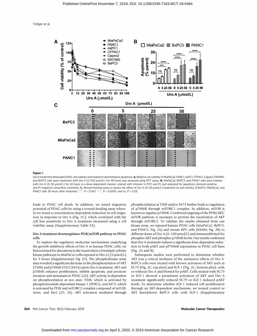
Nine PDAC cell lines were tested for sensitivity to Uro A in vitrousing a cell viability assay (Fig. 1A). IC50 values of Uro A for thesecell lines were determined 48 hours after treatment (Supplemen-tary Table S3). Briefly, Capan1 and HPAC (IC50, 10.23–10.28mmol/L) displayed the most sensitive response, and CFPAC1 andAsPC1 (IC50, 40.39–62.28 mmol/L) showed the least sensitivity toUro A treatment.MiaPaCa2, BxPC3, and PANC1 (IC50, 15.8–24.8mmol/L) cell lines displayed intermediate sensitivity and hencewere chosen to ascertain the antineoplastic effects of Uro A infurther experiments. Other Uro derivatives such as Uro B and C,on the other hand, failed to exhibit growth-inhibitory effects aseffectively as Uro A (Supplementary Fig. S1A and S1B). Previousstudies have established that Uro A doses up to 100 mmol/L areclinically tolerated (22, 23).
In order to investigate the mechanism of PDAC growthinhibition by Uro A, we examined its effect on the inducingapoptosis (Fig. 1B; Supplementary Fig. S2A) and/or cell-cyclealterations by assessing sub-G0, G1, S, and G2 populations(Supplementary Fig. S2B). Uro A treatment resulted in adose-dependent increase in both the sub-G0 (PI positive) andapoptotic (Annexin V–positive/PI-negative) populations atconcentrations at or above 25 mmol/L, suggesting that Uro A
Targeting PI3K/AKT/mTOR Pathway in Pancreatic Cancer
www.aacrjournals.org Mol Cancer Ther; 18(2) February 2019 303
leads to PDAC cell death. In addition, we tested migratorypotential of PDAC cells by using a wound-healing assay where-in we noted a concentration-dependent reduction in cell migra-tion in response to Uro A (Fig. 1C), which correlated with thecell line sensitivity to Uro A treatment measured using a cellviability assay (Supplementary Table S3).
Uro A treatment downregulates PI3K/mTOR pathway in PDACcells
To explore the regulatory molecular mechanisms underlyingthe growth-inhibitory effects of Uro A in human PDAC cells, wefirst screened for alterations in the inactivation ofmultiple cellularkinase pathways inMiaPaCa2 cells exposed to Uro A (25 mmol/L)for 3 hours (Supplementary Fig. S3). The phosphokinase arraydata revealed a significant decrease in the phosphorylation of AKT(T308) and p70S6K (T421/S424) uponUro A treatment. AKT andp70S6K enhance proliferation, inhibit apoptosis, and promoteinvasion and metastasis in PDAC (24). AKT activity is dependenton phosphorylation at two sites: T308, which is activated byphosphoinositide-dependent kinase-1 (PDK1), and S473, whichis activated by PI3K and mTORC2 complex composed of mTOR,rictor, and Sin1 (25, 26). AKT activation mediated through
phosphorylation at T308 and/or S473 further leads to regulationof p70S6K through mTORC1 complex. In addition, mTOR isknown to regulate p70S6K. Combined targeting of the PI3K/AKT/mTOR pathway is necessary to prevent the reactivation of AKTthrough mTORC2. To validate the results obtained from ourkinase array, we exposed human PDAC cells (MiaPaCa2, BxPC3,and PANC1; Fig. 2A) and mouse KPC cells (K8484; Fig. 2B) todifferent doses of Uro A (0–100 mmol/L) and immunoblotted forphospho-AKT andphospho-p70S6K levels.Our results confirmedthat Uro A treatment induces a significant dose-dependent reduc-tion in both pAKT and pP70S6K expressions in PDAC cell lines(Fig. 2A and B).
Subsequent studies were performed to determine whetherAKT was a critical mediator of the antitumor effects of Uro A.BxPC3 cells were treated with known activators of AKT such asSC79 (Fig. 2C, top plot) and IGF-1 (Fig. 2C, bottom plot), withor without Uro A and blotted for pAKT. Cells treated with SC79or IGF-1 showed a prominent activation of AKT and Uro Atreatment significantly reduced SC79 or IGF-1 induced pAKTlevels. To determine whether IGF-1 induced cell proliferationthrough an AKT-dependent mechanism, we treated control orAKT knockdown BxPC3 cells with IGF-1 (Supplementary
Figure 1.
UroA treatment attenuates PDAC cell viability andmigration and enhances apoptosis.A,Relative cell viability inMiaPaCa2, PANC1, AsPC1, CFPAC1, Capan2, SW1990,and BxPC3 cells upon treatment with Uro A (0–100 mmol/L) for 48 hours was assessed using MTT assay. B, MiaPaCa2, BXPC3, and PANC1 cells were treatedwith Uro A (0–50 mmol/L) for 24 hours in a dose-dependent manner, stained with Annexin V–FITC and PI, and analyzed for apoptosis (Annexin-positiveand PI-negative) using flow cytometry. C, Wound-healing assay to assess the effect of Uro A (0–50 mmol/L) treatment on cell motility of BxPC3, MiaPaCa2, andPANC1 cells 36 hours after treatment. ���, P < 0.001; ���� , P < 0.0001; and ns, P > 0.05.
Totiger et al.
Mol Cancer Ther; 18(2) February 2019 Molecular Cancer Therapeutics304
Fig. S4A; Fig. 2D). With the addition of IGF-1, BxPC3 controlcells significantly increased cell proliferation compared withIGF-1 treatment of AKT knockdown cells. Next, AKT knock-down cells showed a significant reduction in IGF-1–mediatedcell proliferation in comparison with its corresponding scram-bled control cells treated with IGF-1. These results confirm thatIGF-1–induced activation of AKT regulates cell proliferation inPDAC cells. Furthermore, Uro A treatment also downregulatedPDK1 (an upstream target of AKT) as well as pGSK3b and p4E-
BP1 (downstream targets of AKT), thereby suggesting that UroA effectively inhibits the PDK1/AKT/mTOR pathway (Supple-mentary Fig. S4B). Further, treatment of MiaPaCa2 cells withthe AKT inhibitor MK2206 or Uro A resulted in an attenuatednumber of colonies (Fig. 2E). The combined treatment withMK2206 and Uro A did not produce further inhibition whencompared with either MK2206 or Uro A single treatments.These results demonstrate that Uro A elicits its anticancer effectson PDAC proliferation that are consistent with AKT inhibition
Figure 2.
UroA treatment downregulates PI3K/AKT/P70S6Kpathway in PDAC cells.A,Western blots demonstrating dose-dependent phosphorylation of AKT and p70S6K inprimary human PDAC cell lines MiaPaCa2, PANC1, and BxPC3 upon Uro A (0–100 mmol/L) treatment for 3 hours. B, Cell line (K8484) established from KPCmouse was treated with Uro A (0–100 mmol/L) for 3 hours. Western blot demonstrating dose-dependent decrease in the levels of phosphorylation of AKT andp70S6K in K8484 cell line upon Uro A treatment. Tubulin was used as Western blots loading control. C, BxPC3 cells were treated with SC79 (10 mmol/L; top)or IGF-1 (50 ng/mL; bottom) for 3 hourswith activatedAKT. The densitometry analyses of pAKT normalized to total AKTproteinwere shown. Cells treatedwithUroA(25 mmol/L) show a significant reduction in the pAKT levels when compared with SC79 or IGF-1-alone–treated cells. D, IGF-1 (50 ng/mL) exposure to AKTknockdownBxPC3 cells shows a significant reduction in the cell proliferationwhen comparedwith scrambled control cells treated with IGF-1 alone. E,MiaPaCa2 cellstreated with either MK2206 or Uro A showed decreased number of colonies. Combination of MK2206 (1 mmol/L) and Uro A did not affect further the number ofcolonies when compared with either MK2206 or Uro A treatment. F, Western blots demonstrating dose-dependent phosphorylation of AKT and p70S6K inprimary human normal pancreas cell lines HPNE and HPNE-KRAS upon Uro A (0–100 mmol/L) treatment for 3 hours. � , P < 0.05; ��, P < 0.01; ���, P < 0.001;���� , P < 0.0001; ns, P > 0.05.
Targeting PI3K/AKT/mTOR Pathway in Pancreatic Cancer
www.aacrjournals.org Mol Cancer Ther; 18(2) February 2019 305
(Supplementary Fig. S4C). Interestingly, Uro A displayed only amild effect on pAKT and pP70S6K expression in normal epi-thelial cell lines HPNE and HPNE-KRAS (Fig. 2F; Supplemen-tary Fig. S5), thereby suggesting potential for cancer cell selec-tivity of Uro A.
Uro A treatment suppresses the growth of pancreatic tumorxenografts in mice
After characterizing the efficacy of Uro A in PDAC cell lines, weproceeded to test this treatment in vivo using a mouse xenograft
model (Fig. 3A). PANC1 (Fig. 3B) and MiaPaCa2 (Fig. 3C)xenografts were generated via subcutaneous flank injection ofPDAC tumor cells into athymic nude mice. These xenograft-bearing mice were then treated with either Uro A or vehicle (Fig.3A). Immunoblotting of Uro A–treated MiaPaCa2 xenografttumor lysates demonstrated significant inhibition of AKT andp70S6K phosphorylation (Fig. 3D). Uro A–treated xenograftmiceexhibited significantly reduced tumor growth in comparison withcorresponding vehicle-treated xenograftmice (Fig. 3B andC). IHCperformed on xenograft specimens revealed a significant decrease
Figure 3.
UroA treatment decreases the growth of pancreatic tumor xenograft.A, Experimental design for Uro A treatment in PANC1 andMiaPaCa2 xenograftmousemodel.Band C, PANC1 (B) and MiaPaCa2 (C) cells were s.c. injected onto the flank of Fox1-nu/nu mice until tumor volume reached 200 to 250 mm3. The xenografttumors were further treated with Uro A (20 mg/kg/daily) or vehicle (10% glucose in water) by oral administration for 42 (B) or 33 (C) days, and their respectivevolumesweremeasured daily (n¼ 5). Both PANC1 and MiaPaCa2 xenografts exhibited significantly reduced tumor growthwith Uro A treatment in comparison withcorresponding vehicle-treated xenografts. By repeated measures t test, PANC1 achieved significance at 22 days of treatment and MiaPaCa2 at 24 days.Growth trajectory was significantly different by linear regression (P < 0.0001 for both cell lines), and treatment was statistically significant by two-way ANOVA forPANC1 (P ¼ 0.0002) and MiaPaCa2 (P ¼ 0.0024). D, Western blot demonstrating levels of pAKT and p70S6K in corresponding resected vehicle and UroA–treated MiaPaCa2 xenograft tissues harvested 33 days after treatment. E, Representative Ki67 and cleaved caspase-3 staining of vehicle or Uro A–treatedMiaPaCa2 flank xenograft tissues (left). Quantification of Ki67 and cleaved caspase-3 staining data obtained from resected vehicle or Uro A–treated MiaPaCa2 flankxenograft tissues (right). � , P < 0.05.
Totiger et al.
Mol Cancer Ther; 18(2) February 2019 Molecular Cancer Therapeutics306
in proliferation and a significant increase in apoptosis, as mea-sured by Ki67 and cleaved caspase-3, respectively (Fig. 3E).Overall, Uro A treatment was well tolerated by murine hosts anddid not have any negative impact on body weight throughouttreatment (Supplementary Fig. S6A and S6B).
UroA inhibits tumor growth and improves survival in PKTmiceTo further explore the effect of UroA treatment onPDAC tumor
growth, we utilized the genetically engineered PKTmousemodel.These mice develop autochthonous PDAC with full penetrancethat reliably recapitulates the clinical andhistopathologic featuresof the human disease. They consistently develop PanIN lesions at3.5 weeks of age which progresses to invasive cancer by 4.5 weeksof age. Median OS of PKT mice is consistently around 59 days(18, 20). Treatment regimens consisting of either Uro A and/orgemcitabine were initiated at 4 weeks of age, and mice werefollowed until moribund for survival (Fig. 4A). Tumor weightwas significantly reduced in all treatment groups compared with
vehicle-treated mice (Fig. 4B). Both monotherapy and combinedUro A/gemcitabine treatments were well tolerated without appre-ciable toxicity, as evidenced by normal body weight (Supplemen-tary Fig. S6C). Similarly, all treatment groups had improvedsurvival compared with vehicle-treated mice (median OS ¼ 53days; Fig. 4C). However, only Uro A (OS¼ 71 days) and Uro Aþgemcitabine (OS ¼ 71 days) groups attained statistical signifi-cance for OS by the Mantel–Cox log-rank test with Bonferroni-adjustedP value. Furthermore, combined administration ofUroAand gemcitabine did not provide any additional survival advan-tage over single-agent administration of Uro A, nor did it providestatistically significant advantage of single-agent treatment withgemcitabine (OS ¼ 62 days). Immunoblot analyses of whole-tumor lysates showed decreased activation of AKT and p70S6K,reduced proliferation, and enhanced cellular apoptosis with UroA treatment (Fig. 4D and E), consistent with the findings seenin vitro and in xenograft tumormodels. Taken together, these datashow that Uro A is an effective single-agent therapy that reduces
Figure 4.
UroA treatment improves survival of PKTmice.A,Experimental design for UroA (20mg/kg/daily), gemcitabine (Gem; 20mg/kg/3 day), or UroAþGem treatment inPKT mice starting at 4 weeks of age (n ¼ 8 per group). B, Tumor weight in the Uro A or Uro AþGem-treated mice was significantly decreased comparedwith vehicle-treated controls. C, Kaplan–Meier survival analysis shows significantly improved OS with Uro A (median 71 days) or Uro AþGem (median 71 days)compared with vehicle control (median 53 days). The log-rank test was used to compare groups, with Bonferroni correction applied to pairwise comparisons toaccount for multiple comparisons. D, Western blot analysis of whole tumor lysates demonstrated decreased expression of pAKT and p70S6K in mice treatedwith Uro A compared with vehicle-treated mice. E, Representative proliferation (Ki67 staining) and apoptosis (cleaved caspase-3) staining of resectedpancreata obtained from Uro A–treated PKT mice (left plot). Proliferation was significantly decreased, and apoptosis was significantly increased with Uro Atreatment when compared with vehicle-treated mice (right plot). �� , P < 0.01; ��� , P < 0.001; and ns, P > 0.05.
Targeting PI3K/AKT/mTOR Pathway in Pancreatic Cancer
www.aacrjournals.org Mol Cancer Ther; 18(2) February 2019 307
tumor burden in vivo through significant reduction in tumor cellproliferation and an increase in apoptosis. Overall, Uro A is moreeffective than gemcitabine in improving OS.
Improved therapeutic response toUro A treatment is associatedwith a reduction in immunosuppressive tumor-associatedmacrophages and Tregs in PKT mice
The immunosuppressive tumor microenvironment (TME) ofPDAC is a substantial deterrent to achieving a durable therapeuticresponse (27). MDSCs, tumor-associated macrophages (TAMs),and Tregs are the major components of this immunosuppressivemilieu (28), as these cell types have been shown to promotesystemic T-cell dysfunction that allows PDAC tumors to escape
immune detection. Tregs are critically dependent on the tran-scription factor FoxP3 (29), and TAMs are primarily identified asthe F4/80þ population (30). Signaling through the AKT pathwayand its corresponding downstream target p70S6Khas been shownto play a critical role in regulating this process (31). PKT tumorsamples were analyzed for F4/80 and FoxP3 by IHC after treat-ment with Uro A or vehicle alone. Our results demonstrated thatUro A treatment significantly reduced the detection of tumor-infiltrating F4/80-positive TAMs as well as the presence of FoxP3-positive Tregs without decreasing the overall CD3-positive T-cellstaining (Fig. 5A). Moreover, Uro A treatment significantlyreduced the number of CD11b/Gr-1–positive MDSCs (Fig. 5B)in pancreatic tumors from PKT mice compared with vehicle-
Figure 5.
Effects of Uro A on TAMs, Tregs, and MDSCs in PKT mice. A, Representative H&E, F4/80, FoxP3, and CD3 staining of pancreatic tissues harvested from UroA–treated PKT mice (left plot). Scale bar, 50 mm. Quantification of relative levels of F4/80, FoxP3, and CD3 IHC staining of Uro A and vehicle-treated PKT micepancreata (right plot; n ¼ 3). B, Representative flow cytometry zebra plots (left plot) and their corresponding bar graphs (right plot) of MDSCs in the singlesuspensions of vehicle or Uro A–treated PKT mice pancreata (n ¼ 3). � , P < 0.05; �� , P < 0.01 and ns, P > 0.05.
Totiger et al.
Mol Cancer Ther; 18(2) February 2019 Molecular Cancer Therapeutics308
treated controls. These findings confirmed that Uro A affected theimmune cell population within the tumor, which was coincidentwith reduced PDAC tumor growth and improved survival.
DiscussionThe PI3K/AKT family is among the most frequently mutated
pathways in human cancer (32). In a retrospective analysis ofpatients treated acrossmultiple early phase clinical trials, PIK3CA-mutant cancers were shown to have an increased response rate toPI3K/AKT/mTORpathway inhibitors (33, 34). The PI3K signalingpathway is a downstream effector of oncogenic KRAS (35), whichis nearly ubiquitous in PDAC. Aberrant activation of the AKTpathway is commonly associated with tumor initiation, diseaseprogression, and the development of chemotherapeutic resistance(32). A recent study which analyzed all 32 cancer types in TheCancer Genome Atlas (TCGA) has identified several genes(including Myc and KRAS) which exert a strong protumorigeniceffect through PI3K/AKT/mTOR pathway activation in numeroushuman malignancies, including PDAC (32). TCGA analysisshowed that the PI3K/AKT/mTOR pathway is overactivated inKRAS-mutant cancers, suggesting that targeting this pathway mayeven be far more effective in PDAC than other non–KRAS-mutantcancers. Furthermore, aberrant activation of the PI3K/AKT signal-ing pathway results in a subsequent activation of the downstreameffector, p70S6 kinase (p70S6K), which primes the ribosome forprotein synthesis (35, 36). Once active, AKT regulates cell growth,proliferation, and survival by phosphorylating a variety of down-stream antiapoptotic and cell-cycle–related proteins as well astranscription factors (37). Inhibition of themTORC1activity loop(i.e., p70S6K at T421/S424) alone is ineffective due to theenhanced activation of the PI3K axis due to loss of mTOR-p70S6K–negative feedback (38). Therefore, identifying a drugcapable of targeting both mTOR and PI3K is necessary to avoidpathway reactivation (24, 32). To this end, dual PI3K/mTORinhibitors are increasingly being considered for clinical use. Ourresults demonstrate that Uro A effectively inhibits simultaneousPI3K/AKT and mTOR activation in PDAC cells and preventspathway reactivation both in vitro and in vivo. This pathwayinhibition effectively reduced tumor growth, proliferation, andmigration in vitro, while it significantly improved OS in thegenetically engineered PKT mouse model of PDAC (refs. 35,36; Fig. 6). These results establish a mechanistic rationale forPI3K/AKT inhibition with Uro A as a potential adjunctive therapyin the treatment of PDAC, for which few effective treatments arecurrently available.
Uro A is known to mediate its antitumor activities throughdownregulation ofWnt and IGF-1 signaling in colon and prostatecancer cells (39, 40). Our findings show that, in PDAC, Uro Amediates its antitumor activities through downregulation ofPI3K/AKT/mTOR signaling and its downstream targets such asGSK-3b and 4E-BP1, disrupting both tumor cell proliferation andthe recruitment of immunosuppressive cells. We posit that ther-apeutic targeting of the PI3K pathway with its downstream targetsAKT and mTOR at multiple levels, as seen from our studies withUro A, provides better antitumor effects than selective inhibitionof a single component of the pathway. We demonstrated that UroA administration inhibits PDAC cell proliferation throughG1-phase cell-cycle suppression and induction of apoptosis in adose-dependent manner. These results are in accordance withrecent studies where it was shown that Uro A exhibited a signif-
icant reduction of cells in G0–G1 phase through downregulationof cell-cycle genes such as CCNB1 and CCNB1/P1 in Caco-2 cells(8). Furthermore, our results show that Uro A has minimalimpact on normal pancreatic epithelial cells such as HPNE andHPNE-KRAS (Fig. 2F), is well-tolerated, and exerts its antitumoreffect at a physiologically appropriate dose. Murine modelsshowed no weight loss or toxic side effects at dose of 20 mg/kg,which is within the range of the concentration of plasma Uro Aseen after consumption of EA rich foods.
A phase I clinical trial of Uro A demonstrated that it is well-tolerated with good bioavailability (11). Unlike its dietaryprecursors, ellagitannins, and EA, Uro A is rapidly absorbedand reaches peak plasma concentration 2 hours after ingestion(41). These features make it an ideal candidate for adjunctivedietary interventions for patients with PDAC. In addition toexamining its effects as a single-therapy regimen, we also testedwhether Uro A could enhance the cytotoxic effects of gemci-tabine, the FDA-approved mainstay of PDAC treatment. Ourresults show combined administration of Uro A and gemci-tabine did not provide any survival advantage over mono-therapy with Uro A alone. These results could be due toplausible reactivation of resistance pathways such as cyclicAMP response element binding protein (CREB; SupplementaryFig. S3). In our previous studies, we have shown that CREB is acritical regulator of PDAC progression (42), and future studiesbased on combination of Uro A and a CREB inhibitor may helpin further improving the OS for PDAC. Overall, the relative
Figure 6.
Biological schematic demonstrating that Uro A intercedes its antitumor effectsby targeting PI3K/AKT/mTOR kinase pathways. In addition, Uro A acts onmacrophages, Tregs, and MDSCs and thus inhibits tumor growth whichenhances survival.
Targeting PI3K/AKT/mTOR Pathway in Pancreatic Cancer
www.aacrjournals.org Mol Cancer Ther; 18(2) February 2019 309
superiority of Uro A to gemcitabine in our preclinical studyhighlights the need to study Uro A as a chemopreventive agentand pursue Uro A treatment in clinical trials versusgemcitabine.
PDAC is generally considered an immunologically coldtumor wherein the immunosuppressive milieu (comprised ofMDSCs, TAMs, and Tregs) limits the activity of chemothera-peutic agents and blunts the host immune response (43, 44).These protumorigenic immune cells play a major role in pro-moting disease progression and/or drug resistance and hencehave become a major focus of targeted therapeutics. Our resultsdemonstrated an increase in the accumulation of MDSCs inuntreated PDAC tumors, which could be attributed to an excessof proinflammatory factors within the TME. Uro A treatmenteffectively suppressed infiltration of MDSCs as well as Tregs andTAMs, suggesting that Uro A in part limits immunosuppressionin PDAC (Fig. 5).
Collectively, our study demonstrated that the natural com-pound Uro A inhibited the PI3K/AKT/mTOR pathway andinduced a strong antiproliferative and proapoptotic effect bothin vitro and in vivo. Uro A monotherapy significantly improvedsurvival, providing evidence for its potential as a promisingtherapeutic approach in PDAC treatment. Given that dualPI3K/mTOR inhibitors are increasingly being considered forclinical use, the findings presented here suggest the potential useof Uro A, a natural, well-tolerated compound which is a potentinhibitor of the PI3K/mTORpathway, as a novel treatment optionin PDAC.
Disclosure of Potential Conflicts of InterestNo potential conflicts of interest were disclosed.
Authors' ContributionsConception and design: T.M. Totiger, S. Srinivasan, V.R. Jala, P. Lamichhane,A.R. Dosch, P.K. Vemula, M. VanSaun, N.B. Merchant, N.S. NagathihalliDevelopment of methodology: T.M. Totiger, S. Srinivasan, P. Lamichhane,M. VanSaun, N.B. Merchant, N.S. NagathihalliAcquisition of data (provided animals, acquired and managed patients,provided facilities, etc.): T.M. Totiger, S. Srinivasan, P. Lamichhane, A.R.Dosch,C. Joshi, S. Rangappa, J. Castellanos, N. Kashikar, N.S. NagathihalliAnalysis and interpretation of data (e.g., statistical analysis, biostatistics,computational analysis): T.M. Totiger, S. Srinivasan, P. Lamichhane,A.R. Dosch, A.A. Gaidarski III, S. Rangappa, J. Castellanos, X. Chen, D. Kwon,M. VanSaun, N.B. Merchant, N.S. NagathihalliWriting, review, and/or revision of the manuscript: S. Srinivasan, V.R. Jala,P. Lamichhane, A.R. Dosch, A.A. Gaidarski III, C. Joshi, D. Kwon, N. Kashikar,M. VanSaun, N.B. Merchant, N.S. NagathihalliAdministrative, technical, or material support (i.e., reporting or organizingdata, constructing databases): T.M. Totiger, S. Srinivasan, P.K. Vemula,M. VanSaun, N.S. NagathihalliStudy supervision: N.S. Nagathihalli
AcknowledgmentsThe authors thank Dr. Xizi Dai, Dr. Kumaraswamy Honnenahally, and
Yanhua Xiong for their technical and administrative assistance.This work was supported by the NIH NCI R21 CA209536, American Cancer
Society IRG 98-277-13, and Stanley Glaser Foundation Research Award (UMSJG 2017-24) to N.S. Nagathihalli, R01 CA161976 and NIH T32 CA211034 toN.B. Merchant, and NCI R21 CA216090 to V.R. Jala. Histopathology CoreService was performed through the Sylvester Comprehensive Cancer Center(SCCC) support grant (N.S. Nagathihalli).
The costs of publication of this articlewere defrayed inpart by the payment ofpage charges. This article must therefore be hereby marked advertisement inaccordance with 18 U.S.C. Section 1734 solely to indicate this fact.
ReceivedMay 1, 2018; revised September 6, 2018; acceptedOctober 29, 2018;published first November 7, 2018.
References1. Siegel RL, Miller KD, Jemal A. Cancer statistics, 2016. CA Cancer J Clin
2016;66:7–30.2. Ryan DP, Hong TS, Bardeesy N. Pancreatic adenocarcinoma. N Engl J Med
2014;371:2140–1.3. Fokas E, O'Neill E, Gordon-Weeks A, Mukherjee S, McKennaWG, Muschel
RJ. Pancreatic ductal adenocarcinoma: from genetics to biology to radio-biology to oncoimmunology and all the way back to the clinic. BiochimBiophys Acta 2015;1855:61–82.
4. Atanasov AG,Waltenberger B, Pferschy-Wenzig EM, Linder T,Wawrosch C,Uhrin P, et al. Discovery and resupply of pharmacologically active plant-derived natural products: A review. Biotechnol Adv 2015;33:1582–614.
5. Newman DJ, Cragg GM. Natural products as sources of new drugs from1981 to 2014. J Nat Prod 2016;79:629–61.
6. Patridge E, Gareiss P, Kinch MS, Hoyer D. An analysis of FDA-approveddrugs: natural products and their derivatives. Drug Discov Today2016;21:204–7.
7. Veeraraghavan J, Natarajan M, Lagisetty P, Awasthi V, Herman TS,Aravindan N. Impact of curcumin, raspberry extract, and neem leafextract on rel protein-regulated cell death/radiosensitization in pancre-atic cancer cells. Pancreas 2011;40:1107–19.
8. Gonzalez-Sarrias A, Espin JC, Tomas-Barberan FA, Garcia-Conesa MT.Gene expression, cell cycle arrest and MAPK signalling regulation inCaco-2 cells exposed to ellagic acid and its metabolites, urolithins.Mol Nutr Food Res 2009;53:686–98.
9. Cheng H, Lu C, Tang R, Pan Y, Bao S, Qiu Y, et al. Ellagic acid inhibits theproliferation of human pancreatic carcinoma PANC-1 cells in vitro and invivo. Oncotarget 2017;8:12301–10.
10. Lei F, Xing DM, Xiang L, Zhao YN, Wang W, Zhang LJ, et al. Pharma-cokinetic study of ellagic acid in rat after oral administration ofpomegranate leaf extract. J Chromatogr B Analyt Technol Biomed LifeSci 2003;796:189–94.
11. Heilman J, Andreux P, Tran N, Rinsch C, Blanco-BoseW. Safety assessmentof Urolithin A, a metabolite produced by the human gut microbiota upondietary intake of plant derived ellagitannins and ellagic acid. Food ChemToxicol 2017;108:289–97.
12. Gossage L, Eisen T. Targeting multiple kinase pathways: a change inparadigm. Clin Cancer Res 2010;16:1973–8.
13. Tomas-Barberan FA,Gonzalez-Sarrias A,Garcia-Villalba R,Nunez-SanchezMA, Selma MV, Garcia-Conesa MT, et al. Urolithins, the rescue of "old"metabolites tounderstanda "new" concept:metabotypes as a nexus amongphenolic metabolism, microbiota dysbiosis, and host health status.Mol Nutr Food Res 2017;61.
14. Garrido-Laguna I, Hidalgo M. Pancreatic cancer: from state-of-the-arttreatments to promising novel therapies. Nat Rev Clin Oncol 2015;12:319–34.
15. Oettle H, Post S, Neuhaus P, Gellert K, Langrehr J, Ridwelski K, et al.Adjuvant chemotherapy with gemcitabine vs observation in patientsundergoing curative-intent resection of pancreatic cancer: a randomizedcontrolled trial. JAMA 2007;297:267–77.
16. Saha P, Yeoh BS, Singh R, Chandrasekar B, Vemula PK, Haribabu B, et al.Gut microbiota conversion of dietary ellagic acid into bioactive phy-toceutical urolithin a inhibits heme peroxidases. PLoS One 2016;11:e0156811.
17. Nagathihalli NS, Beesetty Y, LeeW, WashingtonMK, Chen X, Lockhart AC,et al. Novel mechanistic insights into ectodomain shedding of EGFRLigands Amphiregulin and TGF-alpha: impact on gastrointestinal cancersdriven by secondary bile acids. Cancer Res 2014;74:2062–72.
18. Nagathihalli NS, Castellanos JA, Shi C, Beesetty Y, Reyzer ML, Caprioli R,et al. Signal transducer and activator of transcription 3, mediated remodel-ing of the tumor microenvironment results in enhanced tumor drugdelivery in a mouse model of pancreatic cancer. Gastroenterology2015;149:1932–43.
Totiger et al.
Mol Cancer Ther; 18(2) February 2019 Molecular Cancer Therapeutics310
19. Deer EL, Gonzalez-Hernandez J, Coursen JD, Shea JE, Ngatia J, Scaife CL,et al. Phenotype and genotype of pancreatic cancer cell lines. Pancreas2010;39:425–35.
20. Ijichi H, Chytil A, Gorska AE, Aakre ME, Fujitani Y, Fujitani S, et al.Aggressive pancreatic ductal adenocarcinoma in mice caused by pancre-as-specific blockade of transforming growth factor-beta signaling in coop-eration with active Kras expression. Genes Dev 2006;20:3147–60.
21. Wu J, Houghton PJ. Interval approach to assessing antitumor activity fortumor xenograft studies. Pharm Stat 2010;9:46–54.
22. Nunez-SanchezMA,Garcia-VillalbaR,Monedero-SaizT,Garcia-TalaveraNV,Gomez-SanchezMB, Sanchez-AlvarezC, et al. Targetedmetabolic profilingofpomegranate polyphenols and urolithins in plasma, urine and colon tissuesfrom colorectal cancer patients. Mol Nutr Food Res 2014;58:1199–211.
23. Espin JC, Larrosa M, Garcia-Conesa MT, Tomas-Barberan F. Biologicalsignificance of urolithins, the gut microbial ellagic Acid-derived metabo-lites: the evidence so far. Evid BasedComplement AlternatMed2013;2013:270418.
24. Engelman JA. TargetingPI3K signalling in cancer: opportunities, challengesand limitations. Nat Rev Cancer 2009;9:550–62.
25. Zoncu R, Efeyan A, Sabatini DM.mTOR: from growth signal integration tocancer, diabetes and ageing. Nat Rev Mol Cell Biol 2011;12:21–35.
26. Dienstmann R, Rodon J, Serra V, Tabernero J. Picking the point ofinhibition: a comparative review of PI3K/AKT/mTOR pathway inhibitors.Mol Cancer Ther 2014;13:1021–31.
27. Clark CE, Beatty GL, Vonderheide RH. Immunosurveillance of pancreaticadenocarcinoma: insights from genetically engineered mouse models ofcancer. Cancer Lett 2009;279:1–7.
28. Bayne Lauren J, BeattyGregory L, JhalaN,ClarkCarolyn E, RhimAndrewD,Stanger Ben Z, et al. Tumor-derived granulocyte-macrophage colony-stim-ulating factor regulates myeloid inflammation and T cell immunity inpancreatic cancer. Cancer Cell 2012;21:822–35.
29. Fontenot JD, Rudensky AY. A well adapted regulatory contrivance: regu-latory T cell development and the forkhead family transcription factorFoxp3. Nat Immunol 2005;6:331–7.
30. Mills CD, Kincaid K, Alt JM, Heilman MJ, Hill AM. M-1/M-2 macrophagesand the Th1/Th2 paradigm. J Immunol 2000;164:6166–73.
31. Huijts CM, Santegoets SJ, Quiles Del Rey M, de Haas RR, Verheul HM, deGruijl TD, et al. Differential effects of inhibitors of the PI3K/mTORpathway on the expansion and functionality of regulatory T cells. ClinImmunol 2016;168:47–54.
32. Zhang Y, Kwok-Shing Ng P, Kucherlapati M, Chen F, Liu Y, Tsang YH, et al.A pan-cancer proteogenomic atlas of PI3K/AKT/mTOR pathway altera-tions. Cancer Cell 2017;31:820–32e3.
33. Janku F, Wheler JJ, Naing A, Falchook GS, Hong DS, Stepanek VM, et al.PIK3CAmutationH1047R is associatedwith response to PI3K/AKT/mTORsignaling pathway inhibitors in early-phase clinical trials. Cancer Res2013;73:276–84.
34. Samuels Y, Wang Z, Bardelli A, Silliman N, Ptak J, Szabo S, et al. Highfrequency of mutations of the PIK3CA gene in human cancers. Science2004;304:554.
36. Wong MH, Xue A, Baxter RC, Pavlakis N, Smith RC. Upstream anddownstream co-inhibition of mitogen-activated protein kinase andPI3K/Akt/mTORpathways in pancreatic ductal adenocarcinoma.Neoplasia2016;18:425–35.
38. O'Reilly KE, Rojo F, She QB, Solit D, Mills GB, Smith D, et al. mTORinhibition induces upstream receptor tyrosine kinase signaling and acti-vates Akt. Cancer Res 2006;66:1500–8.
39. Sharma M, Li L, Celver J, Killian C, Kovoor A, Seeram NP. Effects of fruitellagitannin extracts, ellagic acid, and their colonicmetabolite, urolithin A,on Wnt signaling. J Agric Food Chem 2010;58:3965–9.
40. Vicinanza R, Zhang Y, Henning SM, Heber D. Pomegranate juicemetabolites, ellagic acid and urolithin a, synergistically inhibit andro-gen-independent prostate cancer cell growth via distinct effects on cellcycle control and apoptosis. Evid Based Complement Alternat Med2013;2013:247504.
41. Seeram NP, Aronson WJ, Zhang Y, Henning SM, Moro A, Lee RP, et al.Pomegranate ellagitannin-derived metabolites inhibit prostate cancergrowth and localize to the mouse prostate gland. J Agric Food Chem2007;55:7732–7.
42. Srinivasan S, Totiger T, Shi C, Castellanos J, Lamichhane P, D'oschRA, et al. Tobacco carcinogen-induced production of GM-CSFactivates CREB to promote pancreatic cancer. Cancer Res 2018;78:6146–58.
43. Takeuchi S, Baghdadi M, Tsuchikawa T, Wada H, Nakamura T, Abe H,et al. Chemotherapy-derived inflammatory responses accelerate theformation of immunosuppressive myeloid cells in the tissue micro-environment of human pancreatic cancer. Cancer Res 2015;75:2629–40.
44. Winograd R, Byrne KT, Evans RA, Odorizzi PM, Meyer AR, Bajor DL, et al.Induction of T-cell immunity overcomes complete resistance to PD-1 andCTLA-4 blockade and improves survival in pancreatic carcinoma. CancerImmunol Res 2015;3:399–411.
www.aacrjournals.org Mol Cancer Ther; 18(2) February 2019 311
Targeting PI3K/AKT/mTOR Pathway in Pancreatic Cancer
2019;18:301-311. Published OnlineFirst November 7, 2018.Mol Cancer Ther Tulasigeri M. Totiger, Supriya Srinivasan, Venkatakrishna R. Jala, et al. Pathway in Pancreatic CancerUrolithin A, a Novel Natural Compound to Target PI3K/AKT/mTOR
Updated version
10.1158/1535-7163.MCT-18-0464doi:
Access the most recent version of this article at: