2 year outcome for 8 year old female managed with partial cystectomy forprimary bladder clear cell carcinoma
Edward C. Diaza,∗,1, Monica G. Velasqueza, Chia-Sui Kaob, Hsi-Yang Wua
a Department of Urology, Stanford University Medical Center, 300 Pasteur Drive, RM-S-287, Grant Building, 2nd Floor, Stanford, CA, 94305, USAbDepartment of Pathology, Stanford University Medical Center, 300 Pasteur Drive, RMH1402, MC 5626, Stanford, CA, 94305, USA
Bladder cancer is rare in the pediatric population, and clear cell carcinoma is extremely rare with one otherpediatric case reported. Here we report the clinical outcome for a medically complicated pediatric patient withmuscle invasive clear cell carcinoma treated with partial cystectomy without neoadjuvant or adjuvant therapy.Final pathology was stage T2bN0M0 with negative margins. At 2 years, there is no disease recurrence by cy-stoscopy, chest and abdominal imaging. Postoperative issues have been related to reduced bladder capacity andcompliance and the patient is currently managed with continuous urinary diversion and will require futuredefinitive lower tract reconstruction.
Introduction
Clear cell carcinoma of the bladder is rare. There is a higher in-cidence in women with a mean reported age of 57 years.1 Histologicreview of bladder clear cell carcinoma demonstrates characteristics si-milar to mullerian derived malignancies. Its etiology remains unknown,but is suspected to arise from mullerian remnants in the bladder ororiginate or differentiate from urothelial cells.2 Herein is a case of clearcell carcinoma of the bladder in a pediatric patient with multiple co-morbidities treated with partial cystectomy.
Presentation
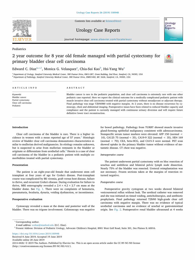
The patient is an eight-year-old female that underwent stem celltransplant at four years of age for Crohn's disease. Post-transplantcourse was complicated by BK viremia, graft versus host disease, failureto thrive, and recurrent Crohn's disease. During evaluation for failure tothrive, MRI enterography revealed a 2.4× 4.2×2.7 cm mass at thebladder dome. See Fig. 1. There were no complaints of hematuria,pneumaturia, fecaluria, dysuria, voiding dysfunction, or incontinence.
Preoperative evaluation
Cystoscopy revealed a mass at the dome and posterior wall of thebladder. There was no trigone involvement. Colonoscopy was negative
for bowel pathology. Pathology from TURBT showed muscle invasivegland-forming epithelial malignancy consistent with adenocarcinoma.Nonspecific serum tumor markers were elevated: AFP 150 (normal <10), CA125 75 (normal< 35), CA19-9 322 (normal < 35), HE4 169(normal < 70); CEA, beta-HCG, and CA15-3 were normal. PET scanshowed uptake in the primary bladder tumor without evidence of me-tastatic disease. CT chest was negative.
Intraoperative course
The patient underwent partial cystectomy with en bloc resection ofurachus and umbilicus and bilateral pelvic lymph node dissection.Nearly 75% of the bladder was resected. Ureteral reimplantation wasnot necessary. Frozen sections taken at the margins of resection re-turned negative.
Postoperative course
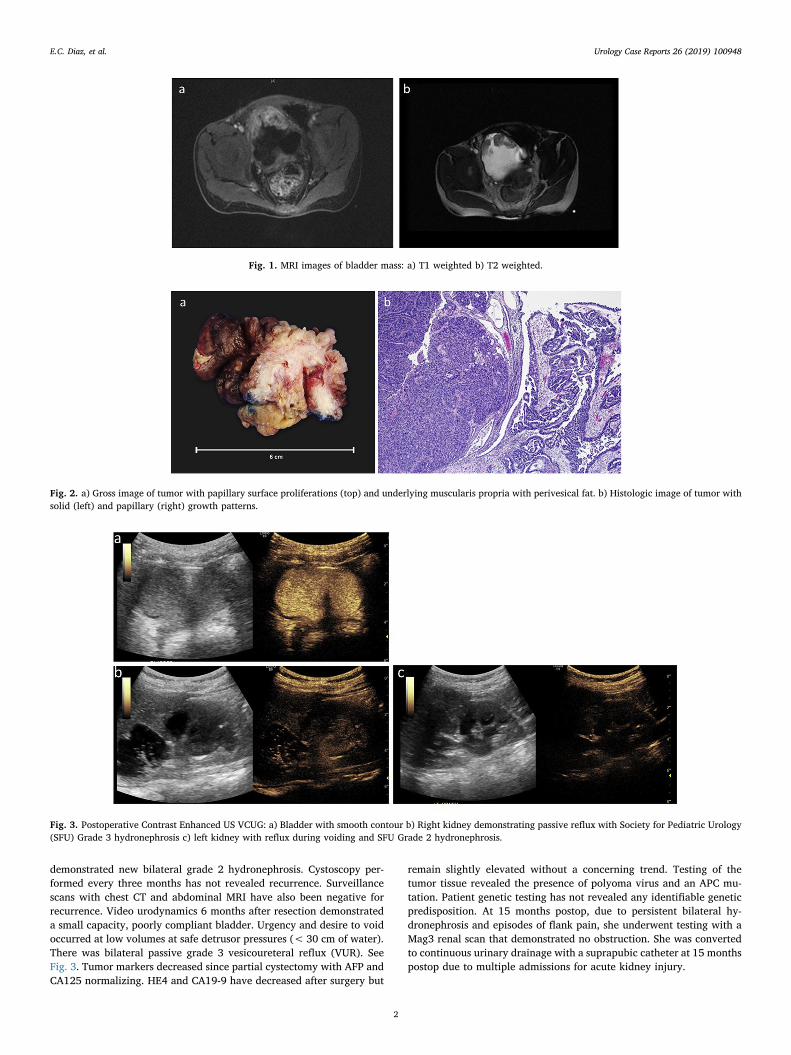
Postoperative gravity cystogram at two weeks showed bilateralvesicoureteral reflux without leak. The urethral catheter was removedand she was initiated on timed voiding, anticholinergics, and antibioticprophylaxis. Final pathology returned T2bN0 high-grade clear cellcarcinoma with negative margin. There was no evidence of typicalurothelial carcinoma and no evidence of urachal or gastrointestinalorigin. See Fig. 2. Postoperative renal bladder ultrasound at 4 weeks
https://doi.org/10.1016/j.eucr.2019.100948Received 8 June 2019; Accepted 19 June 2019
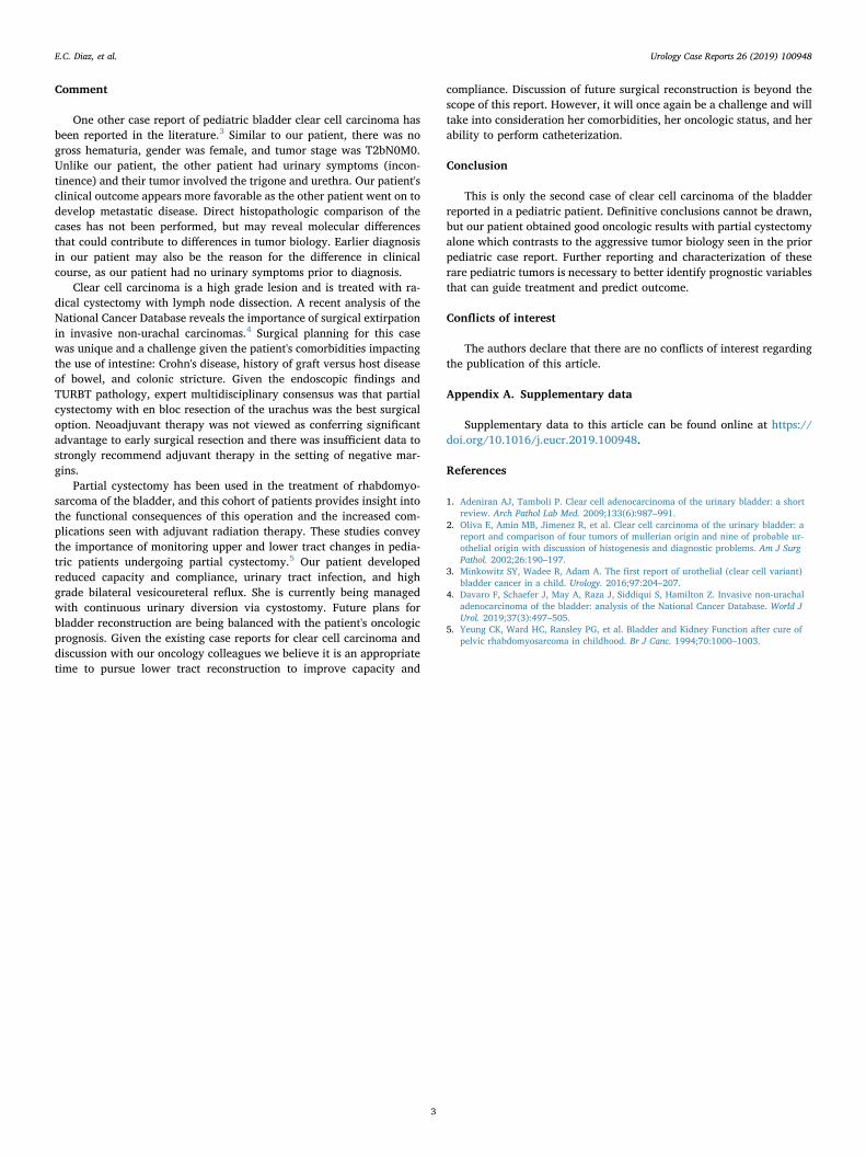
demonstrated new bilateral grade 2 hydronephrosis. Cystoscopy per-formed every three months has not revealed recurrence. Surveillancescans with chest CT and abdominal MRI have also been negative forrecurrence. Video urodynamics 6 months after resection demonstrateda small capacity, poorly compliant bladder. Urgency and desire to voidoccurred at low volumes at safe detrusor pressures (< 30 cm of water).There was bilateral passive grade 3 vesicoureteral reflux (VUR). SeeFig. 3. Tumor markers decreased since partial cystectomy with AFP andCA125 normalizing. HE4 and CA19-9 have decreased after surgery but
remain slightly elevated without a concerning trend. Testing of thetumor tissue revealed the presence of polyoma virus and an APC mu-tation. Patient genetic testing has not revealed any identifiable geneticpredisposition. At 15 months postop, due to persistent bilateral hy-dronephrosis and episodes of flank pain, she underwent testing with aMag3 renal scan that demonstrated no obstruction. She was convertedto continuous urinary drainage with a suprapubic catheter at 15 monthspostop due to multiple admissions for acute kidney injury.
Fig. 1. MRI images of bladder mass: a) T1 weighted b) T2 weighted.
Fig. 2. a) Gross image of tumor with papillary surface proliferations (top) and underlying muscularis propria with perivesical fat. b) Histologic image of tumor withsolid (left) and papillary (right) growth patterns.
Fig. 3. Postoperative Contrast Enhanced US VCUG: a) Bladder with smooth contour b) Right kidney demonstrating passive reflux with Society for Pediatric Urology(SFU) Grade 3 hydronephrosis c) left kidney with reflux during voiding and SFU Grade 2 hydronephrosis.
E.C. Diaz, et al. Urology Case Reports 26 (2019) 100948
2
Comment
One other case report of pediatric bladder clear cell carcinoma hasbeen reported in the literature.3 Similar to our patient, there was nogross hematuria, gender was female, and tumor stage was T2bN0M0.Unlike our patient, the other patient had urinary symptoms (incon-tinence) and their tumor involved the trigone and urethra. Our patient'sclinical outcome appears more favorable as the other patient went on todevelop metastatic disease. Direct histopathologic comparison of thecases has not been performed, but may reveal molecular differencesthat could contribute to differences in tumor biology. Earlier diagnosisin our patient may also be the reason for the difference in clinicalcourse, as our patient had no urinary symptoms prior to diagnosis.
Clear cell carcinoma is a high grade lesion and is treated with ra-dical cystectomy with lymph node dissection. A recent analysis of theNational Cancer Database reveals the importance of surgical extirpationin invasive non-urachal carcinomas.4 Surgical planning for this casewas unique and a challenge given the patient's comorbidities impactingthe use of intestine: Crohn's disease, history of graft versus host diseaseof bowel, and colonic stricture. Given the endoscopic findings andTURBT pathology, expert multidisciplinary consensus was that partialcystectomy with en bloc resection of the urachus was the best surgicaloption. Neoadjuvant therapy was not viewed as conferring significantadvantage to early surgical resection and there was insufficient data tostrongly recommend adjuvant therapy in the setting of negative mar-gins.
Partial cystectomy has been used in the treatment of rhabdomyo-sarcoma of the bladder, and this cohort of patients provides insight intothe functional consequences of this operation and the increased com-plications seen with adjuvant radiation therapy. These studies conveythe importance of monitoring upper and lower tract changes in pedia-tric patients undergoing partial cystectomy.5 Our patient developedreduced capacity and compliance, urinary tract infection, and highgrade bilateral vesicoureteral reflux. She is currently being managedwith continuous urinary diversion via cystostomy. Future plans forbladder reconstruction are being balanced with the patient's oncologicprognosis. Given the existing case reports for clear cell carcinoma anddiscussion with our oncology colleagues we believe it is an appropriatetime to pursue lower tract reconstruction to improve capacity and
compliance. Discussion of future surgical reconstruction is beyond thescope of this report. However, it will once again be a challenge and willtake into consideration her comorbidities, her oncologic status, and herability to perform catheterization.
Conclusion
This is only the second case of clear cell carcinoma of the bladderreported in a pediatric patient. Definitive conclusions cannot be drawn,but our patient obtained good oncologic results with partial cystectomyalone which contrasts to the aggressive tumor biology seen in the priorpediatric case report. Further reporting and characterization of theserare pediatric tumors is necessary to better identify prognostic variablesthat can guide treatment and predict outcome.
Conflicts of interest
The authors declare that there are no conflicts of interest regardingthe publication of this article.
Appendix A. Supplementary data
Supplementary data to this article can be found online at https://doi.org/10.1016/j.eucr.2019.100948.
References
1. Adeniran AJ, Tamboli P. Clear cell adenocarcinoma of the urinary bladder: a shortreview. Arch Pathol Lab Med. 2009;133(6):987–991.
2. Oliva E, Amin MB, Jimenez R, et al. Clear cell carcinoma of the urinary bladder: areport and comparison of four tumors of mullerian origin and nine of probable ur-othelial origin with discussion of histogenesis and diagnostic problems. Am J SurgPathol. 2002;26:190–197.
3. Minkowitz SY, Wadee R, Adam A. The first report of urothelial (clear cell variant)bladder cancer in a child. Urology. 2016;97:204–207.
4. Davaro F, Schaefer J, May A, Raza J, Siddiqui S, Hamilton Z. Invasive non-urachaladenocarcinoma of the bladder: analysis of the National Cancer Database. World JUrol. 2019;37(3):497–505.
5. Yeung CK, Ward HC, Ransley PG, et al. Bladder and Kidney Function after cure ofpelvic rhabdomyosarcoma in childhood. Br J Canc. 1994;70:1000–1003.
E.C. Diaz, et al. Urology Case Reports 26 (2019) 100948