Page 1
CAMPBELL BIOLOGY IN FOCUS
© 2014 Pearson Education, Inc.
Urry • Cain • Wasserman • Minorsky • Jackson • Reece
Lecture Presentations by Kathleen Fitzpatrick and Nicole Tunbridge
Unit 6.2
Animal Nutrition
Page 2
© 2014 Pearson Education, Inc.
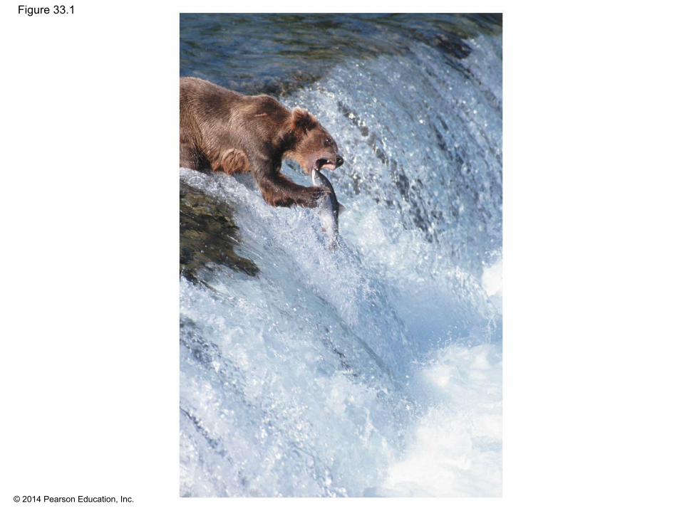
▪ Food is taken in, taken apart, and taken up in the process of animal nutrition
▪ In general, animals fall into three categories▪ Herbivores eat mainly plants and algae▪ Carnivores eat other animals▪ Omnivores regularly consume animals as well as
plants or algae
▪ Most animals are also opportunistic feeders
Overview: The Need to Feed
Page 3
© 2014 Pearson Education, Inc.
Figure 33.1
Page 4
© 2014 Pearson Education, Inc.
Concept 33.1: An animal’s diet must supply chemical energy, organic molecules, and essential nutrients
▪ An animal’s diet provides▪ Chemical energy, which is converted into ATP to
power cellular processes▪ Organic building blocks, such as organic carbon and
organic nitrogen, to synthesize a variety of organic molecules
▪ Essential nutrients, which are required by cells and must be obtained from dietary sources
Page 5
© 2014 Pearson Education, Inc.
Essential Nutrients
▪ Essential nutrients must be obtained from an animal’s diet
▪ There are four classes of essential nutrients▪ Essential amino acids▪ Essential fatty acids▪ Vitamins▪ Minerals
Page 6
© 2014 Pearson Education, Inc.
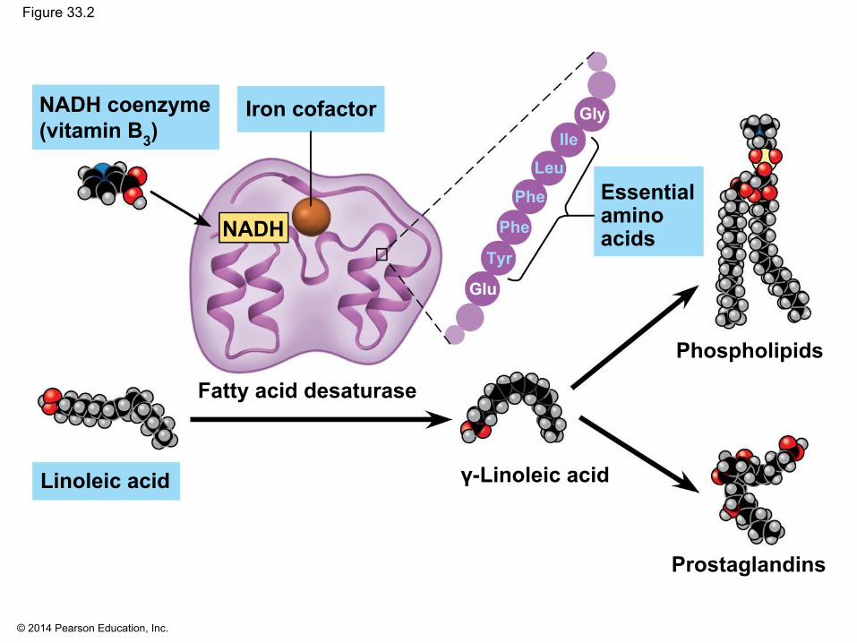
Figure 33.2
Iron cofactor
Essentialaminoacids
NADH coenzyme(vitamin B3)
NADH
Fatty acid desaturase
γ-Linoleic acid
Phospholipids
Prostaglandins
GlyIle
LeuPhe
Phe
Tyr
Glu
Linoleic acid
Page 7
© 2014 Pearson Education, Inc.
▪ In animals, fatty acids are converted into a variety of cellular components, such as membrane phospholipids, signaling molecules, and storage fats
▪ Essential fatty acids can be synthesized by plants▪ Deficiencies of essential fatty acids are rare
Essential Fatty Acids and Amino Acids
Page 8
© 2014 Pearson Education, Inc.
▪ Animals require 20 amino acids and can synthesize about half from molecules in their diet
▪ The remaining amino acids, the essential amino acids, must be obtained from food in preassembled form
▪ Meat, eggs, and cheese provide all the essential amino acids and are thus “complete” proteins
Page 9
© 2014 Pearson Education, Inc.
▪ Most plant proteins are incomplete in amino acid composition
▪ Individuals who eat only plant proteins need to eat specific plant combinations to get all the essential amino acids
Page 10
© 2014 Pearson Education, Inc.
Vitamins
▪ Vitamins are organic molecules required in the diet in small amounts
▪ Thirteen vitamins are essential for humans▪ Vitamins are grouped into two categories: fat-soluble
and water-soluble
Page 11
© 2014 Pearson Education, Inc.
Minerals
▪ Minerals are simple inorganic nutrients, usually required in small amounts
▪ Ingesting large amounts of some minerals can upset homeostatic balance
Page 12
© 2014 Pearson Education, Inc.
Dietary Deficiencies
▪ Malnutrition results from the long-term absence from the diet of one or more essential nutrients
Page 13
© 2014 Pearson Education, Inc.
Deficiencies in Essential Nutrients
▪ Deficiencies in essential nutrients can cause deformities, disease, and death
▪ Animals may consume salt, minerals, shells, or stones to prevent mineral deficiencies
Page 14
© 2014 Pearson Education, Inc.
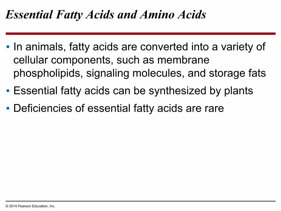
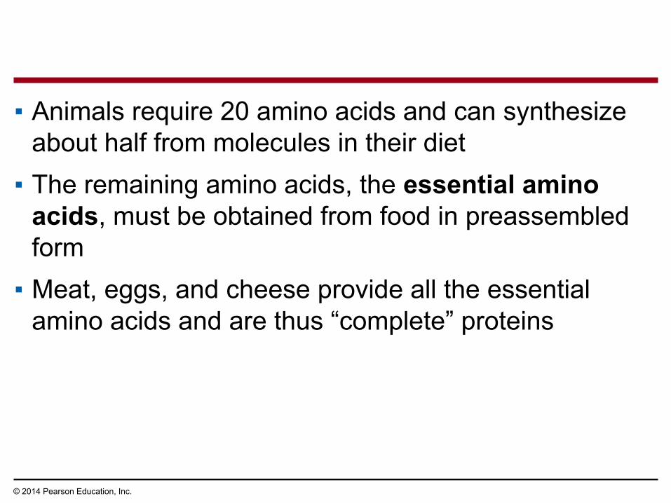
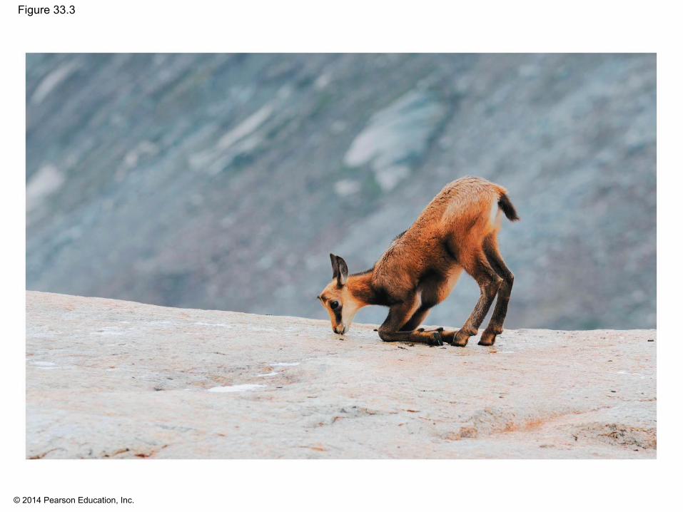
Figure 33.3
Page 15
© 2014 Pearson Education, Inc.
▪ A diet with insufficient amounts of one or more amino acids is the most common type of malnutrition among humans
▪ Individuals subsisting on simple rice diets are often deficient in vitamin A
▪ To overcome this, scientists have engineered a strain of rice that synthesizes beta-carotene, which is converted to vitamin A in the body
Page 16
© 2014 Pearson Education, Inc.
▪ Undernutrition results when a diet does not provide enough chemical energy
▪ An undernourished individual will▪ Use up stored fat and carbohydrates▪ Break down its own proteins▪ Lose muscle mass▪ Suffer protein deficiency of the brain▪ Die or suffer irreversible damage
Undernutrition
Page 17
© 2014 Pearson Education, Inc.
Assessing Nutritional Needs
▪ Genetic defects that disrupt food uptake provide information about human nutrition
▪ For example, hemochromatosis causes iron buildup without excessive iron intake
▪ Insights into human nutrition have come from epidemiology, the study of human health and disease in populations
▪ Neural tube defects were found to be the result of a deficiency in folic acid in pregnant mothers
Page 18
© 2014 Pearson Education, Inc.
Concept 33.2: The main stages of food processing are ingestion, digestion, absorption, and elimination
▪ Food processing can be divided into four distinct stages
Page 19
© 2014 Pearson Education, Inc.
Figure 33.4
Mechanicaldigestion
Nutrient moleculesenter body cells
Chemicaldigestion(enzymatichydrolysis) Undigested
material
EliminationAbsorptionDigestionIngestion1 2 3 4
Page 20
© 2014 Pearson Education, Inc.
▪ Ingestion is the act of eating or feeding▪ Strategies for extracting resources from food differ
widely among animals
Page 21
© 2014 Pearson Education, Inc.
Figure 33.5
Filter feeders Substrate feeders Fluid feeders
Bulk feeders
Caterpillar Feces
Baleen
Page 22
© 2014 Pearson Education, Inc.
Figure 33.5a
Filter feeders
Baleen
Page 23
© 2014 Pearson Education, Inc.
Figure 33.5b
Substrate feeders
Caterpillar Feces
Page 24
© 2014 Pearson Education, Inc.
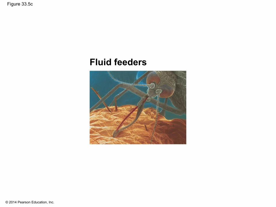
Figure 33.5c
Fluid feeders
Page 25
© 2014 Pearson Education, Inc.
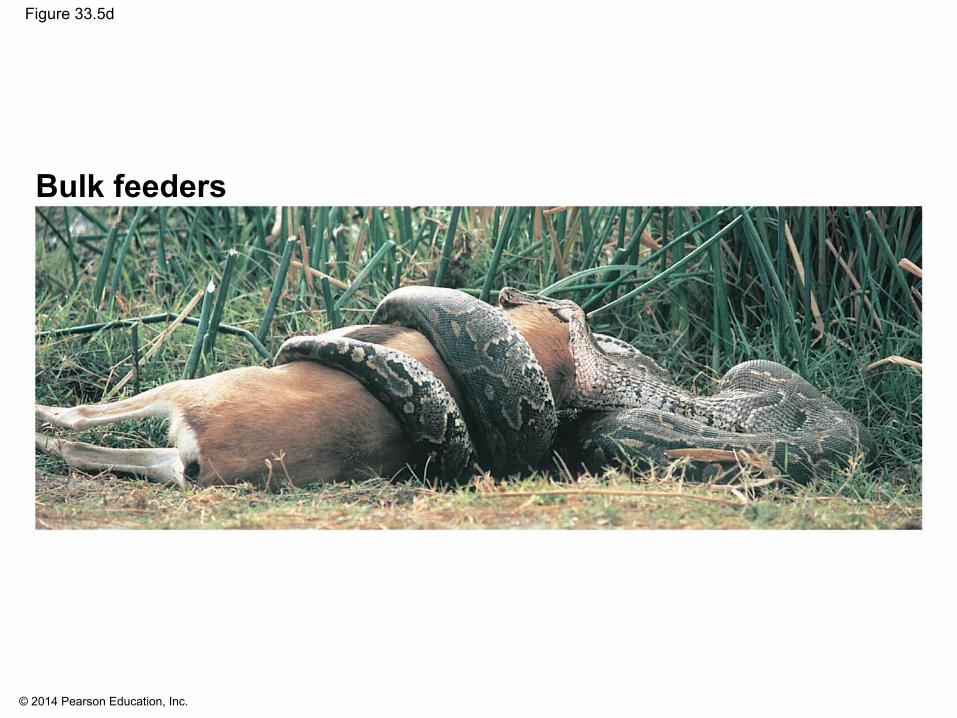
Figure 33.5d
Bulk feeders
Page 26
© 2014 Pearson Education, Inc.
▪ Digestion is the process of breaking food down into molecules small enough to absorb
▪ Mechanical digestion, including chewing, increases the surface area of food
▪ Chemical digestion splits food into small molecules that can pass through membranes
▪ In chemical digestion, the process of enzymatic hydrolysis splits bonds in molecules with the addition of water
Page 27
© 2014 Pearson Education, Inc.
▪ Absorption is uptake of nutrients by body cells▪ Elimination is the passage of undigested material
out of the digestive system
Page 28
© 2014 Pearson Education, Inc.
Digestive Compartments
▪ Most animals process food in specialized compartments
▪ These compartments reduce the risk of an animal digesting its own cells and tissues
Page 29
© 2014 Pearson Education, Inc.
Intracellular Digestion
▪ In intracellular digestion, food particles are engulfed by phagocytosis
▪ Food vacuoles, containing food, fuse with lysosomes containing hydrolytic enzymes
Page 30
© 2014 Pearson Education, Inc.
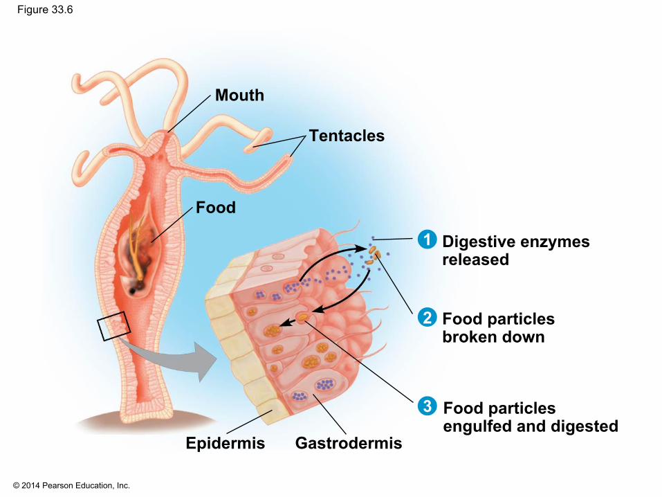
Extracellular Digestion
▪ Extracellular digestion is the breakdown of food particles outside of cells
▪ It occurs in compartments that are continuous with the outside of the animal’s body
▪ Animals with simple body plans have a gastrovascular cavity that functions in both digestion and distribution of nutrients
Page 31
© 2014 Pearson Education, Inc.
Figure 33.6
Mouth
Tentacles
Digestive enzymesreleased
Food particlesbroken down
Food particlesengulfed and digested
GastrodermisEpidermis
Food
1
2
3
Page 32
© 2014 Pearson Education, Inc.
▪ More complex animals have a complete digestive tract or an alimentary canal with a mouth and an anus
▪ The alimentary canal can have specialized regions that carry out digestion and absorption in a stepwise fashion
Page 33
© 2014 Pearson Education, Inc.
Figure 33.7
CropGizzard
Intestine
Anus
Esophagus
Pharynx
Mouth(a) Earthworm
Esophagus Crop
StomachGizzard
Intestine
Anus
Mouth
(c) Bird(b) Grasshopper
MouthCrop Gastric
cecae
AnusRectumEsophagus
Foregut Midgut Hindgut
Page 34
© 2014 Pearson Education, Inc.
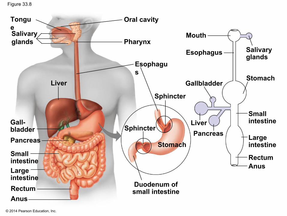
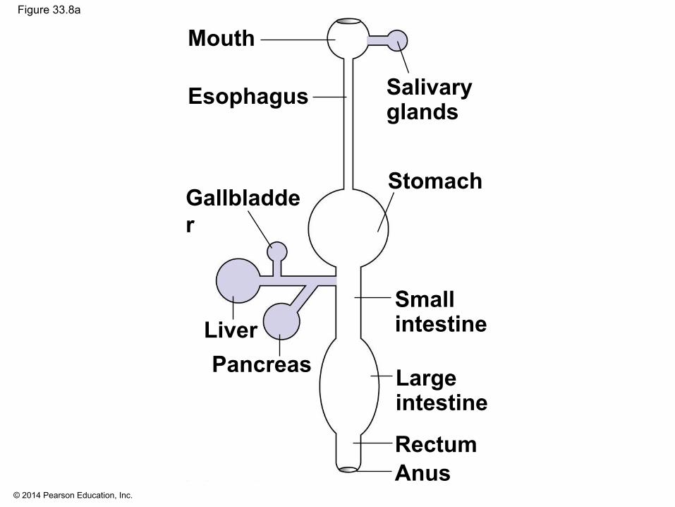
Concept 33.3: Organs specialized for sequential stages of food processing form the mammalian digestive system
▪ The mammalian digestive system consists of an alimentary canal and accessory glands that secrete digestive juices through ducts
▪ Mammalian accessory glands are the salivary glands, the pancreas, the liver, and the gallbladder
Page 35
© 2014 Pearson Education, Inc.
Figure 33.8
TongueSalivaryglands
Liver
Gall-bladderPancreas
SmallintestineLargeintestineRectumAnus
Oral cavity
Pharynx
Esophagus
Sphincter
Sphincter
Stomach
LiverPancreas
Gallbladder
Duodenum ofsmall intestine
Stomach
Smallintestine
Largeintestine
RectumAnus
SalivaryglandsEsophagus
Mouth
Page 36
© 2014 Pearson Education, Inc.
Figure 33.8a
LiverPancreas
Gallbladder
Stomach
Smallintestine
Largeintestine
RectumAnus
SalivaryglandsEsophagus
Mouth
Page 37
© 2014 Pearson Education, Inc.
▪ Food is pushed along by peristalsis, rhythmic contractions of muscles in the wall of the canal
▪ Valves called sphincters regulate the movement of material between compartments
Page 38
© 2014 Pearson Education, Inc.
The Oral Cavity, Pharynx, and Esophagus
▪ The first stage of digestion is mechanical and takes place in the oral cavity
▪ Salivary glands deliver saliva to the oral cavity through ducts
▪ Teeth chew food into smaller particles that are exposed to salivary amylase, initiating breakdown of glucose polymers
▪ Saliva also contains mucus, a viscous mixture of water, salts, cells, and glycoproteins
Page 39
© 2014 Pearson Education, Inc.
▪ The tongue shapes food into a bolus and provides help with swallowing
▪ The throat, or pharynx, is the junction that opens to both the esophagus and the trachea
▪ The esophagus connects to the stomach▪ The trachea (windpipe) leads to the lungs
Page 40
© 2014 Pearson Education, Inc.
▪ Swallowing must be carefully choreographed to avoid choking
▪ The esophagus conducts food from the pharynx down to the stomach through rhythmic cycles of contraction
▪ The form of the esophagus fits its function and varies among species
Page 41
© 2014 Pearson Education, Inc.
Digestion in the Stomach
▪ The stomach stores food and secretes gastric juice, which converts a meal to a mixture of food and digestive juice called chyme
Page 42
© 2014 Pearson Education, Inc.
Figure 33.9
Esophagus
Sphincter
Sphincter
Stomach
Folds ofepithelialtissue
Smallintestine
EpitheliumProduction of gastricjuice
Pepsinogen andHCI secreted intolumenHCI convertspepsinogen topepsin.
Pepsin activatesmore pepsinogen,starting a chainreaction.
Parietalcell
Pepsin(activeenzyme)
Chiefcell
Pepsinogen1
2
3
1
2
3
Gastric gland
Mucous cell
Chief cell
Parietal cell
Gastric piton the interiorsurface ofstomach
10 μ
m
HCI
H+
Cl−
Page 43
© 2014 Pearson Education, Inc.
Chemical Digestion in the Stomach
▪ Gastric juice has a low pH of about 2, which kills bacteria and denatures proteins
▪ Gastric juice is made up of hydrochloric acid (HCl) and pepsin
▪ Pepsin is a protease, or protein-digesting enzyme, that cleaves proteins into smaller peptides
Page 44
© 2014 Pearson Education, Inc.
Figure 33.10
Fat digestion
Fat(triglycerides)
Glycerol,fatty acids,monoglycerides
Pancreaticlipase
Pancreaticnucleases
Nucleotidases
Pepsin
Pancreatic trypsinand chymotrypsin
Pancreaticcarboxypeptidase
Dipeptidases,carboxypeptidase,and aminopeptidaseDisaccharidases
Pancreatic amylases
Disaccharides
Monosaccharides
Amino acids
Small peptides
Smallerpolypeptides
Small polypeptides
Nucleotides
Nucleosides
Nitrogenous bases,sugars, phosphates
Nucleosidasesandphosphatases
DNA, RNA
Nucleic aciddigestion
Protein digestion
Proteins
MaltoseSmallerpolysaccharides
Polysaccharides(starch, glycogen)
Disaccharides(sucrose,
lactose)
Carbohydrate digestionOral cavity,pharynx,esophagus
Smallintestine(enzymesfrompancreas)
Stomach
Smallintestine(enzymesfromepithelium)
Salivary amylase
Page 45
© 2014 Pearson Education, Inc.
▪ Mucus protects the stomach lining from gastric juice▪ Also, cell division adds a new epithelial layer every
three days, to replace any cells damaged by digestive juices
▪ Gastric ulcers, lesions in the stomach lining, are caused mainly by the bacterium Helicobacter pylori
Page 46
© 2014 Pearson Education, Inc.
Stomach Dynamics
▪ Coordinated contraction and relaxation of stomach muscle churn the stomach’s contents
▪ Sphincters prevent chyme from entering the esophagus and regulate its entry into the small intestine
▪ Stomach contents typically pass into the small intestine 2–6 hours after a meal
Page 47
© 2014 Pearson Education, Inc.
Digestion in the Small Intestine
▪ The small intestine is the longest section of the alimentary canal
▪ It is the major organ of digestion and absorption▪ The first portion of the small intestine is the
duodenum▪ Here, chyme from the stomach mixes with digestive
juices from the pancreas, liver, gallbladder, and the intestinal wall
Page 48
© 2014 Pearson Education, Inc.
Pancreatic Secretions
▪ The pancreas produces proteases trypsin and chymotrypsin, which are activated in the lumen of the duodenum
▪ Its solution is alkaline and neutralizes the acidic chyme
Page 49
© 2014 Pearson Education, Inc.
Bile Production by the Liver
▪ In the small intestine, bile aids in digestion and absorption of fats
▪ Bile is made in the liver and stored in the gallbladder
▪ Bile also destroys nonfunctional red blood cells
Page 50
© 2014 Pearson Education, Inc.
Secretions of the Small Intestine
▪ The epithelial lining of the duodenum produces several digestive enzymes
▪ Enzymatic digestion is completed as peristalsis moves the chyme and digestive juices along the small intestine
▪ Most digestion occurs in the duodenum; the jejunum and ileum function mainly in absorption of nutrients and water
Page 51
© 2014 Pearson Education, Inc.
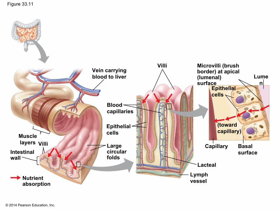
Figure 33.11
Vein carryingblood to liver
Microvilli (brushborder) at apical(lumenal) surface
Villi
Lumen
Basalsurface
Lymphvessel
Capillary
Epithelialcells
(towardcapillary)
Bloodcapillaries
Epithelialcells
Largecircularfolds
Muscle layers Villi
Intestinalwall
Nutrientabsorption
Lacteal
Page 52
© 2014 Pearson Education, Inc.
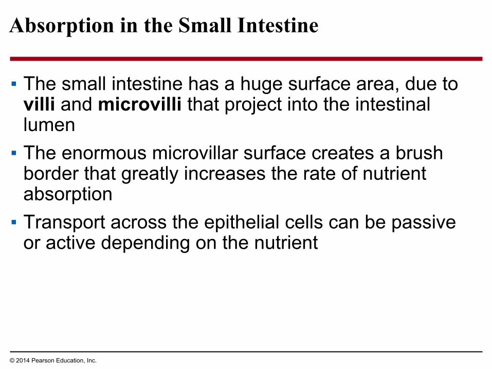
Absorption in the Small Intestine
▪ The small intestine has a huge surface area, due to villi and microvilli that project into the intestinal lumen
▪ The enormous microvillar surface creates a brush border that greatly increases the rate of nutrient absorption
▪ Transport across the epithelial cells can be passive or active depending on the nutrient
Page 53
© 2014 Pearson Education, Inc.
Figure 33.12
Triglyceridesare broken downto fatty acids andmonoglyceridesby lipase.
Monoglyceridesand fatty acids diffuseinto epithelial cellsand are re-formed intotriglycerides.
Triglycerides areincorporated intochylomicrons.
Chylomicrons enterlacteals and are carriedaway by lymph.
4
3
2
1
Triglycerides
Chylomicron
Lacteal
Phospholipids,cholesterol,and proteins
Triglycerides
Fatty acids Mono-glycerides
Epithelialcell
LUMENOF SMALLINTESTINE
Page 54
© 2014 Pearson Education, Inc.
▪ The hepatic portal vein carries nutrient-rich blood from the capillaries of the villi to the liver, then to the heart
▪ The liver regulates nutrient distribution, interconverts many organic molecules, and detoxifies many organic molecules
Page 55
© 2014 Pearson Education, Inc.
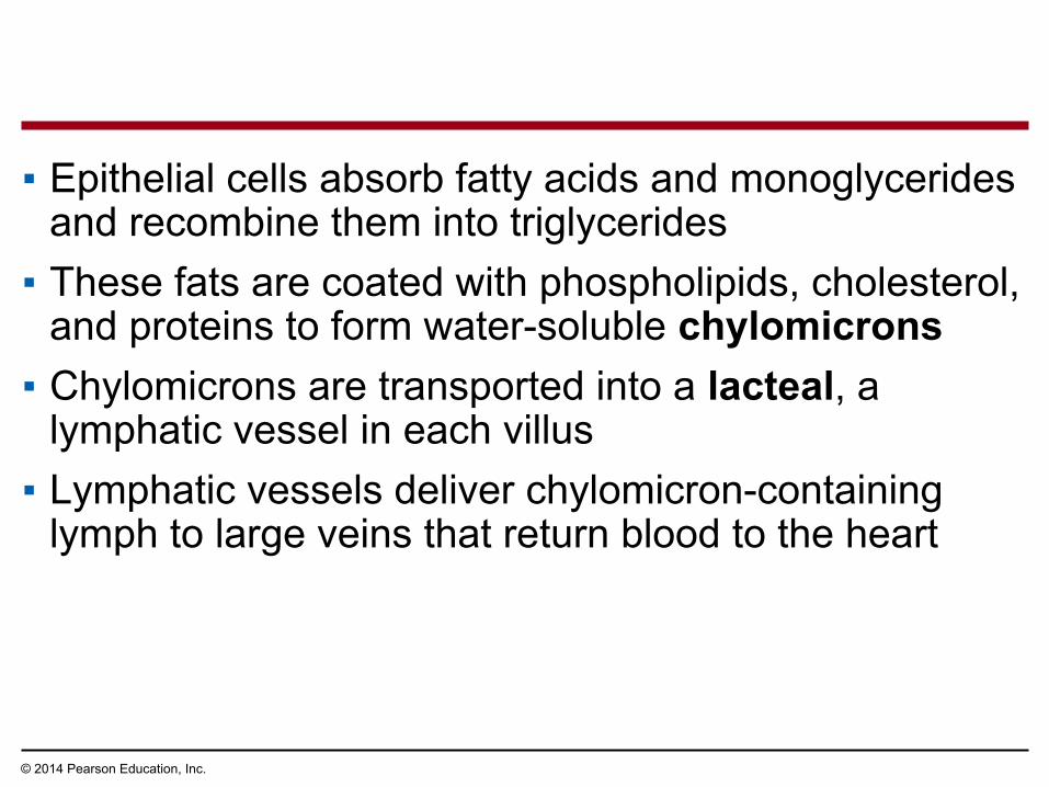
▪ Epithelial cells absorb fatty acids and monoglycerides and recombine them into triglycerides
▪ These fats are coated with phospholipids, cholesterol, and proteins to form water-soluble chylomicrons
▪ Chylomicrons are transported into a lacteal, a lymphatic vessel in each villus
▪ Lymphatic vessels deliver chylomicron-containing lymph to large veins that return blood to the heart
Page 56
© 2014 Pearson Education, Inc.
Absorption in the Large Intestine
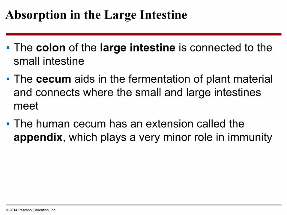
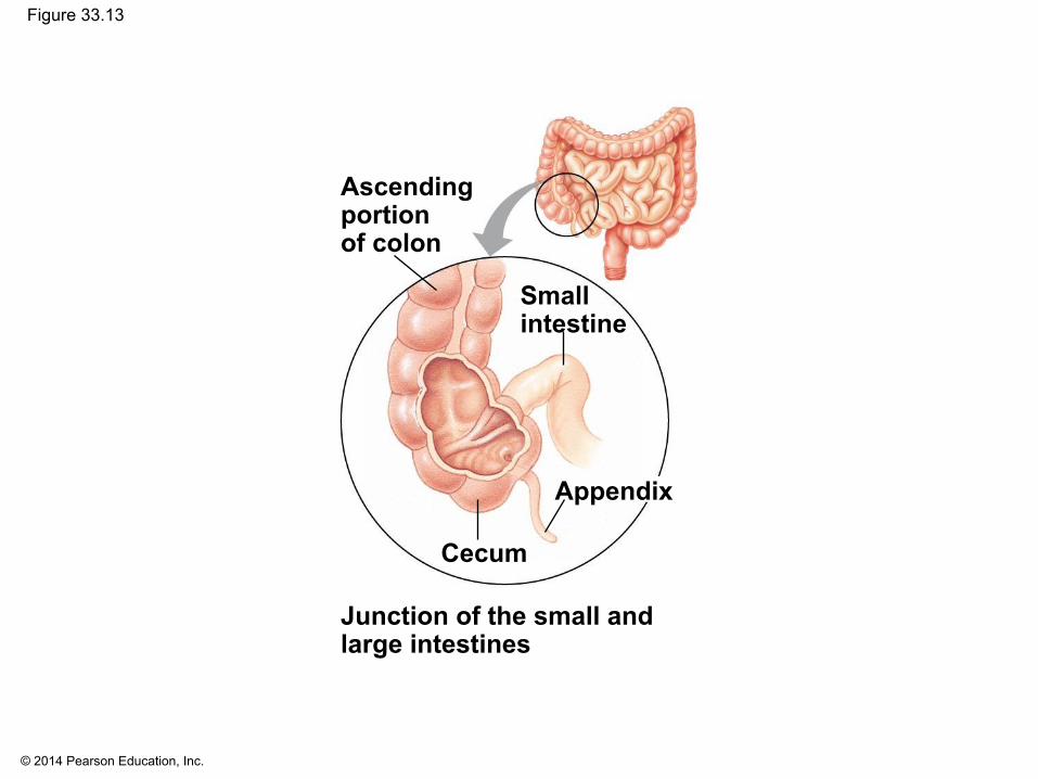
▪ The colon of the large intestine is connected to the small intestine
▪ The cecum aids in the fermentation of plant material and connects where the small and large intestines meet
▪ The human cecum has an extension called the appendix, which plays a very minor role in immunity
Page 57
© 2014 Pearson Education, Inc.
Figure 33.13
Ascendingportionof colon
Smallintestine
Appendix
Cecum
Junction of the small andlarge intestines
Page 58
© 2014 Pearson Education, Inc.
▪ A major function of the colon is to recover water that has entered the alimentary canal
▪ The colon houses bacteria (e.g., Escherichia coli) that live on unabsorbed organic material; some produce vitamins
▪ Feces, including undigested material and bacteria, become more solid as they move through the colon
Page 59
© 2014 Pearson Education, Inc.
▪ Feces are stored in the rectum until they can be eliminated through the anus
▪ Two sphincters between the rectum and anus control bowel movements
Page 60
© 2014 Pearson Education, Inc.
Concept 33.4: Evolutionary adaptations of vertebrate digestive systems correlate with diet
▪ Digestive systems of vertebrates are variations on a common plan
▪ However, there are intriguing adaptations, often related to diet
Page 61
© 2014 Pearson Education, Inc.
Dental Adaptations
▪ Dentition, an animal’s assortment of teeth, is one example of structural variation reflecting diet
▪ The success of mammals is due in part to their dentition, which is specialized for different diets
▪ Nonmammalian vertebrates have less specialized teeth, though exceptions exist
▪ For example, the teeth of poisonous snakes are modified as fangs for injecting venom
Page 62
© 2014 Pearson Education, Inc.
Figure 33.14
Carnivore Herbivore
Omnivore
Incisors Canines
Premolars Molars
Page 63
© 2014 Pearson Education, Inc.
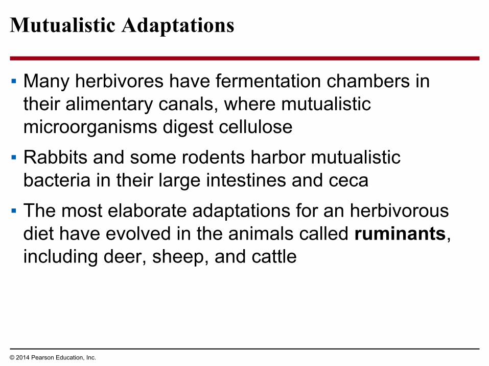
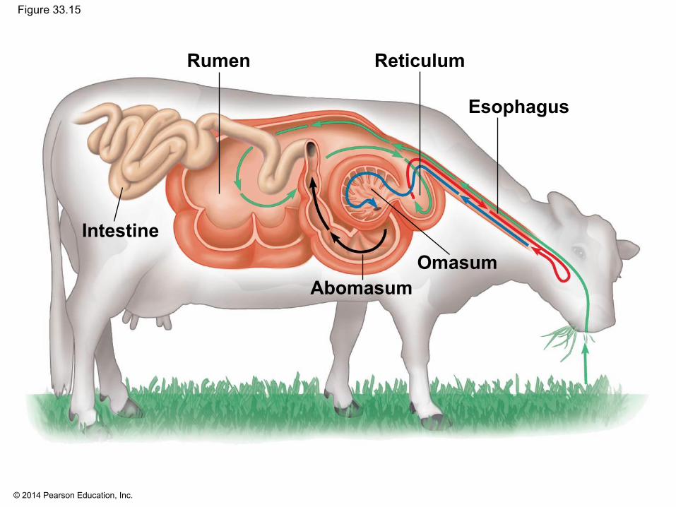
Mutualistic Adaptations
▪ Many herbivores have fermentation chambers in their alimentary canals, where mutualistic microorganisms digest cellulose
▪ Rabbits and some rodents harbor mutualistic bacteria in their large intestines and ceca
▪ The most elaborate adaptations for an herbivorous diet have evolved in the animals called ruminants, including deer, sheep, and cattle
Page 64
© 2014 Pearson Education, Inc.
Figure 33.15
Rumen Reticulum
Esophagus
OmasumAbomasum
Intestine
Page 65
© 2014 Pearson Education, Inc.
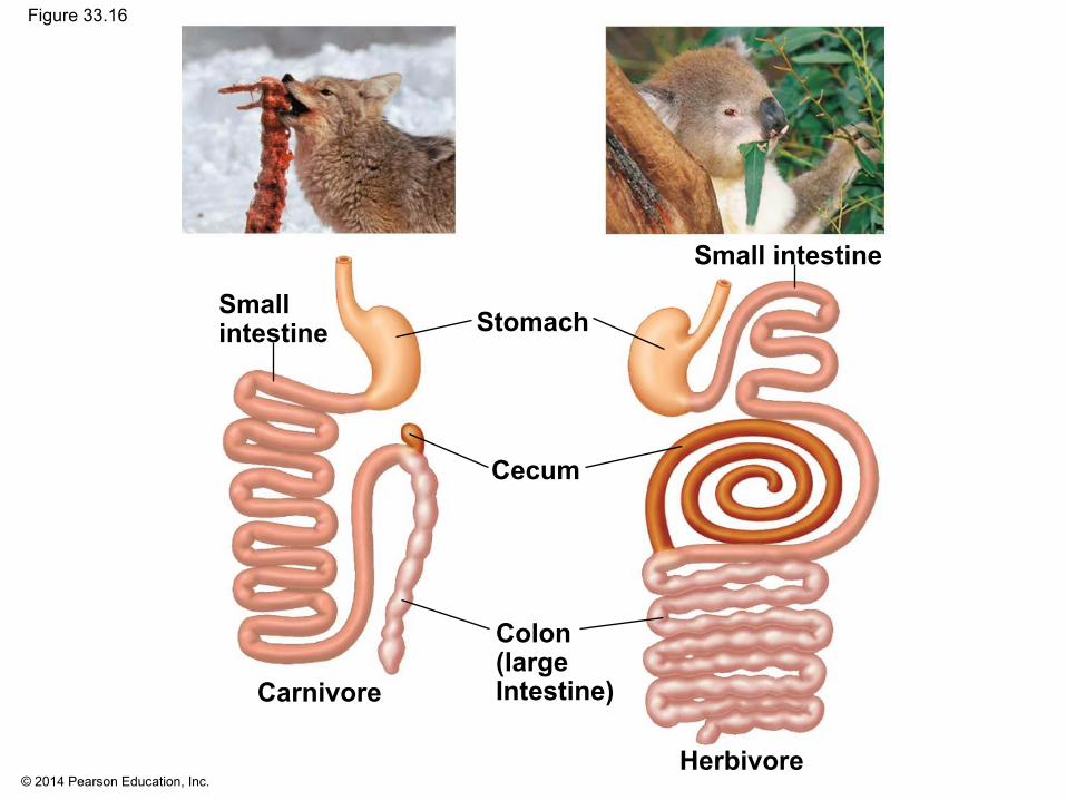
Stomach and Intestinal Adaptations
▪ Many carnivores have large, expandable stomachs▪ Herbivores and omnivores generally have longer
alimentary canals than carnivores, reflecting the longer time needed to digest vegetation
Page 66
© 2014 Pearson Education, Inc.
Figure 33.16
Smallintestine
Small intestine
Stomach
Cecum
Colon(largeIntestine)Carnivore
Herbivore
Page 67
© 2014 Pearson Education, Inc.
Concept 33.5: Feedback circuits regulate digestion, energy allocation, and appetite
▪ An animal’s intake of food and use of nutrients are matched to circumstance and need
Page 68
© 2014 Pearson Education, Inc.
Regulation of Digestion
▪ Each step in the digestive system is activated as needed
▪ The enteric division of the nervous system helps to regulate the digestive process
▪ The endocrine system also regulates digestion through the release and transport of hormones
Page 69
© 2014 Pearson Education, Inc.
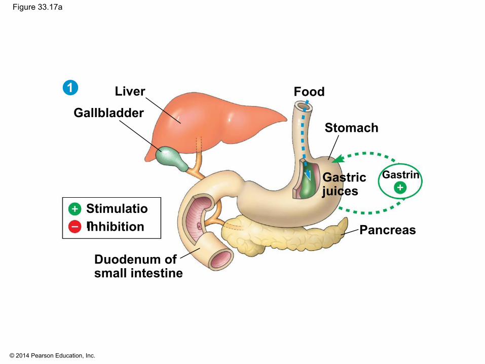
Figure 33.17
Food
Stomach
GastrinGastricjuices
Pancreas
LiverGallbladder
StimulationInhibition
1
Duodenum ofsmall intestine
Bile
Chyme
HCO3−, enzymes
32
CCK
CCK
Secretinand CCK
Secretin
Gastricjuices
Page 70
© 2014 Pearson Education, Inc.
Figure 33.17a
Food
Stomach
GastrinGastricjuices
Pancreas
LiverGallbladder
StimulationInhibition
1
Duodenum ofsmall intestine
Page 71
© 2014 Pearson Education, Inc.
Figure 33.17b
Bile
Chyme
HCO3−, enzymes
CCK
CCK
SecretinStimulationInhibition
2
Page 72
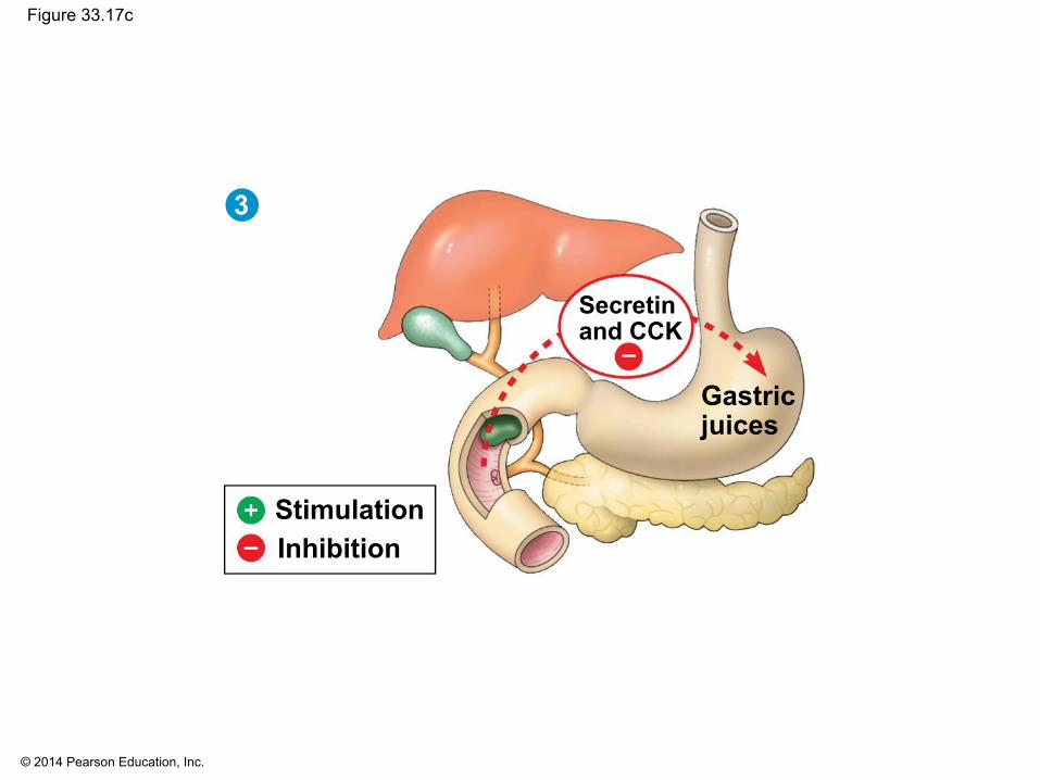
© 2014 Pearson Education, Inc.
Figure 33.17c
Secretinand CCK
Gastricjuices
3
StimulationInhibition
Page 73
© 2014 Pearson Education, Inc.
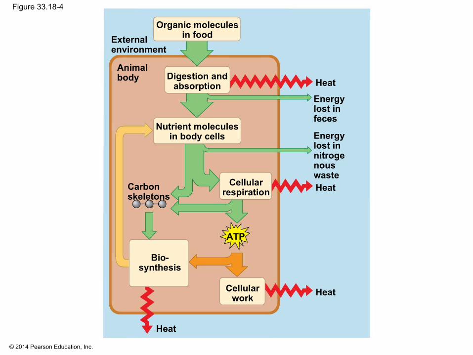
Energy Allocation
▪ The flow and transformation of energy in an animal—its bioenergetics—determine nutritional needs
▪ An animal’s energy use per unit of time is called its metabolic rate
▪ Metabolic rate can be determined by monitoring an animal’s rate of heat loss, the amount of O2 consumed, or the amount of CO2 produced
Page 74
© 2014 Pearson Education, Inc.
Figure 33.18-4
Organic moleculesin food
Heat
Energylost infeces
Energylost innitrogenouswasteHeatCellular
respiration
Digestion andabsorption
Nutrient moleculesin body cells
Animalbody
Externalenvironment
Carbonskeletons
ATP
Bio-synthesis
Cellularwork Heat
Heat
Page 75
© 2014 Pearson Education, Inc.
Minimum Metabolic Rate
▪ Animals must maintain a minimum metabolic rate for basic cell functions
▪ Basal metabolic rate, BMR, is the minimum metabolic rate of a nongrowing endotherm that is at rest, has an empty stomach, and is not experiencing stress
▪ The metabolic rate of a fasting, nonstressed ectotherm at a particular temperature is called standard metabolic rate, SMR
Page 76
© 2014 Pearson Education, Inc.
▪ Endothermy is more energetically costly than ectothermy
▪ For ectotherms and endotherms, activity greatly affects metabolic rate
Page 77
© 2014 Pearson Education, Inc.
Regulation of Energy Storage
▪ When an animal takes in more energy than is needed for metabolism and activity, excess energy is stored
▪ In humans, the liver and muscle cells are used first; energy is stored as glycogen
▪ When glycogen depots are full, additional excess energy is stored as fat in adipose cells
▪ When fewer calories are taken in than expended, the body expends liver glycogen, muscle glycogen, and then fat, in that order
Page 78
© 2014 Pearson Education, Inc.
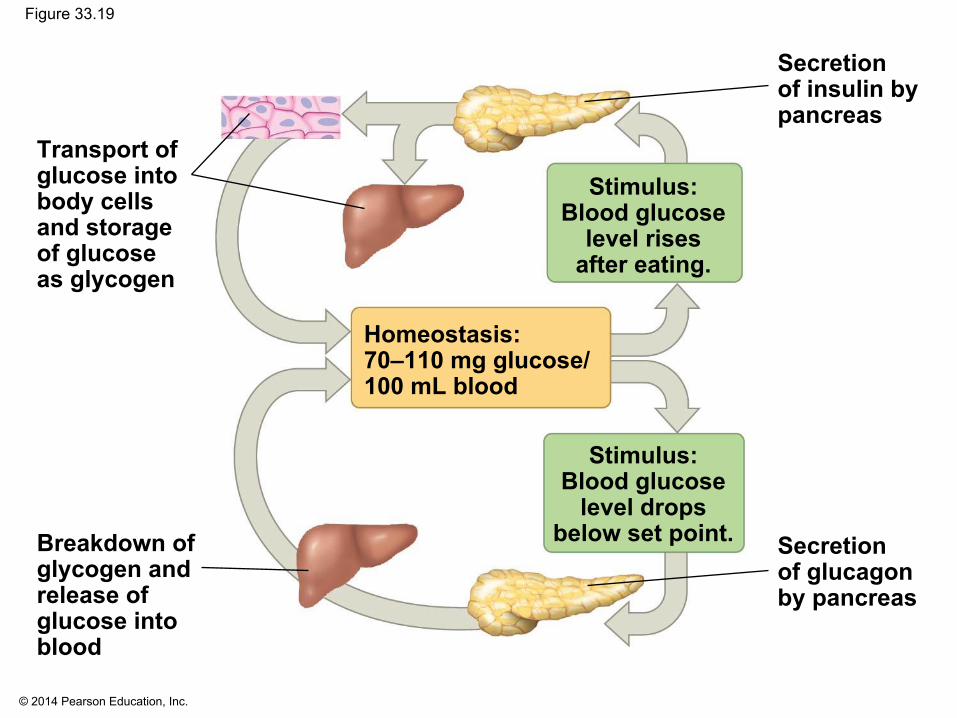
Glucose Homeostasis
▪ Insulin and glucagon together maintain glucose levels ▪ Insulin levels rise after a carbohydrate-rich meal, and
glucose entering the liver through the hepatic portal vein is used to synthesize glycogen
▪ When glucose concentration is low in the hepatic portal vein, glucagon stimulates the liver to break down glycogen and release glucose into the blood
▪ Insulin and glucagon are produced in the pancreas in beta cells and alpha cells, respectively
Page 79
© 2014 Pearson Education, Inc.
Figure 33.19
Transport ofglucose intobody cellsand storageof glucoseas glycogen
Breakdown ofglycogen andrelease ofglucose intoblood
Secretionof insulin bypancreas
Secretionof glucagonby pancreas
Stimulus:Blood glucose
level dropsbelow set point.
Stimulus:Blood glucose
level risesafter eating.
Homeostasis:70–110 mg glucose/100 mL blood
Page 80
© 2014 Pearson Education, Inc.
Diabetes Mellitus
▪ Diabetes mellitus is a disease caused by a deficiency of insulin or a decreased response to insulin in target tissues
▪ Cells are unable to take up glucose to meet their metabolic needs
▪ Fat becomes the main substrate for cellular respiration
Page 81
© 2014 Pearson Education, Inc.
▪ Type 1 diabetes is an autoimmune disorder in which the immune system destroys the pancreatic beta cells
▪ Type 2 diabetes is characterized by a failure of target cells to respond normally to insulin
▪ Heredity is a factor in type 2 diabetes▪ Excess body weight and lack of exercise increase
the risk
Page 82
© 2014 Pearson Education, Inc.
Regulation of Appetite and Consumption
▪ Overnourishment causes obesity, which results from excessive intake of food energy with the excess stored as fat
▪ Obesity contributes to diabetes (type 2), cancer of the colon and breasts, heart attacks, and strokes
▪ Researchers have discovered several of the mechanisms that help regulate body weight
Page 83
© 2014 Pearson Education, Inc.
▪ Ghrelin, a hormone secreted by the stomach wall, triggers a feeling of hunger before meals
▪ Insulin and PYY, a hormone secreted by the small intestine after eating, both suppress appetite
▪ Leptin, a hormone produced by adipose (fat) tissue, also suppresses appetite and may regulate body fat levels