Use of an EZ-Tn5-Based Random Mutagenesis System to Identify a Novel Toxin Regulatory Locus in Clostridium perfringens Strain 13 Jorge E. Vidal 1 , Jianming Chen 1 , Jihong Li 1 , Bruce A. McClane 1,2,3 * 1 Department of Microbiology and Molecular Genetics, University of Pittsburgh School of Medicine, Pittsburgh, Pennsylvania, United States of America, 2 Center for Vaccine Research, University of Pittsburgh School of Medicine, Pittsburgh, Pennsylvania, United States of America, 3 Australian Research Council Centre of Excellence in Structural and Functional Microbial Genetics, Department of Microbiology, Monash University, Melbourne, Victoria, Australia Abstract Background: Although useful for probing bacterial pathogenesis and physiology, current random mutagenesis systems suffer limitations for studying the toxin-producing bacterium Clostridium perfringens. Methodology/Principal Findings: An EZ-Tn5-based random mutagenesis approach was developed for use in C. perfringens. This mutagenesis system identified a new regulatory locus controlling toxin production by strain 13, a C. perfringens type A strain. The novel locus, encoding proteins with homology to the AgrB and AgrD components of the Agr quorum sensing system of Staphylococcus aureus and two hypothetical proteins, was found to regulate early production of both alpha toxin and perfringolysin O (PFO) by strain 13. PFO production by the strain 13 DagrB mutant could be restored by genetic complementation or by physical complementation, i.e. by co-culture of the strain 13 DagrB mutant with a pfoA mutant of either strain 13 or C. perfringens type C CN3685. A similar AgrB- and AgrD-encoding locus is identifiable in all sequenced C. perfringens strains, including type B, C, D, and E isolates, suggesting this regulatory locus contributes to toxin regulation by most C. perfringens strains. In strain 13, the agrB and agrD genes were found to be co-transcribed in an operon with two upstream genes encoding hypothetical proteins. Conclusions/Significance: The new Tn5-based random mutagenesis system developed in this study is more efficient and random than previously reported C. perfringens random mutagenesis approaches. It allowed identification of a novel C. perfringens toxin regulatory locus with homology to the Agr system of S. aureus and which functions as expected of an Agr- like quorum sensing system. Since previous studies have shown that alpha toxin and perfringolysin O are responsible for strain 13-induced clostridial myonecrosis in the mouse model, the new agr regulatory locus may have importance for strain 13 virulence. Citation: Vidal JE, Chen J, Li J, McClane BA (2009) Use of an EZ-Tn5-Based Random Mutagenesis System to Identify a Novel Toxin Regulatory Locus in Clostridium perfringens Strain 13. PLoS ONE 4(7): e6232. doi:10.1371/journal.pone.0006232 Editor: Adam J. Ratner, Columbia University, United States of America Received April 6, 2009; Accepted June 15, 2009; Published July 14, 2009 Copyright: ß 2009 Vidal et al. This is an open-access article distributed under the terms of the Creative Commons Attribution License, which permits unrestricted use, distribution, and reproduction in any medium, provided the original author and source are credited. Funding: This work was supported by grant R01 AI056177-06 from National Institute of Allergy and Infectious Diseases. JEV thanks a generous support from the Mexican National Council of Science and Technology (CONACyT). The funders had no role in study design, data collection and analysis, decision to publish, or preparation of the manuscript. Competing Interests: The authors have declared that no competing interests exist. * E-mail: [email protected]Introduction Clostridium perfringens is a major pathogen of humans and other animals, causing a spectrum of serious enteric and histotoxic infections ranging from clostridial myonecrosis to Clostridium perfringens type A food poisoning [1]. The virulence of this Gram-positive, spore-forming anaerobe is largely attributable to its prodigious toxin production, with the literature reporting at least 17 different C. perfringens toxins [1]. However, toxin production varies from strain-to-strain, allowing individual C. perfringens isolates to be classified into types A–E, based upon their production of four typing toxins (alpha, beta, iota and epsilon toxins). Besides being an important pathogen, C. perfringens is also ubiquitously distributed in the environment [1]. This bacterium is commonly found amongst the normal intestinal flora of most animal species, including humans [1,2]. C. perfringens is also a common inhabitant of soils, both in its spore and vegetative forms [3]. Due to its presence in feces and ability to form resistant spores, C. perfringens has been used as an indicator organism for fecal water pollution [4]. C. perfringens is the most genetically tractable of all pathogenic clostridial species. Using allelic exchange-based techniques, it has been possible for .15 years to construct directed null mutants in transformable strains of this bacterium [5]. More recently, adaptation of group II introns (Targetrons) has greatly improved the efficacy of directed C. perfringens mutant construction [5–7]. For example, Targetron technology facilitated rapid construction of several C. perfringens single and double toxin null mutants [6,8,9], or mutants unable to express an acid soluble protein important for spore resistance properties [10], providing new understanding of C. perfringens virulence, pathogenesis and physiology. PLoS ONE | www.plosone.org 1 July 2009 | Volume 4 | Issue 7 | e6232
Transcript
Use of an EZ-Tn5-Based Random Mutagenesis System toIdentify a Novel Toxin Regulatory Locus in Clostridiumperfringens Strain 13Jorge E. Vidal1, Jianming Chen1, Jihong Li1, Bruce A. McClane1,2,3*
1 Department of Microbiology and Molecular Genetics, University of Pittsburgh School of Medicine, Pittsburgh, Pennsylvania, United States of America, 2 Center for
Vaccine Research, University of Pittsburgh School of Medicine, Pittsburgh, Pennsylvania, United States of America, 3 Australian Research Council Centre of Excellence in
Structural and Functional Microbial Genetics, Department of Microbiology, Monash University, Melbourne, Victoria, Australia
Abstract
Background: Although useful for probing bacterial pathogenesis and physiology, current random mutagenesis systemssuffer limitations for studying the toxin-producing bacterium Clostridium perfringens.
Methodology/Principal Findings: An EZ-Tn5-based random mutagenesis approach was developed for use in C. perfringens.This mutagenesis system identified a new regulatory locus controlling toxin production by strain 13, a C. perfringens type Astrain. The novel locus, encoding proteins with homology to the AgrB and AgrD components of the Agr quorum sensingsystem of Staphylococcus aureus and two hypothetical proteins, was found to regulate early production of both alpha toxinand perfringolysin O (PFO) by strain 13. PFO production by the strain 13 DagrB mutant could be restored by geneticcomplementation or by physical complementation, i.e. by co-culture of the strain 13 DagrB mutant with a pfoA mutant ofeither strain 13 or C. perfringens type C CN3685. A similar AgrB- and AgrD-encoding locus is identifiable in all sequenced C.perfringens strains, including type B, C, D, and E isolates, suggesting this regulatory locus contributes to toxin regulation bymost C. perfringens strains. In strain 13, the agrB and agrD genes were found to be co-transcribed in an operon with twoupstream genes encoding hypothetical proteins.
Conclusions/Significance: The new Tn5-based random mutagenesis system developed in this study is more efficient andrandom than previously reported C. perfringens random mutagenesis approaches. It allowed identification of a novel C.perfringens toxin regulatory locus with homology to the Agr system of S. aureus and which functions as expected of an Agr-like quorum sensing system. Since previous studies have shown that alpha toxin and perfringolysin O are responsible forstrain 13-induced clostridial myonecrosis in the mouse model, the new agr regulatory locus may have importance for strain13 virulence.
Citation: Vidal JE, Chen J, Li J, McClane BA (2009) Use of an EZ-Tn5-Based Random Mutagenesis System to Identify a Novel Toxin Regulatory Locus in Clostridiumperfringens Strain 13. PLoS ONE 4(7): e6232. doi:10.1371/journal.pone.0006232
Editor: Adam J. Ratner, Columbia University, United States of America
Received April 6, 2009; Accepted June 15, 2009; Published July 14, 2009
Copyright: � 2009 Vidal et al. This is an open-access article distributed under the terms of the Creative Commons Attribution License, which permitsunrestricted use, distribution, and reproduction in any medium, provided the original author and source are credited.
Funding: This work was supported by grant R01 AI056177-06 from National Institute of Allergy and Infectious Diseases. JEV thanks a generous support from theMexican National Council of Science and Technology (CONACyT). The funders had no role in study design, data collection and analysis, decision to publish, orpreparation of the manuscript.
Competing Interests: The authors have declared that no competing interests exist.
encoding proteins of metabolic pathways or protein biosynthesis
(45%) and rRNA genes (18%).
Disruption of the C. perfringens agrB gene using EZ-Tn5transposon mutagenesis
To demonstrate the usefulness of the new EZ-Tn5 system, our
strain 13 mutant library was screened by growth on blood agar
plates or egg yolk agar plates for reduced or lost PFO-induced b-
hemolysis or CPA phospolipase activity, respectively. One EZ-
Tn5-carrying mutant, named CPJV501, exhibited a complete loss
of PFO-induced b-hemolysis halo when growing on blood agar
plates and a reduced phospholipase C (alpha toxin)-induced halo
when growing on egg yolk agar plates (data not shown). PCR
analyses, using primers shown in Table 2, indicated (data not
shown) that the transposon present in this mutant had not
disrupted either its pfoA gene (including its promoter and virR
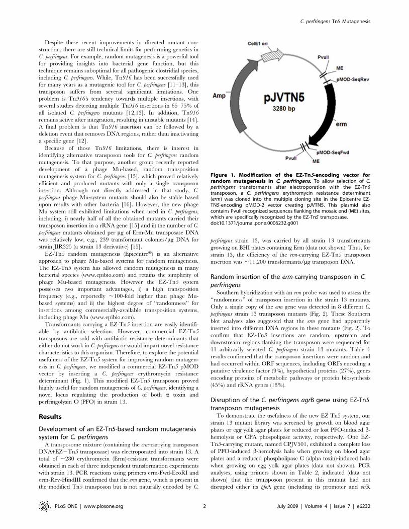
Figure 1. Modification of the EZ-Tn5-encoding vector forrandom mutagenesis in C. perfringens. To allow selection of C.perfringens transformants after electroporation with the EZ-Tn5transposon, a C. perfringens erythromycin resistance determinant(erm) was cloned into the multiple cloning site in the Epicentre EZ-TN5-encoding pMOD-2 vector creating pJVTN5. This plasmid alsocontains PvuII-recognized sequences flanking the mosaic end (ME) sites,which are specifically recognized by the EZ-Tn5 transposase.doi:10.1371/journal.pone.0006232.g001
C. perfringens Tn5 Mutagenesis
PLoS ONE | www.plosone.org 2 July 2009 | Volume 4 | Issue 7 | e6232
boxes), the plc gene, or the virS/virR operon that had previously
been shown to regulate pfoA and plc transcription [11,17,18].
Therefore, the DNA flanking the erm-modified EZ-Tn5 transpo-
son in CPJV501 was sequenced, which revealed that the transposon
had inserted into strain 13 ORF CPE1561 (Table 1). Southern blot
analyses showed that this mutant carries only one copy of the
transposon integrated in its genome (Fig. 3A). PCR analysis using
primers that amplify the wt CPE1561 gene (642 bp) in strain 13
then further demonstrated the presence of the transposon-disrupted
CPE1561 gene (1600 bp) in CPJV501 (Fig. 3B).
A homolog of the putative protein encoded by the CPE1561
gene has been annotated as AgrB for C. perfringens strain ATCC
13124 [19], which is found in other Gram-positive bacteria,
including some other clostridial species. In S. aureus, AgrB and the
secreted peptide AgrD are part of a well-characterized quorum
sensing (QS) system that is involved in regulating the expression of
virulence genes, including several toxins [20]. Bioinformatics
analyses using the Pathema website further revealed that a
sequence with 100% homology to the agrB ORF in C. perfringens
strain 13 and ATCC 13124 is also present in all other sequenced
C. perfringens isolates, including those classifying as types A–E (data
not shown). Moreover, the region between ORFs CPE1561 and
CPE1560 in strain 13 contains a small (135 bp) ORF (Fig. 3C)
sharing ,99% homology with a putative agrD gene from C.
perfringens type A strain SM101 and ATCC13124 [21].
The C. perfringens agr locus regulates PFO productionResults presented above suggested that PFO production is
regulated by the C. perfringens agr locus. A hemoglobin release (Hb)
assay [22] was used to examine the kinetics of this regulation. In
this assay, the ability of C. perfringens supernatants to lyse horse
erythrocytes and release Hb is specifically attributable to PFO
activity [11], as further supported by our observation that the
culture supernatant of a C. perfringens strain 13 pfoA-null mutant is
incapable of inducing Hb release from horse erythrocytes (Fig. 4A).
Using this horse erythrocyte assay, the appearance of PFO activity
in the supernatant of CPJV501 was delayed by 2 h compared to wt
strain 13 (Fig. 4A and 4B). This difference was not due to growth
differences between CPJV501 and wt strain 13 (Fig. 4C). The absence
of PFO activity in the early growth phase supernatants of CPJV501
involved an inhibition of early pfoA transcription in CPJV501.
Specifically, quantitative RT-PCR analyses revealed that a 2 hour
CPJV501 culture contains ,700-fold less pfoA mRNA than does the
equivalent culture of wt strain 13 (Fig. 5A).
Co-culture with DpfoA strains can restore PFOproduction by strain 13 agrB mutant
In S. aureus, AgrD is a secreted factor that activates the agr-mediated
regulatory network [23]. In the current study, we found that PFO
production by CPJV501 could be restored by coincubating CPJV501
with either a strain 13 pfoA-null mutant or a C. perfringens type C
CN3685 pfoA-null mutant, both of which retain an intact agr locus
(Fig. 6A and 6B). These physical complementation results suggested
that toxin regulation by the agr locus involves either cell to cell contact
or a secreted factor(s). To distinguish between those two possibilities,
CPJV501 and strain 13 pfoA-null mutant were inoculated into the
same 100 mm tissue culture dish but physically separated by a
Transwell filter. As shown in Fig. 6C, inoculation of the strain 13
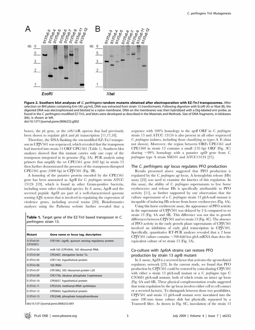
Figure 2. Southern blot analyses of C. perfringens random mutants obtained after electroporation with EZ-Tn5 transposomes. Afterselection on BHI plates containing Erm (40 mg/ml), DNA was extracted from strain 13 transformants. Following digestion with EcoRI (A) or XbaI (B), thedigested DNA was electrophoresed and blotted to a nylon membrane. DNA on the membranes was then hybridized with a Dig-labeled erm probe, asfound in the C. perfringens-modified EZ-Tn5, and blots were developed as described in the Materials and Methods. Size of DNA fragments, in kilobases(kb), is shown at left.doi:10.1371/journal.pone.0006232.g002
Table 1. Target gene of the EZ-Tn5 based transposon in C.perfringens strain 13.
Mutant Gene name or locus tag, description
S13Tn5-01(CPJV501)
CPE1561 (agrB), quorum sensing regulatory protein
S13Tn5-03 rrnB-16S (CPEr004), 16S ribosomal RNA,
S13Tn5-04 CPE2407, elongation factor Tu
S13Tn5-05 CPE1142, hypothetical protein
S13Tn5-06 16S RNAr
S13Tn5-07 CPE1892, 50S ribosomal protein L20
S13Tn5-09 CPE1736, ribulose phosphate 3-epimerase
S13Tn5-10 CPE0027, hypothetical protein
S13Tn5-11 CPE2524, methionyl-tRNA synthetase
S13Tn5-12 CPE0643, hypothetical protein
S13Tn5-13 CPE2348, phosphate butyryltransferase
doi:10.1371/journal.pone.0006232.t001
C. perfringens Tn5 Mutagenesis
PLoS ONE | www.plosone.org 3 July 2009 | Volume 4 | Issue 7 | e6232
pfoA-null mutant into the top well of the dish, and CPJV501 into the
bottom well of the dish, restored PFO activity to similar levels as
shown by the wt strain 13, i.e. this physical complementation involves
a secreted factor produced by both strain 13 and CN3685.
The C. perfringens agr locus also regulates CPAproduction
Relative to wt strain 13, production of CPA in culture
supernatants was also reduced in 2 h cultures of CPJV501, as
detected by an ELISA assay (Fig. 7A). Western blot analyses of
bacterial lysates demonstrated that this reduction of CPA
supernatant levels was due to decreased intracellular production
of CPA by CPJV501, rather than impaired CPA secretion
(Fig. 7B). qRT-PCR analyses demonstrated that this reduced
CPA production involved transcriptional regulation; compared to
wt strain 13, levels of cpa mRNA were reduced about 7-fold in
2 hour cultures of this agrB mutant (Fig. 5B). Together, these
results indicated that the agr locus is involved in regulating pfoA
and cpa transcription, and thus PFO and CPA production, during
the early logarithmic growth phase.
Evidence that C. perfringens agrB and agrD gens are co-transcribed in an operon
In S. aureus, the agr locus is encoded by an operon consisting of
four genes, agrB, agrD, agrC, and agrA [23–25]. The agrA and agrC
genes encode a response regulator and histidine kinase, respec-
tively, of a two-component regulatory system (TCRS). This TCRS
responds to a small peptide named an autoinducer (AI), which is
encoded by the agrD gene. The agrB gene encodes the enzyme
cleaving and modifying the AI [24].
To confirm that the C. perfringens agr locus contributes to early
regulation of PFO and CPA expression in strain 13, and to assess
whether agrB and agrD are expressed by C. perfringens as part of an
operon, the agrB gene alone, the agrB and agrD genes alone, or the
agrB, agrD and two ORFs (CPE1563 and CPE1562, which are
annotated as encoding hypothetical proteins) that lie upstream of
agrB were cloned into the E. coli-C. perfringens shuttle vector
pJIR750, creating the plasmids P1, P2 or P3 respectively (Fig. 3C).
Those plasmids were then individually electroporated into
CPJV501 to create the strains CPJVp1, CPJVp2 or CPJVp3.
PCR analyses confirmed the genotype of these CPJV501
complementing strains (Fig. 3B).
Neither CPJVp1 (encoding agrB alone) nor CPJVp2 (encoding
agrB and agrD) were able to restore PFO or CPA production
(Figs. 4B and 7A). However, complementation with CPJVp3
(encoding CPE1563, CPE1562, agrB and agrD) did restore 2 h
supernatant PFO activity and CPA levels to approximately those
found in culture supernatants of the wt strain 13 (Figs. 4B and 7A).
qRT-PCR analyses confirmed that 2 h cultures of CPJVp3
exhibited similar levels of pfoA and cpa mRNA as found in 2 h
cultures of the wt strain 13 (Fig. 5). In contrast, CPJVp1 and
Table 2. Primers used in this study.
Primer Sequence Reference
erm-Fwd-EcoRI AAGGGAATTCCTAAAAATTTGTAATTAAGAAGGAGT This study
agrBF TTACGAATTCGATGTTAGCCATGTATGCTTTCG This study
agrBR TAGAGGATCCTCATTTAACTCATCCCCTCAAG
agrF1 TTACGAATTCTTAGCTCTTTATATTGGATATACAG This study
agrR1 TAGAGGATCCCCGGTTTAAAACCGACCTTTAG
pfoAF1 ATCCAACCTATGGAAAAGTTTCTGG [22]
pfoAR1 CCTCCTAAAACTACTGCTGTGAAGG
cpaF GCTAATGTTACTGCCGTTGA [45]
cpaR CCTCTGATACATCGTGTAAG
polCJVL AATATATGATACTGAAGAGAGAGTAA This study
polCJVR TCTAAATTATCTAAATCTATGTCTACT
agr101L TAAATTTGCTCCAGTAGATACTAA This study
agrDR TATTCATCTCTTAAAGATTTTGGT
agr102L TTCAAGTTTGATATTGGTATTAGT This study
agr101R CAAAGCTTCTAAAGCTATATTAAA
agr103L ATGATAGGAACAAGTACAGTAAAA This study
agr102R AACTTGAAATTAAATATTCCTTCT
agr104L AAATTTAAAACTTGTTATTGGAGT This study
agr103R GGCTTTAAACTATATCCTTTTATT
pfoA81L CCCAGTTATTCACGATTAAAG This study
pfoA82R AGTAATACTAGATCCAGGGTATAAA
doi:10.1371/journal.pone.0006232.t002
C. perfringens Tn5 Mutagenesis
PLoS ONE | www.plosone.org 4 July 2009 | Volume 4 | Issue 7 | e6232
CPJVp2 exhibited substantially less, if any, complementation in
these qRT-PCR analyses.
Results presented above suggested that agrB and agrD may be
transcribed as an operon that also includes the two upstream
ORFs CPE1563 and CPE1562, which are annotated as encoding
hypothetical proteins (Fig. 8). To assess further whether agrB and
agrD might be transcribed as part of an operon, RT-PCR analyses
were performed using RNA extracted form the wt strain 13. These
RT-PCR analyses also used primers that would produce a product
only if every two ORF’s from CPE1564 through agrD are co-
transcribed. Fig. 8 results showed evidence for significant levels of
co-transcription of CPE1563 and CPE1562, CPE1562 and agrB
(CPE1561), and agrB and agrD, strongly suggesting that all four of
these genes are co-transcribed in an operon. Interestingly, mRNA
transcript was also detected for CPE1564 and CPE1563, but the
signal was less intense than for the other transcripts, possibly
suggesting a weak promoter can also independently co-transcribe
these two ORFs.
Discussion
This study reports the development of a simple EZ-Tn5-based
approach for random mutagenesis in Clostridium perfringens. All
screened EZ-Tn5 mutants obtained by this method contained only
a single transposon insertion and were stable over at least 10
sequential overnight culturings. This new approach produced
mutants at high efficiency, i.e., 11,200 CFU/mg DNA for strain
13. This mutant yield was 46-fold higher than recently reported
for phage Mu-based random mutagenesis of a C. perfringens strain
13 derivative [15], which is consistent with previous reports
comparing mutant yields for other bacteria when using EZ-Tn5
vs. phage Mu-based random mutagenesis approaches (www.
epibio.com).
Besides better efficiency, EZ-Tn5 random mutagenesis possesses
a second advantage over the phage Mu-based system. When
applied to C. perfringens, phage Mu-based transpositions favored
insertion into rRNA genes, with nearly 45% of the phage Mu-
Figure 3. Generation of a C. perfringens agrB mutant and complementing strains. A) Southern blot analyses, as described in Fig. 2, usingEcoRI-digested DNA from CPJV501 and a Dig-labeled probe that detected a single copy of the erm gene. Size of DNA fragments, in kilobases (kb) isshown at left. B) PCR was performed with DNA extracted from the indicated strain and the following pair of primers, agrBFwd and agrBRev inreactions containing DNA from strain 13 (S13), CPJV501 and CPJVp1; agrBFwd and argDR for CPJVp2 and agrF1 and agrD100R for CPJVp3. DNAladders (100 bp or 1 kb) were included in the first and last lane of the gel. Asterisks show the expected PCR product when the primers amplified theTn5-disprupted agrB gene. C) Genes cloned in the E. coli-C. perfringens shuttle plasmid pJIR750 to complement the agrB transposon mutant. Asshown, P1 encodes the agrB gene alone, P2 the agrB and agrD genes and P3 encodes two-genes (CPE1562 and CPE1563) upstream the agrB gene(CPE1561) and agrB and agrD.doi:10.1371/journal.pone.0006232.g003
C. perfringens Tn5 Mutagenesis
PLoS ONE | www.plosone.org 5 July 2009 | Volume 4 | Issue 7 | e6232
Figure 4. The C. perfringens agrB locus regulates PFO production. A and B) Hemoglobin (Hb) release assay. Culture supernatants obtained, atthe indicated time point, from strain 13 (S13), S13 pfoA-null mutant (S13DpfoA), CPJV501 (DagrB), CPJVp1 (DagrB/P1), CPJVp2 (DagrB/P2) or CPJVp3(DagrB/P3), were incubated (1:1) with a 1% suspension of horse red blood cells for 30 min at 37uC. Non-inoculated TGY or 0.1% saponin (Saponin)was included as negative or positive control, respectively. PFO-induced Hb release was detected by obtaining the absorbance at 570 (A570). C) Foreach time point, the OD600 of the cultures is shown. For all panels, error bars represent the standard error of the mean calculated using data fromthree independent experiments.doi:10.1371/journal.pone.0006232.g004
C. perfringens Tn5 Mutagenesis
PLoS ONE | www.plosone.org 6 July 2009 | Volume 4 | Issue 7 | e6232
based C. perfringens mutants carrying an insertion into a rRNA gene
[15]. Since another 12% of those phage Mu-based mutants carried
an insertion into an intergenic region, only ,45% of the C.
perfringens mutants in that phage Mu-based library had the desired
outcome, i.e., a transposon insertion into a protein-encoding ORF.
While the EZ-Tn5 transposon exhibited somewhat higher
insertion rates into C. perfringens rRNA genes than would be
expected by mere chance, this preference was much less than
observed for phage Mu-based random mutagenesis. Specifically,
only 18% of the screened EZ-Tn5 C. perfringens mutants carried a
transposon insertion into a rRNA gene (rRNA genes represent
about 1.5% of total genes in C. perfringens). Since none of the EZ-
Tn5 C. perfringens strain 13 mutants happened to carry a
transposon insertion in an intergenic region (although limited
intergenic EZ-Tn5 insertion was observed with another C.
perfringens strain, data not shown), ,73% of the transposons in
the screened C. perfringens strain 13 EZ-Tn5 carrying mutants had
single insertions in a protein encoding gene.
The current study then directly demonstrated the utility of the
new EZ-Tn5 random mutagenesis system by identifying a new
locus involved in controlling early log-phase production of a toxin
and PFO by C. perfringens strain 13. Prior to the current work,
regulation of PFO and a toxin expression in strain 13 was known
to involve a classical bacterial two component regulatory system
named VirS/VirR, where VirS is the membrane sensor and VirR
is the transcriptional regulator [11,17,18,26–30]. When phosphor-
ylated, the VirR protein binds directly to VirR boxes located
upstream of the pfoA gene encoding PFO. However, VirR boxes
are not present upstream of the plc gene encoding a toxin [31–34];
instead, a regulatory RNA (named VR-RNA), whose transcription
is itself regulated by VirS/VirR, is involved in control of a toxin
expression [28]. In addition, previous studies have implicated the
LuxS quorum sensing system in the regulation of a toxin and PFO
expression by strain 13 [35].
The current study reveals a new level of complexity in the
regulation of a toxin and PFO expression by strain 13.
Specifically, the current results demonstrated that the early log-
phase regulation of a toxin and PFO expression by this C.
perfringens strain involves a locus containing ORFs with homology
to S. aureus agrB and agrD. A recent bioinformatics search had
identified the presence of agr ORFs in many firmicutes, including
C. perfringens [21], but it had not yet been evaluated whether this
system is functional or important for regulating C. perfringens
virulence factor expression. As mentioned in the Results, a similar
agr locus is well-established in quorum sensing regulation of S.
aureus virulence, where the agr locus controls expression of several
toxins, as well as some surface virulence factors [20]. A similar agr
locus was also recently implicated in Listeria monocytogenes virulence,
where agrD-dependent quorum sensing regulates biofilm forma-
tion, Caco-2 cell invasion and mouse virulence [36].
In S. aureus, the agr locus is a four gene operon transcribed
primarily from a promoter named P2 [20]. This S. aureus agr
operon encodes for a TCRS (that includes the AgrA transcrip-
tional regulator and the AgrC membrane sensor), the agrD
signaling peptide, and an AgrB transmembrane protein involved
in AgrD processing. Once activated and secreted, extracellular
AgrD binds to (and activates) AgrC, which then phosphorylates
AgrA. The phosphorylated AgrA then binds to P2 and to another
promoter named P3 [25,37]. This P3 binding leads to production
of a regulatory RNA (named RNAIII) encoded by a gene adjacent
to the agr locus. RNAIII then modulates expression of several
exotoxins and surface proteins.
The agr systems of C. perfringens and S. aureus apparently share
some similarities. Our results clearly demonstrated that, as in S.
Figure 5. Early transcription of pfoA and plc genes is regulated by the C. perfringens agr locus. Total RNA was extracted from a 2 h TGYculture of the wt strain 13 (S13), CPJV501 (DagrB), CPJVp1, CPJVp2 or CPJVp3. Quantitative RT-PCR was then performed with 20 ng of each RNA andprimers that amplified the (A) pfoA gen (pfoAF1 and pfoAR1) or the (B) plc gene (cpaF and cpaR). Average CT values were normalized to the polC geneand the fold differences were calculated using the comparative CT method (22DDC
T) [44]. Values below each bar indicate the calculated fold changerelative to the wt strain 13. Panels shown are representative of three independent experiments.doi:10.1371/journal.pone.0006232.g005
C. perfringens Tn5 Mutagenesis
PLoS ONE | www.plosone.org 7 July 2009 | Volume 4 | Issue 7 | e6232
Figure 6. A C. perfringens secreted factor(s) regulates PFO production. A and B) Physical complementation of the DagrB mutant by co-culturewith a DpfoA mutant of strain 13 or CN3685. C. perfringens strain 13 (S13), CPJV501 (DagrB), S13 pfoA-null mutant (S13DpfoA), CPJV501 and S13 pfoA-null mutant (DagrB/S13DpfoA) or CPJV501 and CN3685 pfoA-null mutant (DagrB/CN3685DpfoA) were inoculated in TGY and incubated at 37uC forthe indicated time. Culture supernatants obtained, at the indicated time point, were incubated (1:1) with a 1% suspension of horse red blood cells for30 min at 37uC. Non-inoculated TGY or 0.1% saponin was included as negative or positive control, respectively (not shown). PFO-induced Hb releasewas detected by obtaining the absorbance at 570 nm (A570). C) The physical complementation shown in panels A and B requires a secreted factor toregulate PFO production. Strain 13 (S13), CPJV501 (DagrB) or S13 pfoA-null mutant (DpfoA) was inoculated in 100 mm tissue culture dishescontaining 25 ml of TGY. Another 100 mm tissue culture dish containing a transwell filter device (0.4 mm pore size) received 25 ml of TGY. Then, theS13 pfoA-null mutant was inoculated into the top chamber and CPJV501 was inoculated into the bottom chamber of the dish (bottom DagrB/TopS13DpfoA) and incubated for the indicated time. Culture supernatants obtained at the indicated time points were incubated (1:1) with a 1%suspension of horse red blood cells for 30 min at 37uC. PFO-induced Hb release was detected by obtaining the absorbance at 570 nm (A570). For allpanels, error bars represent the standard error of the mean calculated using data from three independent experiments.doi:10.1371/journal.pone.0006232.g006
C. perfringens Tn5 Mutagenesis
PLoS ONE | www.plosone.org 8 July 2009 | Volume 4 | Issue 7 | e6232
aureus, the agr locus (via a secreted factor) transcriptionally regulates
toxin production by C. perfringens strain 13. Since the agr locus is
involved in controlling early log-phase expression of a-toxin and
PFO and these two toxins are important for C. perfringens-induced
gas gangrene [12,38,39], the agr locus may play an important role
in C. perfringens virulence, although this needs to be experimentally
confirmed. Another similarity, revealed by our RT-PCR and
complementation results, is that both C. perfringens and S. aureus co-
transcribe agrB and agrD as part of an operon.
There also appear to be some differences between the agr
systems of S. aureus versus C. perfringens. One variation concerns the
arrangement of the agrB and agrD genes within the agr operon. In
S. aureus, agrB and agrD are the first two transcribed genes in the
operon [20,25], but in C. perfringens these two agr genes appear to
be downstream of other genes in the operon. It is unclear whether
the two ORFs adjacent to agrB and agrD in C. perfringens encode a
TCRS, as in S. aureus, although those two ORFs co-transcribed
with agrB and agrD in the C. perfringens agr operon are currently
annotated as encoding ‘‘hypothetical proteins’’ and thus do not
possess obvious characteristics of a TCRS. However, future studies
should evaluate this possibility. A final difference between the agr
systems of C. perfringens versus S. aureus is that the RNAIII-encoding
gene lies in close proximity to the agr operon in S. aureus [25], but
no readily identifiable homolog of the RNAIII-encoding gene is
located near the C. perfringens agr operon or is readily identifiable
elsewhere in the strain 13 genome.
If the two upstream ORFs co-transcribed with the agrB and agrD
genes in C. perfringens do not encode a TCRS, sorting out the
signaling cascade may require some effort. The strain 13 genome
contains 28 known or putative histidine kinase sensors [40], which
coupled with one or more of the 20 known or putative response
regulators, could mediate AgrD signaling. Whether the C. perfringens
agr locus acts via the VirS/VirR TCRS, which is known to regulate
a toxin and PFO production, should be assessed. If C. perfringens
resembles S. aureus by using its Agr system to upregulate the
production of a regulatory RNA, that regulatory RNA should be
identified. A previous study [28] identified a C. perfringens regulatory
RNA named VR-RNA that is involved in strain 13 production of atoxin. However, VR-RNA alone would not appear to readily
explain agr-mediated signaling since, this regulatory RNA is not
known to control PFO production [28], yet our agrB mutant cannot
produce PFO during early log-phase growth. Therefore, further
studies might evaluate whether several other putative regulatory
RNAs of C. perfringens [18] mediate the agr locus signal. Also, sorting
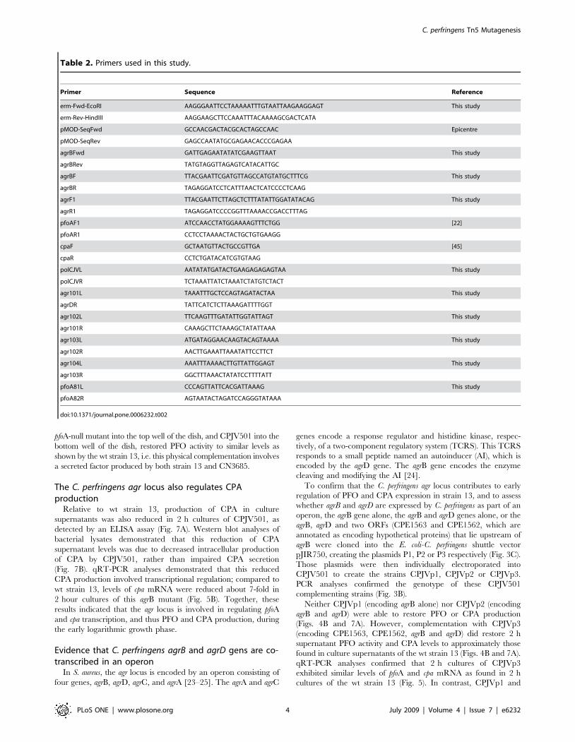
Figure 7. The C. perfringens agrB locus regulates CPA production. A) ELISA analyses. Culture supernatants obtained, at the indicated timepoint, from strain 13 (S13), CPJV501 (DagrB), CPJVp1 (DagrB/P1), CPJVp2 (DagrB/P2), CPJVp3 (DagrB/P3) or purified CPA was used to coat a 96-wellmicroplate overnight at 4uC. The wells were incubated with a mouse monoclonal anti-CPA antibody followed by a HRP-conjugated anti-mouseantibody. The bound antibody was detected with a TMB substrate solution and the color reaction stopped with sulphuric acid (0.18 M). A450 wasdetermined using an ELISA reader. Error bars represent the standard error of the mean calculated using data from three independent experiments. B)Western blot showing the agr locus regulates production of CPA. Strain 13 (S13), CPJV501 (DagrB) or CPJVp3 (DagrB/P3) was inoculated in TGY andincubated at 37uC for 4 h. Bacteria were then pelleted by centrifugation, resuspended in lysis buffer and sonicated. Equal amount (25 ml) of bacteriallysates was run in a 12% SDS-PAGE, transferred to nitrocellulose membrane and western blotted with a monoclonal anti-CPA antibody. As a control,25 ml of CPA-containing concentrated supernatant proteins was added to the gel. The expected molecular weight in kDa of CPA is shown at the left.Shown is a representative figure of three independent experiments.doi:10.1371/journal.pone.0006232.g007
C. perfringens Tn5 Mutagenesis
PLoS ONE | www.plosone.org 9 July 2009 | Volume 4 | Issue 7 | e6232
out the hierarchy of toxin expression control between the agr locus,
LuxS and the VirS/VirR TCRS will require further studies to fully
understand how C. perfringens regulates production of its toxins.
Additionally, future studies should also examine whether expression
of other toxins produced by some C. perfringens strains are regulated
by the Agr system. Finally, it would be interesting to identify the
environmental cues that signal the onset of agr operon transcription
in C. perfringens. While many additional studies are clearly needed to
fully understand it roles in C. perfringens, linkage of the agr locus to
PFO and a toxin production opens a new chapter towards
understanding toxin gene regulation by the important pathogen
C. perfringens.
Materials and Methods
Strains and bacterial culture mediaStrain 13, a genome-sequenced, highly transformable C.
perfringens type A strain [40] was used for transposon mutagenesis
experiments. A strain 13 pfoA-null mutant and CN3685 pfoA-null
mutant were constructed using our previously described Targe-
tronH technology [6,8]. The bacterial culture media used
throughout this study included FTG (fluid thioglycolate medium;
NaCl), LB agar (1.5% agar [Becton-Dickinson]) and brain heart
infusion (BHI) agar (Becton-Dickinson). E. coli Top10 cells
(Invitrogen) were used as the cloning host. When indicated,
ampicillin (Amp, 100 mg/ml), erythromycin (Erm [100 mg/ml] or
[40 mg/ml]) or chloramphenicol (Cm [15 mg/ml]) was added to
the culture medium.
Construction of the modified EZ-Tn5 transposon vectorand transposome preparation
To modify the EZ-Tn5-carrying plasmid pMOD-2 (Epicentre)
for use in C. perfringens, a single colony of an E. coli strain encoding
the EZ-Tn5 pMOD-2 vector was inoculated into 10 ml of LB
broth supplemented with ampicillin (LBA) and then incubated
overnight at 37uC with shaking (250 RPM). The plasmid was
extracted with a QIAprep Spin plasmid extraction kit (Qiagen)
and simultaneously digested with EcoRI and HindIII (New
England Biolabs). The erythromycin resistance gene (erm) from
the E. coli-C. perfringens shuttle vector pJIR751 [41] was amplified
by PCR using JumpStart REDTaq ready mix (Sigma-Aldrich) and
primers erm-Fwd-EcoRI and erm-Rev-HindIII (Table 2). The
PCR product was run on a 1.5% agarose gel, purified using a
QIAquick gel extraction kit (Qiagen), and then simultaneously
digested with EcoRI and HindIII. The digested EZ-Tn5-carrying
pMOD-2 plasmid and the erm gene PCR product were ligated
overnight at 4uC with T4 DNA ligase (New England Biolab). The
resulting plasmid, named pJVTN5, was then transformed into
chemically competent E. coli Top10 cells (Invitrogen) and
inoculated onto LB agar plates supplemented with erythromycin
and ampicillin. To prepare the transposome, pJVTN5 was
digested with PvuII at 37uC for 1 h (see Fig. 1). The erm-carrying
Figure 8. Organization and RT-PCR analysis of the agr operon. A) RT-PCR reactions were performed with 50 ng of RNA extracted from anovernight TGY culture of the wt strain 13. RT-PCR reactions included (+) or not (2) retrotranscriptase (RT). The following pair of primers were used todetect mRNA transcripts from every two-adjacent ORF’s, agr104L and agr103R (L4-R3, which should generate a 321 bp PCR product), agr103L andagr102R (L3-R2, which should generate a 315 bp PCR product), agr102L and agr101R (L2-R1, which should generate a 420 bp PCR product) oragr101L and agrDR (B–D, which should generate a 520 bp PCR product). A 100-bp DNA ladder is shown at left. B) Schematic representation of the agrlocus showing primers used for RT-PCR reactions.doi:10.1371/journal.pone.0006232.g008
C. perfringens Tn5 Mutagenesis
PLoS ONE | www.plosone.org 10 July 2009 | Volume 4 | Issue 7 | e6232
EZ-Tn5 transposon fragment (,900 bp) was purified from an
agarose gel as described earlier and DNA concentration was
quantified. Two ml of the erm-modified EZ-Tn5 Transposon DNA
(100 mg/ml in TE Buffer [10 mM Tris-HCl (pH 7.5), 1 mM
EDTA]) were mixed with 4 ml of the EZ-Tn5 transposase
(Epicentre) and 2 ml of glycerol. The mixture was incubated for
30 min at room temperature, to allow the transposase to stably
bind to the erm-modified EZ-Tn5 Transposon DNA; that mixture
was then stored at 220uC.
Transposome electroporation into C. perfringens strainsFollowing a standard procedure [6,42], 1 ml of the transposome
was electroporated into a 4 hour TGY culture of highly
transformable C. perfringens strain 13 [40]. For this purpose,
electrocompetent cells (400 ml) were mixed, in a 0.2 cm electro-
poration cuvette (Biorad), with 1 ml of the transposome and
incubated 5 min at 4uC. Electroporation was performed using a
BioRad Gene PulserTM with pulse controller set at 200V, 25 mF
and 1.5 kV. Electroporated transposome-containing bacteria were
grown in 3 ml of pre-warmed TGY for 3 h at 37uC to allow them
complete recovery, plated onto BHI agar plates with erythromycin
(40 mg/ml), and incubated at 37uC for 18 h under anaerobic
conditions. All colonies were propagated in BHI agar plates with
erythromycin (40 mg/ml). To confirm the presence of the erm gene,
a PCR was performed with 2 ml of cell lysate as DNA template.
This PCR used primers erm-Fwd-EcoRI and erm-Rev-HindIII
and the following PCR conditions: 1 cycle of 95uC for 5 min, 35
cycles of 95uC for 30 s, 55uC for 45 s, and 68uC for 1 min; and a
single extension of 68uC for 10 min.
Sequencing of the EZ-Tn5 target gene in selectedmutants
After electroporation of the transposon, C. perfringens strain 13
Erm-resistant transformants, which were also erm-positive by PCR,
were randomly chosen for sequencing. Total DNA was extracted
and 1 mg of each DNA was mixed with 10 pmol of primers
pMOD-SeqFwd or pMOD-SeqRev (Epicentre) and sent for
sequencing at the University of Pittsburgh Genomics and
Proteomics Core Laboratory. C. perfringens sequences flanking the
erm-carrying transposon were determined using the nucleotide
BLAST program on the National Center for Biotechnology
Information (NCBI) web site and the J. Craig Venter Institute’s
Pathema website programs.
Complementation of the agrB mutantDNA was isolated from strain 13 using a Master PureTM Gram
Positive DNA purification Kit (Epicentre). The primers agrBF and
agrBR (Table 2) were added (at a 5 mM final concentration) to a
PCR mixture containing 1 ml of purified DNA template and 25 ml
26Taq mixture (NEB). Those reaction mixtures, with a total
volume of 50 ml, were placed in a thermal cycler (Techne) and
subjected to the following amplification conditions: 1 cycle of 95uCfor 2 min, 35 cycles of 95uC for 30 s, 55uC for 40 s, and 68uC for
3 min, and a single extension of 68uC for 5 min. The PCR
products were cloned into a TOPO vector (Invitrogen) and
sequenced at the University of Pittsburgh Core Sequencing
Facility. Using EcoRI and BamHI, the insert was removed from
the TOPO vector and ligated into pJIR750, forming a plasmid
named P1 (which is 1072 bp and contains agrB and a 403 bp
upstream sequence). Using the same method, two other comple-
menting plasmids were created, including P2 (created using agrBF-
agrR1 primers and which has a 1230 bp insert containing agrB
and agrD, along with a 403 bp upstream sequence) and P3 (created
using agrF1-agrR1 and which has a 2893 bp insert containing
agrB, agrD and two upstream ORFs encoding hypothetical
proteins). Plasmids P1, P2 and P3 were separately introduced,
by our standard electroporation techniques, into the agrB mutant
of strain 13. Chloramphenicol (15 mg/ml) resistant transformants
were then selected. The resultant transformants were designated
CPJVp1, CPJVp2 and CPJVp3.
Southern blot analysesC. perfringens DNA was isolated using the MasterPure gram-
positive DNA purification kit (Epicentre, Wisconsin). Each isolated
DNA sample (2.5 mg) was then digested overnight with EcoRI or
XbaI, according to the manufacturer’s (New England Biolabs)
instructions. The digested DNA samples were electrophoresed on
a conventional 1% agarose gel, and the separated DNA digestion
products were then transferred onto nylon membranes (Roche) for
hybridization with an erm-specific probe. After hybridization of the
erm probe, the Southern blots were developed using reagents from
the DIG DNA labeling and detection kit (Roche), according to the
15. Lanckriet A, Timbermont L, Happonen LJ, Pajunen MI, Pasmans F, et al.
(2009) Generation of single-copy transposon insertions in Clostridium perfringens byelectroporation of phage mu DNA transposition complexes. Appl Environ
Microbiol 75: 2638–2642.
16. Pajunen MI, Pulliainen AT, Finne J, Savilahti H (2005) Generation oftransposon insertion mutant libraries for Gram-positive bacteria by electropo-
ration of phage Mu DNA transposition complexes. Microbiology 151:
1209–1218.
17. Shimizu T, Ba-Thein W, Tamaki M, Hayashi H (1994) The virR gene, a
member of a class of two-component response regulators, regulates theproduction of perfringolysin O, collagenase, and hemagglutinin in Clostridium
perfringens. J Bacteriol 176: 1616–1623.
18. Okumura K, Ohtani K, Hayashi H, Shimizu T (2008) Characterization of genesregulated directly by the VirR/VirS system in Clostridium perfringens. J Bacteriol
190: 7719–7727.
19. Myers GS, Rasko DA, Cheung JK, Ravel J, Seshadri R, et al. (2006) Skewed
genomic variability in strains of the toxigenic bacterial pathogen, Clostridium
perfringens. Genome Res 16: 1031–1040.
20. Novick RP, Geisinger E (2008) Quorum sensing in staphylococci. Annu Rev
Genet 42: 541–564.
21. Wuster A, Babu MM (2008) Conservation and evolutionary dynamics of the agr
cell-to-cell communication system across firmicutes. J Bacteriol 190: 743–746.
22. Fisher DJ, Fernandez-Miyakawa ME, Sayeed S, Poon R, Adams V, et al. (2006)Dissecting the contributions of Clostridium perfringens type C toxins to lethality in
the mouse intravenous injection model. Infect Immun 74: 5200–5210.
23. Ji G, Beavis RC, Novick RP (1995) Cell density control of staphylococcalvirulence mediated by an octapeptide pheromone. Proc Natl Acad Sci U S A 92:
12055–12059.
24. Novick RP (2003) Autoinduction and signal transduction in the regulation ofstaphylococcal virulence. Mol Microbiol 48: 1429–1449.
25. Novick RP, Projan SJ, Kornblum J, Ross HF, Ji G, et al. (1995) The agr P2operon: an autocatalytic sensory transduction system in Staphylococcus aureus. Mol
Gen Genet 248: 446–458.
26. Ohtani K, Kawsar HI, Okumura K, Hayashi H, Shimizu T (2003) The VirR/VirS regulatory cascade affects transcription of plasmid-encoded putative
virulence genes in Clostridium perfringens strain 13. FEMS Microbiol Lett 222:137–141.
C. perfringens Tn5 Mutagenesis
PLoS ONE | www.plosone.org 12 July 2009 | Volume 4 | Issue 7 | e6232


![Chloroethylating anticancer drug-induced mutagenesis and ...€¦ · ZA2102 uvrA6, malE::Tn5 < 0.1 [20] SW102 uvrA6, malE::Tn5, recA56, srl::Tn10 0.1±0.5 This study a: All mutants](https://static.documents.pub/doc/80x56/6129e36c31dba069ad59cd1b/chloroethylating-anticancer-drug-induced-mutagenesis-and-za2102-uvra6-maletn5.jpg)


