Use of Label-Free Quantitative Proteomics To Distinguish theSecreted Cellulolytic Systems of Caldicellulosiruptor bescii
and Caldicellulosiruptor obsidiansis!†Adriane Lochner,1,3,4 Richard J. Giannone,2,3 Miguel Rodriguez, Jr.,1,3 Manesh B. Shah,2
Jonathan R. Mielenz,1,3 Martin Keller,1,3 Garabed Antranikian,4David E. Graham,1,5* and Robert L. Hettich2,3*
Biosciences Division,1 Chemical Sciences Division,2 and BioEnergy Science Center,3 Oak Ridge National Laboratory, Oak Ridge,Tennessee 37831; Technical Microbiology, Hamburg University of Technology, Kasernenstrasse 12, D-21073 Hamburg,
Germany4; and Department of Microbiology, University of Tennessee, Knoxville, Tennessee 379965
Received 1 December 2010/Accepted 9 April 2011
The extremely thermophilic, Gram-positive bacteria Caldicellulosiruptor bescii and Caldicellulosiruptor obsidi-ansis efficiently degrade both cellulose and hemicellulose, which makes them relevant models for lignocellulosicbiomass deconstruction to produce sustainable biofuels. To identify the shared and unique features of secretedcellulolytic apparatuses from C. bescii and C. obsidiansis, label-free quantitative proteomics was used to analyzeprotein abundance over the course of fermentative growth on crystalline cellulose. Both organisms’ secretomesconsisted of more than 400 proteins, of which the most abundant were multidomain glycosidases, extracellularsolute-binding proteins, flagellin, putative pectate lyases, and uncharacterized proteins with predicted secre-tion signals. Among the identified proteins, 53 to 57 significantly changed in abundance during cellulosefermentation in favor of glycosidases and extracellular binding proteins. Mass spectrometric characterizations,together with cellulase activity measurements, revealed a substantial abundance increase of a few bifunctionalmultidomain glycosidases composed of glycosidase (GH) domain family 5, 9, 10, 44, or 48 and family 3carbohydrate binding (CBM3) modules. In addition to their orthologous cellulases, the organisms expressedunique glycosidases with different domain organizations: C. obsidiansis expressed the COB47_1671 protein withGH10/5 domains, while C. bescii expressed the Athe_1857 (GH10/48) and Athe_1859 (GH5/44) proteins.Glycosidases containing CBM3 domains were selectively enriched via binding to amorphous cellulose. Prep-arations from both bacteria contained highly thermostable enzymes with optimal cellulase activities at 85°Cand pH 5. The C. obsidiansis preparation, however, had higher cellulase specific activity and greater thermo-stability. The C. bescii culture produced more extracellular protein and additional SDS-PAGE bands thatdemonstrated glycosidase activity.
The conversion of lignocellulosic feedstock into biofuels hasgarnered significant interest recently in light of an increasingglobal demand for transportation fuel alternatives. Clearly, thenatural process of plant cell wall degradation by microorgan-isms serves as an informative guideline for the design andoptimization of efficient industrial enzymatic conversion pro-cesses. To this end, there is a strong emphasis on a compre-hensive understanding of complex microbial biomass degrada-tion systems that consist primarily of glycosidases, includingsynergistically acting cellulases and hemicellulases (21). Theincreasing number of whole genome sequences, along withsophisticated experimental and computational technologies,has provided a remarkable glimpse into the molecular pro-cesses by which microorganisms degrade cellulosic material.
One of the leading technologies employed for the systemlevel interrogation of various organisms is mass spectrometry,which during the last decade has revolutionized the large-scale,high-throughput proteomic characterization of both microbialisolates and communities (3, 24). In particular, proteomics hasaccelerated the discovery and quantification of cellulose-de-grading proteins from both aerobic and anaerobic microorgan-isms. For example, mass spectrometry (MS)-based proteomicmeasurements revealed numerous monofunctional glycosidaseproteins found in the secretome of the mesophilic fungusTrichoderma reesei (20, 50), currently the source of most com-mercial cellulose preparations (22, 35). The mesophilic marinegammaproteobacterium Saccharophagus degradans also en-codes multiple glycosidases, many fused to carbohydrate bind-ing modules or cadherin domains; however, only a handfulwere identified by proteomic measurement of the culture su-pernatant (45).
Thermostable glycosidases produced by thermophilic mi-crobes offer many advantages for large-scale biofuel produc-tion, including increased protein stability and cellulose degra-dation rates and a reduced risk of microbial contamination (5,41). Although several thermophilic microbes are able to de-grade cellulosic biomass, they do so by one of several uniquestrategies. The moderate thermophile (55°C) Thermobifida
* Corresponding author. Mailing address for David E. Graham:Biosciences Division, Oak Ridge National Laboratory, Oak Ridge, TN37831-6038. Phone: (865) 574-0559. Fax: (865) 576-8646. E-mail:[email protected]. Mailing address for Robert L. Hettich: ChemicalSciences Division, Oak Ridge National Laboratory, Oak Ridge, TN37831-6131. Phone: (865) 574-4986. Fax: (865) 576-8559. E-mail:[email protected].
† Supplemental material for this article may be found at http://aem.asm.org/.
! Published ahead of print on 15 April 2011.
4042
fusca secretes a diverse set of monofunctional glycosidases,often fused to carbohydrate-binding domains (1, 53). The ther-mophilic (60°C) bacterium Clostridium thermocellum producesglycosidases in cellulosomal complexes (38), while the ex-tremely thermophilic (70°C) bacterium Caldicellulosiruptorsaccharolyticus secretes several multidomain, multifunctionalcellulases (4, 21).
The use of proteomics for system level investigation of cel-lulose degradation strategies goes beyond general proteinidentification. Quantitative proteomics enables the derivationof relative or absolute measurements of protein abundancewithin a given sample. Several unique strategies exist, includingisotopic-label-based methods such as ICAT or iTRAQ and“label-free” methods that utilize spectral counts (SpC), inten-sity, or chromatographic peak area to estimate protein abun-dance (36, 37). With regard to its application to cellulose-degrading thermophiles, quantitative proteomics has beenemployed to study C. thermocellum’s cellulolytic enzymesbased on metabolic labeling of cells grown on cellulose orcellobiose (16), while a label-free approach measured changesin cellulosomal protein composition using cells grown on dif-ferent substrates (38). In both cases, quantitative analysis per-mitted estimation of relative protein abundance, providingvaluable comparisons of cultures grown on different substratesbased on changes in protein expression.
The Caldicellulosiruptor genus of anaerobic Gram-positivebacteria includes C. saccharolyticus, as well as Caldicellulosir-uptor bescii (optimal temperature, 75°C; previously Anaerocel-lum thermophilum DSM 6725) (55) and Caldicellulosiruptorobsidiansis (78°C) (18). C. bescii and C. obsidiansis share 97%16S rRNA gene sequence identity and 88% average nucleotideidentity in their genome sequences (18), and both grow on thesame set of monomeric and polymeric sugars as carbonsources, including pretreated switchgrass and poplar (18, 49).However, C. obsidiansis grows at slightly higher temperaturesthan C. bescii does and only C. obsidiansis produces measur-able amounts of ethanol during growth on switchgrass or cel-lulose (18, 42, 54). Putative cellulase genes in both organismsare concentrated in an island associated with prophage genes.This cluster varies in size and gene composition among theCaldicellulosiruptor species: it comprises 48 kbp in C. saccha-rolyticus (47), 61 kbp in C. obsidiansis (9), and 68 kbp in C.bescii (23).
Genomic, proteomic, and physiological studies have shownthat catabolic enzymes and pathways evolve rapidly throughpositive selection, differentiating closely related microbial spe-cies (28). Proteomic analysis of the secretomes of two evolvedT. reesei strains showed the diversification of glycosidase pro-files in these fungi, as revealed by changes in expression orsecretion efficiency (20). The genetic diversity among Caldicel-lulosiruptor cellulase gene clusters and the rapid evolution ofthermostable, multidomain, multifunctional glycosidases inthis lineage warranted a comparison of the strains’ secretedcellulolytic protein complement.
To compare cellulolytic systems from C. bescii and C. ob-sidiansis, we analyzed their secreted protein profiles over thecourse of crystalline cellulose fermentations. Label-free quan-titative proteomic analysis was performed using two-dimen-sional liquid chromatography (LC)-tandem MS (MS/MS) toidentify secreted proteins. Identified proteins were then quan-
tified by normalized spectral abundance factors (NSAF), amethod based on spectral counting that corrects for proteinsize and run-to-run variability (37). The correlation of theseNSAF measurements with the carboxymethyl cellulase(CMCase) activity of the culture supernatant was assessed, andthe increase in secreted glycosidase proteins over the course offermentation for both organisms was monitored. Cellulose af-finity digestion experiments were employed to enrich cellulose-binding proteins from both organisms to identify the highlythermostable multidomain glycosidases that are shared by andunique to each organism.
MATERIALS AND METHODS
Controlled cultivation and sampling. Fermentations were performed inBIOSTAT Bplus Twin 5-liter jacketed glass fermentors (Sartorius Stedim Bio-tech) using a 4-liter working volume of basal growth medium (18) that contained4.5 mM KCl, 4.7 mM NH4Cl, 2.5 mM MgSO4, 1.0 mM NaCl, 0.7 mMCaCl2 ! 2H2O, 0.25 mg/ml resazurin, 5.6 mM cysteine-HCl ! H2O, 6.0 mMNaHCO3, 1 mM phosphate buffer, 1! ATCC trace minerals, 1.25! MTC me-dium vitamin solution E (57), 0.02% (wt/vol) yeast extract, and 0.5% (wt/vol)Avicel PH-101 (Fluka). The temperature was controlled at 75°C for C. bescii andat 78°C for C. obsidiansis using a Polystat Circulator (Cole-Parmer). Fermentorsand media were sparged overnight at 200 rpm with a N2-CO2 (80:20) gas mixture;the exhaust gas was run through a water trap. The next day, yeast extract, sodiumbicarbonate, and vitamins were added and sparged for an additional 3 to 4 h.Inocula were grown in 125-ml serum bottles and added to the fermentors toachieve an initial cell density of 2.6 ! 106 cells/ml. The agitation was set at 300rpm, and the pH was controlled at 6.8 using a 10% sodium bicarbonate solution.Replicate samples were taken at elapsed fermentation time points of 0, 4, 8, 12,16, 20, 24, 30, 40, and 48 h. These samples were processed as follows. Forty-milliliter samples for proteomic analysis and 10-ml samples for activity assayswere centrifuged at 6,500 ! g for 15 min, and the supernatant was filtered via a0.22-"m syringe filter with a polyethersulfone (PES) membrane to obtain acell-free culture supernatant fraction. Aliquots of 1.8 ml were centrifuged for 5min at 15,500 ! g, and the supernatants and pellets were frozen in liquid nitrogenand stored at #80°C. At 48 h, the fermentations were stopped and the broth washarvested by centrifugation (30 min at 5,500 ! g). Two liters of supernatant wasfiltered through a 0.22-"m PES membrane Steritop device (Millipore), resultingin the cell-free supernatant fraction SN, which was subsequently concentrated 5times via a Quixstand tangential-flow filtration system equipped with a hollow-fiber filter (GE Healthcare) with a 5,000-molecular-weight cutoff. The superna-tant protein concentrate from tangential-flow filtration, designated TFF, wasused for the cellulase enrichment procedure.
Planktonic cell densities were measured using a Petroff-Hausser microscopecounting chamber (Fisher Scientific). Unless stated otherwise, supernatant pro-tein concentrations, as well as concentrations of other protein solutions used inthis study, were estimated by Bradford microassay (6) with bovine serum albuminas the standard. Pellet protein was estimated by the Lowry method with Peter-son’s modification (Sigma) after cell lysis in a 0.2 N NaOH–1% SDS solution asdescribed previously (56).
Cellulase enrichment. The glycosidases in the supernatant protein concentrate(TFF) were enriched via binding to amorphous cellulose and subsequent diges-tion as described previously (38). Briefly, acid-swollen Avicel PH-105 was addedto the TFF fraction of each respective organism in an amount corresponding to50 mg crystalline cellulose. To maintain a similar protein/substrate ratio, con-centrate volumes were adjusted to contain 20 mg total protein. After binding, theamorphous cellulose was separated by centrifugation and resuspended in 10 mlof reaction buffer containing 50 mM sodium acetate, pH 5.5. The remainingprotein solution was referred to as affinity digest supernatant (ADSN). Affinitydigestion was performed with a dialysis membrane (SpectraPor; 6- to 8-kDacutoff) against reaction buffer at 75°C for 5 h with frequent changes of the dialysisbuffer to prevent possible product inhibition. The reaction was considered com-plete after all visible traces of the substrate had disappeared. Residual substratewas removed from the affinity digest protein fraction (AD) by centrifugation.
Zymogram. SDS-PAGE (26) was performed with 4 to 20% Precise proteingels with the BupH Tris-HEPES-SDS running buffer (Thermo-Pierce). Pro-tein bands were stained with Coomassie blue dye. CMCase bands werevisualized using a modification of the zymogram technique as describedpreviously (40). Gels were incubated in 1% carboxymethyl cellulose (CMC)
VOL. 77, 2011 C. BESCII AND C. OBSIDIANSIS CELLULOLYTIC SYSTEMS 4043
solutions (in 50 mM sodium acetate, pH 5.5) instead of incorporating thesesubstrates into the polyacrylamide gel.
Cellulase assays. CMCase activities of the culture supernatant time coursesamples were assayed in 1-ml reaction mixtures containing 50 mM sodium ace-tate, pH 5.5, and 1.3% CMC. The protein concentrations in the assays variedfrom 3 to 10 mg. The reactions were started by combining the substrate solutionwith the protein-buffer mixture after 10 min of separate preincubation and runfor 30 to 90 min, depending on the protein concentration in the assay. Fortemperature and pH optimum and thermostability determination experimentsusing the highly concentrated AD fractions, small volumes of enzyme solutionwere added to the preheated substrate-buffer mixture and the assay was run for20 min. Temperature assays were conducted within a range of 40 to 100°C. ForpH assays at 80°C, 50 mM citrate buffer was used at pH 3.5 to 6.5 and 50 mMpotassium phosphate was used at pH 6.5 to 8.0. To determine thermostability,the protein solutions were incubated for 30, 60, or 90 min each at 75, 85, and95°C prior to reaction start. To stop the reactions, a 250-"l aliquot of sample wasadded to 500 "l of 3,5-dinitrosalicylic acid reagent (2, 31). Reducing sugarconcentrations were determined as glucose equivalents after boiling at 98°C for5 min. The samples were then diluted 1:5 in H2O, and the absorbance wasmeasured at 540 nm. One unit of activity catalyzed the release of 1 "mol glucoseequivalent per minute. A modified version of the IUPAC standard assay wasused to compare the activities of different fractions (SN, TFF, AD, and ADSN)between the two organisms. Enzyme units were determined by assaying differentenzyme dilutions for 60 min at 80°C using 1.3% CMC or 2.0% Avicel PH-101 inorder to identify the enzyme concentration that yields 4% substrate conversionas previously defined (2, 8).
Analysis of peptides by mini-multidimensional protein identification technol-ogy (MudPIT) LC-MS/MS analysis. Cell-free secretome samples were preparedfor LC-MS analysis as follows. Proteins were denatured and reduced by additionof SDS lysis buffer (4% SDS in 100 mM Tris-HCl, pH 8.0, with 10 mM dithio-threitol [DTT]) at a 1:1 (vol/vol) ratio, boiled, and sonicated with a Branson sonicdisruptor (20% amplitude for 2 min; 10-s pulse, 10-s pause). Trichloroacetic acidwas added to a concentration of 20% (wt/vol) to precipitate sample proteins fromdetergent and solutes. Ice-cold, acetone-washed pelleted proteins were resus-pended with 8 M urea in 100 mM Tris-HCl, pH 8.0. The amount of recoveredprotein was measured using the bicinchoninic acid (BCA) assay (Thermo-Pierce). Proteins were reduced with 5 mM DTT, alkylated with 10 mM iodoac-etamide, and digested with two separate and sequential aliquots of sequencinggrade trypsin (Promega) at a 1:100 (wt/wt) enzyme-to-protein ratio. As 8 M ureainhibits trypsin, samples were diluted to 4 M for an overnight digestion, followedby dilution to 2 M urea for a 4-h digestion. Samples were then adjusted to 150mM NaCl and 0.1% formic acid and filtered through a 500-"l 10-kDa cutoff spincolumn filter (VWR brand). Peptide concentrations were then measured usingthe BCA assay.
To compare the extracellular protein complements of the two organisms ateach time point, a 25-"g aliquot of peptides was bomb loaded onto a biphasicMudPIT back column as described previously (30, 52). Loaded peptides werethen washed with solvent A (5% acetonitrile, 95% high-performance liquidchromatography [HPLC] grade water, 0.1% formic acid) for 20 min, followed bya 25-min gradient to solvent B (70% acetonitrile, 30% HPLC grade water, 0.1%formic acid) offline. Desalted peptides were then placed in line with an in-housepulled, reverse-phase packed nanospray emitter and subjected to a 4-step anal-ysis as previously described (13), with modifications (salt pulses at 10%, 25%,50%, and 100% 500 mM ammonium acetate, each followed by a 1-h organicgradient to 50% solvent B), referred to here as mini-MudPIT. LC-separatedpeptides were analyzed via a hybrid LTQ-Velos/Orbitrap mass spectrometer(Thermo Fisher) operating in a data-dependent fashion. Each full scan (2 mi-croscans) generated by the Orbitrap mass analyzer (30,000 resolution) was fol-lowed by 10 parent ion isolations/MS/MS events (2 microscans) by the LTQ-Velos. Two replicate measurements were obtained for each sample.
MS data analysis and evaluation. Acquired MS/MS spectra were assigned tospecific peptide sequences using the SEQUEST search algorithm (11) with aFASTA proteome database specific to either C. obsidiansis (9) or C. bescii (23).Both databases contained common contaminant protein entries, as well as re-versed decoy entries to assess protein-level false-discovery rates. SEQUEST-scored peptide sequence data were filtered and assembled into protein loci usingDTASelect (43) with the following conservative criteria: XCorr, $1 % 1.8, $2 %2.5, and $3 % 3.5; DeltCN, 0.08; and 2 unique peptides per protein identifica-tion. Prior to semiquantitative analysis, SpC were rebalanced to properly distrib-ute shared, nonunique peptides between their potential parent proteins, basedsolely on their unique SpC. NSAF values were then calculated for each protein(59).
NSAF values were imported into the JMP Genomics software package (ver.
4.1; SAS Institute) for statistical analyses (17). After transformation with thenatural logarithm and standardization to correct for signal intensity, one-wayanalysis of variance (ANOVA) was used to identify proteins that show significantdifferences in abundance over time (P & 0.01). Cellulase protein abundance datafor each organism were compared to growth curves and cellulase assays toascertain differences in organism-specific cellulose degradation.
The predicted protein sequences of C. bescii (23) and C. obsidiansis (9) weresubmitted to the SignalP server (ver. 3.0) (10) to predict the presence of signalpeptides. Hidden Markov model analysis, with parameters for Gram-positivebacteria, was used to identify signal peptidase I cleavage sites within the first 70residues of each protein. Probability scores (SProb) were used to distinguishproteins that could be translocated by the Sec-dependent pathway (12). Thisanalysis did not identify proteins translocated by alternative, less common se-cretory pathways.
Orthologous genes in C. bescii, C. obsidiansis, and C. saccharolyticus wereidentified by comparing predicted protein sequences using the BLASTClustprogram (ver. 2.2.21; Ilya Dondoshansky, National Center for BiotechnologyInformation) from the BLAST package with default parameters. Custom Perlscripts were used to sort and interpret the output.
RESULTS
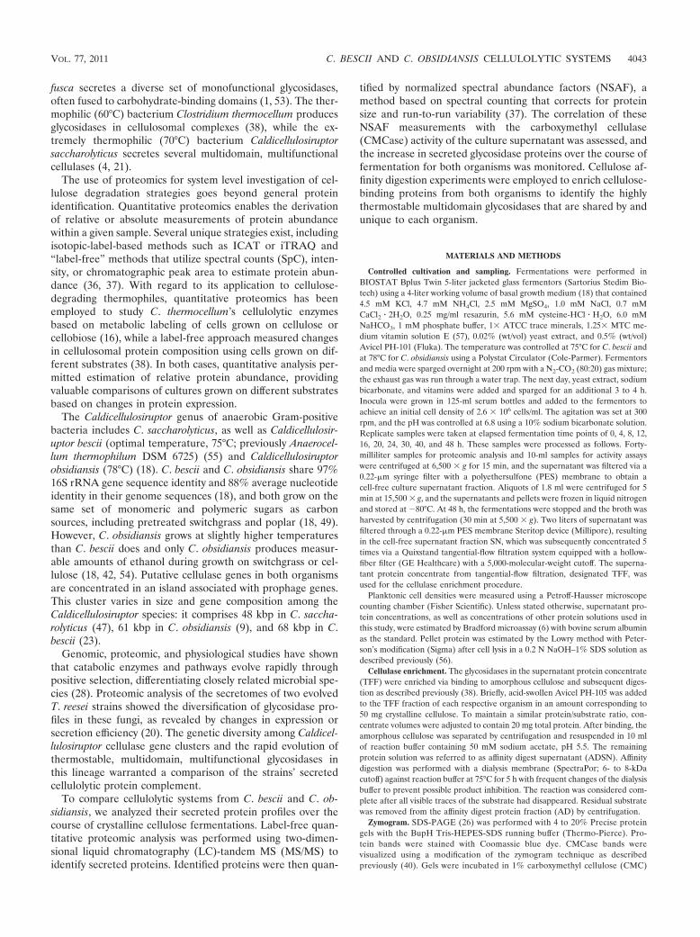
Growth characterization. C. bescii and C. obsidiansis cellswere grown in batch fermentations using crystalline cellulose.Although both fermentors were inoculated with similar celldensities, planktonic cell counts showed a lag phase of about4 h in C. bescii, while C. obsidiansis displayed immediate entryinto the exponential growth phase (Fig. 1A). C. obsidiansisreached a maximal cell density of approximately 109/ml after16 h, and C. bescii did so after 20 h, after which the celldensities of both organisms remained stationary. Pellet proteinconcentrations were below the detection limits of the Lowryassay until 4 h after inoculation (Fig. 1B). In contrast to theplanktonic cell densities, pellet protein concentrations re-mained similar until 12 h, when the C. bescii protein levelsexceeded those of C. obsidiansis by 10 to 30 "g/ml. Proteinconcentrations in the cell-free culture supernatant similarlydiverged after 20 h. The highest supernatant protein concen-trations were measured at the end of the fermentation (48 h),with 47 ' 2 "g/ml for C. bescii and 37 ' 3 "g/ml for C.obsidiansis.
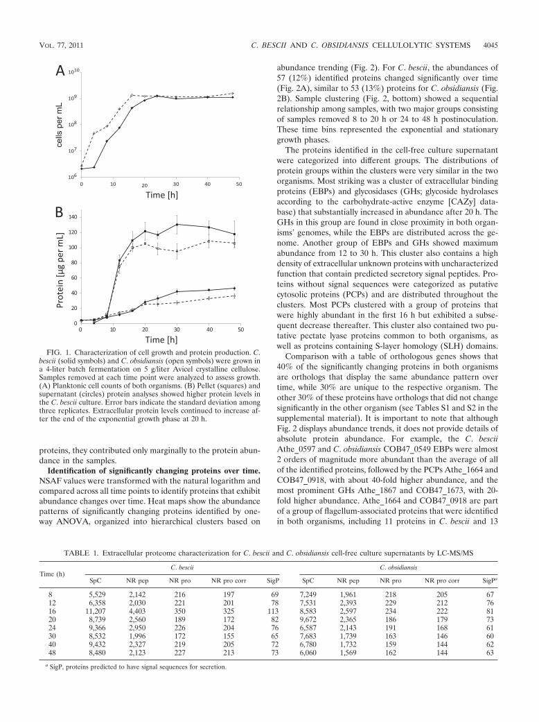
Extracellular proteome characterization. Average values forSpC, nonredundant peptides (NR pep), and nonredundantproteins (NR pro) from cell-free supernatant time course sam-ples of both cultures followed similar trends, although theywere found to be slightly lower in C. obsidiansis than in C.bescii (Table 1, see Table S6 in the supplemental material forboth replicates). At a false-discovery rate of approximately 5%,494 and 418 nonredundant proteins were identified in C. besciiand C. obsidiansis, respectively. The numbers of identified pro-teins in samples taken at 0 and 4 h were too low to be nor-malized together with the subsequent time points. In bothorganisms, the NR pro values were marginally reduced byspectral rebalancing to provide corrected nonredundant pro-tein (NR pro corr) values. SignalP analysis predicted secretorysignal peptide sequences in only one-third of the identifiedproteins (Table 1). NSAF values, derived from SpC, werecalculated for each identified protein and used as a quantita-tive measure of protein abundance. Based on these NSAFvalues, proteins with confidently predicted secretion sequencesmake up 60 to 70% of the sample composition at each timepoint (see Tables S1 and S2 in the supplemental material).Although protein diversity is high among putative nonsecreted
4044 LOCHNER ET AL. APPL. ENVIRON. MICROBIOL.
proteins, they contributed only marginally to the protein abun-dance in the samples.
Identification of significantly changing proteins over time.NSAF values were transformed with the natural logarithm andcompared across all time points to identify proteins that exhibitabundance changes over time. Heat maps show the abundancepatterns of significantly changing proteins identified by one-way ANOVA, organized into hierarchical clusters based on
abundance trending (Fig. 2). For C. bescii, the abundances of57 (12%) identified proteins changed significantly over time(Fig. 2A), similar to 53 (13%) proteins for C. obsidiansis (Fig.2B). Sample clustering (Fig. 2, bottom) showed a sequentialrelationship among samples, with two major groups consistingof samples removed 8 to 20 h or 24 to 48 h postinoculation.These time bins represented the exponential and stationarygrowth phases.
The proteins identified in the cell-free culture supernatantwere categorized into different groups. The distributions ofprotein groups within the clusters were very similar in the twoorganisms. Most striking was a cluster of extracellular bindingproteins (EBPs) and glycosidases (GHs; glycoside hydrolasesaccording to the carbohydrate-active enzyme [CAZy] data-base) that substantially increased in abundance after 20 h. TheGHs in this group are found in close proximity in both organ-isms’ genomes, while the EBPs are distributed across the ge-nome. Another group of EBPs and GHs showed maximumabundance from 12 to 30 h. This cluster also contains a highdensity of extracellular unknown proteins with uncharacterizedfunction that contain predicted secretory signal peptides. Pro-teins without signal sequences were categorized as putativecytosolic proteins (PCPs) and are distributed throughout theclusters. Most PCPs clustered with a group of proteins thatwere highly abundant in the first 16 h but exhibited a subse-quent decrease thereafter. This cluster also contained two pu-tative pectate lyase proteins common to both organisms, aswell as proteins containing S-layer homology (SLH) domains.
Comparison with a table of orthologous genes shows that40% of the significantly changing proteins in both organismsare orthologs that display the same abundance pattern overtime, while 30% are unique to the respective organism. Theother 30% of these proteins have orthologs that did not changesignificantly in the other organism (see Tables S1 and S2 in thesupplemental material). It is important to note that althoughFig. 2 displays abundance trends, it does not provide details ofabsolute protein abundance. For example, the C. besciiAthe_0597 and C. obsidiansis COB47_0549 EBPs were almost2 orders of magnitude more abundant than the average of allof the identified proteins, followed by the PCPs Athe_1664 andCOB47_0918, with about 40-fold higher abundance, and themost prominent GHs Athe_1867 and COB47_1673, with 20-fold higher abundance. Athe_1664 and COB47_0918 are partof a group of flagellum-associated proteins that were identifiedin both organisms, including 11 proteins in C. bescii and 13
FIG. 1. Characterization of cell growth and protein production. C.bescii (solid symbols) and C. obsidiansis (open symbols) were grown ina 4-liter batch fermentation on 5 g/liter Avicel crystalline cellulose.Samples removed at each time point were analyzed to assess growth.(A) Planktonic cell counts of both organisms. (B) Pellet (squares) andsupernatant (circles) protein analyses showed higher protein levels inthe C. bescii culture. Error bars indicate the standard deviation amongthree replicates. Extracellular protein levels continued to increase af-ter the end of the exponential growth phase at 20 h.
TABLE 1. Extracellular proteome characterization for C. bescii and C. obsidiansis cell-free culture supernatants by LC-MS/MS
Time (h)C. bescii C. obsidiansis
SpC NR pep NR pro NR pro corr SigP SpC NR pep NR pro NR pro corr SigPa
a SigP, proteins predicted to have signal sequences for secretion.
VOL. 77, 2011 C. BESCII AND C. OBSIDIANSIS CELLULOLYTIC SYSTEMS 4045
proteins in C. obsidiansis (Athe_1653, Athe_1654, Athe_1675,Athe_1674, Athe_2162, Athe_2165, Athe_2167, Athe_2173,Athe_2174, Athe_2337, Athe_2338, COB47_0906,COB47_0909, COB47_0910, COB47_0930, COB47_0931,COB47_1934, COB47_1943, COB47_1946, COB47_1947,COB47_1948, COB47_1956, COB47_1957, and COB47_2110).
Glycosidase abundance and domain composition. NSAFvalues for selected glycosidases were plotted to compare theirrelative abundances over time (Fig. 3). These GHs were an-notated as putative cellulolytic proteins and exhibited an in-creasing abundance trend over time. Each had a sum of (100 !NSAF) ( 0.5 across all of the time points, indicating substan-tial representation in the extracellular fractions. Athe_1867,
also known as CelA (58), and its ortholog COB47_1673 wereamong the most abundant supernatant proteins identified.Both proteins have a theoretical molecular mass of 195 kDa,and they share 95% amino acid sequence identity (Fig. 4). TheAthe_1865, Athe_1857, COB47_1669, and COB47_1671 GHswere about 60% less abundant than the CelA homologs (Fig.3). Although Athe_1865 and COB47_1669 are orthologs, bothAthe_1857 and COB47_1671 are unique to their respectivestrains (Fig. 4). Another GH unique to C. bescii, Athe_1859, isapproximately an order of magnitude less abundant thanAthe_1867. The GH orthologs Athe_1860 and COB47_1664contribute marginally to the overall cellulolytic component ofeach organism’s secreted proteins (Fig. 3).
FIG. 2. Heat maps showing proteins whose abundances changed significantly in the cell-free culture supernatant. Sets of LC-MS/MS data wereacquired in duplicate for C. bescii (A) and C. obsidiansis (B) from 8 to 48 h. Peptides were identified using SEQUEST, and NSAF values for eachprotein were ln transformed and standardized for one-way ANOVA. Abundances at each time point range from low (green) to high (red). Geneloci are listed without the prefix Athe_ for C. bescii or the prefix COB47_ for C. obsidiansis. Proteins are annotated as EBPs (green), EUP(extracellular unknown proteins; blue), GHs (the CAZy term for glycosidases; red), PCPs (black), PL (pectate lyases; red), or SLH (proteins withSLH domains; orange).
4046 LOCHNER ET AL. APPL. ENVIRON. MICROBIOL.
Cellulase activity correlates with MS data. Time coursesamples were also analyzed for cellulase activity. Cell-free cul-ture supernatant was incubated with CMC, and the rate ofreducing sugar formation was measured. Specific activity in thesample was plotted alongside the summed percent NSAF val-ues of recognized GHs (Fig. 5). Both organisms show a closecorrelation between activity and GH abundance measure-ments, with 97% for C. bescii and 92% for C. obsidiansis,although GH abundance briefly lags activity in C. obsidiansis.
Selective cellulase enrichment. After 48 h, when cellulaseabundance and activity were found to be highest, cells wereharvested by centrifugation and culture supernatant was fil-tered to obtain a cell-free supernatant fraction (SN). The SNwas concentrated 5-fold via tangential-flow filtration, resultingin a retentate that retained roughly 80% of the total protein forC. bescii and 54% for C. obsidiansis (Table 2). Cellulases wereselectively enriched from the TFF fraction via affinity diges-tion, leaving behind proteins in the supernatant that theoreti-cally do not bind to cellulose. In order to compare the fractions
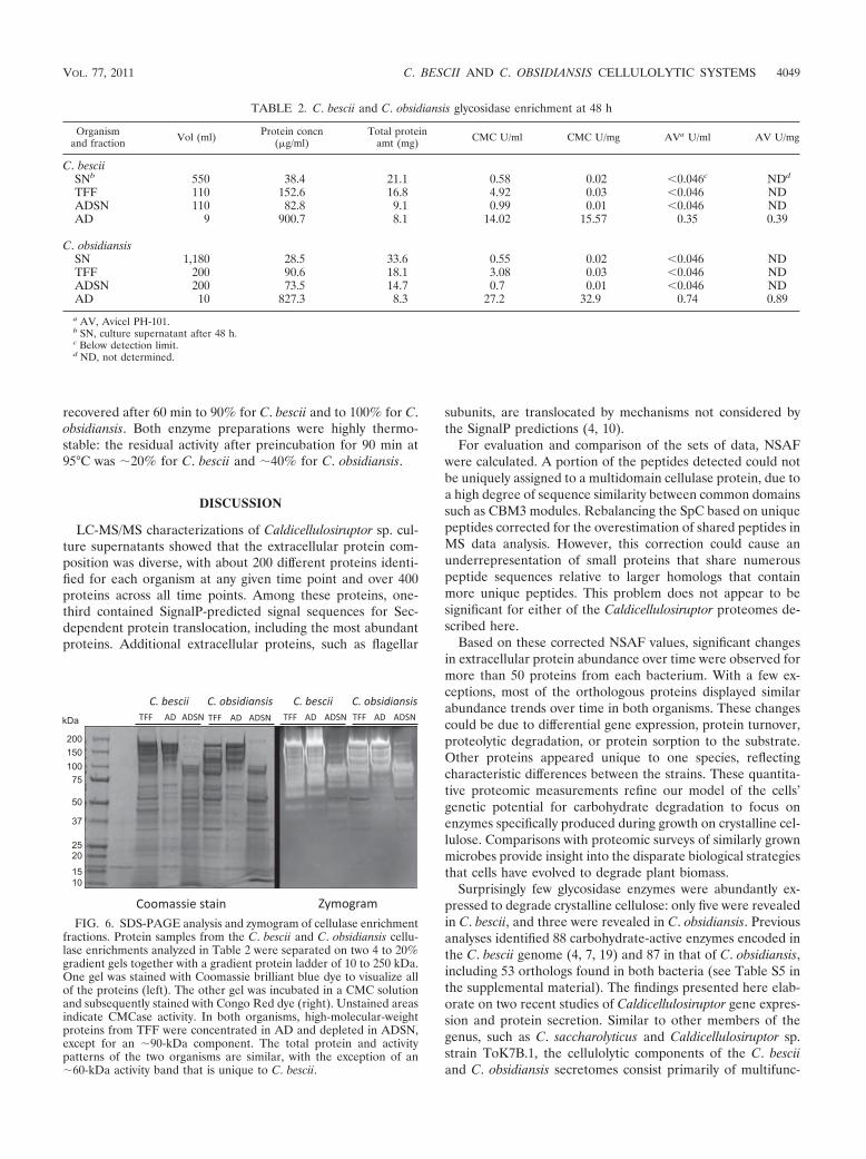
in the two organisms, the protein concentration that converted4% of the CMC or Avicel PH-101 substrate to reducing sugarsin 60 min was determined (designated CMC U or AV U). Thenumber of CMC U per milligram of protein increased 30-foldfor both organisms in TFF. In AD, the activity increased about500-fold for C. bescii and 1,000-fold for C. obsidiansis. Thisindicates that the C. bescii AD fraction (15.57 U/mg) is onlyhalf as active as the one obtained from C. obsidiansis (32.9U/mg) (Table 2). Insoluble Avicel is a more complex andrecalcitrant substrate, and protein concentrations in SN, TFF,and ADSN samples were too low to achieve 4% conversion in60 min, so that activity could not be determined. The numberof AD fraction AV U, however, followed the same trend asobserved in the number of CMC U, with AD more than twiceas active in C. obsidiansis as in C. bescii, 0.89 versus 0.39 U/mg,respectively (Table 2).
Zymogram. The TFF, AD, and ADSN fractions were sepa-rated on two SDS-PAGE gradient gels under the same condi-tions. One gel was stained with Coomassie blue dye to visualizeall proteins, and the other one was stained for cellulase activityin a zymogram (Fig. 6). The zymograms showed a staggeredunstained band pattern of active CMCases with apparentmasses of 60 to 250 kDa for both organisms. A separate bandat 50 kDa was more distinct in C. bescii proteins. The samepattern was retained in the AD fractions. The ADSN fractionsshowed active bands near 90 kDa at substantially reducedintensities. Additionally, C. obsidiansis lacked an active 60-kDaprotein in all three fractions that appeared in the C. bescii TFFand AD fractions. The apparent molecular masses of the gly-cosidases could not be matched to the theoretical masses ofpredicted proteins. Proteins from several gel bands were ex-tracted and analyzed by mass spectrometry; however, the pro-tein diversity was found to be very high and dominated by themost abundant proteins in the samples (data not shown).Larger apparent molecular masses could be due to heteroge-neous protein glycosylation, while smaller ones could haveresulted from proteolytic degradation of the large, multido-main proteins, as suggested previously (58).
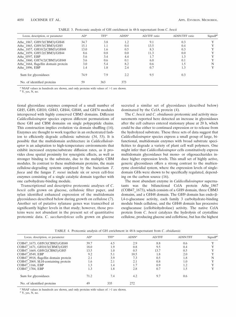
Proteomic analysis of cellulase-enriched fractions. LC-MS/MS measurements of abundant proteins in the TFF, AD,and ADSN fractions are shown in Table 3 for C. bescii and inTable 4 for C. obsidiansis. Tables S3 and S4 in the supplemen-tal material present the complete set of identified proteins. Inboth organisms, the number of identified proteins in the ADfraction was about 6 to 7 times lower than in the TFF andADSN fractions, which have similar numbers of identified pro-teins. Conversely, abundance values for the GHs increasedabout 10-fold compared to those in the TFF fraction and weredepleted 6-fold in the remaining ADSN fraction. Besides theGHs, AD fractions contained several proteins without carbo-hydrate binding domains, such as the EBPs Athe_0597(COB47_0549), Athe_1896 (COB47_1704), and COB47_1166;the flagellin domain protein Athe_1664 (COB47_0918); andthe SLH domain protein COB47_2069. However, none ofthese proteins had been selectively enriched, because the AD/TFF ratio was &1 and the ADSN/TFF ratio was !1. The highabundance of these proteins in the TFF fraction suggests car-ryover through nonspecific binding.
Characterization of cellulase-enriched fractions. The cellu-lase-enriched AD fractions were investigated to determine the
FIG. 3. Glycosidase abundances increased over time. The averagesof two proteomic measurements of C. bescii (A) and C. obsidiansis(B) glycosidase abundances are shown as NSAF values in hundreds.Error bars indicate the standard deviations of duplicate measurements.The signal intensity differs substantially among the enzymes. (A) C.bescii supernatant contained five identified glycosidases, including themost abundant protein Athe_1867 (CelA) and the least abundantprotein Athe_1860. The Athe_1859 (!) protein abundance did notchange significantly during growth but was higher than that ofAthe_1860 after 16 h. (B) C. obsidiansis cell-free culture supernatantcontained four glycosidases, including the most abundant proteinCOB47_1673 (CelA) and the relatively minor protein COB47_1664.
VOL. 77, 2011 C. BESCII AND C. OBSIDIANSIS CELLULOLYTIC SYSTEMS 4047
optimum reaction temperature and pH. Cellulase mixturesobtained from both C. bescii and C. obsidiansis hydrolyzedCMC optimally at 85°C at pH 5 (Fig. 7). Thermostability wasdetermined by preincubating the protein fractions at 75, 85,
and 95°C and then measuring CMCase activity at 80°C. Figure7C and D show that the activity of both cellulase mixturesincreased slightly following preincubation at 75°C. At 85°C, theactivity decreased to 80% after 30 min of preincubation but
FIG. 4. Glycosidase domain composition. The Pfam, CAZy, and nonredundant protein databases were used to identify functional domainswithin the multidomain enzymes. These domains are labeled as follows according to CAZy families: GH, glycosidase; CBM3, carbohydrate-bindingmodule family 3. The Athe_1857 and Athe_1859 proteins have no orthologs in C. obsidiansis, and COB47_1671 has no C. bescii ortholog. aa, aminoacids.
FIG. 5. Total glycosidase abundance correlates with cellulase activity during the course of fermentation. CMCase activity of the cell-freesupernatant was measured for C. bescii (A) and C. obsidiansis (B) from 0 to 48 h after inoculation. Enzyme activity in units per milligram of totalsupernatant protein (solid symbols, solid lines) was plotted together with the sum of NSAF values (in hundreds) for the glycosidases identified inFig. 3 (open symbols, dashed lines). The error bars indicate standard deviations for each value. Abundance and activity increases correlate for bothcultures; however, the correlation is closer for C. bescii data than for C. obsidiansis data, suggesting that an active component may not have beenincluded in the summed NSAF values.
4048 LOCHNER ET AL. APPL. ENVIRON. MICROBIOL.
recovered after 60 min to 90% for C. bescii and to 100% for C.obsidiansis. Both enzyme preparations were highly thermo-stable: the residual activity after preincubation for 90 min at95°C was )20% for C. bescii and )40% for C. obsidiansis.
DISCUSSION
LC-MS/MS characterizations of Caldicellulosiruptor sp. cul-ture supernatants showed that the extracellular protein com-position was diverse, with about 200 different proteins identi-fied for each organism at any given time point and over 400proteins across all time points. Among these proteins, one-third contained SignalP-predicted signal sequences for Sec-dependent protein translocation, including the most abundantproteins. Additional extracellular proteins, such as flagellar
subunits, are translocated by mechanisms not considered bythe SignalP predictions (4, 10).
For evaluation and comparison of the sets of data, NSAFwere calculated. A portion of the peptides detected could notbe uniquely assigned to a multidomain cellulase protein, due toa high degree of sequence similarity between common domainssuch as CBM3 modules. Rebalancing the SpC based on uniquepeptides corrected for the overestimation of shared peptides inMS data analysis. However, this correction could cause anunderrepresentation of small proteins that share numerouspeptide sequences relative to larger homologs that containmore unique peptides. This problem does not appear to besignificant for either of the Caldicellulosiruptor proteomes de-scribed here.
Based on these corrected NSAF values, significant changesin extracellular protein abundance over time were observed formore than 50 proteins from each bacterium. With a few ex-ceptions, most of the orthologous proteins displayed similarabundance trends over time in both organisms. These changescould be due to differential gene expression, protein turnover,proteolytic degradation, or protein sorption to the substrate.Other proteins appeared unique to one species, reflectingcharacteristic differences between the strains. These quantita-tive proteomic measurements refine our model of the cells’genetic potential for carbohydrate degradation to focus onenzymes specifically produced during growth on crystalline cel-lulose. Comparisons with proteomic surveys of similarly grownmicrobes provide insight into the disparate biological strategiesthat cells have evolved to degrade plant biomass.
Surprisingly few glycosidase enzymes were abundantly ex-pressed to degrade crystalline cellulose: only five were revealedin C. bescii, and three were revealed in C. obsidiansis. Previousanalyses identified 88 carbohydrate-active enzymes encoded inthe C. bescii genome (4, 7, 19) and 87 in that of C. obsidiansis,including 53 orthologs found in both bacteria (see Table S5 inthe supplemental material). The findings presented here elab-orate on two recent studies of Caldicellulosiruptor gene expres-sion and protein secretion. Similar to other members of thegenus, such as C. saccharolyticus and Caldicellulosiruptor sp.strain ToK7B.1, the cellulolytic components of the C. besciiand C. obsidiansis secretomes consist primarily of multifunc-
FIG. 6. SDS-PAGE analysis and zymogram of cellulase enrichmentfractions. Protein samples from the C. bescii and C. obsidiansis cellu-lase enrichments analyzed in Table 2 were separated on two 4 to 20%gradient gels together with a gradient protein ladder of 10 to 250 kDa.One gel was stained with Coomassie brilliant blue dye to visualize allof the proteins (left). The other gel was incubated in a CMC solutionand subsequently stained with Congo Red dye (right). Unstained areasindicate CMCase activity. In both organisms, high-molecular-weightproteins from TFF were concentrated in AD and depleted in ADSN,except for an )90-kDa component. The total protein and activitypatterns of the two organisms are similar, with the exception of an)60-kDa activity band that is unique to C. bescii.
TABLE 2. C. bescii and C. obsidiansis glycosidase enrichment at 48 h
VOL. 77, 2011 C. BESCII AND C. OBSIDIANSIS CELLULOLYTIC SYSTEMS 4049
tional glycosidase enzymes composed of a small number ofGH5, GH9, GH10, GH43, GH44, GH48, and GH74 modulesinterspersed with highly conserved CBM3 domains. DifferentCaldicellulosiruptor species express different permutations ofthese GH and CBM domains on single polypeptide chains.This construction implies evolution via domain shuffling (14).Enzymes are thought to work together in an orchestrated fash-ion to efficiently degrade cellulosic substrate (34, 53). It ispossible that the multidomain architecture in Caldicellulosir-uptor is an adaptation to high-temperature environments thatexhibit increased enzyme/substrate diffusion rates, as it pro-vides close spatial proximity for synergistic effects, as well asstronger binding to the substrate, due to the multiple CBMmodules. In contrast to these multidomain proteins, the maincellulose-degrading systems employed by the bacterium T.fusca and the fungus T. reesei include six or seven cell-freeenzymes consisting of a single catalytic domain together withone carbohydrate-binding module.
Transcriptional and descriptive proteomic analyses of C.bescii cells grown on glucose, cellulosic filter paper, andxylan identified enhanced expression of the multidomainglycosidases described below during growth on cellulose (7).Another set of putative xylanase genes was transcribed atsignificantly higher levels in that study; however, those pro-teins were not abundant in the present set of quantitativeproteomic data. C. saccharolyticus cells grown on glucose
secreted a similar set of glycosidases (described below)dominated by the CelA protein (4).
The C. bescii and C. obsidiansis proteomic and activity mea-surements reported here detected an increase in glycosidasesafter the cell cultures entered stationary phase at 20 h, whichcould be due either to continued expression or to release fromthe hydrolyzed substrate. These three sets of data suggest thatCaldicellulosiruptor species express a small group of large, bi-functional, multidomain enzymes with broad substrate speci-ficities to degrade a variety of plant cell wall polymers. Onemight infer that Caldicellulosiruptor cells constitutively expressmultidomain glycosidases but mono- or oligosaccharides in-duce higher expression levels. This small set of highly active,generic glycosidases offers a strong contrast to the multien-zyme clostridial system, where the expression levels of single-domain GHs were shown to be specifically regulated, depend-ing on the carbon source (16).
The most abundant enzyme in Caldicellulosiruptor superna-tants was the bifunctional CelA protein Athe_1867(COB47_1673), which consists of a GH9 domain, three CBM3domains, and a GH48 domain. The GH9 domain has endo-*-l,4-D-glucanase activity, each family 3 carbohydrate-bindingmodule binds cellulose, and the GH48 domain has processiveexoglucanase (cellobiohydrolase) activity. The native CelAprotein from C. bescii catalyzes the hydrolysis of crystallinecellulose, producing glucose and cellobiose, but has the highest
TABLE 3. Proteomic analysis of GH enrichment in 48-h supernatant from C. bescii
Locus, description, or parameter ADa TFFa ADSNa AD/TFF ratio ADSN/TFF ratio SignalPb
No. of identified proteins 49 335 272a NSAF values in hundreds are shown, and only proteins with values of (1 are shown.b Y, yes, N, no.
4050 LOCHNER ET AL. APPL. ENVIRON. MICROBIOL.
activity on CMC (Glu*-134 Glu), *-glucan (Glu*-133,4Glu), and xylan (Xyl*-134 Xyl) (58). A recombinant CelAprotein from C. saccharolyticus did not exhibit xylanase activity(46).
Most cellulolytic organisms express extracellular or cell sur-face-associated GH9 or GH48 protein for crystalline cellulosehydrolysis. Proteomic analysis of the C. thermocellum cellulo-some identified the GH48 (CelS) exoglucanase as the mostabundant enzyme among 14 other less abundant GH9 domain-containing proteins when cultures were grown on crystallinecellulose (25, 38, 51). Cellulose-grown T. fusca also expresses aGH48 (Cel48A) exoglucanase, with concurrent upregulation ofseveral endoglucanases, including a GH9 (Cel9B) protein (1).These findings demonstrate the synergistic importance of theGH9 and GH48 protein domains in the degradation of crys-talline cellulose. Perhaps an exception to the rule, S. degradansproteins lack any detectible GH48 domain. This organism em-ploys mostly endoglucanases, including a GH9 (Cel9B) pro-tein, for the degradation of crystalline cellulose (45).
The CelC-ManB bifunctional glycosidase Athe_1865(COB47_1669) was the second most abundant protein identi-fied by LC-MS/MS. These orthologs contain a GH9 domain,three CBM3 domains, and a GH5 domain. In C. saccharolyti-
cus, the orthologous domains can be found in different openreading frames. Csac_1079 (A4XIF8, CelC) encodes a GH9domain with three CBM3 domains and has been shown to haveendo-l,4-*-D-glucanase activity, while Csac_1080 (A4XIF9,ManB) is a *-mannanase homolog of the GH5 domain (15,33). In addition to its role in cellulose hydrolysis, the bifunc-tional protein may facilitate hemicellulose degradation by cat-alyzing glucomannan hydrolysis. GH5 enzymes seem to beessential for cellulose degradation in T. reesei (Cel5A) (20), T.fusca (Cel5A) (1), and S. degradans (Cel5I and Cel5J) (45),where all three are annotated as endoglucanases.
C. bescii and C. obsidiansis both express GH10 domains, butin nonorthologous multidomain proteins. Athe_1857 containsGH10 and GH48 domains, while in COB47_1671, the GH10domain is associated with another GH5 module. GH10 doi-mains often confer endo-l,4-*-D-xylanase activity, although ahomologous domain from C. saccharolyticus was reported tohave exo-*-l,4-D-glucanase (cellobiohydrolase) activity (39).The COB47_1671 protein is homologous to CelB from C.saccharolyticus (Csac_1078, A4XIF7). This enzyme was re-cently heterologously expressed, purified, and shown to havethe highest hydrolytic activity on xylan and *-glucan, followedby glucomannan and CMC. When expressed separately, the
FIG. 7. Biochemical characterization of glycosidase preparations. The protein fractions isolated by affinity digestion of the cell-free culturesupernatants of C. bescii (solid symbols, solid lines) and C. obsidiansis (open symbols, dashed lines) were examined for optimal reaction parametersand thermostability in triplicate CMCase assays at 80°C (B to D) or at various temperatures of 40 to 100°C (A). To determine the pH optimum,values were measured at pHs of 3.5 to 6.5 in 50 mM citrate buffer (circles) and at pHs of 6.5 to 8 in 50 mM potassium phosphate buffer (squares).The thermostability of the C. bescii (C) and C. obsidiansis (D) enzyme mixtures was measured by incubating the protein samples for 30, 60, or 90min at 75, 85, or 95°C before starting the CMCase assay by substrate addition. Both preparations showed slightly increased activity uponpreincubation for 30 and 60 min. The C. bescii enzyme mixture retained 10 to 20% of its activity upon preincubation for 90 min at 95°C, while theC. obsidiansis mixture retained up to 40%.
VOL. 77, 2011 C. BESCII AND C. OBSIDIANSIS CELLULOLYTIC SYSTEMS 4051
GH5 and GH10 domains both independently exhibited thesame broad substrate specificity, although at decreased rates ofhydrolysis. Combining the single enzymes did not completelyrestore the activity of the full-length version, demonstratingthe synergistic effects of multidomain proteins (48). Neverthe-less, several cellulolytic bacteria express single GH10 domainproteins. C. thermocellum has four xylanolytic GH10 domainsdispersed over several proteins, although their expressionseems to be downregulated on crystalline cellulose (16, 38). Incontrast, T. fusca expresses significant levels of GH10 xylanasetogether with other hemicellulases when grown on cellulose(1), while T. reesii and S. degradans do not encode any GH10domain proteins at all (20, 45).
The C. bescii protein Athe_1859 (ManA) was less abundantthan CelC-ManB and has no ortholog in C. obsidiansis. Itcontains an N-terminal GH5 *-mannanase, two CBM3 do-mains, and a GH44 endo-*-1,4-glucanase domain. The or-thologous C. saccharolyticus protein (Csac_1077, A4XIF6) ex-hibited endo-*-1,4 xylanase activity (15, 27).
The orthologs Athe_1860 and COB47_1664 are present atvery low levels in the supernatant, as well as in the cellulase-enriched fractions. These proteins contain an amino-terminalAsp box repeat commonly found in sialidases, two CBM3 do-mains, and a GH48 processive cellobiohydrolase domain. C.saccharolyticus does not have a full-length ortholog but doesencode a protein that contains a similar N-terminal domain(Csac_1085; A4XIG4). The Asp box repeat forms a *-propel-ler sequence motif that has also been found in GH74 proteinslike the xyloglucanase Xgh74A from C. thermocellum (29). Thenatural function of these proteins may be to facilitate glucoxy-lan hydrolysis in hemicellulose.
Besides the highly abundant glycosidases, other proteins thatwere found in the secretome include putative pectate lyases:Athe_1854 (COB47_1662) and Athe_1855. These genes arefound to be in close genomic proximity to the multifunctionalglycosidase genes; however, their abundance patterns do notcluster with the identified GHs. Rather, they exhibited a re-verse trend, with higher abundances in the early growth stagesthat decreased over time, consistent with previous transcrip-tional analysis of cells grown on cellulose versus glucose (7).Other GHs showed a similar proteomic trend, although theirtranscript abundance was higher during growth on cellulose:those proteins declining in abundance included a putative pul-lulanase, Athe_0609 (OB47_0563); a putative +-amylase,Athe_0610 (COB47_0564); a putative *-xylosidase with a GH3domain, Athe_2354 (COB47_2124); the endo-*-1,4-xylanaseAthe_0089, and the GH73-containing putative peptidoglycanhydrolase Athe_1080 (COB47_1445) (see Tables S1 and S2 inthe supplemental material). Since the abundance of these pro-teins decreased over time, it is possible that their expressionwas triggered either by impurities in the cellulose substrate orby yeast extract components in the medium.
Cellulase protein stability is valuable in enzyme mixtures forlarge-scale cellulose saccharification; therefore, it is notewor-thy that the cellulase-enriched fractions from both organismshad maximum CMCase activity at pH 5 and 85°C, exceedingthe thermoactivity of commercial cellulase preparations (35).In a previous study, a similar pH optimum for CMCase activityof purified C. bescii CelA was measured, while the hydrolyticactivity continued increasing at elevated temperatures, even at
95 to 100°C (58). Enzyme preparations from both Caldicellu-losiruptor spp. appeared equally thermostable following prein-cubation at various temperatures, although C. obsidiansis pro-teins retained slightly more stability after preincubation at95°C. A similar study with Caldicellulosiruptor lactoaceticus cul-ture supernatant reported optimal CMCase activity at 80°Cand pH 6. However, the proteins’ thermostability was lower:60% activity remained after 90 min preincubation at 70°C, 50%at 80°C, and about 30% at 90°C (32). Significant differences inthe assay methods used to measure cellulase activity preventcomparison of Caldicellulosiruptor AD activity data measuredat 80°C with results from previous studies. However, commer-cial cellulase mixtures that were also assayed according to theIUPAC standard show a slightly lower range (5 to 25 CMCU/mg at 50°C [35]) of specific activities.
Most of the abundant glycosidase proteins are expressedfrom loci situated in islands on the chromosomes of C. bescii,C. obsidiansis, and C. saccharolyticus. These 61- to 80-kbpislands are hot spots of genetic recombination, insertion, anddeletion events that produced the diversity of multidomainglycosidases and accessory enzymes observed in these bacteria.These islands include genes COB47_1657 to COB47_1693,Athe_1845 to Athe_1886, or Csac_1060 to Csac_1108 encodingglycosidase integrases, transposases, and pilin or prophage el-ements, as well as numerous hypothetical proteins. However,there are no transporter components in close genomic prox-imity.
ABC transporters are the primary mode of monosaccharideand oligosaccharide uptake by Caldicellulosiruptor cells. Themost abundant extracellular proteins identified in this study,EBPs, bind their cognate substrates and deliver them to themembrane-bound components of ABC transport systems. TheC. saccharolyticus genome contains at least 177 ABC trans-porter genes, including components of 25 putative sugar trans-porters (49). C. bescii and C. obsidiansis share orthologs for 14of these systems, which include the most abundant EBP sub-units detected in these proteomics analyses. In both organisms,the abundance levels of one group of EBPs were low at thebeginning of the fermentations, peaked in the exponentialgrowth stage, and decreased in the stationary phase. Amongthese EBPs are COB47_0357 (orthologous to Athe_0399 andCsac_0440) and COB47_0096 (orthologous to Athe_0105 andCsac_2506). Expression of the orthologous C. saccharolyticusgenes was upregulated during growth on the monosaccharidesxylose, glucose, fructose, and galactose (49). Like the abun-dance of the identified hemicellulases, that of these proteinsdecreased over time and therefore they could also be inducedby impurities in the cellulose substrate or yeast extract com-ponents and/or be derived from a basal level of constitutiveexpression.
Another group of EBPs increased over time in both organ-isms, with the highest levels observed in the late stationaryphase; these proteins clustered and trended with the majorGHs. These EBPs included COB47_0549 (orthologous toAthe_0597 and Csac_0681), which was predicted to transportxyloglucans (49). Due to its high abundance in cellulose-growncultures, it most likely has a substrate specificity for cellooli-gosaccharides. COB47_0569 (orthologous to Athe_0614 andCsac_0692), on the other hand, may bind a broad range ofmonosaccharides (49). These EBPs could work synergistically
4052 LOCHNER ET AL. APPL. ENVIRON. MICROBIOL.
with the major GHs of each species, binding carbohydratesreleased by the multifunctional cellulases.
The flagellin proteins in both species, Athe_1664 andCOB47_0918, decreased in abundance during growth but re-mained the second most abundant extracellular proteins. Fur-thermore, these proteins were abundant in AD fractions. Lessabundant components of the flagellar apparatus were alsoidentified in the supernatant fractions. Electron micrographsof C. bescii and C. obsidiansis did not indicate flagella, andmotility has not been observed in these organisms. However,their genomes contain conserved clusters of flagellar motorand filament assembly genes. In Clostridium difficile, the FliCand FliD proteins mediate cellular attachment to mucus (44),suggesting a potential role for flagella in cellulose attachment,as well as possible motility.
Conclusions. Caldicellulosiruptor spp. have proven to behighly versatile cellulose- and hemicellulose-degrading ther-mophiles. This quantitative proteomic analysis identified thestrategy C. bescii and C. obsidiansis use to degrade crystallinecellulose, i.e., by secreting a small number of cellulose-binding,multifunctional glycosidases dominated by the CelA protein. Afew EBPs are expressed at high levels to sequester releasedglucose and small glucans for cellular metabolism. Predictionsof the hemicellulose-degradative potential of the same glyco-sidases expressed during growth on purified cellulose suggeststhat future studies of differential protein expression in cellsgrown with diverse polymeric substrates will help differentiatethe cells’ general and specific responses to lignocellulosic sub-strates. Closely related species of the same genus differ in theirsecreted enzyme yields, enzymes, and catalytic efficiencies.Therefore, the assumption that genomic similarity equates tophysiological similarity can lead to a missed opportunity togarner biotechnological potential.
ACKNOWLEDGMENTS
We thank Barbara Klippel, Scott Hamilton-Brehm, Babu Raman,and James Elkins for helpful discussions.
This study was funded by the BioEnergy Science Center, a U.S.Department of Energy Bioenergy Research Center supported by theOffice of Biological and Environmental Research in the DOE Office ofScience. Oak Ridge National Laboratory is managed by and this reporthas been authored by University of Tennessee-Battelle, LLC, undercontract DE-AC05-00OR22725 with the U.S. Department of Energy.
REFERENCES1. Adav, S. S., C. S. Ng, M. Arulmani, and S. K. Sze. 2010. Quantitative iTRAQ
secretome analysis of cellulolytic Thermobifida fusca. J. Proteome Res.9:3016–3024.
2. Adney, W. S., and J. O. Baker. 1996. Measurement of cellulase activities.National Renewable Energy Laboratory, Washington, DC.
3. Aebersold, R., and M. Mann. 2003. Mass spectrometry-based proteomics.Nature 422:198–207.
4. Andrews, G., D. Lewis, J. Notey, R. Kelly, and D. Muddiman. 2010. Part I:characterization of the extracellular proteome of the extreme thermophileCaldicellulosiruptor saccharolyticus by GeLC-MS2. Anal. Bioanal. Chem.398:377–389.
5. Blumer-Schuette, S. E., I. Kataeva, J. Westpheling, M. W. Adams, and R. M.Kelly. 2008. Extremely thermophilic microorganisms for biomass conversion:status and prospects. Curr. Opin. Biotechnol. 19:210–217.
6. Bradford, M. M. 1976. A rapid and sensitive method for the quantitation ofmicrogram quantities of protein utilizing the principle of protein-dye bind-ing. Anal. Biochem. 72:248–254.
7. Dam, P., et al. 2011. Insights into plant biomass conversion from the genomeof the anaerobic thermophilic bacterium Caldicellulosiruptor bescii DSM6725. Nucleic Acids Res. 39:3240–3254.
8. Decker, S. R., W. S. Adney, E. Jennings, T. B. Vinzant, and M. E. Himmel.2003. Automated filter paper assay for determination of cellulase activity.Appl. Biochem. Biotechnol. 105–108:689–703.
9. Elkins, J. G., et al. 2010. Complete genome sequence of the cellulolyticthermophile Caldicellulosiruptor obsidiansis OB47T. J. Bacteriol. 192:6099–6100.
10. Emanuelsson, O., S. Brunak, G. von Heijne, and H. Nielsen. 2007. Locatingproteins in the cell using TargetP, SignalP and related tools. Nat. Protoc.2:953–971.
11. Eng., J. K., A. L. McCormack, and J. R. Yates. 1994. An approach tocorrelate tandem mass-spectral data of peptides with amino-acid-sequencesin a protein database. J. Am. Soc. Mass Spectrom. 5:976–989.
12. Erickson, B. K., et al. 2010. Computational prediction and experimentalvalidation of signal peptide cleavages in the extracellular proteome of anatural microbial community. J. Proteome Res. 9:2148–2159.
13. Giannone, R. J., et al. 2007. Dual-tagging system for the affinity purificationof mammalian protein complexes. Biotechniques 43:296–302.
14. Gibbs, M. D., et al. 2000. Multidomain and multifunctional glycosyl hydro-lases from the extreme thermophile Caldicellulosiruptor isolate Tok7B. 1.Curr. Microbiol. 40:333–340.
15. Gibbs, M. D., D. J. Saul, E. Luthi, and P. L. Bergquist. 1992. The *-man-nanase from Caldocellum saccharolyticum is part of a multidomain enzyme.Appl. Environ. Microbiol. 58:3864–3867.
16. Gold, N. D., and V. J. J. Martin. 2007. Global view of the Clostridiumthermocellum cellulosome revealed by quantitative proteomic analysis. J.Bacteriol. 189:6787–6795.
17. Griffin, N. M., et al. 2010. Label-free, normalized quantification of complexmass spectrometry data for proteomic analysis. Nat. Biotechnol. 28:83–89.
18. Hamilton-Brehm, S. D., et al. 2010. Caldicellulosiruptor obsidiansis sp. nov.,an anaerobic, extremely thermophilic, cellulolytic bacterium isolated fromObsidian Pool, Yellowstone National Park. Appl. Environ. Microbiol. 76:1014–1020.
19. Henrissat, B. 1991. A classification of glycosyl hydrolases based on aminoacid sequence similarities. Biochem. J. 280(Pt. 2):309–316.
20. Herpoel-Gimbert, I., et al. 2008. Comparative secretome analyses of twoTrichoderma reesei RUT-C30 and CL847 hypersecretory strains. Biotechnol.Biofuels 1:18.
21. Himmel, M. E., et al. 2010. Microbial enzyme systems for biomass conver-sion: emerging paradigms. Biofuels 1:325–343.
22. Kabel, M. A., M. J. E. C. van der Maarel, G. Klip, A. G. J. Voragen, and H. A.Schols. 2006. Standard assays do not predict efficiency of commercial cellu-lase preparations towards plant materials. Biotechnol. Bioeng. 93:56–63.
23. Kataeva, I. A., et al. 2009. Genome sequence of the anaerobic, thermophilic,and cellulolytic bacterium “Anaerocellum thermophilum” DSM 6725. J. Bac-teriol. 191:3760–3761.
24. Keller, M., and R. Hettich. 2009. Environmental proteomics: a paradigmshift in characterizing microbial activities at the molecular level. Microbiol.Mol. Biol. Rev. 73:62–70.
25. Kruus, K., W. K. Wang, J. T. Ching, and J. H. D. Wu. 1995. Exoglucanaseactivities of the recombinant Clostridium thermocellum CelS, a major cellu-losome component. J. Bacteriol. 177:1641–1644.
26. Laemmli, U. K. 1970. Cleavage of structural proteins during the assembly ofthe head of bacteriophage T4. Nature 227:680–685.
27. Luthi, E., N. B. Jasmat, R. A. Grayling, D. R. Love, and P. L. Bergquist.1991. Cloning, sequence analysis, and expression in Escherichia coli of a genecoding for a *-mannanase from the extremely thermophilic bacterium Cal-docellum saccharolyticum. Appl. Environ. Microbiol. 57:694–700.
28. Lynd, L. R., P. J. Weimer, W. H. van Zyl, and I. S. Pretorius. 2002. Microbialcellulose utilization: fundamentals and biotechnology. Microbiol. Mol. Biol.Rev. 66:506–577.
29. Martinez-Fleites, C., et al. 2006. Crystal structures of Clostridium thermocel-lum xyloglucanase, XGH74A, reveal the structural basis for xyloglucan rec-ognition and degradation. J. Biol. Chem. 281:24922–24933.
30. McDonald, W. H., R. Ohi, D. T. Miyamoto, T. J. Mitchison, and J. R. Yates.2002. Comparison of three directly coupled HPLC MS/MS strategies foridentification of proteins from complex mixtures: single-dimension LC-MS/MS, 2-phase MudPIT, and 3-phase MudPIT. Int. J. Mass Spectrom. 219:245–251.
31. Miller, G. L. 1959. Use of dinitrosalicylic acid reagent for determination ofreducing sugar. Anal. Chem. 31:426–428.
32. Mladenovska, Z., I. M. Mathrani, and B. K. Ahring. 1995. Isolation andcharacterization of Caldicellulosiruptor lactoaceticus sp. nov., an extremelythermophilic, cellulolytic, anaerobic bacterium. Arch. Microbiol. 163:223–230.
33. Morris, D. D., R. A. Reeves, M. D. Gibbs, D. J. Saul, and P. L. Bergquist.1995. Correction of the *-mannanase domain of the CelC pseudogene fromCaldocellulosiruptor saccharolyticus and activity of the gene-product on kraftpulp. Appl. Environ. Microbiol. 61:2262–2269.
34. Nidetzky, B., M. Hayn, R. Macarron, and W. Steiner. 1993. Synergism ofTrichoderma reesei cellulases while degrading different celluloses. Biotech-nol. Lett. 15:71–76.
35. Nieves, R. A., C. I. Ehrman, W. S. Adney, R. T. Elander, and M. E. Himmel.1998. Survey and analysis of commercial cellulase preparations suitable forbiomass conversion to ethanol. World J. Microbiol. Biotechnol. 14:301–304.
VOL. 77, 2011 C. BESCII AND C. OBSIDIANSIS CELLULOLYTIC SYSTEMS 4053
36. Ong, S. E., and M. Mann. 2005. Mass spectrometry-based proteomics turnsquantitative. Nat. Chem. Biol. 1:252–262.
37. Paoletti, A. C., et al. 2006. Quantitative proteomic analysis of distinct mam-malian Mediator complexes using normalized spectral abundance factors.Proc. Natl. Acad. Sci. U. S. A. 103:18928–18933.
38. Raman, B., et al. 2009. Impact of pretreated switchgrass and biomass car-bohydrates on Clostridium thermocellum ATCC 27405 cellulosome compo-sition: a quantitative proteomic analysis. PLoS One 4:e5271.
39. Saul, D. J., L. C. Williams, D. R. Love, L. W. Chamley, and P. L. Bergquist.1989. Nucleotide sequence of a gene from Caldocellum saccharolyticumencoding for exocellulase and endocellulase activity. Nucleic Acids Res.17:439.
40. Schwarz, W. H., K. Bronnenmeier, F. Grabnitz, and W. L. Staudenbauer.1987. Activity staining of cellulases in polyacrylamide gels containing mixedlinkage *-glucans. Anal. Biochem. 164:72–77.
41. Skinner, K. A., and T. D. Leathers. 2004. Bacterial contaminants of fuelethanol production. J. Ind. Microbiol. 31:401–408.
42. Svetlichnyi, V. A., T. P. Svetlichnaya, N. A. Chernykh, and G. A. Zavarzin.1990. Anaerocellum thermophilum gen. nov., sp. nov.—an extremely thermo-philic cellulolytic eubacterium isolated from hot-springs in the Valley ofGeysers. Microbiology 59:598–604.
43. Tabb, D. L., W. H. McDonald, and J. R. Yates. 2002. DTASelect andContrast: tools for assembling and comparing protein identifications fromshotgun proteomics. J. Proteome Res. 1:21–26.
44. Tasteyre, A., M. C. Barc, A. Collignon, H. Boureau, and T. Karjalainen.2001. Role of FliC and FliD flagellar proteins of Clostridium difficile inadherence and gut colonization. Infect. Immun. 69:7937–7940.
45. Taylor, L. E., et al. 2006. Complete cellulase system in the marine bacteriumSaccharophagus degradans strain 2–40T. J. Bacteriol. 188:3849–3861.
46. Te’o, V. S. J., D. J. Saul, and P. L. Bergquist. 1995. celA, another gene codingfor a multidomain cellulase from the extreme thermophile Caldocellumsaccharolyticum. Appl. Microbiol. Biotechnol. 43:291–296.
47. van de Werken, H. J., et al. 2008. Hydrogenomics of the extremely thermo-philic bacterium Caldicellulosiruptor saccharolyticus. Appl. Environ. Micro-biol. 74:6720–6729.
48. Vanfossen, A. L., I. Ozdemir, S. L. Zelin, and R. M. Kelly. 17 February 2011,posting date. Glycoside hydrolase inventory drives plant polysaccharide de-
construction by the extremely thermophilic bacterium Caldicellulosiruptorsaccharolyticus. Biotechnol. Bioeng. [Epub ahead of print.] doi:10.1002/bit.23093.
49. Vanfossen, A. L., M. R. A. Verhaart, S. M. W. Kengen, and R. M. Kelly. 2009.Carbohydrate utilization patterns for the extremely thermophilic bacteriumCaldicellulosiruptor saccharolyticus reveal broad growth substrate prefer-ences. Appl. Environ. Microbiol. 75:7718–7724.
50. Vinzant, T. B., et al. 2001. Fingerprinting Trichoderma reesei hydrolases in acommercial cellulase preparation. Appl. Biochem. Biotechnol. 91–93:99–107.
51. Wang, W. K., K. Kruus, and J. H. D. Wu. 1993. Cloning and DNA sequenceof the gene coding for Clostridium thermocellum cellulase-Ss (CelS), a majorcellulosome component. J. Bacteriol. 175:1293–1302.
52. Washburn, M. P., D. Wolters, and J. R. Yates. 2001. Large-scale analysis ofthe yeast proteome by multidimensional protein identification technology.Nat. Biotechnol. 19:242–247.
53. Wilson, D. B. 2004. Studies of Thermobifida fusca plant cell wall degradingenzymes. Chem. Rec. 4:72–82.
54. Yang, S. J., et al. 2009. Efficient degradation of lignocellulosic plant biomass,without pretreatment, by the thermophilic anaerobe “Anaerocellum thermo-philum” DSM 6725. Appl. Environ. Microbiol. 75:4762–4769.
55. Yang, S. J., et al. 2010. Reclassification of ’Anaerocellum thermophilum’ asCaldicellulosiruptor bescii strain DSM 6725T sp. nov. Int. J. Syst. Evol. Mi-crobiol. 60(Pt. 9):2011–2015.
56. Zhang, Y., and L. R. Lynd. 2003. Quantification of cell and cellulase massconcentrations during anaerobic cellulose fermentation: development of anenzyme-linked immunosorbent assay-based method with application to Clos-tridium thermocellum batch cultures. Anal. Chem. 75:219–227.
57. Zhang, Y. H., and L. R. Lynd. 2005. Regulation of cellulase synthesis in batchand continuous cultures of Clostridium thermocellum. J. Bacteriol. 187:99–106.
58. Zverlov, V., S. Mahr, K. Riedel, and K. Bronnenmeier. 1998. Properties andgene structure of a bifunctional cellulolytic enzyme (CelA) from the extremethermophile Anaerocellum thermophilum with separate glycosyl hydrolasefamily 9 and 48 catalytic domains. Microbiology 144:457–465.
59. Zybailov, B., et al. 2006. Statistical analysis of membrane proteome expres-sion changes in Saccharomyces cerevisiae. J. Proteome Res. 5:2339–2347.