MEDICAL EDUCATION The Journal of the American Osteopathic Association December 2014 | Vol 114 | No. 12 918 From the A.T. Still University–School of Osteopathic Medicine in Arizona (Mesa). Financial Disclosures: None reported. Support: This research was funded in part by an osteopathic educational development grant from the John C. Lincoln Health Foundation (2008, DMH). Address correspondence to Inder Raj S. Makin, MD, PhD, A.T. Still University– School of Osteopathic Medicine in Arizona, 5850 E Still Cir, Mesa, AZ 85206-3618. E-mail: [email protected]Submitted January 30, 2014; revision received May 7, 2014; accepted June 3, 2014. Use of Real-Time Physiologic Parameter Assessment to Augment Osteopathic Manipulative Treatment Training for First-Year Osteopathic Medical Students Deborah M. Heath, DO; Inder Raj S. Makin, MD, PhD; Chandhana Pedapati, BS; and Jonathon Kirsch, DO Context: The first 2 years of osteopathic medical school involve training in osteo- pathic principles and practice, including understanding the tenets of osteopathic medi- cine and developing palpatory skills for clinical application. Although this training emphasizes the link between somatic dysfunction and physiologic function, it does not include the opportunity for students to quantitatively assess the physiologic effect of osteopathic manipulative treatment (OMT) using physiologic measurements. Objective: To evaluate an approach for integrated OMT training coupled with physi- ologic measurements of relevant parameters, whereby first-year osteopathic medical students assess the quantitative, real-time changes in specific physiologic signals during instruction. Methods: During mandatory musculoskeletal and cardiovascular demonstration labo- ratories at a single osteopathic medical school, students were divided into small groups and performed OMT on each other while recording real-time measurements of physi- ologic functions such as maximum clench force, time to fatigue for the forearm flexor muscles, heart rate, and peripheral vascular flow. After data were collected, students analyzed pre- and post-OMT measurements and discussed underlying physiologic principles in a large group format. At the end of the sessions, students completed a brief survey on the usefulness of the integrated laboratories. Results: Overall, 13 of 28 student groups (46.4%) measured a pre- to post-OMT increase in maximum clench force, and 16 (57.1%) observed an increase in time to fatigue for the forearm flexor muscles. Twenty-three of 27 student groups (85.2%) observed a reduction in heart rate and 19 (70.4%) measured an increase in peripheral vascular flow after OMT. Student satisfaction was generally favorable, with overall mean (SD) ratings of 6.38 (1.86) for the musculoskeletal laboratory and 7.81 (1.69) for the cardiovascular laboratory out of a maximum of 10 points. In open-ended com- ments, students deemed the combined laboratories as clinically applicable but desired more time for completing the laboratories. Conclusion: Measurement of specific physiologic musculoskeletal and cardiovascular parameters before and after OMT enabled quantification of physiologic responses to OMT. Students’ favorable feedback indicated that the quality of learning in the labora- tories was enhanced by the addition of physiologic measurements. J Am Osteopath Assoc. 2014;114(12):918-929 doi:10.7556/jaoa.2014.179
Transcript
MEDICAL EDUCATION
The Journal of the American Osteopathic Association December 2014 | Vol 114 | No. 12918
Use of Real-Time Physiologic Parameter Assessment to Augment Osteopathic Manipulative Treatment Training for First-Year Osteopathic Medical StudentsDeborah M. Heath, DO; Inder Raj S. Makin, MD, PhD; Chandhana Pedapati, BS; and Jonathon Kirsch, DO
Context: The first 2 years of osteopathic medical school involve training in osteo-pathic principles and practice, including understanding the tenets of osteopathic medi-cine and developing palpatory skills for clinical application. Although this training emphasizes the link between somatic dysfunction and physiologic function, it does not include the opportunity for students to quantitatively assess the physiologic effect of osteopathic manipulative treatment (OMT) using physiologic measurements.
Objective: To evaluate an approach for integrated OMT training coupled with physi-ologic measurements of relevant parameters, whereby first-year osteopathic medical students assess the quantitative, real-time changes in specific physiologic signals during instruction.
Methods: During mandatory musculoskeletal and cardiovascular demonstration labo-ratories at a single osteopathic medical school, students were divided into small groups and performed OMT on each other while recording real-time measurements of physi-ologic functions such as maximum clench force, time to fatigue for the forearm flexor muscles, heart rate, and peripheral vascular flow. After data were collected, students analyzed pre- and post-OMT measurements and discussed underlying physiologic principles in a large group format. At the end of the sessions, students completed a brief survey on the usefulness of the integrated laboratories.
Results: Overall, 13 of 28 student groups (46.4%) measured a pre- to post-OMT increase in maximum clench force, and 16 (57.1%) observed an increase in time to fatigue for the forearm flexor muscles. Twenty-three of 27 student groups (85.2%) observed a reduction in heart rate and 19 (70.4%) measured an increase in peripheral vascular flow after OMT. Student satisfaction was generally favorable, with overall mean (SD) ratings of 6.38 (1.86) for the musculoskeletal laboratory and 7.81 (1.69) for the cardiovascular laboratory out of a maximum of 10 points. In open-ended com-ments, students deemed the combined laboratories as clinically applicable but desired more time for completing the laboratories.
Conclusion: Measurement of specific physiologic musculoskeletal and cardiovascular parameters before and after OMT enabled quantification of physiologic responses to OMT. Students’ favorable feedback indicated that the quality of learning in the labora-tories was enhanced by the addition of physiologic measurements.
J Am Osteopath Assoc. 2014;114(12):918-929
doi:10.7556/jaoa.2014.179
MEDICAL EDUCATION
The Journal of the American Osteopathic Association December 2014 | Vol 114 | No. 12 919
family medicine residencies than in Accreditation Council for Graduate Medical Education programs. In an effort to strengthen student palpatory skills and to improve understanding of the broader spectrum of indications for OMT, a variety of strategies have been devised by osteopathic medical school faculty. For ex-ample, palpatory training models have been used to help students gauge diagnostic accuracy on premeasured lumbar and pelvic static models.6-9 In addition, the Vir-tual Haptic Back simulation technology uses touch screens to give real-time feedback about digital pressures and diagnostic interpretations.10 Some schools have in-corporated clinical case discussions into the second-year osteopathic medical school curricula to enhance students’ understanding of the clinical relevance of OMT.11,12 Man-dating OPP lecture and laboratory courses during clinical clerkship rotations is another strategy to foster integra-tion of OMT.13,14 Even with these strategies, trainees’ use of OMT in clinical practice ranges widely, depending on preceptor availability and skill level.4 Preceptor-based, hands-on OPP training has been described by Gevitz15 as “a craft model of education” whereby “[t]he master teaches the student how he or she performs a particular task, and the student imitates the master until gaining the desired degree of proficiency.” He also proposed that students can gain greater insight into systemic and functional influence of OMT when their instruction is paired with physiologic measure-ments.15 This type of teaching approach can help demon-strate to students the physiologic influence of OMT, such as findings reported in studies by Denslow16,17 and Korr.18 We describe one osteopathic medical school’s ap-proach to integrate physiologic measurements with OMT training during mandatory first-year OPP laboratories. Distinct from training using simulated mannequins and models, the training used in the present laboratories in-volved students performing OMT on fellow students while continuously monitoring specific physiologic pa-rameters, thereby obtaining immediate human-response feedback to OMT.
All osteopathic medical students are taught the tenets of osteopathic medicine1 as part of the distinctive osteopathic principles and
practice (OPP) curriculum. During the first 2 years of osteopathic predoctoral education, students learn the clinical relevance and application of these tenets in OPP laboratories, during which they develop proficien-cy in osteopathic palpatory diagnosis and manipulative treatment skills. A unique component of OPP training is the identification of somatic dysfunction and an ap-preciation of its relevance to underlying physiology and systemic complaints. This relationship of somatic dysfunction to physiology is conceptually taught as somatovisceral, somatosomatic, and viscerosomatic reflexes. Because students learn osteopathic manipu-lative treatment (OMT) primarily by performing it on their asymptomatic peers, OMT’s potential influ-ence on health and disease is difficult to demonstrate. Although students might occasionally encounter and treat the peer with neuromuscular complaints, their un-derstanding of OMT for managing illness is acquired primarily through textbooks and other publications. Further, within the traditional curricular framework for osteopathic medical education, students usually learn about OMT as a tool for benefiting health through qualitative observations. Few opportunities to practice and observe the clinical effectiveness of OMT before residency training limits students’ view of OMT and thus influences these stu-dents’ future use of OMT in residency and clinical prac-tice.2-4 Survey-based research of osteopathic family physicians and specialists has been conducted to attain a better understanding of whether OMT is becoming a “lost art.”3 Johnson and Kurtz2 reported that more than 50% of osteopathic physicians practiced OMT on less than 5% of their patients. Among other aspects, the training environment influences whether residents per-form OMT. For example, a 2005 study5 found that the rate of residents practicing OMT was much greater in American Osteopathic Association (AOA)–approved
MEDICAL EDUCATION
The Journal of the American Osteopathic Association December 2014 | Vol 114 | No. 12920
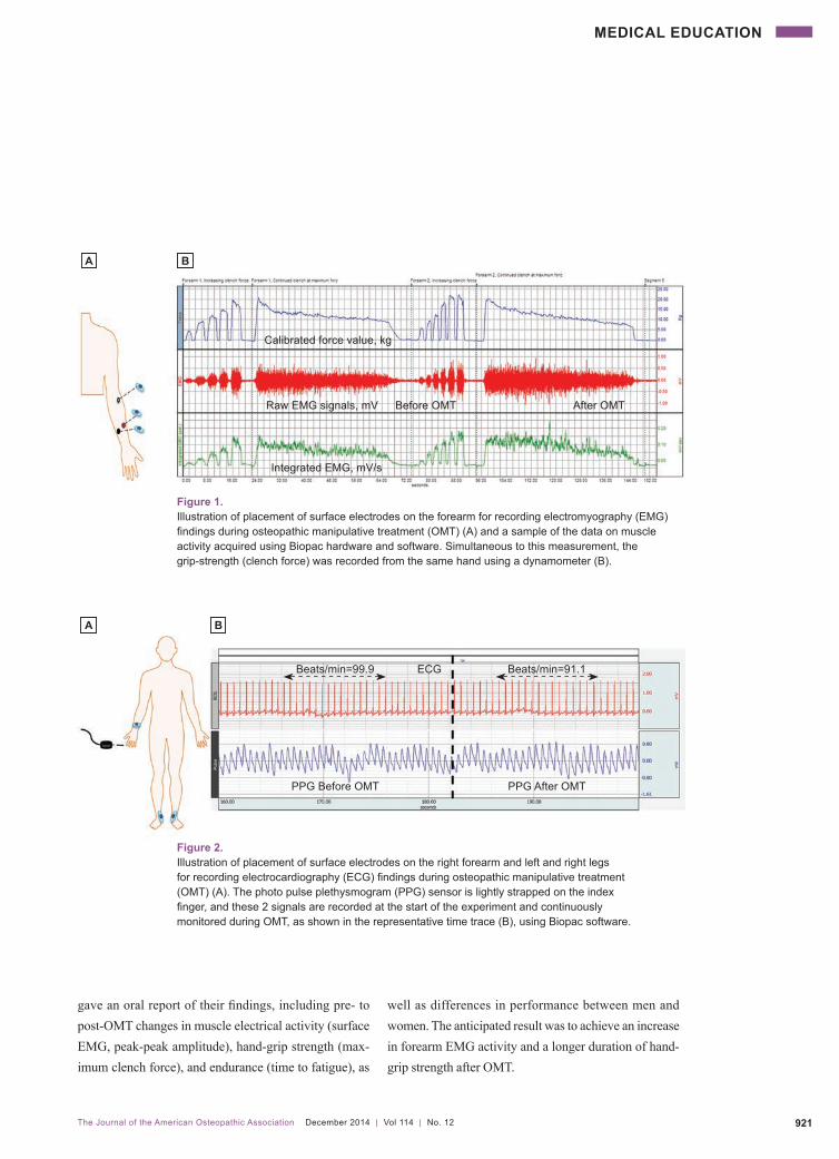
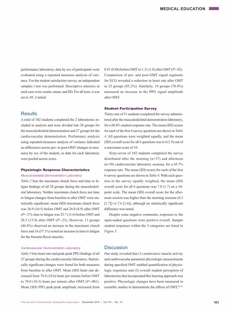
on their peers in small groups of 3 to 5 students. Within each group, students served as either the OMT operator, the test participant, or the computer operator. A multi-channel physiology education software system (Biopac, Inc) with various relevant sensors and a connected computer was used for physiologic measure-ments as schematically shown in Figure 1 and Figure 2. Similar to those described in other reports,19-21 laboratory instruction manuals were adapted to integrate specified OMT procedures with standard physiologic measure-ments. The stepwise instructional material for operating the instrumentation was intended to minimize com-plexity and to focus the students’ effort to analyze and interpret the experimental results. During these laboratories, students performed OMT and obtained measurements within 30-minute time slots at 5 identical test stations. Morning and afternoon labo-ratories accommodated the total class size of 102 stu-dents. Table 1 details the steps in a typical integrated OMT laboratory session.
Musculoskeletal Demonstration Laboratory
For the musculoskeletal demonstration laboratory, stu-dents recorded baseline and post-OMT measurements in real time using electromyography (EMG) for forearm muscle electrical activity and a clench force transducer for hand grip strength.22,23 In the laboratory, faculty guided the students in iden-tifying and treating specific forearm tender points using the strain-counterstrain OMT procedure.24 Surface elec-trodes were placed on the forearm of the test participant (Figure 1). Clench force was measured by using a dyna-mometer within the same set up. At the end of the session, students used the Biopac software to analyze post-OMT changes in the forearm of the test participant. For this signal, the peak force as well as the time to fatigue was assessed. Time to fatigue was defined as the time it took to reach 50% of the maximum force during a sustained clench of the dynamometer. Results were recorded on a worksheet, and each group
In the present study, 2 aspects of this integrated physi-ologic measurement-OPP model were assessed: (1) whether noninvasive physiologic measurement during specified OMT enabled quantification of physiologic responses and (2) whether student perception of the quality of the laboratory experience was positive.
Methods After institutional review board approval, 5 mandatory physiology demonstration laboratories were integrated into the first-year OPP curriculum at A.T. Still Univer-sity–School of Osteopathic Medicine in Arizona (Mesa) between 2010 and 2012. During the beginning of the academic year, the first laboratory sessions were used to fine-tune the teaching approach, logistics, allocation of appropriate student group size per station, and student feedback forms. In the present study, we analyzed data from 2 laboratory sessions in 2012 that occurred later in the academic year, during which the instruction sessions could be successfully completed and all information recorded. To evaluate the success of these laboratories, we (1) compared student-recorded pre- and post-OMT physio-logic measurements to assess whether noninvasive physiologic measurement during OMT enabled quantifi-cation of physiologic responses and (2) administered a survey to students after completion of the new laborato-ries to assess student perception of the quality of the laboratory experience.
Physiologic Demonstration Laboratories
The first laboratory solely introduced the students to basic physiologic signals and the link between effective OMT and expected changes in corresponding musculo-skeletal parameters (musculoskeletal demonstration) or cardiovascular parameters (cardiovascular demonstra-tion). The remaining 4 laboratories involved interactive teaching exercises—2 musculoskeletal and 2 cardiovas-cular—during which students practiced OMT procedures
MEDICAL EDUCATION
The Journal of the American Osteopathic Association December 2014 | Vol 114 | No. 12 921
gave an oral report of their findings, including pre- to post-OMT changes in muscle electrical activity (surface EMG, peak-peak amplitude), hand-grip strength (max-imum clench force), and endurance (time to fatigue), as
well as differences in performance between men and women. The anticipated result was to achieve an increase in forearm EMG activity and a longer duration of hand-grip strength after OMT.
Calibrated force value, kg
Raw EMG signals, mV
Integrated EMG, mV/s
Before OMT After OMT
A B
Figure 1.Illustration of placement of surface electrodes on the forearm for recording electromyography (EMG) findings during osteopathic manipulative treatment (OMT) (A) and a sample of the data on muscle activity acquired using Biopac hardware and software. Simultaneous to this measurement, the grip-strength (clench force) was recorded from the same hand using a dynamometer (B).
A B
Beats/min=99.9 ECG Beats/min=91.1
PPG Before OMT PPG After OMT
Figure 2.Illustration of placement of surface electrodes on the right forearm and left and right legs for recording electrocardiography (ECG) findings during osteopathic manipulative treatment (OMT) (A). The photo pulse plethysmogram (PPG) sensor is lightly strapped on the index finger, and these 2 signals are recorded at the start of the experiment and continuously monitored during OMT, as shown in the representative time trace (B), using Biopac software.
MEDICAL EDUCATION
The Journal of the American Osteopathic Association December 2014 | Vol 114 | No. 12922
student population, not all groups had a student with a high heart rate. Sensors for noninvasive, real-time re-cording of ECG and PPG findings were placed on the extremities (Figure 2). After baseline data were acquired from the test participant, under instructor guidance, stu-dents performed indirect balanced ligamentous tension to the occipitoatlantal and atlantoaxial joints.26 Contin-uous cardiovascular parameters were recorded, with time markers used to indicate when OMT occurred. The real-time tracing coinciding with OMT provided visual feed-back for ideal hand placement and application of force to achieve desired outcomes. Once the OMT session was completed, the data for pre- to post-OMT changes in heart rate (beats per minute) and digital blood volume (PPG signal; peak-peak amplitude) were downloaded for analysis. Expected responses included a reduction in heart rate and an increase in peripheral vascular flow.
Student Participation Survey
At the end of the sessions, students were administered a 9-item survey that asked them to rate the following items using a 10-point Likert scale, with 1 indicating “poor” and 10 indicating “excellent”: presentation and instruc-tional materials, time allowed to practice, amount of provided material, complexity of the material, organiza-tion of the laboratory, and faculty. In addition, students were asked to provide comments regarding the strong points of laboratory, the clinical applicability of the labo-ratory, and suggestions to improve the laboratory. The survey was administered during the afternoon musculo-skeletal laboratory session and during the morning and afternoon cardiovascular laboratory sessions.
Data Collection and Statistical Analysis
Data were obtained from student laboratory worksheets and student satisfaction forms. Statistical comparison (using SPSS statistical software, version 22) of the pre- and post-OMT musculoskeletal and cardiovascular pa-rameters for the 2 different laboratories was conducted using dependent samples t tests. For the musculoskeletal
Cardiovascular Demonstration Laboratory
During the cardiovascular demonstration laboratory, pre- and post-OMT measurements for heart rate from electro-cardiography (ECG) findings and peripheral vascular flow from photo pulse plethysmograph (PPG) signals27 were recorded. In this laboratory, students with a high heart rate, preferably greater than 80 beats per minute, were se-lected to be the test participant in their group when pos-sible. Because of the relatively young and healthy
Table 1. Activity Sequence of a Typical Physiologic Demonstration Laboratory Completed by First-Year Osteopathic Medical Students
Step Allotted Time Activity
1 40 min The OPP instructor describes the learning objectives, relevant anatomy, and physiologic mechanisms related to OMT procedures.
2 20 min The OPP instructor describes relevant diagnosis and OMT procedures.
3 20 min The OPP instructor reviews a handout of step-by-step instructions, describes the physiologic measurements that will be taken, and demonstrates how to operate the instrument and collect data.
4 30 to 40 min Students break into 5 groups (3 to 5 students per group) and perform the OMT exercise, collect physiologic measurements, perform data analysis, and complete a worksheet.
5 Repeat testing with next student batch.
6 10 min Students give oral presentations on their findings and clinical indications to the large group.
7 20 to 30 min Data from all groups are displayed, and the OPP instructor leads a discussion on clinical relevance, physiologic mechanisms, and possible research design and methodologies.
Abbreviations: OMT, osteopathic manipulative treatment; OPP, osteopathic principles and practice.
MEDICAL EDUCATION
The Journal of the American Osteopathic Association December 2014 | Vol 114 | No. 12 923
0.91 (0.86) before OMT to 1.11 (1.0) after OMT (P=.02). Comparison of pre- and post-OMT signal segments for ECG revealed a reduction in heart rate after OMT in 23 groups (85.2%). Similarly, 19 groups (70.4%) measured an increase in the PPG signal amplitude after OMT.
Student Participation Survey
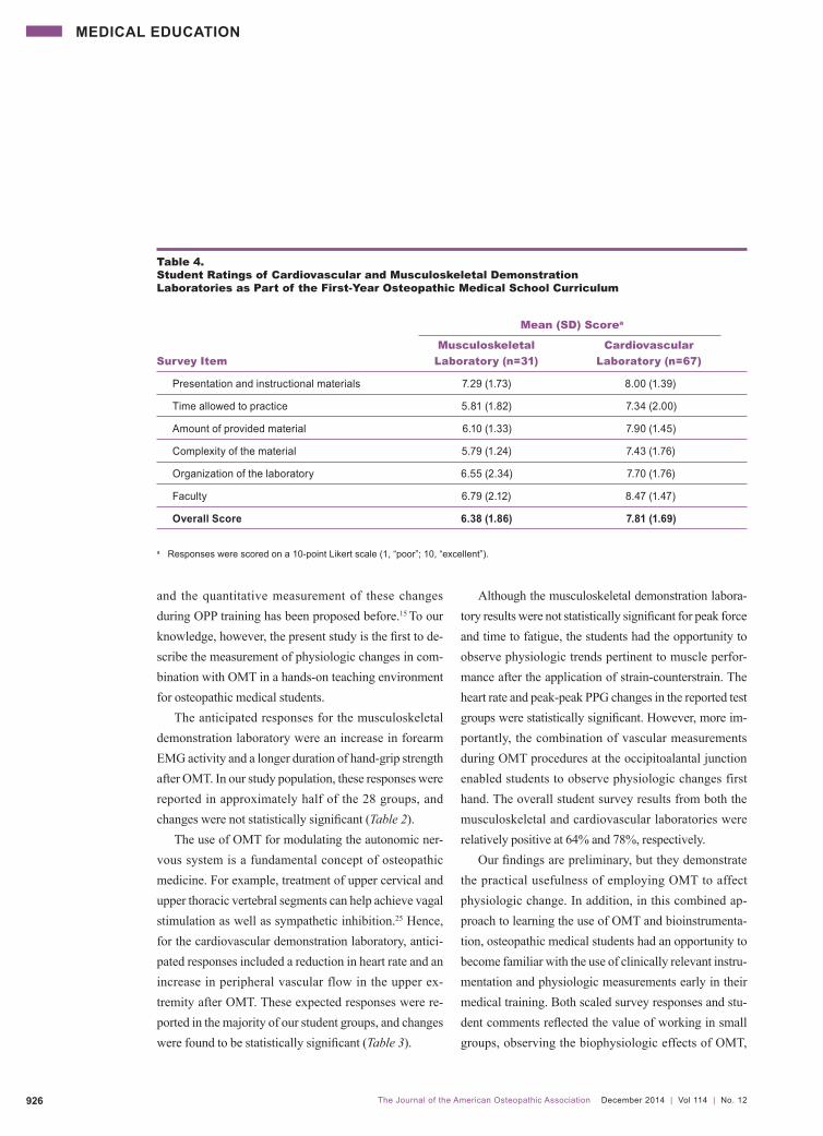
Thirty-one of 51 students completed the survey adminis-tered after the musculoskeletal demonstration laboratory, for a 60.8% student response rate. The mean (SD) scores for each of the first 6 survey questions are shown in Table 4. All questions were weighted equally, and the mean (SD) overall score for all 6 questions was 6.4 (1.9) out of a maximum score of 10. Sixty-seven of 102 students completed the survey distributed after the morning (n=37) and afternoon (n=30) cardiovascular laboratory sessions, for a 65.7% response rate. The mean (SD) scores for each of the first 6 survey questions are shown in Table 4. With each ques-tion in the survey equally weighted, the mean (SD) overall score for all 6 questions was 7.8 (1.7) on a 10-point scale. The mean (SD) overall score for the after-noon session was higher than the morning sessions (8.2 [1.7]) vs 7.6 [1.6]), although no statistically significant difference was noted. Despite some negative comments, responses to the open-ended questions were positive overall. Sample student responses within the 3 categories are listed in Figure 3.
Discussion Our study revealed that (1) noninvasive muscle activity and cardiovascular parameter physiologic measurements during specified OMT enabled quantification of physio-logic responses and (2) overall student perception of laboratories that incorporated this learning approach was positive. Physiologic changes have been measured in scientific studies to demonstrate the effects of OMT,28-31
performance laboratory, data by sex of participants were evaluated using a repeated measures analysis of vari-ance. For the student satisfaction survey, an independent samples t test was performed. Descriptive statistics in each case were counts, mean, and SD. For all tests, α was set at .05, 2-tailed.
Results A total of 102 students completed the 2 laboratories in-cluded in analysis and were divided into 28 groups for the musculoskeletal demonstration and 27 groups for the cardiovascular demonstration. Preliminary analysis using repeated-measures analysis of variance indicated no differences across pre- to post-OMT changes in mea-sures by sex of the student, so data for each laboratory were pooled across sexes.
Physiologic Response Characteristics
Musculoskeletal Demonstration Laboratory
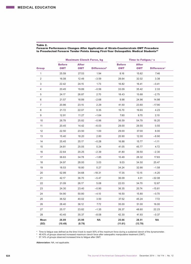
Table 2 lists the maximum clench force and time to fa-tigue findings of all 28 groups during the musculoskel-etal laboratory. Neither maximum clench force nor time to fatigue changes from baseline to after OMT were sta-tistically significant: mean (SD) maximum clench force was 26.9 (10.5) before OMT and 26.0 (8.9) after OMT (P=.37); time to fatigue was 25.7 (11.6) before OMT and 28.5 (13.8) after OMT (P=.23). However, 13 groups (46.4%) observed an increase in the maximum clench force and 16 (57.1%) noted an increase in time to fatigue for the forearm flexor muscles.
Cardiovascular Demonstration Laboratory
Table 3 lists heart rate and peak-peak PPG findings of all 27 groups during the cardiovascular laboratory. Statisti-cally significant changes were found for both measures from baseline to after OMT. Mean (SD) heart rate de-creased from 76.8 (10.6) beats per minute before OMT to 70.0 (10.3) beats per minute after OMT (P<.001). Mean (SD) PPG peak-peak amplitude increased from
MEDICAL EDUCATION
The Journal of the American Osteopathic Association December 2014 | Vol 114 | No. 12924
Table 2. Forearm Performance Changes After Application of Strain-Counterstrain OMT Procedure to Preselected Forearm Tender Points Among First-Year Osteopathic Medical Students24
Maximum Clench Force, kg Time to Fatigue,a s
Before After Before After
Group OMT OMT Differenceb OMT OMT Differencec
1 25.59 27.53 1.94 8.16 15.62 7.46
2 16.08 12.49 –3.59 28.64 32.02 3.38
3 22.42 24.15 1.73 16.82 16.41 –0.41
4 20.45 19.89 –0.56 33.09 35.42 2.33
5 24.17 26.87 2.70 18.43 15.68 –2.75
6 21.57 18.89 –2.68 9.98 24.96 14.98
7 20.86 23.15 2.29 41.50 23.60 –17.90
8 21.72 22.07 0.35 15.70 19.93 4.23
9 12.91 11.27 –1.64 7.60 9.70 2.10
10 26.78 25.82 –0.96 36.59 54.79 18.20
11 20.45 14.42 –6.03 29.00 29.55 0.55
12 22.50 23.50 1.00 29.00 37.00 8.00
13 15.40 18.20 2.80 20.90 12.00 –8.90
14 25.45 25.17 –0.28 16.88 15.77 –1.11
15 24.81 25.05 0.24 41.05 45.77 4.72
16 22.64 20.25 –2.39 41.80 39.50 –2.30
17 36.63 34.78 –1.85 10.49 28.32 17.83
18 24.97 28.00 3.03 9.03 34.50 25.47
19 18.53 18.80 0.27 34.24 32.66 –1.58
20 52.99 34.68 –18.31 17.35 13.15 –4.20
21 42.17 36.70 –5.47 36.09 4.01 –32.08
22 21.09 26.17 5.08 22.03 34.70 12.67
23 24.30 23.40 –0.90 36.35 20.74 –15.61
24 54.90 50.80 –4.10 16.50 15.80 –0.70
25 36.52 40.02 3.50 37.52 45.24 7.72
26 28.40 36.12 7.72 35.00 51.00 16.00
27 25.17 23.82 –1.35 26.37 48.60 22.23
28 43.45 35.37 –8.08 42.30 41.93 –0.37
Mean 26.89 25.98 NA 25.66 28.51 NA (SD) (10.49) (8.79) (11.61) (13.79)
a Time to fatigue was defined as the time it took to reach 50% of the maximum force during a sustained clench of the dynamometer.b 46.43% of groups observed increased maximum clench force after osteopathic manipulative treatment (OMT).c 57.14% of groups observed increased time to fatigue after OMT.
Abbreviation: NA, not applicable.
MEDICAL EDUCATION
The Journal of the American Osteopathic Association December 2014 | Vol 114 | No. 12 925
Table 3. Heart Rate and Upper Limb Digital Blood Flow Changes After Application of Balanced Ligamentous Tension to Occipitoatlantal and Atlantoaxial Cervical Segments Among First-Year Osteopathic Medical Students26
Heart Rate, beats/min Digital Blood Flow, peak-peak amplitude, mV
Before After Before After
Group OMT OMT Differenceb OMT OMT Differencec
1 51.00 46.00 –5.00 0.49 0.64 0.15
2 63.49 68.18 4.69 0.16 0.19 0.04
3 74.22 60.84 –13.38 0.86 0.41 –0.45
4 83.00 75.00 –8.00 0.54 1.90 1.36
5 76.60 64.90 –11.70 1.66 1.92 0.26
6 71.43 68.18 –3.25 1.02 0.95 –0.07
7 78.00 71.00 –7.00 0.38 0.53 0.16
8 74.30 70.18 –4.12 1.57 2.95 1.38
9 69.80 70.60 0.80 0.13 0.13 –0.01
10 76.92 69.77 –7.15 2.24 2.02 –0.22
11 74.08 66.30 –7.78 2.33 2.29 –0.04
12 94.50 88.00 –6.50 1.61 1.47 –0.14
13 94.00 69.42 –24.58 0.17 0.30 0.13
14 68.18 54.05 –14.13 1.84 1.99 0.15
15 89.00 81.00 –8.00 0.55 0.89 0.34
16 63.98 67.98 4.00 0.21 0.66 0.45
17 89.85 78.08 –11.77 0.50 0.26 –0.24
18 82.00 81.60 –0.40 0.10 0.15 0.06
19 86.55 82.01 –4.55 0.58 0.91 0.33
20 93.48 90.80 –2.68 0.48 0.86 0.38
21 72.29 67.39 –4.90 3.67 4.41 0.75
22 80.41 65.72 –14.69 0.40 0.46 0.06
23 86.54 82.01 –4.53 0.58 0.91 0.33
24 66.60 55.19 –11.41 1.10 1.18 0.08
25 71.70 58.90 –12.80 0.21 0.37 0.16
26 67.67 70.63 2.97 1.00 0.94 –0.06
27 74.46 67.00 –7.46 0.30 0.34 0.03
Mean 76.82 70.03 NA 0.91 1.11 NA (SD) (10.55) (10.26) (0.86) (0.99)
a 85.19% of groups observed a decrease in heart rate after osteopathic manipulative treatment (OMT). b 70.37% of groups observed an increase in digital blood flow after OMT.
Abbreviation: NA, not applicable.
MEDICAL EDUCATION
The Journal of the American Osteopathic Association December 2014 | Vol 114 | No. 12926
Although the musculoskeletal demonstration labora-tory results were not statistically significant for peak force and time to fatigue, the students had the opportunity to observe physiologic trends pertinent to muscle perfor-mance after the application of strain-counterstrain. The heart rate and peak-peak PPG changes in the reported test groups were statistically significant. However, more im-portantly, the combination of vascular measurements during OMT procedures at the occipitoalantal junction enabled students to observe physiologic changes first hand. The overall student survey results from both the musculoskeletal and cardiovascular laboratories were relatively positive at 64% and 78%, respectively. Our findings are preliminary, but they demonstrate the practical usefulness of employing OMT to affect physiologic change. In addition, in this combined ap-proach to learning the use of OMT and bioinstrumenta-tion, osteopathic medical students had an opportunity to become familiar with the use of clinically relevant instru-mentation and physiologic measurements early in their medical training. Both scaled survey responses and stu-dent comments reflected the value of working in small groups, observing the biophysiologic effects of OMT,
and the quantitative measurement of these changes during OPP training has been proposed before.15 To our knowledge, however, the present study is the first to de-scribe the measurement of physiologic changes in com-bination with OMT in a hands-on teaching environment for osteopathic medical students. The anticipated responses for the musculoskeletal demonstration laboratory were an increase in forearm EMG activity and a longer duration of hand-grip strength after OMT. In our study population, these responses were reported in approximately half of the 28 groups, and changes were not statistically significant (Table 2). The use of OMT for modulating the autonomic ner-vous system is a fundamental concept of osteopathic medicine. For example, treatment of upper cervical and upper thoracic vertebral segments can help achieve vagal stimulation as well as sympathetic inhibition.25 Hence, for the cardiovascular demonstration laboratory, antici-pated responses included a reduction in heart rate and an increase in peripheral vascular flow in the upper ex-tremity after OMT. These expected responses were re-ported in the majority of our student groups, and changes were found to be statistically significant (Table 3).
Table 4. Student Ratings of Cardiovascular and Musculoskeletal Demonstration Laboratories as Part of the First-Year Osteopathic Medical School Curriculum
Mean (SD) Scorea
Musculoskeletal Cardiovascular
Survey Item Laboratory (n=31) Laboratory (n=67)
Presentation and instructional materials 7.29 (1.73) 8.00 (1.39)
Time allowed to practice 5.81 (1.82) 7.34 (2.00)
Amount of provided material 6.10 (1.33) 7.90 (1.45)
Complexity of the material 5.79 (1.24) 7.43 (1.76)
Organization of the laboratory 6.55 (2.34) 7.70 (1.76)
Faculty 6.79 (2.12) 8.47 (1.47)
Overall Score 6.38 (1.86) 7.81 (1.69)
a Responses were scored on a 10-point Likert scale (1, “poor”; 10, “excellent”).
MEDICAL EDUCATION
The Journal of the American Osteopathic Association December 2014 | Vol 114 | No. 12 927
Despite differences in students’ familiarity with in-strumentation and computer skill level, most students are able to successfully complete the laboratories using the Biopac software. The laboratories are time-limited and must be run efficiently to ensure that all groups finish their OMT exercises, collect and report their data, and allow sufficient time for large-group discussion. We have found that for the optimum laboratory experience, all supervising faculty and fellows must be trained in using the equipment, troubleshooting technical glitches, and carefully guiding the students’ use of OMT. In the large-group discussions, instructors emphasize that the observed findings illustrate the relationship of OPP with physiologic responses. The students are re-minded that these demonstration laboratories are not in-tended as research studies because they lack a detailed, formal study design and proper controls. However, the laboratories do introduce students to basic research ques-tions and provide an opportunity for faculty to discuss double-blind, randomized control designs, as well as the role of placebos and sham procedures, in osteopathic clinical research. After completing the demonstration laboratories, a few students have been motivated to design and seek funding for their own clinical research studies. The current study has a number of limitations, weak-nesses, and confounding factors. First, it included a small number of physiologic performance data sets from EMG and cardiovascular measurements after OMT. A greater number of student data sets must be analyzed before de-finitive conclusions can be made about the efficacy of particular OMT procedures in producing the corre-sponding physiologic output. In addition, more student feedback is needed to fully understand the value of com-bining OMT with physiologic signal monitoring. Con-founding factors include students’ ability to operate computer-based instrumentation, perform OMT, and un-derstand certain physiologic concepts, as well as the lack of a controlled scientific experimental environment. De-spite these limitations, the success of the present combined approach for training first-year osteopathic medical stu-
and optimizing OMT procedures (Table 4 and Figure 3). No differences in survey responses were observed be-tween the morning and afternoon sessions, indicating no change in instructor readiness or student familiarity with the steps as the day progressed. During the implementation stage of the current inte-grative teaching approach, workflow in the laboratories evolved, and the worksheets and expectations from the students were revised. Instructions were simplified, and student group sizes were reduced. Initially, the learning groups were 10 students per station, which was deter-mined to be too large for optimum participation. Students were also not required to formally report their acquired data, explain their results, or submit their laboratory records. Students are now expected to scientifically inter-pret their outcomes and submit a worksheet. The current 3- to 5-person group size ensures greater student partici-pation in the exercises. In addition, the present laboratory structure provides precise, easy-to-follow instructions for operating instrumentation, acquiring data accurately, and analyzing post-OMT data efficiently. Since completion of the current study, A.T. Still Uni-versity–School of Osteopathic Medicine in Arizona has continued to include 5 physiologic demonstrations as part of its 25 mandatory first-year OPP laboratory sessions. The sessions are timed to integrate within curriculum content and are designed to be appropriate for the student skill level. For example, the musculoskeletal demonstra-tion laboratory coincides with the neuromusculoskeletal coursework that covers upper extremity muscle anatomy and physiology, and the cardiovascular demonstration laboratory coincides with coursework on the cardiovas-cular system and the basics of ECG. In addition, the OMT procedures selected for the laboratories are safe and ap-propriate for a novice level. In contrast to the complex designs of detailed research protocols from studies related to recording musculoskeletal and cardiovascular function with OMT,28-31 the laboratories described in the present study are simple, noninvasive, and easy to implement into first-year osteopathic medical student training.
MEDICAL EDUCATION
The Journal of the American Osteopathic Association December 2014 | Vol 114 | No. 12928
ConclusionResults from 2 laboratory exercises performed by osteopathic medical students during the first-year cur-riculum demonstrated an increase in musculoskeletal parameters in response to strain-counterstrain proce-dures in approximately half of student groups and im-proved cardiovascular parameters from occipitoatlantal decompression in the majority of student groups. These findings, combined with positive student feedback, indicate that these combined laboratories are useful in demonstrating the clinical significance of OMT to first-year osteopathic medical students.
AcknowledgmentWe thank Professor Curtis Bay, PhD, for performing the statistical analysis for the present study.
Author ContributionsAll authors provided substantial contributions to conception and design, acquisition of data, or analysis and interpretation of data; Dr Heath, Dr Makin, and Ms Pedapati drafted the article or revised it critically for important intellectual content; and all authors gave final approval of the version of the article to be published.
References1. Seffinger MA, King HH, Ward RC, Jones JM, Rogers FJ,
Patterson MM. Osteopathic philosophy. In: Chila A, executive ed. Foundations of Osteopathic Medicine. 3rd ed. Baltimore, MD: Lippincott Williams & Wilkins; 2010:3.
2. Johnson SM, Kurtz ME. Diminished use of osteopathic manipulative treatment and its impact on the uniqueness of the osteopathic profession. Acad Med. 2001;76(8):821-828.
3. Johnson SM, Kurtz ME, Kurtz JC. Variables influencing the use of osteopathic manipulative treatment in family practice. J Am Osteopath Assoc. 1997;97(2):80-87.
4. Gamber RG, Gish EE, Herron KM. Student perceptions of osteopathic manipulative treatment after completing a manipulative medicine rotation. J Am Osteopath Assoc. 2001;101(7):395-400.
5. Allee BA, Pollack MH, Malnar KF. Survey of osteopathic and allopathic residents’ attitudes toward osteopathic manipulative treatment. J Am Osteopath Assoc. 2005;105(12):551-561.
6. Stovall BA, Bae S, Kumar S. Anterior superior iliac spine asymmetry assessment on a novel pelvic model: an investigation of accuracy and reliability. J Manipulative Physiol Ther. 2010;33(5):378-385. doi:10.1016/j.jmpt.2010.05.005.
dents is promising. The aforementioned limitations will be addressed in future laboratories by further simplifying in-structions and obtaining more student feedback. In addi-tion, future research is needed to correlate students’ OMT skill level to measureable physiologic responses as they acquire further training. Future studies are also needed to investigate whether integrated OPP training affects stu-dents’ readiness to use OMT in clinical practice.
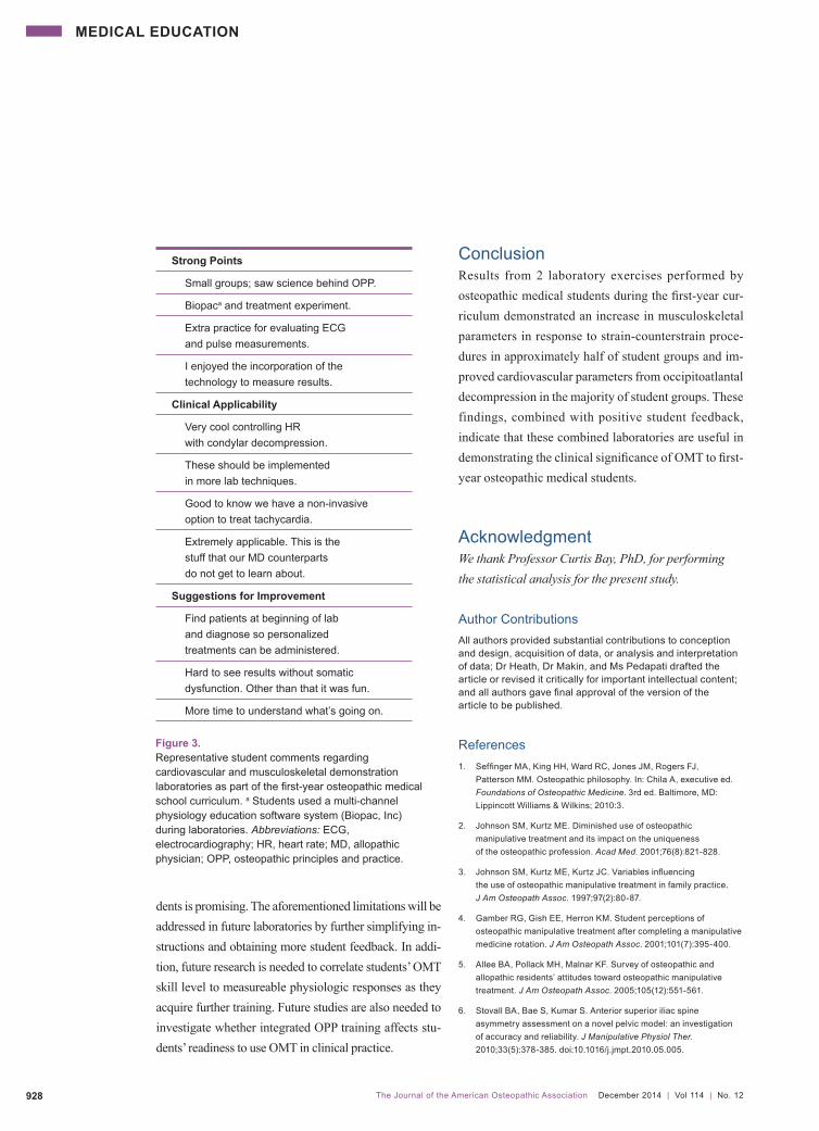
Figure 3.Representative student comments regarding cardiovascular and musculoskeletal demonstration laboratories as part of the first-year osteopathic medical school curriculum. a Students used a multi-channel physiology education software system (Biopac, Inc) during laboratories. Abbreviations: ECG, electrocardiography; HR, heart rate; MD, allopathic physician; OPP, osteopathic principles and practice.
Strong Points
Small groups; saw science behind OPP.
Biopaca and treatment experiment.
Extra practice for evaluating ECG and pulse measurements.
I enjoyed the incorporation of the technology to measure results.
Clinical Applicability
Very cool controlling HR with condylar decompression.
These should be implemented in more lab techniques.
Good to know we have a non-invasive option to treat tachycardia.
Extremely applicable. This is the stuff that our MD counterparts do not get to learn about.
Suggestions for Improvement
Find patients at beginning of lab and diagnose so personalized treatments can be administered.
Hard to see results without somatic dysfunction. Other than that it was fun.
More time to understand what’s going on.
MEDICAL EDUCATION
The Journal of the American Osteopathic Association December 2014 | Vol 114 | No. 12 929
23. Suzuki M, Yamazaki Y, Matsunami K. Relationship between force and electromyographic activity during rapid isometric contraction in power grip. Electorencephal Clin Neurophysiol. 1994;93(3):218-224.
25. Beal MC. Palpatory testing for somatic dysfunction in patients with cardiovascular disease. J Am Osteopath Assoc. 1983:(11)82:822-831.
26. Kimberly PE. Outline of Osteopathic Manipulative Procedures: The Kimberly Manual. Marceline, MO: Walsworth Publishing Company; 2006:72-77.
27. Allen J. Photoplethysmography and its application in clinical physiological measurement [published online February 20, 2007]. Physiol Meas. 2007;28(3):R1-R39.
28. Richards DG, McMillin DL, Mein EA, Nelson CD. Osteopathic regulation of physiology. Am Acad Osteopath J. 2001;11(3):34-38.
29. Purdy WR, Frank JJ, Oliver B. Suboccipital dermatomyotomic stimulation and digital blood flow. J Am Osteopath Assoc. 1996;96(5):285-289.
30. Hoyt WH, Shaffer F, Bard DA, et al. Osteopathic manipulation in the treatment of muscle-contraction headache. J Am Osteopath Assoc. 1979;78(5):322-325.
31. Fryer G, Bird M, Robbins B, Fossum C, Johnson JC. Resting electromyographic activity of deep thoracic transversospinalis muscles identified as abnormal with palpation. J Am Osteopath Assoc. 2010;110(2):61-68.
7. Fossum C, Snider E, Fryer G, Gillette N, Degenhardt B. The introduction of a novel approach to the teaching and assessment of osteopathic manipulative medicine assessment skills. Int J Osteopath Med. 2008;11(4):149-168.
8. Rajendran D, Gallagher D. The assessment of pelvic landmarks using palpation: a reliability study of undergraduate students. Int J Osteopath Med. 2011;14(2):57-60.
9. Kmita A, Lucas NP. Reliability of physical examination to assess asymmetry of anatomical landmarks indicative of pelvic somatic dysfunction in subjects with and without low back pain. Int J Osteopath Med. 2008;11(1):16-25.
10. Howell JN, Conatser RR, Williams RL II, Burns JM, Eland DC. Palpatory diagnosis training on the virtual haptic back: performance improvement and user evaluations. J Am Osteopath Assoc. 2008;108(1):29-36.
11. Chamberlain NR, Yates HA. Use of computer-assisted clinical case (CACC) SOAP note exercise to assess students’ application of osteopathic principles and practice. J Am Osteopath Assoc. 2000;100(7):437-440.
12. Chamberlain NR, Yates HA. A prospective study of osteopathic medical students’ attitudes toward use of osteopathic manipulative treatment in caring for patients. J Am Osteopath Assoc. 2003;103(10):470-478.
13. Teng AY, Terry RR, Blue RJ. Incorporating a mandatory osteopathic manipulative medicine (OMM) curriculum in clinical clerkships: impact on student attitudes toward using OMM. J Am Osteopath Assoc. 2011;111(4):219-224.
14. Steele KM, Baker HH. Clearly distinct: outcomes of initiative to increase integration of osteopathic principles and practice at West Virginia School of Osteopathic Medicine. J Am Osteopath Assoc. 2009;109(11):579-590.
15. Gevitz N. Center or periphery? the future of osteopathic principles and practices. J Am Osteopath Assoc. 2006;106(3):121-129.
16. Denslow JS. Pathophysiologic evidence for the osteopathic lesion: the known, unknown, and controversial. J Am Osteopath Assoc. 1975;75(4):415-421.
17. Denslow JS. Neural basis of the somatic component in health and disease and its clinical management. J Am Osteopath Assoc. 1972;72(2):149-156.
18. Peterson B, ed. The Collected Papers of Irvin M. Korr. Colorado Springs, CO: American Academy of Osteopathy; 1979:77-89.
19. Carvalho H, West CA. Voluntary participation in an active learning exercise leads to a better understanding of physiology. Advan Physiol Educ. 2010;35(1):53-58. doi:10.1152/advan.00011.2010.
20. Heilman KJ, Porges SW. Accuracy of the LifeShirt (Vivometrics) in the detection of cardiac rhythms [published online April 29, 2007]. Biol Psychol. 2007;75(3):300-305.
21. Cotter PA, Rodnick KJ. Fishing for an ECG: a student- directed electrocardiographic laboratory using rainbow trout. Adv Physiol Educ. 2007;31(2):211-217.
22. Roman-Liu D, Tokarski T. EMG of arm and forearm muscle activities with regard to handgrip force in relation to upper limb location. Acta Bioeng Biomech. 2002;4(2):33-48.