44
Use of the Sicat Surgical Guide In Reconstruction of the Hemi-Mandilectomy Patient Dr. Michael J. Maginnis Dr. Glenn E. Appleton Prosthodontics and Family Dentistry www.drsmanda.com
| Date post: | 20-Aug-2018 |
| Category: |
Documents |
| Upload: | truongthien |
| View: | 215 times |
| Download: | 0 times |
Use of the Sicat Surgical Guide
In Reconstruction
of the
Hemi-Mandilectomy Patient
Dr. Michael J. Maginnis
Dr. Glenn E. Appleton
Prosthodontics and Family
Dentistry
www.drsmanda.com
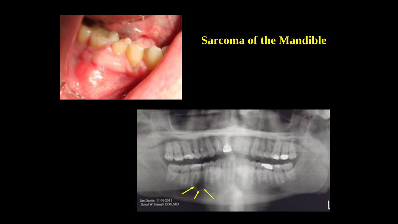
Sarcoma of the Mandible
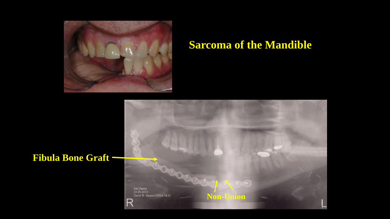
Sarcoma of the Mandible
Non-Union
Fibula Bone Graft
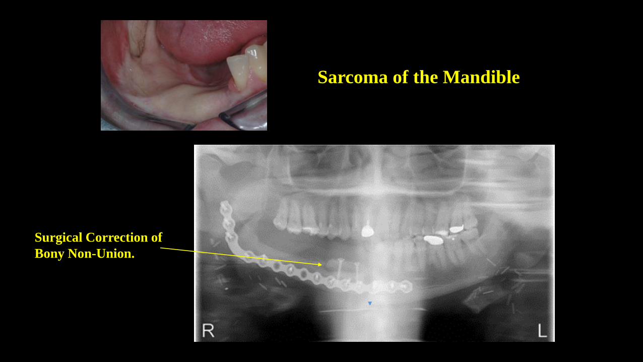
Sarcoma of the Mandible
Surgical Correction of
Bony Non-Union.
SS
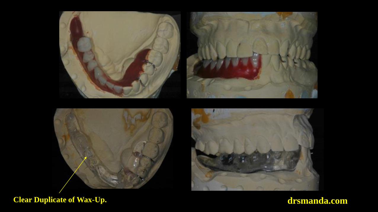
drsmanda.comClear Duplicate of Wax-Up.
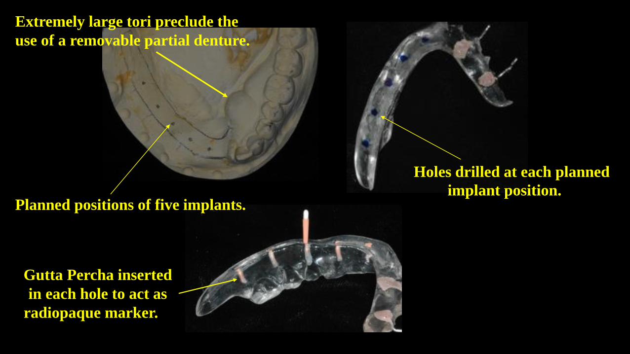
Planned positions of five implants.
Gutta Percha inserted
in each hole to act as
radiopaque marker.Gu
Holes drilled at each planned
implant position. H
Extremely large tori preclude the
use of a removable partial denture.
drsmanda.com
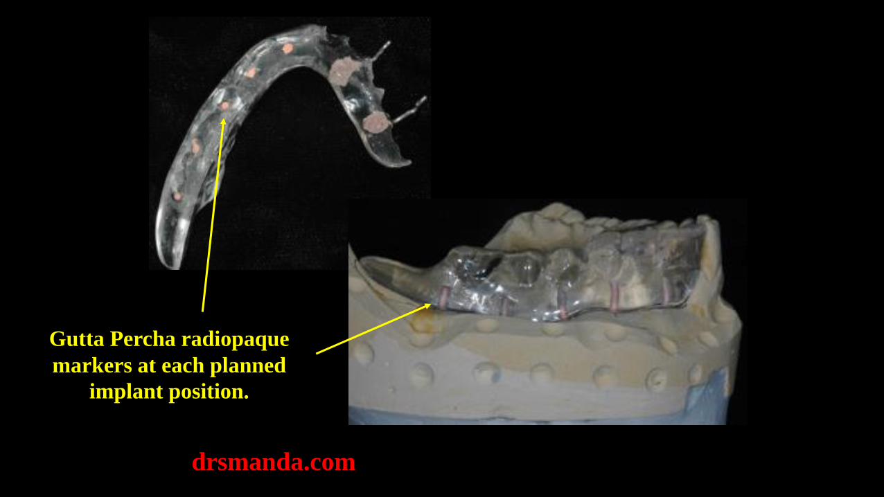
Gutta Percha radiopaque
markers at each planned
implant position.
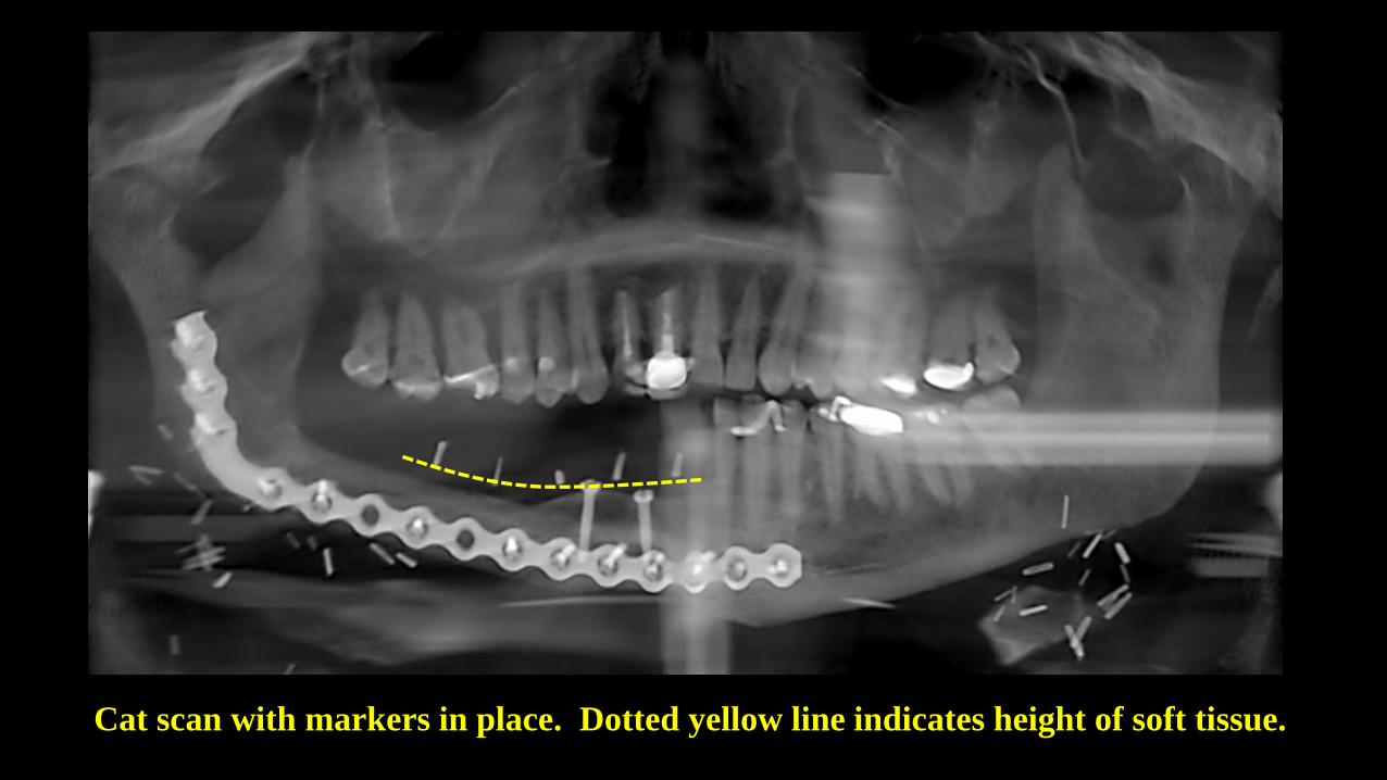
Cat scan with markers in place. Dotted yellow line indicates height of soft tissue.
The use of gutta percha points and a clear duplicate of the
preliminary wax-up is useful in assessing tissue thickness,
bone height and width, as well as surgical screw positions.
To construct a Sicat surgical guide, the manufacturer prefers
a radiographic template in the shape of the planned
prosthesis and made of radiopaque resin. The following
slides demonstrate construction of such a template.
www.drsmanda.com
Author’s Note:
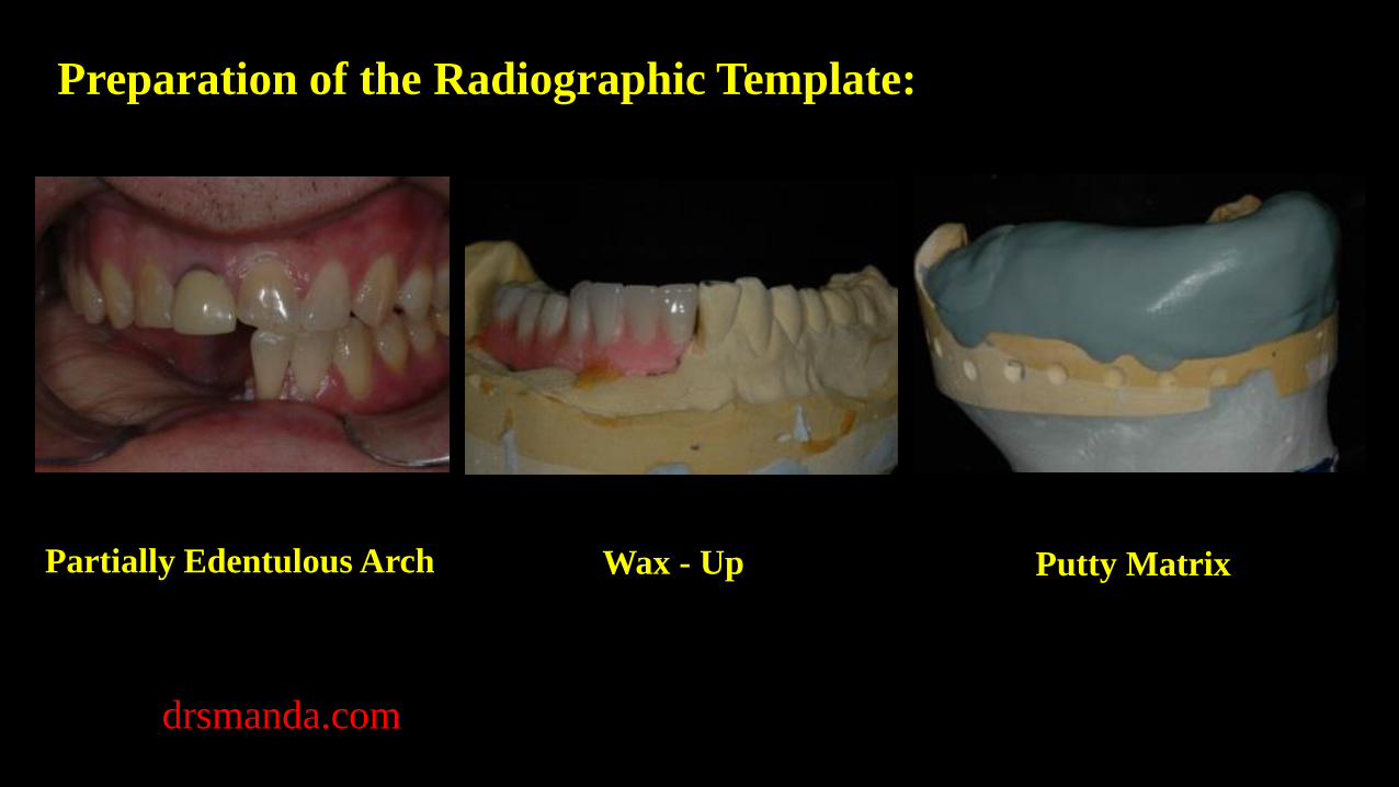
Preparation of the Radiographic Template:
Partially Edentulous Arch Wax - Up Putty Matrix
drsmanda.com
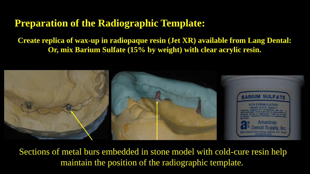
Preparation of the Radiographic Template:
Create replica of wax-up in radiopaque resin (Jet XR) available from Lang Dental:
Or, mix Barium Sulfate (15% by weight) with clear acrylic resin.
Sections of metal burs embedded in stone model with cold-cure resin help
maintain the position of the radiographic template.
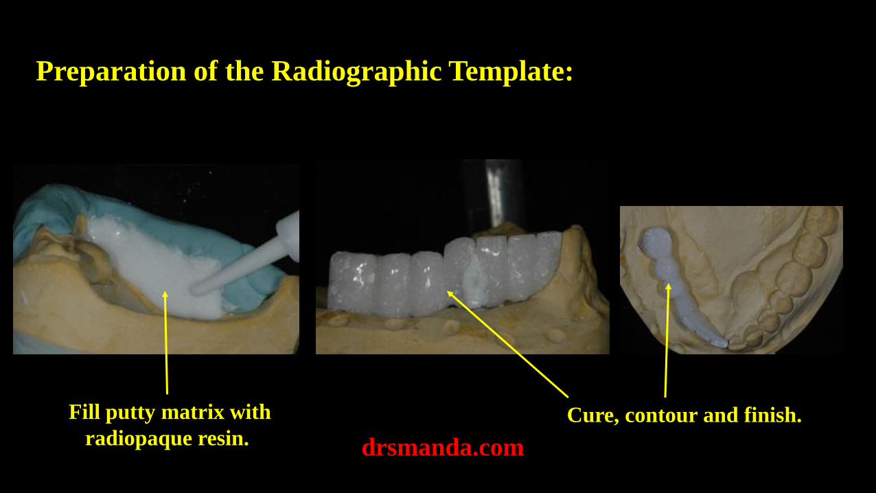
Preparation of the Radiographic Template:
drsmanda.com
Fill putty matrix with
radiopaque resin.Cure, contour and finish.
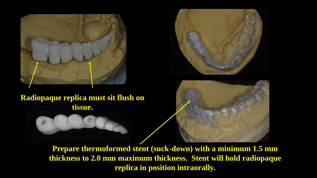
Prepare thermoformed stent (suck-down) with a minimum 1.5 mm
thickness to 2.0 mm maximum thickness. Stent will hold radiopaque
replica in position intraorally.
Radiopaque replica must sit flush on
tissue.
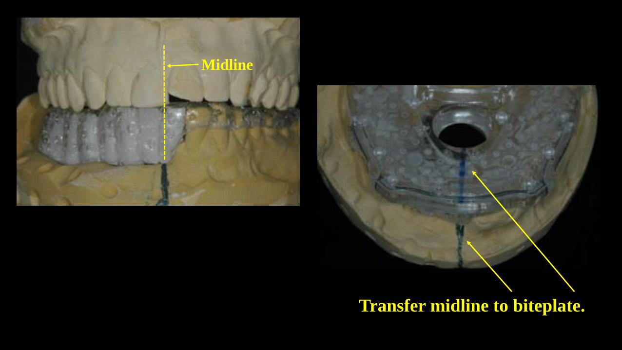
Midline
Transfer midline to biteplate.
drsmanda.com
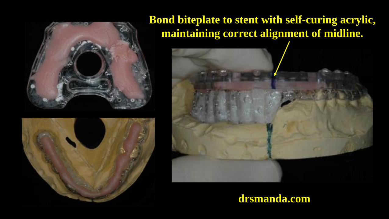
Bond biteplate to stent with self-curing acrylic,
maintaining correct alignment of midline.
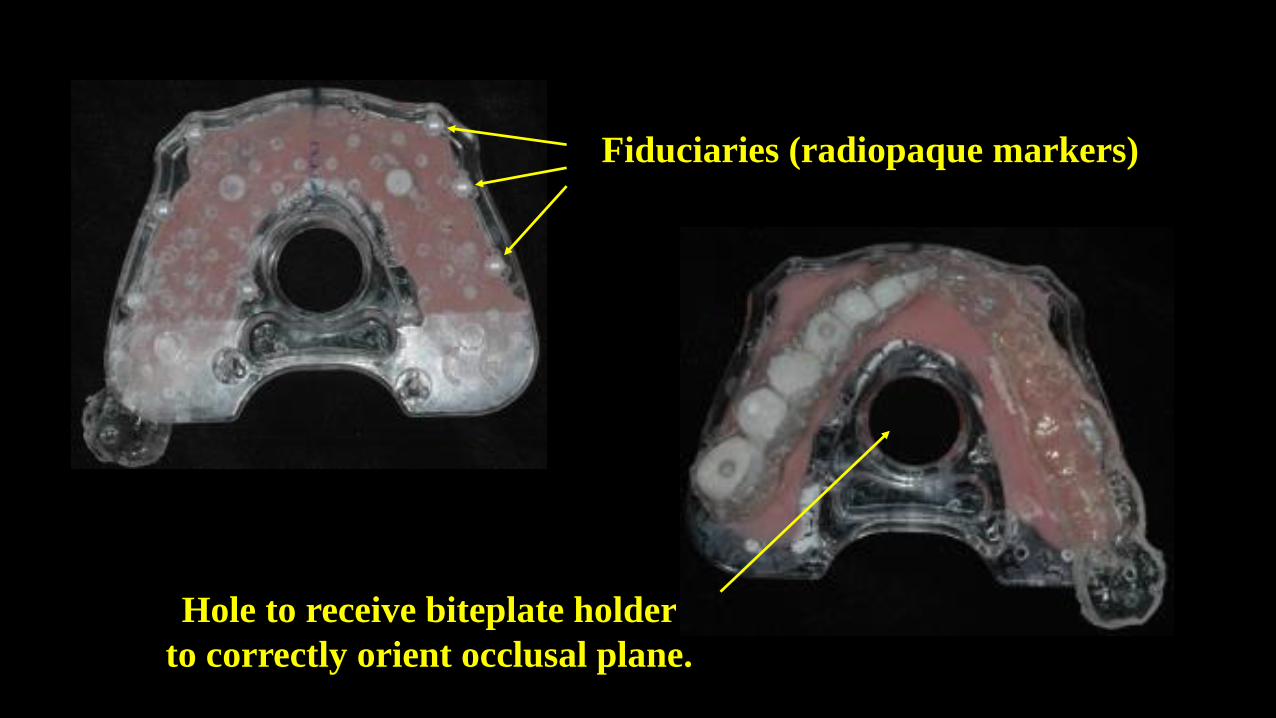
Fiduciaries (radiopaque markers)
Hole to receive biteplate holder
to correctly orient occlusal plane.
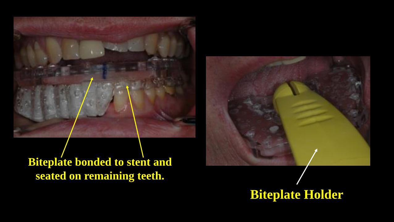
Biteplate Holder
Biteplate bonded to stent and
seated on remaining teeth.
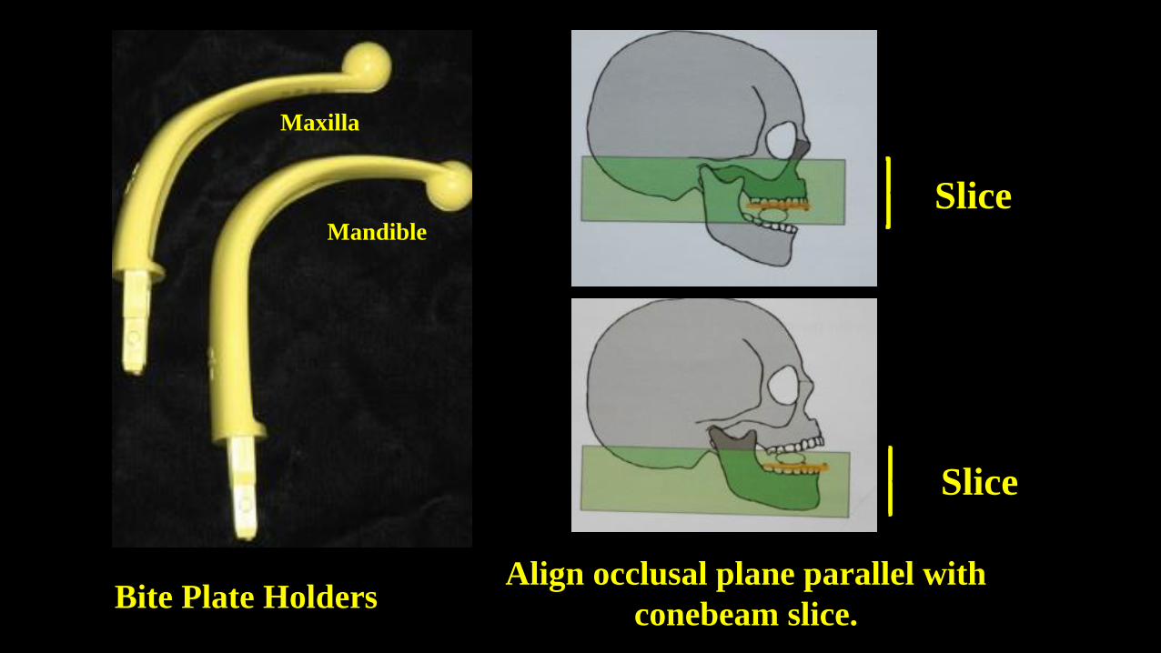
Bite Plate Holders
Maxilla
Mandible
Slice
Slice
Align occlusal plane parallel with
conebeam slice.

drsmanda.com
Biteplate holder attached to scanner to
properly orient occlusal plane with
conebeam slice.
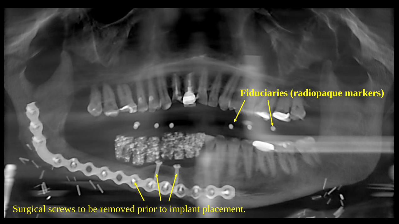
Fiduciaries (radiopaque markers)
Surgical screws to be removed prior to implant placement.
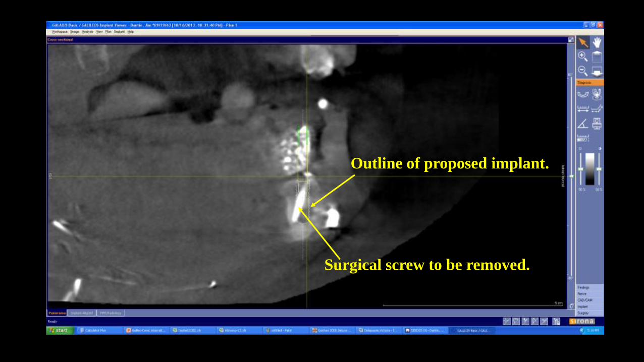
Outline of proposed implant.
Surgical screw to be removed.
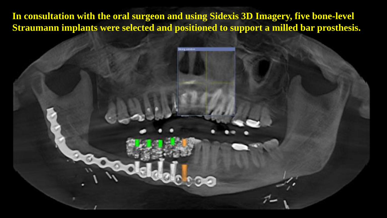
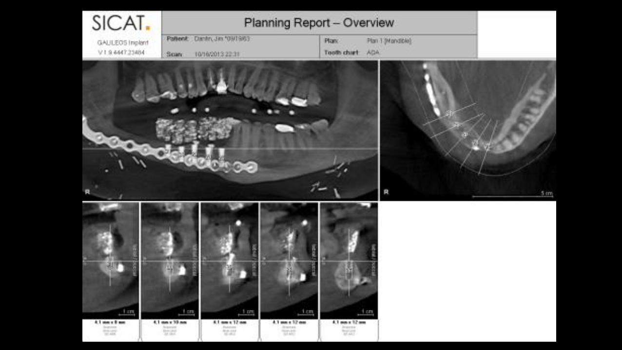
In consultation with the oral surgeon and using Sidexis 3D Imagery, five bone-level
Straumann implants were selected and positioned to support a milled bar prosthesis.
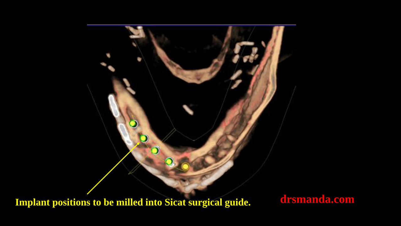
drsmanda.comImplant positions to be milled into Sicat surgical guide.
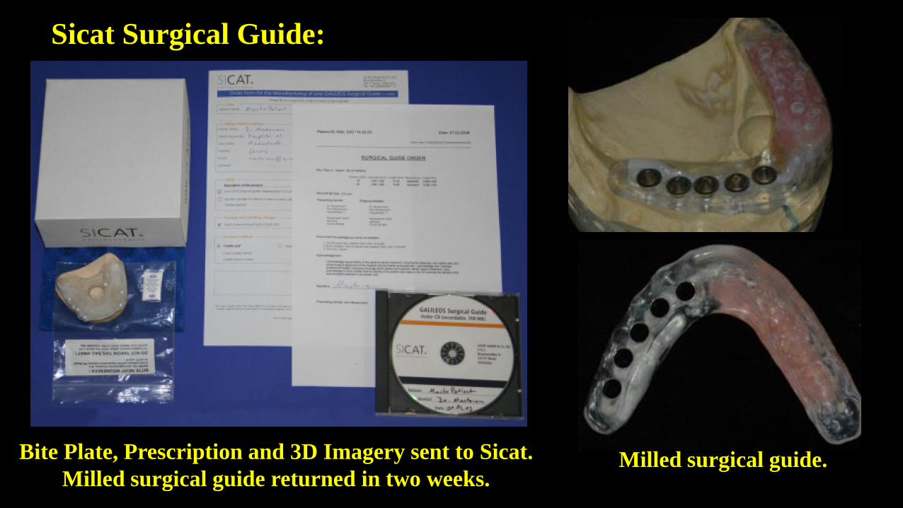
Sicat Surgical Guide:
Milled surgical guide.Bite Plate, Prescription and 3D Imagery sent to Sicat.
Milled surgical guide returned in two weeks.
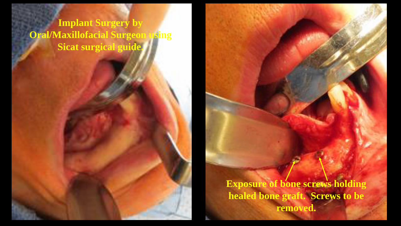
Implant Surgery by
Oral/Maxillofacial Surgeon using
Sicat surgical guide.
Exposure of bone screws holding
healed bone graft. Screws to be
removed.

Surgical placement of five Straumann bone-level implants using Sicat Surgical Guide
Sicat Surgical Guide
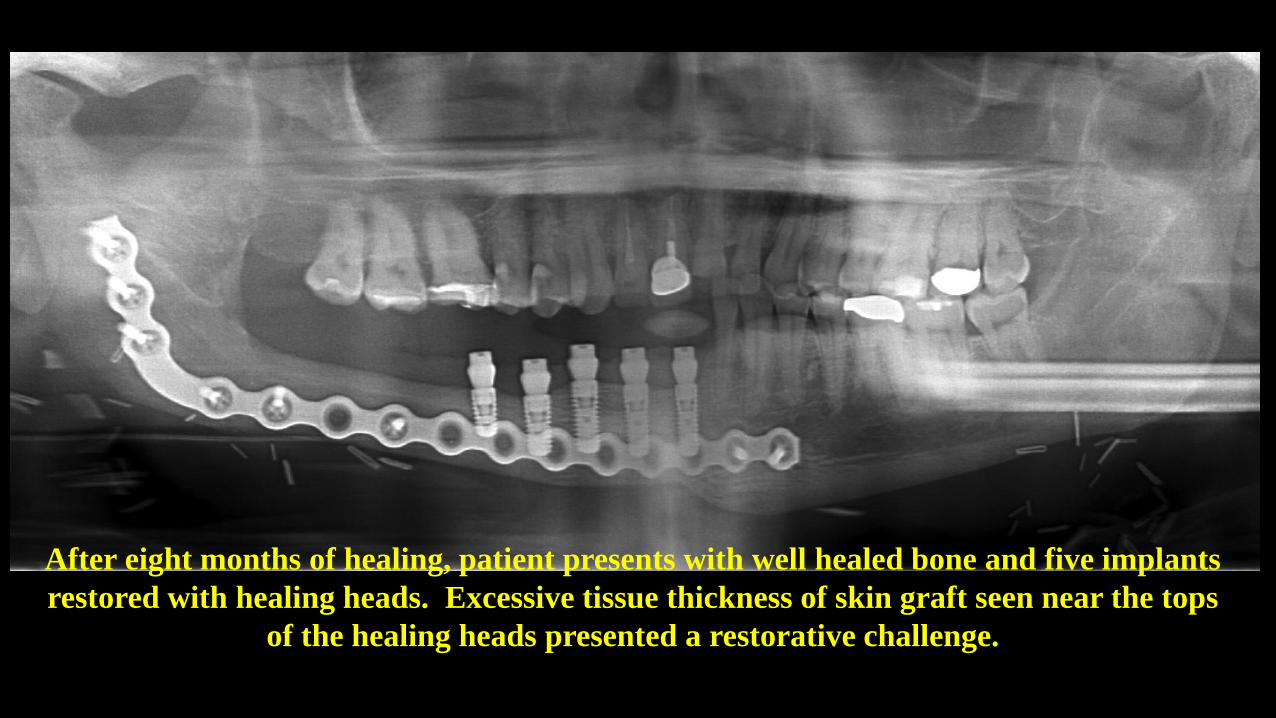
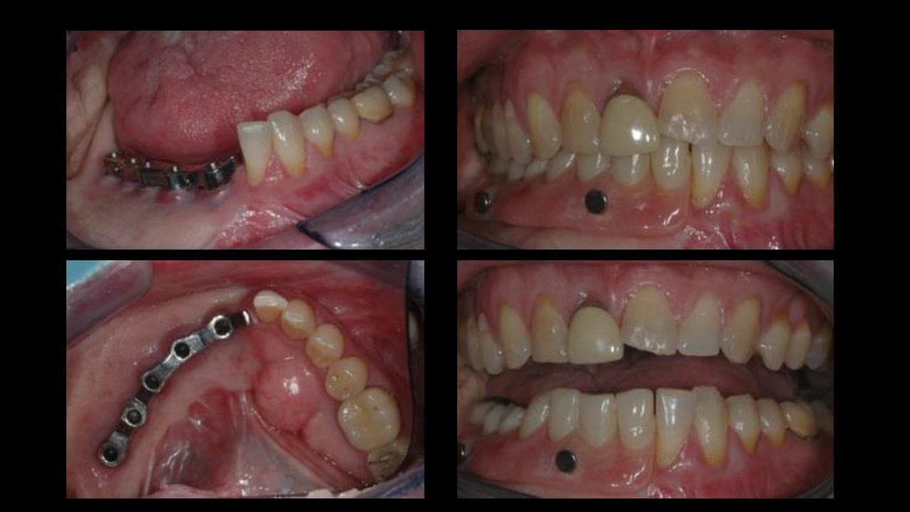
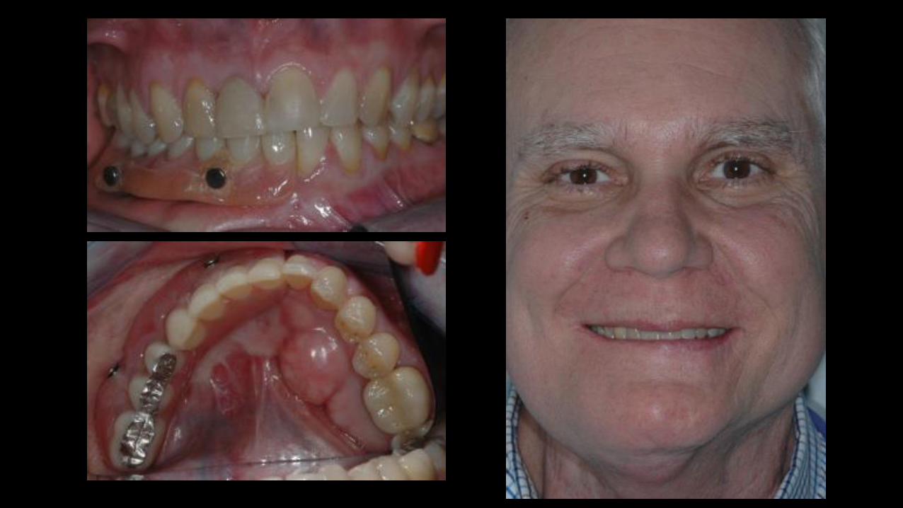
After eight months of healing, patient presents with well healed bone and five implants
restored with healing heads. Excessive tissue thickness of skin graft seen near the tops
of the healing heads presented a restorative challenge.
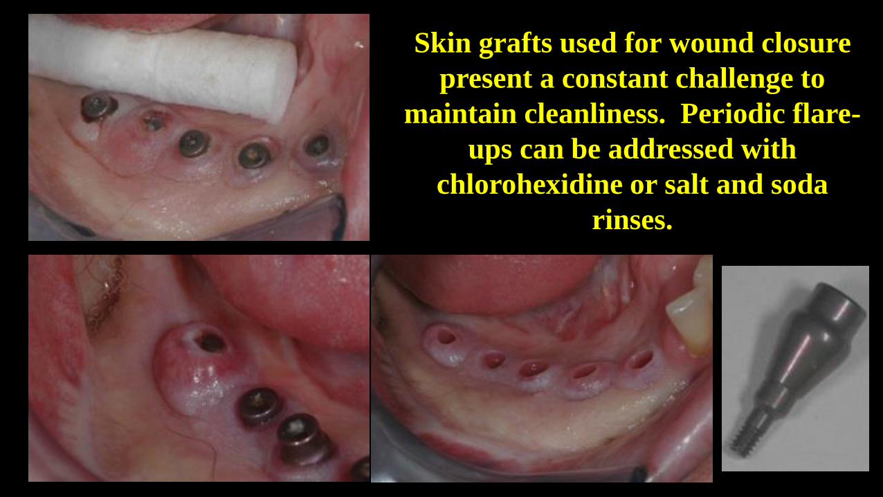
Skin grafts used for wound closure
present a constant challenge to
maintain cleanliness. Periodic flare-
ups can be addressed with
chlorohexidine or salt and soda
rinses.
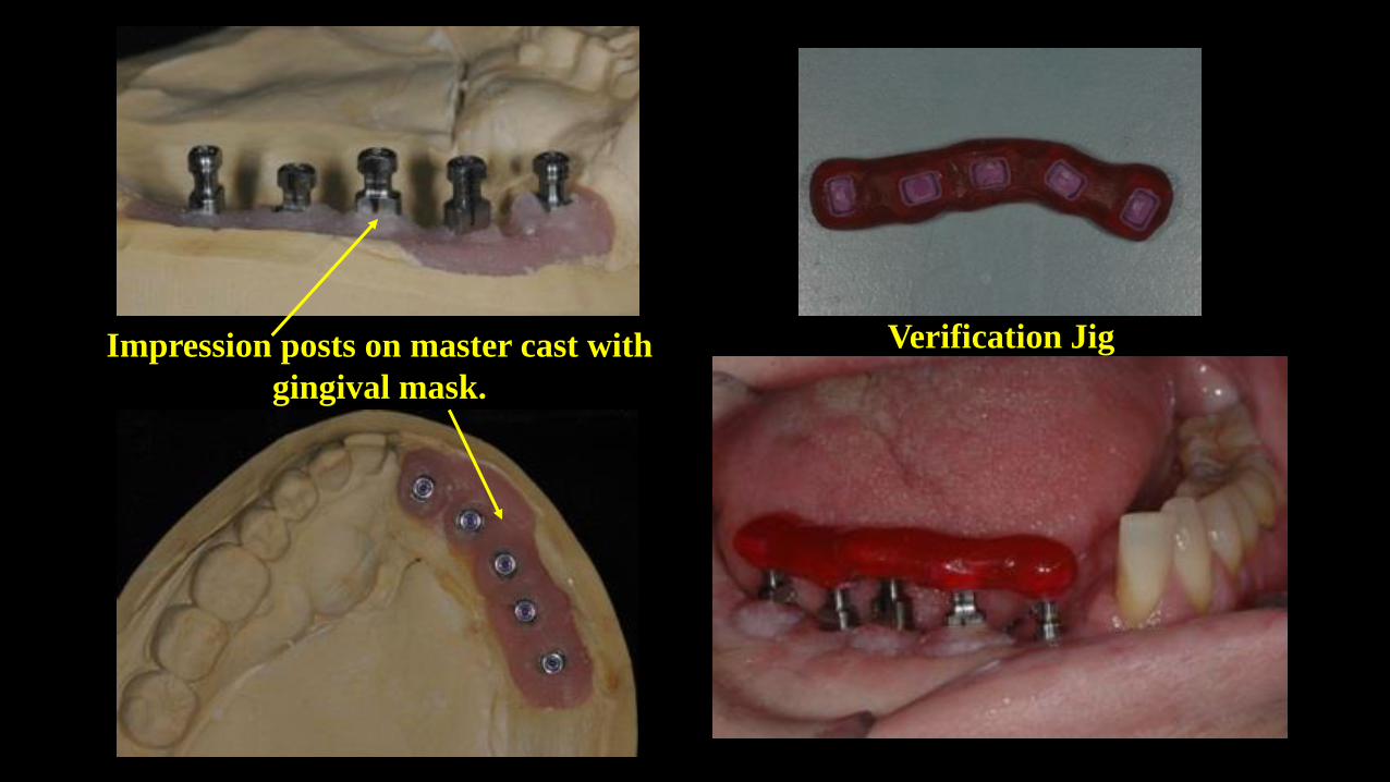
Verification JigImpression posts on master cast with
gingival mask.
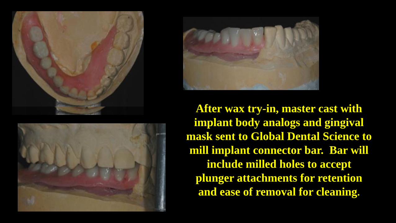
After wax try-in, master cast with
implant body analogs and gingival
mask sent to Global Dental Science to
mill implant connector bar. Bar will
include milled holes to accept
plunger attachments for retention
and ease of removal for cleaning.
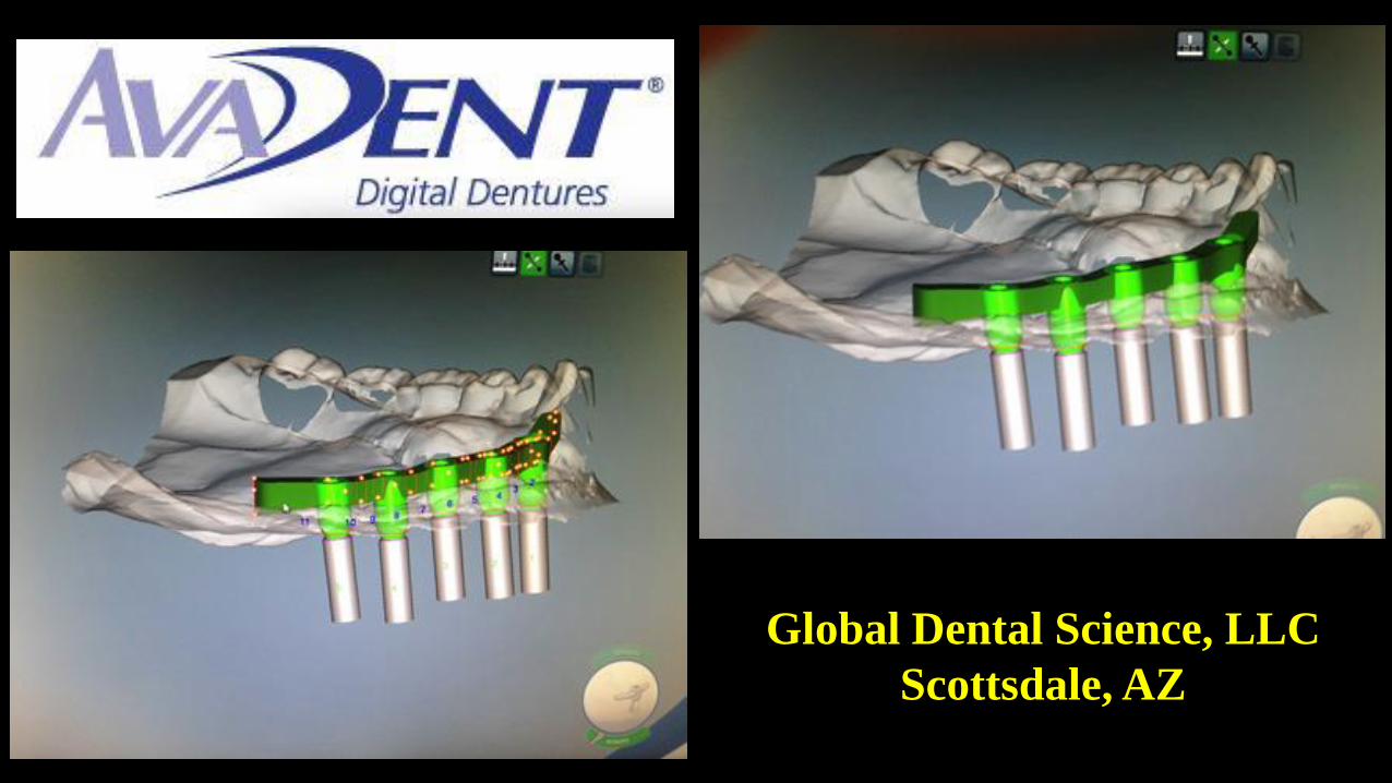
Global Dental Science, LLC
Scottsdale, AZ
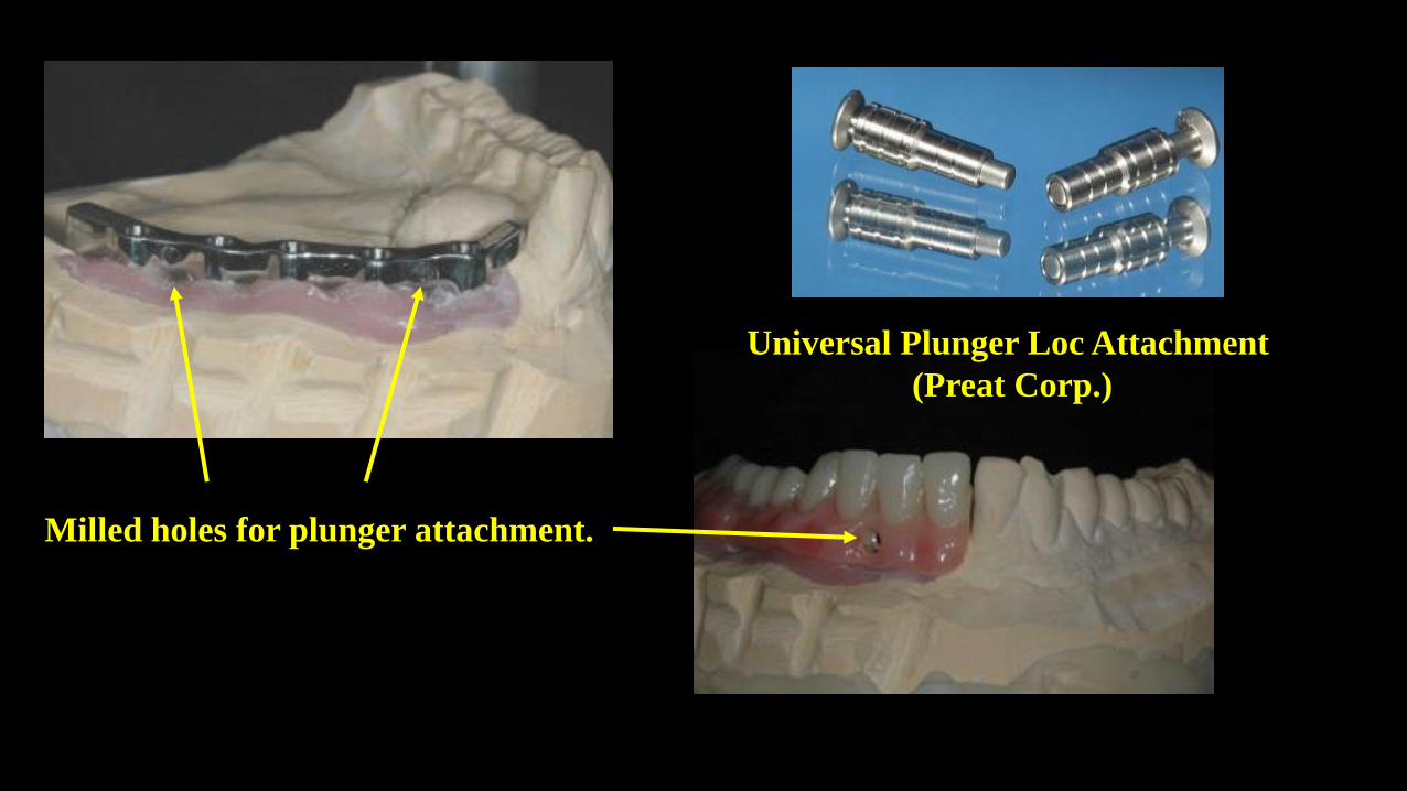
Universal Plunger Loc Attachment
(Preat Corp.)
Milled holes for plunger attachment.
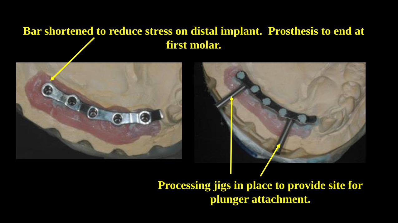
Bar shortened to reduce stress on distal implant. Prosthesis to end at
first molar.
Processing jigs in place to provide site for
plunger attachment.
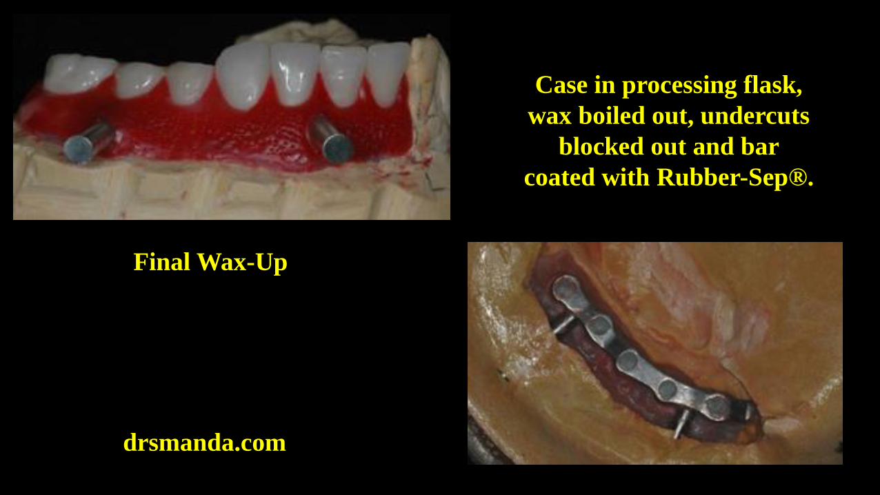
Final Wax-Up
Case in processing flask,
wax boiled out, undercuts
blocked out and bar
coated with Rubber-Sep®.
drsmanda.com
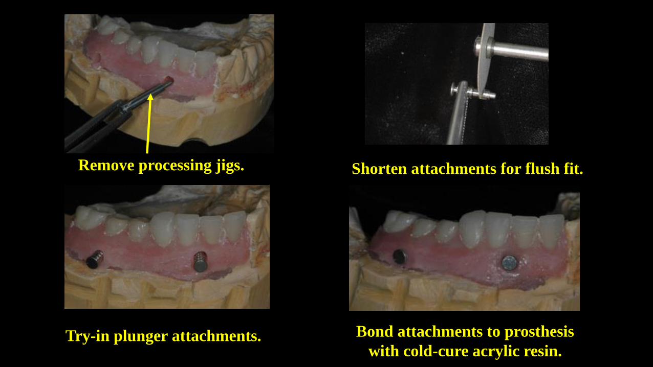
Remove processing jigs.
Try-in plunger attachments.
Shorten attachments for flush fit.
Bond attachments to prosthesis
with cold-cure acrylic resin.
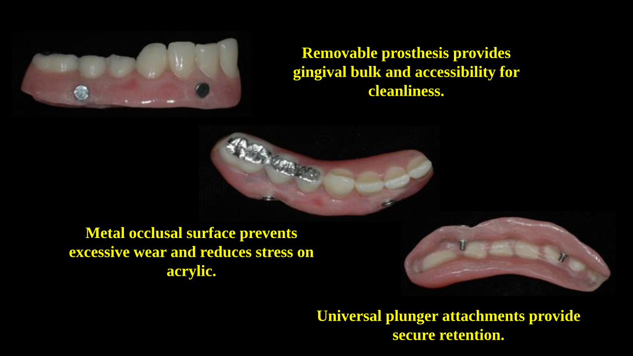
Removable prosthesis provides
gingival bulk and accessibility for
cleanliness.
Metal occlusal surface prevents
excessive wear and reduces stress on
acrylic.
Universal plunger attachments provide
secure retention.

www.drsmanda.com
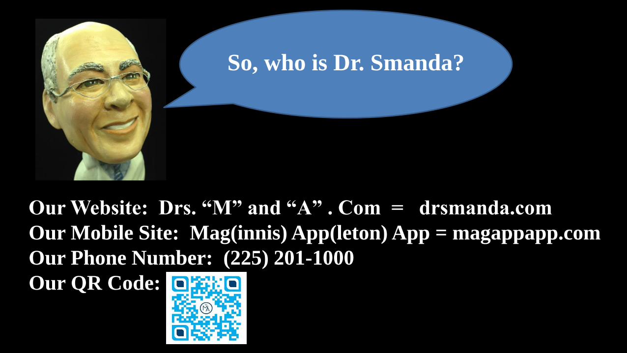
So, who is Dr. Smanda?
Our Website: Drs. “M” and “A” . Com = drsmanda.com
Our Mobile Site: Mag(innis) App(leton) App = magappapp.com
Our Phone Number: (225) 201-1000
Our QR Code:
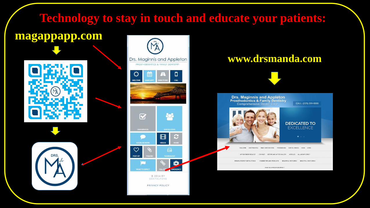
magappapp.com
Technology to stay in touchTechnology to stay in touch and educate your patients:
www.drsmanda.com
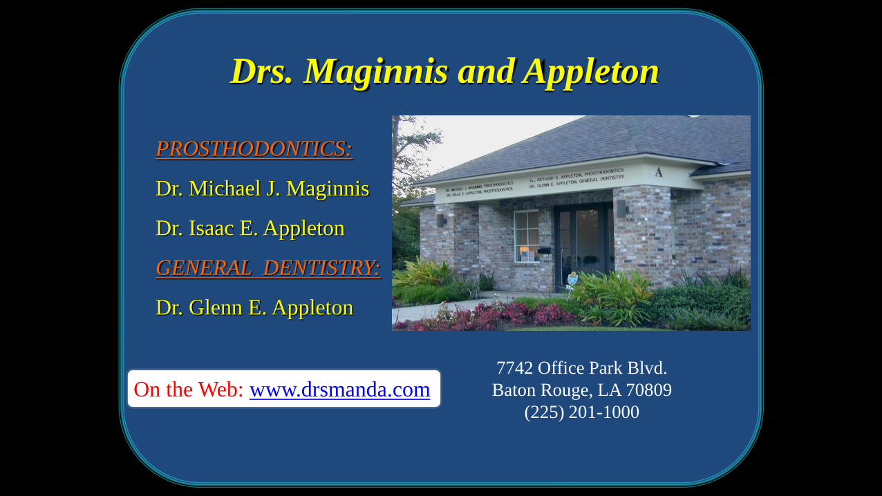
Drs. Maginnis and Appleton
PROSTHODONTICS:
Dr. Michael J. Maginnis
Dr. Isaac E. Appleton
GENERAL DENTISTRY:
Dr. Glenn E. Appleton
On the Web: www.drsmanda.com7742 Office Park Blvd.
Baton Rouge, LA 70809
(225) 201-1000


