Page 1
SECTION 11
_____________________________________________
Use of Wireless Phones and Evidence for Increased
Risk of Brain Tumors
2012 Supplement
Lennart Hardell, MD, PhD, Professor
Department of Oncology, University Hospital, Örebro, Sweden
Michael Carlberg, MSc
Department of Oncology, University Hospital, Örebro, Sweden
Kjell Hansson Mild, PhD, Professor
Department of Radiation Physics. Umeå University, Umeå, Sweden
Prepared for the BioInitiative Working Group
November 2012
Page 2
I. INTRODUCTION
In May 2011 the International Agency for Research on Cancer (IARC) at WHO categorised the
radiofrequency electromagnetic fields (RF-EMF) from mobile phones, and from other devices
that emit similar non-ionising electromagnetic fields, as a Group 2B, i.e. a ‘possible’, human
carcinogen (Baan et al., 2011, IARC, 2011). Nine years earlier IARC had also classified
extremely low frequency (ELF) magnetic field as Group 2B carcinogen (IARC, 2002).
The IARC decision on mobile phones was based mainly on case-control studies from the
Hardell group in Sweden and the IARC Interphone study. Both provided supportive results on
positive associations between two types of brain tumors; glioma and acoustic neuroma, and
exposure to RF-EMF from wireless phones.
The final IARC decision was confirmed by voting of 29 scientists (one not present during
voting) at the meeting. A large majority of participants voted to classify RF-EMF radiation as
‘possibly carcinogenic’ to humans, Group 2B. The decision was also based on occupational
studies. We present in this paper an updated review of evidence of the association between use
of wireless phones and brain tumors including also papers published after the IARC evaluation.
The Nordic countries were among the first countries in the world to widely adopt the wireless
telecommunications technology. Analogue phones (NMT; Nordic Mobile Telephone System)
were introduced in the early 1980s using both 450 and 900 Megahertz (MHz) frequencies. NMT
450 was used in Sweden from 1981-2007, NMT 900 operated during 1986-2000.
The digital system (GSM; Global System for Mobile Communication) using dual band, 900 and
1800 MHz, started to operate in 1991 and dominates now the market. The third generation of
mobile phones, 3G or UMTS (Universal Mobile Telecommunication System), using 1 900/2 100
MHz RF fields has been introduced worldwide in recent years, in Sweden in 2003. Currently the
fourth generation, 4G (Terrestrial 3G), operating at 800/2600 MHz and Trunked Radio
Communication (TETRA 380-400 MHz) are being established in Europe. Nowadays mobile
phones are used more than landline phones in Sweden
(http://www.pts.se/upload/Rapporter/Tele/2011/sv-telemarknad-halvar-2011-pts-er-2011-
21.pdf). Worldwide, an estimate of 5.9 billion mobile phone subscriptions was reported at the
Page 3
end of 2011 by the International Telecommunication Union (ITU; http://www.itu.int/ITU-
D/ict/facts/2011/material/ICTFactsFigures2011.pdf).
Desktop cordless phones (DECT) have been used in Sweden since 1988, first using analogue
800-900 MHz RF fields, but since early 1990s using a digital 1900 MHz system. These cordless
phones are becoming more common than traditional landlines. They emit RF-EMF radiation
similar to that of mobile phones. Thus when human health risks are evaluated it is also
necessary to consider the use of cordless phones along with mobile phones.
The real increase in use and exposure to radiation fields from wireless phones (mobile phones
and cordless phones) in most countries has occurred since the end of the 1990s. The brain is the
main target organ during use of the handheld phone (Cardis et al., 2008). Fear of an increased
risk for brain tumors has dominated the debate during the last one or two decades. While RF-
EMFs do not have sufficient energy to break chemical bonds like ionising radiation, at least not
directly, they can nevertheless have harmful effects on biological tissues. Plausible biological
mechanisms for these effects include DNA damage, impairment of DNA repair mechanisms,
and epigenetic changes to DNA (see also chapters by H. Lai (Genotoxicity) and I. Belyaev
(Physical and Biological Mechanisms).
Primary brain tumors (central nervous system; CNS) constitute of a heterogeneous group of
neoplasms of different histological types depending on tissue of origin with different growth
patterns, molecular markers, anatomical localisations, and age and gender distributions. The
clinical appearance, treatment and prognosis are quite different depending on tumor type.
There are few established risk factors for brain tumors besides ionising radiation (Preston Martin
et al., 2006). Higher socio-economic status tends to be related to higher incidence and some rare
inherited cancer syndromes account for a small fraction of tumors (Preston Martin et al., 2006).
Familial aggregation of glioma has also been reported (Scheurer et al., 2010).
We base this review primarily on the Hardell group papers and the WHO Interphone study
(Interphone Study Group, 2010, 2011, Cardis et al., 2011). More discussion of the results and
responses, agreements and disagreements of the findings for the Hardell group and Interphone
studies can be found in Hardell et al., (2012, 2013).
Page 4
II. MATERIALS AND METHODS
The PubMed database (www.ncbi.nlm.nih.gov) was used for an up-dated search of published
studies in this area using mobile/cellular/cordless telephone and brain tumour/neoplasm/acoustic
neuroma/meningioma/glioma as searching terms. Personal knowledge of published studies was
also used in order to get as up-to-date review as possible.
III. RESULTS
Brain tumors overall
Exposure to the radiation from the phones is generally higher in the temporal lobe, the part of
the brain that is near to the ear (Cardis et al., 2008). For tumors located in the temporal, occipital
or temporoparietal lobe areas of the brain an increased risk was found for ipsilateral exposure,
that is the telephone was mostly used on the same side of the head as the tumor appeared,
yielding OR = 2.42, 95 % CI = 0.97-6.05 (Hardell et al., 2001). This was the first study in the
world that indicated an association between use of mobile phones and an increased risk for brain
tumors. However, the results were based on low numbers of exposed subjects and different
histopathological types of brain tumors so no firm conclusions could be drawn. Furthermore,
this first study did not include use of cordless phones, see also Hardell et al., (1999).
Glioma
Glioma is the most common malignant brain tumor and represents about 60 % of all central
nervous system tumors. The most common glioma subtype is astrocytoma. Astrocytic tumors
are divided in two groups depending on the malignant potential; low-grade (WHO grades I-II)
and high-grade (WHO grades III-IV). Low-grade astrocytoma has a relatively favourable
prognosis, whereas survival is shorter for patients with high-grade glioma. Glioblastoma
multiforme (WHO grade IV) accounts for 60-75 % of all astrocytoma.
The Hardell group in Sweden studied the association between use of mobile and cordless phones
and brain tumors diagnosed during 1997-2003. First, cases diagnosed during 1 January 1997 to
30 June 2000 were included (Hardell et al., 2002, 2003). The next study period included 1 July
2000 to 31 December 2003 (Hardell et al., 2005, 2006a). The methods were the same with the
same inclusion criteria and an identical questionnaire in both studies.
Page 5
In short, both men and women aged 20-80 years at the time of diagnosis were included and all
were alive at the time of inclusion in the study. They were reported from cancer registries and
had all a brain tumor verified by histopathology. The Swedish Population Registry was used for
identification of matched controls. In addition to other exposures use of wireless phones was
carefully assessed by a self-administered questionnaire supplemented over the phone. The ear
that had mostly been used during calls with mobile phone and/or cordless phone was assessed
by separate questions. This information was checked during the supplementary phone calls and
finally also by a separate letter with good agreement between these three methods.
Use of the wireless phone was defined as ipsilateral (> 50 % of the time), or contralateral (< 50
% of the time) in relation to tumor side. The matched control was assigned the same side as the
tumor of the respective case. Use of hands free devices was also assessed as well as use in a car
with external antenna. Such use was not included in the calculation of cumulative number of
hours for life time use. Latency time was defined as the period from the year of first use until
diagnosis (corresponding year for the matched control).
Medical records including computer tomography (CT) and/or magnetic resonance imaging
(MRI) were used to define tumor localisation in the brain. Further details can be found in the
publications.
As a response to a critique from Boice and McLaughlin (2002) that the exclusion of deceased
cases was a source of bias in our studies we performed a study on the cases with a malignant
brain tumor that had died before inclusion in the case-control studies 1997-2003. These cases
represented patients with a poor prognosis, mostly with astrocytoma WHO grade IV
(glioblastoma multiforme). Controls were selected from the Death Registry in Sweden. The
study encompassed 464 cases and 464 controls that had died from a malignant disease and 463
controls with other causes of death. Exposure was assessed by a questionnaire sent to the next of
kin to each deceased case and control. The questionnaire was similar as in previous studies. This
investigation confirmed the previous results of an association between use of mobile phones and
malignant brain tumors (Hardell et al., 2010).
Page 6
We have previously published pooled analysis of malignant brain tumors diagnosed during the
period 1997-2003 (Hardell et al., 2006b). These results were updated including also results for
the deceased cases with malignant brain tumors (Hardell et al., 2011a, Carlberg, Hardell 2012).
The results on use of wireless phones were based on 1,251 cases with malignant brain tumor
(response rate 85%) and 2,438 controls (response rate 84%). Most cases had glioma (n=1,148)
so we present in the following results for that type of tumor. Latency was divided in three
categories, >1-5 years, >5-10 years, and > 10 years from first use of a wireless phone until
diagnosis of glioma.
Both use of mobile and cordless phone gave an increased risk overall, highest in the latency
group >10 years, increasing further for ipsilateral use yielding for mobile phone OR = 2.9, 95 %
CI = 1.8-4.7 and for cordless phone OR = 3.8, 95 % CI = 1.8-8.1 (Table 1). Highest ORs were
found in the > 10 year latency group for total wireless phone use as well, OR = 2.1, 95 % CI =
1.6-2.8.
OR increased statistically significant for glioma for cumulative use of wireless phones per 100
h; OR = 1.014, 95 % CI = 1.008-1.019, and per year of latency; OR = 1.056, 95 % CI = 1.037-
1.075 (Carlberg and Hardell, 2012). Separate calculations of mobile phone and cordless phone
use yielded similar results with statistically significant increasing risks.
The Interphone study was conducted at 16 research centres in 13 countries during varying time
periods between 2000 and 2004 under the guidance of IARC. An increased risk for brain tumor
was found in some separate country studies and decreased risk in other studies as we have
discussed elsewhere (Hardell et al., 2008, 2009). After several years of delay the overall
Interphone results were finally published in May 2010 (Interphone Study Group, 2010).
In total 4,301 glioma cases were included in Interphone and the final results were based on
2,708 participating cases (response rate 64 %, range by centre 36-92 %). In total 14,354
potential controls were identified and interviews were completed with 7,658 (53 %, range 42-74
%). The low participation rates in some centres may have created selection bias, see Hardell et
al., (2008).
Regular use of mobile phone in the past > 1 year gave for glioma OR = 0.81, 95 % CI = 0.70-
0.94 (Table 1). Subgroup analyses showed statistically significant increased risk in the highest
Page 7
exposure group, i.e. those with cumulative mobile phone use > 1,640 hours, OR = 1.40, 95 % CI
= 1.03-1.89. The risk increased further for glioma in the temporal lobe yielding OR = 1.87, 95 %
CI = 1.09-3.22. In the same exposure category, cumulative use > 1,640 hours and ipsilateral
exposure produced OR = 1.96, 95 % CI = 1.22-3.16 in total (no data given for temporal lobe).
In Appendix 2 (Interphone Study Group, 2010, available on the web) analysis was restricted to
ever-regular users of mobile phones. Cumulative call time > 1,640 hours gave OR = 1.82, 95 %
CI = 1.15-2.89 compared with use < 5 hours. Time since start of regular use (latency) > 10 years
produced OR = 2.18, 95 % CI = 1.43-3.31; reference entity 1-1.9 years.
The Interphone study group concluded: “However, biases and errors limit the strength of the
conclusions we can draw from these analyses and prevent a causal interpretation.” In an
editorial accompanying the Interphone results the main conclusion of the Interphone results was
described as “both elegant and oracular…(which) tolerates diametrically opposite readings”
(Saracci and Samet 2010). Several methodological reasons why the Interphone results were
likely to have underestimated the risks were discussed including the short latency period since
first exposures became widespread; less than 10 % of the Interphone cases had more than 10
years exposure. “None of the today’s established carcinogens, including tobacco, could have
been firmly identified as increasing risk in the first 10 years or so since first exposure”.
Estimated RF-EMF dose in the tumor area from mobile phone use was associated with an
increased risk of glioma in parts of the Interphone study (Cardis et al., 2011). OR increased with
increasing total cumulative dose of specific energy (J/kg) absorbed at the estimated tumor centre
for more than 7 years before diagnosis giving OR = 1.91, 95 % CI = 1.05-3.47 (p trend = 0.01)
in the highest quintile of exposure. A similar study based on less clear methods was later
published by another part of the Interphone study group (Larjavaara et al., 2011). The results
seemed not to support the findings of Cardis et al., (2011). However, only 42 cases had used a
mobile phone for more than 10 years and no analysis was made of the most exposed group with
longest duration of use.
Based on Hardell et al (2011b) and Interphone Study Group (2010) we made meta-analysis of
glioma and use of mobile phones. Random-effects model was used based on test for
heterogeneity in the overall (≥10 years and ≥1,640 hours) groups. We used published results in
Page 8
Interphone since we do not have access to their database. Our results were recalculated to these
groups of exposure. The meta-analysis yielded for mobile phone use OR = 1.71, 95 % CI =
1.04-2.81 for glioma in the temporal lobe in the > 10 years latency group. Ipsilateral mobile
phone use > 1,640 h in total gave the highest risk, OR = 2.29, 95 % CI = 1.56-3.37 (Hardell et al
2012). This meta-analysis strengthens a causal association between use of mobile phones and
glioma.
Meningioma
Meningioma is the most common benign brain tumor. It develops from the pia and arachnoid
that covers the central nervous system. Meningioma is an encapsulated and well-demarked
tumor more common in women than in men. It is rarely malignant.
A pooled analysis of benign brain tumors from the two case-control studies from the Hardell
group as discussed above (Hardell et at., 2006c, Hardell and Carlberg, 2009) gave regarding
meningioma and use of mobile phone OR = 1.1, 95 % CI = 0.9-1.3, and cordless phone OR =
1.1, 95 % CI = 0.9-1.4 (Table 2). Using > 10 year latency period OR increased; for mobile
phone to OR = 1.5, 95 % CI = 0.98-2.4, and for cordless phone to OR = 1.8, 95 % CI = 1.01-3.2.
Ipsilateral mobile phone use in the > 10 years latency group yielded OR = 1.6, 95 % CI = 0.9-
2.9, and cordless phone OR = 3.0, 95 % CI = 1.3-7.2. These results were based on rather low
numbers of exposed cases, however.
Regular use of mobile phone produced in the Interphone study (2010) a statistically significant
decreased risk for meningioma, OR = 0.79, 95 % CI = 0.68-0.91, Table 2. The risk increased
somewhat with cumulative use > 1,640 hours and ipsilateral mobile phone use to OR = 1.45, 95
% CI = 0.80-2.61. Analysis restricted to tumors in the temporal lobe or to the group of ever-
regular use did not change the overall pattern of no increased risk.
We performed meta-analysis of meningioma for use of mobile phone based on results in the
Hardell group and Interphone results similarly as for glioma. No statistically significant
decreased or increased risk was found (Hardell et al., 2012). These results support the
conclusion that up to latency > 10 years or cumulative use >1,640 hours there is no consistent
pattern of an association between use of mobile phones and meningioma.
Page 9
Acoustic neuroma
Acoustic neuroma or Vestibular Schwannoma is a slow growing benign tumor located in the
eighth cranial nerve in the auditory canal. It grows gradually out into the cerebellopontine angle
with potential compression of vital brain stem centres. Tinnitus and hearing problems are usual
first symptoms of acoustic neuroma. The eighth cranial nerve is located close to the handheld
wireless phone when used, so there is particular concern of an increased risk for neuroma
development due to exposure to EMF-RF emissions during use of these devices.
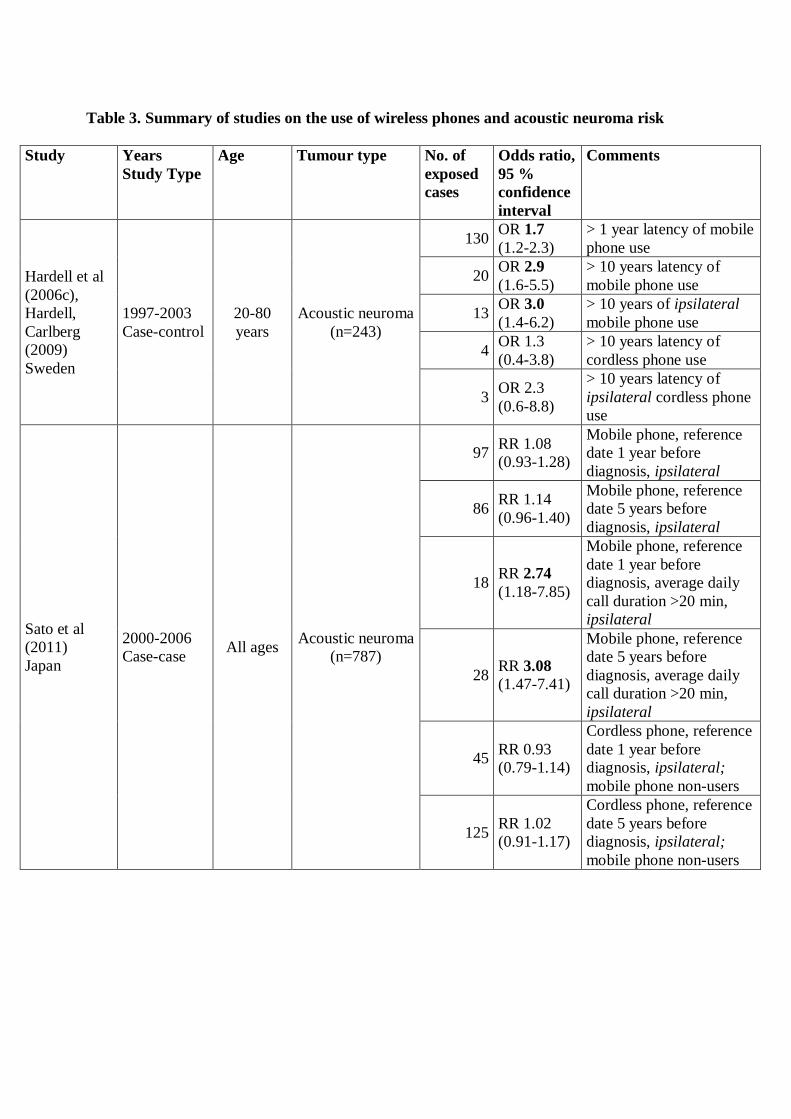
The pooled analysis of the Hardell group studies yielded regarding use of mobile phones for
acoustic neuroma OR = 1.7, 95 % CI = 1.2-2.3 increasing to OR = 2.9, 95 % CI = 1.6-5.5 with >
10 years latency period, Table 3. Ipsilateral use increased the risk further; in the > 10 years
latency group to OR = 3.0, 95 % CI = 1.4-4.2 (Hardell and Carlberg, 2009). Cordless phone use
gave OR = 1.5, 95 % CI = 1.04-2.0 increasing to OR = 1.7, 95 % CI =1.2-2.5 for ipsilateral use
in the > 1 year latency group.
In the Interphone study (2011) 1,121 (82 %) acoustic neuroma cases participated, range 70-100
% by centre. Of the controls 7,658 (53 %) completed the interviews, range 35-74 % by centre.
The final matched analysis (1:1 or 1:2) consisted of 1,105 cases and 2,145 controls. Overall no
increased risk was found censoring exposure at one year or at 5 years before reference date, OR
= 0.85, 95 % CI = 0.69-1.04 and OR = 0.95, 95 % CI = 0.77-1.17, respectively (Table 3).
Cumulative number of hours of ipsilateral mobile phone use > 1,640 hours up to 1 year before
reference date gave OR = 2.33, 95 % CI = 1.23-4.40 and contralateral use OR = 0.72, 95 % CI =
0.34-1.53 for acoustic neuroma, see Table 3 (Interphone Study Group, 2011). For cumulative
number of hours of ipsilateral mobile phone use > 1,640 hours up to 5 years before reference
date OR = 3.53, 95 % CI = 1.59-7.82, and for contralateral use OR = 1.69, 95 % CI = 0.43-6.69
were obtained. The risk increased further for cumulative ipsilateral use > 1,640 hours with start
> 10 years before reference date to OR = 3.74, 95 % CI = 1.58-8.83. Contralateral use in that
group yielded OR = 0.48, 95 % CI = 0.12-1.94, however based on only 4 exposed cases and 9
exposed controls. Overall OR = 1.93, 95 % CI = 1.10-3.38 was obtained for long-term use with
start > 10 years before reference date and cumulative call time > 1,640 hours.
Page 10
Similar analyses of the data as in Appendix 2 for glioma (see Interphone Study Group, 2010)
yielded highest OR for acoustic neuroma in the shortest latency group, 2-4 years before
reference date, OR = 1.41, 95 % CI = 0.82-2.40. Lower OR was calculated in the > 10 years
group, OR = 1.08, 95 % CI = 0.58-2.04. Somewhat higher risk than in total, OR = 1.32, 95 % CI
= 0.88-1.97, was found for cumulative mobile phone use > 1,640 hours; OR = 1.74, 95 % CI =
0.90-3.36, in this analysis restricted to only regular users. No results were given for ipsilateral
use.
We performed meta-analysis of the results for use of mobile phone and the association with
acoustic neuroma based on results by the Hardell group and Interphone study (Hardell et al
2012). For the latency group > 10 years highest risk was obtained for ipsilateral use, OR = 1.81,
95 % CI = 0.73-4.45. The risk increased further for cumulative use > 1,640 hours yielding OR =
2.55, 95 % CI = 1.50-4.40 for ipsilateral use. The meta-analysis strengthens a causal association
between use of mobile phones and acoustic neuroma.
A case-case study was performed in Japan (Sato et al., 2011). The cases were identified during
2000-2006 at 22 participating neurosurgery departments. The diagnosis was based on
histopathology or CT/MRI imaging. Of 1,589 cases 816 (51 %) agreed to participate and
answered a mailed questionnaire. The final analysis included 787 cases, Cases with ipsilateral
use were regarded as exposed and those with contralateral use were assumed to be unexposed
and were used as the reference category. Overall no increased risk was found. However, for
average daily call duration > 20 minutes with reference date 1 year Risk Ratio (RR) = 2.74, 95
% CI = 1.18-7.85 was found increasing to OR = 3.08, 95 % CI = 1.47-7.41 with reference date 5
years before diagnosis (Table 3). Unfortunately no results were given for cumulative number of
hours for use over the years. For cordless phones no increased risk was found but the analysis
was not very informative.
Risks to children and adolescents
The developing brain is more sensitive to toxins (Kheifets et al., 2005) and it is still developing
until about 20 years of age (Dosenbach et al., 2010). Children have smaller head and thinner
skull bone than adults. Their brain tissue has also higher conductivity and these circumstances
give higher absorption from RF-EMF than for adults (Cardis et al., 2008, Christ et al., 2010,
Gandhi et al., 2012). Use of wireless phones is widespread among children and adolescents
Page 11
(Söderqvist et al., 2007, 2008). The greater absorption of RF energy per unit of time, the greater
sensitivity of their brains, and their longer lifetimes with the risk to develop a brain tumor leaves
children at a higher risk than adults from mobile phone radiation.
We have published results regarding brain tumor risk for different age groups at the time of
diagnosis (Hardell et al., 2004) or age at first use of wireless phones (Hardell and Carlberg,
2009, Hardell et al., 2011a, 2012, 2013). Three age groups for first use of a wireless phone were
used: <20 years, 20-49 years and 50-80 years. Highest risk for glioma was found for first use of
mobile phone or cordless phone before the age of 20 years (Table 4). Thus, mobile phone use
yielded for glioma OR = 3.1, 95 % CI = 1.4-6.7 and cordless phone OR 2.6, 95 % CI = 1.2-5.5.
Also for acoustic neuroma the risk was highest in the youngest age group with OR = 5.0, 95 %
CI = 1.5-16 for use of mobile phone. Only one case had first use of cordless phone before the
age of 20, so no conclusions could be drawn for cordless phones. Regarding meningioma no
clear pattern of age-dependent increased risk was seen.
A multi-centre case-control study was conducted in Denmark, Sweden, Norway, and
Switzerland, CEFALO (Aydin et al., 2011). It included children and adolescents aged 7–19
years and has been commented elsewhere in detail since serious methodological problems exist
in the study design and interpretation of the results (Söderqvist et al., 2011). In CEFALO a
statistically non-significant increased risk for brain tumors among regular users (one call per
week for at least 6 months) of mobile phones was found; OR = 1.36, 95 % CI = 0.92-2.02. This
OR increased somewhat with cumulative duration of subscriptions and duration of calls (Aydin
et al., 2011). No data for long-term use were given; the longest latency period was 5 years.
Further support of a true association was found in the results based on operator-recorded use for
62 cases and 101 controls, which for time since first subscription >2.8 years yielded a
statistically significant OR of 2.15, 95 % CI = 1.07-4.29, with a statistically significant trend
(p=0.001).
Use of cordless phones was covered only in the first 3 years of use. No explanation was given
for this most peculiar definition. Wireless phone use was not considered, that is use of both
mobile phones and cordless phones as the relevant exposure category, as used by the Hardell
group and adopted by IARC (Baan et al., 2011). Instead Aydin et al., (2011) included use of
Page 12
cordless phones in the ‘unexposed’ category when risk estimates were calculated for mobile
phone use. Similarly, regarding use of cordless phones RF-EMF emissions from mobile phones
were regarded as ‘no exposure’. Thus, an increased risk was potentially concealed.
The authors summarised that they “did not observe that regular use of a mobile phone increased
the risk for brain tumors.” An editorial in the same journal accompanied that conclusion by
stating by that the study showed “no increased risk of brain tumors” (Boice and Tarone, 2011).
This was echoed by a news release from the Karolinska Institute in Stockholm claiming that the
results of no increased risk were ‘reassuring’ (Karolinska Institute, 2011). However the results
indicate a moderately increased risk, in spite of low exposure, short latency period and
limitations in study design and analyses. Certainly it cannot be used as reassuring evidence
against an association, see Söderqvist et al., (2011).
Danish cohort study on mobile phone subscribers
An attempt to establish a cohort of mobile phone users was made in Denmark in co-operation
between the Danish Cancer Society and the International Epidemiology Institute (IEI),
Rockville, MD, USA. It was financed by grants from two Danish telecom operation companies
(TeleDenmark Mobil and Sonafon), IEI, and the Danish Cancer Society. The source of money
for IEI has not been disclosed.
The Danish study on brain tumor risk among mobile phone subscribers has so far resulted in
four publications (Johansen et al., 2001, Schüz et al., 2006, Frei et al., 2011, Schüz et al., 2011).
It included subjects from January 1, 1982 until December 31, 1995 identified from the
computerised files of the two Danish operating companies, TeleDenmark Mobil and Sonofon. A
total of 723,421 subscribers were initially identified but the final cohort consisted of only 58 %
of these subjects. Due to lack of names of individual users 200,507 corporate users were
excluded.
We have discussed elsewhere several shortcomings in the Danish cohort study such as exclusion
of corporate users, no individual exposure data, users of cordless phones are included in the
reference category, no control for use of mobile phones in the population after the establishment
of the cohort, and no operator-verified data on years of subscription is available (Söderqvist et
al., 2012). These limitations are likely to have led to an underestimate of any risk in this study.
Page 13
One would also expect considerable misclassification of mobile phone use both among
subscribers and the reference population since no new subscribers were included in the exposed
cohort after 1995.
The IARC working group concluded that the methods used could have resulted in considerable
misclassification in exposure assessment in the Danish cohort study on mobile phone
subscribers (Baan et al., 2011).
After the outcome of the IARC-evaluation was made public in June 2011 (Baan et al., 2011) two
additional reports on the Danish cohort were published (Frei et al., 2011, Schüz et al., 2011).
Both were new up-dates of the initial cohort and included more information on risk related to
longer follow-up. One focused on acoustic neuroma (Schüz et al., 2011) while the other gave
results both for all cancers and separately for glioma and meningioma (Frei et al., 2011). This
time the number of the cohort was reduced to 358,403 (49.5 %) of the initially identified
subscribers (n=723,421). The major additional exclusion (n=54,350) was due to record linkage
with the Danish so-called CANULI cohort on socioeconomic factors (Dalton et al., 2008).
The authors of the Danish study have themselves pointed out the main causes of considerable
exposure misclassifications (Frei et al., 2011). While at least non-response and recall bias can be
excluded the study has serious limitations related to exposure assessment (Söderqvist et al.,
2012). In fact, these limitations cloud the findings of the four reports to such an extent they are
uninformative at best. At worst, they may be used in a seemingly solid argument against an
increased risk; as reassuring results from a large nationwide cohort study.
Brain tumor incidence
It has been suggested that overall incidence data on brain tumors for countries show no
increasing trends and may be used to disqualify the association between mobile phone use and
brain tumors observed in the case-control studies (Aydin et al., 2011; Ahlbom, and Feychting,
2011; Deltour et al., 2012; Little et al., 2012).
However, by now several studies show increasing incidence of brain tumors. In Denmark a
statistically significant increase in incidence rate per year for brain and central nervous system
Page 14
tumors (combined) was seen during 2000-2009; in men +2.7 %, 95 % CI = +1.1 to 4.3 % and in
women +2.9 %, 95 % CI = +0.7 to 5.2 % (NORDCAN). Updated results for brain and central
nervous system tumors have been released in Denmark. The age-standardized incidence of brain
and central nervous system tumors increased with 40 % among men and 29 % among women
during 2001-2010 (Sundhedsstyrelsen, 2010). A more recent news release based on the Danish
Cancer Register stated that during the last 10 years there has been an increasing number of cases
with the most malignant glioma type, glioblastoma multiforme (astrocytoma WHO grade IV),
especially among men
(http://www.cancer.dk/Nyheder/nyhedsartikler/2012kv4/Kraftig+stigning+i+hjernesvulster.htm)
.
Little et al., (2012) studied the incidence rates of glioma during 1992-2008 in the United States
and compared with ORs for glioma associated with mobile phone use in the 2010 Interphone
publication (Interphone Study Group, 2010) and our pooled results published in 2011 (Hardell et
al., 2011a). Since our results are discussed and questioned by Little et al their study needs to be
reviewed in more detail. Our response to the journal (BMJ) was never accepted for publication
in the journal and cannot be found via PubMed, only on the web
(http://www.bmj.com/content/344/bmj.e1147/rr/578564).
First, one important methodological issue that was not stated in the abstract or in the article
[Figures 2-4] by Little et al., (2012), but can be found in the web appendix, is that observed rates
were based on men aged 60-64 years from the Los Angeles SEER registry as the baseline
category. These data were used to estimate rates in the entire dataset, men and women aged > 18
years and all 12 SEER registries. Thereby numerous assumptions were made as pointed out by
Kundi (2012) and Davis et al., (2012).
Using only men, as Little et al., did, ignores the fact that women had less frequent use of mobile
phones than men in our studies (Table 5). Overall 31 % of women reported such use versus 57
% of men. Furthermore, use varies with age group with a large difference according to age, as
we have explored in our publications (Hardell and Carlberg, 2009, Hardell et al., 2011a). Thus,
the age group 60-64 year old men is not valid to use for these calculations.
Page 15
There are several other points that may be added. Another example is that the results for
anatomical localisations and tumor grade [in Table 5 in the article] by Little et al are based on
numerous assumptions from SEER data, Interphone and the Hardell group studies. The authors
seem not to have paid attention to the fact that the fraction of mobile phone users differs for
gender and age, see Table 5.
One interesting result that was not commented further by Little et al., (2012) was the finding of
a statistically significant yearly increasing incidence of high-grade glioma (WHO grades III-IV)
in the SEER data for 1992-2008, +0.64%, 95% CI = +0.33 to 0.95 %. On the contrary, the
incidence of low-grade glioma (WHO grades I-II) decreased with –3.02 %, 95 % CI = –3.49 to –
2.54 %. Little et al., (2012) found also a statistically significant increasing yearly trend for
glioma in the temporal lobe, +0.73 %, 95 % CI = +0.23 to 1.23 %.
Zada et al., (2012) studied incidence trends of primary malignant brain tumors in the Los
Angeles area during 1992-2006. The overall incidence of primary malignant brain tumors
decreased over the time period with the exception of glioblastoma multiforme (astrocytoma
WHO grade IV). The annual age adjusted incidence rate of that tumor type increased statistically
significant in the frontal lobe with Annual Percentage Change (APC) +2.4 % to +3.0 % (p <
0.001) and temporal lobe APC +1.3 % to +2.3 % (p < 0.027) across all registries. In the
California Cancer Registry the incidence of glioblastoma multiforme increased also in
cerebellum, APC +11.9 % (p < 0.001). For lower grade astrocytoma decreases of annual age
adjusted incidence rates were observed. The authors concluded that there was a real increase in
the incidence of glioblastoma multiforme in frontal and temporal lobes and cerebellum, areas of
the brain with the highest absorbed dose of RF-EMF emissions from handheld mobile phones
(Cardis et al., 2008).
Of interest is also the report by de Vocht et al., (2011) from England that showed for the time
period 1998 to 2007 a statistically significant increasing incidence of brain tumors, the majority
glioma, in the temporal lobe for men and women (p < 0.01), and frontal lobe for men (p < 0.01).
The incidence increased also for women in the frontal lobe, although not statistically significant
(p = 0.07). The incidence decreased in other parts of the brain.
Page 16
Deltour et al., (2012) reported increasing glioma incidence rates in Denmark, Finland, Norway,
and Sweden for the time period 1979-2008. APC increased for men with +0.4 %, 95 % CI +0.1
to 0.6 % and for women with +0.3 %, 95 % CI +0.1 to 0.5 %. A study from Australia for the
time period 2000-2008 showed that APC for malignant brain tumors increased statistically
significant +3.9 %, 95 % CI +2.4 to 5.4 % (Dobes et al., 2011). An increase was seen among
both men and women. The APC for benign tumors increased with +1.7 %, 95 % CI -1.4 to +4.9
%, thus not statistically significant.
From urban Shanghai an increasing incidence of brain and nervous system tumors for the time
period 1983-2007 was reported with APC +1.2 %, 95 % CI +0.4 to 1.9 % in males and APC
+2.8 %, 95 % CI +2.1 to 3.4 % in females (Ding and Wang, 2011).
We reported increasing incidence of astrocytoma WHO grades I-IV during 1970-2007 in
Sweden. In the age group > 19 years the annual change was +2.16 %, 95 % CI +0.25 to 4.10 %
during 2000-2007, for further details see Hardell and Carlberg (2009).
IV. DISCUSSION
As pointed out by IARC (Baan et al., 2011) the most comprehensive results on use of wireless
phones and the association with brain tumors come from the Hardell group in Sweden and the
international Interphone study. Results for latency time of 10 years or more have been published
from both study groups.
Both were case-control studies and the cases were recruited during similar time periods, 1997-
2003 in the Hardell group and during 2000-2004 in Interphone, with somewhat different years in
the varying study regions. There was no overlapping of cases in the Hardell group studies and
the Swedish part of Interphone.
The Hardell group included cases aged 20-80 years whereas eligible cases in Interphone were
aged 30-59 years at diagnosis. One control subject matched on age, gender and geographical
area (region) to each case in the Hardell group studies was drawn from the national population
register. In Interphone one control was selected for each case from a ‘locally appropriate
population-based sampling frame’. In Germany two controls were selected and the centres used
Page 17
individual matching or frequency matching. Regarding the Interphone study on acoustic
neuroma some centres sampled special controls to the cases, other draw controls from the pool
of controls in the glioma and meningioma studies, or used a mixture of both methods. In UK
general practioners’ lists (Hepworth et al 2006) and in Japan random digit dialling were used
(Takebayashi et al., 2006, 2008). Certainly the methods used in Interphone may introduce
selection bias.
Use of wireless phones and other exposures were carefully assessed by a self-administered
questionnaire in the Hardell et al., studies. The information was supplemented over the phone by
trained interviewers thereby using a structured protocol. This was done blinded as to case or
control status. After the interviews all personal data like names and addresses were removed
from the questionnaires so that only an id-number that did not disclose if it was a case or a
control was shown. Thus, coding of the data for statistical analysis was performed without
personal data of the individual.
On the contrary information on past mobile phone use was collected during face-to-face
interviews in Interphone obviously disclosing if it was a case or a control that was interviewed.
These interviews were performed by a large number of interviewers at different participating
centres. Experienced interviewers were defined as those who conducted at least 20 interviews. In
fact, in the sensitivity analysis the risk increased for glioma for cumulative mobile phone use >
1,640 hours from OR = 1.40, 95 % CI 1.03-1.89 to OR = 1.50, 95 % CI = 1.10-2.06 if
‘experienced interviewers only’ were considered. The higher risk restricting analysis to
‘experienced interviewers’ in Interphone indicates observational bias during assessment of
exposure decreasing the risk.
In the Hardell group studies few persons conducted all interviews of the 1,251 participating
cases with malignant brain tumor, 1,254 cases with benign brain tumor, and 2,438 controls (total
4,942; note one case had both a malignant and a benign brain tumor). All interviewers were first
educated; they used a defined protocol and gained considerable experience as interviewers. In
fact, they were obliged to carry out the interviews extensively to fulfil the quality in data
assessment according to the structured protocol. It is obvious that the few interviewers in the
Hardell group study must have been much more experienced than the diversity of interviewers
in Interphone.
Page 18
In the personal interviews in Interphone a computer program that guided the interview with
questions read by the interviewer from a laptop computer screen was used. The answers were
entered directly into the computer by the interviewer. Using computer based face-to-face
interviews may be a stressful situation for the patients. In fact patients scored significantly lower
than controls due to recalling of words (aphasia), problems with writing and drawing due to
paralysis in the Danish part of Interphone (Christensen et al., 2005). Furthermore, it has not been
disclosed how the personal interviews were performed in sparsely populated areas, e.g. in the
Northern Sweden. Did the interviewers travel long distances for interviews of controls in rural
areas or were all controls living in the largest cities thereby creating selection bias?
In the Hardell group studies the response rate was 85 % (n=1,251) for cases with malignant
brain tumor, 88 % (n=1,254) for cases with benign brain tumor, and 84 % (n=2,438) for controls
(Hardell et al., 2006c, Carlberg and Hardell, 2012). Lower response rates were obtained in
Interphone study, 64 %, range by centre 36-92 %, (n=2,765) for glioma cases, 78 %, range 56-
92 %, (n=2,425) for meningioma cases, 82 %, range 70-100 % (n=1,121) for acoustic neuroma
cases, and 53 %, range 42-74 %, (n=7,658) for controls (Interphone Study Group, 2010; 2011).
These low response rates may have created the possibility of considerable selection bias
(Hardell et al., 2008). Not responding controls in Interphone tended to be less frequent users of
mobile phone than participating controls leading to underestimation of the risk.
The Hardell group studies included subjects aged 20-80 years, versus 30-59 years in Interphone.
We have shown that restricting the age group to 30-59 years and considering subjects that used a
cordless phone as unexposed in the Hardell group studies reduced the ORs and produced results
quite similar to Interphone (Hardell et al., 2011b). Latency time > 10 years for glioma in the
temporal lobe yielded OR = 1.40, 95 % CI = 0.70-2.81 in the Hardell group studies and OR =
1.36, 95 % CI = 0.88-2.11 in Interphone (latency > 10 years). Thus, excluding exposure to RF-
EMFs from cordless phones as in the Interphone study as well as excluding the younger and
older subjects biased the ORs towards unity in Interphone, which likely dilutes the ability to see
health risks.
By placing a strong emphasis on incidence data an association between use of wireless phones
and brain tumors has been challenged (Swerdlow et al., 2011). The authors considered that if the
Page 19
increased risks seen in case-control studies reflect a causal relationship, there would already be
an increase in incidence of brain and central nervous system tumors. As discussed above by now
increasing incidence rates, especially for certain brain tumor types and anatomical localisations
of relevance, have been reported. The natural history of most glioma from earliest events to
clinical manifestation is unknown, but most likely several decades. The exposure duration in
most studies is thus incompatible with a tumor initiating effect. If the exposure on the other hand
acts as a promoter, this would decrease latency time for already existing tumors, giving a
temporary but not a continuous increase in incidence (Kundi, 2010).
The first case in the world on worker’s compensation for a brain tumor after long-term use of
wireless phones was the ruling 12 October 2012 by the Italian Supreme Court. A previous ruling
that the Insurance Body for Work (INAIL) must grant compensation to a businessman who had
used wireless phones for 12 years and developed a neurinoma in the brain was affirmed
(http://www.applelettrosmog.it/public/news.php?id_news=44 ; www.microwavenews.com). He
had used both mobile and cordless phones for five to six hours per day preferably on the same
side as the tumour developed. The neurinoma was located in the trigeminal Gasser’s ganglion in
the brain. This 5th
cranial nerve controls facial sensations and muscles. It is the same type of
tumour as the acoustic neuroma in the 8th
cranial nerve located in the same area of the brain. No
further appeal of the Supreme Court decision is possible.
V. CONCLUSIONS
Based on epidemiological studies there is a consistent pattern of increased risk for glioma and
acoustic neuroma associated with use of mobile phones and cordless phones. The evidence
comes mainly from two study centres, the Hardell group in Sweden and the Interphone Study
Group. No consistent pattern of an increased risk is seen for meningioma. A systematic bias in
the studies that explains the results would also have been the case for meningioma. The different
risk pattern for tumor type strengthens the findings regarding glioma and acoustic neuroma.
Meta-analyses of the Hardell group and Interphone studies show an increased risk for glioma
and acoustic neuroma. Supportive evidence comes also from anatomical localisation of the
tumor to the most exposed area of the brain, cumulative exposure in hours and latency time that
all add to the biological relevance of an increased risk. In addition risk calculations based on
estimated absorbed dose give strength to the findings.
Page 20
In summary:
There is reasonable basis to conclude that RF-EMFs are bioactive and have a potential to
cause health impacts.
There is a consistent pattern of increased risk for glioma and acoustic neuroma
associated with use of wireless phones (mobile phones and cordless phones) mainly
based on results from case-control studies from the Hardell group and Interphone Final
Study results.
Epidemiological evidence gives that RF-EMF should be classified as a human
carcinogen.
Based on our own research and review of other evidence the existing FCC/IEE and
ICNIRP public safety limits and reference levels are not adequate to protect public
health.
New public health standards and limits are needed.
Authors’ contributions
Lennart Hardell was responsible for drafting of the manuscript and Michael Carlberg made all
statistical calculations. Michael Carlberg and Kjell Hansson Mild read and gave valuable
comments on the manuscript. All authors have read and approved the final version. No conflicts
of interest reported. Supported by grants from Cancer- och Allergifonden, Cancerhjälpen, and
Örebro University Hospital Cancer Fund.
Page 21
VI. REFERENCES
Ahlbom A, Feychting M. 2011. Mobile telephones and brain tumours. BMJ 343:d6605.
Aydin D, Feychting M, Schüz J, Tynes T, Andersen TV, Schmidt LS, et al. 2011. Mobile phone
use and brain tumors in children and adolescents: a multicenter case-control study. Journal of
the National Cancer Institute 103(16):1264-1276.
Baan R, Grosse Y, Lauby-Secretan B, El Ghissassi F, Bouvard V, Benbrahim-Tallaa L, et al.
2011. Carcinogenicity of radiofrequency electromagnetic fields. Lancet Oncology 12(7):624-
626.
Boice JD Jr, McLaughlin JK. 2002. Epidemiologic Studies of Cellular Telephones and Cancer
Risk - A Review. SSI Publication 2002:16, accessed at
http://www.stralsakerhetsmyndigheten.se/Publikationer/Rapport/Stralskydd/2002/200216/
Boice JD Jr, Tarone RE. 2011. Cell phones, cancer, and children. Journal of the National Cancer
Institute 103(16):1211-1213.
Cardis E, Deltour I, Mann S, Moissonnier M, Taki M, Varsier N, et al. 2008. Distribution of RF
energy emitted by mobile phones in anatomical structures of the brain. Physics in Medicine and
Biology 53(11):2771–2783.
Cardis E, Armstrong BK, Bowman JD, Giles GG, Hours M, Krewski D, et al. 2011. Risk of
brain tumours in relation to estimated RF dose from mobile phones: results from five Interphone
countries. Occupational and Environmental Medicine 68(9):631-640.
Carlberg M, Hardell L. 2012. On the association between glioma, wireless phones, heredity and
ionising radiation. Pathophysiology 19(4):243-252.
Christ A, Gosselin MC, Christopoulou M, Kühn S, Kuster N. 2010. Age-dependent tissue-
specific exposure of cell phone users. Physics in Medicine and Biology 5(7):1767-1783.
Christensen HC, Schüz J, Kosteljanetz M, Poulsen HS, Boice JD Jr, McLaughlin JK, et al. 2005.
Cellular telephones and risk for brain tumors: a population-based, incident case-control study.
Neurology 64(7):1189-1195.
Davis DL, Miller AB, Philips A. 2012. Association of mobile phone use with adult brain cancer
remains plausible. BMJ 344:e3083.
Dalton SO, Steding-Jessen M, Gislum M, Frederiksen K, Engholm G, Schüz J. 2008. Social
inequality and incidence of and survival from cancer in a population-based study in Denmark,
1994-2003: Background, aims, material and methods. European Journal of Cancer 44(14):1938-
1949.
Deltour I, Auvinen A, Feychting M, Johansen C, Klaeboe L, Sankila R, et al. 2012. Mobile
phone use and incidence of glioma in the Nordic countries 1997-2008: Consistency Check.
Epidemiology 23(2):301-307.
Page 22
de Vocht F, Burstyn I, Cherrie JW. 2011. Time trends (1998-2007) in brain cancer incidence
rates in relation to mobile phone use in England. Bioelectromagnetics 32(5):334-339.
Ding L-X, Wang Y-X. 2011. Increasing incidence of brain and nervous tumours in urban
Shanghai, China, 1983-2007. Asian Pacific Journal of Cancer Prevention 12(12):3319-3322.
Dobes M, Shadbolt B, Khurana VG, Jain S, Smith SF, Smee R, et al. 2011. A multicenter study
of primary brain tumor incidence in Australia (2000-2008). Neuro-Oncology 13(7):783-790.
Dosenbach NU, Nardos B, Cohen AL, Fair DA, Power JD, Church JA, et al. 2010. Prediction of
individual brain maturity using fMRI. Science 329(5997):1358-1361.
Frei P, Poulsen AH, Johansen C, Olsen JH, Steding-Jessen M, Schüz J. 2011. Use of mobile
phones and risk of brain tumours: update of Danish cohort study. BMJ 343:d6387.
Gandhi OP, Morgan LL, de Salles AA, Han YY, Herberman RB, Davis, DL. 2012. Exposure
limits: the underestimation of absorbed cell phone radiation, especially in children.
Electromagnetic Biology and Medicine 31(1):34-51.
Hardell L, Näsman Å, Påhlson A, Hallquist A, Hansson Mild K. 1999. Use of cellular
telephones and the risk for brain tumours: A case-control study. International Journal of
Oncology 15(1):113-116.
Hardell L, Hansson Mild K, Påhlson A, Hallquist A. 2001. Ionizing radiation, cellular
telephones and the risk for brain tumours. European Journal of Cancer Prevention 10(6):523-
529.
Hardell L, Hallquist A, Hansson Mild K, Carlberg M, Påhlson A, Lilja A. 2002. Cellular and
cordless telephones and the risk for brain tumours. European Journal of Cancer Prevention 11(4)
377-386.
Hardell L, Hansson Mild K, Carlberg M. 2003. Further aspects on cellular and cordless
telephones and brain tumours. International Journal of Oncology 22(2):399-407.
Hardell L, Hansson Mild K, Carlberg M, Hallquist A. 2004. Cellular and cordless telephone use
and the association with brain tumors in different age groups. Archives of Environmental Health
59(3):132-137.
Hardell, L, Carlberg M, Hansson Mild K. 2005. Case-control study on cellular and cordless
telephones and the risk for acoustic neuroma or meningioma in patients diagnosed 2000-2003.
Neuroepidemiology 25(3):120-128.
Hardell L, Carlberg M, Hansson Mild K. 2006a. Case-control study of the association between
the use of cellular and cordless telephones and malignant brain tumors diagnosed during 2000-
2003. Environmental Research 100(2):232-241.
Hardell L, Carlberg M, Hansson Mild K. 2006b. Pooled analysis of two case-control studies on
use of cellular and cordless telephones and the risk for malignant brain tumours diagnosed in
1997-2003. International Archives of Occupational and Environmental Health 79(8):630-639.
Page 23
Hardell L, Carlberg M, Hansson Mild K. 2006c. Pooled analysis of two case-control studies on
the use of cellular and cordless telephones and the risk of benign brain tumours diagnosed
during 1997-2003. International Journal of Oncology 28(2):509-518.
Hardell L, Carlberg M, Hansson Mild K. 2008. Methodological aspects of epidemiological
studies on the use of mobile phones and their association with brain tumors. Open
Environmental Sciences 2:54-61.
Hardell L, Carlberg M, Hansson Mild K. 2009. Epidemiological evidence for an association
between use of wireless phones and tumor diseases. Pathophysiology 16(2-3):113-122.
Hardell L, Carlberg M. 2009. Mobile phones, cordless phones and the risk for brain tumours.
International Journal of Oncology 35(1):5-17.
Hardell L, Carlberg M, Hansson Mild K. 2010. Mobile phone use and the risk for malignant
brain tumors: a case-control study on deceased cases and controls. Neuroepidemiology.
35(2):109-114.
Hardell L, Carlberg M, Hansson Mild K. 2011a. Pooled analysis of case-control studies on
malignant brain tumours and the use of mobile and cordless phones including living and
deceased subjects. International Journal of Oncology 38(5):1465-1474.
Hardell L, Carlberg M, Hansson Mild K. 2011b. Re-analysis of risk for glioma in relation to
mobile telephone use: comparison with the results of the Interphone international case-control
study. International Journal of Epidemiology 40(4):1126-1128.
Hardell L, Carlberg M, Hansson Mild K. 2012. Use of mobile phones and cordless phones is
associated with increased risk for glioma and acoustic neuroma. Pathophysiology.
http://dx.doi.org/10.1016/j.pathophys.2012.11.001.
Hardell L, Carlberg M, Gee D. In press 2013. Mobile phone use and brain tumour risk: early
warnings, early actions? In: Late Lessons from Early Warnings, part 2. European Environment
Agency, Copenhagen, Denmark.
Hepworth SJ, Schoemaker MJ, Muir KR, Swerdlow AJ, van Tongeren MJ, McKinney PA.
2006. Mobile phone use and risk of glioma in adults: case-control study. BMJ 332(7546):883-
887.
IARC. 2002. Non-Ionizing Radiation, Part 1: Static and Extremely Low-Frequency (ELF)
Electric and Magnetic Fields. IARC Monographs on the Evaluation of Carcinogenic Risks to
Humans, Volume 80, IARCPress, Lyon, France.
IARC. 2011. Non-Ionizing radiation, Part II: Radiofrequency Electromagnetic Fields [includes
mobile telephones]. IARC Monographs on the Evaluation of Carcinogenic Risks to Humans,
Volume 102, IARCPress, Lyon, France.
Interphone Study Group. 2010. Brain tumour risk in relation to mobile telephone use: results of
the INTERPHONE international case-control study. International Journal of Epidemiology
39(3):675-694.
Page 24
Interphone Study Group. 2011. Acoustic neuroma risk in relation to mobile telephone use:
results of the INTERPHONE international case-control study. Cancer Epidemiology 35(5):453-
464.
Johansen C, Boice J Jr, McLaughlin J, Olsen J. 2001. Cellular telephones and cancer--a
nationwide cohort study in Denmark. Journal of the National Cancer Institute 93(3):203-207.
Karolinska Institute. 2011. Reassuring results from first study on young mobile users and cancer
risk. (http://ki.se/ki/jsp/polopoly.jsp?d=130&a=125250&l=en&newsdep=130) accessed 5
August, 2012.
Kheifets L, Repacholi M, Saunders R, van Deventer E. 2005. The sensitivity of children to
electromagnetic fields. Pediatrics 116(2):e303-313.
Kundi M. 2010. Essential problems in the interpretation of epidemiologic evidence for an
association between mobile phone use and brain tumours. Comptes Rendus Physique 11(9-
10):556-563.
Kundi M. 2012. Study of mobile phone use and glioma risk was fatally flawed. BMJ.
344:e3078.
Larjavaara S, Schüz J, Swerdlow A, Feychting M, Johansen C, Lagorio S, et al. 2011. Location
of gliomas in relation to mobile telephone use: a case-case and case-specular analysis. American
Journal of Epidemiology 174(1):2-11.
Little MP, Rajaraman P, Curtis RE, Devesa SS, Inskip P, Check DP, et al. 2012. Mobile phone
use and glioma risk: comparison of epidemiological study results with incidence trends in the
United States. BMJ 344:e1147.
NORDCAN, (http://www-dep.iarc.fr/NORDCAN/english/frame.asp) accessed 5 August, 2012.
Preston-Martin S, Munir R, Chakrabarti I. 2006. Nervous system, in: D. Schottenfeld, J.F.
Fraumeni Jr (Eds.), Cancer Epidemiology and Prevention, Oxford University Press, 1173-1195.
Saracci R, Samet J. 2010. Commentary: Call me on my mobile phone...or better not?--a look at
the INTERPHONE study results. International Journal of Epidemiology 39(3):695-698.
Sato Y, Akiba S, Kubo O, Yamaguchi N. 2011. A case-case study of mobile phone use and
acoustic neuroma risk in Japan. Bioelectromagnetics 32(2):85-93.
Scheurer ME, Etzel CJ, Liu M, Barnholtz-Sloan J, Wiklund F, Tavelin B, et al. 2010.Familial
aggregation of glioma: a pooled analysis. American Journal of Epidemiology 172(10):1099-
1107.
Schüz J, Jacobsen R, Olsen JH, Boice JD Jr, McLaughlin JK, Johansen C. 2006. Cellular
telephone use and cancer risk: update of a nationwide Danish cohort. Journal of the National
Cancer Institute 98(23):1707-1713.
Page 25
Schüz J, Steding-Jessen M, Hansen S, Stangerup SE, Cayé-Thomasen P, Poulsen AH, et al.
2011a. Long-term mobile phone use and the risk of vestibular schwannoma: a Danish
nationwide cohort study. American Journal of Epidemiology 174(4):416-422.
Sundhedsstyrelsen. Cancerregisteret 2010.
(http://www.sst.dk/publ/Publ2011/DAF/Cancer/Cancerregisteret2010.pdf) accessed 5 August,
2012.
Swerdlow AJ, Feychting M, Green AC, Kheifets L, Savitz DA. 2011. International Commission
for Non-Ionizing Radiation Protection Standing Committee on Epidemiology. Mobile phones,
brain tumors, and the interphone study: where are we now? Environmental Health Perspectives
119(11):1534-1538.
Söderqvist F, Hardell L, Carlberg M, Hansson Mild K. 2007. Ownership and use of wireless
telephones: a population-based study of Swedish children aged 7-14 years. BMC Public Health
7:105.
Söderqvist F, Carlberg M, Hardell L. 2008. Use of wireless telephones and self-reported health
symptoms: a population-based study among Swedish adolescents aged 15-19 years.
Environmental Health 7:18.
Söderqvist F, Carlberg M, Hansson Mild K, Hardell L. 2011. Childhood brain tumour risk and
its association with wireless phones: a commentary. Environmental Health 10(1):106.
Söderqvist F, Carlberg M, Hardell L. 2012. Review of four publications on the Danish cohort
study on mobile phone subscribers and risk of brain tumors. Reviews on Environmental Health.
27(1):51-58.
Takebayashi T, Akiba S, Kikuchi Y, Taki M, Wake K, Watanabe S, et al. 2006. Mobile phone
use and acoustic neuroma risk in Japan. Occupational and Environmental Medicine 63(12):802-
807.
Takebayashi T, Varsier N, Kikuchi Y, Wake K, Taki M, Watanabe S, et al. 2008. Mobile phone
use, exposure to radiofrequency electromagnetic field, and brain tumour: a case-control study.
British Journal of Cancer 98(3):652-659.
Zada G, Bond AE, Wang Y-P, Giannotta SL, Deapan D. 2012. Incidence trends in the anatomic
location of primary malignant brain tumors in the United States: 1992-2006. World
Neurosurgery 77(3-4):518-524.
Page 26
Table 1. Summary of studies on the use of wireless phones and glioma risk
Study Years
Study Type
Age Tumour type No. of
exposed
cases
Odds ratio,
95 %
confidence
interval
Comments
Hardell et al
(2006b,
2010,
2011a)
Carlberg,
Hardell
(2012)
Sweden
1997-2003
Case-control
20-80
years
Glioma (n=1148)
123 OR 2.5
(1.8-3.3)
>10 year latency, mobile
phone
57 OR 2.9
(1.8-4.7)
>10 year latency, mobile
phone, ipsilateral, only
living
50 OR 2.6
(1.7-4.1)
>10 year latency, mobile
phone only
45 OR 1.7
(1.1-2.6)
>10 year latency, cordless
phone
20 OR 3.8
(1.8-8.1)
>10 year latency, cordless
phone, ipsilateral, only
living
9 OR 1.2
(0.5-2.9)
>10 year latency, cordless
phone only; >5-10 year
latency OR 1.9 (1.3-2.9;
n=55)
150 OR 2.1
(1.6-2.8)
>10 year latency, wireless
phone (mobile and
cordless phone)
Astrocytoma,
high grade
(n=820)
102 OR 3.0
(2.1-4.2)
>10 year latency, mobile
phone
47 OR 3.9
(2.3-6.6)
>10 year latency, mobile
phone, ipsilateral, only
living
37 OR 2.8
(1.7-4.6)
>10 year latency, mobile
phone only
36 OR 2.0
(1.2-3.2)
>10 year latency, cordless
phone
15 OR 5.5
(2.3-13)
>10 year latency, cordless
phone, ipsilateral, only
living
6 OR 0.9
(0.3-2.6)
>10 year latency, cordless
phone only; >5-10 year
latency OR 2.4 (1.6-3,7;
n=44)
121 OR 2.5
(1.8-3.4)
>10 year latency, wireless
phone (mobile and
cordless phone)
Page 27
Table 1. cont.
Study Years
Study Type
Age Tumour type No. of
exposed
cases
Odds ratio,
95 %
confidence
interval
Comments
Interphone
Study
Group
(2010) 13
countries;
Australia,
Canada,
Denmark,
Finland,
France, UK,
Germany,
Israel, Italy,
Japan, New
Zealand,
Norway,
Sweden
2000-2004,
2-4 years
depending on
study region.
Case-control
30-59
years
Glioma (n=2708)
1666 OR 0.81
(0.70-0.94)
Regular use of mobile
phone in the past >1 year
210 OR 1.40
(1.03-1.89)
Cumulative hours mobile
phone > 1640 hours
78 OR 1.87 (1.09-3.22)
Cumulative hours mobile
phone > 1640 hours,
tumors in temporal lobe
100 OR 1.96
(1.22-3.16)
Cumulative hours mobile
phone > 1640 hours,
ipsilateral mobile phone
use
Interphone
Study
Group
(2010)
Appendix 2
Glioma (n=1211)
460 OR 1.68
(1.16-2.41)
Restricted to ever regular
use time since start 2-4
years; 1-1.9 years as
reference entity
468 OR 1.54
(1.06-2.22)
Restricted to ever regular
use time since start 5-9
years; 1-1.9 years as
reference entity
190 OR 2.18
(1.43-3.31)
Restricted to ever regular
use time since start 10+
years; 1-1.9 years as
reference entity
160 OR 1.82
(1.15-2.89)
Restricted to ever regular
use >1640 hours, <5
hours as reference entity
Page 28
Table 2. Summary of studies on the use of wireless phones and meningioma risk
Study Years
Study
Type
Age Tumour type No. of
exposed
cases
Odds ratio,
95 %
confidence
interval
Comments
Hardell et al
(2006c),
Hardell,
Carlberg
(2009)
Sweden
1997-2003
Case-
control
20-80 years Meningioma
(n=916)
347 OR 1.1
(0.9-1.3)
> 1 year latency, mobile
phone use
38 OR 1.5
(0.98-2.4)
> 10 years latency of
mobile phone use
18 OR 1.6
(0.9-2.9)
> 10 years latency of
ipsilateral mobile phone
use
294 OR 1.1
(0.9-1.4)
> 1 year latency,
cordless phone
23 OR 1.8
(1.01-3.2)
> 10 years latency of
cordless phone use
11 OR 3.0
(1.3-7.2)
> 10 years latency of
ipsilateral cordless
phone use
Interphone
Study Group
(2010) 13
countries;
Australia,
Canada,
Denmark,
Finland,
France, UK,
Germany,
Israel, Italy,
Japan, New
Zealand,
Norway,
Sweden
2000-2004,
2-4 years
depending
on study
region.
Case-
control
30-59 years Meningioma
(n=2409)
1262 OR 0.79
(0.68-0.91)
Regular use of mobile
phone in the past >1
year
130 OR 1.15
(0.81-1.62)
Cumulative hours
mobile phone > 1640
hours
21 OR 0.94
(0.31-2.86)
Cumulative hours
mobile phone > 1640
hours, tumors in
temporal lobe
46 OR 1.45
(0.80-2.61)
Cumulative hours
mobile phone > 1640
hours, ipsilateral mobile
phone use
Page 29
Table 2. cont.
Study Years
Study
Type
Age Tumour type No. of
exposed
cases
Odds ratio,
95 %
confidence
interval
Comments
Interphone
(2010)
Appendix 2
2000-2004,
2-4 years
depending
on study
region.
Case-
control
30-59 years Meningioma
(n=842)
362 OR 0.90
(0.62-1.31)
Restricted to ever
regular use time since
start 2-4 years; 1-1.9
years as reference entity
288 OR 0.75
(0.51-1.10)
Restricted to ever
regular use time since
start 5-9 years; 1-1.9
years as reference entity
76 OR 0.86
(0.51-1.43)
Restricted to ever
regular use time since
start 10+ years; 1-1.9
years as reference entity
96 OR 1.10
(0.65-1.85)
Restricted to ever
regular use >1640
hours, <5 hours as
reference entity
Page 30
Table 3. Summary of studies on the use of wireless phones and acoustic neuroma risk
Study Years
Study Type
Age Tumour type No. of
exposed
cases
Odds ratio,
95 %
confidence
interval
Comments
Hardell et al
(2006c),
Hardell,
Carlberg
(2009)
Sweden
1997-2003
Case-control
20-80
years
Acoustic neuroma
(n=243)
130 OR 1.7
(1.2-2.3)
> 1 year latency of mobile
phone use
20 OR 2.9
(1.6-5.5)
> 10 years latency of
mobile phone use
13 OR 3.0
(1.4-6.2)
> 10 years of ipsilateral
mobile phone use
4 OR 1.3
(0.4-3.8)
> 10 years latency of
cordless phone use
3 OR 2.3
(0.6-8.8)
> 10 years latency of
ipsilateral cordless phone
use
Sato et al
(2011)
Japan
2000-2006
Case-case All ages
Acoustic neuroma
(n=787)
97 RR 1.08
(0.93-1.28)
Mobile phone, reference
date 1 year before
diagnosis, ipsilateral
86 RR 1.14
(0.96-1.40)
Mobile phone, reference
date 5 years before
diagnosis, ipsilateral
18 RR 2.74
(1.18-7.85)
Mobile phone, reference
date 1 year before
diagnosis, average daily
call duration >20 min,
ipsilateral
28 RR 3.08
(1.47-7.41)
Mobile phone, reference
date 5 years before
diagnosis, average daily
call duration >20 min,
ipsilateral
45 RR 0.93
(0.79-1.14)
Cordless phone, reference
date 1 year before
diagnosis, ipsilateral;
mobile phone non-users
125 RR 1.02
(0.91-1.17)
Cordless phone, reference
date 5 years before
diagnosis, ipsilateral;
mobile phone non-users
Page 31
Table 3 cont.
Study Years
Study Type
Age Tumour type No. of
exposed
cases
Odds ratio,
95 %
confidence
interval
Comments
Interphone
Study Group
(2011) 13
countries;
Australia,
Canada,
Denmark,
Finland,
France, UK,
Germany,
Israel, Italy,
Japan, New
Zealand,
Norway,
Sweden
2000-2004,
2-4 years
depending
on study
region.
Case-control
30-59
years
Acoustic neuroma
(n=1105)
643 OR 0.85
(0.69-1.04)
Mobile phone regular use
up to 1 year before
reference date
304 OR 0.95
(0.77-1.17)
Mobile phone regular use
up to 5 years before
reference date
77 OR 1.32
(0.88-1.97)
Cumulative hours mobile
phone > 1640 hours up to 1
year before reference date
36 OR 2.79
(1.51-5.16)
Cumulative hours mobile
phone > 1640 hours up to 5
years before reference date
47 OR 2.33
(1.23-4.40)
Cumulative hours mobile
phone > 1640 hours up to 1
year before reference date;
ipsilateral use
27 OR 3.53
(1.59-7.82)
Cumulative hours mobile
phone > 1640 hours up to 5
years before reference date;
ipsilateral use
37 OR 1.93
(1.10-3.38)
Cumulative hours mobile
phone > 1640 hours in the
past start >10 years before
reference date
28 OR 3.74
(1.58-8.83)
Cumulative hours mobile
phone > 1640 hours in the
past start >10 years before
reference date, ipsilateral
225 OR 1.41
(0.82-2.40)
Restricted to ever regular
use time since start 2-4
years; 1-1.9 years as
reference entity
209 OR 1.38
(0.80-2.39)
Restricted to ever regular
use time since start 5-9
years; 1-1.9 years as
reference entity
64 OR 1.08
(0.58-2.04)
Restricted to ever regular
use time since start 10+
years; 1-1.9 years as
reference entity
72 OR 1.74
(0.90-3.36)
Restricted to ever regular
use >1640 hours, <5 hours
as reference entity
Page 32
Table 4. Odds ratio (OR) and 95 % confidence interval (CI) for glioma, meningioma and
acoustic neuroma in different age groups for first use of the wireless phone (Hardell et al
2006b,c, 2010, 2011a). Numbers of exposed cases (Ca) and controls (Co) are given.
Adjustment was made for age, gender, SEI-code, year of diagnosis. For glioma adjustment
was also made for vital status.
Glioma
(n=1148)
Meningioma (n=916) Acoustic neuroma
(n=243)
Ca/Co OR, CI Ca/Co OR, CI Ca/Co OR, CI
Mobile phone 529/963 1.3
(1.1-1.6)
347/900 1.1
(0.9-1.3)
130/900 1.7
(1.2-2.3)
< 20 years old 17/14
3.1
(1.4-6.7)
5/14
1.9
(0.6-5.6)
5/14
5.0
(1.5-16)
20-49 years old 315/581
1.4
(1.1-1.7)
210/555
1.3
(0.99-1.6)
86/555
2.0
(1.3-2.9)
≥ 50 years old 197/368
1.3
(1.01-1.6)
132/331
1.0
(0.8-1.3)
39/331
1.4
(0.9-2.2)
Cordless phone 402/762 1.3
(1.1-1.6)
294/701 1.1
(0.9-1.4)
96/701 1.5
(1.04-2.0)
< 20 years old 16/16
2.6
(1.2-5.5)
2/16
0.5
(0.1-2.2)
1/16
0.7
(0.1-5.9)
20-49 years old 206/437
1.2
(0.9-1.5)
167/416
1.3
(0.98-1.6)
65/416
1.7
(1.1-2.5)
≥ 50 years old 180/309
1.4
(1.1-1.7)
125/269
1.1
(0.8-1.4)
30/269
1.3
(0.8-2.1)
Page 33
Table 5. Gender and age distribution for use of mobile phones among cases aged 20-80
years in the Hardell group studies. Glioma (n=1148).
Men Women Total
Age,
diagnosis No use/1
year latency,
mobile
phones
Use >1 year
latency,
mobile
phones
No use/1
year latency,
mobile
phones
Use >1 year
latency,
mobile
phones
No use/1
year latency,
mobile
phones
Use >1 year
latency,
mobile
phones
20-24 8 7 (47 %) 3 8 (73 %) 11 15 (58 %)
25-29 10 15 (60 %) 5 10 (67 %) 15 25 (63 %)
30-34 11 26 (70 %) 19 8 (30 %) 30 34 (53 %)
35-39 9 23 (72 %) 8 13 (62 %) 17 36 (68 %)
40-44 10 26 (72 %) 16 11 (41 %) 26 37 (59 %)
45-49 14 37 (73 %) 12 16 (57 %) 26 53 (67 %)
50-54 22 61 (73 %) 26 27 (51 %) 48 88 (65 %)
55-59 35 65 (65 %) 59 20 (25 %) 94 85 (47 %)
60-64 41 51 (55 %) 53 15 (22 %) 94 66 (41 %)
65-69 55 46 (46 %) 57 13 (19 %) 112 59 (35 %)
70-74 43 16 (27 %) 41 5 (11 %) 84 21 (20 %)
75-80 27 8 (23 %) 35 2 (5 %) 62 10 (14 %)
All 285 381 (57 %) 334 148 (31 %) 619 529 (46 %)