461 JRRD JRRD Volume 43, Number 4, Pages 461–474 July/August 2006 Journal of Rehabilitation Research & Development Using cranial electrotherapy stimulation to treat pain associated with spinal cord injury Gabriel Tan, PhD, ABPP; 1–2* Diana H. Rintala, PhD; 1–2 John I. Thornby, PhD; 1 June Yang, MD; 1–2 Walter Wade, MD; 1–2 Christine Vasilev, BS 1 1 Michael E. DeBakey Department of Veterans Affairs Medical Center, Houston, TX; 2 Baylor College of Medicine, Houston, TX Abstract—Treatments for chronic pain in persons with spinal cord injury (SCI) have been less than effective. Cranial electro- therapy stimulation (CES), a noninvasive technique that deliv- ers a microcurrent to the brain via ear clip electrodes, has been shown to effectively treat several neurological and psy- chiatric disorders. The present study examined the effects of daily 1-hour active CES or sham CES treatment (randomly assigned) for 21 days on pain intensity and interference with activities in 38 males with SCI. The active CES group (n = 18) reported significantly decreased daily pain intensity compared with the sham CES group (n = 20) (mean change: active CES = –0.73, sham CES = –0.08; p = 0.03). Additionally, the active CES group reported significantly decreased pain interference (–14.6 pre- vs postintervention, p = 0.004) in contrast to the nonsignificant decrease in the sham CES group (–4.7 pre- vs postintervention, p = 0.24). These results suggest that CES can effectively treat chronic pain in persons with SCI. Key words: adults, cranial, cranial electrotherapy stimulation, electric stimulation therapy, male, musculoskeletal, neuro- pathic, pain, rehabilitation, spinal cord injury, veterans. INTRODUCTION Chronic pain following spinal cord injury (SCI) is a significant issue that can affect the clinical outcome of postinjury rehabilitation and, ultimately, overall quality of life in numerous domains (e.g., physical, psychologi- cal, social, and occupational). At least two-thirds of per- sons with SCI experience frequent pain [1–3]. To date, the majority of studies on SCI pain have been conducted during initial rehabilitation or within the first few years after injury. However, most persons with SCI report chronic pain that persists and intensifies for many years following SCI [1]. Several temporal and circumstantially related patterns of SCI pain are notable (e.g., patient age, delayed rehabilitation, and localization of spinal insult), both in regard to the possible pathophysiological mecha- nisms involved and the potential implications for treat- ment and/or management [4–10]. Irrespective of these provocative or exacerbating factors, chronic pain after SCI has been shown to impose additional disability on a given degree of SCI-related disability [2,11–12]. Despite numerous experimental and clinical attempts at control- ling chronic pain in persons with SCI, the vast majority of these treatments have been largely ineffective. Rag- narsson noted that the persistent refractoriness of chronic pain greatly reduces the resultant quality of life [13]. Abbreviations: ASIA = American Spinal Injury Association, BPI = Brief Pain Inventory, CES = cranial electrotherapy stimu- lation, MEDVAMC = Michael E. DeBakey Department of Vet- erans Affairs Medical Center, RA = research assistant, SCI = spinal cord injury, SD = standard deviation, VA = Department of Veterans Affairs. * Address all correspondence to Gabriel Tan, PhD, ABPP; Pain Section, Anesthesiology (145), Michael E. DeBakey VA Medical Center, 2002 Holcombe Boulevard, Houston, TX 77030; 713-794-8794; fax: 713-794-7674. Email: [email protected]DOI: 10.1682/JRRD.2005.04.0066
Transcript
JRRDJRRD Volume 43, Number 4, Pages 461–474
July/August 2006
Journal of Rehabil itation Research & Development
Using cranial electrotherapy stimulation to treat pain associatedwith spinal cord injury
Gabriel Tan, PhD, ABPP;1–2* Diana H. Rintala, PhD;1–2 John I. Thornby, PhD;1 June Yang, MD;1–2 Walter Wade, MD;1–2 Christine Vasilev, BS11Michael E. DeBakey Department of Veterans Affairs Medical Center, Houston, TX; 2Baylor College of Medicine, Houston, TX
Abstract—Treatments for chronic pain in persons with spinalcord injury (SCI) have been less than effective. Cranial electro-therapy stimulation (CES), a noninvasive technique that deliv-ers a microcurrent to the brain via ear clip electrodes, hasbeen shown to effectively treat several neurological and psy-chiatric disorders. The present study examined the effects ofdaily 1-hour active CES or sham CES treatment (randomlyassigned) for 21 days on pain intensity and interference withactivities in 38 males with SCI. The active CES group (n = 18)reported significantly decreased daily pain intensity comparedwith the sham CES group (n = 20) (mean change: active CES =–0.73, sham CES = –0.08; p = 0.03). Additionally, the activeCES group reported significantly decreased pain interference(–14.6 pre- vs postintervention, p = 0.004) in contrast to thenonsignificant decrease in the sham CES group (–4.7 pre- vspostintervention, p = 0.24). These results suggest that CES caneffectively treat chronic pain in persons with SCI.
Chronic pain following spinal cord injury (SCI) is asignificant issue that can affect the clinical outcome ofpostinjury rehabilitation and, ultimately, overall qualityof life in numerous domains (e.g., physical, psychologi-cal, social, and occupational). At least two-thirds of per-sons with SCI experience frequent pain [1–3]. To date,
the majority of studies on SCI pain have been conductedduring initial rehabilitation or within the first few yearsafter injury. However, most persons with SCI reportchronic pain that persists and intensifies for many yearsfollowing SCI [1]. Several temporal and circumstantiallyrelated patterns of SCI pain are notable (e.g., patient age,delayed rehabilitation, and localization of spinal insult),both in regard to the possible pathophysiological mecha-nisms involved and the potential implications for treat-ment and/or management [4–10]. Irrespective of theseprovocative or exacerbating factors, chronic pain afterSCI has been shown to impose additional disability on agiven degree of SCI-related disability [2,11–12]. Despitenumerous experimental and clinical attempts at control-ling chronic pain in persons with SCI, the vast majorityof these treatments have been largely ineffective. Rag-narsson noted that the persistent refractoriness of chronicpain greatly reduces the resultant quality of life [13].
Abbreviations: ASIA = American Spinal Injury Association,BPI = Brief Pain Inventory, CES = cranial electrotherapy stimu-lation, MEDVAMC = Michael E. DeBakey Department of Vet-erans Affairs Medical Center, RA = research assistant, SCI =spinal cord injury, SD = standard deviation, VA = Departmentof Veterans Affairs.*Address all correspondence to Gabriel Tan, PhD, ABPP;Pain Section, Anesthesiology (145), Michael E. DeBakey VAMedical Center, 2002 Holcombe Boulevard, Houston, TX77030; 713-794-8794; fax: 713-794-7674.Email: [email protected]: 10.1682/JRRD.2005.04.0066
Cranial electrotherapy stimulation (CES) is a nonin-vasive technique used for treating various conditions.The analgesic action of subperceptive levels of CES hasbeen demonstrated in various clinical pain models [14–15]. Extracellular recording techniques indicated thatCES modifies noxious stimuli-evoked responses in theregions of the rat brain that are involved in nociceptiveprocessing [16–17]. In humans, the mechanism of actionof CES is not fully understood; however, it has beenshown to stabilize neurotransmitter turnover [18], stimu-late production of insulin growth factor-1,* and facilitatenormalization of monoamine levels following experi-mentally induced noxious stress.†
Additionally, CES has been shown to enhance anes-thetic effects in humans: CES increased nitrous oxidepotency by approximately 37 percent [19] and reduced therequired analgesic dose of fentanyl by approximately33 percent in patients undergoing urologic surgery [20].CES also has anxiolytic [21–22] and mood-enhancingeffects [23] in humans. CES has been demonstrated toeffectively decrease spinal [24], headache [25–27], dental[28–29], and muscle pain and spasms [30–32], as wellas control several conditions often associated with pain(e.g., anxiety, depression, insomnia, and generalizedstress) [33]. A recent double-blind, placebo-controlledstudy of fibromyalgia showed that CES was as effective aspharmacotherapy in reducing pain and, unlike pharmaco-therapy, did not incur the risk of adverse side effects or thepotential for polypharmacy [34]. In addition to pain reduc-tion, CES also significantly improved the sleep, feelings ofwell-being, and reported quality of life of the patients withfibromyalgia. Donaldson et al. reported that CES modifiedthe specific quantitative electroencephalographic “signa-tures” associated with fibromyalgia and this may havebeen partly responsible for, or reflective of, a significantdecrease in pain and associated symptoms [35].
One advantage of CES is the apparent absence ofadverse side effects. Prior to 1990, a few reports indi-cated mild burns at the electrode site [36], transient blur-ring of vision when electrodes were placed over the eyes
[37–42], slight dizziness [30], headache [43–44], giddi-ness [43], and tooth pain [43]. Such side effects appear tobe related to the use of higher voltages and the placementof electrodes on the eyes. One should note that currentiterations of CES consistently use lower voltage deliveryand the method of placing electrodes over the eyes wasdiscontinued nearly 30 years ago. More recently, Smithreported that of 23 psychiatric outpatients, 1 (4.3%) criedduring treatment and 1 (4.3%) reported skin irritationbehind the ears when the electrode gel began drying out[45]. In two postmarketing surveys (1995 and 1998), 47physicians reported the results of CES use by 500patients for various conditions [46]. Six patients (1.2%)reported dizziness and two (0.4%) reported nausea, bothof which normally occur if the current is set too high.Three patients (0.6%) reported skin irritation, and oneeach (0.2%) reported anger, a metallic taste, a heavy feel-ing, and intensified tinnitus. Several studies conductedduring the 1990s, with a combined total of 259 partici-pants, reported no side effects that could reasonably beattributed to CES use [22–23,47–48].
Given the demonstrated success of CES in treatingfibromyalgia, which involves a centrally mediated painthat is typically triggered by physical trauma (as is cen-tral neuropathic pain in persons with SCI), CES has beenhypothesized to effectively reduce chronic pain afterSCI and lower the burden of long-term pharmacologicmanagement.
Capel et al. found that CES decreased the intensity ofpain (of mixed etiology) and medication use in personswith SCI who received active CES treatment as comparedwith sham CES treatment [49]. In the first arm of thestudy, 14 participants received active CES and 13received sham CES for 2 hours twice a day for 4 days.The active CES group reported less pain during andimmediately after CES and reported that they used lesspain medication. After an 8-week washout period, bothgroups received active CES for 2 hours twice a day for4 days. The participants who had initially received shamCES showed significant improvement when they receivedactive CES in the second arm of the study. Such results,while certainly encouraging, are somewhat limitedbecause the mixed pain etiology in the investigationleaves unaddressed questions about the mechanistic basisof the effect(s). Furthermore, Capel et al.’s study usedvery small amounts of current (12 μA), administered CESfor a relatively long time each day (4 hours), and had avery short intervention period (4 days), which leaves
*Smith RB, Ryser CA. Important things we learn when research goesawry. In: Proceedings of the International Oxidative Medicine Asso-ciation Conference; 2000 Aug 16–20; Denver, CO.
†Gold MS, Pottash ALC, Sternbach H, Barbaban J, Asunitto W. Anti-withdrawal effect of alpha methyl dopa and cranial electrotherapy.Presented at the Annual Meeting of the Society for Neuroscience;1982 Oct 31–Nov 5; Minneapolis, MN.
463
TAN et al. Cranial electrotherapy stimulation and SCI
unanswered questions about the optimal current, durationof daily treatment, and number of treatment days. Inapparent recognition of these limitations, the authors con-cluded that larger studies of the effectiveness of CES fortreating specific types of chronic pain in persons with SCIare needed [49].
Thus, given the potential of previous CES findings,together with the paucity of similar data on CES-basedpain therapeutics in SCI, we undertook the present pilotstudy to assess the efficacy of CES on the intensity andspecific behavioral correlates of musculoskeletal andneuropathic pain associated with SCI in a sample of vet-erans who had received care at a Department of VeteransAffairs (VA) SCI center. In addition, we exploredthe feasibility and logistics of having the participantsself-administer the daily treatments in their homes over a21-day period. This is particularly important given themobility limitations that result from SCI and the need foradequate pain relief in the noninpatient care setting.
METHODS
ParticipantsA group of 40 veterans who were 6 months to
60 years post-SCI and with chronic musculoskeletal orneuropathic pain was recruited from the Michael E.DeBakey VA Medical Center (MEDVAMC) SCI CareLine in Houston, Texas. The inclusion criteria were—1. Diagnosis of SCI with chronic pain of at least 3-months
duration and moderate-to-severe intensity (i.e., a self-reported pain rating of 6 or above on a numeric scalefrom 0 to 10).
2. At least 6 months post-SCI.3. Signed institutional-review-board–approved informed
consent form.4. Ability and willingness to comply with instructions,
treatment regimen, and other study requirements.5. Ability to travel to MEDVAMC two to three times,
depending on group assignment.The exclusion criteria were—1. Documented history of noncompliance with past treat-
ment or research studies (e.g., person missed follow-upappointments and did not take medications asdirected).
2. Evidence of substance abuse (e.g., confirmed violationof medical orders, inappropriate dose escalation, pro-curement against medical advice, and/or current sub-
stance abuse disorder as diagnosed by Diagnostic andStatistical Manual of Mental Disorders, Fourth Edi-tion–Text Revision).
3. History of severe cognitive and/or mental disorder thatmight interfere with the treatment regimen.
Overall attrition was limited to two participants.
Experimental DesignThe study used a double-blind, sham-controlled
design with random assignment of participants to eitheran active CES or sham CES treatment group. The investi-gators, research assistant (RA), and participants wereblinded to treatment type until the end of the initialphase.
CES EquipmentThe CES equipment used was the Alpha-Stim® 100
(Electromedical Products International Inc, MineralWells, Texas), a prescription medical technology that theU.S. Food and Drug Administration has approved for themanagement of pain, anxiety, depression, and insomnia.The Alpha-Stim® 100 is illustrated in Figure 1.
Alpha-Stim® technology has been commerciallyavailable since 1981 and has been the subject of morethan 50 studies to date. The unit uses microcurrent elec-trical therapy, and although a slight tingling sensation issometimes felt under the electrodes, the observed/reported treatment effect(s) is not contingent upon thisliminal sensory input because many patients report com-plete absence of sensory stimulation with Alpha-Stim®
100 treatment. The therapy is delivered through easilyapplied ear clip electrodes. The manufacturer providedthe CES units for the study.
Half the CES units delivered active CES and halfdelivered sham CES. Participants were unable to deter-mine whether they were receiving active or sham CES,since the amount of electrical stimulation was set at a sub-threshold level and could not be changed by the partici-pants. The manufacturer also provided a third set of CESunits for use in an open-label phase for those participantswho were originally in the sham CES group. These unitswere the same as those available on the market; thus, theparticipant could adjust the stimulation level from 100 to500 μA. The manufacturer trained the research staff onproper use of the CES units.
464
JRRD, Volume 43, Number 4, 2006
ProceduresParticipants were recruited by telephone from a list
of patients with SCI who were on the registry at theMEDVAMC. Persons who agreed to participate met withthe RA who explained the study, obtained informed con-sent, and explained the financial compensation thatwould assist with travel to the medical center (i.e., $25each data collection point). The RA also conducted astructured interview that included the preinterventionquestionnaire packet that is described in the “Measures”section. The RA instructed the participants in the self-administration of CES and use of the daily pre- and post-session pain rating form. The participants were then ran-domly assigned to either the active or sham CEStreatment groups. Participants in both groups received aCES unit to take home and were instructed to self-admin-ister the treatment daily for 21 consecutive days. Partici-pants who had the active CES units received 1 hour a dayof 100 μA subthreshold CES. The RA regularly con-
tacted the participants by telephone, generally weekly, toanswer questions and ensure that they were followingstudy instructions.
After the initial 21-day trial, the participants returnedto the clinic and completed a postintervention interviewthat included completion of a postintervention question-naire packet. Then, the CES unit number was checkedagainst a list (maintained by someone not directlyinvolved in the study) for determining whether the unithad been programmed to deliver active or sham CEStreatment. If the latter, the participant was offered theopportunity to participate in the open-label phase with anactive CES unit. Those who chose to participate in theopen-label phase were given the same instructions to self-administer the CES treatment 1 hour a day and recordtheir pain ratings immediately before and after each dailytreatment session for another 21 consecutive days.Although these open-label units allowed the participantsto adjust the level of current intensity at their own discre-tion, the participants were not required to record theintensity used. After the 21 days, the participants againreturned to the MEDVAMC to complete another packetof questionnaires and return the CES unit.
MeasuresWe obtained demographic and injury-related infor-
mation by self-report. These data included age, race/ethnicity, educational status, marital status, date of SCI,and etiology of SCI.
We obtained the level and completeness of SCI frommedical records. This data included the American SpinalInjury Association (ASIA) Impairment Scale grade,which indicates degree of completeness [50].
A physiatrist who cared for persons with SCI con-ducted a brief examination to determine whether theparticipants’ chronic pain was neuropathic or musculo-skeletal. Siddall et al. have proposed a three-tiered taxon-omy for post-SCI pain [51–52]. The first tier categorizespain as nociceptive or neuropathic based on acceptedoperational definitions of pain that reflect distinct periph-eral and/or central mechanisms [53]. The second tierclassifies nociceptive pain as being of musculoskeletaland/or visceral origin. The third tier classifies neuro-pathic pain according to site of occurrence or experiencerelative to the level of SCI.
Participants used daily pain rating forms to recordpain immediately before and after each daily 1-hourtreatment session. A numeric rating scale from 0 to 10
Figure 1.Alpha-Stim® 100 (Electromedical Products International Inc, MineralWells, Texas) cranial electrotherapy stimulation unit used in study.
465
TAN et al. Cranial electrotherapy stimulation and SCI
was used; 0 indicated “no pain” and 10 indicated “pain asbad as you can imagine.”
As part of the preintervention questionnaire packet,we assessed pre- and postintervention pain intensity withthe Pain Intensity subscale of the Brief Pain Inventory(BPI) [54]. Similar to the daily pain estimates, the BPIasked patients to rate on a 0 to 10 rating scale their pain:(1) “at its worst in the past 24 hours,” (2) “at its least inthe past 24 hours,” (3) “on average,” and (4) “right now.”For each rating scale, 0 indicated “no pain” and 10 indi-cated “pain as bad as you can imagine.”
Originally developed to assess cancer pain [54], the BPIwas recently validated for the evaluation of nonmalignantchronic pain [55]. However, while the BPI has been usedwith persons diagnosed with chronic low-back and amputa-tion pain [56–59], we are aware of no reports of the use ofthe BPI in patients with SCI. In the present study,Cronbach α for the BPI Pain Intensity subscale was 0.91 atpreintervention and 0.92 at postintervention.
We assessed pre- and postintervention pain interfer-ence with a version of the Pain Interference subscale ofthe BPI that was modified for persons with physical dis-ability [60]. This modified scale has 10 items that are ratedon a 0 to 10 numeric rating scale. Participants were askedto rate the degree to which pain in the past week had inter-fered with 10 quality-of-life domains: general activity,mood, mobility, work, relations with other people, sleep,enjoyment of life, self-care, recreational activities, andsocial activities. For each item scale, 0 indicated that pain“does not interfere” and 10 indicated that pain “interferescompletely.” Data support the reliability and validity ofthe BPI for assessing pain interference in patients withcancer [54,61], and preliminary evidence supports the reli-ability of the modified Pain Interference subscale forassessing pain interference in patients with pain secondaryto physical disability [60,62–63]. For the present study,Cronbach α for the BPI Pain Interference subscale was0.95 at preintervention and 0.96 at postintervention.
Data AnalysesDescriptive statistics (mean, standard deviation [SD],
and range for continuous variables; number and percent-age for categorical variables) were obtained for eachdemographic and injury-related variable.
Daily Pain RatingsWe calculated each participant’s average presession
and postsession pain ratings (across the 21 daily sessions).
The difference between the presession and postsessionaverage pain ratings was calculated and yielded one meanchange score for each participant. We performed a two-sample t-test to determine whether the average changescore for the active CES group significantly differed fromthat of the sham CES group. Additionally, within eachgroup (active, sham, and open-label), we performedpaired t-tests to determine whether pain ratings signifi-cantly changed from pre- to postsession.
Brief Pain Inventory Pain Intensity and Pain InterferenceFor each data collection point (preintervention,
postintervention, post-open-label), the 4 items on the BPIPain Intensity subscale were summed for a compositepain intensity score and the 10 items on the BPI PainInterference subscale were summed for a composite paininterference score. Change scores were calculated foreach variable from pre- to postintervention for all 38 par-ticipants and from postintervention to post-open-label forthe 17 participants in the open-label phase.
We conducted two-group t-tests to determinewhether the active CES group pre- to postinterventionchange significantly differed from the sham CES group.Additionally, paired t-tests were performed separately foreach group (active, sham, and open-label) for determin-ing whether the individual subscale items and compositescores from pre- to postintervention were significantlydifferent within each group.
Exploratory AnalysesWe performed exploratory analyses (t-test, analysis
of variance, and Pearson correlation) to assess whether(1) etiology of SCI (traumatic or nontraumatic), (2) leveland completeness of injury (tetraplegia with ASIA gradeA, B, or C; paraplegia with ASIA grade A, B, or C; ortetraplegia/paraplegia with ASIA grade D), (3) type ofpain (musculoskeletal or neuropathic), and/or (4) base-line levels of each measure were related to the amount ofchange in pain intensity or pain interference.
RESULTS
A total of 38 participants completed the study; 18were randomly assigned to the active CES group and 20to the sham CES group. The characteristics of the twogroups are shown in Table 1. When participants in the
466
JRRD, Volume 43, Number 4, 2006
sham CES group were later asked to participate in theopen-label phase, 17 (85%) agreed to do so.
Daily Pain RatingsThe active and sham CES groups did not differ sig-
nificantly with regard to their average presession painratings (mean = 6.46 active CES vs 6.08 sham CES). Thetwo groups also did not differ significantly with regard totheir average postsession pain ratings (mean = 5.73active CES vs 6.00 sham CES). However, the results of atwo-sample t-test indicated that the average change indaily pain intensity from pre- to postsession was signifi-cantly larger for the active CES group (mean = –0.73)
than the sham CES group (mean = –0.08, p = 0.03). Thetreatment effect size was medium to large (Cohen d =0.76) as defined by Cohen [64]. Furthermore, the resultsof paired t-tests within each group indicated that partici-pants who received sham CES did not show significantlyreduced pain (p = 0.34), whereas participants whoreceived active CES did show significantly reduced pain(p = 0.02). In other words, the sham CES group’s averagedaily postsession pain rating was 98.7 percent of the pre-session rating, whereas the active CES group’s postses-sion rating was 88.7 percent of the presession rating.Finally, the 17 sham CES participants who subsequentlyparticipated in the open-label phase reported significant
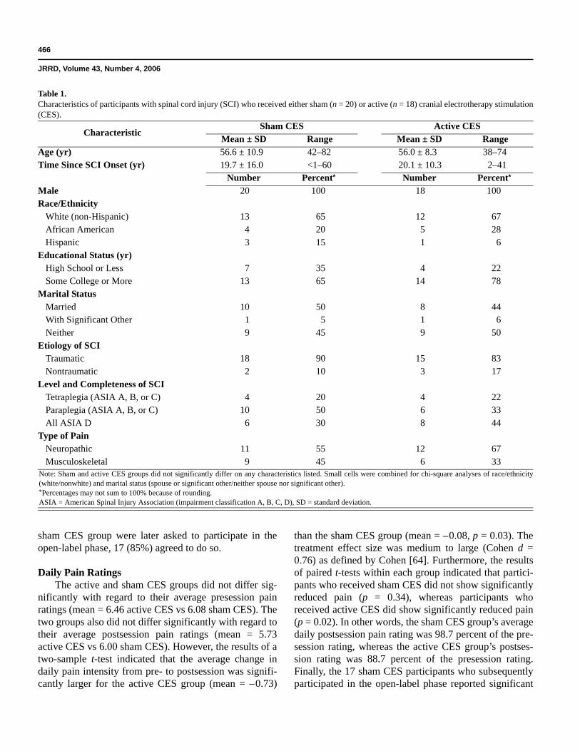
Table 1.Characteristics of participants with spinal cord injury (SCI) who received either sham (n = 20) or active (n = 18) cranial electrotherapy stimulation(CES).
CharacteristicSham CES Active CES
Mean ± SD Range Mean ± SD RangeAge (yr) 56.6 ± 10.9 42–82 56.0 ± 8.3 38–74Time Since SCI Onset (yr) 19.7 ± 16.0 <1–60 20.1 ± 10.3 2–41
Number Percent* Number Percent*
Male 20 100 18 100Race/Ethnicity
White (non-Hispanic) 13 65 12 67African American 4 20 5 28Hispanic 3 15 1 6
Educational Status (yr)High School or Less 7 35 4 22Some College or More 13 65 14 78
Level and Completeness of SCITetraplegia (ASIA A, B, or C) 4 20 4 22Paraplegia (ASIA A, B, or C) 10 50 6 33All ASIA D 6 30 8 44
Type of PainNeuropathic 11 55 12 67Musculoskeletal 9 45 6 33
Note: Sham and active CES groups did not significantly differ on any characteristics listed. Small cells were combined for chi-square analyses of race/ethnicity(white/nonwhite) and marital status (spouse or significant other/neither spouse nor significant other).*Percentages may not sum to 100% because of rounding.ASIA = American Spinal Injury Association (impairment classification A, B, C, D), SD = standard deviation.
467
TAN et al. Cranial electrotherapy stimulation and SCI
postsession pain reduction (p = 0.003). These findingsare summarized in Table 2.
Pain ratings before and after the daily treatment ses-sions for the active and sham CES groups are shown inFigures 2 and 3, respectively. Of the 20 individuals orig-inally assigned to the sham CES group, 17 participated inthe open-label phase. Mean pain ratings before and afterdaily treatment sessions for these 17 participants are dis-played in Figure 4.
Brief Pain Inventory
Pain IntensityTwo-sample t-tests revealed that neither degree of
change in any of the four BPI Pain Intensity subscaleitems nor the composite pain intensity score significantlydiffered between the active and Sham CES groups. How-ever, paired t-tests within treatment groups revealed thatamong the four BPI Pain Intensity subscale items, theworst pain item decreased the most and thereforeappeared most sensitive to CES. However, none of thechanges in the BPI Pain Intensity subscale items was sta-tistically significant for any of the three groups (active,sham, and open-label).
Pain InterferenceTwo-sample t-tests revealed no significant difference
between the active and sham CES groups with regard tochange from pre- to postintervention in any of the 10 BPIPain Interference subscale items or the composite paininterference score. However, in paired t-tests for theactive CES group, 7 of the 10 individual Pain Interferencesubscale items significantly changed and reflected smallto moderate effect sizes: general activity (Cohen d =0.67), self-care (Cohen d = 0.58), sleep (Cohen d = 0.53),
social activities (Cohen d = 0.51), normal work (Cohend = 0.45), enjoyment of life (Cohen d = 0.42), and recre-ational activities (Cohen d = 0.38). A paired t-test withinthe active CES group revealed that the composite paininterference score decreased significantly (mean change =–14.6, p = 0.004, Cohen d = 0.50). For the sham CESgroup, neither the individual BPI Pain Interference sub-scale items nor the composite pain interference scorechanged significantly (mean change = –4.7, p = 0.24);however, during the open-label phase, pain interferencewith sleep decreased significantly (Cohen d = 0.40). Thefindings for the three BPI Pain Interference subscaleitems with effect sizes > 0.50 and the composite paininterference score are displayed in Figures 5–8. Althoughthe active CES group showed a significantly reducedcomposite pain interference score from pre- to postinter-vention, the change scores between the active and shamCES groups were not significantly different, partlybecause both groups showed decreased composite paininterference scores from pre- to postintervention. In otherwords, the slope for the active CES group was significantand the slope for the sham CES group was not significant;however, the two slopes were not significantly differentfrom each other.
Exploratory analyses of the effectiveness of CESwere performed only on the data from the initial activeCES group (n = 18). The relationship of effectiveness toetiology of SCI, level and completeness of SCI, type ofpain, and initial pain ratings was examined.
Etiology of Spinal Cord InjuryOnly three participants in the active CES group had
nontraumatic SCI. The mean change in daily pain ratingsfrom pre- to postsession was –1.04 for the 3 participantswith nontraumatic SCI and –0.67 for the 15 participants
Table 2.Average daily pain ratings before and after 21-day cranial electrotherapy stimulation (CES). Participants rated pain on scale from 0 (“no pain”) to10 (“pain as bad as you can imagine”).
CESCondition n
Before After Change*t-Test
Mean ± SD Range Mean ± SD Range Mean ± SD RangeActive 18 6.46 ± 1.95 2.94 to 10.00 5.73 ± 2.56 1.24 to 10.00 –0.73 ± 1.15 –4.14 to 0.00 2.69†
Sham 20 6.08 ± 2.42 1.93 to 10.00 6.00 ± 2.41 1.60 to 10.00 –0.08 ± 0.38 –1.20 to 0.67 0.98Open-Label 17 5.97 ± 2.35 1.95 to 10.00 5.51 ± 2.51 0.95 to 10.00 –0.46 ± 0.54 –1.48 to 0.19 3.47‡
*Mean after-session rating minus mean before-session rating (negative change scores indicate decreased pain intensity).†p < 0.05.‡ p < 0.01.SD = standard deviation.
468
JRRD, Volume 43, Number 4, 2006
Figure 2.Daily pre- and postsession pain ratings for participants with spinalcord injury who received active cranial electrotherapy stimulation(n = 18). Participants rated pain on scale from 0 (“no pain”) to 10(“pain as bad as you can imagine”).
Figure 3.Daily pre- and postsession pain ratings for participants with spinalcord injury who received sham cranial electrotherapy stimulation (n =20). Participants rated pain on scale from 0 (“no pain”) to 10 (“pain asbad as you can imagine”).
Figure 4.Daily pre- and postsession pain ratings for 17 participants with spinalcord injury who received sham cranial electrotherapy stimulation(CES) in first arm of study and active CES in second (open-label)arm. Participants rated pain on scale from 0 (“no pain”) to 10 (“painas bad as you can imagine”).
Figure 5.Pre- and postintervention pain interference with general activity (asmeasured by Pain Interference subscale of Brief Pain Inventory) forparticipants who received either sham or active cranial electrotherapystimulation. Participants rated pain interference on scale from 0(“does not interfere”) to 10 (“interferes completely”).
Figure 6.Pre- and postintervention pain interference with enjoyment of life (asmeasured by Pain Interference subscale of Brief Pain Inventory) forparticipants who received either sham or active cranial electrotherapystimulation. Participants rated pain interference on scale from 0(“does not interfere”) to 10 (“interferes completely”).
Figure 7.Pre- and postintervention pain interference with social activities (asmeasured by Pain Interference subscale of Brief Pain Inventory) forparticipants who received either sham or active cranial electrotherapystimulation. Participants rated pain interference on scale from 0(“does not interfere”) to 10 (“interferes completely”).
469
TAN et al. Cranial electrotherapy stimulation and SCI
with traumatic SCI. Decrease in pain intensity, as measuredby the BPI Pain Intensity subscale, was greater for the non-traumatic vs traumatic SCI participants in the individualsubscale items, as well as the composite pain intensityscore (–2.33 vs –1.93, respectively). Decrease in pain inter-ference was also greater for the nontraumatic vs traumaticSCI participants for 5 of the 10 BPI Pain Interference sub-scale items; the two items that decreased significantly wereenjoyment of life (–5.33 vs –0.73, respectively, p < 0.001)and social activities (–4.57 vs –1.20, respectively, p =0.03); the composite pain interference scores were not sig-nificantly different (–21.33 vs –13.20, respectively).
Level and Completeness of Spinal Cord InjuryIn the active CES group, the mean difference
between the daily pre- and postsession ratings wasgreater for those participants with less impairment fromthe SCI. Participants with tetraplegia with ASIA grade A,B, or C (n = 4) had the smallest pain decrease (meanchange = –0.22), participants with paraplegia with ASIAgrade A, B, or C (n = 6) had a mean change of –0.46, andparticipants with ASIA grade D (n = 8) had a meanchange of –1.19. This same pattern was observed for theaverage pain and worst pain items of the BPI Pain Inten-sity subscale. However, when change in pain interferencewas examined, participants with paraplegia with ASIAgrade A, B, or C had the greatest decreases for 9 of the 10individual BPI Pain Interference subscale items and thecomposite pain interference score.
Type of PainIn the active CES group, 6 participants had musculo-
skeletal pain and 12 had neuropathic pain. Change in the
daily pre- and postsession pain intensity ratings was largerfor the neuropathic than the musculoskeletal group (–0.81vs –0.57, respectively); however, because of the smallnumber of participants in each group, this difference wasnot statistically significant. The findings for pain intensityas measured by the BPI Pain Intensity subscale weremixed. Participants with musculoskeletal pain had greaterpre- to postsession changes for two of the four items(worst pain and pain now) and the composite pain inten-sity score, while participants with neuropathic pain hadgreater pre- to postsession changes for the other two items(average pain and least pain). For pain interference,musculoskeletal pain improved more than neuropathicpain for 8 of the 10 BPI Pain Interference subscale itemsand the composite pain interference score (–19.5 vs –12.1,respectively), but the differences were not significant.
Relation of Change in Pain Ratings to Level of Initial Pain Ratings
Examination of the relationship between the meanpresession score and the mean daily change scorerevealed that change in pain during the sessions wasgreater for participants who had less intense presessionpain, particularly those whose mean presession pain ratingwas 7 or less. However, when we examined the relation-ships between the preintervention BPI Pain Intensity sub-scale items and the change in pain intensity, participantswith higher initial pain intensity had greater improvementin pain intensity during the 3-week study period. A similarpattern was found for the BPI Pain Interference subscaleitems and the composite pain interference score: partici-pants with higher preintervention scores improved moreduring the 3-week period.
DISCUSSION
This study extended the knowledge base regardingeffectiveness of CES for pain in persons with SCI. Weestablished that persons with SCI can and will use theCES device at home for a 3-week period. This period ofuse is longer than that in other studies and this differencemay be important. Some persons may need a longer trialperiod before experiencing any treatment effects. Thetechnology of the specific CES device used in this study isquite different from the devices used in other studies. Inthis pilot study, the participants who received active CESreported, on average, significant pain reduction after each
Figure 8.Pre- and postintervention composite pain interference scores (totalscore for 10 items on modified Pain Interference subscale of BriefPain Inventory) for participants who received either sham or activecranial electrotherapy stimulation.
470
JRRD, Volume 43, Number 4, 2006
of the 21 daily sessions, but those who received shamCES did not (Figures 2–3), indicating a medium to largeeffect size. In separate paired t-tests, a significant differ-ence was demonstrated between the pre- and postsessionpain ratings for the active CES group but not the shamCES group. Furthermore, when the 17 participants origi-nally in the sham CES group participated in the subse-quent open-label phase, their pre- versus postinterventionratings significantly differed (Figure 4). The pain reduc-tion in this study was not as great as in Capel et al.’s studyin terms of pain intensity as a percentage of baseline [49].In the first arm of their study, the pain rating on the finalday was about 50 percent of baseline for the active CEStreatment group, whereas our study found only an 11 per-cent change.
Many factors may allow the relatively small changein average daily pain ratings to be viewed in perspective.First, change scores varied considerably more in theactive CES group than the sham CES group (SD = 1.15vs 0.38, respectively; range = 4.14 vs 1.20, respectively).Second, we set the dose at a subthreshold of 100 μA tomaintain the double-blind design. Possibly, higher dosesresult in greater improvement in pain; however, to date,no study has examined the dose-response effect of CESon pain.
Post hoc exploratory examination of the daily painrating data suggested that persons with nontraumatic SCI,lower level and/or less complete SCI, neuropathic pain,and mild-to-moderate pain intensity may get more imme-diate benefit from CES than persons with traumatic SCI,higher and/or more complete SCI, musculoskeletal pain,and more severe pain. Thus, the size of change in painratings likely increases when specific subgroups of per-sons with SCI are targeted for CES treatment. Additionalstudies are required for identifying the patients mostlikely to benefit from CES and for investigating the pos-sible mechanistic basis of such effects. Studies are alsoneeded for determining the duration of pain relief aftereach session. Figure 2 suggests that improvements inpain dissipated substantially by the next session. How-ever, no known reason exists that CES treatment couldnot be used indefinitely every day.
The findings of paired t-tests within groups revealedthat the observed reductions in the individual and com-posite BPI Pain Intensity subscale scores were not signifi-cant. However, the results showed that the composite paininterference score and several individual BPI Pain Inter-ference subscale items decreased significantly for the
active CES group but not for the sham CES group frompre- to postintervention.
Thus, these data indicate that CES effectivelyreduced pain intensity immediately after each treatmentsession, but its long-term effects on pain reduction werenot statistically supported. Furthermore, exploratoryanalyses indicated that the group that benefited mostfrom CES differed depending on the outcome measureexamined (i.e., average daily decrease in pain intensity,3-week decrease in pain intensity, or 3-week decrease inpain interference. Short-term relief of pain intensity wasgreater in persons with less severe pain, while longerterm relief of pain intensity and interference was greaterin persons with more severe pain. Such differences ineffect may possibly be due to the small sample size.Future research should replicate the findings using alarger sample size and should more closely examine theissue of long-term treatment effects.
The fact that active CES significantly altered short-term pain intensity and long-term pain interference butnot long-term pain intensity should not compromise theimportance of the findings. Other studies have shown that(1) pain interference plays a central role in mediating therelationship between negative emotions (such as depres-sion) and disability [65], (2) pain interference mediatesthe effect of pain severity on depression [65], and (3) per-ceived control over pain interference with daily activitiesis more strongly associated with functioning than per-ceived control over pain intensity [66]. The relationshipsamong pain intensity, pain interference, depression, anddisability should be examined carefully in future studieson the effects of CES treatment.
One important limitation of the present study is thatthe participants were all male veterans who were receiv-ing care at a VA healthcare facility. Generalizing the find-ings to female patients and other persons with SCI outsideof the VA population should be done cautiously becauseof the somewhat unique sociodemographic factors inher-ent to our patient group and its type(s) and access to lon-gitudinal care. Furthermore, this study did not address anumber of important factors, including (1) the impact ofCES on psychological distress and quality of life, (2) pos-sible reduction in analgesic consumption, and (3) theamenability of patients to using the device long term if itwere available. Finally, while many statistical tests wereperformed, thereby increasing the probability of chancefindings, the preliminary nature of this study and the useof post hoc analysis of many variables was justifiable so
471
TAN et al. Cranial electrotherapy stimulation and SCI
as to better inform or suggest domains of inquiry forfuture studies.
CONCLUSIONS
In conclusion, the findings of this study, if replicatedwith larger samples, support the use of CES as a practicaland effective treatment for particular types of SCI-induced pain.
ACKNOWLEDGMENTS
We wish to thank Daniel L. Kirsch, PhD, of Electro-medical Products International, Inc (Mineral Wells,Texas) for the generous provision of Alpha-Stim® 100units for use in this study. Alpha-Stim® provided adviceand consultation upon request from the authors. As well,we acknowledge the scientific advice and assistance ofJames Giordano, PhD, on this article.
Dr. Yang is now with the Washington, DC, VAMC,and Ms. Vasilev is now with the University of Texas M.D. Anderson Cancer Center.
This material was based on work supported by theVA Rehabilitation Research and Development ServiceCenter of Excellence on Healthy Aging with Disabilities,MEDVAMC, grant E0802-C.
The authors have declared that no competing inter-ests exist.
2. Rintala DH, Loubser PG, Castro J, Hart KA, Fuhrer MJ.Chronic pain in a community-based sample of men withspinal cord injury: prevalence, severity, and relationshipwith impairment, disability, handicap, and subjective well-being. Arch Phys Med Rehabil. 1998;79(6):604–14.[PMID: 9630137]
3. Rintala DH, Holmes SA, Fiess RN, Courtade D, LoubserPG. Prevalence and characteristics of chronic pain in veter-ans with spinal cord injury. J Rehabil Res Dev. 2005;42(5):573–84. [PMID: 16586183]
4. Richards JS, Meredith RL, Nepomuceno C, Fine PR, Ben-nett G. Psycho-social aspects of chronic pain in spinal cordinjury. Pain. 1980;8(3):355–66. [PMID: 7402693]
5. Davidoff G, Roth E, Guarracini M, Sliwa J, Yarkony G.Function-limiting dysesthetic pain syndrome among trau-matic spinal cord injury patients: a cross-sectional study.Pain. 1987;29(1):39–48. [PMID: 3588000]
6. Rintala DH, Hart KA, Fuhrer MJ. Self-reported pain in per-sons with chronic spinal cord injury [abstract]. J Am Para-plegia Soc. 1991;14(2):83.
7. Burke DC. Pain in paraplegia. Paraplegia. 1973;10(4):297–313. [PMID: 4697003]
8. Sie IH, Waters RL, Adkins RH, Gellman H. Upper extrem-ity pain in the postrehabilitation spinal cord injured patient.Arch Phys Med Rehabil. 1992;73(1):44–48.[PMID: 1729973]
11. Tan G, Young S. Psychosocial and vocational issues inrehabilitation. In: Monga TN, Grabois M, editors. Painmanagement in rehabilitation. New York (NY): DemosMedical Publishing; 2002. p. 35–58.
12. Loubser PG, Donovan WH. Chronic pain associated withspinal cord injury. In: Narayan RK, Wilberger JE, Pov-lishock JT, editors. Neurotrauma. New York (NY):McGraw Hill; 1996. p. 1311–22.
13. Ragnarsson KT. Management of pain in persons with spi-nal cord injury. J Spinal Cord Med. 1997;20(2):186–99.[PMID: 9144608]
14. Wilson OB, Hamilton RF, Warner RL, Johnston CM,deFriece R, Harter L, Schweitzer C, Talaverra J, Hymel CM,Skolnick MH. The influence of electrical variables on analge-sia produced by low current transcranial electrostimulationof rats. Anesth Analg. 1989;68(5):673–81. [PMID: 2719297]Erratum in: Anesth Analg. 1990;70(4):474.
15. Capel ID, Dorrell HM, Spencer EP. The application of sub-perception electrical stimuli elicits a temporally distinctresponse from restraint stress: I. Antinociceptive charac-teristics. J Bioelectricity. 1990;9:167–76.
16. Qiao JT, Skolnick MH, Dafny N. Dorsal raphe and externalelectrical stimulation modulate noxious input to single neu-rons in nucleus parafascicularis thalami. Brain Res Bull.1988;21(4):671–75. [PMID: 3208154]
17. Dong WQ, Wilson OB, Skolnick MH, Dafny N. Hypotha-lamic, dorsal raphe and external electrical stimulation modu-late noxious evoked responses of habenula neurons.Neuroscience. 1992;48(4):933–40. [PMID: 1630629]
18. Pozos RS, Strack LE, White RK, Richardson AW. Electros-leep versus electroconvulsive therapy. In: Reynolds DV,Sjoberg AE, editors. Neuroelectric research. Springfield(IL): Charles Thomas; 1971. p. 221–25.
19. Stanley TH, Cazalaa JA, Limoge A, Louville Y. Transcutane-ous cranial electrical stimulation increases the potency ofnitrous oxide in humans. Anesthesiology. 1982;57(4):293–97.[PMID: 6982009]
20. Stanley TH, Cazalaa JA, Atinault A, Coeytaux R, Limoge A,Louville Y. Transcutaneous cranial electrical stimulationdecreases narcotic requirements during neurolept anesthesiaand operation in man. Anesth Analg. 1982;61(10):863–66.[PMID: 7125252]
21. Braverman E, Smith R, Smayda R, Blum K. Modificationof P300 amplitude and other electrophysiological parame-ters of drug abuse by cranial electrical stimulation. CurrTher Res. 1990;48(4):586–96.
22. Krupitsky EM, Burakov AM, Karandashova GF, Katsnel-son J, Lebedev VP, Grinenko AJ, Borodkin JS. The admin-istration of transcranial electric treatment for affectivedisturbances therapy in alcoholic patients. Drug AlcoholDepend. 1991;27(1):1–6. [PMID: 2029855]
23. Philip P, Demotes-Mainard J, Bourgeois M, Vincent JD.Efficiency of transcranial electrostimulation on anxiety andinsomnia symptoms during a washout period in depressedpatients. A double-blind study. Biol Psychiatry. 1991;29(5):451–56. [PMID: 2018818]
24. Kulkarni AD, Smith RB. The use of microcurrent electricaltherapy and cranial electrotherapy stimulation in pain con-trol. Clin Pract Altern Med. 2001;2(2):99–103.
25. Brotman P. Low-intensity transcranial electrostimulationimproves the efficacy of thermal biofeedback and quietingreflex training in the treatment of classical migraine head-ache. Am J Electromed. 1989;6(5):120–23.
26. Solomon S, Elkind A, Freitag F, Gallagher RM, Moore K,Swerdlow B, Malkin S. Safety and effectiveness of cranialelectrotherapy in the treatment of tension headache. Head-ache. 1989;29(7):445–50. [PMID: 2668227]
27. Romano TJ. The usefulness of cranial electrotherapy in thetreatment of headache in fibromyalgia patients. Am J PainManag. 1993;3:15–19.
28. Clark MS, Silverstone LM, Lindenmuth J, Hicks MJ, Aver-bach RE, Kleier DJ, Stoller NH. An evaluation of the clini-cal analgesia/anesthesia efficacy on acute pain using thehigh frequency neural modulator in various dental settings.Oral Surg Oral Med Oral Pathol. 1987;63(4):501–5.[PMID: 3554095]
29. Hochman R. Neurotransmitter modulator (TENS) for con-trol of dental operative pain. J Am Dent Assoc. 1988;116(2):208–12. [PMID: 3278031]
30. Forster S, Post BS, Benton JG. Preliminary observations onelectrosleep. Arch Phys Med Rehabil. 1963;44:481–89.[PMID: 14050721]
31. Magora F, Beller A, Aladjemoff L, Magora A, Tannen-baum J. Observations on electrically induced sleep in man.Br J Anaesth. 1965;37(7):480–91. [PMID: 5318569]
32. Kirsch DL, Lerner FN. Electromedicine: The other side ofphysiology. In: Weiner RS, editor. Innovations in pain man-agement: a practical guide for clinicians. Boca Raton (FL):St. Lucie Press; 1998.
33. Kirsch DL, Smith RB. The use of cranial electrotherapystimulation in the management of chronic pain: a review.NeuroRehabilitation. 2000;14(2):85–94. [PMID: 11455071]
34. Lichtbroun AS, Raicer MC, Smith RB. The treatment offibromyalgia with cranial electrotherapy stimulation. J ClinRheumatol. 2001;7(2):72–78.
35. Donaldson CCS, Sella GE, Mueller HH. The neural plas-ticity model of fibromyalgia: theory, assessment, and treat-ment. Pract Pain Manag. 2001;25–29.
37. Frankel BL, Buchbinder R, Snyder F. Ineffectiveness ofelectrosleep in chronic primary insomnia. Arch Gen Psy-chiatry. 1973;29(4):563–68. [PMID: 4748315]
38. Koegler RR, Hicks SM, Barger JH. Medical and psychiat-ric use of electrosleep. Transcerebral electrotherapy. DisNerv Syst. 1971;32(2):100–104. [PMID: 5313321]
39. Levitt EA, James NM, Flavell P. A clinical trial of electros-leep therapy with a psychiatric inpatient sample. Aust N ZJ Psychiatry. 1975;9(4):287–90. [PMID: 769773]
40. McKenzie RE, Rosenthal SH, Driessner JS. Some psycho-physiologic effects of electrical transcranial stimulation(electrosleep). In: Wulfsohn NL, Sances A, editors. Thenervous system and electric currents. New York (NY): Ple-num; 1976. p. 163–67.
41. Rosenthal SH, Wulfsohn NL. Electrosleep. A preliminarycommunication. J Nerv Ment Dis. 1970;151(2):146–51.[PMID: 5457619]
42. Rosenthal SH, Wulfsohn NL. Studies of electrosleep withactive and simulated treatment. Curr Ther Res Clin Exp.1970;12(3):126–30. [PMID: 4985490]
43. Singh K. Sleep inducing devices a clinical trial with a Rus-sian machine. Int J Neuropsychiatry. 1967;3(4):311–18.[PMID: 6064118]
44. Matteson MT, Ivancevich JM. An exploratory investigationof CES as an employee stress management technique.J Health Hum Resour Adm. 1986;9:93–109.
45. Smith RB. Cranial electrotherapy stimulation in the treat-ment of stress related cognitive dysfunction, with an eigh-teen month follow up. J Cogn Rehabil. 1999;17(6):14–18.
46. Kirsch DL. Postmarketing survey of Alpha-Stim CESpatients. In: Kirsch DL, editor. The science behind cranialelectrotherapy stimulation. Edmonton (Canada): MedicalScope Publishing; 1999. p. 148.
47. Smith RB, Tiberi A, Marshall J. The use of cranial electro-therapy stimulation in the treatment of closed-head-injuredpatients. Brain Inj. 1994;8(4):357–61. [PMID: 8081350]
TAN et al. Cranial electrotherapy stimulation and SCI
48. Overcash SJ. A retrospective study to determine the effectof cranial electrotherapy stimulation (CES) on patientssuffering from anxiety disorders. Am J Electromed. 1999;16(1):49–51.
49. Capel ID, Dorrell HM, Spencer EP, Davis MW. The ame-lioration of the suffering associated with spinal cord injurywith subperception transcranial electrical stimulation. Spi-nal Cord. 2003;41(2):109–17. [PMID: 12595874]
50. American Spinal Injury Association. International stan-dards for neurological and functional classification of spi-nal cord injury. 5th ed. Chicago, IL: American SpinalInjury Association; 1996.
51. Siddall PJ, Yezierski RP, Loeser JD. Pain following spinalcord injury: clinical features, prevalence, and taxonomy.IASP Newsletter. 2000;(3):3–7.
52. Siddall PJ, Taylor DA, Cousins MJ. Classification of painfollowing spinal cord injury. Spinal Cord. 1997;35(2):69–75.[PMID: 9044512]
53. Giordano J. The neurobiology of nociceptive and anti-noci-ceptive systems. Pain Physician. 2005;8(3):277–90.
54. Cleeland CS. Measurement of pain by subjective report. In:Chapman CR, Loeser JD, editors. Issues in pain meas-urement. New York (NY): Raven Press; 1989. p. 391–403.
55. Tan G, Jensen MP, Thornby JI, Shanti BF. Validation of theBrief Pain Inventory for chronic nonmalignant pain. J Pain.2004;5(2):133–37. [PMID: 15042521]
56. Small EJ, Smith MR, Seaman JJ, Petrone S, Kowalski MO.Combined analysis of two multicenter, randomized, pla-cebo-controlled studies of pamidronate disodium for thepalliation of bone pain in men with metastatic prostate can-cer. J Clin Oncol. 2003;21(23):4277–84. [PMID: 14581438]
57. Lang E, Liebig K, Kastner S, Neundorfer B, HeuschmannP. Multidisciplinary rehabilitation versus usual care forchronic low back pain in the community: Effects on qualityof life. Spine J. 2003;3(4):270–76. [PMID: 14589185]
58. Marshall HM, Jensen MP, Ehde DM, Campbell KM. Painsite and impairment in individuals with amputation pain.Arch Phys Med Rehabil. 2002;83(8):1116–19.[PMID: 12161833]
59. Gammaitoni AR, Galer BS, Lacouture P, Domingos J,Schlagheck T. Effectiveness and safety of new oxycodone/acetaminophen formulations with reduced acetaminophenfor the treatment of low back pain. Pain Med. 2003;4(1):21–30. [PMID: 12873275]
60. Tyler EJ, Jensen MP, Engel JM, Schwartz L. The reliabilityand validity of pain interference measures in persons withcerebral palsy. Arch Phys Med Rehabil. 2002;83(2):236–39.[PMID: 11833028]
61. Cleeland CS. Pain assessment in cancer. In: Osoba D, edi-tor. Effect of cancer on quality of life. Boca Raton (FL):CRC Press; 1991. p. 293–305.
62. Engel JM, Schwartz L, Jensen MP, Johnson DR. Pain incerebral palsy: the relation of coping strategies to adjust-ment. Pain. 2000;88(3):225–30. [PMID: 1068109]
63. Jensen MP, Ehde DM, Hoffman AJ, Patterson DR, Czerni-ecki JM, Robinson LR. Cognitions, coping and social envi-ronment predict adjustment to phantom limb pain. Pain.2002;95(1–2):133–42. [PMID: 11790476]
64. Cohen J. Statistical power analysis for the behavioral sci-ences. 2nd ed. Hillsdale (NJ): Lawrence Earlbaum Associ-ates; 1988.
65. Tan G, Thornby JI, Jensen MP. Revisiting negative emo-tions, pain, and functioning [abstract]. J Pain. 2004;5(3 Suppl 1):124.
66. Tan G, Jensen MP, Robinson-Whelen S, Thornby JI,Monga T. Measuring control appraisals in chronic pain.J Pain. 2002;3(5):385–93. [PMID: 14622742]
Submitted for publication April 11, 2005. Accepted inrevised form December 30, 2005.