110
Uso de Modelos In Vitro e In Vivo en el Diseňo de Nuevas Estrategias Terapéuticas Dirigidas a Blancos Moleculares En Enfermedades Cardiovasculares. SILVIA S. PIERANGELI, Ph.D. Professor
| Date post: | 22-Dec-2015 |
| Category: |
Documents |
| Upload: | elaine-greene |
| View: | 217 times |
| Download: | 0 times |
Uso de Modelos In Vitro e In Vivo en el Diseňo de Nuevas Estrategias Terapéuticas
Dirigidas a Blancos Moleculares En Enfermedades Cardiovasculares.
SILVIA S. PIERANGELI, Ph.D.
Professor
What is the Antiphospholipid Antibody Syndrome ?
An acquired autoimmune thrombophilia, characterized by:
a) vascular thrombosis.
b) recurrent pregnancy losses.
c) thrombocytopenia.
d) laboratory evidence for:
-antibodies against phospholipids or
phospholipid-binding protein cofactors.
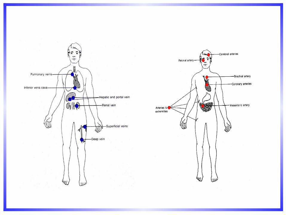
APS morbidity
APS is the most common cause of acquired thrombophilia. Prevalence in general population: 2-4%
15-20% of all DVT with or without PE. 1/3 of new strokes in patients < 50 years age. 10-15% women with recurrent pregnancy losses. APS: significant proportion of thromboembolic disease and
pregnancy loss in SLE. APL Abs present in 30-40% SLE. One third of those patients
have clinical manifestations of APS. aCL positivity may precede a more severe form of SLE.
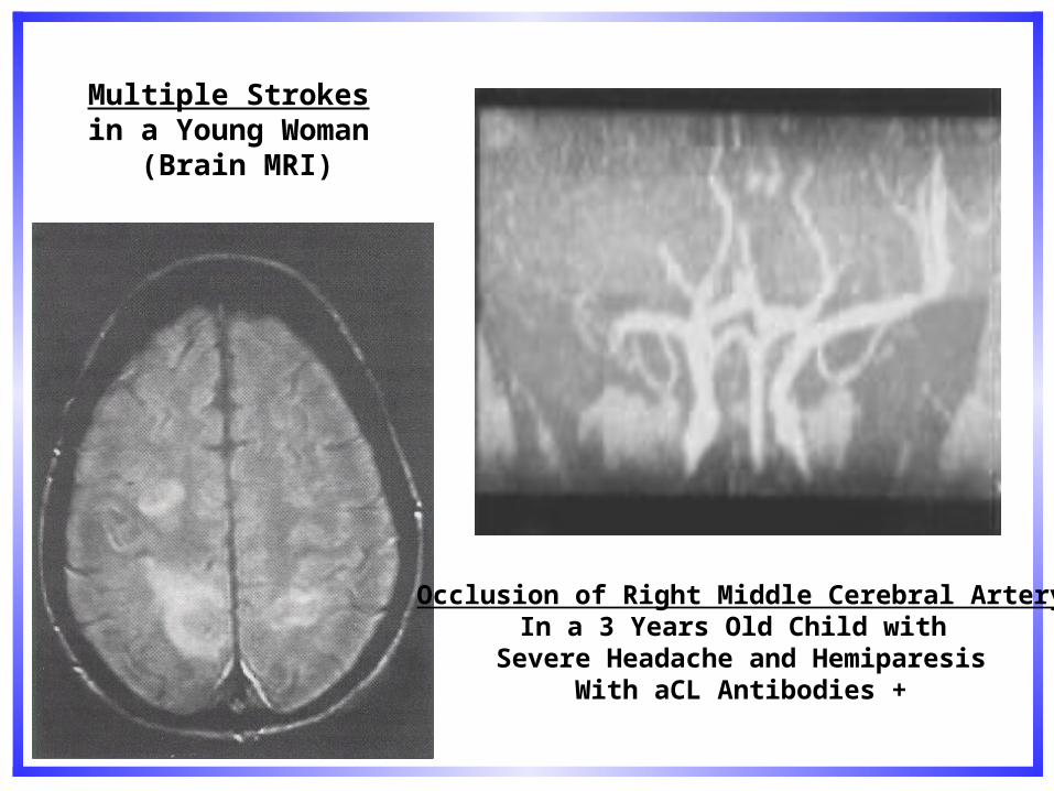
Multiple Strokes in a Young Woman
(Brain MRI)
Occlusion of Right Middle Cerebral ArteryIn a 3 Years Old Child with
Severe Headache and HemiparesisWith aCL Antibodies +
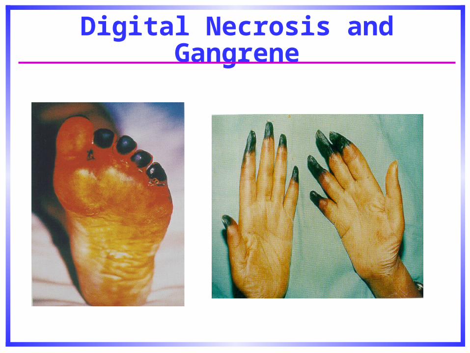
Digital Necrosis and Gangrene
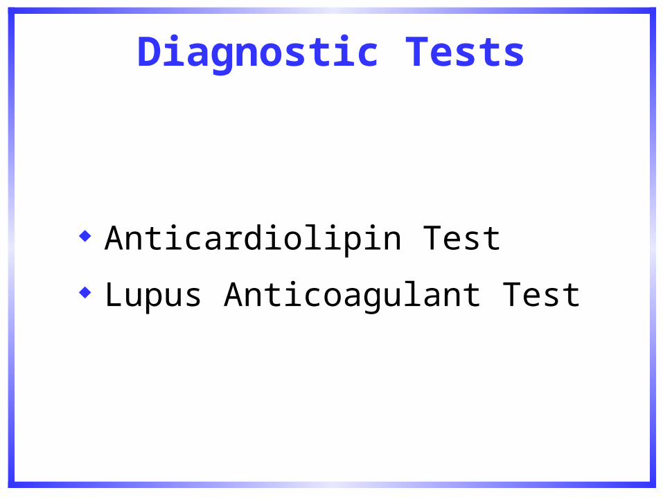
Diagnostic Tests
Anticardiolipin Test
Lupus Anticoagulant Test
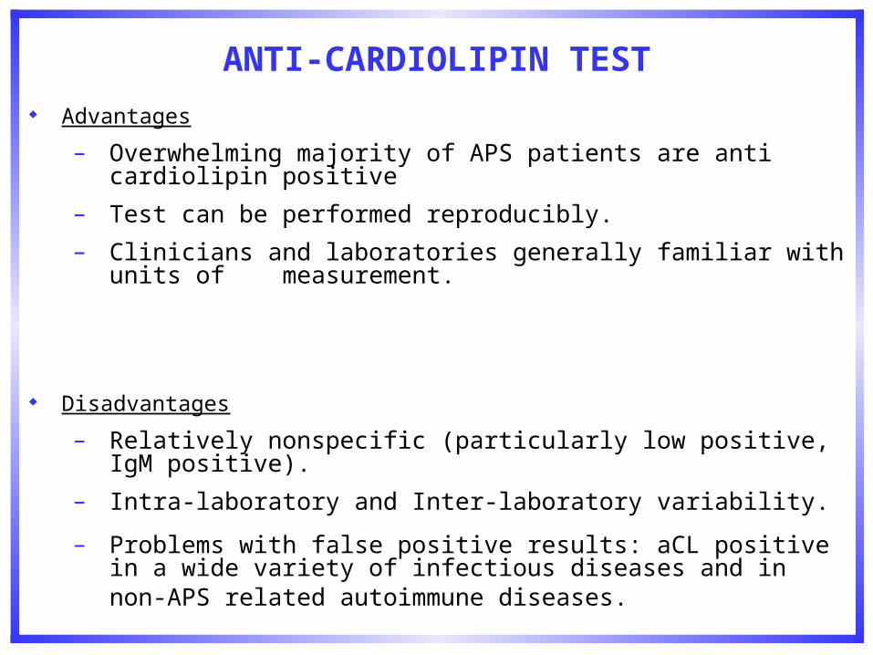
ANTI-CARDIOLIPIN TEST
Advantages
– Overwhelming majority of APS patients are anti cardiolipin positive
– Test can be performed reproducibly.
– Clinicians and laboratories generally familiar with units of measurement.
Disadvantages
– Relatively nonspecific (particularly low positive, IgM positive).
– Intra-laboratory and Inter-laboratory variability.
– Problems with false positive results: aCL positive in a wide variety of infectious diseases and in non-APS related autoimmune diseases.
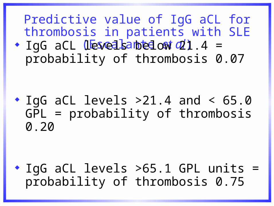
Predictive value of IgG aCL for thrombosis in patients with SLE (Escalante et al)
IgG aCL levels below 21.4 = probability of thrombosis 0.07
IgG aCL levels >21.4 and < 65.0 GPL = probability of thrombosis 0.20
IgG aCL levels >65.1 GPL units = probability of thrombosis 0.75
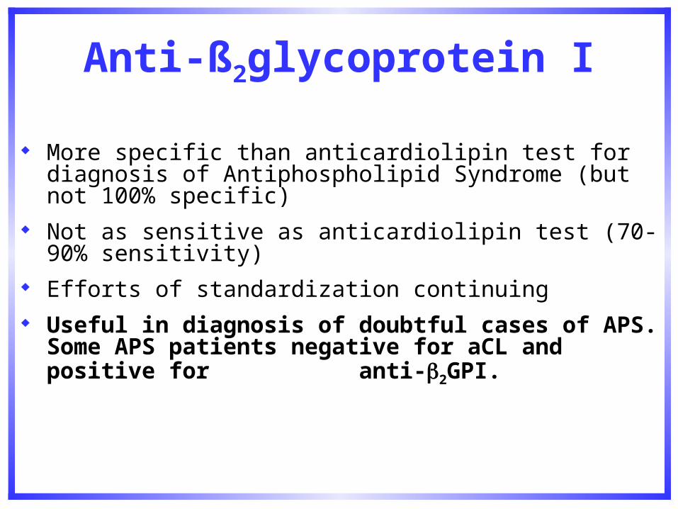
Anti-ß2glycoprotein I
More specific than anticardiolipin test for diagnosis of Antiphospholipid Syndrome (but not 100% specific)
Not as sensitive as anticardiolipin test (70-90% sensitivity) Efforts of standardization continuing Useful in diagnosis of doubtful cases of APS. Some APS
patients negative for aCL and positive for anti-2GPI.
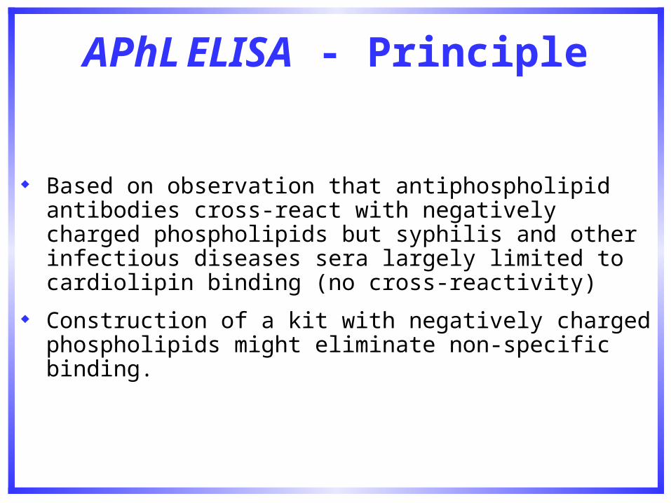
APhL ELISA - Principle
Based on observation that antiphospholipid antibodies cross-react with negatively charged phospholipids but syphilis and other infectious diseases sera largely limited to cardiolipin binding (no cross-reactivity)
Construction of a kit with negatively charged phospholipids might eliminate non-specific binding.
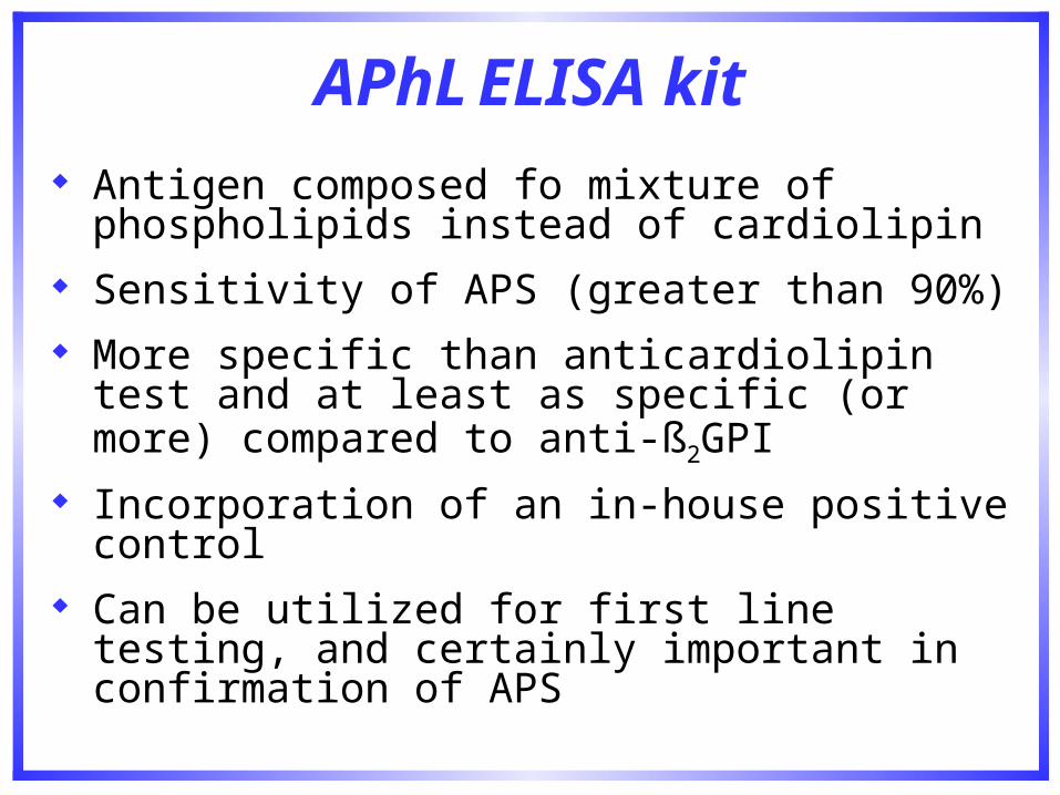
APhL ELISA kit
Antigen composed fo mixture of phospholipids instead of cardiolipin
Sensitivity of APS (greater than 90%) More specific than anticardiolipin test and at least
as specific (or more) compared to anti-ß2GPI Incorporation of an in-house positive control Can be utilized for first line testing, and certainly
important in confirmation of APS
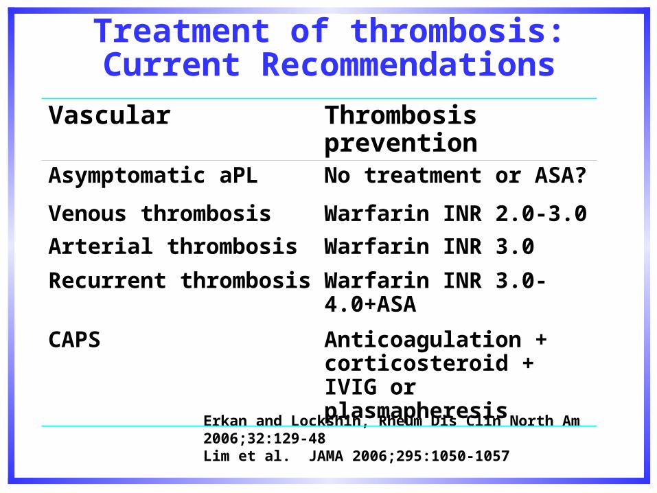
Erkan and Lockshin, Rheum Dis Clin North Am 2006;32:129-48Lim et al. JAMA 2006;295:1050-1057
Vascular Thrombosis prevention
Asymptomatic aPL No treatment or ASA?
Venous thrombosis Warfarin INR 2.0-3.0
Arterial thrombosis Warfarin INR 3.0
Recurrent thrombosis Warfarin INR 3.0-4.0+ASA
CAPS Anticoagulation + corticosteroid + IVIG or plasmapheresis
Treatment of thrombosis:Current Recommendations
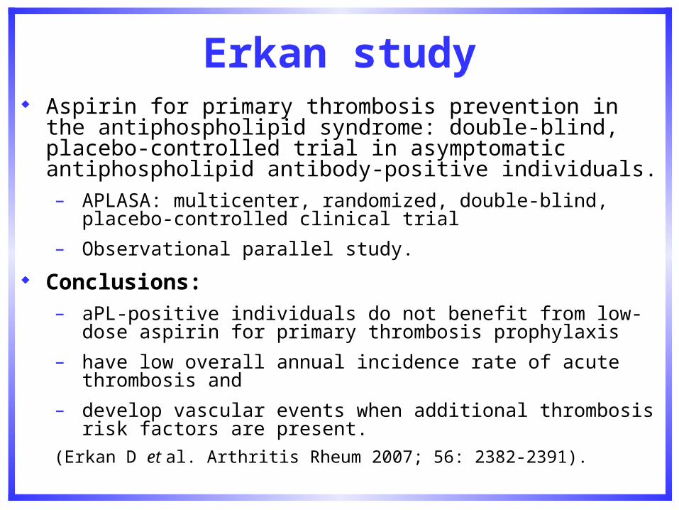
Erkan study Aspirin for primary thrombosis prevention in the
antiphospholipid syndrome: double-blind, placebo-controlled trial in asymptomatic antiphospholipid antibody-positive individuals.– APLASA: multicenter, randomized, double-blind, placebo-
controlled clinical trial
– Observational parallel study.
Conclusions: – aPL-positive individuals do not benefit from low-dose aspirin for
primary thrombosis prophylaxis
– have low overall annual incidence rate of acute thrombosis and
– develop vascular events when additional thrombosis risk factors are present.
(Erkan D et al. Arthritis Rheum 2007; 56: 2382-2391).
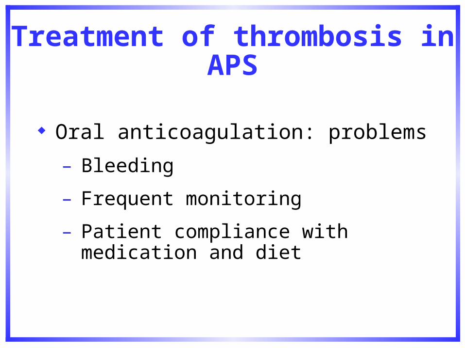
Treatment of thrombosis in APS
Oral anticoagulation: problems
– Bleeding
– Frequent monitoring
– Patient compliance with medication and diet
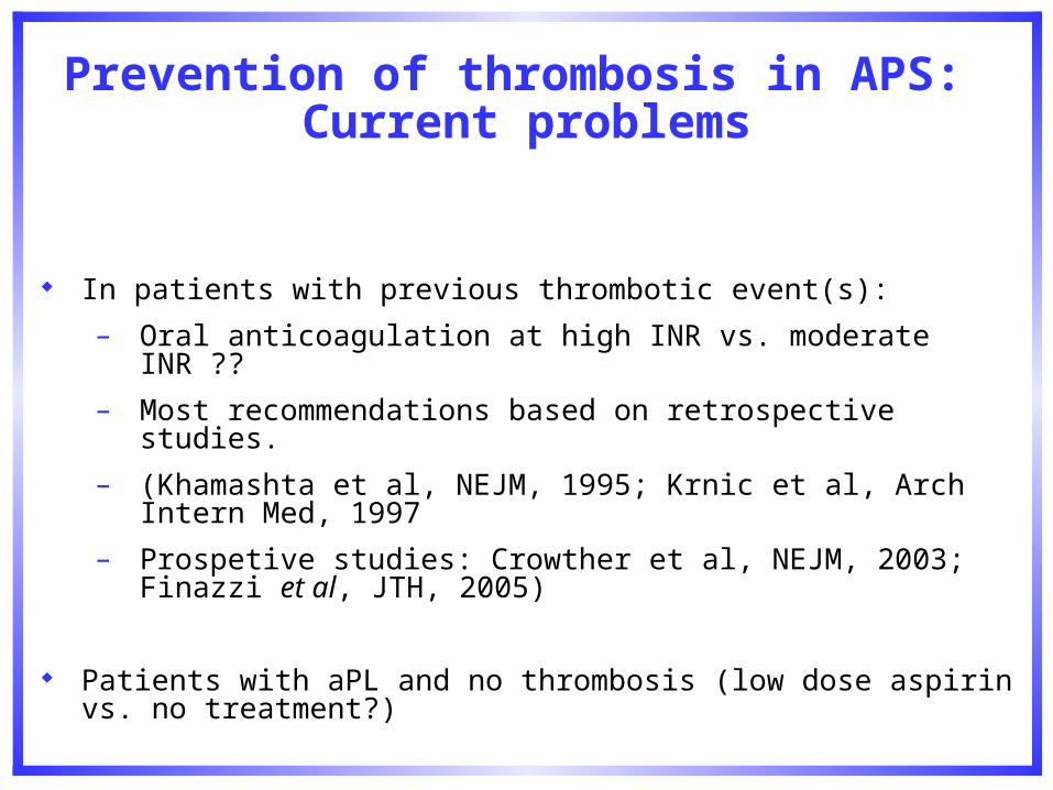
Prevention of thrombosis in APS: Current problems
In patients with previous thrombotic event(s):
– Oral anticoagulation at high INR vs. moderate INR ??
– Most recommendations based on retrospective studies.
– (Khamashta et al, NEJM, 1995; Krnic et al, Arch Intern Med, 1997
– Prospetive studies: Crowther et al, NEJM, 2003; Finazzi et al, JTH, 2005)
Patients with aPL and no thrombosis (low dose aspirin vs. no treatment?)
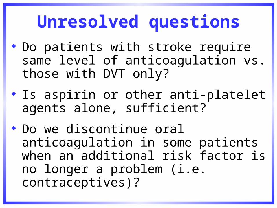
Unresolved questions
Do patients with stroke require same level of anticoagulation vs. those with DVT only?
Is aspirin or other anti-platelet agents alone, sufficient?
Do we discontinue oral anticoagulation in some patients when an additional risk factor is no longer a problem (i.e. contraceptives)?
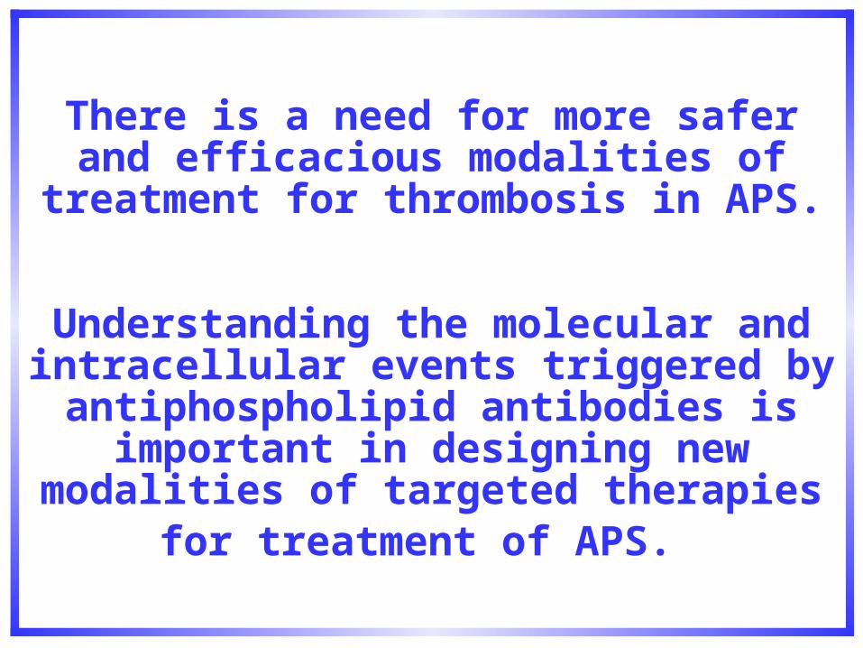
There is a need for more safer and efficacious modalities of treatment for
thrombosis in APS.
Understanding the molecular and intracellular events triggered by
antiphospholipid antibodies is important in designing new modalities of targeted
therapies for treatment of APS.
Do aPL antibodies cause/induce thrombosis?
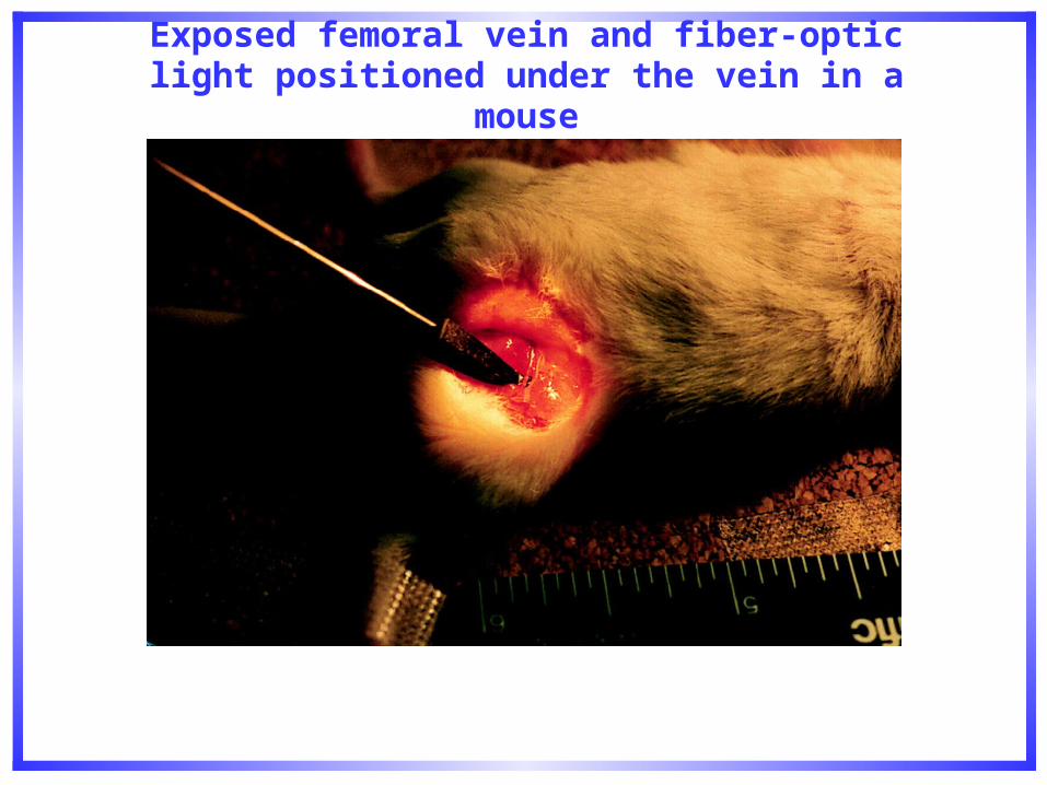
Pierangeli, S. S. et al. Circulation 1996;94:1746-1751
Exposed femoral vein and fiber-optic light positioned under the vein in a mouse
Pierangeli, S. S. et al. Circulation 1996;94:1746-1751
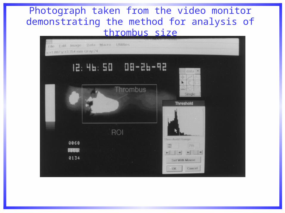
Photograph taken from the video monitor demonstrating the method for analysis of thrombus size
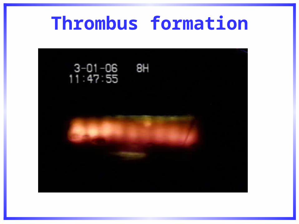
Thrombus formation
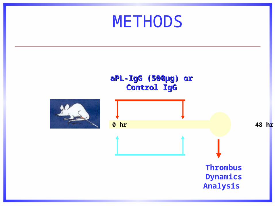
METHODS
aPL-IgG (500aPL-IgG (500µg)µg) or orControl IgGControl IgG
0 hr 48 hr 72 hr
Thrombus DynamicsAnalysis
Antiphospholipid Antibodies Promote Clot Formation in Mice

Un Visitante Ilustre…
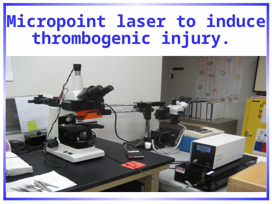
Micropoint laser to induce thrombogenic injury.
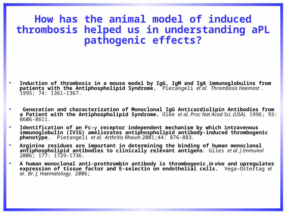
How has the animal model of induced thrombosis helped us in understanding aPL pathogenic effects?
Induction of thrombosis in a mouse model by IgG, IgM and IgA immunoglobulins from patients with the Antiphospholipid Syndrome. Pierangeli et al. Thrombosis Haemost . 1995; 74: 1361-1367.
Generation and characterization of Monoclonal IgG Anticardiolipin Antibodies from a Patient with the Antiphospholipid Syndrome. Olee et al. Proc Nat Acad Sci. (USA). 1996; 93: 8606-8611.
Identification of an Fc- receptor independent mechanism by which intravenous immunoglobulin (IVIG) ameliorates antiphospholipid antibody-induced thrombogenic phenotype. Pierangeli et al. Arthritis Rheum 2001;44: 876-883.
Arginine residues are important in determining the binding of human monoclonal antiphospholipid antibodies to clinically relevant antigens. Giles et al. J Immunol 2006; 177: 1729-1736.
A human monoclonal anti-prothrombin antibody is thrombogenic.in vivo and upregulates expression of tissue factor and E-selectin on endothelial cells. Vega-Ostertag et al. Br. J. Haematology. 2006;
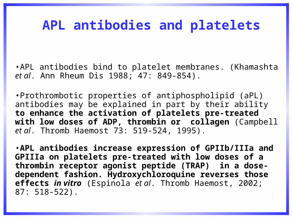
APL antibodies and platelets
•APL antibodies bind to platelet membranes. (Khamashta et al. Ann Rheum Dis 1988; 47: 849-854).
•Prothrombotic properties of antiphospholipid (aPL) antibodies may be explained in part by their ability to enhance the activation of platelets pre-treated with low doses of ADP, thrombin or collagen (Campbell et al. Thromb Haemost 73: 519-524, 1995). •APL antibodies increase expression of GPIIb/IIIa and GPIIIa on platelets pre-treated with low doses of a thrombin receptor agonist peptide (TRAP) in a dose-dependent fashion. Hydroxychloroquine reverses those effects in vitro (Espinola et al. Thromb Haemost, 2002; 87: 518-522).
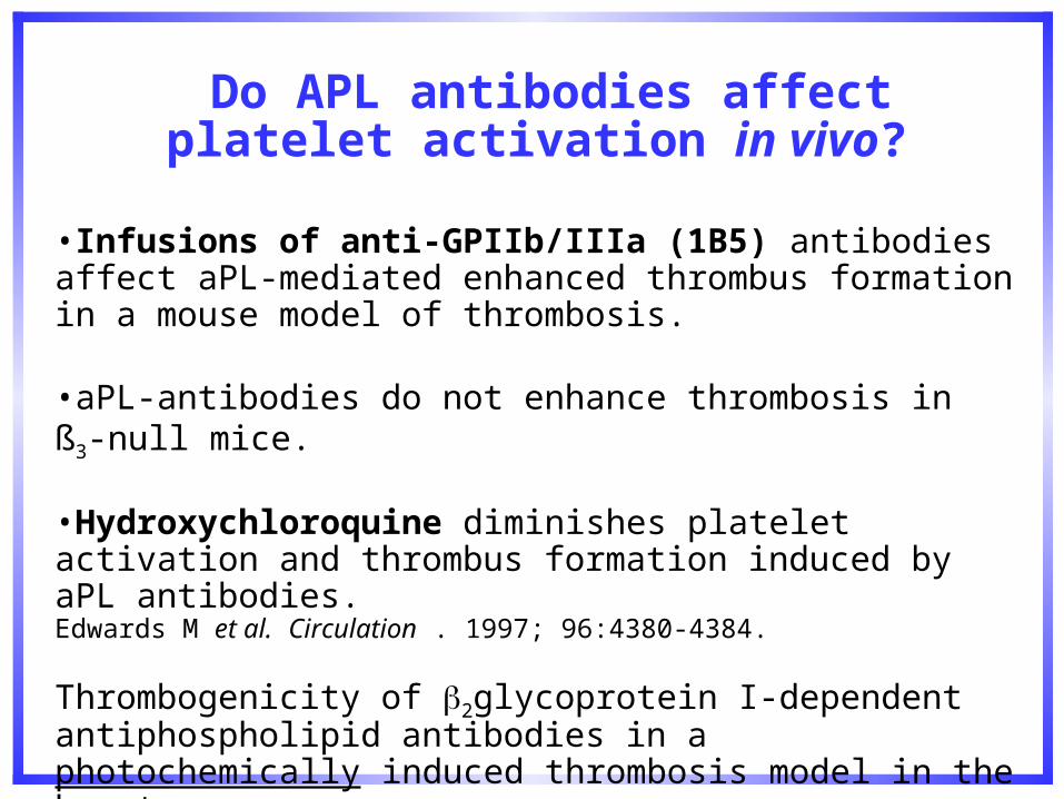
Do APL antibodies affect platelet activation in vivo?
•Infusions of anti-GPIIb/IIIa (1B5) antibodies affect aPL-mediated enhanced thrombus formation in a mouse model of thrombosis.
•aPL-antibodies do not enhance thrombosis in ß3-null mice.
•Hydroxychloroquine diminishes platelet activation and thrombus formation induced by aPL antibodies. Edwards M et al. Circulation . 1997; 96:4380-4384.
Thrombogenicity of 2glycoprotein I-dependent antiphospholipid antibodies in a photochemically induced thrombosis model in the hamster. Jankowski et al. Blood 2003; 101: 157-162.
Hydroxychloroquine in APS
Yoon KH. Sufficient evidence to consider hydroxychloroquine as an adjunct therapy in antiphospholipid (Hughes’) syndrome. J Rheumatol 2002; 29: 1222-1226.
Wallace DJ. Does hydroxychloroquine protect against clot formation in systemic lupus erythematosus? Arthritis Rheum 1987; 30: 11435-1436.
Petri M. Hydroxychloroquine use in the Baltimore Lupus Cohort: effects on lipids, glucose and thrombosis. Lupus 1996; 5: S16-22.
McCarty GA and Cason TE. Use of hydroxychloroquine in antiphospholipid antibody syndrome at three academic rheumatology units over two years: improvement in antibody titer and symptoms management (abstract). 7th International Congress on SLE and Related condictions. Abstracts Book, NY 2004.. pM17A.

Question
aPL antibodies enhance platelet activation in vitro and in vivo.
What are the intracellular pathways involved in aPL-mediated platelet
activation?
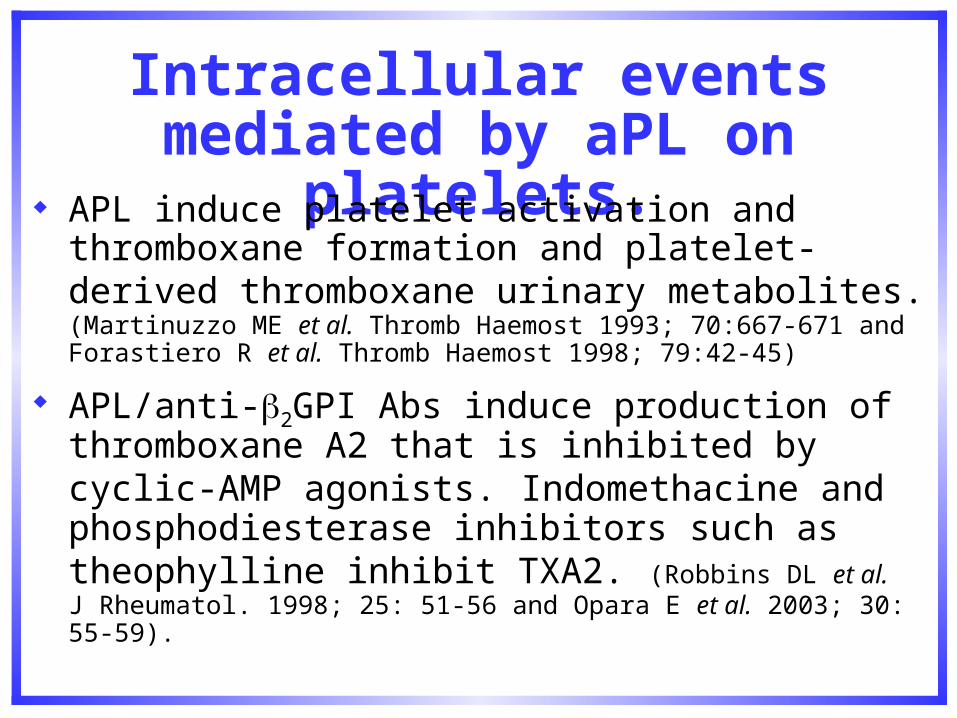
Intracellular events mediated by aPL on platelets.
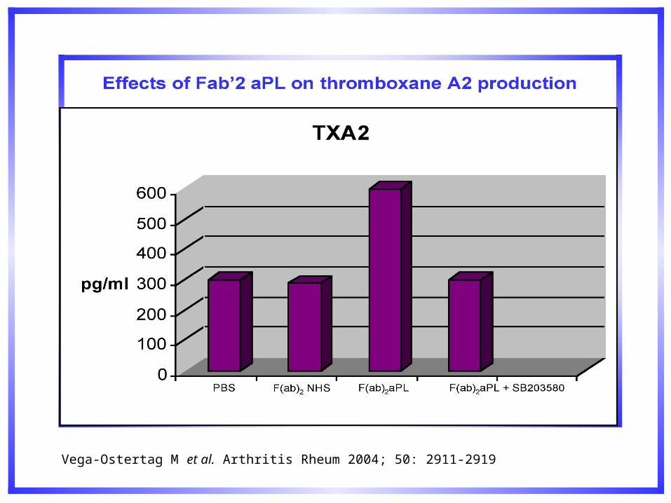
APL induce platelet activation and thromboxane formation and platelet-derived thromboxane urinary metabolites. (Martinuzzo ME et al. Thromb Haemost 1993; 70:667-671 and Forastiero R et al. Thromb Haemost 1998; 79:42-45)
APL/anti-2GPI Abs induce production of thromboxane A2 that is inhibited by cyclic-AMP agonists. Indomethacine and phosphodiesterase inhibitors such as theophylline inhibit TXA2. (Robbins DL et al. J Rheumatol. 1998; 25: 51-56 and Opara E et al. 2003; 30: 55-59).
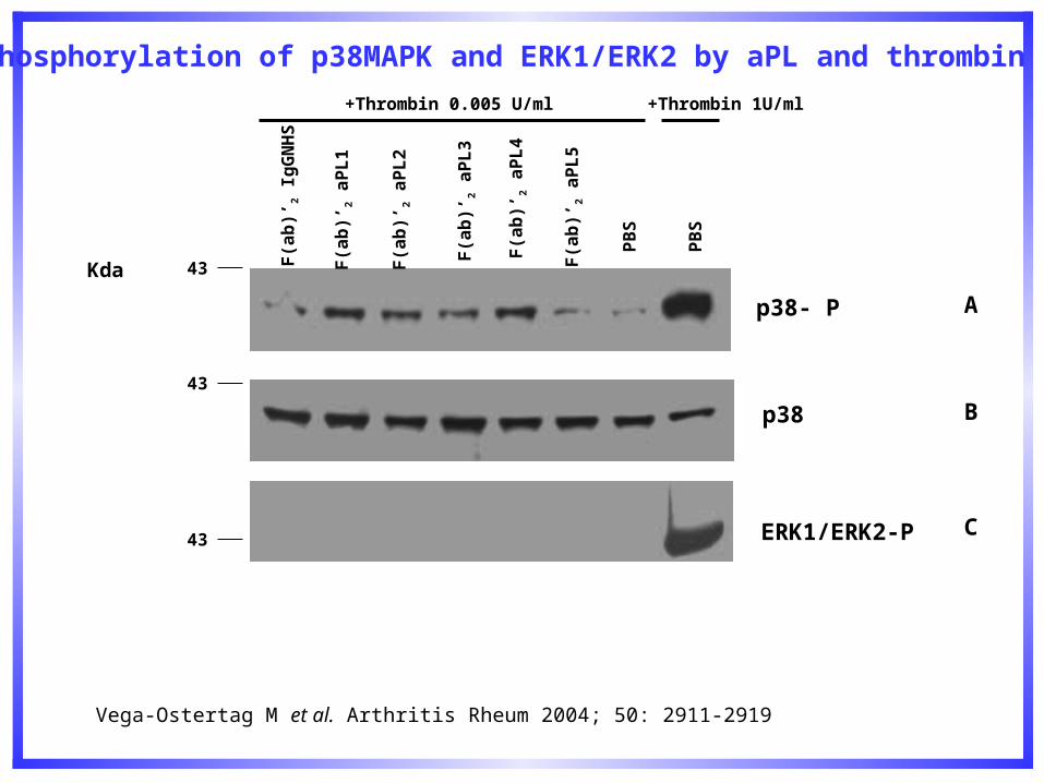
p38- P
p38
ERK1/ERK2-P
F(a
b)’
2 Ig
GN
HS
F(a
b)’
2 a
PL
1
F(a
b)’
2 a
PL
2
F(a
b)’
2 a
PL
3
F(a
b)’
2 a
PL
4
F(a
b)’
2 a
PL
5
PB
S
PB
S
+Thrombin 0.005 U/ml +Thrombin 1U/ml
43
43
43
Kda
A
B
C
Phosphorylation of p38MAPK and ERK1/ERK2 by aPL and thrombin
Vega-Ostertag M et al. Arthritis Rheum 2004; 50: 2911-2919
Vega-Ostertag M et al. Arthritis Rheum 2004; 50: 2911-2919
Vega-Ostertag M et al. Arthritis Rheum 2004; 50: 2911-2919
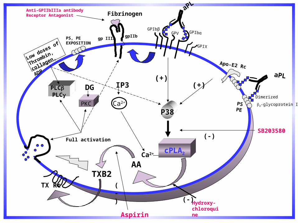
P38
cPLA2
AA
PKC
Fibrinogen
gp IIIagpIIb
Ca2+
PLC PLC
DG IP3
Ca2+
TXB2
(+)
TX Rc
Dimerized
2-glycoprotein I
Full activation
SB203580
Aspirin
Hydroxy-chloroquine
(-)
(-)
Anti-GPIIbIIIa antibodyReceptor Antagonist
(-)
Low doses of
Thrombin,
collagen, ADP
PS, PEEXPOSITION
GPIbβ
aPL
GPV GPIbα
GPIX
Apo-E2 Rc
aPL
PSPE
(+)
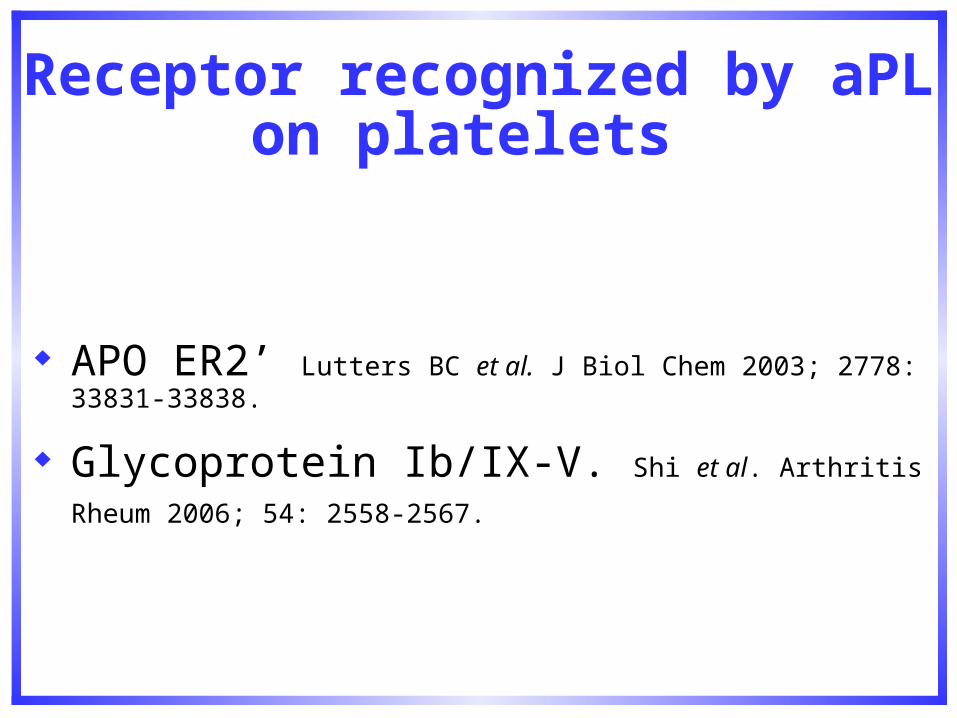
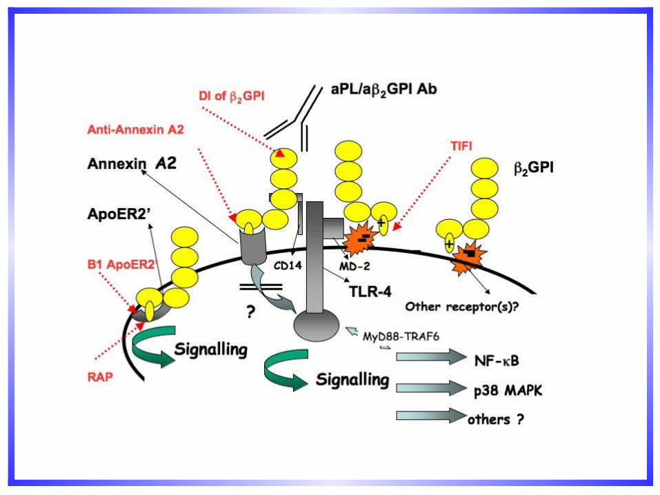
Receptor recognized by aPL on platelets
APO ER2’ Lutters BC et al. J Biol Chem 2003; 2778: 33831-33838.
Glycoprotein Ib/IX-V. Shi et al. Arthritis Rheum 2006; 54:
2558-2567.
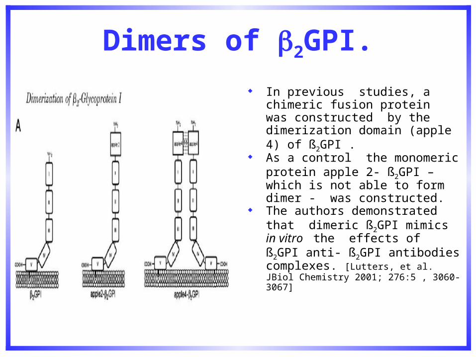
Dimers of 2GPI.
In previous studies, a chimeric fusion protein was constructed by the dimerization domain (apple 4) of ß2GPI .
As a control the monomeric protein apple 2- ß2GPI – which is not able to form dimer - was constructed.
The authors demonstrated that dimeric ß2GPI mimics in vitro the effects of ß2GPI anti- ß2GPI antibodies complexes. [Lutters, et al. JBiol Chemistry 2001; 276:5 , 3060-3067]
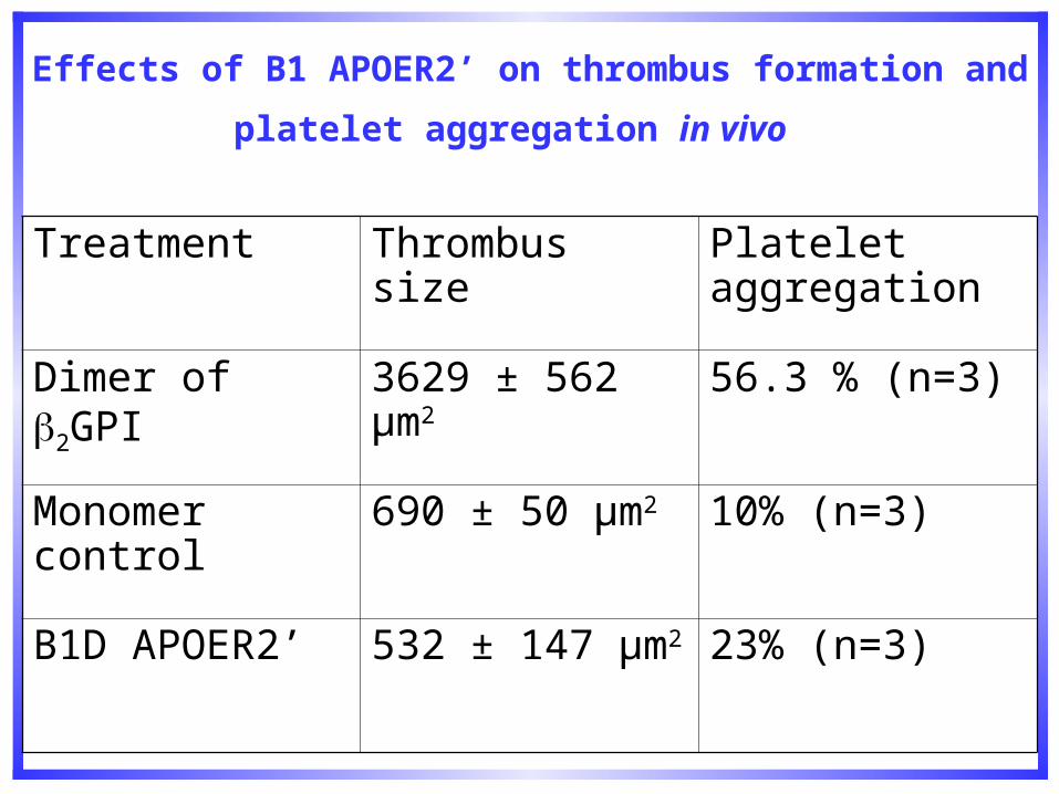
Effects of B1 APOER2’ on thrombus formation and platelet
aggregation in vivo
Treatment Thrombus size Platelet aggregation
Dimer of 2GPI 3629 ± 562 μm2 56.3 % (n=3)
Monomer control 690 ± 50 μm2 10% (n=3)
B1D APOER2’ 532 ± 147 μm2 23% (n=3)
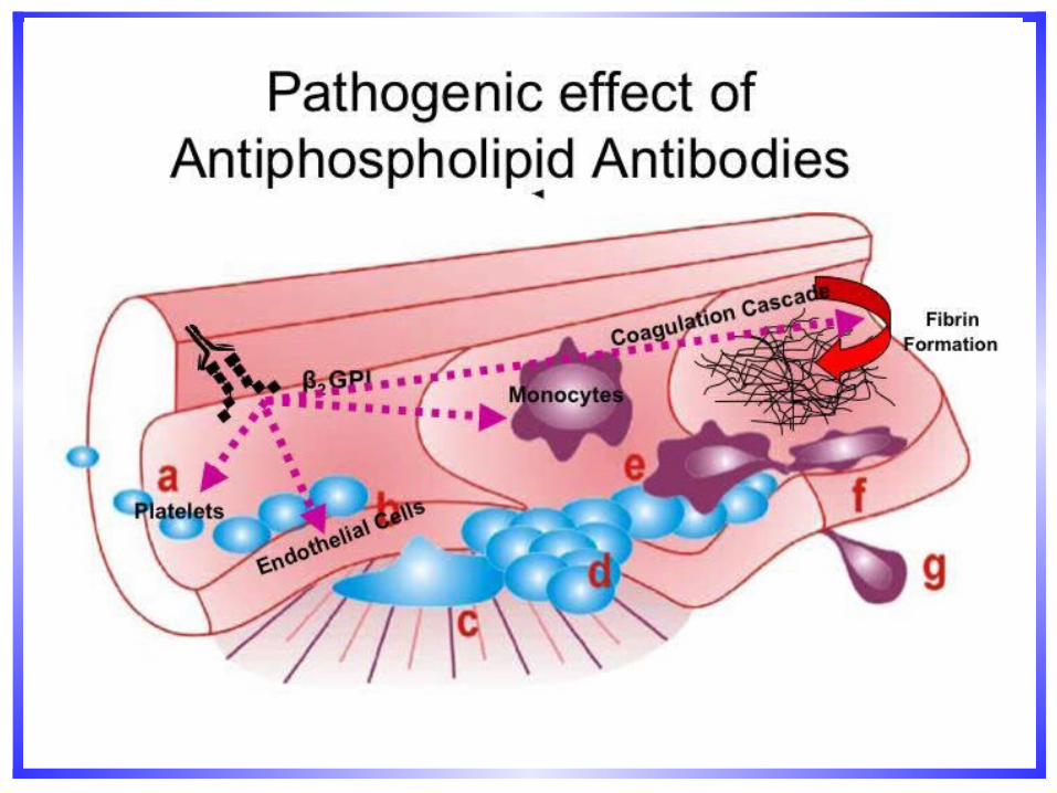
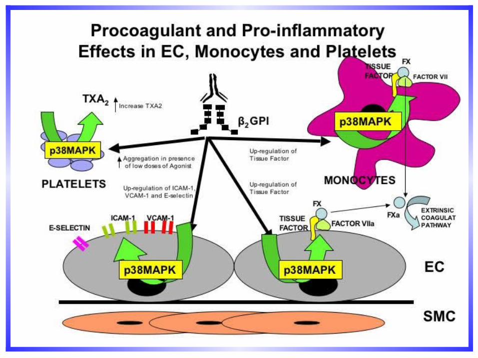
Antiphospholipid Antibodies and Endothelial Cells
APL antibodies activate endothelial cells in vitro and in vivo.
aPL or anti-2GPI antibodies upregulate EC adhesion molecules and this effect is related to aPL binding to EC (Del Papa N et al Arthritis Rheum 1997; 40: 551-561.
aPL-induced upregulation of ICAM-1, VCAM-1 and E-selectin on HUVEC and increased adhesion of monocytes to EC in the presence of 2GPI (Simantov R et al J Clin Invest 1995; 96: 2211-2219).
Soluble levels of VCAM-1 significantly increased in plasma of patients with APS and recurrent thrombosis (Kaplanski G et al 2000; 43: 55-64).
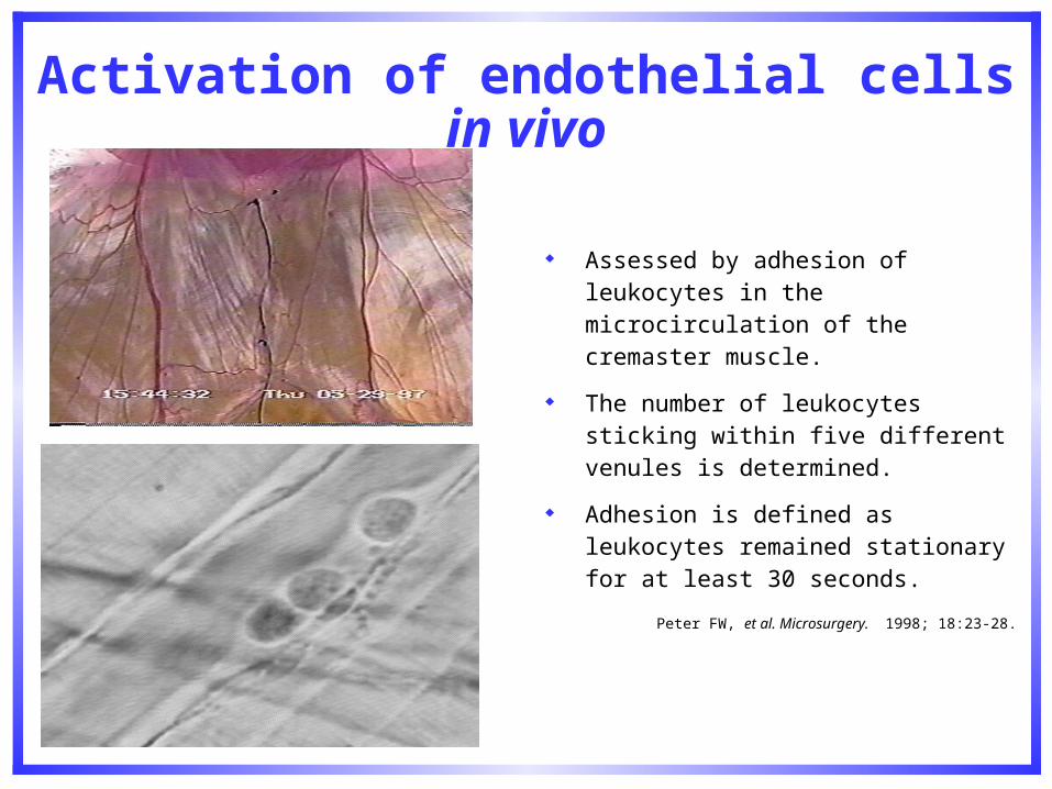
Activation of endothelial cells in vivo
Assessed by adhesion of leukocytes in the microcirculation of the cremaster muscle.
The number of leukocytes sticking within five different venules is determined.
Adhesion is defined as leukocytes remained stationary for at least 30 seconds.
Peter FW, et al. Microsurgery. 1998; 18:23-28.
APL antibodies enhance thrombus formation and this correlates with activation of endothelial cells in vivo
Antiphospholipid antibodies from patients with Antiphospholipid Syndrome activate endothelial cells in vitro and in vivo. Pierangeli et al Circulation, 1999; 99:1997-2002.GDKV-induced antiphospholipid antibodies enhance thrombosis and activate endothelial cells in vivo. Gharavi et al J Immunol 1999; 163: 2922-2927.
Functional analysis of patient-derived IgG monoclonal anticardiolipin antibodies using in vivo thrombosis and in vivo microcirculation Models. Pierangeli et al. Thrombosis Haemost 2000; 84:388-395.
• Thrombogenic effects of antiphospholipid (aPL) antibodies are mediated by intercellular cell adhesion molecule-1(ICAM-1), vascular cell adhesion molecule-1 (VCAM-1) and P-selectin. Pierangeli et al. Circ Res 2001; 88: 245-250.
• E-selectin mediates pathogenic effects of antiphospholipid antibodies. Espinola et al Thromb Haemost. 2003; 1:843-848.
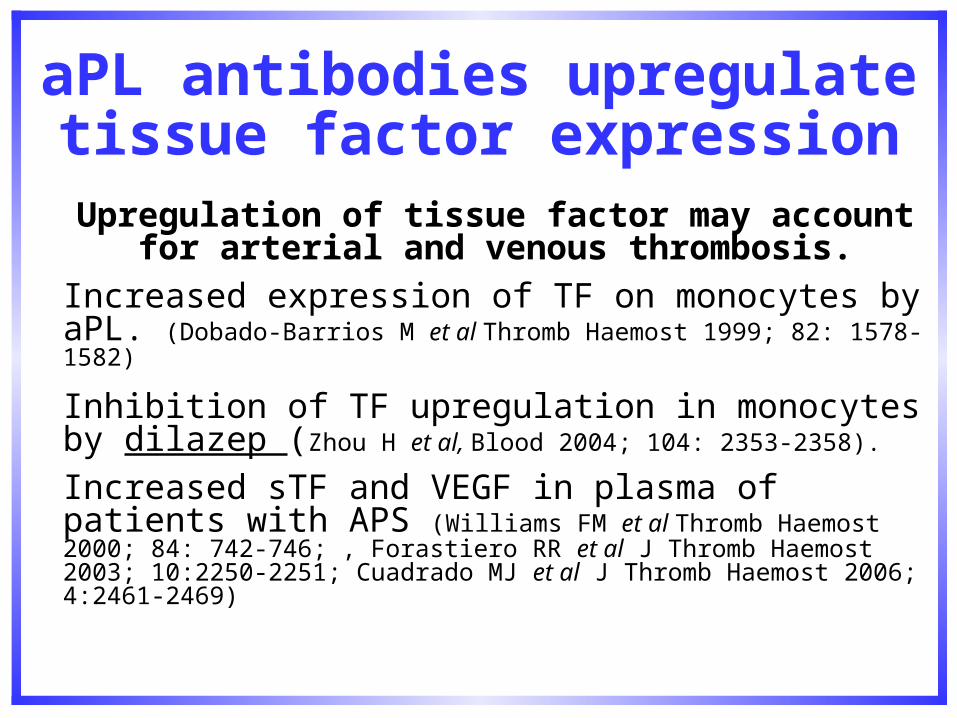
aPL antibodies upregulate tissue factor expression
Upregulation of tissue factor may account for arterial and venous thrombosis.
Increased expression of TF on monocytes by aPL. (Dobado-Barrios M et al Thromb Haemost 1999; 82: 1578-1582)
Inhibition of TF upregulation in monocytes by dilazep (Zhou H et al, Blood 2004; 104: 2353-2358).
Increased sTF and VEGF in plasma of patients with APS (Williams FM et al Thromb Haemost 2000; 84: 742-746; , Forastiero RR et al J Thromb Haemost 2003; 10:2250-2251; Cuadrado MJ et al J Thromb Haemost 2006; 4:2461-2469)
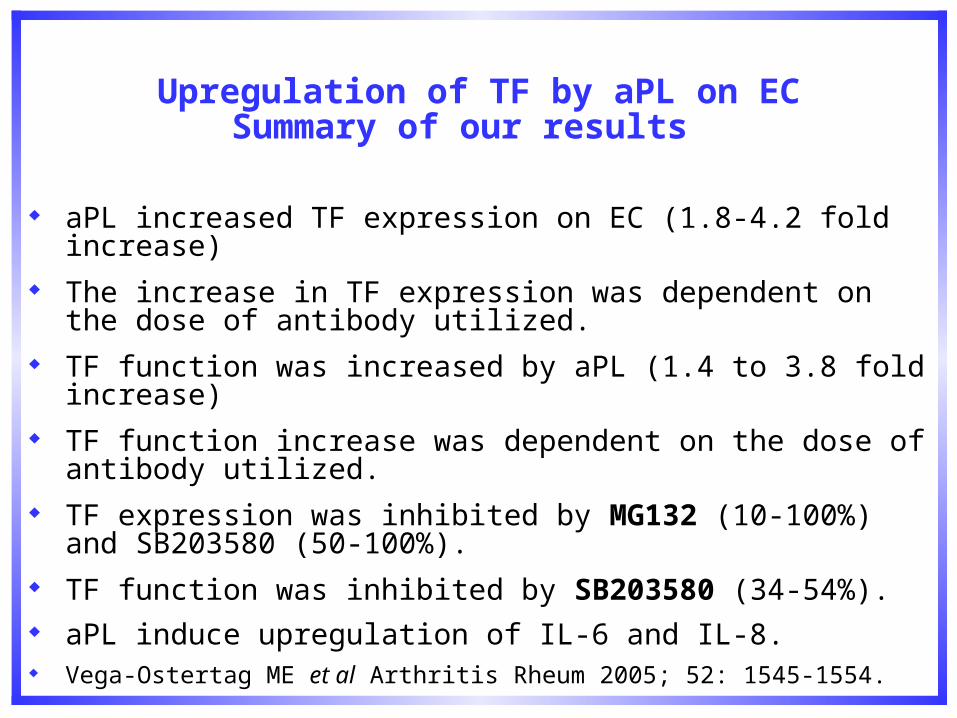
Upregulation of TF by aPL on ECSummary of our results
aPL increased TF expression on EC (1.8-4.2 fold increase) The increase in TF expression was dependent on the dose of
antibody utilized. TF function was increased by aPL (1.4 to 3.8 fold increase) TF function increase was dependent on the dose of antibody
utilized. TF expression was inhibited by MG132 (10-100%) and SB203580
(50-100%). TF function was inhibited by SB203580 (34-54%). aPL induce upregulation of IL-6 and IL-8. Vega-Ostertag ME et al Arthritis Rheum 2005; 52: 1545-1554.
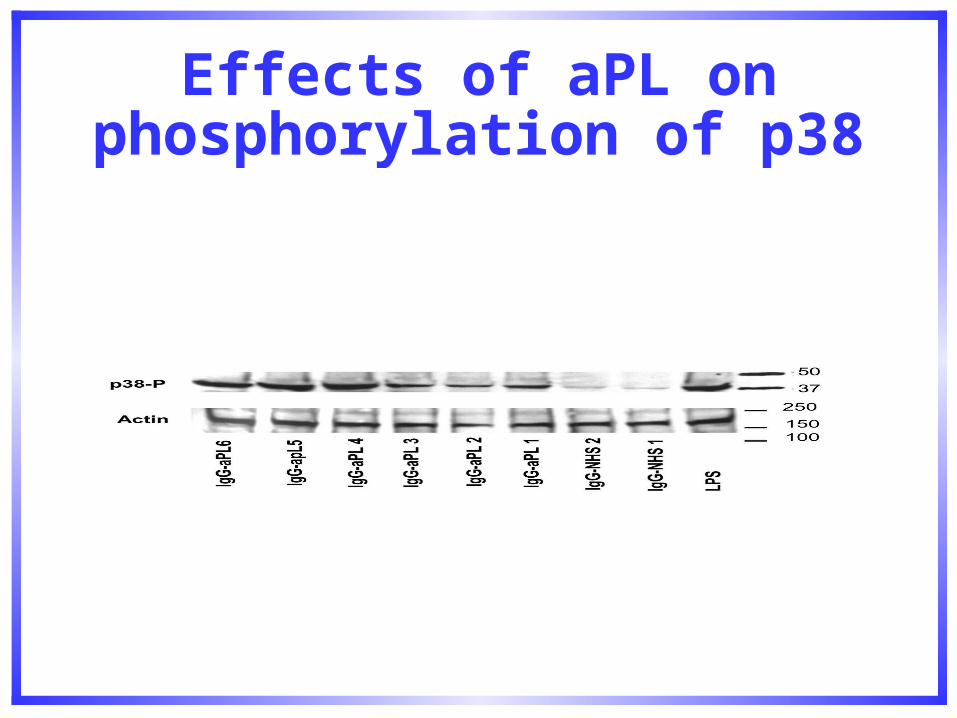
Effects of aPL on phosphorylation of p38 MAPK
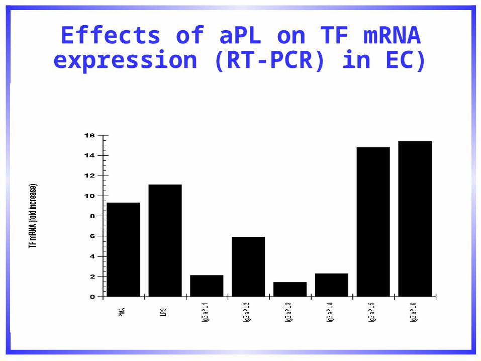
Effects of aPL on TF mRNA expression (RT-PCR) in EC)


Conclusions
– aPL induce phosphorylation of p38MAPK
– aPL induce iNOS
– aPL induce transcription of TF mRNA and this effect is inhibited by SB 203580 in a dose-dependent fashion.
(Vega Ostertag M, et al. Arthritis Rheum, 2005; 52: 5: 1545-1554)
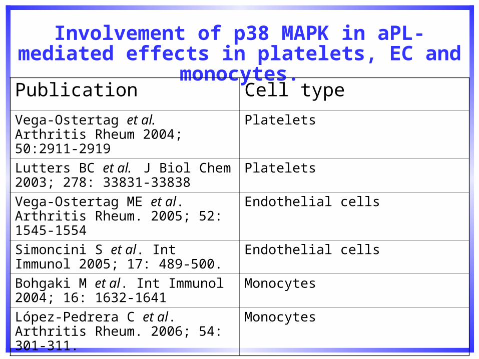
Involvement of p38 MAPK in aPL-mediated effects in platelets, EC and monocytes.
Publication Cell type
Vega-Ostertag et al. Arthritis Rheum 2004; 50:2911-2919
Platelets
Lutters BC et al. J Biol Chem 2003; 278: 33831-33838
Platelets
Vega-Ostertag ME et al. Arthritis Rheum. 2005; 52: 1545-1554
Endothelial cells
Simoncini S et al. Int Immunol 2005; 17: 489-500.
Endothelial cells
Bohgaki M et al. Int Immunol 2004; 16: 1632-1641
Monocytes
López-Pedrera C et al. Arthritis Rheum. 2006; 54: 301-311.
Monocytes
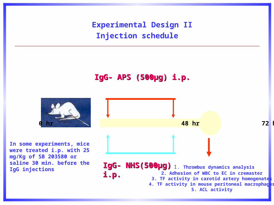
Experimental Design II
Injection schedule
IgG- APS (500µg) i.p.IgG- APS (500µg) i.p.
0 hr 48 hr 72 hr
1. Thrombus dynamics analysis2. Adhesion of WBC to EC in cremaster
3. TF activity in carotid artery homogenates4. TF activity in mouse peritoneal macrophages
5. ACL activity
IgG- NHS(500µg) i.p.IgG- NHS(500µg) i.p.
In some experiments, mice were treated i.p. with 25 mg/Kg of SB 203580 or saline 30 min. before the IgG injections
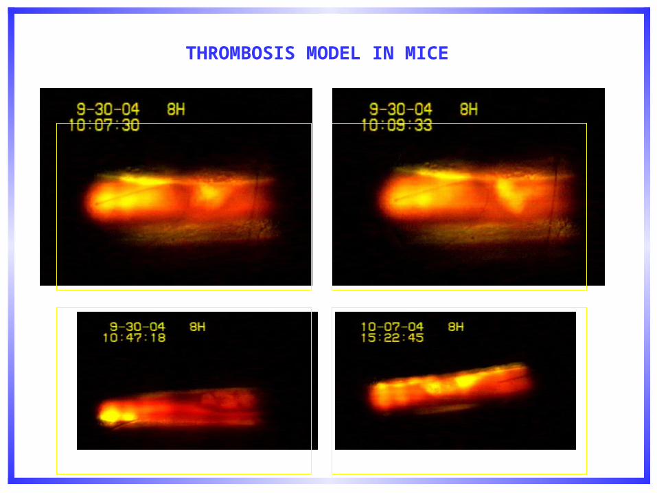
THROMBOSIS MODEL IN MICE
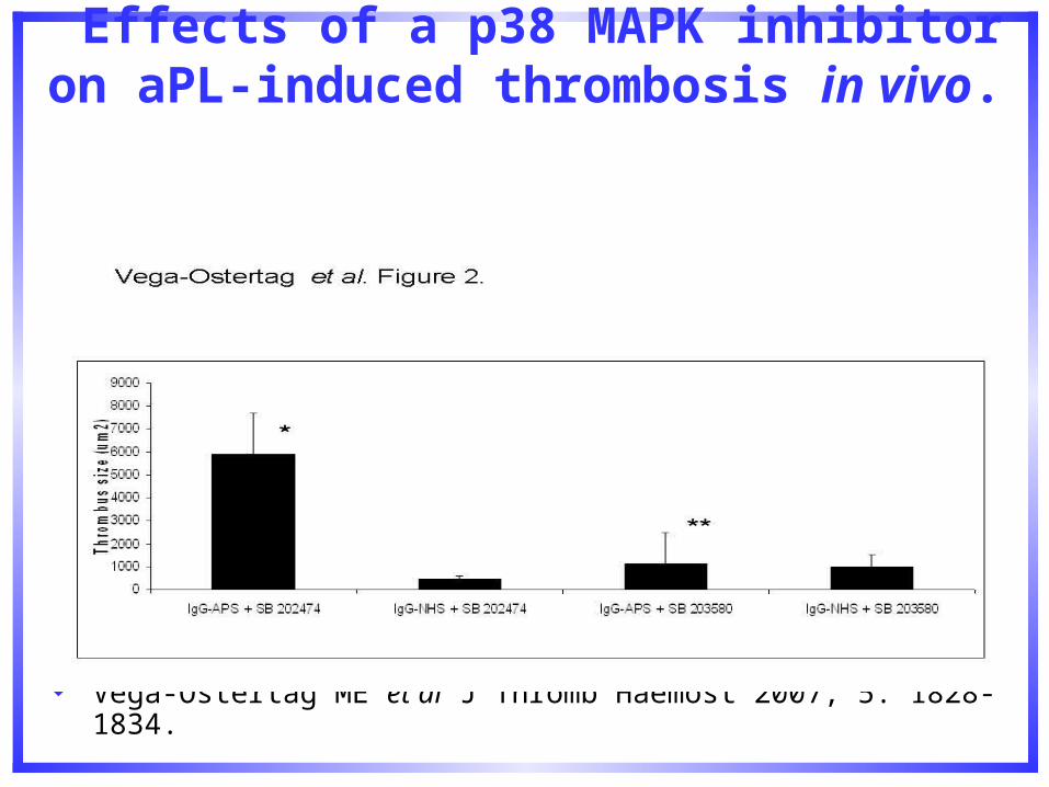
Effects of a p38 MAPK inhibitor on aPL-induced thrombosis in vivo.
Vega-Ostertag ME et al J Thromb Haemost 2007; 5: 1828-1834.
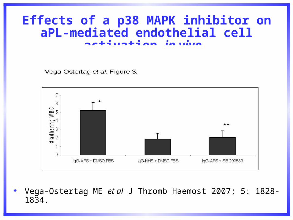
Effects of a p38 MAPK inhibitor on aPL-mediated endothelial cell activation in vivo.
Vega-Ostertag ME et al J Thromb Haemost 2007; 5: 1828-1834.
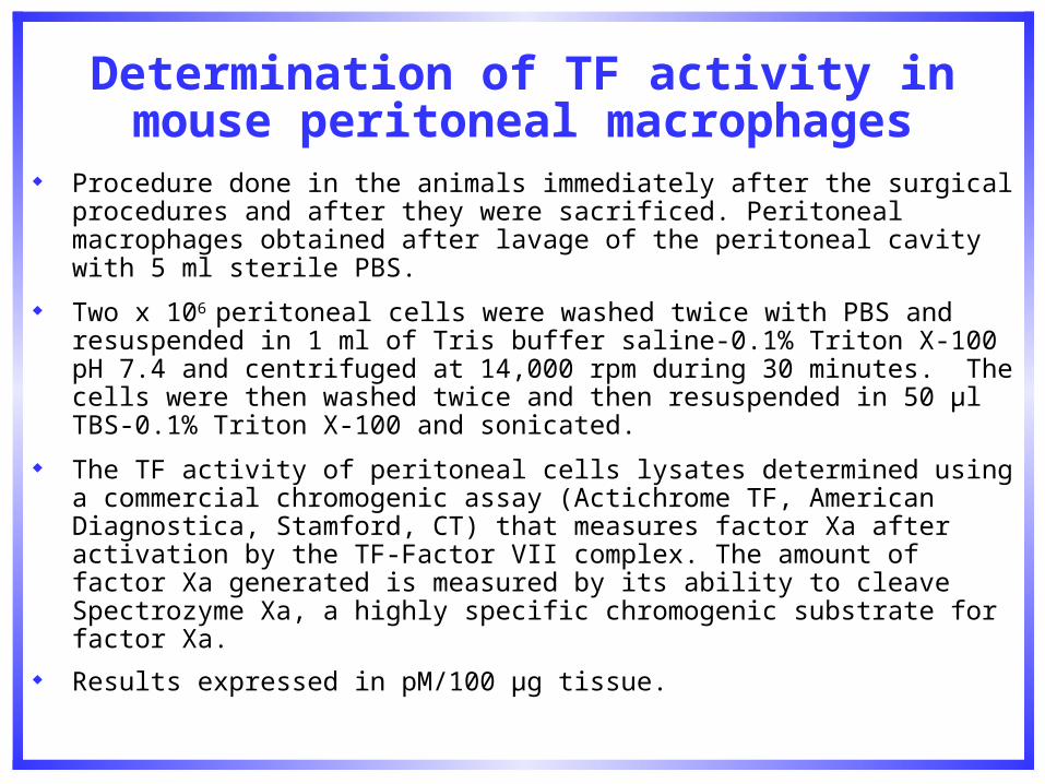
Determination of TF activity in mouse peritoneal macrophages
Procedure done in the animals immediately after the surgical procedures and after they were sacrificed. Peritoneal macrophages obtained after lavage of the peritoneal cavity with 5 ml sterile PBS.
Two x 106 peritoneal cells were washed twice with PBS and resuspended in 1 ml of Tris buffer saline-0.1% Triton X-100 pH 7.4 and centrifuged at 14,000 rpm during 30 minutes. The cells were then washed twice and then resuspended in 50 µl TBS-0.1% Triton X-100 and sonicated.
The TF activity of peritoneal cells lysates determined using a commercial chromogenic assay (Actichrome TF, American Diagnostica, Stamford, CT) that measures factor Xa after activation by the TF-Factor VII complex. The amount of factor Xa generated is measured by its ability to cleave Spectrozyme Xa, a highly specific chromogenic substrate for factor Xa.
Results expressed in pM/100 µg tissue.

Effects of a p38 MAPK inhibitor on aPL-induced TF activity in mouse peritoneal macrophages
Vega-Ostertag ME et al J Thromb Haemost 2007; 5: 1828-1834.
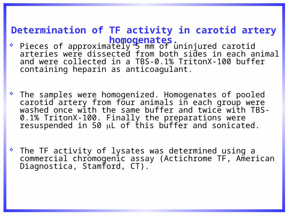
Determination of TF activity in carotid artery homogenates.
Pieces of approximately 5 mm of uninjured carotid arteries were dissected from both sides in each animal and were collected in a TBS-0.1% TritonX-100 buffer containing heparin as anticoagulant.
The samples were homogenized. Homogenates of pooled carotid artery from four animals in each group were washed once with the same buffer and twice with TBS-0.1% TritonX-100. Finally the preparations were resuspended in 50 L of this buffer and sonicated.
The TF activity of lysates was determined using a commercial chromogenic assay (Actichrome TF, American Diagnostica, Stamford, CT).
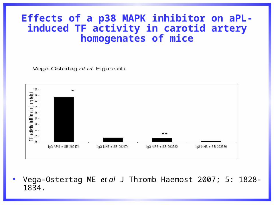
Effects of a p38 MAPK inhibitor on aPL-induced TF activity in carotid artery homogenates of mice
Vega-Ostertag ME et al J Thromb Haemost 2007; 5: 1828-1834.
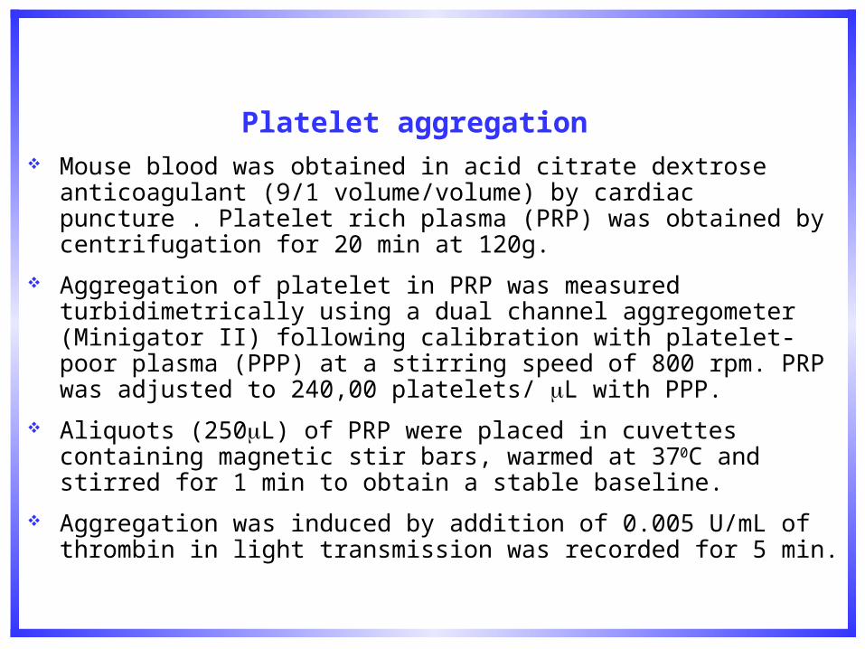
Platelet aggregation
Mouse blood was obtained in acid citrate dextrose anticoagulant (9/1 volume/volume) by cardiac puncture . Platelet rich plasma (PRP) was obtained by centrifugation for 20 min at 120g.
Aggregation of platelet in PRP was measured turbidimetrically using a dual channel aggregometer (Minigator II) following calibration with platelet-poor plasma (PPP) at a stirring speed of 800 rpm. PRP was adjusted to 240,00 platelets/ L with PPP.
Aliquots (250L) of PRP were placed in cuvettes containing magnetic stir bars, warmed at 370C and stirred for 1 min to obtain a stable baseline.
Aggregation was induced by addition of 0.005 U/mL of thrombin in light transmission was recorded for 5 min.
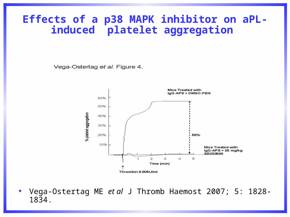
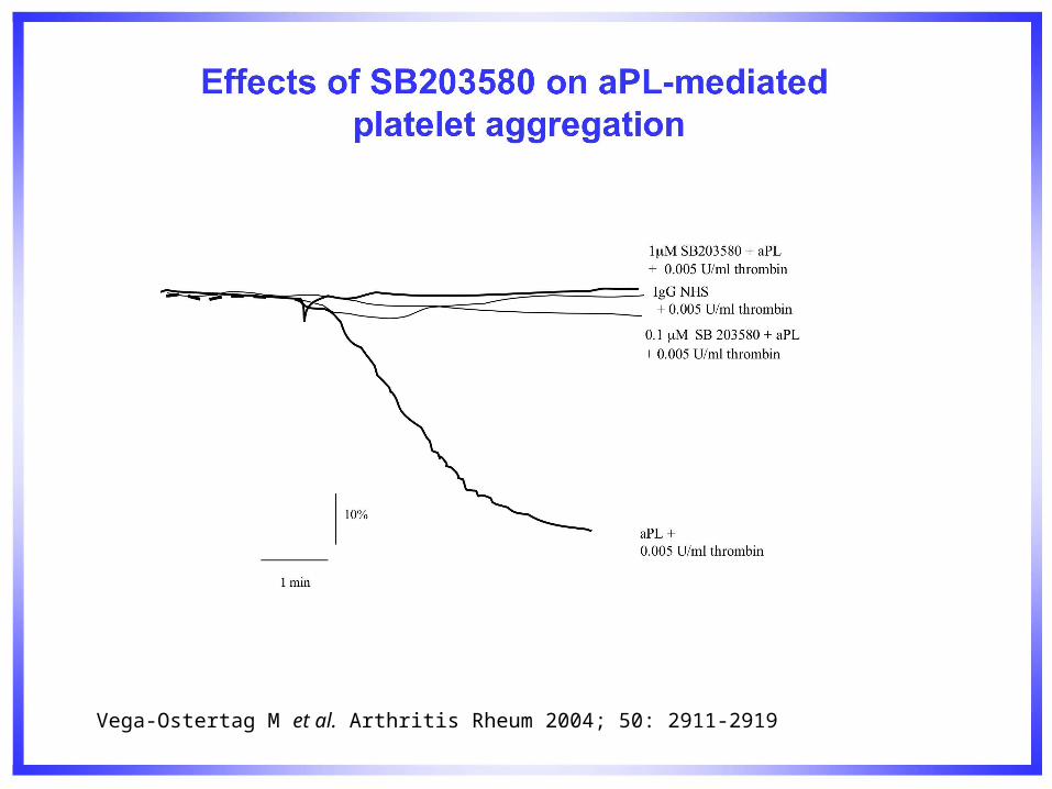
Effects of a p38 MAPK inhibitor on aPL-induced platelet aggregation
Vega-Ostertag ME et al J Thromb Haemost 2007; 5: 1828-1834.
Experimental Design Ex vivo experiments:
Determination of VCAM-1 in flat aorta preparations of mice using Qdot conjugates and dual photon confocal microscopy.
Mouse arteries were pressure-perfused with 10% formalin. After fixation, the arteries were washed three times with PBS.
The immunohistochemical procedure was done on a 24 well-plate. Blocking was done with 2% BSA/5% goat serum for one hour. Primary antibodies were incubated overnight at 4ºC. Washing steps were done to remove primary antibody excess. Qdot-conjugated secondary antibody (Qdot Corp) was incubated for one hour. Finally, nuclear visualization was done with Hoechst stain.
Image collection was done using a Zeiss LSM 510 Meta two-photon microscope equipped with a near-infrared (NIR) titanium-sapphire femtosecond laser (Mira 900 Ti:S Coherent) tuned and mode-locked at 750 nm. The separation of the emission signals was performed by acquisition of lambda stacks with posterior selection of reference spectra using the META detector.
The following primary antibodies were used:, monoclonal rat anti-mouse VCAM-1 IgG (1:50 dilution, BD Pharmingen) and a nonimmune primary used to address the contribution of nonspecific Fc receptor-mediated binding (nonimmune purified rat IgG2a (BD Pharmingen™). The following Qdot-bioconjugate was used in the experiments: Qdot® 655 goat F(ab')2 anti-rat IgG Conjugate

Effects of a p38 MAPK inhibitor on aPL-induced VCAM-1 expression in aorta of mice ex vivo: nano crystals Q dot conjugates and dual photon
confocal microscopy.
Vega-Ostertag ME et al J Thromb Haemost 2007; 5: 1828-1834.
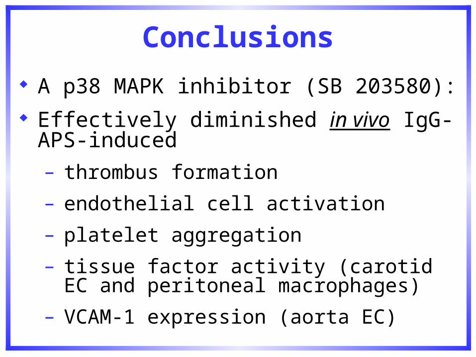
Conclusions
A p38 MAPK inhibitor (SB 203580): Effectively diminished in vivo IgG-APS-
induced
– thrombus formation
– endothelial cell activation
– platelet aggregation
– tissue factor activity (carotid EC and peritoneal macrophages)
– VCAM-1 expression (aorta EC)
NF-B
NF-B is a complex group of heterodimeric and homodimeric transcription factors that are trapped in the cytoplasm as an inactive complex by I-B.
NF-B involved in transcription of inflammatory genes such as: IL-6, IL-8, TNF- and IL-1and in induction of adhesion molecules on EC (VCAM-1, E-sel and ICAM-1) and in recruitment of inflammatory cells to extravascular sites.
NF-B associated with rheumatoid arthritis and other autoimmune diseases.
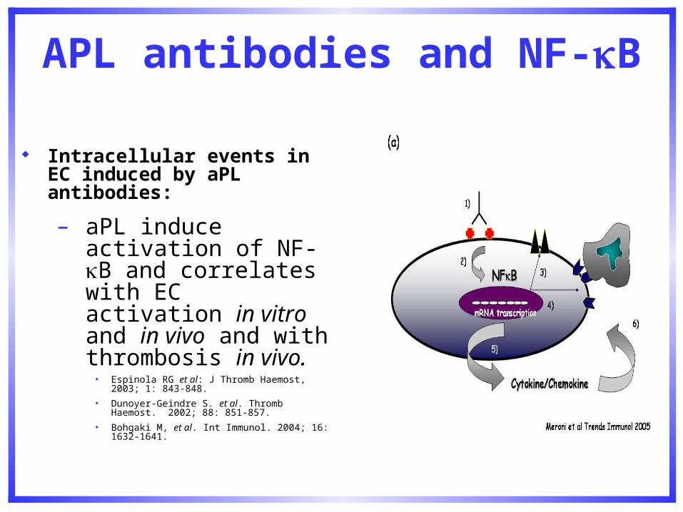
APL antibodies and NF-B
Intracellular events in EC induced by aPL antibodies:
– aPL induce activation of NF-B and correlates with EC activation in vitro and in vivo and with thrombosis in vivo.
• Espinola RG et al: J Thromb Haemost, 2003; 1: 843-848.
• Dunoyer-Geindre S. et al. Thromb Haemost. 2002; 88: 851-857.
• Bohgaki M, et al. Int Immunol. 2004; 16: 1632-1641.
MG 132
NF-B inhibitors used in RA and other autoimmune and inflammatory diseases.
MG 132 = Carbobenzoxyl-leucinyl leucinylleucinal (Z-Leu-Leu-Leu-aldehyde; Z-LLL-CHO).
MG 132 is a potent 20 S proteasome inhibitor that has been shown effective in
suppressing NF-B activation in different cellular systems. Several studies have shown beneficial effects of MG132 on models of
rheumatoid arthritis, suggesting that this inhibitor may provide a new approach in the treatment of this autoimmune disease.
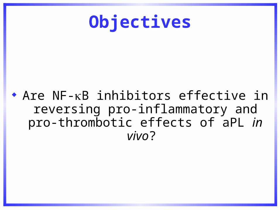
Objectives
Are NF-B inhibitors effective in reversing pro-inflammatory and pro-thrombotic effects
of aPL in vivo?
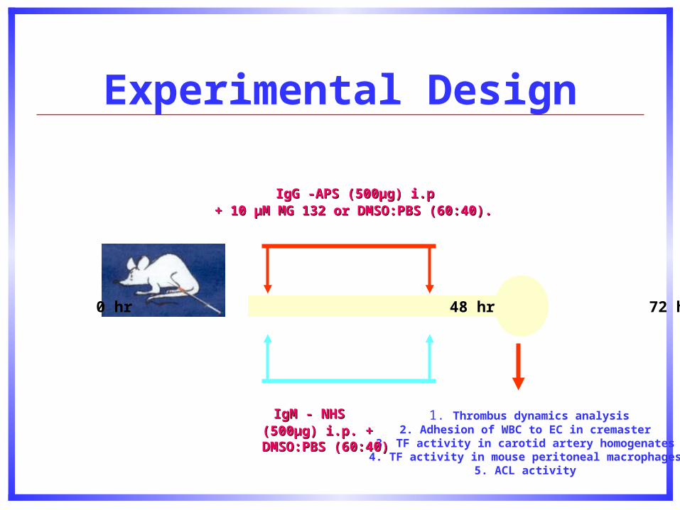
Experimental Design
IgG -APS (500µg) i.p IgG -APS (500µg) i.p + 10 µM MG 132 or DMSO:PBS (60:40).+ 10 µM MG 132 or DMSO:PBS (60:40).
0 hr 48 hr 72 hr
1. Thrombus dynamics analysis2. Adhesion of WBC to EC in cremaster
3. TF activity in carotid artery homogenates4. TF activity in mouse peritoneal macrophages
5. ACL activity
IgM - NHSIgM - NHS (500µg) i.p. (500µg) i.p. + DMSO:PBS (60:40)+ DMSO:PBS (60:40)
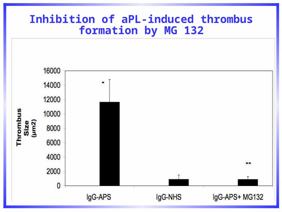
Inhibition of aPL-induced thrombus formation by MG 132
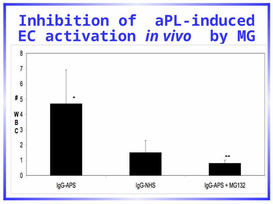
Inhibition of aPL-induced EC activation in vivo by MG 132
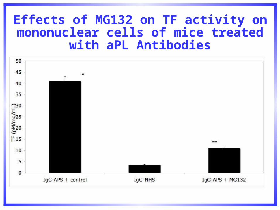
Effects of MG132 on TF activity on mononuclear cells of mice treated with aPL
Antibodies
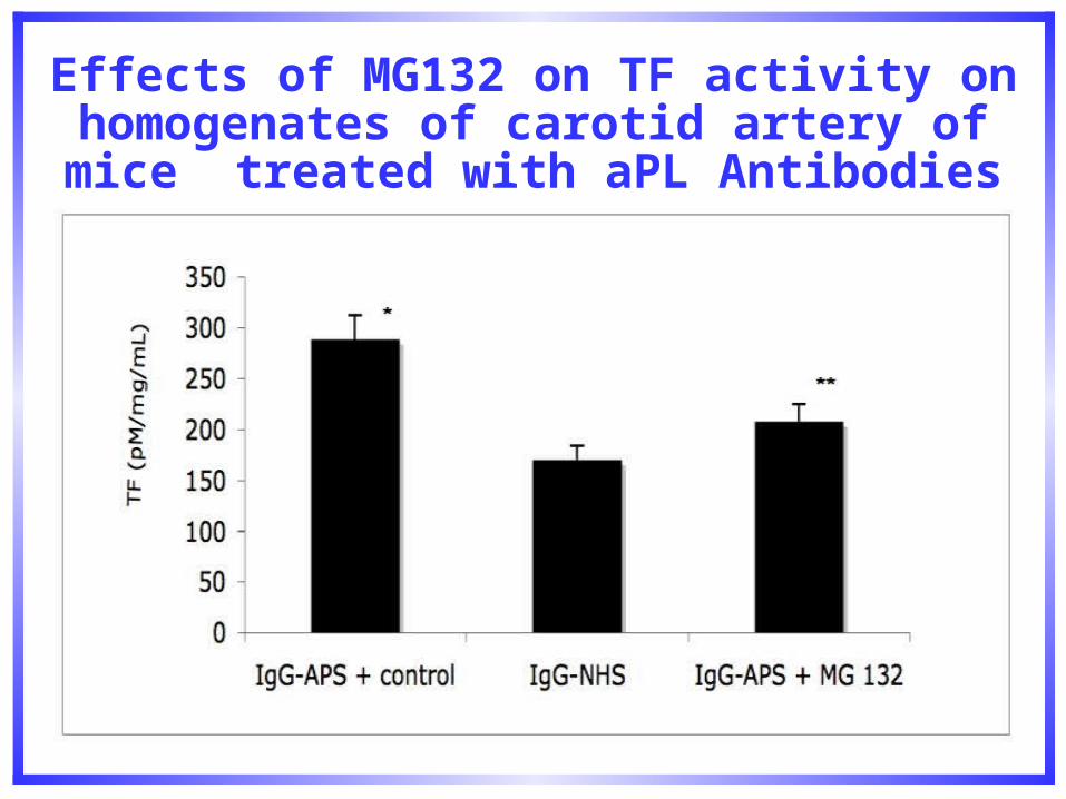
Effects of MG132 on TF activity on homogenates of carotid artery of mice
treated with aPL Antibodies
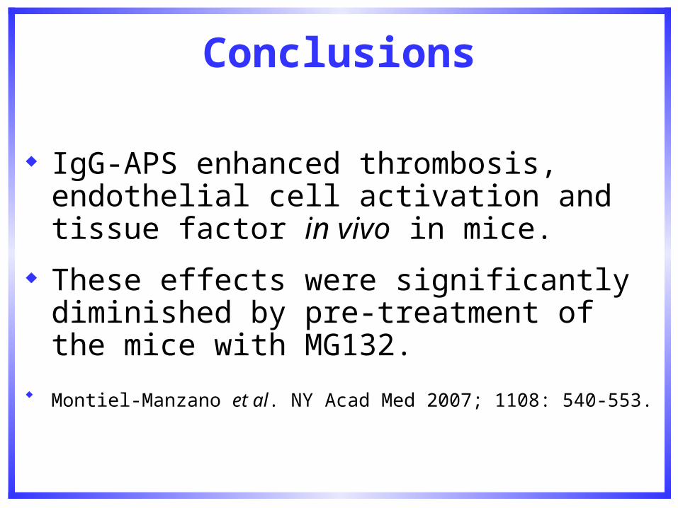
Conclusions
IgG-APS enhanced thrombosis, endothelial cell activation and tissue factor in vivo in mice.
These effects were significantly diminished by pre-treatment of the mice with MG132.
Montiel-Manzano et al. NY Acad Med 2007; 1108: 540-553.
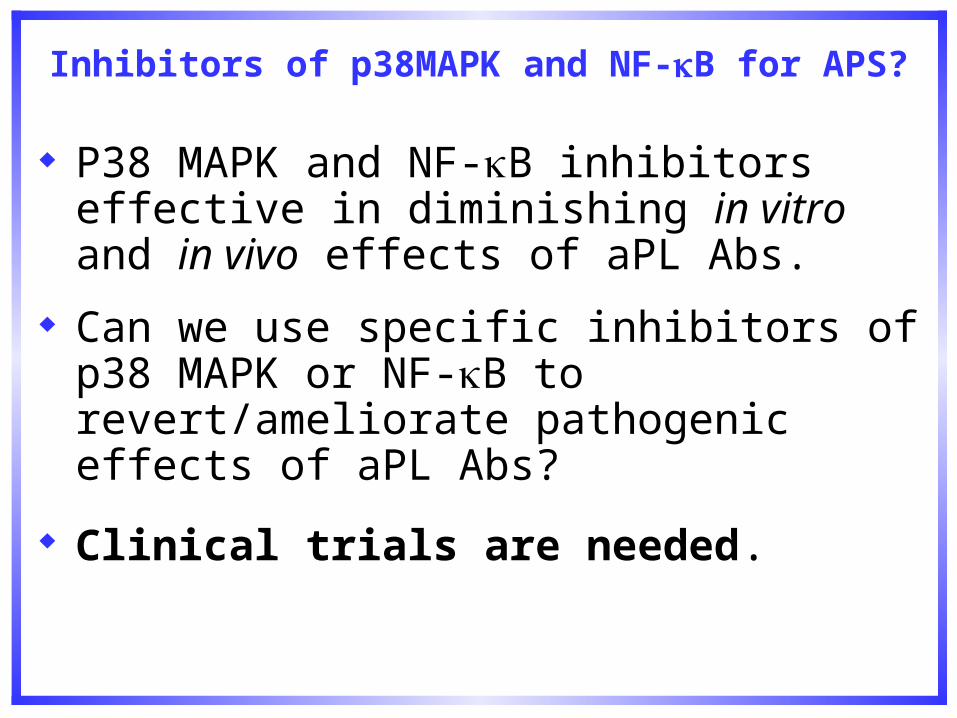
Inhibitors of p38MAPK and NF-B for APS?
P38 MAPK and NF-B inhibitors effective in diminishing in vitro and in vivo effects of aPL Abs.
Can we use specific inhibitors of p38 MAPK or NF-B to revert/ameliorate pathogenic effects of aPL Abs?
Clinical trials are needed.
Antiphospholipid Antibodies and the statins
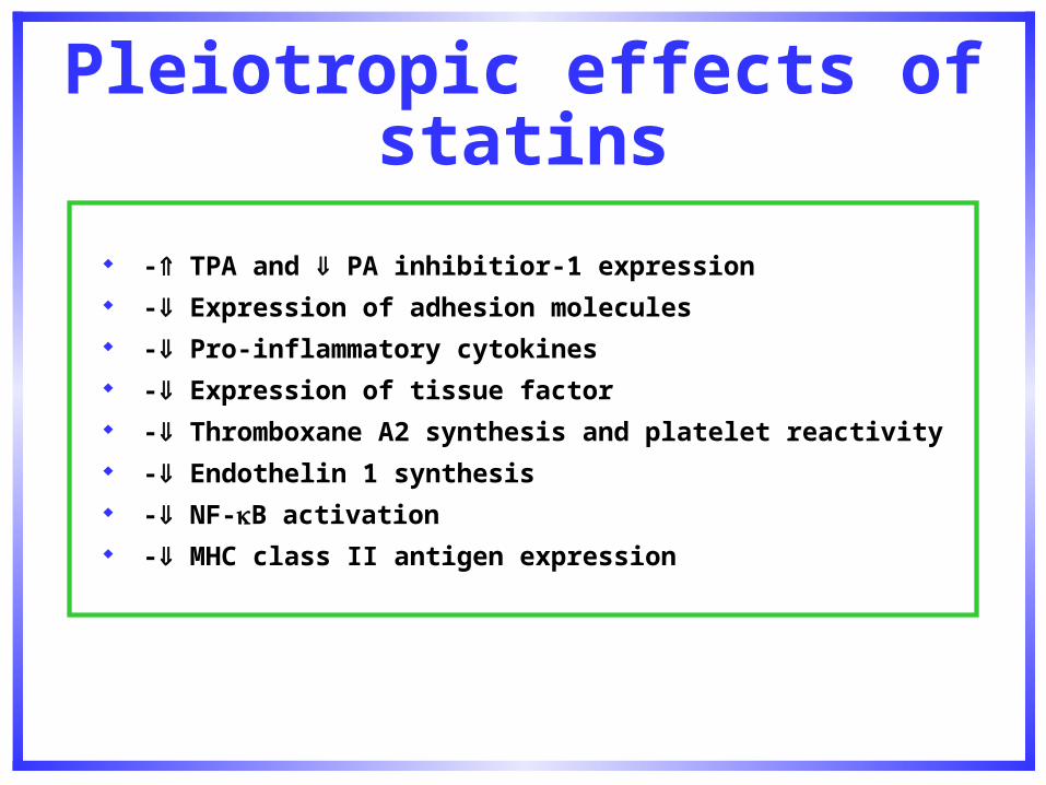
Pleiotropic effects of statins
- TPA and PA inhibitior-1 expression - Expression of adhesion molecules - Pro-inflammatory cytokines - Expression of tissue factor - Thromboxane A2 synthesis and platelet reactivity - Endothelin 1 synthesis - NF-B activation - MHC class II antigen expression
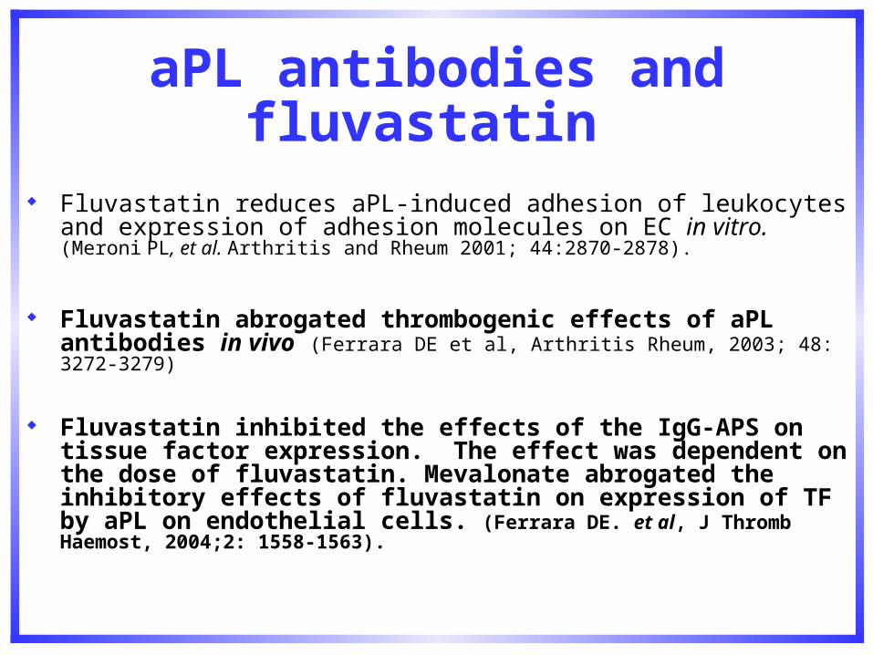
aPL antibodies and fluvastatin
Fluvastatin reduces aPL-induced adhesion of leukocytes and expression of adhesion molecules on EC in vitro. (Meroni PL, et al. Arthritis and Rheum 2001; 44:2870-2878).
Fluvastatin abrogated thrombogenic effects of aPL antibodies in vivo (Ferrara DE et al, Arthritis Rheum, 2003; 48: 3272-3279)
Fluvastatin inhibited the effects of the IgG-APS on tissue factor expression. The effect was dependent on the dose of fluvastatin. Mevalonate abrogated the inhibitory effects of fluvastatin on expression of TF by aPL on endothelial cells. (Ferrara DE. et al, J Thromb Haemost, 2004;2: 1558-1563).
Implications
These findings may have important implications in designing new modalities of treatment and prevention of recurrent thrombosis in patients with APS.
Well designed clinical trials are needed to investigate and confirm these findings in APS patients

Objectives of the study
To determine the effects of statins on pro-thrombotic and pro-inflammatory markers in patients with aPL Abs
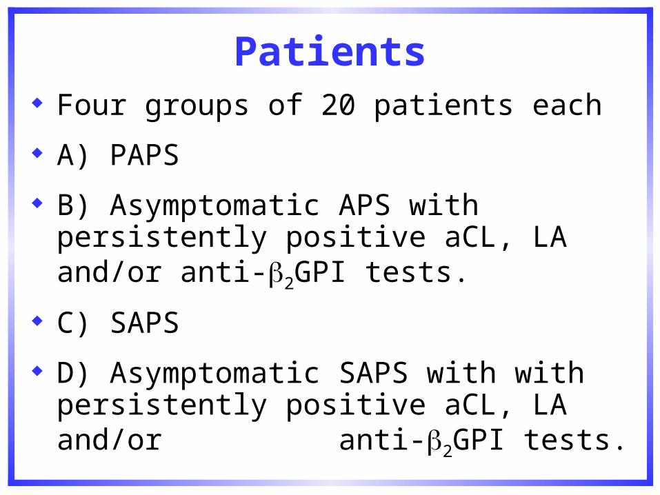
Patients
Four groups of 20 patients each
A) PAPS
B) Asymptomatic APS with persistently positive aCL, LA and/or anti-2GPI tests.
C) SAPS
D) Asymptomatic SAPS with with persistently positive aCL, LA and/or anti-2GPI tests.
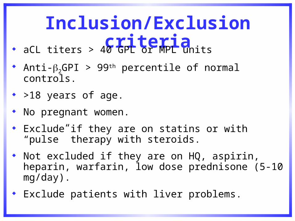
Inclusion/Exclusion criteria
aCL titers > 40 GPL or MPL units
Anti-2GPI > 99th percentile of normal controls.
>18 years of age.
No pregnant women.
Exclude if they are on statins or with “pulse” therapy with steroids.
Not excluded if they are on HQ, aspirin, heparin, warfarin, low dose prednisone (5-10 mg/day).
Exclude patients with liver problems.
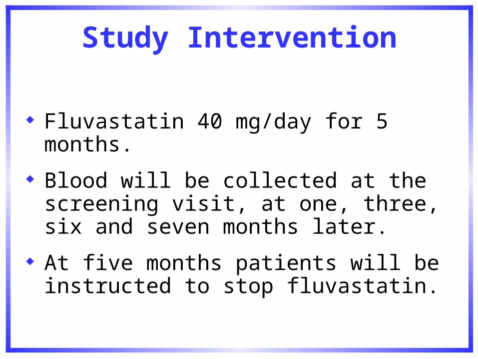
Study Intervention
Fluvastatin 40 mg/day for 5 months.
Blood will be collected at the screening visit, at one, three, six and seven months later.
At five months patients will be instructed to stop fluvastatin.
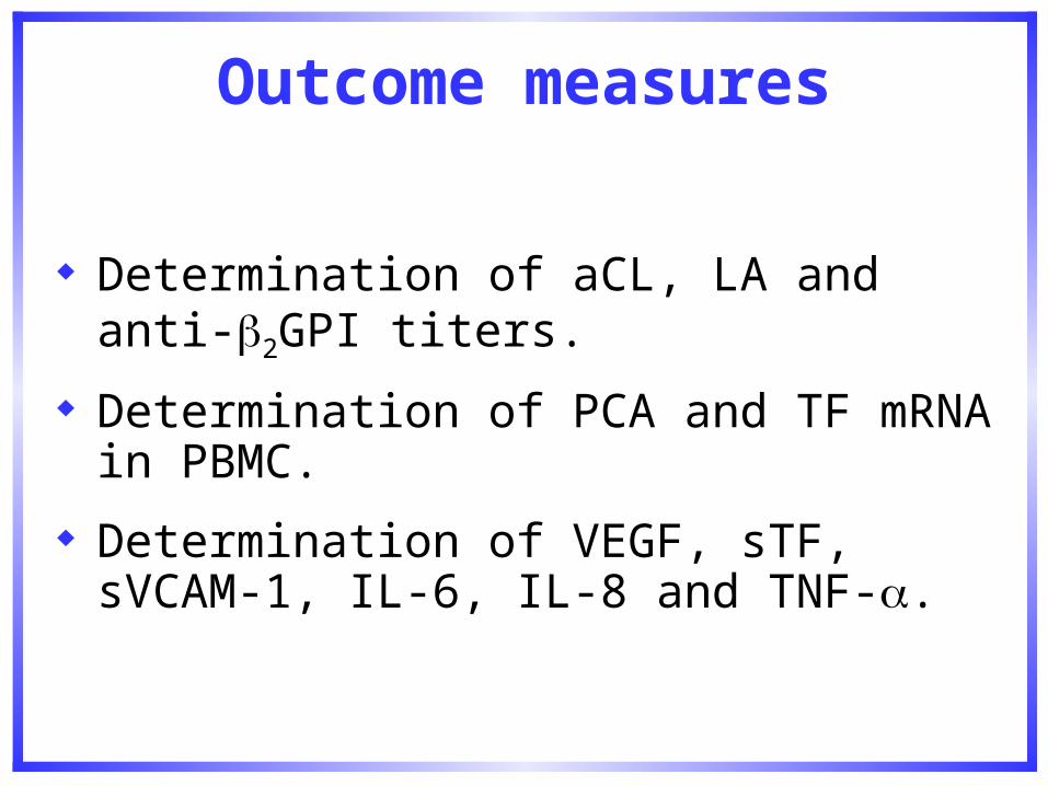
Outcome measures
Determination of aCL, LA and anti-2GPI titers.
Determination of PCA and TF mRNA in PBMC.
Determination of VEGF, sTF, sVCAM-1, IL-6, IL-8 and TNF-.
aPL, complement, endothelial cell activation and thrombosis
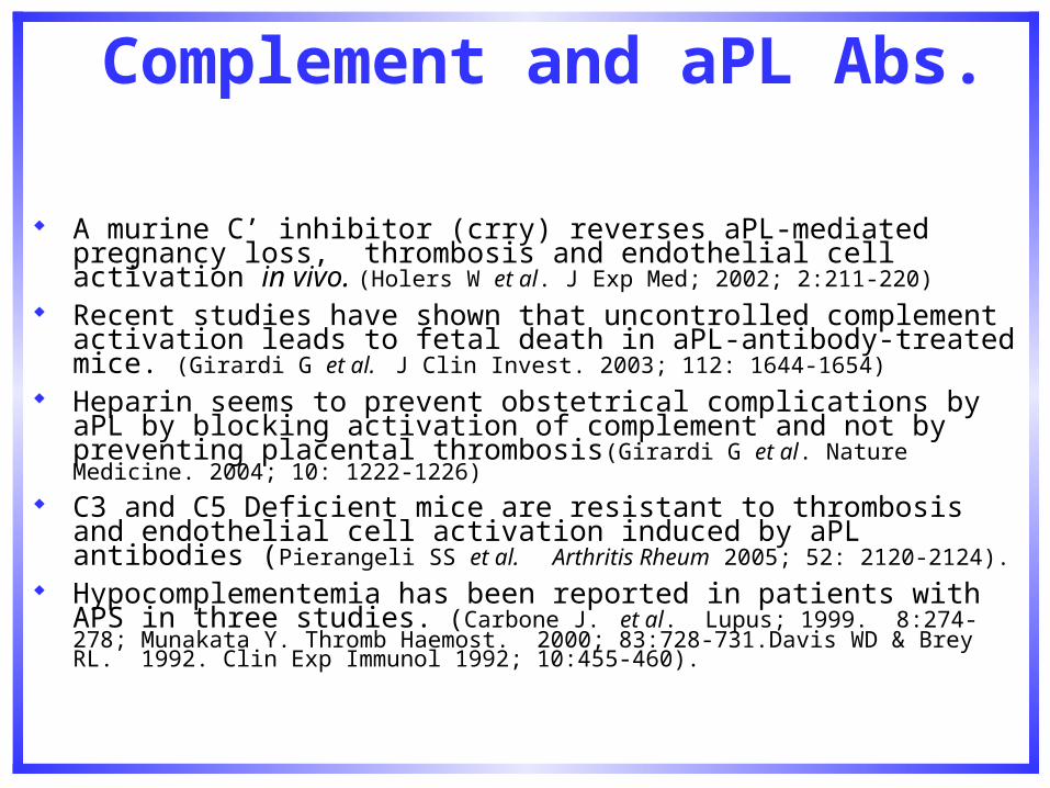
Complement and aPL Abs.
A murine C’ inhibitor (crry) reverses aPL-mediated pregnancy loss, thrombosis and endothelial cell activation in vivo. (Holers W et al. J Exp Med; 2002; 2:211-220)
Recent studies have shown that uncontrolled complement activation leads to fetal death in aPL-antibody-treated mice. (Girardi G et al. J Clin Invest. 2003; 112: 1644-1654)
Heparin seems to prevent obstetrical complications by aPL by blocking activation of complement and not by preventing placental thrombosis(Girardi G et al. Nature Medicine. 2004; 10: 1222-1226)
C3 and C5 Deficient mice are resistant to thrombosis and endothelial cell activation induced by aPL antibodies (Pierangeli SS et al. Arthritis Rheum 2005; 52: 2120-2124).
Hypocomplementemia has been reported in patients with APS in three studies. (Carbone J. et al. Lupus; 1999. 8:274-278; Munakata Y. Thromb Haemost. 2000; 83:728-731.Davis WD & Brey RL. 1992. Clin Exp Immunol 1992; 10:455-460).
Objective
Does an anti-C5 MoAb prevent aPL-mediated thrombosis in mice?
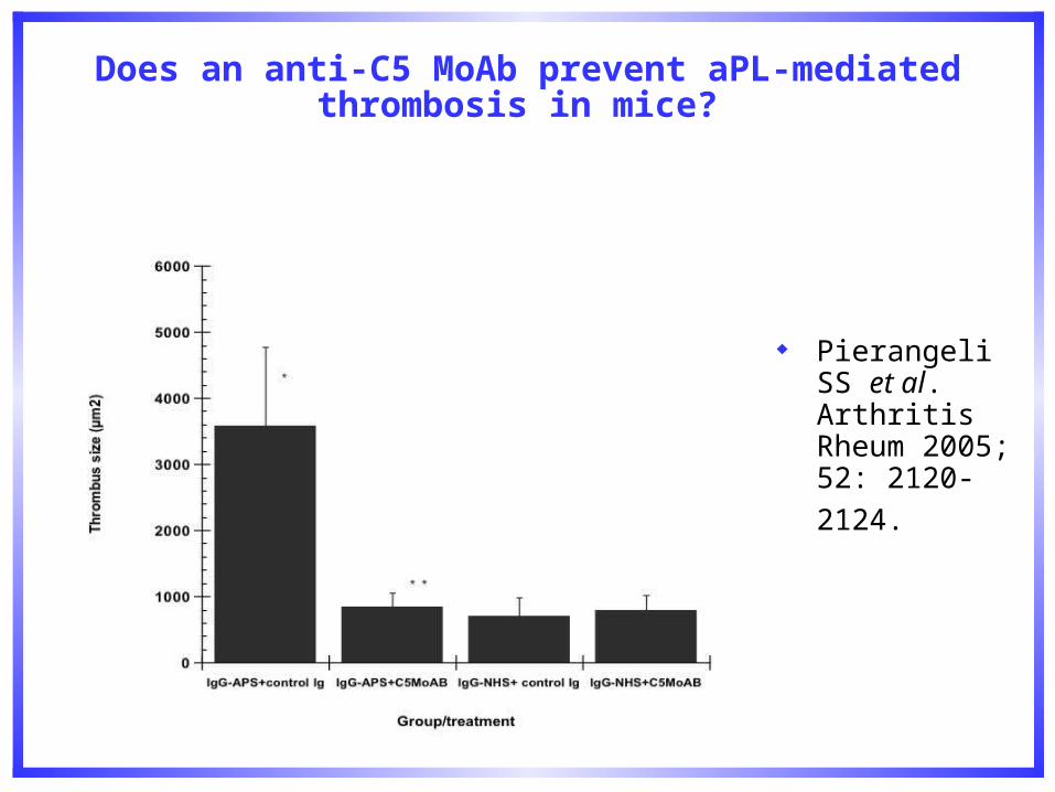
Does an anti-C5 MoAb prevent aPL-mediated thrombosis in mice?
Pierangeli SS et al. Arthritis Rheum 2005;
52: 2120-2124.

Effects of aPL Abs on thrombosis in C5aR deficient mice. C5aR-/- + IgG-APS 3400 ± 1681 108.9 ± 33.4
C5aR-/- + IgG-NHS 777.3 ± 270.4 0.8 ± 0.4
C5aR +/+ + IgG-APS 3507 ± 965 80.3 ± 17.6
C5aR+/+ + IgG-NHS 1321 ± 798 0.8 ± 0.5
C5aR -/- + IgM-APS *676 ± 690 96.4 ± 30.8
C5aR-/- + IgM-NHS 958 ± 388 0.0 ± 0.0
C5aR +/+ + IgM-APS 3198 ± 2361 99.8 ± 31.4
C5aR+/+ + IgM-NHS 585 ± 460 00.0 ±0.1
Romay-Penabad Z et al. NY Acad Sci 2007; 1108: 554-566.
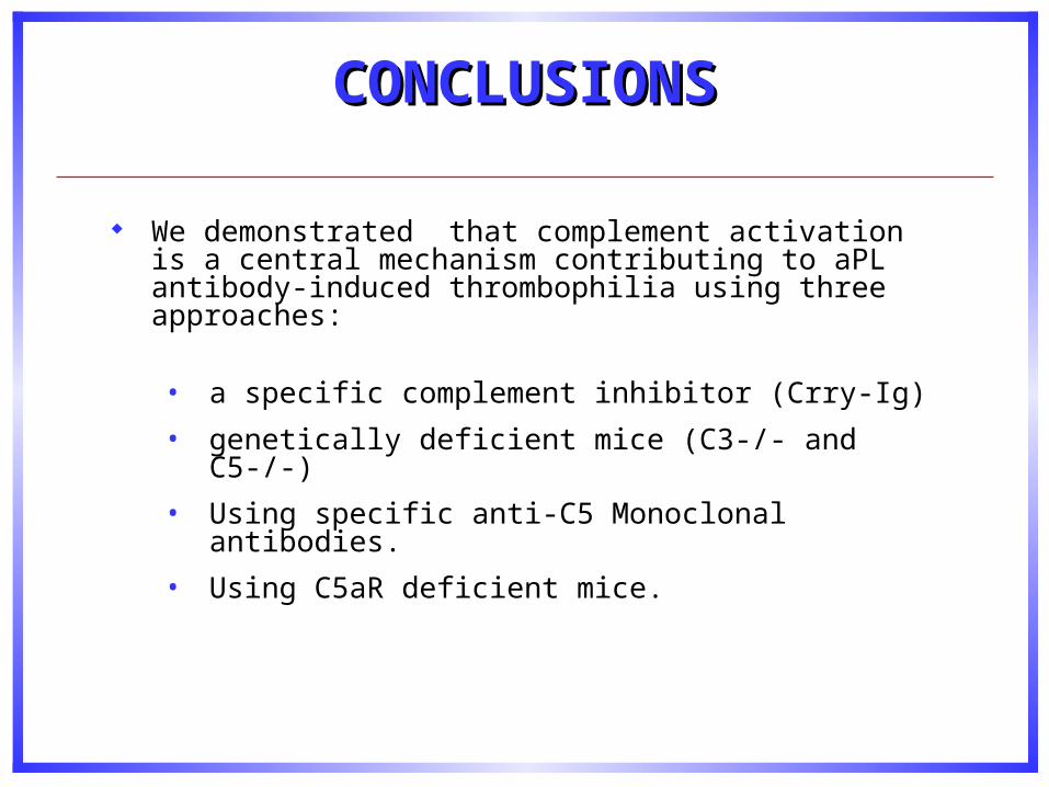
We demonstrated that complement activation is a central mechanism contributing to aPL antibody-induced thrombophilia using three approaches:
• a specific complement inhibitor (Crry-Ig)
• genetically deficient mice (C3-/- and C5-/-)
• Using specific anti-C5 Monoclonal antibodies.
• Using C5aR deficient mice.
CONCLUSIONSCONCLUSIONS
Further evidence of complement involvement
Thrombus formation induced by antibodies to 2glycoprotein I is complement dependent and requires a priming factor (Fischetti et al Blood 2005; 106: 2340-2346).

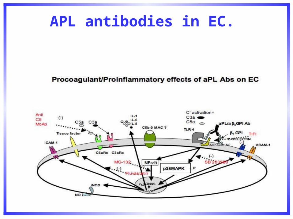
Pathogenic aPL antibodies, in addition to their direct effects on platelet and endothelial cell targets, induce complement activation, generating complement split products which attract inflammatory cells that may induce then thrombosis and tissue injury
Activation of complement may be a critical proximal effector mechanism in aPL-associated thrombosis. In APS patients, due to aPL IgG deposition targeted to the endothelium,
complement activation is increased locally and overwhelms normally adequate inhibitory mechanisms.
Therefore, inhibition of complement activation should ameliorate aPL-mediated vascular thrombosis.
IMPLICATIONSIMPLICATIONS
APL antibodies in EC.
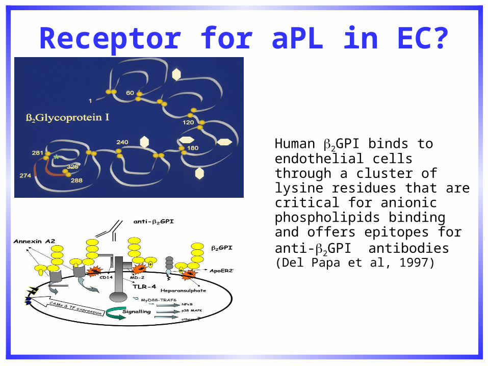
Receptor for aPL in EC?
Human 2GPI binds to endothelial cells through a cluster of lysine residues that are critical for anionic phospholipids binding and offers epitopes for anti-2GPI antibodies (Del Papa et al, 1997)
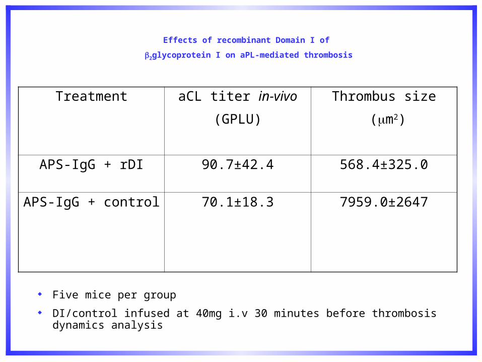
Treatment aCL titer in-vivo
(GPLU)
Thrombus size
(m2)
APS-IgG + rDI 90.7±42.4 568.4±325.0
APS-IgG + control 70.1±18.3 7959.0±2647
Five mice per group
DI/control infused at 40mg i.v 30 minutes before thrombosis dynamics analysis
Effects of recombinant Domain I of
2glycoprotein I on aPL-mediated thrombosis
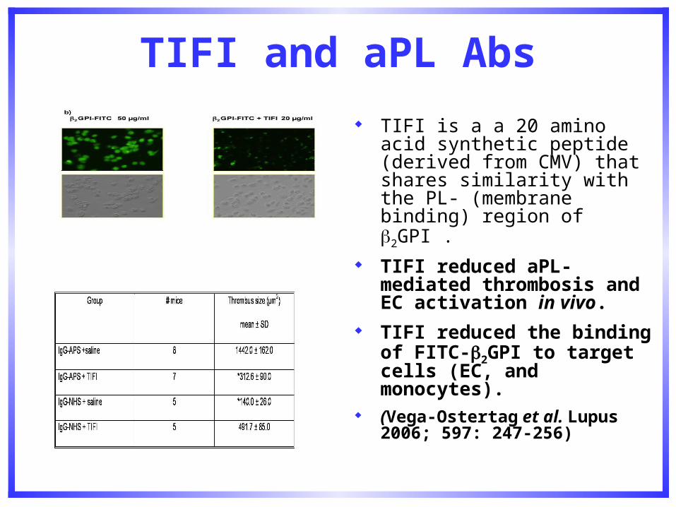
TIFI and aPL Abs
TIFI is a a 20 amino acid synthetic peptide (derived from CMV) that shares similarity with the PL- (membrane binding) region of 2GPI .
TIFI reduced aPL-mediated thrombosis and EC activation in vivo.
TIFI reduced the binding of FITC-2GPI to target cells (EC, and monocytes).
(Vega-Ostertag et al. Lupus 2006; 597: 247-256)
TLR-4: Receptor for aPL on EC?
MyD88 signaling cascade - associated to TLR-4 - is triggered by aPL reacting with 2GPI on the endothelial cell surface membrane (Raschi et al. Blood 2003; 101: 3495-3500).
APL Abs are not thrombogenic in LPS -/- mice and EC activation and TF are diminished. (Pierangeli SS et al Ann Rheum Dis. 2007; epub ahead of press)
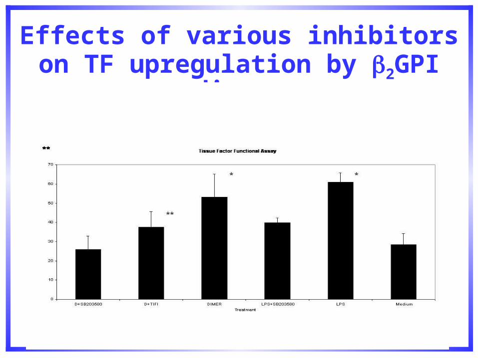
Effects of various inhibitors on TF upregulation by 2GPI dimer.
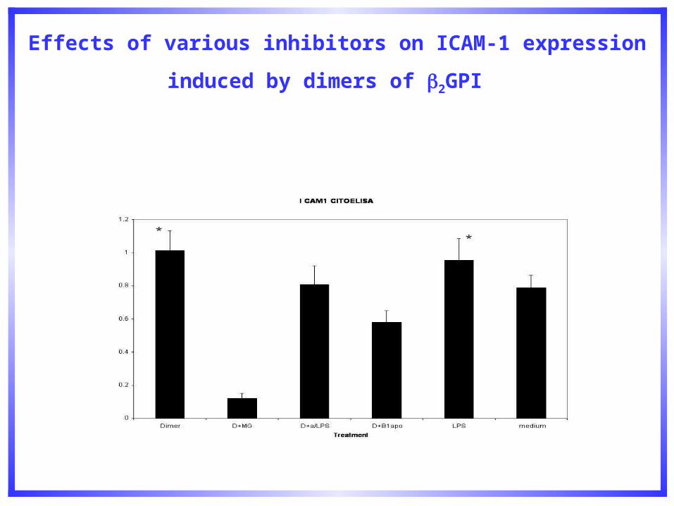
Effects of various inhibitors on ICAM-1 expression induced by
dimers of 2GPI
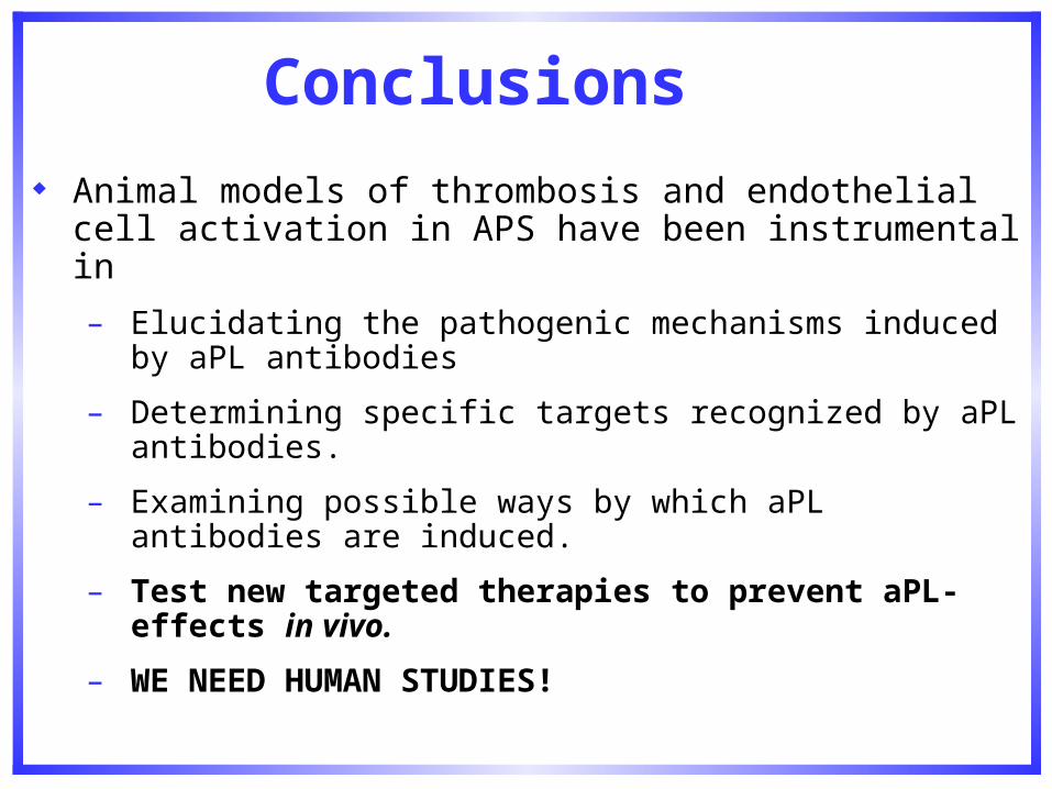
Conclusions
Animal models of thrombosis and endothelial cell activation in APS have been instrumental in
– Elucidating the pathogenic mechanisms induced by aPL antibodies
– Determining specific targets recognized by aPL antibodies.
– Examining possible ways by which aPL antibodies are induced.
– Test new targeted therapies to prevent aPL-effects in vivo.
– WE NEED HUMAN STUDIES!
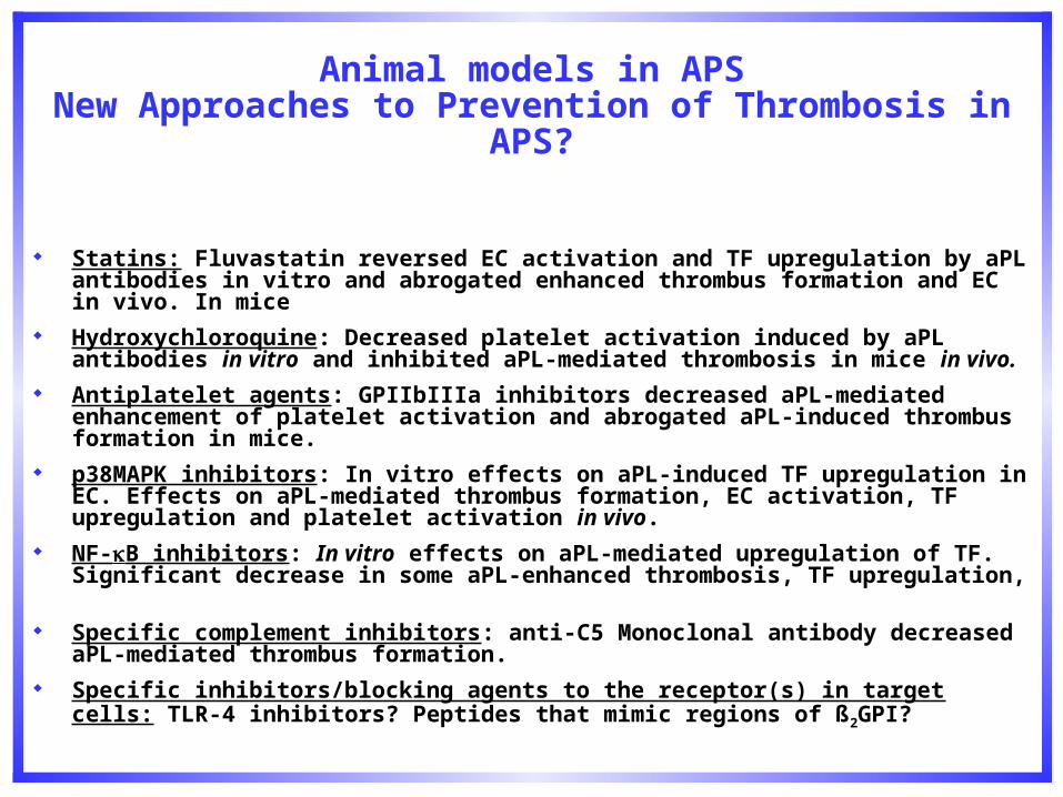
Animal models in APSNew Approaches to Prevention of Thrombosis in APS?
Statins: Fluvastatin reversed EC activation and TF upregulation by aPL antibodies in vitro and abrogated enhanced thrombus formation and EC in vivo. In mice
Hydroxychloroquine: Decreased platelet activation induced by aPL antibodies in vitro and inhibited aPL-mediated thrombosis in mice in vivo.
Antiplatelet agents: GPIIbIIIa inhibitors decreased aPL-mediated enhancement of platelet activation and abrogated aPL-induced thrombus formation in mice.
p38MAPK inhibitors: In vitro effects on aPL-induced TF upregulation in EC. Effects on aPL-mediated thrombus formation, EC activation, TF upregulation and platelet activation in vivo.
NF-B inhibitors: In vitro effects on aPL-mediated upregulation of TF. Significant decrease in some aPL-enhanced thrombosis, TF upregulation,
Specific complement inhibitors: anti-C5 Monoclonal antibody decreased aPL-mediated thrombus formation.
Specific inhibitors/blocking agents to the receptor(s) in target cells: TLR-4 inhibitors? Peptides that mimic regions of ß2GPI?

New Approaches to Prevention of Thrombosis in APS?
ACE inhibitors: Inhibit monocyte TF expression
Dilazep, dipyridamole: Adenosine uptake inhibitor; antiplatelet; inhibits monocyte TF expression.
LJP 1082: ß2GPI-specific B cell toleragen; decreases anti-ß2GPI-specific B cell toleragen;decreases anti- ß2GPI antibody levels.
Rituximab: Anti CD20.
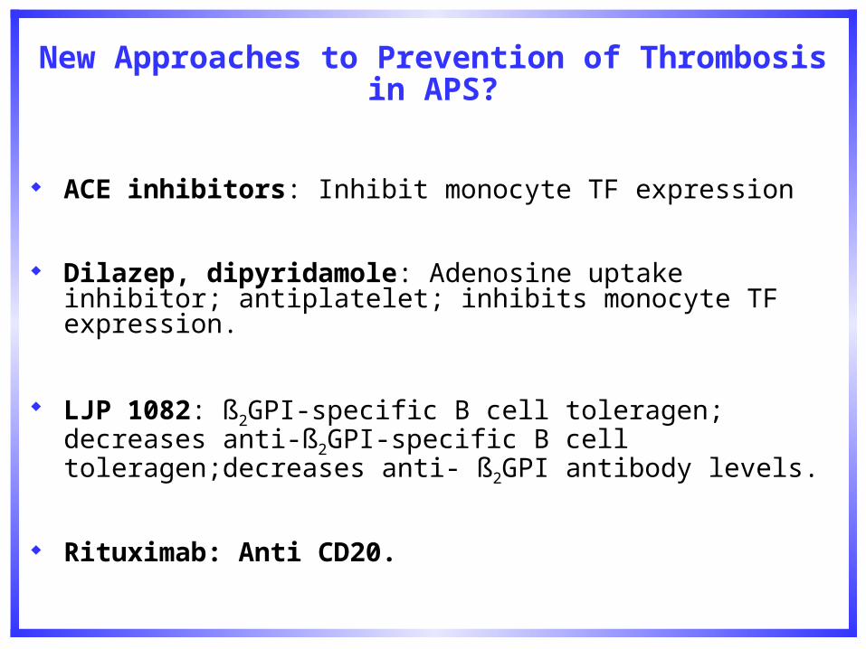
Collaborators
Mariano Vega-Ostertag, MS Morehouse School of Medicine Zurina Romay-Penabad, PhD UTMB Guadalupe Montiel, B.S. UTMB Elizabeth Papalardo, B.S. UTMB Dardo E. Ferrara, MD Morehouse School of Medicine R.G. Espinola, MD Morehouse School of Medicine X. Liu, MD Morehouse School of Medicine Ian P. Giles University College London Robert Swerlick, MD Emory University School of Medicine Pier Luigi Meroni, MD University of Milan Guillermina Girardi, PhD Hosp Spec Surgery Jane Salmon, MD Hosp Spec Surgery VM Holers, MD Univ Colorado,Denver Philip deGroot Utrecht University.
The 13th International Congress on antiphospholipid antibodies: Galveston, TX. Spring 2010.

Acknowledgements
These studies were partially funded by a Research Centers in Minority Institutions - National Institutes of Health grant # G12-RR03034 and a Minority Biomedical Research Support Grant from the National Institutes of Health (GM58268-02) and a multidisciplinary clinical research grant NIH grant #: 2P60AR047785-06.


